Embed Size (px)
Citation preview
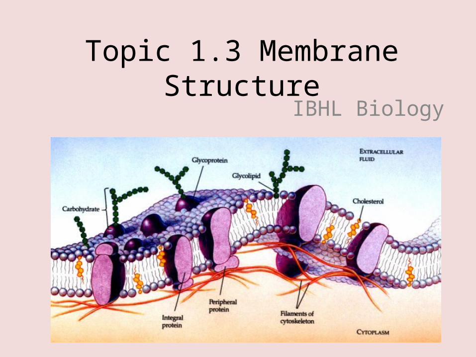
Topic 1.3 Membrane StructureIBHL Biology
Cell Membrane
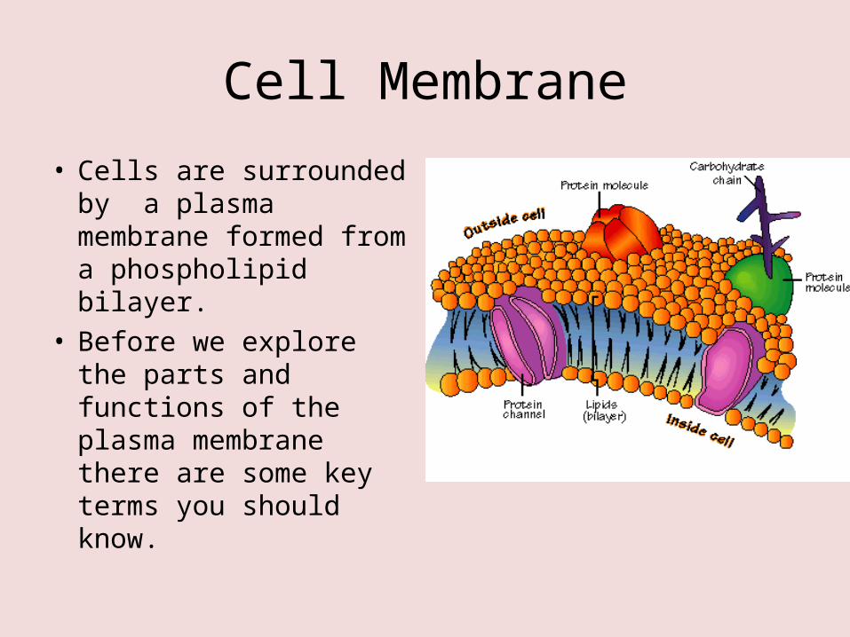
• Cells are surrounded by a plasma membrane formed from a phospholipid bilayer.
• Before we explore the parts and functions of the plasma membrane there are some key terms you should know.
Hydrophobic
• Having an aversion to water; tending to coalesce and form droplets in water.
• Hydrophobic is a term used to describe how molecules react with water. Molecules that are hydrophobic have an aversion to water and are often referred to as “water fearing”.
Hydrophilic
• Having an affinity for water.• Hydrophilic is a term used to describe how
molecules react with water. Molecules that are hydrophilic have an affinity for water. Hydrophilic is often referred to as “water loving”.
Integral protein
• Typically a transmembrane protein with hydrophobic regions that completely spans the hydrophobic interior of the membrane.
• The integral proteins in the plasma membrane penetrate the lipid bilayer from one side to the other.
• These proteins control the entry and exit of specific molecules from the cell. They also have a hydrophobic and hydrophilic region which helps keep them in place in the membrane.
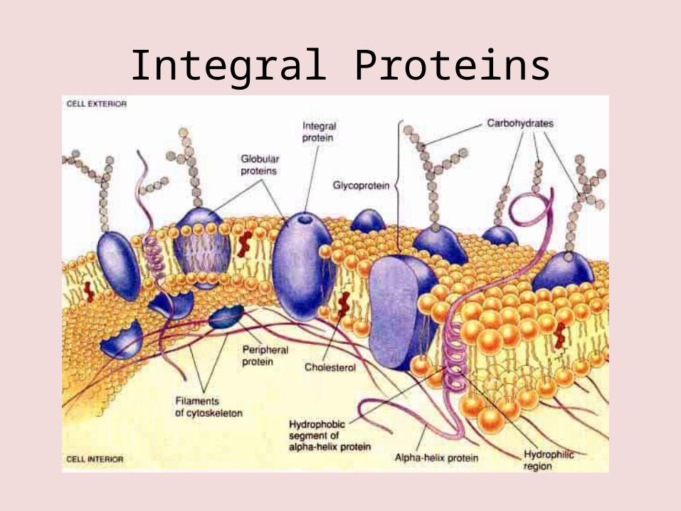
Integral Proteins
Peripheral protein
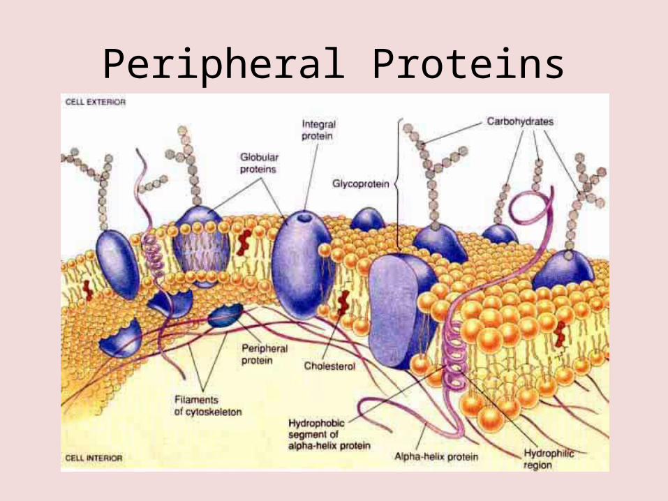
• A protein appendage loosely bound to the surface of a membrane and not embedded in the lipid bilayer.
• The protein molecules are not fixed in one spot of the membrane and they actually float in the fluid phospholipid bilayer or are attached to an integral protein.
• Peripheral proteins are known as glycoproteins because they have a carbohydrate attached. The function in immune responses and are involved in cell to cell recognition.
Peripheral Proteins
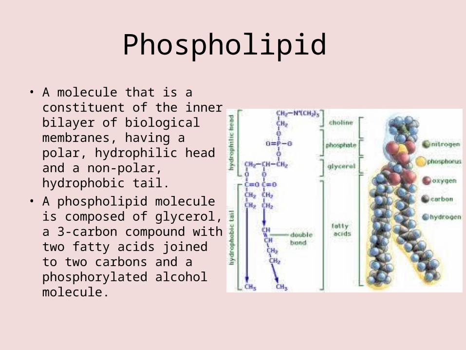
Phospholipid • A molecule that is a
constituent of the inner bilayer of biological membranes, having a polar, hydrophilic head and a non-polar, hydrophobic tail.
• A phospholipid molecule is composed of glycerol, a 3-carbon compound with two fatty acids joined to two carbons and a phosphorylated alcohol molecule.
1.3 (A3) Analysis of evidence from electron microscopy that led to the proposal of the Davson-Danielli model.
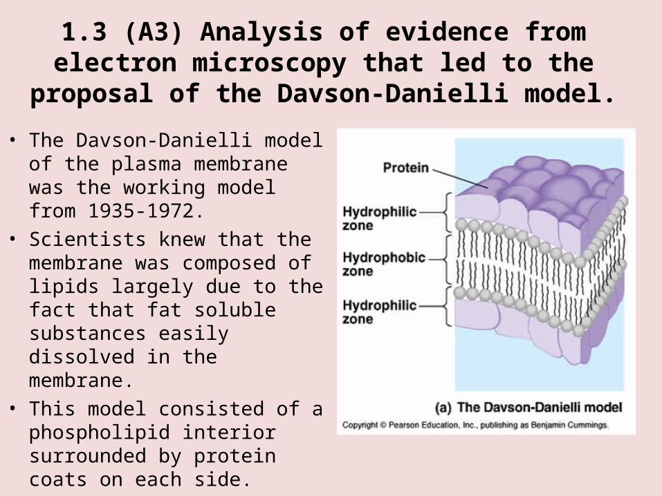
• The Davson-Danielli model of the plasma membrane was the working model from 1935-1972.
• Scientists knew that the membrane was composed of lipids largely due to the fact that fat soluble substances easily dissolved in the membrane.
• This model consisted of a phospholipid interior surrounded by protein coats on each side.
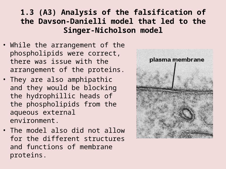
1.3 (A3) Analysis of the falsification of the Davson-Danielli model that led to the Singer-Nicholson model
• While the arrangement of the phospholipids were correct, there was issue with the arrangement of the proteins.
• They are also amphipathic and they would be blocking the hydrophillic heads of the phospholipids from the aqueous external environment.
• The model also did not allow for the different structures and functions of membrane proteins.

Singer-Nicholson Model
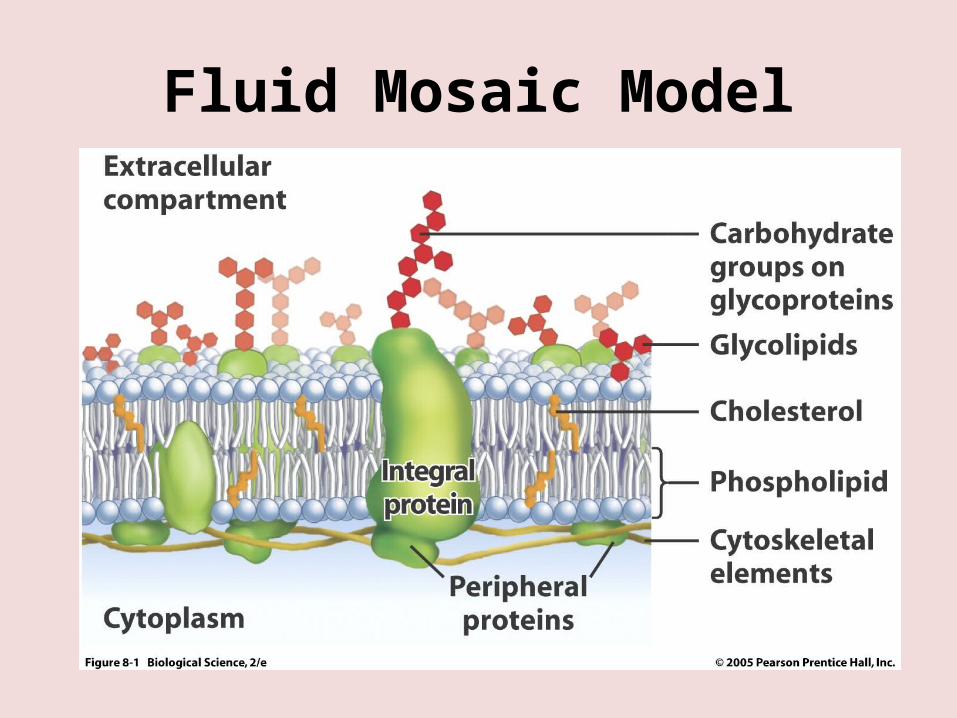
• The Singer-Nicholson model was proposed in 1972 and remains the current accepted model of the plasma membrane. It is known as the fluid mosaic model and while the arrangements of the phospholipids remain the same, it is the location of the proteins that differs.
• This model shows them embedded in the bilayer as either integral or peripheral proteins.
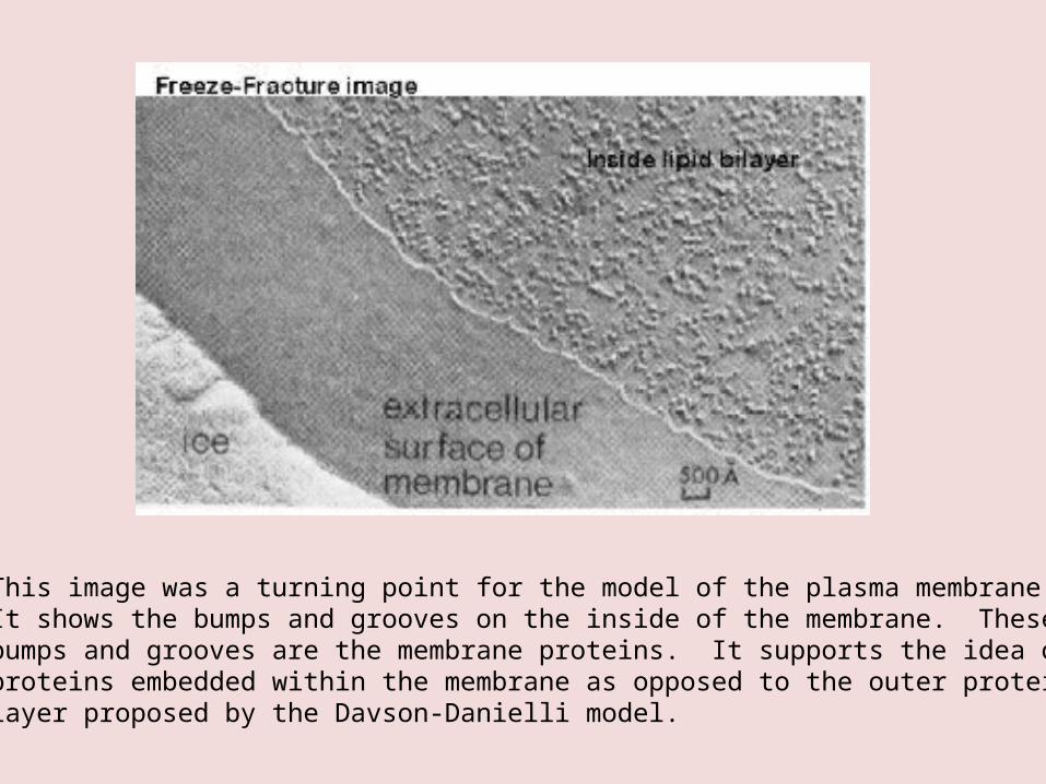
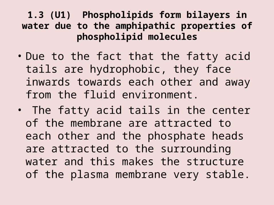
This image was a turning point for the model of the plasma membrane. It shows the bumps and grooves on the inside of the membrane. These bumps and grooves are the membrane proteins. It supports the idea of proteins embedded within the membrane as opposed to the outer protein layer proposed by the Davson-Danielli model.
1.3 (A2) Drawing of the fluid mosaic model
• You must be able to draw the fluid mosaic model which is the current model of the plasma membrane. Ensure you draw each part to scale and include all relevant labels.
• Your drawing should include a clear drawing of individual phospholipid molecules using circles for the head and parallel lines for the tails.
• You must also include a range of membrane proteins (integral and peripheral) including glycoproteins.
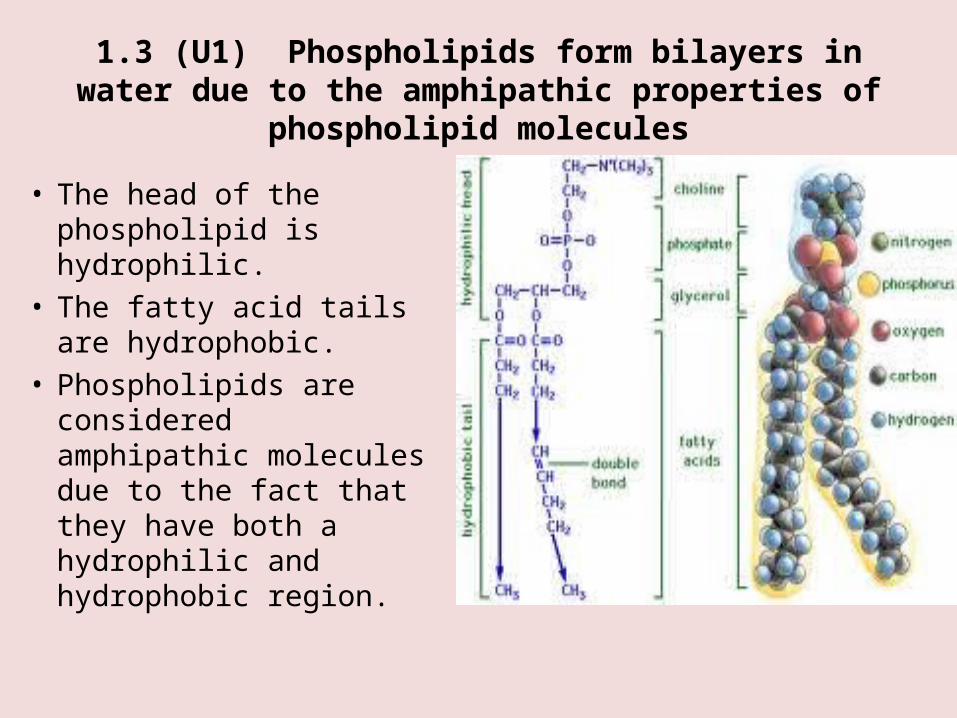
1.3 (U1) Phospholipids form bilayers in water due to the amphipathic properties of phospholipid molecules
• The head of the phospholipid is hydrophilic.
• The fatty acid tails are hydrophobic.
• Phospholipids are considered amphipathic molecules due to the fact that they have both a hydrophilic and hydrophobic region.
1.3 (U1) Phospholipids form bilayers in water due to the amphipathic properties of phospholipid molecules
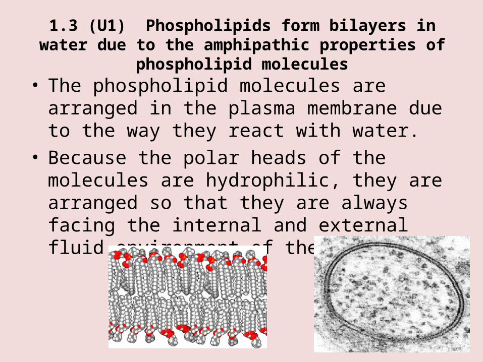
• The phospholipid molecules are arranged in the plasma membrane due to the way they react with water.
• Because the polar heads of the molecules are hydrophilic, they are arranged so that they are always facing the internal and external fluid environment of the cell.
1.3 (U1) Phospholipids form bilayers in water due to the amphipathic properties of phospholipid molecules
• Due to the fact that the fatty acid tails are hydrophobic, they face inwards towards each other and away from the fluid environment.
• The fatty acid tails in the center of the membrane are attracted to each other and the phosphate heads are attracted to the surrounding water and this makes the structure of the plasma membrane very stable.
1.3 (U2) Membrane proteins are diverse in terms of structure, position in the membranes and function
• There are many different types of membrane proteins. They occupy different positions in the plasma membrane depending on their structure and function.
• The following slide summarizes the different types of membrane proteins and their function.
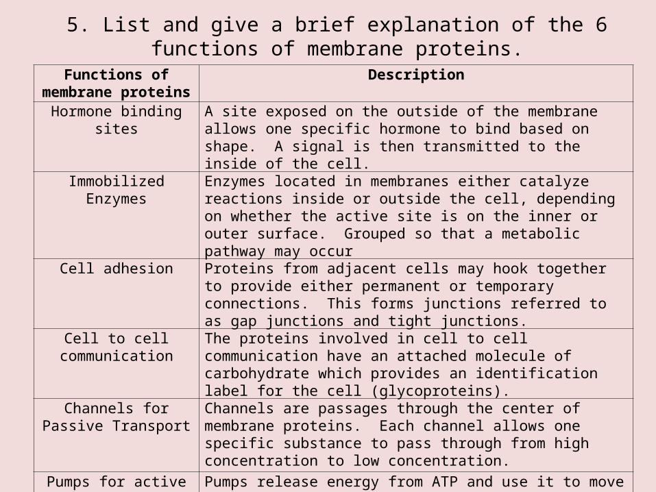
5. List and give a brief explanation of the 6 functions of membrane proteins.
Functions of membrane proteins
Description
Hormone binding sites A site exposed on the outside of the membrane allows one specific hormone to bind based on shape. A signal is then transmitted to the inside of the cell.
Immobilized Enzymes Enzymes located in membranes either catalyze reactions inside or outside the cell, depending on whether the active site is on the inner or outer surface. Grouped so that a metabolic pathway may occur
Cell adhesion Proteins from adjacent cells may hook together to provide either permanent or temporary connections. This forms junctions referred to as gap junctions and tight junctions.
Cell to cell communication
The proteins involved in cell to cell communication have an attached molecule of carbohydrate which provides an identification label for the cell (glycoproteins).
Channels for Passive Transport
Channels are passages through the center of membrane proteins. Each channel allows one specific substance to pass through from high concentration to low concentration.
Pumps for active transport
Pumps release energy from ATP and use it to move specific substances across the plasma membrane. The energy is used to change the shape of the protein.
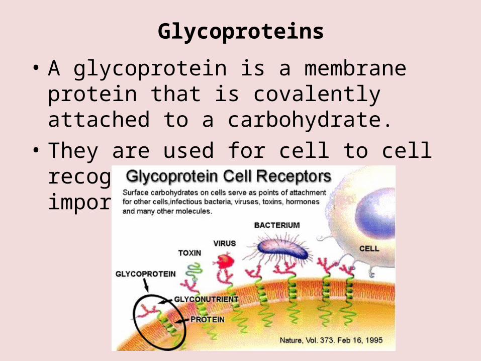
Glycoproteins
• A glycoprotein is a membrane protein that is covalently attached to a carbohydrate.
• They are used for cell to cell recognition and play an important role in immunity.
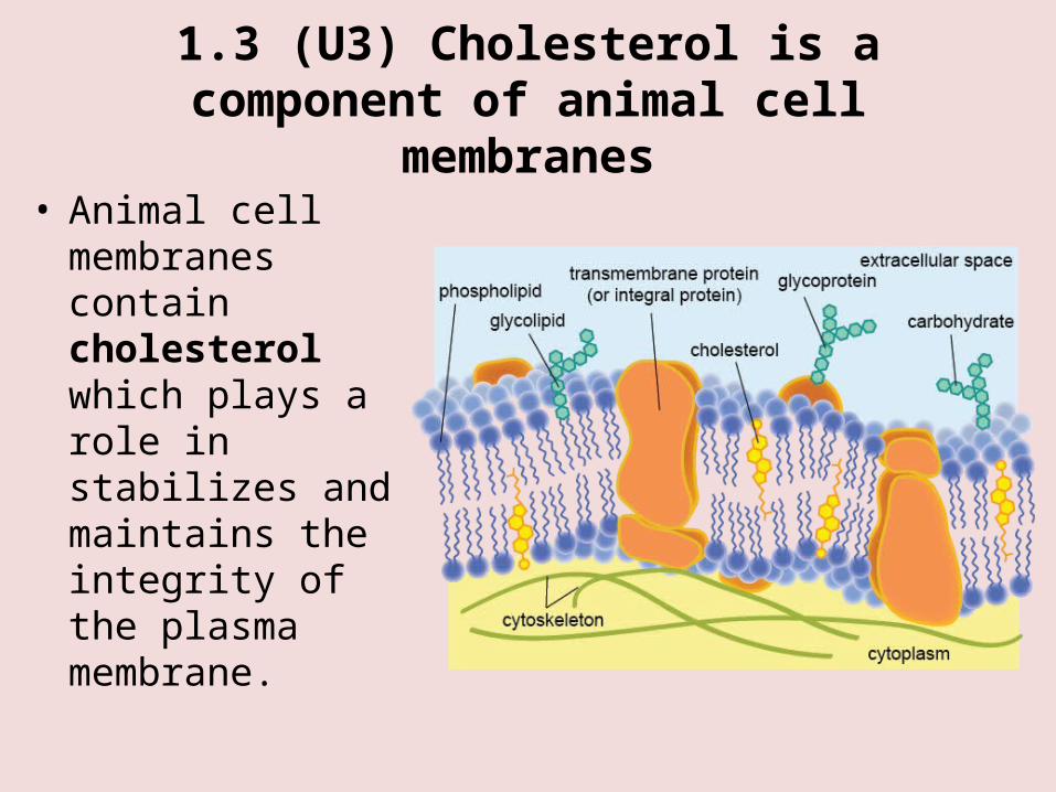
1.3 (U3) Cholesterol is a component of animal cell membranes
• Animal cell membranes contain cholesterol which plays a role in stabilizes and maintains the integrity of the plasma membrane.
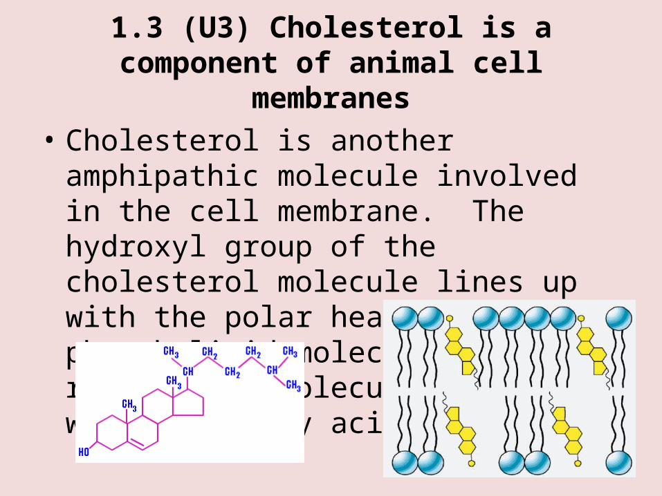
1.3 (U3) Cholesterol is a component of animal cell membranes
• Cholesterol is another amphipathic molecule involved in the cell membrane. The hydroxyl group of the cholesterol molecule lines up with the polar heads of the phospholipid molecules. The rest of the molecule tucks in with the fatty acid tails.
1.3 (A1) Cholesterol in mammalian membranes reduces membrane fluidity and permeability to some solutes.
• Cholesterol will affect the fluidity of the membrane and it has different affects at different temperatures.
• At moderate temperatures the cholesterol will reduce phospholipid movement which reduces the fluidity of the membrane.
• The location of the cholesterol molecules prevents the close packing of the phospholipids. This helps prevent the membrane from solidifying if temperatures drop.