Embed Size (px)
Citation preview

TOMOSSÍNTESE RASTREAMENTO E DIAGNÓSTICO
BEATRIZ BOHRER DO AMARAL


APLICAÇÕES DA TOMOSSÍNTESE
RASTREAMENTO
DIAGNÓSTICO


• Sechopoulos I et al, Radiology, abril 2012
• Compararam a dose de radiação da mamografia digital de campo total (DR) e da tomossíntese: 1,2 mGy X 1,3mGy = 2,5 mGy.
• Conclusão: a baixa dose de radiação viabiliza a tomossíntese para o rastreamento de câncer de mama.

• Breast Cancer Screening Using Tomosynthesis in Combination With Digital Mammography
• Sarah M. Friedewald et al
• JAMA. 2014;311(24):2499-2507.
• Análise retrospectiva de 13 diferentes centros com um total de 454.850 exames (mamo: 281.187; mamo+tomo: 173.663)

MAMOGRAFIA x MAMO+TOMOSSÍNTESE (por 1.000 pacientes)
• RECONVOCAÇÃO: 107 x 91 -15%
• DETECÇÃO DE CA: 4,2 x 5,4 29%
• CA INVASIVO: 2,9 x 4,1 41%
• CDIS: 1,4 x 1,4 0%


FFDM vs. DBT + FFDM in Oslo Tomosynthesis Screening Trial Breast Cancer Res Treat, 10 Feb 2018
FFDM FFDM + DBT
Recall rate 42.3 33.7
Screen-detected cancer rate 6.3 9.3
Interval cancer rate 2.0 2.1
Sensitivity 76.2% 80.8%
Specificity 96.4% 97.5%
Recalls per screen-detected cancer 6.7 3.6
Rates per 1,000 screens.

Performance of digital mammography alone vs. DM + DBTThe Reggio Emilia Tomosynthesis Randomized Trial Radiology 2018
Measure DM DM + DBT
Invasive cancers found 39 69
Cancer detection rate per 1,000 cases 4.5 8.6
Positive predictive value (cancers found in total number of recalled women) 13% 24.1%
Recall rate 3.5% 3.5%
False positives per 1,000 cases 30 27
Median radiation dose 4.84 mGy 11.24 mGy
Median read time, negative studies 34 sec 56 sec
Median read time, recalls 104 sec 114 sec
Lesion size < 10 mm 16 31
Lesion size ≥ 10 mm to < 20 mm 14 31
DCIS 5 14
Cancers found according to BI-RADS density
A (fatty) 2 4
B (scattered fibroglandular) 12 25
C (heterogeneously dense) 18 27
D (extremely dense) 7 16

Comparison of synthesized 2D DBT, FFDM and 2D DBTRadiology, feb 2017

Tomosynthesis: Comparison of Full-Field Digital Mammography with Digital Breast Tomosynthesis
TMIST
• Desenhado por representantes da ACS, BCSC, ACR e NCI, coordenado por Etta Pisano , USC e Martin Yaffe, Universidade de Toronto.
• Comparação do número de cânceres avançados (tumores estágio II ou mais e todos os tumores > 6 mm de alto grau) detectados por mamografia digital e tomossíntese.
• 67.000 mulheres acima de 40 anos, examinadas com um exame ou outro por 3 anos consecutivos.
• Objetivo de investigar a eficácia do rastreamento atual do câncer de mama.


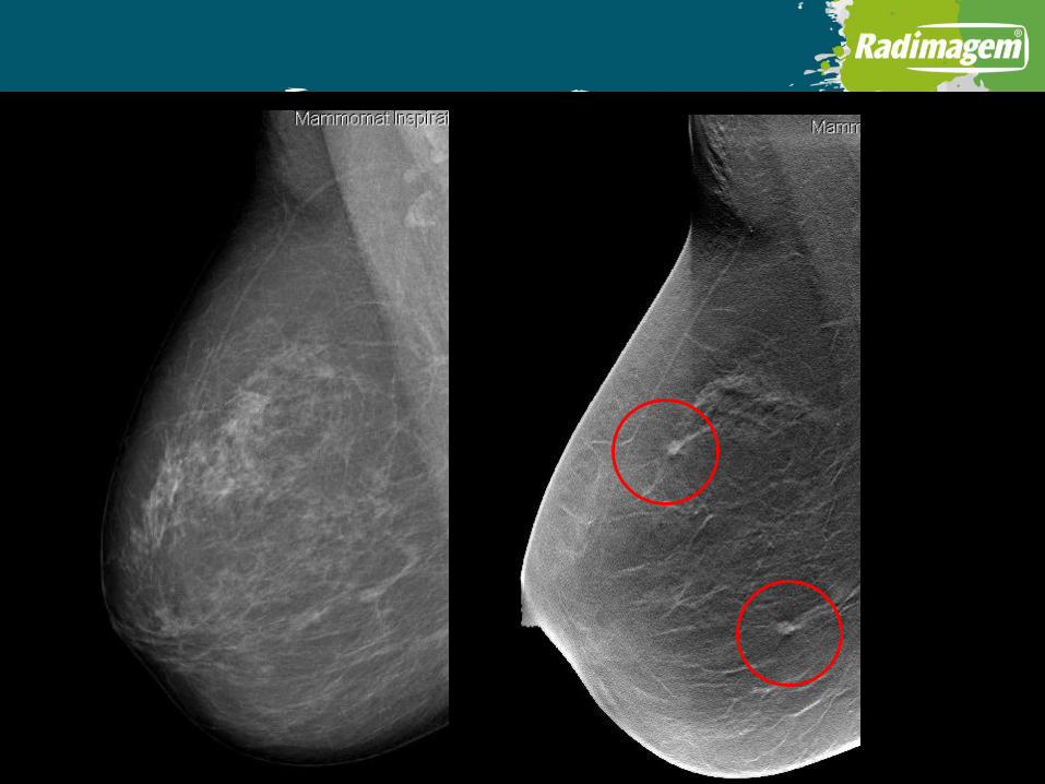
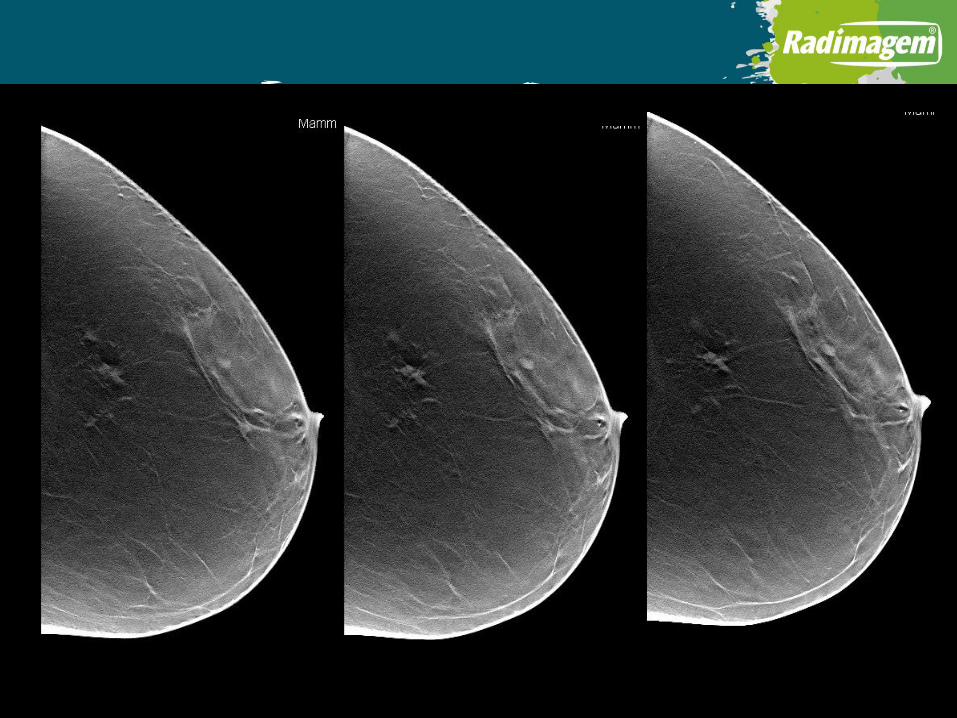
Compressão seletiva x Tomossíntese

Assimetria Focal Nova

Nódulo com características benignas

2D versus 3D mammography for diagnostic use
Vijapura et al, ARRS,2018
2D diagnostic mammography
3D diagnostic mammography
p-value
Cancer detection rate (per 1,000 exams) 31.3 38.2 p < 0.01
False-positive rate 7.3% 6.5% p < 0.01

Findings leading to false-positive exams on diagnostic mammography
2D diagnostic mammography 3D diagnostic mammography
Architectural distortion 1% 5%
Asymmetry 18% 12%
Calcifications 37% 27%
Masses 20% 33%
Other 24% 23%

TOMOSSÍNTESE
PRÓS:AUMENTO DA SENSIBILIDADE E ESPECIFICIDADE (RASTREAMENTO/DIAGNÓSTICO)
CONTRAS:AUMENTO DA RADIAÇÃO (SOLUÇÃO: 2DS) AUMENTO DE CUSTO (EQUIPAMENTO E TEMPO DE INTERPRETAÇÃO)
FUTURO:AVALIAR OS BENEFÍCIOS DA TOMOSSÍNTESE A LONGO PRAZO (PROGNÓSTICO E
SOBREDIAGNÓSTICO)RASTREAMENTO DE CÂNCER DE MAMA SERÁ TOMOSSÍNTESE MAIS
MAMOGRAFIA 2D SINTETIZADA

Comparison of synthesized 2D DBT, FFDM and 2D DBTRadiology, feb 2017