Embed Size (px)
Citation preview

Introduction To The New AACPDM Hip Surveillance Care Pathway For Children With Cerebral Palsy:
What's the Consensus? How Can It Be Implemented?
September 15, 2017Montreal Quebec Canada
1

CARE PATHWAYS
HIP SURVEILLANCE
Section I: Evidence Summary
DEFINITIONS:
Hip surveillance is defined as: “The process of monitoring and identifying the critical early indicators of hip displacement." Hip displacement refers to the displacement of the femoral head laterally out of the acetabulum and is measured using a migration percentage (MP). Hip subluxation refers to hip displacement where the femoral head is partially displaced from under the acetabulum while hip dislocation refers to hip displacement where the femoral head is completely displaced from under the acetabulum.
IMPACT: WHY IS HIP SURVEILLANCE IMPORTANT?
Hip displacement and dislocation can lead to pain, reduced function and reduced quality of life.
Children with cerebral palsy (CP) have an increased likelihood of hip displacement.
Hip surveillance allows for early detection of hip displacement.
Early detection enables referral for assessment and/or management.
Hip surveillance for children with CP should be completed using a systematic approach.
Target Population: Pediatric/Children & Youth Population (age ≤19 years) diagnosed with CP or those children not yet diagnosed with CP but for whom there is a clinical suspicion of having CP.
Target Clinical Providers: Pediatricians including Pediatric Sub-Specialists, Radiologists and Pediatric Orthopedic Surgeons, Therapists, Radiology Technicians, and Nurses providing musculoskeletal care for children/youth with CP.
ASSESSMENT
Hip surveillance involves a multi-step process for every child with CP.
Surveillance consists of two components: a clinical examination and a radiographic examination which are completed at surveillance intervals which vary
according to risk. The clinical examination involves determining/re-confirming, age, Gross Motor Function Classification System (GMFCS) level and Winters, Gage, Hicks (WGH) gait type at each surveillance interval in addition to inquiring re: pain during history taking. Hip abduction passive range of motion (PROM) is also measured with attention given to presence of pain on assessment.
Radiographic examination consists of measurement of migration percentage (MP) from a supine AP pelvis radiograph with standardised positioning.
FREQUENCY
Surveillance frequency is based on a child’s age, GMFCS level, and WGH gait type. Surveillance is ideally initiated by 2 years of age, when a CP diagnosis is provided, or when CP is suspected. Surveillance frequency increases with increasing GMFCS level; frequency modifiers are based on absolute MP value and percentage change in MP.
Discharge criteria vary depending on GMFCS level and WGH gait type. Children at GMFCS levels III to V and those with a WGH Gait Type IV hemiplegia are discharged at skeletal maturity except those with a MP greater than 30% or those with pelvic obliquity in the presence of increasing scoliosis where continued surveillance is recommended. Children at GMFCS levels I and II are discharged earlier if MP is stable and under 30%.
REFERRAL
Referral to a pediatric physiatrist, developmental paediatrician or pediatric orthopedic surgeon with experience treating hip displacement in children with CP is recommended when there is presence of hip pain on history and/or physical examination. When the migration percentage is greater than 30% and/or there is less than 30 degrees of hip abduction with or without other findings, referral to a pediatric orthopaedic surgeon is recommended.

*Do [does] you [your child] have hip pain? You may notice this when you move [your child moves] your [their] hip or after prolonged activity, when
changing your [your child’s] position, when you move your [your child’s] leg or when looking after your [your child’s] personal care. 1GMFCS : Palisano R, Rosenbaum P, Walter S et al., 1997. 2WGH Gait Type IV: Winters T, Gage J & Hicks R, 1987. xClinicians collaborate to ensure planned approach to ongoing surveillance.
`
Determine Gross Motor Function Classification System (GMFCS)1 Level
Measure Migration Percentage from AP Pelvis
Complete AP Pelvis with standardized positioning
See Frequency Table to Determine Need for Radiograph
Complete Radiological Examination (if indicated)
Determine Winters, Gage, Hicks (WGH)2 Gait Type for children with hemiplegia
Inquire re: presence of pain in history taking *
Yesx
Complete Clinical Assessment
Child/Youth with Cerebral Palsy OR
Clinical Suspicion of Cerebral Palsy
HIP
SU
RV
EILL
AN
CE
CO
NTE
NT
Provide Information to Family Initiate Hip Surveillance
Use Frequency Table to Determine Timing of
Next Clinical & Radiological Exams Based on:
Strength of Evidence
Effective
Possibly or Probably Effective
Data Inadequate (Recommendation based
on expert opinion)
WGH Type Age + +
Continue Surveillance
GMFCS
Refer to Physician#+
#+Physician refers to pediatric orthopedic
surgeon+, pediatric physiatrist#,
developmental pediatrician# with experience
treating hip displacement in children with CP.
Determine Age
Assess pain during physical examination
Measure hip abduction passive range of motion
No
Is one or more of the following present? - Positive response to question re: pain#+
- Pain on physical examination#+
- Migration percentage > 30% on AP pelvis+
- Hip abduction passive range of motion <30+

The purpose of this document is to provide health care professionals with recommendations for hip surveillance of children and youth with cerebral palsy. This summary was produced by the AACPDM Hip Surveillance Care Pathway Team (M O’Donnell (team lead), T Mayson (project manager and clinical examination sub-group leader), S Miller (radiology sub-group leader), R Cairns, K Graham, S Love, F Miller, K Mulpuri, U Narayanan, H Read, B Shore, K Stannage, P Thomason, J Vargus-Adams, L Wiggins, K Willoughby, M Wynter). The summary is based on current evidence and expert consensus when evidence was insufficient. The care pathway and the methodology used to create it will be submitted for peer-reviewed publication. However, health care professionals should continue to use their own judgement and take into account additional relevant factors and context. The AACPDM is not liable for any damages, claims, liabilities, or costs arising from the use of these recommendations including loss or damages arising from any claims made by a third party.
3
Legend: : Clinical Examination : AP Pelvis Radiograph GMFCS: Gross Motor Function Classification System (Palisano R et al., 1997. Illustrations reproduced with permission and copyright © Bill Reid, The Royal Children’s Hospital, Melbourne, AUS.) WGH Gait Type IV: Winters, Gage, Hicks Gait Type IV Hemiplegia (Winters T, Gage J, Hicks R, 1987; Rodda J & Graham HK, 2001. Illustrations reproduced with permission and copyright © Bill Reid, The Royal Children’s Hospital, Melbourne, AUS.)
HIP
SU
RV
EILL
AN
CE
FREQ
UEN
CY
Age (Years)
GMFCS I GMFCS II GMFCS III GMFCS IV & V Any GMFCS Level with
Winters Gage Hicks Gait Type IV
2.0 years or at ID #
2.5
3
3.5
4
*
5
*
6
*
7
*
8
* *
9
*
10
* *
11
*
12 to 16 or
Skeletal Maturity
(SM)
Bi-Annually
to SM†
Bi- Annually
* to SM†
Annually
to SM†
Annually
* to SM†
Bi-Annually
to SM†
Bi-Annually
to SM†
Skeletal Maturity (SM) is defined as closure of the triradiate cartilage.
Notes re: Initiation
If CP is diagnosed or suspected after age 2 but before 4 years, begin surveillance immediately. Do not wait until 4 years of age.
If CP is diagnosed or suspected after age 2, immediately begin 12- monthly schedule for a minimum of 24 months.
If CP is diagnosed or suspected after age 2, immediately begin 6-monthly schedule and continue for a minimum of 24 months at that frequency.
If CP diagnosed or suspected after age 2 but before age 4, begin surveillance immediately.
#If there is any doubt of the GMFCS level, follow the recommendation for the higher level.
Frequency Modifiers
* Do not reduce from previous higher frequency if: (1) 24 months of surveillance have not yet been completed based on a child's surveillance start date; (2) stability is not yet achieved over a period of 2 years. Stability is defined as < 10% change in MP over a 12 month period; OR (3) MP > 30%.
.
Discharge
Discharge if MP ≤30% at age 10 (unless WGH Gait Type IV).
Discharge if skeletally mature and MP ≤30%.
† In the presence of pelvic obliquity associated with clinical or radiographic evidence of increasing scoliosis, the hip/s continue to be at risk and should ideally be monitored even beyond skeletal maturity.

4
Section II: Published Evidence
The published literature was reviewed to determine the effectiveness of hip surveillance in children with cerebral palsy as well as
the elements that should be included in hip surveillance and their recommended frequency. The Hip Surveillance Care Pathway is
based on this current best evidence as well as expert consensus when evidence was not available. The care pathway and the
methodology used to create it will be submitted for peer-reviewed publication.
Evidence for Hip Surveillance Effectiveness:
The literature supports the completion of hip surveillance in children and youth with cerebral palsy.
Gordon GS & Simkiss DE. A systematic review of the evidence for hip surveillance in children with cerebral palsy. J Bone Joint Surg-Br
2006; 88-B: 1492-6. doi:10.1302/0301-620X.88B11. 18114.
Hagglund G, Alriksson-Schmidt A, Lauge-Pedersen H, Rodby-Bousquet E, Wagner P, Westbom L. Prevention of dislocation of the hip
in children with cerebral palsy: 20-year results of a population-based prevention programme. Bone Joint J 2014; 96-B: 1546-52. doi:
10.1302/0301-620X.96B11.34385.
Wynter M, Gibson N, Willoughby KL, et al. Australian hip surveillance guidelines for children with cerebral palsy: 5-year review. Dev
Med Child Neurol 2015; 57: 808-20. doi: 10.1111/dmcn.12754.
Evidence for Hip Surveillance Clinical Examination and Hip Health Practical Tool Content:
The literature supports that hip surveillance be based on a child’s age and Gross Motor Function Classification System. The
additional content of the clinical examination component of the Hip Surveillance Care Pathway as well as the Hip Health Practical
Tool was established through expert consensus after review of the literature.
Wynter M, Gibson N, Willoughby KL, et al. Australian hip surveillance guidelines for children with cerebral palsy: 5-year review. Dev
Med Child Neurol 2015; 57: 808-20. doi: 10.1111/dmcn.12754.
Evidence for Hip Surveillance Radiographic Examination and Radiography Practical Tool Content:
The literature supports hip surveillance based on a child’s migration percentage as measured on an AP pelvis radiograph taken with
standardized positioning. The content of the Hip Surveillance Care Pathway and the Radiography Practical Tool was established
through expert consensus after review of the literature. A review of this literature will be submitted for peer-reviewed publication.
Hagglund G, Lauge-Pedersen H, Persson M. Radiographic threshold values for hip screening in cerebral palsy. J Child Orthop 2007;
1: 43–7.
Parrott J, Boyd RN, Dobson F, et al. Hip displacement in spastic cerebral palsy: repeatability of radiologic measurement. J Pediatr
Orthop 2002; 22: 660–7.
Pons C, Remy-Neris O, Medee B, Brochard S. Validity and reliability of radiological methods to assess proximal hip geometry in
children with cerebral palsy: a systematic review. Dev Med Child Neurol 2013; 5: 1089-102. doi: 10.1111/dmcn.12169.

5
Evidence for Hip Surveillance Frequency:
The content of the care pathway was established through expert consensus after review of the literature and comparison of
currently existing international hip surveillance programs in Australia (Consensus Statement on Standards of Care), Canada (British
Columbia Provincial Hip Surveillance Program; Holland Bloorview Kids Rehabilitation Hospital, Ontario; Grandview Children’s Centre,
Ontario); USA (Boston Children’s Hospital, Boston, MA; Alfred I. DuPont Hospital for Children, Wilmington, DW); Sweden (CPUP);
United Kingdom (Bristol, Scotland). Program comparisons will be submitted for peer-reviewed publication.
Wynter M, Gibson N, Willoughby KL, et al. Australian hip surveillance guidelines for children with cerebral palsy: 5-year review. Dev
Med Child Neurol 2015; 57: 808-20. doi: 10.1111/dmcn.12754.

6
Section III: Practical Tools
Plain Language Summary for Families
Hip surveillance is a plan for regular check-ups to watch for signs that your child’s hip may be moving out of joint (this is
called hip displacement). Your child is at risk for hip displacement if your child has cerebral palsy. Cerebral palsy (CP)
affects a child’s ability to move. When children are late to stand and walk or can only do so with help, the hip joint may
not develop as expected. In addition, the muscles that pull the legs together and up are often tight or stiff and may
affect the muscle balance around the hip. Hip displacement can lead to the hip coming completely out of the joint (hip
dislocation). Hip displacement and dislocation can cause pain, difficulty moving the hip, and problems with sitting,
standing, and walking.
Hip Surveillance includes clinical examinations and hip x-rays at regularly scheduled times. Clinical examinations include
asking you and your child about any hip pain, measuring hip movement and noting any pain on movement. Hip x-rays
are done to view the hip joint because hip displacement can occur without any signs or symptoms. Taking part in Hip
Surveillance allows your child’s health care team to find hip displacement early and help your child before the hip
becomes dislocated.
Your child should begin Hip Surveillance when they are diagnosed or suspected of having CP. How often your child
requires Clinical Examinations and x-rays after that depends on their ability to move. We use a scale called the Gross
Motor Function Classification System (GMFCS) to help us with this.
The GMFCS is used to describe a child’s ability to move and includes five levels from Roman numeral I (1) to V (5). Your
child’s physiotherapist, occupational therapist, family doctor, or pediatrician can help you determine your child’s GMFCS
level in just a few minutes.
Risk for hip displacement is directly related to GMFCS level.
Children whose ability to move is at GMFCS Level I have the lowest risk of hip displacement. They receive the
fewest Clinical Examinations and x-rays.
Children whose ability to move is at GMFCS Level V have the highest risk of hip displacement (8 out of 10
children that are at GMFCS Level V will have hip displacement). Clinical Examinations and x-rays are done most
often for children that are at GMFCS Levels IV and V.
In addition to GMFCS, children with hemiplegia (one side of the body affected) who walk with one hip turned
and pulled inward (this is called a Gait Type IV pattern of walking) are at higher risk for hip displacement.
The table in the care pathway shows how often children need clinical examinations and hip x-rays.
Children at low risk will stop Hip Surveillance at age 6 years (ability to move at GMFCS I) or 10 years (ability to move at
GMFCS II). Because hip displacement can occur while children and youth are growing, children who are at higher risk
(ability to move is at GMFCS Levels III, IV, and V or a Hemiplegia Gait Type IV pattern of walking) take part in Hip
Surveillance until an x-ray determines that their bones have stopped growing.
If your child’s health care team finds signs of hip displacement, they can refer your child to a doctor with experience
treating hip displacement in children with CP to determine suitable next steps to prevent hip dislocation.
The following summary, aimed to support parents and families, was provided by the Child Health BC Hip Surveillance Program for
Children with Cerebral Palsy in British Columbia, Canada. If replicated please acknowledge appropriately.

7
Gross Motor Function Classification System (GMFCS)
For information regarding the GMFCS, please see:
Palisano R, Rosenbaum P, Walter S, Russell D, Wood E, Galuppi B. Development and reliability of a system to classify gross motor
function in children with cerebral palsy. Dev Med Child Neurol 1997; 3: 214-23.
https://canchild.ca/en/resources/42-gross-motor-function-classification-system-expanded-revised-gmfcs-e-r.
Winters, Gage, Hicks (WGH) Type IV Gait Pattern in Children with Hemiplegia
For information regarding the WGH Type IV gait in children with hemiplegia, please see:
Winters T, Gage J, Hicks R. Gait patterns in spastic hemiplegia in children and young adults. J Bone Joint Surg Am. 1987;69: 437-441.
Rodda J & Graham HK. Classification of gait patterns in spastic hemiplegia and spastic diplegia: a basis for a management algorithm.
Eur J Neurol. 2001; 8(S5):S98-108.

8
Hip Health Addendum Practical Tool
Hip surveillance is one component in the musculoskeletal care of children with cerebral palsy. As part of the ‘Hip Surveillance
Pathway’ consensus building process, various measures, tests and clinical questions were identified by the expert panel as being
helpful in supporting hip/musculoskeletal health.
While these items were not identified as being necessary for hip surveillance, the consensus panel agrees that the following list of
measures are worthy of your consideration as you evaluate hip health on an ongoing basis for children with cerebral palsy.
Passive Range of Motion
Hip Flexion
Hip Extension
Hip Internal Rotation
Hip External Rotation
Hip Abduction
Popliteal angle
Tone
Hip Flexors
Hip Adductors
Hamstrings
Special Tests Femoral Neck Anteversion
Asymmetry Assessments
Pelvic Obliquity
Spinal Deformity
Leg Length Discrepancy
Questions
Question re: Deterioration in Ability to Care
Question re: Decrease in Ability to Weight-Bear through Hip
Question re: Deterioration in Ability to Walk
Other Melbourne Cerebral Palsy Hip Classification Scale - Expanded Revised

9
Radiography Addendum Practical Tools
1. Anterio-posterior (AP) Pelvis Radiograph Positioning
A supine AP pelvis radiograph in a standardized position is required to accurately measure migration percentage (MP). MP values
can be impacted by hip abduction (MP may measure low) and adduction (MP may measure high) so femurs should be positioned in
neutral adduction/abduction. Measurement of MP requires that the triradiate cartilages be visible and therefore anterior and
posterior pelvic tilt must be corrected. Position with neutral pelvic obliquity and flattened lordosis. Position as close to optimal as
possible.
Reproduced with permission and copyright © Bill Reid, The Royal Children’s Hospital, Melbourne, Australia.
2. Field of View & Gonad Protection
An AP pelvis radiograph is recommended for hip surveillance. The ideal field of view will include the proximal femurs; this allows for
the assessment of whether the femurs were positioned in neutral abduction/adduction, as required for measuring migration
percentage. Visualization of the iliac crests is recommended as it may be helpful in determining pelvic obliquity and in assessing
skeletal maturity using the Risser sign. Local gonad protection guidelines should be adopted. An example of the preferred field of
view is provided.

10
3. Measuring Migration Percentage
Reimer’s migration percentage (MP) is the recommended measurement to assess hip displacement. MP represents the portion
of the ossified femoral head that is not covered by the ossified acetabular roof.
1. Draw Hilgenreiner's line (H), a horizontal line between the superior aspect of the triradiate cartilages. When the triradiate cartilages are closed, after skeletal maturity, the most useful horizontal line is through the most inferior points of the acetabular teardrops.
2. Draw Perkin’s line (P), perpendicular to Hilgenreiner’s line, at the lateral edge of the acetabulum (see notes on identification
of Gothic arch).
3. Draw lines, parallel to Perkin’s line, along the medial and lateral side of the ossified femoral head.
4. Measure distance between medial and lateral sides of the femoral head (B).
5. Measure distance between Perkin’s line and lateral side of the femoral head (A).
6. Calculate MP (A/B x 100%).
Reproduced with permission from Wynter M et al. Australian Hip Surveillance Guidelines for Children with CP; 2014.

11
4. Identification of Gothic Arch
A “Gothic arch” results when eccentric pressure from a displaced femoral head causes the inhibition of ossification of the
superolateral aspect of the cartilaginous acetabulum (Parrott et al., 2002). When present, there is a risk that the measurement of
MP could be underestimated. If a gothic arch is observed on x-ray, significant acetabular dysplasia is present, hip migration is likely
significant, and it is recommended the child be referred to a pediatric orthopaedic surgeon. It is recommended that the midpoint of
the arch be used when placing Perkin’s line (Parrott et al., 2002; Hagglund et al., 2007).

12
Section IV: Acknowledgements
Name Affiliation(s) Location Specialty Expertise
Maureen O’Donnell (Lead)
Child Health BC The University of British Columbia
Vancouver, BC, Canada Developmental Pediatrician
Tanja Mayson (Project manager and
subgroup lead)
Sunny Hill Health Centre for Children The University of British Columbia
Vancouver, BC, Canada Physiotherapist Project Manager
Stacey Miller (Subgroup lead)
British Columbia Children’s Hospital The University of British Columbia
Vancouver, BC, Canada Physiotherapist
Robyn Cairns British Columbia Children’s Hospital The University of British Columbia
Vancouver, BC, Canada Pediatric Radiologist
Kerr Graham The Royal Children’s Hospital Melbourne, VIC, Australia
Pediatric Orthopedic Surgeon
Sarah Love Princess Margaret Hospital for Children Perth, WA, Australia Physiotherapist
Freeman Miller Alfred I. DuPont Hospital for Children Wilmington, DE, USA Pediatric Orthopedic Surgeon
Kishore Mulpuri British Columbia Children’s Hospital The University of British Columbia
Vancouver, BC, Canada Pediatric Orthopedic Surgeon
Unni Narayanan The Hospital For Sick Children & Holland Bloorview Kids Rehabilitation Hospital.
University of Toronto
Toronto, ON, Canada Pediatric Orthopedic Surgeon
Heather Read Greater Glasgow and Clyde Health Board
Glasgow, Scotland Pediatric Orthopedic Surgeon
Benjamin Shore Boston Children’s Hospital Boston, MA, USA Pediatric Orthopedic Surgeon
Kate Stannage Princess Margaret Hospital for Children Perth, WA, Australia Pediatric Orthopedic Surgeon
Pam Thomason The Royal Children’s Hospital Melbourne, VIC, Australia
Physiotherapist
Jilda Vargus-Adams Cincinnati Children’s Hospital Medical Center
Cincinnati, OH, USA Pediatric Physiatrist
Laura Wiggins Greater Glasgow and Clyde Health Board
Glasgow, Scotland Physiotherapist
Kate Willoughby The Royal Children’s Hospital Melbourne, VIC, Australia
Physiotherapist
Meredith Wynter Queensland Paediatric Rehabilitation Service
Brisbane, QLD, Australia Physiotherapist

9/11/2017
1
Introduction To The New AACPDM Hip Surveillance Care Pathway For Children With Cerebral Palsy:
What's the Consensus? How Can It Be Implemented?
September 15, 2017Montreal Quebec Canada
Formal Course Objectives1. Describe the evidence in support of hip surveillance and the clinical
components of hip surveillance.
2. Understand the evidence behind, process for development of and content of the new international pathway.
3. List additional “hip health” adjunctive tips that were created by the international consensus group and may be useful to clinicians.
4. Describe practical “tips” for implementing hip surveillance in a variety of disciplines perspectives and variety of practice settings, including clinic, community, and state/province

9/11/2017
2
Our “Real” Goal Today…• Is to introduce the care pathway• To have a practical conversation about implementation – raise issues, barrier bust…
• That involves you!
AACPDM Care Pathway Team – Robyn Cairns, Vancouver BC, Pediatric Radiology– Kerr Graham, Melbourne Australia, Pediatric Orthopedics– Sarah Love, Perth Australia, Physical Therapy– Tanja Mayson, Vancouver Canada, Physical Therapy (Project Manager & subgroup lead)– Freeman Miller, Wilmington Delaware, Pediatric Orthopedics– Stacey Miller, Vancouver Canada, Physical Therapy (sub‐group lead)– Kishore Mulpuri, Vancouver Canada, Pediatric Orthopedics– Maureen O’Donnell, Vancouver Canada, Developmental Pediatrics (Project Lead)– Unni Narayanan, Toronto Canada, Pediatric Orthopedics– Heather Read, Glasgow Scotland, Pediatric Orthopedics– Ben Shore, Boston USA, Pediatric Orthopedics– Kate Stannage, Glasgow Scotland, Pediatric Orthopedics– Pam Thomason, Melbourne Australia, Physical Therapy– Jilda Vargus‐Adams, Cincinnati, USA, Pediatric Physiatry– Laura Wiggins, Glasgow Scotland, Physical Therapy– Kate Willoughby, Melbourne Australia, Physical Therapy– Meredith Wynter, Brisbane Australia, Physical Therapy
AACPDM Care Pathway Team – Intro’s– Robyn Cairns, Vancouver BC, Radiology– Kerr Graham, Melbourne Australia, Pediatric Orthopedics– Sarah Love, Perth Australia, Physical Therapy– Tanja Mayson, Vancouver Canada, Physical Therapy (Project Manager& subgroup lead)– Freeman Miller, Wilmington Delaware, Pediatric Orthopedics– Stacey Miller, Vancouver Canada, Physical Therapy (sub‐group lead)– Kishore Mulpuri, Vancouver Canada, Pediatric Orthopedics– Maureen O’Donnell, Vancouver Canada, Developmental Pediatrics (Project Lead)– Unni Narayanan, Toronto Canada, Pediatric Orthopedics– Heather Read, Glasgow Scotland, Pediatric Orthopedics– Ben Shore, Boston USA, Pediatric Orthopedics– Kate Stannage, Glasgow Scotland, Pediatric Orthopedics– Pam Thomason, Melbourne Australia, Physical Therapy– Jilda Vargus‐Adams, Cincinnati, USA, Pediatric Physiatry– Laura Wiggins, Glasgow Scotland, Physical Therapy– Kate Willoughby, Melbourne Australia, Physical Therapy– Meredith Wynter, Brisbane Australia, Physical Therapy

9/11/2017
3
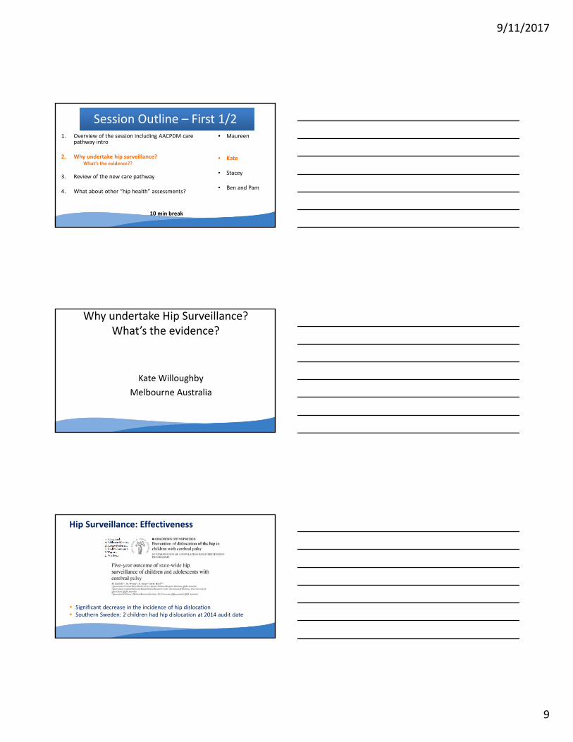
Session Outline – First 1/21. Overview of the session including AACPDM care
pathway intro
2. Why undertake hip surveillance? What’s the evidence??
3. Review of the new care pathway
4. What about other “hip health” assessments?
10 min break
• Maureen
• Kate
• Stacey
• Ben and Pam
Session Outline ‐ Second 1/2Panel Presentation/ Discussion – with you!
‐ “How to” from three jurisdictions perspective (US example, Australia, BC in Canada)
‐ Including questions for the panel…… how do families feel about this?… how to “find” all potential kids?… how to ensure radiology quality?… your questions!
Session Outline – First 1/21. Overview of the session including AACPDM care
pathway intro
2. Why undertake hip surveillance? What’s the evidence??
3. Review of the new care pathway
4. What about other “hip health” assessments?
10 min break
• Maureen
• Kate
• Stacey
• Ben and Pam

9/11/2017
4
• Care pathways work began in 2014
• Working group created• Created process for all care
pathway development• ID’d 6 inaugural care
pathways
What is an AACPDM “Care Pathway”?It is a …• Structured • Multi‐disciplinary• Plan for care (of an individual child )• Aimed to support the implementation of clinical guidelines and best evidence
• Includes a step‐wise sequencing of care (aka algorithm)
What’s the difference from a clinical practice guideline again?
Accessed September 3, 2017https://www.nhlbi.nih.gov/health‐pro/guidelines/about

9/11/2017
5
9/11/2017
6
Care Pathway Development
The Process in a Nutshell• ID area of need• Assemble multi‐disciplinary, (international) working group• *Define the parameters of the question/project well (Project
charter)*• Review/assemble best evidence
– Literature – SR’s, specific lit review re: each branch in algorithm– International jurisdictional review of guidelines and programs
• Come to consensus ‐meetings using tele‐conferencing, Go‐to‐meeting, online survey tools
• Assemble supportive documents
Hip Surveillance• “The process of monitoring and identifying the critical early indicators of hip displacement."

9/11/2017
7
The Process in a Nutshell• ID area of need• Assemble multi‐disciplinary, (international) working group• *Define the parameters of the question/project well (Project
charter)*• Review/assemble best evidence
– Literature – SR’s, specific lit review re: each branch in algorithm– International jurisdictional review of guidelines and programs
• Come to consensus ‐meetings using tele‐conferencing, Go‐to‐meeting, online survey tools
• Assemble supportive documents
Key Decisions in the Project Charter• Hip surveillance should be based on:
Age GMFCS level Classification as WGH gait type IV or not Migration percentage
• Surveillance should consist of radiological exams & clinical assessments• Pathway represents minimum requirements for only hip surveillance• Pathway will guide care for individual children; NOT about how to
operationalize surveillance (case finding, data management, care coordination)
The Process in a Nutshell• ID area of need• Assemble multi‐disciplinary, (international) working group• *Define the parameters of the question/project well (Project
charter)*• Review/assemble best evidence
– Literature – SR’s, specific lit review re: each branch in algorithm– International jurisdictional review of guidelines and programs
• Come to consensus ‐meetings using tele‐conferencing, Go‐to‐meeting, online survey tools
• Assemble supportive documents

9/11/2017
8
e.g. Comparison of Clinical Exam Content
e.g. Comparison of Frequency of Surveillance
The Process in a Nutshell• ID area of need• Assemble multi‐disciplinary, (international) working group• *Define the parameters of the question/project well (Project
charter)*• Review/assemble best evidence
– Literature – SR’s, specific lit review re: each branch in algorithm– International jurisdictional review of guidelines and programs
• Come to consensus ‐meetings using tele‐conferencing, Go‐to‐meeting, online survey tools
• Assemble supportive documents
9/11/2017
9
Session Outline – First 1/21. Overview of the session including AACPDM care
pathway intro
2. Why undertake hip surveillance? What’s the evidence??
3. Review of the new care pathway
4. What about other “hip health” assessments?
10 min break
• Maureen
• Kate
• Stacey
• Ben and Pam
Why undertake Hip Surveillance?What’s the evidence?
Kate WilloughbyMelbourne Australia
Hip Surveillance: Effectiveness
Significant decrease in the incidence of hip dislocation Southern Sweden: 2 children had hip dislocation at 2014 audit date

9/11/2017
10
Hip Health in CP: More than just ‘dislocation’A healthy hip:
Has satisfactory morphology, Is mobile, Does not limit function, Is PAIN FREE
Classifying Hip Morphology
Quantitative Radiographic Measure• Migration percentage
Qualitative Radiographic Features• Break in Shenton’s line• Shape of the femoral head• Development of lateral acetabular margin• Presence of pelvic obliquity
Hip Health at Skeletal Maturity
Associations:
GMFCS and hip morphology (MCPHCS)
MCPHCS and Pain (Likert scales)
MCPHCS and Hip Surveillance
9/11/2017
11
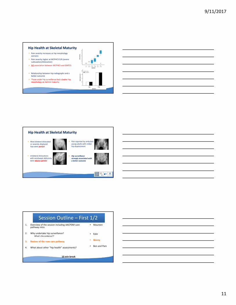
Pain severity increases as hip morphology worsens
Pain severity higher at MCPHCS 5/6 (severe subluxation/dislocation)
NO association between MCPHCS and GMFCS
Relationship between hip radiographs and a better outcome
Those under hip surveillance had a better hip morphology at skeletal maturity
Hip Health at Skeletal Maturity
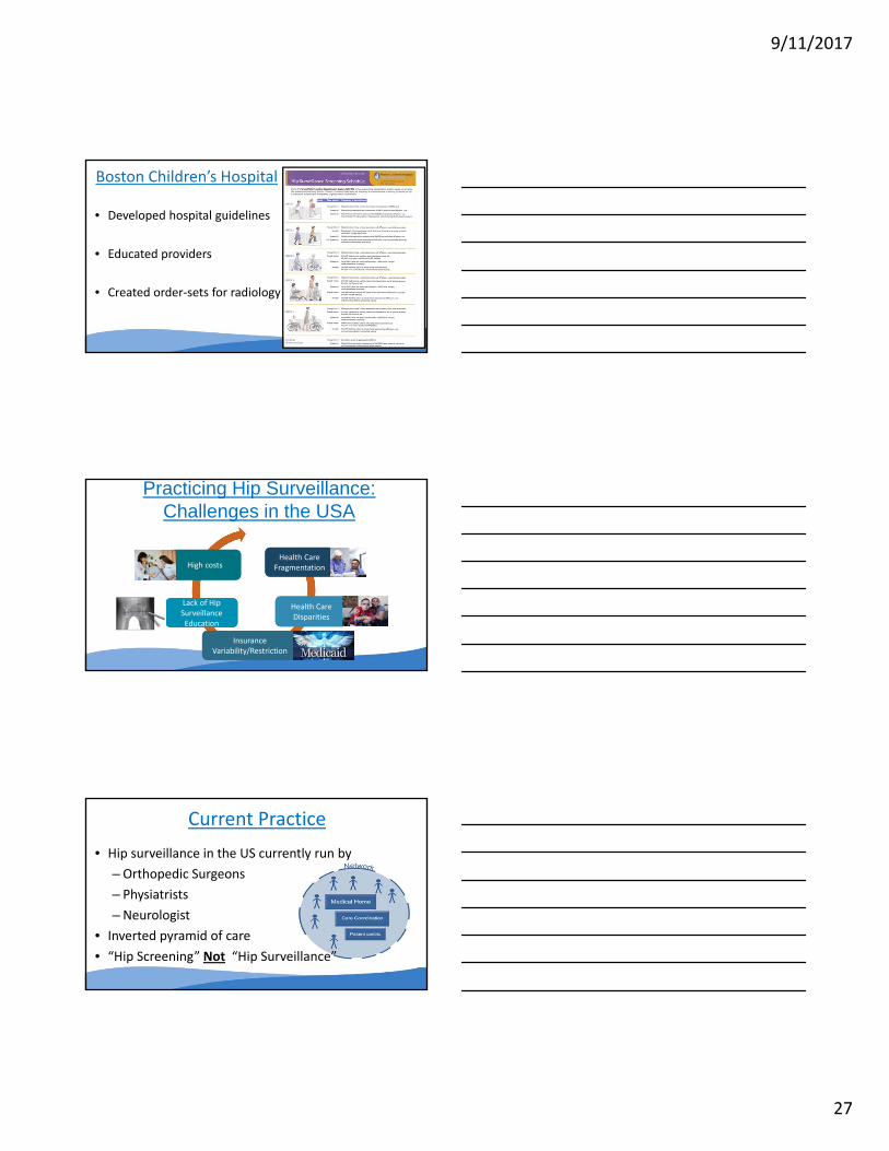
Most bilateral dislocated or severely displaced hips were painful
Unilateral dislocations with windswept deformity were always painful
Pain reported by ambulant young adults with milder hip displacement
Hip surveillance strongly associated with a better outcome
Hip Health at Skeletal Maturity
Session Outline – First 1/21. Overview of the session including AACPDM care
pathway intro
2. Why undertake hip surveillance? What’s the evidence??
3. Review of the new care pathway
4. What about other “hip health” assessments?
10 min break
• Maureen
• Kate
• Stacey
• Ben and Pam

9/11/2017
12
AACPDM Hip Surveillance Care Pathway
AACPDM Hip Surveillance Care Pathway
Clinical Exam

9/11/2017
13
Clinical Exam
If child has hemiplegia, identify if WGH Gait Type IV
Clinical Exam
AACPDM Hip Surveillance Care Pathway

9/11/2017
14
Initiation of Surveillance
GMFCS IV & V
* Do not reduce from previous higher frequency if:
(2) stability is not yet achieved over a period of 2 years. Stability is defined as < 10% change in MP over a 12 month period;
OR(3) MP > 30%.
(1) 24 months of surveillance have not yet been completed based on a child's surveillance start date;

9/11/2017
15
GMFCS III
* Do not reduce from previous higher frequency if:
(2) stability is not yet achieved over a period of 2 years. Stability is defined as < 10% change in MP over a 12 month period;
OR(3) MP > 30%.
(1) 24 months of surveillance have not yet been completed based on a child's surveillance start date;
GMFCS II
GMFCS I

9/11/2017
16
Any GMFCSLevel with
Winters Gage Hicks Gait Type IV
AACPDM Hip Surveillance Care Pathway
Radiological Exam• AP pelvis (supine)
• Positioning– Abduction/adduction: Neutral– Hip rotation: Patellae up
– Neutral Pelvic Obliquity; Flattened lordosis
Reproducedwith permission and copyright © Bill Reid, Royal Children’s Hospital, Melbourne, Australia.

9/11/2017
17
• Measure: Migration Percentage (MP)
• Abnormal MP: MP >30%
• Stable MP: no ∆ in MP >10% in a 12 month period
• Skeletal Maturity: Closure of triradiate
Radiological Exam
• Optimal Field of View
• Gonad Protection– Per local guidelines
Radiological Exam

9/11/2017
18
Discharge/Continue Surveillance
Clarifications?
Session Outline – First 1/21. Overview of the session including AACPDM care
pathway intro
2. Why undertake hip surveillance? What’s the evidence??
3. Review of the new care pathway
4. What about other “hip health” assessments?
10 min break
• Maureen
• Kate
• Stacey
• Ben and Pam

9/11/2017
19
What else to consider for the hip?“Hip Health”
Ben ShorePam Thomason
Hip Health in CP• Hip surveillance
– One component of musculoskeletal care– Primary measures GMFCS and MP
• Musculoskeletal assessment – part of a comprehensive care for children with CP– Primarily not required for hip surveillance– But important to evaluate hip health
Hip health in CP • The hip joint should be:
– Flexible– PAIN FREE– Not limit function
• Femoral head covered by acetabulum

9/11/2017
20
Hip health in the context of the child• Mobility
– Goals vary depending on GMFCS
• Pain• ADL• Weight bearing• Bone health• Medication
• Mood / anxiety• Cardio‐respiratory• Oro‐motor function
• Constipation• Reflux• Epilepsy• Nutrition• Sleep
Questions?• Family best source of information• Deterioration or change in:
– Ability to care– Weight bear through hip– Stand and step– Walk or walking pattern
Physical examinationRange of motion
– Hip abduction– Hip flexion‐extension– Hip rotation– FNA– Popliteal angle
Muscle tone– Hip adductors– Hip flexors– Hamstrings

9/11/2017
21
Importance of symmetry• Spinal deformity• Pelvic obliquity• Leg length discrepancy
Pelvic obliquity: Prevalence and long term consequences
Inter Iliac Crest Line
Inter Teardrop Line
Inter Tuberosity Line
Pelvic obliquity

9/11/2017
22
1. High prevalence but majority mild
2. Increased SAA and MP always high side
3. Unilateral dislocation always high side
Pelvic obliquity
Classification of Hip Morphology• Quantitative Radiographic Measure
• Migration percentage
• Qualitative Radiographic Features• Break in Shenton’s line• Shape of the femoral head• Development of lateral acetabular margin• Presence of pelvic obliquity
Melbourne Cerebral Palsy Hip Classification Scale‐ Expanded and Revised
Normal hip
Near Normal Hip
Dysplastic Hip
Dysplastic ‐Mild Subluxation
Mod‐Severe Subluxation
Dislocated
Salvage Surgery
Melbourne Cerebral Palsy Hip Classification Scale‐ Expanded & Revised

9/11/2017
23
1. Pain severity increases as hip morphology worsens
2. NO association between MCPHCS and GMFCS3. Most bilateral dislocated and severely displaced
hips painful4. Unilateral dislocations with windswept
deformities always painful5. Hip surveillance associated with a better
outcome
Hip Health at Skeletal Maturity
Session Outline – First 1/21. Overview of the session including AACPDM care
pathway intro
2. Why undertake hip surveillance? What’s the evidence??
3. Review of the new care pathway
4. What about other “hip health” assessments?
10 min break
• Maureen
• Kate
• Stacey
• Ben and Pam
Session Outline ‐ Second 1/2Panel Presentation/ Discussion – with you!
‐ “How to” from three jurisdictions perspective (US example, Australia, BC in Canada)
‐ Including questions for the panel…… how do families feel about this?… how to “find” all potential kids?… how to ensure radiology quality?… your questions!

9/11/2017
24
Let’s introduce our panel…
How to implement?
Three examples ‐ from three different contexts
Thing to consider when implementing..
– The decision to start surveillance is made by… … (e.g. who starts things? How does a child get “enrolled”)
– Information for families at the time of initiation/enrollment is provided by or through…– The routine clinical exams of surveillance are done by…– The xray is ordered by…– The xray is read by…– The person who is responsible for putting together the information from the clinical exam and xray
(and who shares this with the family is )– If there are problems with the xray or concerns with the clinical exam and a referral to doc is
needed, this referral is done by…– The way we make sure that kids get the follow‐up in the right frequency is/will be…– We know the population of children with CP in our “jurisdiction” or “catchment” are getting
surveillance done because… – An important thing or two that I would say is a final lesson learned from our approach is…

9/11/2017
25
Implementation:a US example
How Hip Surveillance is Currently Practiced in the United States
Benjamin Shore MD MPH FRCSCBoston Children’s Hospital
Questions we will address• When does hip surveillance begin?• Who initiates surveillance?• Who places the xray orders and who interpretes them?• Who is the person who organizes care?• How do we know who our population is?• Who establishes frequency of follow up and treatment if necessary?

9/11/2017
26
Boston Children’s Hospital• 415 bed hospital• 160 MDs• 1300 RNs• 16,000 inpatient admissions/yr• 60,000 ED visits/yr• >20,000 outpatient surgeries/yr
Boston Children’s Hospital – CP CenterFamily Generated Care Map
OTHER CLINICS
NEUROLOGY
NEUROSURGERY
COMPLEX MEDICINE
ORTHOPEDICS & PHYSIATRY
ORTHOPEDICS/PHYSIATRY& NEUROLOGY
9/11/2017
27
Boston Children’s Hospital
• Developed hospital guidelines
• Educated providers
• Created order‐sets for radiology
Practicing Hip Surveillance: Challenges in the USA
High costsHigh costs
Lack of Hip Surveillance Education
Lack of Hip Surveillance Education
Health Care DisparitiesHealth Care Disparities
Health Care FragmentationHealth Care
Fragmentation
Insurance Variability/Restriction
Insurance Variability/Restriction
Current Practice • Hip surveillance in the US currently run by
–Orthopedic Surgeons– Physiatrists– Neurologist
• Inverted pyramid of care• “Hip Screening” Not “Hip Surveillance”

9/11/2017
28
Moving Forward• Transition from “Screening to Surveillance”• Improve education• Establish a point of entry at the therapist level
– School based enrollment similar to scoliosis
• Create national guidelines and benchmarks
Implementation –Australia’s approach/es
Implementing… Australia style– The decision to start surveillance is made by… … (e.g. who starts things? How does a child get
“enrolled”)– Information for families at the time of enrollment is provided by or through…– The routine clinical exams of surveillance are done by…– The xray is ordered by…– The xray is read by…– The person who is responsible for putting together the information from the clinical exam and xray
(and who shares this with the family is )– If there are problems with the xray or concerns with the clinical exam and a referral to doc is
needed, this referral is done by…– The way we make sure that kids get the follow‐up in the right frequency is/will be…– We know the population of children with CP in our “jurisdiction” or “catchment” are getting
surveillance done because… – An important thing or two that I would say is a final lesson learned from our approach is…
9/11/2017
29
Implementation –A Canadian province’s approach
Building Consensus in BC Provincial meetings in 2011 & 2012 50 participants All regions represented Inter‐disciplinary
• Parents• Pediatric orthopaedic surgeons• Pediatricians• Developmental Pediatricians• GPs• Physiotherapists• Occupational Therapists• Nurse• Radiology• Health administrators• Policy makers

9/11/2017
30
Child Development Centers
School Services
Child Development Centers
School Services
Child Development Centers
School Services
Hip Surveillance Coordinator/Medical Lead
9/11/2017
31
Child Development Centers
School Services
Hip Surveillance Coordinator/Medical Lead
Pediatric Orthopaedic Surgeons
BC Hip Surveillance Program
0100200300400500600700800900
VCH FHA IHA VIHA NHA
Num
ber o
f Children En
rolled
Health Authority
Hip Surveillance Program
Estimated Population2000‐2015
Any clarifications requested of our presenters?

9/11/2017
32
Implementation: Ask our Panel
Implementation question:
What has been your experience regarding how families respond to or feel about hip
surveillance?
Implementation question
What solutions or tips do you have to offer with respect to getting kids enrolled at diagnosis or before age 2 years?
How much does having the “diagnosis” of CP play into this? How do you manage that?

9/11/2017
33
Implementation question
Regarding radiology…
What are your practical tips for x‐rays done well –i.e. with good positioning? Or is this even an issue?
Read/reported well? Or is this even an issue?
Implementation question– from participants
Implementation question(if time permits)
Practical tips for making sure that the clinical exam information and the x‐ray information get put together and shared with family?

9/11/2017
34
Implementation question– from participants
Implementation question(if time permits)
In your experience, what can most help PT’s contribute to and/or drive hip surveillance?
Implementation question
What’s your one final quick tip to those undertaking hip surveillance?

9/11/2017
35
Formal Course Objectives1. Describe the evidence in support of hip surveillance and the clinical
components of hip surveillance.
2. Understand the evidence behind, process for development of and content of the new international pathway.
3. List additional “hip health” adjunctive tips that were created by the international consensus group and may be useful to clinicians.
4. Describe practical “tips” for implementing hip surveillance in a variety of disciplines perspectives and variety of practice settings, including clinic, community, and state/province