Embed Size (px)
Citation preview

1
TO STUDY THE EFFICACY OF HYSTEROSCOPY AS
A SCREENING METHOD IN PATIENTS WITH
ABNORMAL UTERINE BLEEDING
DISSERTATION SUBMITTED FOR
M.D (BRANCH – II) (OBSTETRICS & GYNAECOLOGY)
MARCH 2010
THE TAMILNADU DR.M.G.R. MEDICAL UNIVERSITY
CHENNAI, TAMILNADU

2
BONAFIDE CERTIFICATE
This is to certify that the dissertation entitled “TO
STUDY THE EFFICACY OF HYSTEROSCOPY AS A
SCREENING METHOD IN PATIENTS WITH ABNORMAL
UTERINE BLEEDING” is a bonafide record work done by
Dr. N. KURINJI PRIYA under my direct supervision and
guidance, submitted to the Tamil Nadu Dr. M.G.R. Medical
University in partial fulfillment of University regulation for M.D
Branch II – Obstetrics & Gynaecology.
Dr. DILSHATH. M.D.,D.G.O., Professor & Head of the Department Department of O&G
Madurai Medical College, Madurai.

3
DECLARATION
I Dr. N. KURINJI PRIYA solemnly declare that the
dissertation titled “TO STUDY THE EFFICACY OF
HYSTEROSCOPY AS A SCREENING METHOD IN
PATIENTS WITH ABNORMAL UTERINE BLEEDING” has
been prepared by me. I also declare that this bonafide work or a part of
this work was not submitted by me or any other for any award, degree,
diploma to any other University board either in India or abroad.
This is submitted to The Tamilnadu Dr. M. G. R. Medical
University, Chennai in partial fulfillment of the rules and regulation for
the award of M.D degree Branch – II (Obstetrics & Gynecology) to be
held in March 2010.
Place : Madurai Dr. N. KURINJI PRIYA
Date :

4
ACKNOWLEDGEMENT
My sincere and thankful gratitude to Prof. Dr. DILSHATH,
M.D., D.G.O., Professor and Head of the Department of Obstetrics
and Gynaecology for her expert guidance and support for the
completion of the study.
I am extremely thankful to THE DEAN, Madurai Medical
College and Government Rajaji Hospital, Madurai for granting me
permission to undertake this study.
I am very grateful to Dr. Parvathavarthini, M.D., D.G.O.,
Dr. Subbulakshmi, M.D., D.G.O., Dr. Angayarkanni, M.D.,
D.G.O., Dr. Ambigai Meena, M.D., D.G.O., Dr. Lalitha, M.D.,
D.G.O., Additional Professors, Department of Obstetrics and
Gynaecology, for their valuable suggestions and guidance in
preparing this dissertation.
My grateful thanks to Asst. Professors of Department of
Obstetrics and Gynaecology, for their immense help during this
study. Thanks to my fellow post graduates who had assisted me
throughout the study.
I acknowledge the cooperation of the patients without whom
this study would not have been possible.

5
CONTENTS
S.NO. TITLE PAGE
1. INTRODUCTION 1
2. AIM OF THE STUDY 3
3. REVIEW OF LITERATURE 4
4. PLACE OF DIAGNOSTIC HYSTEROSCOPY
IN AUB
21
5. MATERIALS AND METHODS 24
6. ANALYSIS OF THE STUDY 37
7. DISCUSSION 55
8. SUMMARY 58
9. CONCLUSION 60
BIBLIOGRAPHY
PROFORMA
MASTER CHART
ABBREVIATION

6
INTRODUCTION
Abnormal uterine bleeding (AUB) is a common complaint in
“gynaecological practice. It represents a major proportion of
outpatient attendance. The most probable cause of abnormal
uterine bleeding depends on the patient’s reproductive age and the
likelihood of serious endometrial pathology like malignancy
increases with age. The diagnosis of AUB is by exclusion.
Before hysteroscopy was available, curettage was the primary
method of evaluating abnormal uterine bleeding. Brooks and
Serden have revealed that approximately half of pedunculated
abnormalities such as endometrial polyps were missed by curettage.
It renders endometrial sampling blind and incomplete, so the
diagnostic accuracy of curettage is less than that of hysteroscopy.
For Focal lesions, D & C is less accurate and less reliable
However, it gives histopathological report, which is of immense
value in the treatment of AUB. According to Valle, hysteroscopy

7
is not a substitute for tissue diagnosis. According to Loffer, a
tissue diagnosis is essential. Though, transvaginal sonography is
considered as an initial investigation in patients with AUB, the
prediction of endometrial pathology based on ultrasound scan in
premenopausal women is not reliable because of great overlap
between normal range and those with endometrial pathology. So,
hysteroscopy combined with histological examination is the new
“Gold Standard” for evaluating a case with AUB (Widrich et al ,
Bakour)

8
AIM OF THE STUDY
The objective of the present study is to evaluate the
usefulness of Hysteroscopy as a screening method in patients
with AUB in relation to the histopathology results of the
endometrial biopsy and results of transvaginal sonography.
1. To evaluate the usefulness of hysteroscopy as a
screening method in patients with Abnormal uterine
bleeding.
2. To compare the efficacy of hysteroscopy in diagnosing
of endometrial pathology with transvaginal
sonography and its correlation with histopathology.

9
REVIEW OF LITERATURE
HISTORY OF THE TYPES OF THE ENDOMETRIUM IN DUB
* As far as 1860, Scansoni stated that the common female
disease was chronic metritis and that the bleeding was due to
abnormal brittleness of the arteries.
* Remiche (1897), Slocum (1908) and Findley (1905)
attached importance to the uterine arteriosclerosis as a cause of
bleeding.
* The term “Apoplexy uterus” is said to have been used by
Guveilbein as early as 1882.
* Brenecke described the so called ‘endometritis oophoritis’
which is know as hyperplasia of the endometrium. Then the
hormonal function of the ovary was not even known.
* Pankor (1909) emphasized the term ‘Metropathia
haemorrhagica’ as there was increased elastic tissue in the blood
vessel.
* 1910 Thailbater denoted the defective contractility of
mesometrium to embrace connective tissue as well as the
musculature of the uterus.

10
* Later on in 1915, Schroeder evolved a working concept
of the mechanism involved in Metropathia haemorrhogica, and
William P also contributed to this study in hysterectomised
specimens of AUB.
Endometrial Hyperplasia.
* French surgeons Robert (1846), Robin (1848) and Nelton
(1853), made the first clinical observation of hyperplasia.
* Recamier (1850) the inventor of curette, advised curettage
as a method of treatment and met with strong opposition by Aron
(1958) and Becaucert (1859)
* Olshavsen (1875) described the histology and its
association with uterine bleeding. He called it “endometritis
fungosis” and “chronic hyperplasia”.
* Cullen (1900) described in English and coined the term
“hyperplastic endometrium.”
* Robert Schroeder (1915 & 1919) by his epoch making
work ruled out the older inflammatory theory and correlated
endometrial hyperplasia with persis.

11
* Robert Meyer (1920) described the ovarian changes and
drew attention to graffian follicle maturation and their
disintegration by atresia.
* Anatomic studies of E Novak, Seitre, Baher, Reuge, Novak,
Adler, Martzold, Backmarard, Wilfred shaw were published in
1920.
* Hanghaver (1930) attempted to reproduce hyperplasia by
injecting anterior pituitary extracts to guinea pigs.
* Fluhman (1931) suggested that hyperplasia is due to
unopposed action of estrogen.
* Zucherman and Movie Jeffcoate, Dayne, Taylor Novak
(1936) said that hyperplasia is due to excessive estrogen.
* Clessens and Cowell found 19% of adolescent girls with
menorrhagia having a haemostatic defect while Smith et al found
the incidence to be 33%.
* Falcone et al found only 3.3% of patients admitted for
menorrhagia over a 10 year period.
* Studies by Ash et al, suggested that menstrual cycle
irregularity and hypertension were independently significant risk
factors for detecting abnormal endometrial riskfactors.

12
* Lettererrand Masshoff, 1941 described the glandular
epithelium of endometrium of cystoglandular hyperplasia.
* Gruner 1942 described about the luteinisation of follicular
epithelium that may be reflected in the endometrium as patchy
secretory changes.
* Schmidt 1965, Matihesan 1967 studied about estrogen and its
stimulation of fibrinolytic activity of endometrium
* Kurman et al 1985 have studied the progression of
endometrial hyperplasia to carcinoma
* Sharman and Brown 1979 showed that Grade I and Grade II
endometrial hyperplasia have 22% and grade III has 57% chance
of development of carcinoma endometrium.
ATYPICAL HYPERPLASIA
Cullen as early as 1900 described atypical endometrium
existing with adenocarcinoma. He suggested that it may be the
earliest phase of malignancy, not pathognomonic but a sign of
cancer. Diagnostic criteria was laid down by Schroeder (1925),
Taylor (1932), Schroeder (1929), Novak and Yin (1934) indicated
that there is some relationship between hyperplasia and corporal
cancer. Gueberg (1947) believed that there is a constant

13
relationship between adenomatous hyperplasia, benign and
malignant adenoma and estrogen stimulation. Rutletge and Novak
(1948) showed similarity between atypical hyperplasia and
endometrial carcinoma. Hertig Sommen and Bengloff (1949) in
their extensive study drew attention to focal and generalized
adenomatous hyperplasia and development of cancer. Their studies
bore no time relation to the development of cancer and
adenomatous hyperplasia may regress, though in some it was found
to be the precursor of endometrial cancer. Sperts’ (1952) excellent
paper on premalignant phase of endometrial cancer is noteworthy.
Gusberg (1954), John A wall and Waner’s (1957) paper are for
adenomatous hyperplasia and precancerous condition. Compell
Bastere (1966) and George sipper (1966) laid down criteria for
diagnosing atypical hyperplasia and its importance in treatment.
Proliferative Endometrium
Anovulatory cycles are common in adolescence and before
the menopause. Though the mechanism is not known, the
menstrual bleeding associated with the shedding of proliferative
endometrium in the second half of the cycle should always be
regarded as abnormal.
14
HISTORY OF HYSTEROSCOPY
* Visual examination of the uterine cavity is an old
technique. Pantaleoni, in 1865, was the first to examine the uterus
using a small tube inserted through the external OS of the cervix
with a kerosene lamp or a candle for a light source (Lindemenn
1973)
* This first examination was performed as a 60 year old
woman with intractable post-menopausal bleeding and it was
reported that polyp like growths were found.
* Many technical advances in the optical systems were
developed in Europe at the turn of the century (Fragenhein 1987),
but only in the last 25 years have fibre-optic light sources and the
Hopkins lens system made hysteroscopy a practical diagnostic out-
patient procedure.
Endoscopic procedure in general attained a wider acceptance
after the introduction in 1952, also in France, of the “Cold Light”
concept by Fourestier, Gladu and Vulmiere. This innovation
permitted great improvements to be made in the equipment. In the
same year, Hopkins and Kapany in England introduced fiberoptics
to the field of endoscopy.

15
Hysteroscopy was the earliest form of endoscopy. The
introduction of effective uterine distress media – high molecular
weight dextran (Edstrom and Fernstrom, 1970), CO2 (Lindemann
1970), 5% dextran (Norment and Sykes, 1972) – made
hysteroscopy, a practical procedure.
The microcolpohysteroscope was introduced by Hamou in
1980. The American Association of Gynaecological laprascopists
(AAGL) was found in 1972. This organization has played a major
role in the dissemination of endoscopic knowledge in North
America and in many parts of rest of the world.
Author (PT) was preveledged to be the scientific chairman
for the first practical course in hysteroscopy offered by the AAGL
at the virgin masou clinic in Seattle, Washington in 1982.
HPE OF Endometrium
DUB should be evaluated promptly which is primarily
associated with ovarian dysfunction and or anovulation.
1. By doing HPE of the endometrium
2. By studying the endometrial thickness using
transvaginal probe
3. Direct visualization of the endometrial cavity using
hysteroscopy

16
In evaluation of AUB, the following principles should be
borne is mind.
It is essential to exclude organic diseases if necessary by
repeated examination and special investigation.
The morphology of endometrium lightly responds to ovarian
steroid hormones and changes in the hormones are specifically and
promptly reflected in the endometrium. Endometrial biopsy is the
convenient procedure traditionally used is gynaecology.
Interpretation of endometrial biopsy specimens provides
reasonable accurate measurement to steroid stimuli. The relative
ease with which endometrium may be biopsied has permitted the
histological diagnosis of the disease of the uterus and afforded
opportunity to evaluate the various physiological and
pathophysiological events which have an impact on menstrual
function.

17
HISTOPATHOLOGY OF ENDOMETRIUM IN DUB
Classification of possible endocrine abnormality associated
endometrial histology and typical bleeding pattern in DUB.
Type Endocrine Abnormality and
Endometrial Histology
Typical bleeding
pattern
Normal
ovulatory
* Short cycle – short proliferative
Phase – normal endometrium
* Long cycle – long proliferative
phase – normal endometrium
Polymenorrhea
Menorrhagia
Oligomenorrhea
menorrhagia
Corpus luteum
abnormality
* Insufficiency – short luteal phase
– irregular or deficient secretory
endometrium
* Persistent (Halbans disease)
irregular endometrial shedding
Premenstrual
spotting
Menorrhagia
Anovulatory * Insufficient follicles – short cycle
inadequate proliferative or atrophic
endometrium
* Persistent follicle or PCOS –
prolonged cycle proliferative or
hyperplastic endometrium
Polymenorrhea
Metrorrhagia
Oligomenorrhea
Metropathia
haemorrhagica

18
Endometrial Pattern in DUB
I Ovulatory - Irregular shedding
Irregular ripening
II Anovulatory - Proliferative
Hyperplastic
- Simple cystoglandular hyperplasia
- Complex adenomatous hyperplasia
without atypia
- Atypical (Adenomatous with atypia)
III Atrophic
Abnormal functional bleeding may be associated with any
type of endometrium even normal secretory endometrium. This
may be due to variation in vasculature of some local defect within
the uterus.
Ovulatory endometrium indicates persistence of secretory
changes suggesting a persistence or over activity of corpus luteal
function.
To assess this pattern of bleeding, biopsy should be done in
immediate post-menstrual period.

19
Irregular shedding
On histological examination, there is predominance of
proliferative endometrium.
Irregular Ripening
First described by Traut and Kinder at al in 1935. On
histological examination, there is mixture of both proliferative and
secretory endometrium
Anovulatory endometrium, proliferative phase
The proliferated glandular cells are columnar and the
elongated nucleus has coarse chromatin which fills most of the cell.
The nuclei give a pseudostratified appearance. The cystoplasm is
scanty. Cilia may be present. The stroma is heavily infiltrated with
leucocytes and red cells and the vessels are thin walled and straight.
Endometrial Hyperplasia
In 1988, the nomenclature committee of the international
society of Gynaecologic pathologists adopt the following
modification of terms for endometrial hyperplasia.
1. Simple hyperplasia (Cystoglandular hyperplasia)

20
2. Complex hyperplasia (Adenomatous hyperplasia without
atypia)
3. Atypical hyperplasia (Adenomatous hyperplasia with atypia)
Endometrial Hyperplasia represents a spectrum of morphologic
and biologic alterations of the endometrial glands and stroma,
ranging from an exaggerated physiologic state to carcinoma in situ.
Clinically significant hyperplasias usually evolve within a
back ground of proliferative endometrium as a result of protracted
estrogen stimulation in the absence of progestin influence.
The most recent classifications scheme endorsed by the
International Society of gynaecological pathologists is based on
architectural and cytologic features as well as long term studies that
reflect the natural history of the lesions.
Classification of Endometrial hyperplasias
Type of hyperplasia Progression to carcinoma
Simple (cystic without atyptia) 1%
Complex (adenomatous without atypia) 3%
Atypical
Simple (cystic with atyptia) 8%
Complex (adenomatous with atypia) 29%

21
Simple hyperplasia (Cystoglandular Hyperplasia)
It is seen in perimenopausal women that the glands may be of
varying sized and some are cystic. They are lined by multi-layered
cuboidal or tall columnar epithelium. The stroma is increased and
densely cellular.
Complex Hyperplasia (Adenomatous Hyperplasia without
Atypia)
Microscopically there is an increase in the number and size of
the endometrial glands which are crowded together with scanty
stroma in between. The glands are lined by hyperplasia stratified
epithelium with finger like out pouchings into the stroma as well as
papillary budding of the epithelium into the glandular lumen.
Atypical Hyperplasia (Adenomatous hyperplasia with Atypia)
The lining epithelium of the glands are large, irregularly
heaped up, columnar cells with large irregular nuclei and loss of
polarity.
The cytoplasm has dense, abundant eosinophilic staining.
There is also back to back glandular crowding with reduction
and papillary infolding of the glandular epithelium.

22
Atrophic endometrium
The glands are found dilated and they are lined by flat
cuboidal epithelium. The stroma is edematous and sparsely
cellular.
Apart from the fact that hyper estrogenic level is responsible
for abnormal bleeding, psychological factors also play a role in
patients perceiving uterine bleeding as excessive.
The studies with special stains expands the parameters of one
observations of ovulatory and anovulatory endometrium.
The positive staining for glycogen in secretory endometrium
indicates ovulatory endometrium and the positive staining for
collagen in hyperplastic endometrium indicates anovulatory
endometrium.

23
ROLE OF TVS IN AUB
Probably the commonest complaint in a woman is irregular
bleeding disorders, menorrhagia etc. This forms the commonest
indication for the Ultrasound examination before any invasive
method.
Ultrasound scan, particularly the transvaginal route is used to
assess endometrial thickness, endometrial and myometrial
consistency and abnormalities of endometrial morphology like
submucosal fibroid or polyp etc.
It has been recommended that in patients with menorrhagia,
the uterine cavity should initially be investigated using transvaginal
sonography. Transvaginal sonography is preferable to pelvic ultra
sonography because of better quality of its image. This is achieved
because of its higher frequency which allows greater image
resolutions at the expense of decreased depth of penetrations. So
transvaginal sonography can be considered a first line of
investigation for women with AUB.
Ultrasonography especially transvaginal is an inexperience,
non invasive, convenient way to indirectly visualize the

24
endometrial cavity. It is a safer, painless, convenient method of
diagnosing those with pathology.
Any women who presents with AUB cannot be labeled as
AUB usually as clinical examination alone can only detect 9 %
percent of anatomic findings as compard to 31 % by ultrasound.
USG is helpful in excluding many patients with anatomic
findings not detected by physical examination, eg. Intramural,
subserous, submucous fibroids, endometrial or fibroid polyp,
PCOS,congenital anomalies of uterus and endometrial carcinoma.
It also helps in the evaluation of endometrium.
Thus USG has an increasingly important role in evaluating
certain disorders of the endometrium. It also guides in the need for
correction of anovulation depending on the age group. Thus, USG
examinations provides a method of evaluating symptomatic
patients and for identifying any remaining endometrium that could
later on become symptomatic.
• Atrophic endometrium – very thin bright interface in the
centre of the uterus.
• Endometrial Hyperplasia – Echogenic thick endometrium not
correlating to the day of menstrual cycle.

25
An endometrial thickness of more than 18 mm at perimenopause
carries a 20% risk of endometrial malignancy. If these patients are
on HRT, the cut off thickness is 10 mm.
• Endometrial polyps – These polyps can be microscopic or
macroscopic. A polyp of 5 mm can be visualized by TVS. If there
is fluid in the endometrial canal these polyps are better delineated.
Intra-luminal polyps can be diagnosed to almost 100% accuracy by
high resolution.
Misplaced IUCDS
TVS can detect myometrial burying partial perforation and
cervical displacement of any type of IUCD. This will help in
proper planning of hysteroscope or laparoscopic removal of the
misplaced IUCD.

26
PLACE OF DIAGNOSTIC HYSTEROSCOPY IN AUB
The diagnosis of AUB is by exclusion. The specific
diagnostic approach depends on whether the patient is
premenopausal, or postmenopausal. In perimenopausal patients,
endometrial biopsy and other methods of detecting endometrial
hyperplasia or carcinoma must be considered early in the
investigation. Thus in this age group, endometrial biopsy or
transvaginal ultrasonography is included in the initial investigation
followed by hysteroscopy.
Hysteroscopy and USG are now being increasingly used not
only for detecting functional disorders of endometrium but also for
excluding various unsuspected organic diseases of the endometrium
like cancer and Tuberculosis.
The way to perform a thorough hysteroscopic inspection of
the uterus has been well described. It is important to perform the
procedure under direct vision without routine blind dilatation,
which can lead to pain and suboptimal visualization, secondary to
bleeding from genital tract trauma. The operator should obtain a
panoramic view of the whole cavity and orientate in relation to
uterine landmarks – cornua, tubal ostia, fundus. In most cases this
27
is a simple procedure although a small but significant number
become problematic. Good technique and an understanding of the
limitation of hyperoscopic diagnosis are important.
Hysteroscopy is the standard procedure for detecting
intrauterine structural pathology (Widrich). Macroscopic inspection
of the uterine cavity cannot, however, make histological diagnosis
directly. Within the clinical context, therefore, hysteroscopic
diagnosis incorporating morphological features would be better
classified as normal, abnormal (thickened), abnormal (suspicion) or
abnormal (cancerous), consistent with the capability of the
technology. (Bakour).
Studies comparing hysteroscopy with traditional D and C
have suggested that the former is superior. However the existing
data supporting the use of hysteroscopy is generally related to
detection of benign disease and structural pathology. Hysteroscopy
appears to be better than ultra-sonography in detecting intra-uterine
structural pathology (clerk) although results with saline infusion
sonography are similar (Soares).
Subgroup analysis suggests that diagnostic accuracy of both
outpatient and inpatient hysteroscopy was high for endometrial

28
cancer, although there was a trend towards higher accuracy in the
outpatient setting (Clark). The video imaging and small diameter
endoscopes (<5mm diameter) also improved accuracy.
The introduction of outpatient of diagnostic procedure has
benefited both patients and the health service in terms of
convenience, reduced morbidity and costs. Following this
diagnostic process and inpatient surgical intervention such as D&C,
polypectomy, endoscopic procedure or hysterectomy is often
recommended. Intra-uterine structural pathology (eg.endometrial
polyps and sub-mucous fibroids) is found in around 25 percent of
women with abnormal uterine bleeding and accounts for much of
this surgical activity.
The same advances in endoscopic technology that helped
outpatient hysteroscopic diagnosis happen now offer the possibility
of outpatient hysteroscopic treatment for such conditions like
polyps, intracavity fibroids, septas and adhesions. However
inpatient endoscopic treatment of these conditions has been
recommended (RCOG press).

29
MATERIALS AND METHODS
PATIENT POPULATION
Our study includes 50 women age varying between 28 to 55
years who were admitted. We included only the patients with
technical success in transvaginal sonography and Hysteroscopy in
this study. Finally 50 patients with pathological confirmation were
included in this study. All 50 patients were first evaluated with
transvaginal sonography followed by Hysteroscopy and traditional
curettage with cervical biopsy performed after 2 days following
transvaginal sonography. The pathological findings are then
correlated with ultra-sound finding and diagnosis by hysteroscopy.
Criteria for Selection
1. Patients who had AUB for more than 6 months were
selected
2. Patients in Reproductive and perimenopausal age were
selected
3. Both Nulliparous and parous women were included
4. Patients in pubertal age group were excluded
5. Both treated and untreated patients were taken

30
6. Patients with any temporary method for the past 6 months
were excluded
7. Patients who underwent abortion were excluded
8. Patients with any other medical or surgical illness were
excluded
9. Lactating mothers were excluded
Ethical committee approval and informed consent from the
patients taken. Patients are also investigated if they have Intra-
uterine device or not, oral contraceptive use, regular drug or
hormonal preparation usage. Former gynaecological operations
and applications recorded. Patients detected to be pregnant are not
included in the study.
Transvaginal ultra-sonography is applied with 7 MHZ
vaginal probe to all patients who were selected for research.
Cervix, cervical canal, myometrium and ovaries were examined is
saggital and coronal section. After inspection of morphological
pattern of endometrium, endometrial thickness of external borders
measured and recorded in thickest place in longitudinal place.

31
DIAGNOSTIC HYSTEROSCOPY
INSTRUMENTATION :
TELESCOPES
The most important piece of equipment for hysteroscopy is the
lens or telescope. The optics as well as the fiberoptic illumination
bundles are packaged together in this single instrument. Most rigid
telescopes range in diameter from 2 to 4 mm (o.d.). The best light
shower and optical resolution are likely to be found in 4 mm (o.d.)
instruments. However, for office hysteroscopy, as well as operative
procedures, a 3 mm telescope can Produce an acceptable video
image particularly when magnified by means of a zoom video
camera.
The smaller telescope permits the operator to employ a
smaller sheath. Although flexible telescopes are now finding their
way onto the market, they suffer from inferior resolution compared
to that of the rigid equipment. The most convenient length for the
hysteroscopic telescope is 35 cm. Shorter instruments offer no
advantage and some distinct disadvantages when coupled to
operative sheaths. The telescope consists of three major parts:

32
• The magnifying eyepiece.
• The transmitting lens system.
• The objective lens
The most commonly used terminal objective lenses provide
a straight on view (00) or offset view (300). Selection of the lens is
a matter of personal preference but for the best panoramic operative
view the 00 lens is recommended, particularly when using laser
fibers or flexible or semirigid operating accessories. Rigid lenses
are fragile and must be handling, steam autoclaving, improper
liquid disinfection and inadequate cleaning will shorten the life of
the telescope and require expensive repairs as well as system down
time. A properly cared for lens will last a lifetime and provide
excellent service over and over again. Located just below the
eyepiece is the fiberoptic coupling connection. At this location the
fiberoptic cable joins, the telescope. Each lens manufacturer has a
unique coupling. Several companies supply attachments that
permit a variety of cables to join their particular instrument. The
latter is an advantage since any light generator and light cable can
be used with a given telescope.

33
FIBEROPTIC LIGHT CABLE
Fiberoptic cables transmit intense cold light to the telescope
and form a conduit, which connects the high intensity (heat
producing) generator to the telescope. The cable is filled with
many incoherent drawn-out glass fibers capable of conducting light
from the generator to the terminus of the cable. Obviously these
cables are fragile and can be easily damaged if not handled
carefully. Inspection of the end of the cable will readily determine
whether fibers or groups of fibers have been broken. These are
indicated by dark spots in an otherwise intense light shower.
Inspection of the periphery of the cable in a darkened room
can also reveal fiber disruption. This appears as light transmission
through the sides of the cable. Poor light at the end of the telescope
is almost always due to a damaged cable. The only alternative to
substandard light is to replace the cable. Cable should be
disinfected by soaking in cidex (glutaraldehyde) for 15-20 minutes,
followed by thorough washing in sterile water. At the end of the
case, the fiber should be washed again, resoaked and cleansed again
with water. The cable should then be stored dry in a protective
container. Most hospitals and surgicenters have replaced liquid

34
disinfect on with gas sterilization. The only drawback to the latter
is the long period required for aeration.
Light Generator
Several varieties of light generators are available on the
market. These range from simple and inexpensive tungsten light
generators (US $500-700) to costly xenon generators US$2000-
5000). For office use a simple apparatus will suffice, however,
hysteroscopy performed in the operating room under video control
demands intense light. Xenon (300W) generators produce white
light which is most favourable when coupled with endoscopic
television cameras (Baggish, 1997). Characteristically the simpler
tungsten light generators produce an orange tinted light which
creates a rather poor color on the video monitor. Between these
two types of generator are the 250 W metal halide generators.
These produce intense light, which is adequate for video images,
but characteristically give off a bluish tinge. The light bulbs in all
these generators produce a lot of heat, so a fan is built into the
cabinet to dissipate the heat and prolong bulb life. .
Light generators and fiberoptic cables can be used
interchangeably for hysteroscopy or laparoscopy. Obviously the

35
more powerful generators are required for laparoscopy. All
generators should be appropriately grounded and should be
periodically inspected (and indexed) biomedical engineering for
low frequency electrical leakage.
Hysteroscopic Sheaths
Two general categories of sheaths are used for hysteroscopic
procedures: diagnostic and operating. A sheath is required for
panoramic hysteroscopy in order to serve as a conduit through
which to instill the distending medium into the uterine cavity. The
diagnostic sheath fulfils this singular requirement and measures
approximately 4-5m in outer diameter (when coupled to a 3-4 mm
telescope). The sheath is essentially a hollow stainless steel tube
equipped with a proximal port through which the distending
medium is injected. The telescope must couple tightly to the sheath
with sufficient seal to prevent medium leakage at the
telescope/sheath, interface. The objective lens of the telescope
should fit precisely, flush with the end of the sheath to produce an
unobstructed view. Therefore each given manufacturer’s lens must
be matched correspondingly to the same manufacturer’s sheath.

36
The coupling mechanisms differ for different hysteroscopes,
preventing the interchange of lenses and sheaths.
A 5 mm sheath ordinarily will allow passage through
nulliparous cervices without resorting to dilatation. A 4 mm sheath
equipped with a 3 mm telescope can negotiate the endocervical
canal most easily than the 5 mm sheath., and is ideal in the office
setting. They are therefore ideally suited to office hysteroscopy.
Hysteroscopic sheaths are sturdy and stand up to routine handling.
They may be steam autoclaved. Obviously they should be
thoroughly flushed and cleansed after usage and stored away clean.
The stopcock mechanism should be disassembled, cleaned,
lubricated and reassembled after each usage. The stopcock should
be turned to the open position when stored, from time to time the
sheath as well as the shaft of the telescope should be polished with
a high quality.
ABNORMAL UTERINE BLEEDING
Abnormal uterine blooding is the most common complaint
of patients consulting the gynecologist and provides the most
frequent indication for hysterectomy. D and C has been the
diagnostic method of choice for many decades for these cases.

37
However, for diagnosing non-malignant intrauterine pathology
such as endometrial polyps. and intrauterine leiomyoms which may
cause uterine blooding disorders, D and C appears to be unreliable
(Wamsteker, 1977, 1984a; Gimpelson and Rappold. 1988. Loffer
1989; Motashaw and Dave. 1990).
Direct hysteroscopic inspection with adequate distention and
visualization discloses almost every intrauterine abnormality with
high accuracy. Additionally, it enables exact localization of the
pathology and determination of its intracavitary extent. However,
for the diagnosis of endometritis and adenomyosis, conclusive
hysteroscopic criteria are still lacking.
For histologic examination selective samples of any abnormal
tissue can be obtained by visually controlled biopsies. A
significant percentage of benign intrauterine pathology disclosed by
hysteroscopic diagnosis in patients with abnormal uterine bleeding
can be treated with minimally invasive transcervical hysteroscopic
endosurgery.
As the majority of intrauterine disorders resulting in
abnormal uterine bleeding in the reproductive phase of life, the
climacteric and the postmenopausal period, are benign types of

38
pathology, D and C can be longer hold its position as the primary
diagnostic method for patients with abnormal uterine bleeding.
Today ambulant or outpatient hysteroscopy with visually directed
biopsies or directed curettage is to be recommended as the
diagnostic method of choice for these cases.
With the more recently developed continuous how (CF)
technique the surface structure of the endometriun can be observed
with very low intrauterine pressure, which prevents compression of
the soft tissue of the mucosa and also reduces the transtubal flow of
the distention medium.
Extensive studies have not indicated any negative effect of
abdominal spill of the gas or liquid used for distention of the
uterine cavity in panoramic hysteroscopy in cases of endometrial
cancer (Wamsteker, 1977, Neis et al ,1994.
Review of the literature does not indicate that hysteroscopy
with abdominal spill of the distention medium should be considered
more hazardous in cases of endometrial cancer than D and C used
alone. Although D and C will seldom fail to disclose endometrial
cancer and hyperplasia, hysteroscopic investigation additionally
enables the early detection of small endometrial cancers and

39
determination of the localization, size and extent of the neoplasia
and /or its precursors. Notwithstanding the above-mentioned
considerations it seems to be sensible to recommend reducing
transabdominal spill of distention medium as much as possible in
these cases. This can be achieved reducing the intrauterine
working pressure during hysteroscopy in cases suspected of
endometrical cancer.
Specific indications for hysteroscopic diagnosis in patients
with abnormal uterine bleeding are:
• Hypermenorrhea or menorrhagia.
• Metrorrhagia
• Intermenstrual bleeding
• Postmenopausal bleeding
• Intrauterine or endometrial abnormality on TVS or HSG.
In cases of cervical dysplasia or malignancy accurate in vivo
diagnosis on a cellular level can be performed with contact
microcolpohysteroscope (Hamou, 1981). However, experience with
determining cytologic pathology is a prerequisite for this technique.

40
Patients admitted, evaluated to rule out systemic illness and
then subjected to hysteroscopy after informed consent. Under strict
aseptic precautions, under Total intra-venous Anaesthesia, patient
put in Lithotomy position KARL STORZ hysteroscope introduced.
Cervical canal examined followed by a panoramic inspection of
whole endometrial cavity in relation to uterine landmarks – cornea,
tubal ostia and fundus.
Atophic endometrium is thought, when the cavity is thin,
pale, smooth surface and sometimes tiny petechial bleeding seen.
Endometrial tissue covering smooth surface – pedunculated
or non-pedunculated structure are evaluated as polyp. Lesions not
concerned with endometrium, shiny as pearl, sessile appearing and
vascularisation are submucous myomas.
If endometrial surface is smooth or thickened in polypoidal
appearance, when pressed wit hysteroscope, endometrial groove is
seen it is called Hyperplasia. When this thickening is together with
high grade irregularity, it is called Atypical Hyperplasia. If
necrotic areas, glandular and vascular disorganization is present in
addition to this appearance endometrial carcinoma is thought of.

41
Dark red coloured relatively with smooth surface or lesions
seen independent from necrotic endometrium are evaluated as
retention of placental tissue. All other finding except from above
are accepted as normal endometrium.
Normal endometrium and atrophic endometrium are accepted
as normal hysteroscopic findings. Dilatation and curettage is
performed to all patients. Obtained material is preserved in
formulin and sent to Histopathology laboratory for confirmation of
diagnosis. Endometrial hyperplasia, polyp, myoma and placental
polyps are accepted as pathological results.
When no material is obtained and histological finding as
secretory endometrium, proliferative endometrium, normal and
Atrophic endometrium are evaluated. Chi square and Fischer
extract tests are used to compare the rates P value < 0.05 is
accepted as significant.

42
ANALYSIS OF THE STUDY
Table – 1
Age Distribution
Age in Years No.of patients Percentage
Reproductive age group
20 - 30 yrs
30 – 40 yrs
10
25
20 %
50 %
Perimenopausal age group
40 – 50 yrs
15
30%
Age Distribution :
Analysis of women age wise revealed that 10 out of 50
patients belonged to 20-30 years.
Among 50 patients, 35 of them belonged to
reproductive age group and 15 of them to perimenopausal age
group.
Most of the people in the study belonged to 30-40 yrs of
age.

43
Table – 2
Socio economic Status
I II III IV V
Reproductive
age group 0 0 0
13
(26%)
22
(44%)
Perimenopausal
age group 0 0
2
(4%)
5
(10%)
8
(16%)
Among reproductive age group, 44% of the patients belong to
class V, socio economic status 26% belong to class IV socio
economic status. Among perimenopausal age group, 16% patients
belong to class V socio economic status, 10% belong to class IV
and 4% belong to class III socio economic status.

44
Table – 3
Previous Treatment
Treated Not treated
No. % No. %
Reproductive
age group 7 14 28 56
Perimenopausal
age group 8 16 7 14
14% of patients in reproductive age group had treatment
previously while 16% of patients in perimenopausal age group had
been treated previously for AUB.

45
Table - 4
Among the treated patients, type of treatment given
Placebo Hormones Non hormone D&C
No % No % No % No %
Reproductive
age group
20 - 30 yrs
30 – 40 yrs
2
2
4
4
0
2
0
4
0
0
0
0
0
1
0
2
Perimenopausal
age group
40 – 50 yrs
5
10
1
2
2
4
0
0
Most of the patients in reproductive age group were given
hormonal treatment when compared to patients in perimenopausal
age group. Placebo was given to many patients. Non hormonal
treatment like antifibrinolytics were given to 4% of perimenopausal
age group. In 2 % of the patients in reproductive age group,
dilatation and curettage was done.

46
Table – 5
Pattern of Cycle
Regular Irregular
No % No. %
Reproductive
age group 27 54 8 16
Perimenopausal
age group 4 8 11 22
54% of patients in reproductive age group had regular
menstrual cycle while 16% patients had irregular cycle.
Among perimenopausal age group, 22% of patients had
irregular cycles and 8% of the patients had regular cycle.
47
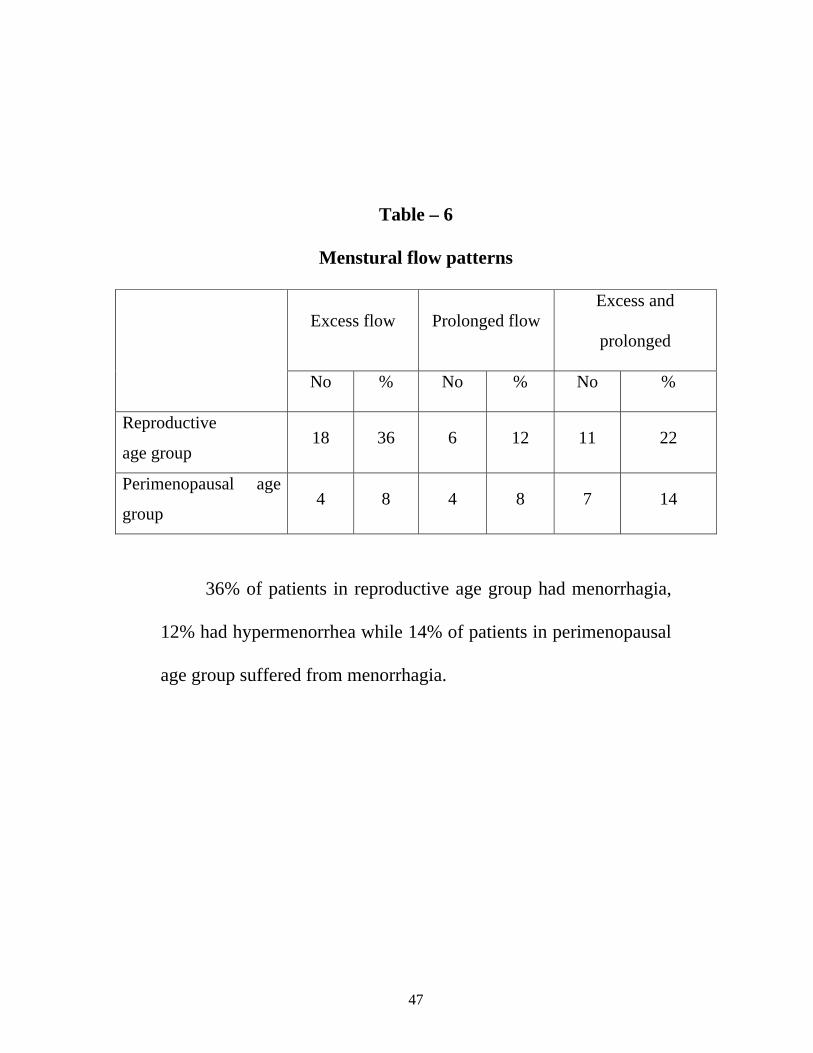
Table – 6
Menstural flow patterns
Excess flow Prolonged flow Excess and
prolonged
No % No % No %
Reproductive
age group 18 36 6 12 11 22
Perimenopausal age
group 4 8 4 8 7 14
36% of patients in reproductive age group had menorrhagia,
12% had hypermenorrhea while 14% of patients in perimenopausal
age group suffered from menorrhagia.

48
Table – 7
Parity
Nullipara Unipara Multiparous
(2-5)
Multiparous
(>5)
No % No % No % No %
Reproductive
age group 3 6 9 18 23 46 0 0
Perimenopausal
age group 1 2 5 10 7 14 2 4
In reproductive age group, 6% of patients were nulliparous,
18% uniparous and 46% were multiparous women.
In perimenopausal age group, 2% patients were nullipara,
10% were uniparous and 4% patients multipara.

49
Table – 8
Contraception and DUB
Temporary No
contraception
Permanent
contraception DC IUCD Others
No % No % No % No. % No %
Reproductive
age group 10 20 15 30 3 6 7 14 0 0
Perimenopausal
age group 3 6 12 24 0 0 0 0 0 0
Patients who have undergone sterlisation had increased
incidence of AUB. Probable reason may be
1. The blood supply to the ovary is altered
2. The axis of ovary is sometimes changed due to
adhesions.
50
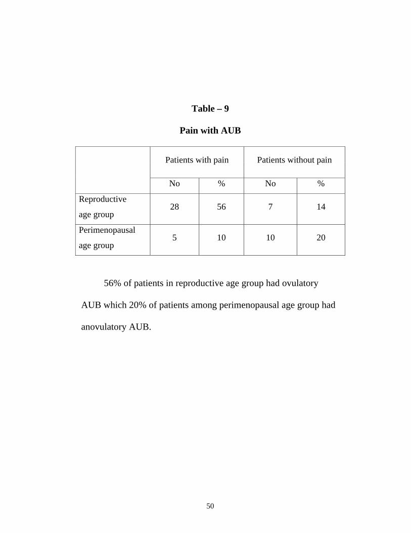
Table – 9
Pain with AUB
Patients with pain Patients without pain
No % No %
Reproductive
age group 28 56 7 14
Perimenopausal
age group 5 10 10 20
56% of patients in reproductive age group had ovulatory
AUB which 20% of patients among perimenopausal age group had
anovulatory AUB.

51
Table – 10
Weight of the patient
Within the ideal weight More than Ideal weight
No % No %
Reproductive
age group 22 44 13 26
Perimenopausal
age group 4 8 11 22
44% of the patients among reproductive age group were
within ideal weight while most of the perimenopausal patients with
AUB had more weight.

52
Table – 11
Blood grouping
Rh Typing A B O AB +ve -ve
No % No % No % No % No % No %Reproductive
age group 2 4 27 54 5 10 1 2 33 66 2 4
Perimenopausal
age group 4 8 7 14 2 4 2 4 14 28 1 2
It was an incidental finding that among the study group, most
of them belong to the B group.
No other bleeding disorders were noted among the study
group. No other thyroid dysfunction was detected.

53
Table – 12
TVS Endometrial thickness
Endometrial Thickness
1 mm
1-3 mm
3- 5 mm
5 – 8 mm
8 – 10 mm
> 1 cm
No % No % No % No % No % No %Reproductive
age group 0 0 15 30 19 38 1 2 0 0 0 0
Perimenopausal
age group 0 0 5 10 1 2 5 10 3 6 1 2
In the study of the endometrial thickness of the reproduction
age group, pencil line endometrium was nil, 15 had 1-3 mm, 19
patients 3-5mm thickness, 1 had 5-8mm thickness, non had > 1 cm
thickness.
Among perimenopausal age group, pencilline endometrium
was nil, 5 had 1-3mm thickness, patient had 3-5mm thickness and 3
patients had 8-10mm thickness and 1 patient had thickness > 1cm.
54
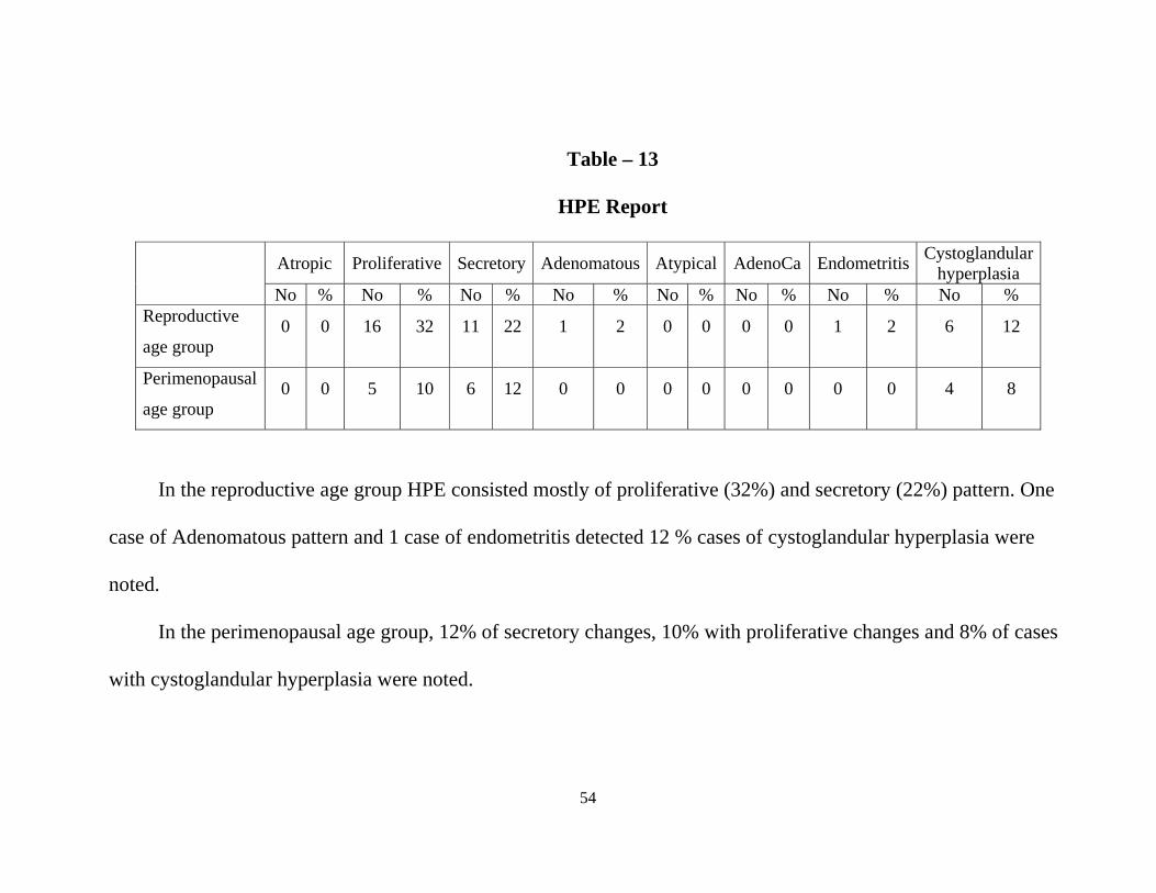
Table – 13
HPE Report
Atropic Proliferative Secretory Adenomatous Atypical AdenoCa Endometritis Cystoglandular hyperplasia
No % No % No % No % No % No % No % No % Reproductive
age group 0 0 16 32 11 22 1 2 0 0 0 0 1 2 6 12
Perimenopausal
age group 0 0 5 10 6 12 0 0 0 0 0 0 0 0 4 8
In the reproductive age group HPE consisted mostly of proliferative (32%) and secretory (22%) pattern. One
case of Adenomatous pattern and 1 case of endometritis detected 12 % cases of cystoglandular hyperplasia were
noted.
In the perimenopausal age group, 12% of secretory changes, 10% with proliferative changes and 8% of cases
with cystoglandular hyperplasia were noted.

55
Table – 14
Biopsy Ectocervix
Normal cervix Inflammmatory
condition Cervicitis Dysplasia
No % No % No % No % Reproductive
age group 12 24 18 36 5 10 0 0
Perimenopausal
age group 2 4 9 18 3 6 1 2
Among reproductive age group, 12 cases were normal, 18
cases of ectocervical biopsy revealed inflammatory changes, 5 cases
– cervicitis. Among perimenopausal age group, 2 cases were with
the normal ectocervix, 9 cases showed inflammatory changes, 3
cases were with dysplasia, 1 case with dysplastic change.

56
Table - 15
Distribution of uterine pathology regarding TVS
Uterine Pathology No.of cases %
Hyperplasia 9 18
Polyp 5 10
Myoma 2 4
Polyp + Myoma 0 0
Cancer 0 0
No pathology 34 68
In both reproductive and perimenopausal age group, according
to transvaginal sonography, 9 cases showed endometrial hyperplasia,
5 cases were with endometrial polyp, 2 cases showed submucous
myoma and the remaining 34 cases were with nil pathology.

57
Table - 16
Distribution of Uterine pathology regarding hysteroscopy
Uterine Pathology No.of cases %
Hyperplasia 15 30
Polyp 12 24
Myoma 2 4
Polyp + Myoma 1 2
Cancer 0 0
No pathology 20 40
According to hysteroscopy, 15 cases were diagnosed with
endometrial hyperplasia, 12 cases showed endometrial polypoidal
growths, 2 cases were diagnosed as submucosa myoma, 1 case as
with both endometrial polyp and submucosa myoma and 20 cases
showed no pathology.

58
Table – 17
Distribution of Uterine pathology regarding D & C
Uterine Pathology No.of cases %
Hyperplasia 10 20
Polyp 5 10
Myoma 0 0
Polyp + Myoma 0 0
Cancer 0 0
Adenomatous 1 2
Endometritis 1 2
No pathology 33 66
According to dilatation and curettage biopsy, 10 cases (20%)
showed cystoglandular hyperplasia, 5 cases (10%) showed
polypoidal growth, 1 case (2%) of endometritis and 33 cases (66%)
were with normal histopathology.

59
Table - 18
Distribution of Intra uterine pathology according to TVS, D&C
hysteroscopy
TVS and D&C Hysteroscopy and D&C
No % No % Hyperplasia 9 18 15 30
Polyp 5 10 12 24
Myoma 2 4 2 4
Polyp + Myoma 0 0 1 2
Cancer 0 0 0 0
No pathology 34 68 20 40

60
DISCUSSION
Abnormal uterine bleeding is a common problem (Couher) and
is the main indicator for hysteroscopic evaluation. Hysteroscopy
and USG are now being increasingly used not only for detecting
functional disorders of endometrium, but also for excluding various
unsuspected organic diseases of the endometrium like cancer and
tuberculosis.
In this series 50 patient with AUB were taken for study. For
all 50 patients, TVS, hysteroscopy and D&C were done in the
premenstrual phase and analysis of the study has been done. The
study period consisted of 2 years – during the year 2008 – 2009 in
Government Rajaji Hospital. Majority of the patients belong to
reproductive age between 30-40%. Only 30% belong to the age
group of 20-30 years.
All the 50 patients were married, out of which 6 were
widowed.
Regarding the parity, in the reproductive age group, most of
them are multiparus (46%), uniparous 18%, and nulliparaous
6% . In the perimenopausal age group, multiparous were 18%,
uniparous 10% and nulliparous accounted for 2%.

61
The flow pattern and cycles varied and were analysed
according to the individual picture.
It was found that most of the patients around were sterlised.
In the reproductive age, 44% were within ideal weight and in
premenopausal age, there was 22% increased weight.
Most of them belonged to the B group according to our study.
In all the patients, endometrial thickness measured by
Sonogram was compared with HPE.
Comparing the distribution and diagnosis of intra uterine
pathology according to transvaginal sonography, D&C and
hysteroscopy showed that 30% hyperplasia was detected by
combination of hysteroscopy and D&C whereas TVS and
D&C could detect only 18% of hyperplasia.
Combination of hysteroscopy and D&C could detect 24% of
cases with endometrial polyp while TVS and D&C could
detect only 10% of cases with endometrial polyp.
Both combination of transvaginal sonography and D&C could
detect 4% of cases as with sub mucous myoma but
combination hysteroscopy and D&C in addition, could detect
2% of case as with both myoma and polyp.

62
40% of cases showed no pathology according to diagnostic
hysteroscopy and D&C whereas TVS and D&C could not
detect any pathology in 68% of AUB cases.
Thus combination of diagnostic hysteroscopy and D&C has
better diagnostic value when compared with transvaginal
sonography and D&C.
63
SUMMARY
A total of 50 cases of DUB have been selected. 35 from
reproductive age group and 15 from perimenopausal age
group. These patients were subjected to TVS, hysteroscopy
and D&C on two successive days in the premenstrual phase.
Among the reproductive age group, 20% belong to the age of
20-30 year and 50% belong to age between 30-40 years.
Married and widowed women were included.
Nulliparous, Uniparous and multiparous were taken into study.
Other diseases like bleeding disorders were excluded.
TVS, diagnostic hysteroscopy and D&C were done in
premenstrual phase.
There was excellent correlation of endometrial thickness with
that of HPE of endometrium. Most patients with 3-5mm
thickness were of secretory and proliferative pattern.
There were no complications encountered during the above
said procedures.
Blood group of B was an incidental finding and there is no
literature to support a particular group in AUB.

64
Most of the endometrial hyperplasia, myomas and polyps were
diagnosed by diagnostic hysteroscopy when compared to TVS
or D&C.
Diagnostic hysteroscopy can be done in an office setting as a
day care procedure and is considered as a safe procedures with
minimal complications.
Women with menorrhagia under 40 may require hysteroscopic
investigation if intra uterine pathology is suspected following
transvaginal ultrasound scan or due to severity and persistence
of symptoms despite recommended medical treatment
(RCOG)
Hysteroscopy combined with histological examination is
considered as “Gold standard” for evaluating a case with
abnormal uterine bleeding. (Widrich et al, Bakour).

65
CONCLUSION
Abnormal uterine bleeding (AUB) is one of the most
commonly encountered condition in OPD. The most probable cause
of abnormal uterine bleeding depends on the patients reproductive
age and the likelihood of endometrial pathology.
Achary V, Mehte S and Rander A is March 2003, studied 100
patients and concluded that for detecting submucous myomas and
endometrial polyp, hysteroscopy has 100 percent sensitivity,
specificity and very high positive and negative predictive values.
They concluded that in the diagnosis and management of AUB
cases, the non invasive TVS should be of first choice. But
hysteroscopy followed by curettage and histopathology will improve
the accuracy of clinical diagnosis.
Organic pathologies which cause abnormal uterine bleeding
such as endometrial polyp, myoma and endometrial hyperplasia –
that are commonly not identified by blind dilatation and curettage
are diagnosed readily by hysteroscopy.

66
Since the incidence of focal lesions in patient with AUB is
high, it seems that most beneficial approach is to proceed with
hysteroscopy complemented by endometrial biopsy early in the
assessment of AUB. So, hysteroscopy with endometrial biopsy is
the gold standard investigation for AUB.
Before hysteroscopy was available, curettage was the primary
method of evaluating abnormal uterine bleeding. Brooks and Serden
have revealed that approximately half of pedunculated abnormalities
such as endometrial polyp were missed by curettage. It renders
endometrial sampling blind and incomplete, so that the diagnostic
accuracy of curettage is less than that of hysteroscopy followed by
curettage biopsy. So, hysteroscopy combined with histological
examination is considered better than TVS and D&C.

67
BIBLIOGRAPHY
1. Brooks PG, Serden SP. Hysteroscopic findings after unsuccessful
D and C for abnormal uterine bleedings. Am J Obstet Gynecol
1988;158:1354-57.
2. Valle RE Hysteroscopic evaluation of patients with abnormal
uterine bleeding. Surg Gynecol Obstet 1981;153:521-23.
3. Laffer FD. Hysteroscopy with selective endometrial samplipg
compared with D and C for c: abnormal uterine bleeding. Obstet
and GynecoI1989;73:16'-20. .
4. Widrich T, Bradley LD, Mitchi_on AR, Collins RL. Comparison of
saline infusion sonography with office hysteroscopy for the
evaluation of the endometrium. Am J Obstet Gynecol
1996;174:1327-34.
5. Bakour SH, Dwarakanath LS, Khan K5, Newton JR. The diagnostic
accuracy of outpatient miniature hysteroscopy in predicting
premalignant and malignant endometrial lesions. Gynaecological
Endoscopy) 1999;8:143-48.
6. Emanuel MH, Verdel MI, Wamsteker K et al. A prospective comparison of
Transvaginal _ ultrasonography and diagnostic hysteroscopy in the
evaluation of patients with abnormal uterine bleeding. An1 J
Obstet Gyni_col, _995i172:547-,5!.
7. Acharya V, Mehta 5, Randar A. Evaluation of DUB by
hysteroscopy, histopathology, TVS J Obstet, Gynecol Ind
2003;53(2):170-77.
8. Clark TI, Khan KS, Gupta JK. Current practice for the treatment of
benign intrauterine polyps: A quetionnaire survey of consultant
gynecologists in the UK. Eur J Obstet.Gynecol Rep'lrod'Biol
02;103:65-67.

68
9. De Iaco P, Marabini A, Stefane':ti Ni, Del Vecchio C, Bovicelli L.
Acceptability and Pall1 of outpatient. hysteroscopy. J Am Assoc
Gynecol Laparosc2000;7:71-75.
10. Kremer C, Duffy 5, Moroney M. Patient satisfaction with
outpatient hysteroscopy versus day case hysteroscopy: Randomised
controlled trial. BM] 2000;320:279
11. Coulter A, Kelland A, Long A.The management of menorrhagia.
Effective Health Care 1995;1(9).Department of Health. Referral
guidelines for suspected cancer. London: DOH; 2000.
[www.doh.gov.uk/pub/docs/doh/guidelines.pdf].
12. Royal College of Obstetricians and Gynaecologists.The Management
of Menorrhagia in Secondary Care. Evidence-Based Clinical
guideline No.5. Londori:RCOG Press; 1999.
13. Van del' Pas H. Hysteroscopy as an outpatient procedure. In:
Siegler AM, Lindemann HI, (Editors). Hysteroscopy - principles
and practice. Philadelphia: Lippincott; 1984;104-05..
14. Clark TJ,Voi_ D, Song F, Hyd_ C, GuptaJK, Khan KS.Accuracy
of hysteroscopy in the diagnosis of endometnal cancer and
diseas_e: A systematic review;JAMA 2002 (in press).
15. Glimpelson RJ, Rappoid HO. A comparative study between
panoramic hysteroscopy with directed biopsies and dilatation and
curettage. A review of 276 cases. Am Obstet Gynecol
1988;158:489
16. Soares SR, Barbosa dos Reis.MM, Camargos AF: Diagnostic
accuracy of sonohysterography, transvaginal sonography, and
hysterosalprngograph in patients with uterine cavity diseases. Fertil
Steril 2000 ; 73:406-11.

69
17. Darwish AM, Makhlouf AM, Youssof AA,.Gad_lla HA.
Hysteroscopic r4yometrial biopsy in, unexplained abnormal
bleeding. Eur J Obstet Gynecol Reprod BioI1999;8p(2),139-43.,
18. Bakour SH, Khan KS, Gupta K.The risk of premalignant and
malignant pathology in endometrial polyps. Acta Obstet Gynecol
Scand 2000;79:317-20.
19. Vilos GA. Intrauterine surgery using a new coaxial bipolar
electrode in normal saline solution (Versapoint):A pilot-study.
Fertil Steril1999;72:740-43.
20. Wren BG. Dysfunctional uterine bleeding Aust Fam Physician.
1998;27(5): 371-77.
21. Goldstein SR, Zeltser I, Horan CK, Snyder JR, Schwartz LB. Ultrasonography
based triage for perim2nopausal patients with abnormal uterine bleeding.
Am J Obstet Gynecol1997i77(1): 102- 08.
22. Callen PW, Demartini WJ, Filli RA. The central uterine cavity echo: A useful
ultrasonographic evaluation of the female pelvis. Radiology
1979;131:187-90. ,
23. Mendelson EB, Bohm-Velez M, Joseph N, Neiman He. Endometrial
abnormalities: Evaluation with transvaginal sonography AJR
1988;150:139-42.
24. Fldscher AC, Kalemeris GC, Entmen 55. Sonographic depiction of endmetrium
cycles. Ultrasound Med Biol'1986;J 2:271_77.
25. FleischerAC, Kalemeris GC;Machn JE, et al. Sonographic depiction
of normal and abnormal endometrium with histopathologic correlation. J
Ultrasound Med 1986;5:445-52.
26. Deichert U, Hackeloer BJ, Daume E. The sonographic and
endocrinologic evaluation of the endometrium in the luteal phase.
Hum Reprod 1986i1:2i19-2

70
27. Demas BE, Hricak Ii, Jaffe RB: Uterine MRimaglng: Effects of
hormonal stimulation. Radiology 1986;159:123-26.
28. Giorlandino C Fher N, Nanni C, et al.The sonographic picture of
endometrium in spontaneous and induced cycles. Fertil steril
l987;47:50 - 11
29. Salm R: MucH1 production of normal and abnormal endometrium.
Arch Pathol1962;?3:3O-39.
30. Dodson MG. lJse of transvaginal ultrasound in diagno;.ing the
etiology of menometorrhagia. J Reprod Meld 1994;39(5):362-72.
31. Oriel KA, S4rager S. Abnormal uterine bleeding. Am.Fam Physician 19991;
60(5):1371-80, 1381,82.
32. Ash SJ,Parrell SA, Flowerdew G. Endometrial biopsy in DUB. J
Reprod Med 1996;41(12):892-96.
33. Debora_ A Hall, Isabel C Yoder. Ultrasound evaluation of the
uterus. In Ultrasonography in Obstetncs and Gynaecology (3rd ed
Peter Callen). Harcourt Brace Asia Saunders, 594. .
34. Fleischer AC, Entman 55. Sonogl'apWc evaluation of the uterus and related
disorders. In: Artscher, Roberto Romero, FrankA Manning,
Philppe Jeanty, A Everette, James JR (Eds). the
Principles and Practice of USG in Obstetrics and Gynaecology.
35. Perrella R_, McLu_as B, Ragavendra N, Tessler FN, Schiller VL,
Grant EG. Sonographic findings after surgIcal ablatlOn of the
endometrium. AJR Am J _entgenol.__92i159:1239-41.
36. Baggish S I 979 Contact hysteroscopy. ObsteIrics and Gynecology
54: 350-354
37. Baggish S, Barbot J 1980 Contact hysteroscopy for easier
diagnosis. Conremporary Obstetrics and Gynaecology 16: 3-11
38. Bardot J, Parent B, Dubuisson jB 1980 Contact hysteroscopy.

71
American J ouma! of Obstetrics and GYI1ecolo_ 136: 721-726
39. Brun_J 1983 P.blockas a contraceptive method. In: Hysteroscopy,
MTP Press, Boston, ppI37_141
40. De Maeyfr J F D E 1983 Trancervical hysteroscopic sterilisation.
In: Hysteroscopy. MTP Fress, 1.kJsroI1, pp 191-]99
41. Frangenheim H 1988 The history of laparoscopy. In: Gordon A,
Lewis B V (eds) Gynaecological "endoscopy, Chapman Hall,
London.
42. Goldrath H, F-nller T A, Segal S ] 981 Laser photovaporisation of
the endometrium for the treatment of menorrhagia. American
Journa1 of Obstetrics and Gynecology ]40: 14- 19
43. Hamon J 1981 Microhysteroscopy. Journal of Reproductive
Medicine 26: 375-382.

72
PROFORMA
Name OP/ IP No. Unit
Age Income Date of Adm.
Address Date of Dis.
Date of Operation :
Socio economic Status of the Patient:
Parity :
Use of oral Contraception :
Complaints with Duration :
• Previous Medical/ Surgical History : • Family History : • Gynaec. History : • Menarche : • Period : Cycle Length: Duration of flow: • Amount of flow: Moderate: Profuse: Scanty: • Pain with Menstruation : • Other symptoms : • Intermenstrual spotting : • Marital History Married Widow • Obstetric History : Type of Deliveries LCB • Contraception : Temporary
Permanent • General Examinations :
Obesity : Anaemia : Breast : Thyroid :
• Systemic Examination : 1. PR 2. BP 3. CVS 4. RS 5. Abdomen 6. CNS
• Gynaecological Examination: P/A : P/V : S/E : P/R :
• Investigation : Ht: Wt: Hb% Blood Grouping: Urine: Albumin Sugar BT: CT: USG Hystroscope Findings: Follow up
1. D & C 2. HPE

73
ABBREVIATIONS
AUB ABNORMAL UTERINE BLEEDING
TVS TRANSVAGINAL SONOGRAPHY
D&C DILATATION AND CURETTAGE
HPE HISTOPATHOLOGICAL EXAMINATION
USG ULTRASONOGRAPHY
CGH CYSTOGLANDULAR HYPERPLASIA
EH ENDOMETRIAL HYPERPLASIA
EP ENDOMETRIAL POLYP
SM SUBMUCOUS MYOMA
T TREATED
NT NOT TREATED
R REGULAR
IR IRREGULAR
N NORMAL STUDY

74
HYSTEROSCOPY

75
ENDOMETRIAL POLYP

76
SUBMUCOUS MYOMA

77
ENDOMETRIAL HYPERPLASIA

78
ENDOMETRIAL HYPERPLASIA

79
RIGHT TUBAL OSTIA

80

S.N
o.
Nam
e
IP N
o
Age
Parit
y
SE s
tatu
s
Wei
ght
H/o
Pre
viou
s tre
atm
ent
Men
stru
al
cycl
e pa
ttern
Con
trace
ptio
n us
ed
Pain
with
AU
B
Bloo
d gr
oup
Endo
met
rial
thic
knes
s
USG
find
igns
Hys
tero
scop
y fin
ding
s
HPE
Rep
rot
Biop
sy
ecto
cerv
ix
D&C
+ T
VS
D&C
+
Hys
tero
scop
y
1 Janaki 415197 47 3 V 60 T IR - + B +ve 9 EH EH CGH Cervicitis EH EH2 Backiam 155726 42 3 V 73 NT R + - A +ve 3 N N Proliferation Inflammatory N N3 Chandra 470248 30 1 IV 42 NT R - - B +ve 3 N N Secretory N N N4 Maryma 470235 32 2 IV 40 NT R + + B +ve 3 S. Myoma S. Myoma Proliferation Cervicitis SM SM5 Vasantha 490395 31 1 V 45 NT IR + - O +ve 2 N N Secretory N N N6 Murugeswari 41515 30 2 IV 67 NT R - + O +ve 3 End.polyp EP+SM Proliferation&polyp N EP EP&SM7 Tamilselvi 70971 48 3 IV 72 T IR + - B +ve 6 N EP Proliferation Inflammatory N EP8 Selvi 70989 32 0 IV 32 T R + + B +ve 6 N EP Proliferation&polyp N N EP9 Murugeswari 18349 33 1 V 60 NT IR - + B +ve 2 N N Secretory N N N10 Saraswathy 36410 40 4 V 70 NT R + + O +ve 7 N EH Proliferation Inflammatory N EH11 Guruvammal 41360 43 3 IV 48 NT R + + B +ve 6 N EH Proliferation Inflammatory N EH12 Pandiammal 41347 48 1 V 52 T IR - + B +ve 8 EH EH Proliferation Cervicitis EH EH13 Alagu 76171 40 4 V 48 NT R - + B +ve 2 N N Secretory N N N14 Petchiammal 700613 40 3 V 62 NT R - + O +ve 3 N EP Proliferation&polyp Inflammatory N EP15 Arumugam 41632 45 6 V 78 T IR + - B +ve 7 Myoma SM Proliferation Inflammatory SM SM16 Kalaiyarasi 41615 40 2 V 55 NT R + + B +ve 3 EH EH Secretory N EH EH17 Meenammal 40105 35 1 IV 60 T R + - B +ve 6 N EP Proliferation N N EP18 Gomathi 10564 43 1 IV 52 NT IR + + A +ve 6 End.polyp EP Proliferation N EP EP19 Saraswathy 37217 29 4 IV 48 NT IR - - B +ve 2 N N Secretory Cervicitis N N20 Maheswari 41627 28 2 IV 75 T R - + O +ve 2 N N Secretory Inflammatory N N21 Shanthi 41946 35 1 V 62 NT R + + B +ve 3 N N Adenomatous Inflammatory N N22 Vasantha 42041 28 3 IV 72 NT IR + - B +ve 2 N EP Proliferative Inflammatory N N23 Jeyakodi 42044 37 4 V 55 NT R - + B +ve 3 EH EH CGH Inflammatory EH EH
MASTER CHART

24 Valli 19731 47 1 V 40 NT IR + - B +ve 2 N N Secretory Inflammatory N N25 Maruthi 105697 45 1 III 42 NT IR + + B +ve 2 N N Secretory Inflammatory N N26 Karuthaveena 107538 45 3 V 55 NT R + - B +ve > 1cm EH EH CGH Inflammatory EH EH27 Deepa 107555 28 5 III 80 T IR - - A +ve 3 N N endometritis Cervicitis N N28 Shanthi 10078 35 1 IV 70 NT R + + B +ve 3 N N Secretory Inflammatory N N29 Ponnu 79518 38 5 V 45 NT R + + O +ve 3 EH EH CGH N EH EH30 Nageswari 79507 27 1 V 50 T IR - + B +ve 3 N EH CGH Cervicitis N EH31 Pappathy 11102 44 1 IV 52 T R + - B +ve 2 N Secretory N N N32 Muthumari 97019 41 3 V 40 NT R + - A +ve 9 EH EH CGH Inflammatory EH EH33 Selvi 12782 33 0 V 72 NT R - + B +ve 3 EH EH CGH Inflammatory EH EH34 Rajalakshmi 13989 30 2 IV 75 NT IR - - B +ve 3 EH EP Proliferative+polyp N EH EP35 Taj Nisha 18385 37 4 V 71 T R - + B +ve 3 EP EP Proliferative+polyp Inflammatory EP EP36 Kokila 45608 45 3 IV 88 NT R + - O +ve 2 N N Secretory Inflammatory N N37 Savithiri 98072 53 0 III 90 T IR - - B +ve 2 N N Secretory Dysplasia N N38 Gnanam 95058 26 1 V 48 T R + + AB +ve 3 N EP Proliferative Cervicitis N EP39 Latha 90312 32 3 V 45 NT R + + B +ve 3 N EH Secretory Inflammatory N EH40 Andichi 53757 30 4 V 47 NT R + + B +ve 2 N EP Proliferative+polyp Inflammatory N EP41 Rajathi 91641 28 2 IV 55 NT IR - + A +ve 3 EP EP Proliferative N E Polyp EP42 Chinnakaruppu 913217 40 2 V 48 NT IR + + B +ve 2 N EH Proliferative Inflammatory N EH43 Kaliammal 42021 38 3 V 60 NT R + + O +ve 2 N N Secretory N N N44 Ponnuthai 73676 25 1 V 68 T R + + A +ve 2 N N Secretory Inflammatory N N45 Ponnu 79518 38 4 V 72 NT R - + B +ve 2 N EH CGH Inflammatory N EH46 Nageswari 79509 27 0 IV 46 NT R + + B +ve 3 N N Secretory Inflammatory N N47 Jeya 79799 40 2 V 88 NT IR + + AB +ve 3 N EH Proliferative Inflammatory N EH48 Panchavarnam 85130 35 3 V 42 NT R + + B +ve 3 EP EP Proliferative Inflammatory EP EP49 Eswari 85136 37 5 V 58 T R + + B +ve 2 N N Secretory Inflammatory N N50 Rathi 43133 43 6 V 63 NT IR + - B +ve 2 N N Secretory Inflammatory N N


24
27
75
2 1 2
33
14
2 10
5
10
15
20
25
30
35
A B O AB Rh +ve Rh -ve
BLOOD GROUPING
Reproductive age group Perimenopausal age group


