Embed Size (px)
Citation preview

Tl.Jl.!JRS OF THE GASTRO- INTF.STilfAL TRACT
Tumor Pathology Loan Collection
KANSAS UNIVERSITY HliDICAL CR1TER
KANSAS CITY, KANSAS
This case material constitutes the seventh in a set of Tumor Loan collec.tions prepar.cd and distributed by the Departmept, of Pathology and Oncology of the University of Kansas ~1edical School aided by a Cancer Control Grant (CS- 9209) from the National Cancer Institute of the National Institutes of Health, United States Department of Health, Education and \felfare. This material is provided on a permanent "loan" basis .
rumor Pathology Loan Collection Tumors of ~stro -intestinal Tract
Case I <qua~.ous Cell Carcinoma of Esophagus
University of Kansas Hedical Center Kansas Ci t y, Ka~sas Aut o psy ~!o .: 940
Hi story : This 72-year-old colored male was in good health until 8 months befor e
deat h when he began haVing a persi stent cold, cough, and l ow grade fever . These
symptoms became worse and he lost 30 pounds . About 3 months before death he be-
came bedfast with anor exia, nausea, and occasional vomiting of solid food . The
cou:;h became rore severe and produced about 1 CU!>ful of milky thick fluid oer day.
l!o hemoptysis was noted . He had choking spells at night lasti ng several minutes
Nhich 1-1ere relieved after expector ation of !lputum . He a l so had chest pain t<hich
Has accentuated by deep r espirati on . ~lith ~hese complaints he was admitted t o
I( .U. Hedical Center about 2 months bef ore death .
Ther-e was a past history of excessive alcohol consulll?t ion i n :vouth . He
smoked 2 to 4 cigars a day. Except for moderate 1·1asting a physical examination
sho1~cd no abnormalities . There 1~as no l.ymnh adenopathy. '3ronchoscopy and lar.vn-
~scony were negative . Esophagoscopy dieclosed a rungating tumor with a cons"ric-
tion 5 em . belo11 the cri coid cartilage .
Labor atory studies 1~ere negative except .for slight anemia ( TL '3 .C. 3 .·2
mi lli on), an e l evated sedimentati on rate and posi tive ser ol ogic terr~s .for syphilis .
Cytol ogical study of a bronchial asni rate ~<as negative .
X- ray examinat ion r evealed diminished aeration of the right upper lobe, 0\ll-
monary emphysema and complet~ obstruction of the esophagus extending downward
12 em . from the aortic arch . On one occasion, barium was seen to flow into a
~inor bronchus of the right lung through a fistula .
Tho patient ' s t emperat ur e ranged b'et1•een 98 and 100°F . His condition pre-
c.luded surgery and ~e was t r eated symptomatically . X- ray t he rapy, totalling

Case 1 - page 2
2119 r . i ncepth, and aimed at the tumor from several ports was given about J
11ceks bef ore death . The patient, however, bec.'ll1!e increasingly weaker and expired
quietly.
Gross Fatholof.(ic Findings : The tumor involved tho mid- portion of the esophagus .
'!'here: was un annular fungating lesion adherent to the adjacent trachea and
bronchi but "'ith no detectable comunication bet\~een the lumen of the esonhagus
and a oronchus . Scattered throughout the periesophageal and neritracheal nodes ,
and the lungs were multiple tiny foci of metastatic tumor . tietastases were also
f'ound in the liver, spleen, and uppt.r lumbar vertebrae .
Diatmosis : Squamous Cell Carcinoma of Esophagus .
-Corunent : Sinca t he a]:>peal"ance of the tumor in the metast atic lesions >las the
SIII1c as that in the esot>hagus, radiation effect on the r.:'Orphology am:arently can
be excluded . This bizarre poorl y differentiated t ype of w~croeconic morphology
is not uncommonl y seen in unirradia·~ed epidermoid carcinomas of the esophagus .
Reference : (1) Ack~rman, L.V. rJld dol Rog~to, J .A.: Concor; pages 5~-521,
The cv·Nosby. Co . , St . Louis, 1954 .

Tumor Pathology Loan Collect ion Tumors of t he Gastro-intestinal Tract
Case 2 Leiomyosarcoma of stomach
Universit y of Kansa-s Medica l Center Kansas City, Kansas Surgical Pathology ~fo . : 2939- 51
and 1916- 54
Hi sto r y : This 48- year-ol d 1-1hite male 'ttas well until 9 \<eeks Prior to t his
admission, when he suddenly experienced i nability to s11allow his food properly .
He coul d eat a littl e br eakfast, less lunch , and 1·1ould vomit all bi s dinner .
The dy5phagia rapi dly progressed urttil he wa·s able t o s>lallo•t( only liqui ds .
Dur i ng t he pe riod he los t 29 pounds .
P;ist and f arni ly histories are non- contri butory .
Physi cal examination sho1·1ed no abnormalities .
Labor at ory f indings were essentially normal except for compl ete a chlor hydria
r <;vea led by histamine anal ysi s .
X-ray examination revealed dis pl acement of the lo~1er pa r t of t he e:,ophagus
by an extrinsic n~ss . No evi dence of infilt rative change i n or dest ructi on of
the mucosa v1as ·noted. Fairly normal ruga l pattern of the cardia of the stomach
;~as described . Gas t r i c empt ying v1as s a tis factor y .
The pos t erior !Jediast i num tfaS expl ored 1rom t he l eft and a tumor mass 6 ern.
in diameter Ytas found at the gastt'ic cardia to the left of t he esophagus, and
extending into the abdominal cavit:v . Eso~:>hagogastrectom:y vtas done. The post-
operat ive course v1as uncompl icated and, on the 9t h postoperat.i ve day, he was
i!ischarged.
Gross Pathol o_gic F' ndings : There v1as a t umor mass, 12 em . in len~th and 7 em .
in Nidt h on the greater clll'vatur e of the gastric cardia and fundus l<~i t h sligjlt
extension into the >·!all of the distal esophagus . The mass 1·1a s f i r m, rubbery , and

Case 2 - page 2
lobula. ted . The overlying gastri c mucosa presented a deep ul cerated l esion
9 em. in greatest diamet er 1·1ith elevated >~hite hardened edge . The gastric
r.JUcosa within 4 em . distal to the lesion sho~rs no rugae, othe.nrise gas tric
l!l1d E!SOphageal rnuco,sae vrere unremarkable . The tumor v1as composed of drnse v.tti;!B t iSsue
arrr~ged in l obules as l arge as 2 em. in diameter . The centers of the lobul es
vren; dark- red and necrotic . There v1as a distinct capsule surrounding the tumor.
t!o. ~ross i nvasion \~as found . The regional lymphnodes showed no t umor metastasis
on microscopi c exronir1at i on .
The patien t di d ·.·1ell hut 2 l/2 years after t he operation he not.iced a lu.mp
on the back of his neck. This incr eased about 3- 4 times in si ze in the f ollo·,;ing
3 months and he 1~as again admitted .
Physical examination sho1~ed only a 3 x 5 em. slight movable , f i r m, non- t ender
mass attached to the occiput .
.X-ray study r evealed no r ecurrence of the tumor in the stomach and a \-!ell
functioni ng gastroesophageal anast omosis .
The l!'oass 'in the neck 1~as re rrpved under local anesthesia. It Neighed 20 gms .
and measured 5 .5 x 3 .5 x 3.5 em . It 1-1as very soft, and fragmented when handled.
It was com:posed of yello1<, hemorrhagic, very soft mater ial 1-1hi ch vias rather gran
ular in appear ance . No a r eas of caseat ion or definit e necrosis '"ere note<) .
The patient ~<as in good health V~hen last heard from, 3 1/2 year s a.ftcr the
gastrectomy and 8 months after t he excision .o f the neck tumor .
The slide A i s t he t .umor of the stomach; sl ; de B is the tumor i n t he neck .
Pia®osis : Leiomyosarcoma of stomach wi t h metast asis to subocci nital soft tissues .
Cor.unent: In this case the hi stologj ca'l. a pnear<mce is not di"ficul t to inter pr et
( See case 2l) . Gas tric leiom,yosarcoma i.s usuall y rather benign in its progress

Case 2 - page 3
a~ com~·ed with gastric carcinoma (1, 2) . Golden renor ts metastasis of gastric
.~icmyosarcoma to the lung in 2 out of 5 cases, but t o other soft tissue (3) .
Ir. this case, the suboccipital lesion was interpreted as a metastasis in vie11 of
;;te morphological similarity to the gastric neoplasm. Ho\~ever the secondary mass
is composed of less differentiated and rr.ore malignax1t appeari11g !)lump cells shol~
i.ng many mitoses as compared to the more guiescent and mature appearing cells of
~he primary tumor mass . The; latter exhibits some variation in cell type, mitoses,
and fusion •;lith muscle layers in certain areas .
References : ( 1 ) ~!nrsha.ll, S .F. and Meissner, l/ .A. Sarcoma of the Stomach . Ann .
S1rrg . 131:824, ~950 .
(2) Lumb, G. Smooth liuscJ.c Tumors of the G;;::.;tro-intcstinal Tract and Ret ro
peritoneal Tissue Presenting as Large Cystic J;asses . J . Path . Bact. 63 :139, 1951.
(3) Gold!Jn, T . , and Stout, A. P. Smooth Husclc Tumors of t he Gastro- intestinal
T~act and Retroperitoneal Tissue . Surg . q;~ . Obs . 73 :784, 1941 .

Tumor Pathology Loan Collecti on Tumors of the Gastro- i ntestinal Tract
CRSB 3 Lymphosarcoma of Stomach
Univers i t y of Kansas l4edical Center Kansas Ci ty, Kansas Surgical ~1o .: 2603- 51 Autonsy 1io .: XXXVII-193
Historv: This 64 year old uhite fe.>nale ~:as aani tted t•'ith complaints of nausea
and vomiting of 6 months durati on . ll cholocystl'lctomy had been performed 1 year
pruviously . 1'he r e had been no hernatemcsi s . She ha.d lost 65 potlnds dur ing t he
y ar prior t o admission .
Physical exa."'ination revealed a oass in the mid epi g'!lstriwn Hhich ~tas mod-
cratoly tender, measured about 6 x 6 em . in sizo and seemed to be connected •d.th
l.h•J sto1.1ach .
Laborat o'cy examino.tiona sho1·1ed moderato anomia and a total ser wn protei n of
4. ··l i911 ·%. The aner.;i a was improved bj' blood bransfusi ons . X- ray examination
.hot·cd a tumor of tho :;IJ:Imach in the region of the pylorus .
The patient had sevo.;rc 'hc-rnar.ernesis 1Alich required numerous transfusi ons .
A 'O'll'tial gastr ectomy and hepatc.ctomy vtcre done . She •·:as discharged on the 19th
~lOU~OfiO I'ative day .
Gt'OCG Pathol oJ<i c Findin.as : On the mucosal surface of the stomach 1~as an oval
lc~ion 7 em . x 4 em . 1rl th & red ole-, a ted rim, 1 cr. . in hei~" surrounding a ccn-
tral necrotic area . It ~1as comoosed of pink tissue lllOttled by foci or recent
~"-Pt'l'\J.~ge . The l esion ox:tendGd into tile duodenum. No microscoric lymphnode
involve.ment was f ound .
About . 1 1·:8ek later, t.hc patient noticed a _oo.rtial disruot.ion of the upper
part of t.he inci s i on ui t.h drainage of foul smelling material. She Has lethargic
,'fith blood p r essure 80/60 and zerur.; potassium 4 .0 mF.q/1, non- protein nitrogen
'19 mg ,,;, and chloride 80- 96 rnEq/lit er . After transf usions and venoclyses with

Case 3 - page 2
addition of potassium chloride, the patient 1s condition was greatl y improved .
The drainage, which was largely bile>, subsided considerably. Although the
atient was thought to be orogressins; well in every respect, she '•as found dead
in bed one morning, 31 days oostoper ati voly .
Autopsy revealed acute locali~cd neritonit is, chronic omental cellulitis,
acute and chronic suppurative ucl·i - hep;.ltitis , chroni c SU!lPUrative sigmoid
diverticulitis with chronic p<'rforation and localized pelvic peritonitis .
At no place •:las any ,·emaining neoplastic tissue se en . None of the abdomine.l
l !flllOhnodes si1o1-1od neoplasi.ic changes . No leukemic cells \vore f ound in the blood
stream .
Diap,nosi s : Primary lymphosarc(>Ol<.l of stomach .
Co1roncnt : Autopsy revealed this lymnhosarcotla to be primary i n t h e stomach
rather than a gastric manifC5\.ation of a generalized disea$e . '!'his i:J not too
:.lnusua1 (1, 2) . A reticulin ("ilder) stain showed some anparont proliferation
of r~~iculin, fjnc and coarse . The value of such s t aining is controversial (3) ,
:l..!d the tumor might be considered similar to"reticul.um cell lymphosarcoma" (1) .
Ref.:r~o;ncos : (1) stout., A.P . Tumors of the Stomach . Atl as of Tumor PatholottV
?~sicle 21: 78-87, 1?53.
(2) J.:arshl<ll, S . F. , and r;eissner, l! .A. &arcoma of the Stomach . Ann . Sux-r.
131:821, , 1950 .
(3) \Iillis, R.A.: Patholor.v of Twnors; '3utten;orth, London, 2nd Edition
panes 761-783 , 1953 .
Tumor Pathology Loan Collecti on Tumor ::; of the Castro-intestinal Tract
G,.se 4 Atrophic Castritjs with ;Jif£\tse
Carcinoma
University of Kansas Hedical Center ::·nsas City , Kansas Autopsy Ko . : 996
Hiswry: Tilis 69- year-old •mite female had a 25-year history of perniciou!O
an.:lria controlled '>y liver and n:ore recently by vitatin '3 12 theraoy. Th& ')Q.tj•n•
had never noted numbne!:s and tingling, rtifficulty in walking, sor& .nouth or
tongue . She had 11flu 11 about 1 year before death but r e cover ed quickly •.it! 3.Ut'- o-
nwcin . Since t at time, t'1c patient suff,;red bloating, abdominal grir.ing pains,
and some nautnla bttt no vomit,jng .
At t he time of admission fj ve mont'•.s pri or t o death, t he abdomen llt1.s r:ro-
tuber ant but, moder11t ely soft and ~ympanitic an-t eriorly t•!it'l shii't i ng dul lness in
t.~e flanks but no dofini Le fluid wave . i!o lymph nc>des were nalpabl e . ~leurolo('ical
"xanlinatian l·tas I'.OJ"mal. Laboratory f indings v:ere es·sentially negative except £or
:light anemia (U: 6 gm. h~,;:hO('lo·,in) . Liver function tests Nero normal. l~o ''ccult
blood was detectable in i '· e stool . An X- ray exanlinati on of the gast rointestinal
tract sho\'lcd the stomac1 to have a "leather bottle" al)pearance.
Paracentesis was done and cytological study of the al'citic fbi.a :-evealed
~r..~cnant. cells . Her condition 1~as thour'lt to prohibit SW"F.ery and so 160
:lillicurics of !'a<tioactive gold ·.-~ere claced in the abdomen to control fluid
accunrulation .
Her condition became progressively worse and several paracenteses ~·ere nee-
cssitated .
She v1as readmitted becau;;e of ascites and hydrotlo)orax 2 months before 1\<;ath
at v1:1ich time a sofL st~el.Linr vta:; fotmcl in the left subclavicular a r ea . IJo ot:l()r
l.YJnp::oadenopathy waa noted. '!'be rir'1t chest show·ed dullness to nercussion and
Case 4 - page 2
absence of bx·eath sounds to the l evel of 5t 11 rib . The abdomen was di s t ended and
ther e lifaS a f luid wa ve . There v1as pitti ng edema of both ankles and l ov1er l e gs .
Neurologica l exami nation was nor mal. Cytological study of t he pleura l fluid
again reveal ed mal i gnant cell s .
She was maintai ne d on a l ow salt diet, vit amin 131;z , dir,italis, a nd mercuri al
dJ uretics . Frequent abdominal and t horaci c paracentes es ~~e re done . She became
s t eadil y 11or se, was una bl e to t a ke any f ood or f l ui ds orall y , devel oped r e current
r espi rat ory tra ct inf ection, and ex pired qui et l y .
Gros s Pathol oki c<\l Findings : Autopsy r evea l ed 1800 cc . of a s cites a nd hydr othorax
1200 on t he right and 700 cc . on the l eft . Ther e was a diffuse thickeni ng of the
abdowinal serosa liit h multi pl e int r a- abdominal a dhe si ons which had caused a ·vol
~ilus of a l arge por~ion of the smal l i ntes t ine . Anot her ~ss consisting of
intestine and pel vic or gans compressed both ureter s and produced bila t eral hydro
nenhr osi s and pyel onenhr itis . The lung showed emphyseJ!l.a and l ympha t i c oormeati on
by tumor . The bone marro1~ 1~as pa l e and decreas ed in c el lular i ty.
The Hhol,;, sto!:!a.ch f r om the cardia t o t he pylor us was i nvol ved by a f i rm
t !rickened f ibrot ic mass . The thicknes s of t he NaJ.l mea sur es about 8 mm . The
ruga e 'tlere f lattened a nd t he wall o f the stomach s eemed "t o form a rigi d t ube .
l.icros_copically, metast ases v~ere f ound i n the r egio11e l lymnhnodes , diaphr·aron,
l.unr,s , ut erus , ova ries, gal lbl adder and peri toneum.
DiaFmosis: Dif.tuse carcinoma of the stomach devel opi ng in a pati ent vrith a t r o ohic
r;a strihs a nd l ong standi ng oer nici ous anemia .
Comuent : This i s off er e d as a reminder of the si j;nif icantly !rir!her incidence of
c;>rci n oma of the s t Qmach i n pati ents witl~ uenrici ous anemia (1, 2) .

Case 4 - page 3
References: (l) Rigler , L .G. , Pernicious Anemia and Tumor s of the Stomach .
J . Nat . Cancer Institute, 7 :327., 19b7 .
( 2) Brown, L . The Patholo~y of the Gastro- i ntest inal t.ract in pernicious
anemia and Subacute combined degeneration of the spinal cord. , Ne1·1 Eng. Hed . J .
210:473, 1934 .

Tumor Pathology Loan Collecti on Tumors of the Gastro-intestinal •rract
Case 5 .~denocarcinoma preceded by long- standing
Ulc.er like Symptoms
Uni versi ty of. Kansas ~ledical Center Kansas City, Kansas Surgical Patl·olor y No .: 671-52 Autopsy No . XXXITIII-/47
llistory: This 48 year old \>Jhit e male had epigastric pain for about 4 years bef ore
admission . The pain was at first relieved by ingestion of f ood but later i t 11as
r elieved only by pr essure on the abdomen 1,-hile in a sitt ing posit i on . He had
been hospitalized several times else1Yhere f or thi s compl aint but had ref used op-
eration . In the 3 to 4 mo.ntns before admission he lost 40 lbs . in Height . About
10 days before adnussion, he f irs t noticed t arry stools . Epigastric distress
became cont inuous and he vomitt ed coff ee ground material.
Famil y and past hi s t ory '"ere non- contril:utory.
Physical examination revealed onl y t ende rness to palpation and percussion
acros.s t he Ullper abdomen .
Laboratory studies Nere normal .
'I'he patient was pu·t t o bed but continued t o have occasional hematemesi s and
r,!' pass tarry stools . Therefore 1-Ji thout previous X-r ay examination of the gastro-
intestinal tract, expl orator y laparot omy v;as done. At t hat time a large uleerating
carcinoma of the l esser curvature of t he s t omach extending into the esophagus 1·1US
found . Total gastrectomy, j~ju.nostomy and esophagojejunosiomy were tlerformed .
~ross Patholop;ic FindinP.:s : The lower esophag~ts and cardia c por(,ion of the stomach
1·1ere i nvo l ved by a hard penetrating t umorous process t~hich also extended do1om the
l esser curvature for a distance of 6 em . I r, t he posterior aspect of the stomach
the tumor v1as bordered by nn ulcer ? em. in diameter which penetrated into t he
adher ent r etroperitoneal structures . The slide •nas taken through th e ulcer and
adja-cent tumor . Tumor· is seen i n peri - neural lymphatic spaces .

Case 5 - page 2
Tnree days after oneration he became definitely ~~aker and comQlained of
sovere abdominal pain . Despite Vifl;orou~J an t ibiotic t.herapy, he graduall,y bec<lllte
n.uch ~·Jeaker and exuired 6 days after the oDerat.ion .
Autopsy revealed acute general i zed peritoniti~ followi ng runture of an cso
;1haeo.iejunal ana~'tomosis , f..etastases vrF.>re seen only in the h.Llar l ymph nodes and
they •.;ere of mucous adenocarcinoma ,
Jiagnosis: Adenocarcinoma of stom~ch , oossibly derived from the border of a
pcmtic ul cer .
Conment : The chronic f ibrosis and fusion of musc•tlaris tmcosa and muscle laycrr;
uggest l ong-Gtandin.g peptic ulcer, It cannot however be said whether this ulcer
ant<ldated or followed t he ad(1noearcj 11011\D. although the 4-year hi$tory i::~ mor•e
plausibl e for ulcer than for carcinoma . The lwoplasm is found only at t.hc ?order
of ~he ulcer and does not lnvolve its b:1se; t hi s ;)gain does not constitute proof
as to which lesion antedated or r:>redisnosed to [,110 other . One may profitnbl.v
consider some of the crit-eria f or, and t.t·e objections to the dbr,nosis of' carcinor.>.a
,ri:;ing in A pr·e- existent peptic ulcer (1) , and t'•e ·dgorous (but not .undisnuted)
ota.tement thn.t "the v•idest field for inmrovement in manaNemen t of (!ast ric c.oncers
lies in the promnt resect-ion or all gastric ulcet•s" (2) .
:~oferences : (l) Ack.:.rman, L V.: Surl'ic:1.l PJ.thology; p~cs 282- 28b, !:Osby,
St . Louis, 1953 .
(2) Boyce, F' , F ,: 'l'h" :fonsurgic··.l I:Ortalit.y of Carcinoma of the Stomch ;
A Probl~;,m of R ... sponsibil.i.ty; Am. J . Caotroont.orolor.:v 24:36- 52, 1955.
(3) Pl'.ck, G. T. , The R<Jl;\t:conship of Gastric Ulcor to GilGtric Gancor . (Pnncl
1iscussion), cancer, 3 :515, 1950.

Tumor Pathology Loan Collection Tumors of the Gastro- intestinal Tract
Case 6 Mienomatous Polyp of Stomach
University of Kansas Hedical C(mter Kansas Ci ty, Kansas ~urgical ?athology No .: 2666-50
Historv: Thio 7.3-year- old white male noticed epigastric !)ain described as "heart
burn" .3- 4 months bef ore admission . This was present intennittenHy and not
relat"'rl. to exercise o r food ingestion . No chills, fever, jaundice or dy51>hac,ia
were noticed .
His oother died of carcinoma of stomach at 55 .
Phyoical cxaninations were negative.
Labor-a~ory studies : Stool v1as n••gative for blood . Gastric anal.vsis shoued
no free acid and ve ry low total acid, 6 units o
X-ray J'ovealed multipl e pol yps of ~he s tomach .
Jo.xploratory laparotomy disclosed mult.iple intralumbal polyps, involving
r;he louer half of the stomach o A gastrect()l!\y was done . He had a satisfactory
)Xlstooerat.ive course and 1·1as discharged on the 11th postooe-rative day .
Grv,.s Pathologic Findings: eeven nol.vns H<>r·c found alon11: the am.erior and greater
curvature surface of the stomach . The largest 11as 2 em . in dirune·~er, pcduncula~ed
P.nc\ :>oft .
Loym,)h nodes showed onl y h;yrel'"lf.H~ ~:I.e rolHcles on l!licro$conic exAminntion .
'The family re')orted thac the oatirmt died at horne about 13 mon~hs after
discharge . No autoPsy was performed and the c;mse of death is not kno~m o
Dias.nosis: Adenoreatous pclyp of stom:1ch o
Co1'f1ent : ilo evidence of mali(;llancy ,.as found . Gastric nolyposis can oroduce
ga~ t!'ic di::~tress or bleeding whicn rt:quire~ surgical intervention o l!alimo.nt
tran:;formation is said to be si~nificently high 1'l.1.rticular l.,y i n casAS of multipl e
pol~rpoo.in (1, 2) .

Case 6 - page 2
Cur ly and Lyle state that atrophic mucosa and a bsence of free hydrochloric
:;cid ar e associar.ed with mor e frequen t malignant alteraLion of the pol yps (3) .
'1 o1:ever, the same aut< !tors later state that a r.alignant !JOlyp shows a character
ist.:.c appearance on gas eros cony from its beginning and !P.alignant transformation
of the benign type polyp is unlikely (4) . I n the case of congenital 110lyps, t he
JSC''1aris m cosae actually enters tlle polyp, but i n inflarr;na.tory hyperplasia i t
docs net (2 ) .
ilcfer ences : (1) Brunn, H. and Pear l , F.L. Diffuse C~stric Pol yposi s Adena
papillomatosi s Oastrica, Surg . Gyn . Ob . 43:559, 1926 .
( 2) Pearl, F .L . and Brunn , ll. I;ultipl e C-as ~ric Polyposi s, Sunt. O,yn . Ob .
76:257' 1943 .
(3)
(4)
Ca rly, J . B. o.nd L~rle, H.
Ca1·ly, J .B. , and Lyle, H.
Gast r i c Polyps , Gastroenter ol. 10:102, 1948 .
Gastric Polyps , Gastroenter ol . lh :280, 1950 .
Tumor Pathology Loan Goll@ction ~umors of the C~stro-intestinal Tract
Case 7 Adeoocarcinomq. of the Amuu.lla o£ Vater
University of Kansas MeiUcal Cent-er Kansas City, Kansas .Surgical Pat,holoey ~lo .: 3537- 53
Bistor:r: This 80 year ol d Negro male had da.r k urine and jaundice for 2 months
prior to admission . fje also had weig!1t. loss of 20 pounds , anorexi.a a>1d pains in
t 'lc right 3nd l eft ).ower ouadrant . The pain gradually be came more severe around
the umbilicus , but Has never sharply localized.
Physical examination 1·1as essentially norms.l except for jaundice .
Laboratory studies shol•ted a total s<;>r um bi liru'oin of 13 .4 ltigll! .:t, and a direct
bilirubin oi' 8 .4 mgm.%. Thymol turbidity ~ras 5 units , total cholesterol Has 300
mg.%, and there Nas 40% of cholesterol ester . - Prothrombin time wa s $0-90": of
X- ray reveal€l d no abnormalit ies in the upper gastrointestinal tract . Chol -
arigio.gr am revealed an obstruct i on of the distal col!il1\on bile d,uct .
A laparotomy l..,..d.s done . The gallblo.dder vtas found to be dilated t o about 3
t imes its nom.al si ze 1·tith some adhesions .to adjacent st,l:'uctures . Tile 1·ral11~as
only slightly thickened . Th<:> cystic and common ducts 1•ere dilated '3.p'tlarentl.v due to
an obstruction in the region of the ampulla of Vater . There was no tumor i n
l·.he pancreas . No Rbnormal. lym.p~ nodes could be !lalpated at any s ite . After
.:;pcning the duodenum, ·i t 1·1as found that t he obstruction ~<as a discr ete hard tumor
invol ving the ampulla of Vat er. l"'rozen section indicated malignancy and wedge
excision oi the cluorlenal v;all lrith the ampulla of Vater viaS nerformed, 1./ith r e-
implantation of conunon duct and pancreatic duct stump5 . After cholecyst.o- je.juno-
~tomy and jejunojejunostomy, the abdomen l·tas closed . The postoperative course
1;as r <?Jnar ka.bl.y good and 19 days arter operat ion, the patient '..ras discharged, being

Case 7 - page 2
a.mc\1.latory and nearly free of jaundice and other signs . Aft er discharge, the
~t.ient had inter•.li. ttent crampinf! abdominal pain and on 2 occasions was seen at.
the ltospit:U. for trcatmem. with continuous suction and IV fluid replacement . His
hepatogra~ was entirdy normal , and hP improved on both occasions . The ::mtient
died at home 1 1/2 years after the operation. Autopsy was not done .
,ross l'athological F'indinP,s: The specimen was the ~~edge from the ~1all of the
duodenum measuring J x 2 . 5 em . including the ampulla o f Vater . The orifices of
tr.e com.11:on bile ducts and the pancreatic duct were i dentifi ed . The ampulla wao
occluded by D. firm well circumscribed r ounded tumor no dule measuring l em . in
diam<~ter .
-Diagnosis : Adenocarcinoma of t he Ampt!lla of Vater .
CDlm!lent: Gross and micro$copic f i ndings perw.i t t h is diagnosis . fumors oJ: this
',ype- have tho best pror.nosis al<long the p!l'riampullary carcinomas .
In this particular reg:! on carcinoma can arise i'r om various sources and only
''.en ti1e site of the carcinoma produces early obstructi on of the bilo duct can
"e origin be verified . Absolute hit,tological indenti!ication of the origin can
, cot 0e made (1) . Tho dii'f~rent.ial diae;nosis of suecimens from this region requires
r"eat care since t.hc subzecuent. surrical nrocedures are risky and varied (1) .
liaN•:!.y a benign atlcnor..a may b'i! r..isinlercrcted as a carcinona (l) .
"he surgical asnects of l.wnors in thi s region are discussed in the rcf<>rence~
(2, 3, 4) .

~ac-o '7 - page 3
~deromcos : (1) Ackerman, L . and Rcgato, J .A. Cancer . p . 718- 723. t:Osby, St .
::.t.luis, 1954.
(2) Ev.:,rson, T.C. and Cole, 'I.H . , Ten Year Survival Follo,.'ing i"ancreato
Juodenectouw for Carcinoll'.a of the Ampulla of 'later . Surp;ery 37 :.260, 1955 .
(3) Hiller, E.l .. , Dockorty. t .3 . , :follaeg<:r, .E .E. and •!e.ugh, J .l:., Carcinoma
:.:::. th Region of Papilla of Vater, SurP,. G,vn . Ob . 92:172, 1951.
(1,) Orr, T .C. , Some Observations on the Treatment of Carcinoma of the Pan
ere~~. Sur~ery, 32 :933, 1952 .

Tumor Pathology Loan Collection Tumors of the Gastro- intestinal Tract
::ase 8 ).naplastic Carcinoma of .Jejunum
University of Kansas ~•edical Center Kansas City, Kansas Surgical Patholo~ No .: 2781- 55
"istorv: ':his 50 year old white fe:":ale had recurring eni~odes of crar.t!>ing
abcbminal pain J -4 years prior to admi"sion . 'I'his 1~as 10reated as "spas&ic
coli tis" else>~here , apparently ~r.itl· success . How;ver , J - 4 months prior to
ad'llission , she again su.!'fered intemittE:nt diffuse abdominal pain with headache,
ratigue, some anorexia and peri- o rbHn.l edema . Her heJ:Joglobin was 7 gm .~ and
she Has s t a rted on vitamin 612 and 'ron pills for her anemia . C:he had hacl a ten-
dency to~1ard anemia all hor life and had taken iron i ntermittently. The e,astl"o-
intestinal tra<::t ~1as s.1i cl to be nol'rnal bv X-l"<'~Y but for 2 11eeks pri or· to admission,
~:n- had inc'reasing constipation, blacl; stools and more severe constant abdominal
•;ain . Her a.ppetito wars increMinRlY poor and :<l1e lost 10-15 pounds in t ho month
l)rior to admission . licr family pl':vsician fow1d a lesion in t''e colon and c.l:~
~1as admi tt"d for furthur study anrl surgery,
No enlarged lymph nodeo were Palpated on p"ysica1 examination. Tl•ere >~as a
roderatel.y t.endet firm irref(Ular 5 x 5 em . mass in the left upoer quadrant of t.• c
::bdo~en with a definite sharp inferior edge just beneatn the costal margin .
Red blood count was 2 .29 million . Hemoglobin t-·as 6 .2-13 .6 gm . \\'hite blood
count l'tas 6 .200 . Differential \ill>' within nonnallimit~ . Serum bilirubin : direct
2 .0, total 4 .7 mg .,; , Cephalin-cholesterol flocculation was nep,ative. 'lloorl el<·c-
troly1.es were within normal. limits .
;rith the patient in t.he supine nosition, there ••as a filling defect seen b;v
l<- ray in the antnl'ior por~ion of the grE>.ater curvature side of the stomAch , approx-
imatdy over the area of !,he mass which could be palpated. The stomach e1wt.lcd
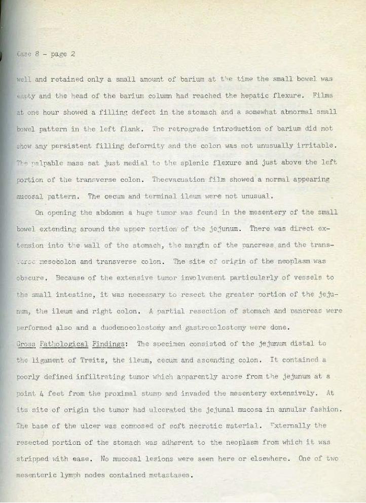
la.sc 8 - page 2
well and r etained only a small G.mount of bari um at t ' •e time the small bo,,rel \•as
b.L!JtY and the head of t he bar ium column had r eached the hepati .c .flexure. Fil ms
at one hour shm(f)d a i'ill.illf, defect in the s t omach and ::>. somevihat a bnoTm;ll small
bowel pattern in the l eft flank . The retrop.;rade i ntroduction of barium eli<;! not
&bow any persistent filling defomi,ty ?nd the colon >las not unusually irritable .
1'r" nalpabl e mass sat j~o~st medial to tl1e splenic flexure and just above the l eft
porti on oi t he tramwerse col on . Theevacuation fiiJn showed a normal appearing
mucosal pattern . The cecum and termina.l ilelllli Here not unusual.
On openi ng the abdomen a huge tumor >~as found in the mesentery of' t he small
boHel extendin~; a.round the upper terti on of the je,lunum . Thera 1~as direct ex:
teasion into t l'e Nall of the stomach, the margin of t he l'}ancreas and t ·lie trans-
. Gr:...:: nesoColon and transverse co lon . The s i te of orip,in of the neoplasm was
ob$CUre . Becattse of t he extensive tlm;or· involvement particularly oC vessels to
t.he sma11 intestine, i t v1as necessary to resect ~he gr.e~.ter oo.rti•~n of' t fJe jeju
num, tlte ileum and right colon . A r,>ar ti.a.l resection of stomach and 1;lancr ea:: i·ler e
oer formed .;tlso and a duodenooolcstomy and gastrccolostomy 1·1ere done .
Cross Patholotical FindintlS : The soeci:nen consisted of the jej unum distal to
the l.ig:Jllleht of Treitz, the ileum, cecum and ascending col on . I t contained a
]loorly define d infil t r;;l.ting tttmor v1hich a:flparen<.ly arose from t<te jejunum at a
point 4 feet from the prox:i.mal stumn and im·aded the mesentery extensively. At
Us ai t.e of origin the tumor ho.d t1l cerated the je,;unal mucosa i n annular fashion .
The base of t he ulcer was comoosed of soft nect·otic materia.l. rxternally tfJe
r esected portion of the stomach vras adher ent to the neoplasm from ~1hich it \•las
stri.pped 1-r.it.h ease . No mucosal lesions were seen her e or el se1·1her e . One of two
mesenteric l ymph nodes contained metal"tases .

Case B - page 3
llia~mosie : Anaplastic carcinoma of jejunum. liS ~wtl-
Co=ent: Soecial stains disclosed the nresence of mucus in t.:J1l!Or cells . .,o
argattophilic cells were discovered .
The patient's postoperative co..arse was storli\}' but s~•e surVived. \!it.h care
f\11 dietary management and nut•sin!l' care she '1as remained reasonably ~rell for tt·ro
r.tontl,s .
Tllis case is included since carcinoma of t he small bowel is a sur!>risingly
infreouent lesi on excepL in the nerirunpullary region.
RefGrenco : (l) Ingrnm , I . N. , ".nd Clayton, D. '.V . Prirr~.-,ry Cnrcinorn.o. of thu
Jej unum . Am . J . Surg. 07, 71..7, 1954.
I

Case 9
Tumor ratholOIW Loan Collcct.ion Tumors of the Gastro- intestinal Tract
Lvrnp'1or:.~ (Lympltosarcoma) of Ileum
Univel'si ty of l<ansas r;edical Center Kansas City, Kansas Sur¢. cal Patholo~zy No . : 3986-51.
llistonr: Thi s 10-yoar- ol d whiLo male was in good health unt.il 7 mont 1te before
a·lr'li~sion ·~nen he suffered from a s•cmach ache for 2 Heeks . Five weeks befor e
~dnnssion he began vomiting occasi onally and had abdominal p3in a nd fever up to
... o2o F at night . There '"a~ pain lrt the area of the umbi licus throu(lhout thi:;
~er·iod . He had about. two watery stools per day and gradually became "eal<er .
'I'he patient was slightly lis~less and a.patr etl.c . l!o lymphadenopathy ~<as
l'oted. Trere was considerable ril.'idi. ty in -.he right side of the abdomen Hith
~~nderMss in t ho rigtrt loHer auadrant and r i gHt flnnk. A mao$ 6 em . i n diameter
•nas fEll. in the rir)lt lo;·:et· quadrant and there was rebound tcndernes~< in this
.'~' 4 mas(; ~·1as felt on rectal e}(amin'tion in the right l oNer Quadrant .
Red blood coun~ was 4 . 5 ncllli.on, homorlobin ' ·las 12 .4 gm. , and 1;hite bl oo1
COllflt, rell.ehed 28, aoo 1\'i.th e6;, polys . '3looil chemistry was normal. AgJ!lutinat.i ons
rr.·' t yphoid, pa ratyphoid, and br·ucella ~~el'e negat:Lve .
7..- "'bY exarn:i.nat i.on shm•ed obsctl!'eci o:soas muscle on the ri•:ht . The rctro'!rade
.i.ntr odu ction of barium revealed a norrml colon 110 ~o the rlcscendin_., oortion <i'rE:>re
a rather prontinent <lef ect \~a" .:ncoltntt:red . The ao;cenc\ir>~; colon M!s eli s LP.nded
:.r.d a n~gative shado>l projectt>d into the lume·n of the ascendin,. col on . Suira1
Lyoe shndows Her e ~!:!en i n ~hi~ area , The patient comnlained of considerable pain
"· t!rc ar·ea v1as examined . The evacua-.i n and air study aga1n demon~trated the
• •.>ft tiSl$UB mass >)rejecting into the ascending colon . Those nicturos :Jurr.ested
an in~ussusception .

C~se 9 - pa~e 2
Exploratory laparot.omy ";as done l•ri.th a _oreopera.tive diagrrosi ::: of appendiceal
absces~ and possi ble intussuscep~ion . About 500 n>~ . of turbid pur.ulent fluid
v1as asoi rated, J r om whi ch a culture 1·u~s negati\'e . The t erminal ileum ha d int.us
suscepted into the cecum . The appendix •:•as acutely inflamed and ther e 1~as marked
mesenteric lymphadenopathy. 1'he int us-sesception was deliver ed , but r educt i on
•~ao impossible and B inches of ileum were resect.ed.
Gros~ Patholo,tic Findings: The mass, measuring 8 x 8 x 4 .. 5 em. , was comoosed of
cne distal pox·tion of the intussusceuted -resected ileum . The wall of tile ileum
11as grea tly thi ckened. The ser osal SU l'face 1·1as smooth and g,listenin~ red in color
and congested . The tissue occupying the '<all of t,he iletllli ·v;a s f i rm and yel lm: .
Ther e were a1;eas of hemorrhage and ulceration ex1;0sing musculari s on the mucosal
s'ide o:f' the viscus .
Oia~mosis : Lymphoma (Lymoho sarcoma) of ileum vlith intussusceot.ion .
Co~ent : The postoperati ve Cottrse "as complicated by pnewnonia and t,hromhopl,leb
i tis . A to t al of l , OOOR depth. dose of X radiation v;as adndnistered to l,he abdomen .
The pati ent did poorly an d died one won~h ~.fter oueration . Terminally he devel
ol.>ed a right lm;er quadrant a '.>dominal :rre.ss, abdc>miL~al distension , a hemorrhagi c
diatlle$is ))ara,lrsia of the left .:u·m anc\ generalizl."d convulsi ons . Jln'toT)sy vtas
not done .
Sarcomas in the small intestine are rather tare <L1'1d among them lymnhosarcoma
is by i'<J.r the most frequent . Ll'lllr>hotna i $ almost ,"<S f r enuent as car cinoma in th.~
mrnll intestine . Among l Jilnphomas, ;,ne reticulum cell type is very i nfre0uen t .
This pa r t icul ar tumor shaHs folli cular, l _ymphoc;yt.ic and lymphoblastic areas and
al,;o sho't!S positi ve reticuli n st.ain (':!ilcter ) even i n subserosa:L nodules (see
eore.ent on Case 3) .

Cu c 9 - page :3
t(d'er!!l1ces : (l) Frank, L.l·! . , !>'.iller, A. J . , and 9ell, J .C. , Sarcoma of the Small
lnl.estihe, Ann . Sur!( . 115, 544, 1942 .
(2) Willis, R.A. , Pathology of Twnors; 3ut.ten·IOrths, London, 2nd Edition,
"'t;es 761- 783, 1953 .

Tumor Pathology ~~an Collection Tumors of the Gastro-intestinal Tract
Cuoe 10 .~donocarcinorna of Appendix
University of Kansas lledical Center Knns:\s City, Kansas :-urfP.cal Pathology No .: 2509-53
!l~:ltory : This 50 year old Hhl.te female had the sudden onset of chills, vollliti ne-
'l.nd diarrhea 5 months before admission . She r ecove red from this epi sode . Four
'·lt'<!k'1 l.at er she had a very sharp intermitten~ and cramping pain in the abdomen
>lhich she compared with a labor pain . At thia time she had diarrhea a nd vomi ted
oile-like m"terial . She had frequent subsequent r ecurrences of euc._. enisodes and
finally the diarrhe~ and vomiting became conetant . No h~~temesis or melena was
noted .
Pltysical examinahnn revealed on ly a cl.i.B t.•;mdecl and tender abdomen .
Labora t ory findings Hero 11ithin normal limi'ts .
A-ray exar.ination disclosed intestinal obstruction involvinR the s~ll
int£>stine .
A laparot omy '~as done . The ileum sl1o>1ed the effects of chronic distension
~~~r.h a ~hickened edemat ous ~1all . A ttUllor Jllass otcts found involvin11 the ileo-cecal
'Fa1ve and t he cecum . The ri!<'ht fall o'>ian tube, ovary and ureter werl' adherent
to this lesion . The mass extended retroneritoneally, no identifiable apnendix
~in~ found . After re~ection cf t~e tumor and part of the cecum and ileum, an
end to end a nastomosi s between the i l eum and cecum HaS done . Pos t oncrativel:y t he
Pa l,icn ~ did '"ell and was ctl ~Jcharged .
'iross Pat,hol o!tical Findlngs : The snecimen consi!.lted o f 5 em . of cecum with 7 em .
of io~.CIIJ':l , and the right ovary with 2 = · of fa ..lopian tube attached . Attached
1-xtcrnally in the region of the ileocecal valve was a P?inted bullet shaned r.>.ass
int,Cl'preied ae the appendix mea&'Uring I, . 5 em. lonp and 4 em . in cJjameter at the

.:aa(j J..O - page 2
baae . It 11as dirty yello~l and, on nalpation, had a ruboery consistency. At the
oa.se of the appendix anteriorly was a small yello~r c;y5 t - like nro,jection of l em.
di31Jleter . The l\unen of the inte$t inc was cuite nA.:rro>~ed in the r·eg:i.on of Lho
'leo- cecal valve . The lli.i ts of che valve were not di s cernible and a p!"Obe could
not be passed i nto t he lumen of tJ•e <tpoanctix . The mucosa of the ileum aopeared
>lT.I' 1.
The appendix contained a ~:euddy yellow material Hith snctcty red discolorations .
At. its base was a firm, white glis Lening mass measurinr- 3 x 5 x 5. 5 em . The wall
el,;~\lhere N8S 0 .1 em . thick . The white mass com;ained y ellow n l aaues maasurinp;
oar> to 0 .5 em . The slide is through ileurr., base of aproendix and mucocelr: area of
.nmor .
icdcr oscooically l out of 7 r egional lyrnphnodes shoved metastasis .
The tumor grew into the mesentery but none of the adherent organs had been
!'IV:Ided .
Five months after the operation, the patient dcveloned r~in on terminntion
f micturi tion . Pelvic exruninati on revealed an ulcerated lesion of the nosterior
fornix and biopsy reveal ed adenocarcj n(>ma. :Ci.r,ht months after the first operation,
the patient was r eadmitted and j leo- colastol11.V, ur~>teroureterostonzy-, oartJ 'll uret.-
r~ctomy, bilateral salpingectomy, oooherecvorr,v, ysterectomy, and excision of
U)l'lt1r portion of Lhe vagina were dono . L8.ter on rir.ht ne~hrectomv ;;as neccssi t.ated
.ue tc the hydronephrosis . Patholop:' CAl study r evealed adenocarcinoma. invr'llvin~
il«ur,., colon, cervi...x, rectum and va,::ina but t>he kidney and ureter were not <nvo:!.ved .
The ratient improved Md ;ta s discharged . On neriodical checking, !XIlpnble
t:.anor was found at the •taginal closure and rif')lt ili'lc fossa 7 months after tiN
second operation . ~1e patient died at home 8 months aft er th& ~econd operation
o1· 16 months a .rter .tile f irst Ol)eration . No autopsy wao done .

r
Case 10 - page 3
J>aP,nosis : Adenocarcinoma nf th~ aopendix.
;;orr.mcnt : 'l'oe original tumor was found at the base of the appendix. Thi'l is t;ht
s i t e of preference for this tumor (l) . The distal w rtion of the appendix formed
3 sort of n:ucocele containing "'ali£11a.nt cells . I:Ulticle sec1:.ion:; taken frol" the
adjacent areas of t.he cecum and ilewn >1h0>1ed tur.:<)r growing in from Hi. thout 1.nd
1:. .e c,ucosa uas seen to be intact . '3ased on these findings t he above diamosis tsas
r.tade . However, this case was too advanced to demonstrate actual site of ori~in
in the lumen of the appendix or t o comnletely nle out IX'ssibilities of !l.dcnocar
cinoroa of the cecum invading and obstructing the base of the appendix .
Adenocarcinoma o r Lhe apoendbc i !> r:~other rare . ( 2)
References : (1 ) McCollum, \·! ., 'l.hd Pund, E.R.: Pr einv;tsivc Adenocarcinoma af ·bhe ·
Appc.ndix; Report of 16 Casas; Cc.nc0r 4 :261- 26q., 1951. .
( 2 ) Epst ein, H. J . , Mt.ndoll, T. H. , and Conston, A.S . Pr imn:ry Adenocarcinoma
t)f thu App.ondix:, Am. J . Digest . Db . 20:284, 1953 .

Tumor Pathology Loan Collection ·rumors of t)1e Gastro- intes-t inal Tract
Mtu.oWl.U...c-. + t.MV,I.,IJ'\~ II'> Case ll ~·~' ~W-kplW' ~ Carcinoma of Cecum (probably a malignant carci noi d cumor)
University of Kansas tledic!<l Center Kansas Cit y , Kansas Surgical Pathology No . : 3421-51.
fiistor:v: This 56 year old ~;hite male ha d an unevent.f.ul aPpendectomy 21 years
before t he adrnission ah,d was asymptomati c; until about 1 year before admi ssion .
He then de,rel oped epigastric pain t<ith some t ende rness i n the region of the apl'len-
dilctom;y scar . Four months befoJ'e admis si on, he noted a painful s•t~eJ_ling under
the a ppendectomy scar i nt e rpreted as an abs cess of the a bdominal •trall . This Has
drained one month before admission and about 10 cc . of pus >:1as r emoved. Since
his condition did not improve, he t~as a.dr\j:l;tecf for surgery.
A tvell heal ed r i ght pararectal sca r was present in the lower abdomen . Under-
lying this 1·1as a mass a ppro:xima tely 3 em. x 2 ern •
Laboratory examination was normal..
A l aparotomy v1as don e . There 1~as one hole :in the wall of the cecum ;:rnmrently
•:ltere it had been held. to the r,nterior t•lall by adhesions . The entire >Ja.ll of t he
cectun was extrel!!ely f riable and consisted of' -:r anulomatous mat erial r esembUng
s.carred omentum. The f istula. 2nd adjoining abdonrl.nalt~all toge·bher >rith a few of
the large def ects i n ·~he cecum to~er.> resected and an end- to- s i de ileo-colos~omy
·:IIJ:S done .
The postoperative course t~as uneventful except for a mild superficial wound
inf ect i on ~1hich r esponded to l ocal moist heat and he ·.vas d:Ls charged.
Gross ?atbolo.rdcal Findint<s : The ileo- cecal valv e ;1a.s nor mal . The cecum 11as
ilemorrhagic "nd edematous . Distd to the ileo-cecal val ve, there W.<S an irreguler,
ragged ulcerated tumor that measured 2 . 5 x 1.. .5 em. in s urface area and 2-4 em. in

Case 11 - page 2
depth . The edge of the ulcer \YB.S slightly <:!leva ted, rolled and greatly il'regular .
In one area> necrosis beneath the tumo:r extended throu.(ltl all layers cf t he bom;;l
<md the s er osa ~•as adherent . Dilated l ymphat:ics in ~his area ,.,ere filled with '"hit&.
material .
Di a gnosis : Invasive tumor of the cecum, !'robably invasive carcinoid.
Co;:-_,-,ent: Regional lymphnodes v1ere not, involved by- the tumor although tumor cells
Here present in the tissues ~<hich oonstituted the f i stul ous tract bet~<een bhe
'lerforated ce cum and the ant erior abdominal 1~all. This tumo r sho~<s an invasive
solid type of gt'ol·rth as ><ell as a glandular type of growth . J.\ucus secretion
( Pf,S positi ve Jnaterial) is found freouentl.,v \'lith in the lumina of the glandular
structu res, and very infrequentl:y ~d.thin the tumor cells . Mitoses are seen.
\'/hen this tumor was first examined, the sections sho1·1ed cel l >:atterns ex
clutively of the solid type and t he diagnosis of mali gna'1t carcinoi d ~1as rmt e r
tained . However, .further sections {one of Nh:i.cn is in.cluded in this ser ies )
revealed areas of J]landular r:attern . i:oreover the s ilver stain is neg~>tiv·e except
in transi tiona~ areas where t he f e\t scattered cells uhich contain argentaffine
granul es may be normal. cells enveloped by tumor gr-owt h .
Carcinoids in t he large l:xn•el are usually l ess typical than those of the
appel'ld:ix and Slllall intes t i ne . Occa.sionally one i s found in which the tyoical
solid cell pa t tern, presence of arr.entaffine granules , and lack of mu cus secretion
n~ke the diagnosi s easy.
Stout reported probable ca r cinoids of large bo~tel :i.n whi ch neither argent;U'fine
granules nor mucin secretion were detectable {1), sug"esting tha t the tumor cells
corresponded to the pre- enterochromi c stage of bas igranular cell development . One
111ighb a lso propos<J that these tumor cells mil',ht be in an undifferent.iat ed orerm1cou.:
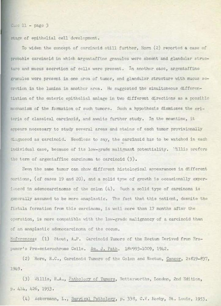
ll - page J
!'tage of epithelial cell develo!):nent .
To 1'1iden the concept of cili"cinoid still fur-ther, !iorn (2) rel)Ortcd a case of
probo.ble carcinoid in ~;hicb a r genr.affine granules were absent and glandul-ar struc
lurc and muct.~s secret i on of cell!'! •l(ere present . Ir. anot her case, ar gentaffine
granules wor e present in one !j!'ca of tl.llllcr, and gland1.1lar st ructure wH h rnucus se
crr:>ti on in the lwnil1a in anoth<.r area . He suggested t he si mul tan.eous diff<.:ron
tiation of tho enteric epithelial anlage in two diff er ent directions as a possit1~
occhanisc of the fonnation of Sl.ICh tumors . Such a hyoothesis dismi;;ses the cri
terill of classical carcinoid, and a\r"lit.S further study . In the meantime, it
appears necessary to study several areas and stains of each t~mor provisional~y
<'li••llJ'lO$Cd as carcinoid. Needless to :Jn,y, t.he carci noid has to be lo!~<otchcd in euch
individual caso, because of its lo~l-grll.(le mulirnant potent i alUy . · 'illis iWOfcrs
the term of argentaffine carcinoma ~o carcinoid (.3) .
i!:vGn Lhc same tumor can show dif.fo1·ont histoloP.ical apnearanccs in different
oonion:~, (cf cases 19 and 20), 311d a solid t,yne of gro>.1.h is occasionally oxpcr
.. nc.:od "~n adenocarcinomas of th" colon ( 4) . Stach a mlid type of carcinoma is
g.:m·Jra~y as~Juntt~d to be more anaplastic . The fact that this uo.ti cnt, desnite the
fi:;tula formation from t!U.s carcinoma, ls 1·1\J.Ll rore than 17 months ntter the
r>P'!Jrati<>n, is mora comp.:ttibl e 'ti~h t.h(" l.ow- grarlc malignanc~t of a carcinoid Lhan
of '\11 !lnaplast.ic adenocarcinoma of L.ho cocum .
~kf<?rences : (1) Stout, A.P. Ca:·cinoid 'l\m1ors of the Rectum Derivud from r.:rs
pamcr' :J Pre-cnterochrome Cells . A.•11. J . Path . 18:993- 1009, 191,2 .
(2) Horn, R.C. , Carcinoia Tumors of th" Colon and Rectun, Canccz·, 2 :819- 837,
1949 .
(3) .Iillis, R. A. , ?atholopy of Tumors, "11ttt.E>rworths, London, 2nd Edition,
p . '"l'·· 1,26, 1953 0
(l, ) i1ckermann, L . , Sur[;ical Patl\oloi<Y, p . 338, C.V. Hosby, St.. Lorl"ls, L953 .

Tumor Pa thology fua,n Collection 'Illlnors of the Gas<,ro-intestina.l Tract
Ca!;e 12 f aw.:i Hal Pol;}'po si s of Colon ~;i th
Adenocarcinoma of Si gmoid
Uni versity oi' Kansas hedical Center Kans:l.s City, Kansas S\U•gicd i'atholop.:y No . : 3795- 54
HistDry: Thi s 37 year old l·Ihite mal e noted the onset of persistent di arrh<la 11,
year's pri or to admission . Three years later a di agnosi s of familial pol.~i'posis.
wae made and total colectomy v1a>; said to have been done >lith ilea- r ectal anasto1~o-
sis . He •~as told that, there were two separate small carcinomas in the l eft colon .
After ·~he operation, his bo>~els moved o.bo1Jt t1·~Lce a day. Hovrever about 1 year
befot'e thi s admi ssi on, he again hegan haVing diarri1ea l<ith o r:casi onal Dlood
f1 ecl<s . He had occas i onal fulP,Uration of r-:>ctaJ. nolyp.~.
His father died of co.rcinoma of the col on ;dth multiJJle nolyrosi s at the age
of 37 . The patient sai d that other dist<1nt relati v es had t he smne . disease .
About L, em. above th« s:ymphyei:; oubis, there 1-:as a slightl ;v· tender area wi tb
t.!le sensation of a mass . Otherwise physical e.Y~-un:Lnation \'las norwal.
Laboratory stlldies were 1·d. thin nor!IJAl limits exceot for 3+ blood in the stool .
Bariwn enema revealed polyps in the rectum and sigmoid and a tyoical a•Jneari nH
sarci noma of thtl s igmoi d colon just distal to the old anastomosis .
Abdomin<:> -per ineal resection oi' the ::>igmoid and rectum >~aS done ''oith great
difficulty, due to many aclhesions . There was an apparent carcinoma of the s i gl!loid
.ius~; dist al to the ileos:Lgmoid Mastomosis . Ho l ymph nodes v1ere palpable . '!"ne
operation was completE'ld vrith ileostonw . Post- operative course ''las complicated by
t!;e fai lure of functi.on.i.ng of the ileostomy a11d fistula forma.tion . These were
corrected !100 he 11as discharged.
Seven months aft er · the oper<~tion, +,be patient. was examined. l~s condition ~!as
good and the ileost ow,y '·las functioning excellently. ·

Case 12 - page 2
Gro»s PatholoRical Findinns: The colon sho1·1ed many sTh>ll discrete and confl uent:
sessile nolypoid lesions me~surintt 0 .1-0 .8 em . in diameter. T'nere >·;as a larl{e
t:unor nearly surrounding the rectal wall . This had a large centra::. ulcer measuring
5 em . ·.;hieh >;as surrounded by a .firm rolled border of mucosa about 1 em . in
t~ckness . The mucosa in the entire area of the tumor was adherent and not freely
movable over the underlying "':.Js cuuris . llo free t umor was seen in the serosal
£ttrface and no lymoh nod~.> mctastazes •:~ere found .
Jia gnosis : Adenocarcinoma of sirmoid colon . i'olyposis of sigmoid colon .
Comment : The section oho1~s transition of the normal e~ithelium to a denocarcinomn .
'l'hr i.m!10r t.ance of polyps oi' the colon as a r.oj:.ential pr ecancerous lesion i:. ~<ol.l
lmovm . The adenomatous ool~rP i s rnot'f> f requent than the villous type ( see cas<> II.) .
References : (1) SHington, N.\1. · .md Do~no, ~·f. A ., The· :Oi :mif':i.onncc J.nd Treatment
of Polyps of tht> Colon :md Rcctl.un . N~~' Enr. . tied . J . 249 :673, 1953 .
(2) Ride!' , J .A. Polyps of thu Colon :lnd Re ctum : Their L-,cidcncc .:>.nd Rc
l".ticn!lhip to C~rcinomc . Am. J . J:od. 16: 555, 1954.
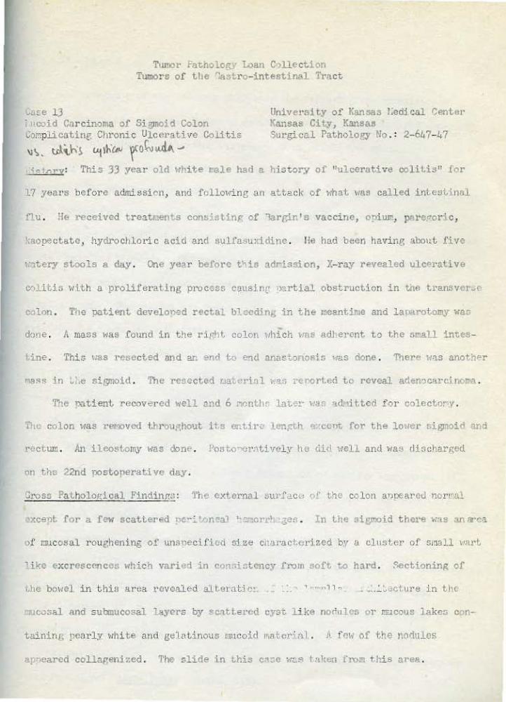
Twnor f'a,;nolor;v Loan Crlllect.ion Tumors of the ".astr o- intestinal Tract
Case 13 i ~>coid Carcinoma of Sigmo' d Colon Corr.plicatint: Chronic Ulcerative Co.Litis
'l\ ~ t.rl'-\-\i l.<jlh"cn-~ ro~~ .... University of Kansas !;edi cal ')enter Kansas City, Kansas Surgical Pat;;holoeY No .: 2-647- 47
"" lr.ry: Tl>is 33 year olrl ~1hite r.ale had a IJiswl'y of "ulcerative colitis" for
17 years before admissi cm, ami folloHing an attack of 1·;hat. •~as called intesti nal
f1 u . H& recei vcd t reat.nen .s consj,, ting of 1argin 1 s vaccine, o;>iu.m, pare~Cor:i c,
kaopectate, hydrochloric acid and sulfasuxidine . He had been havine; "\bout !'lvc
· · ... tery stools a da,y . One year before tT·is adri.ssion, X-ray revealed ulc»rative
cr>li t:i. n with a proliferating proceGs C;J.t'.SinL' 'Jartial obstruction in tne transvcrwe:
C<Jlon . 1'1-te patient develoned recta ... bl_ecd:i.ng in th& l:le2.lltime and laJ.tarotamy war.
done . A mass ~1as found in t!1e rirJ1 ~ col on 11hich VIIS adherent to t,he small im:es-
t.ine. This uas resected and an ~ncl to e?ld armstollosis ';as done . '1'11cr" Has anoth"r
r~~;>,;s in .I.e sigmo.l.d . The resected r.Ja• eri, l ~.'l.fi rc r>ort.ed to revel!.]. aden?c~rc.i.noma .
The nati en"L recovered 1·1ell nnd 6 l'lontlic lilt-.!· li3F 3.d'lri t.tod f Dr colcctor-y.
HLc colon \IllS ret·:oved t.hrou,-;hout its entir,:, 1~:-n;n:h ~·cvr.. for the lc\ler sigmoi<' ·!lid
>·cctum . An i 1 eost.omy ~o~as done. J'ost.o--er:,ti vel y 11 e ciirl ,_qell and "a3 rli s charg<'d
on thz 22nd nostorerativc day.
Gross Patholo,gical Find:i.nf!:J : ~'hC' external ~~ur.faco of the colon anoeareu norJ .. al
of mucosal roughening of unsnecifio.-1 size clwrae"t.cr1 zed by a clrLSter of small 1•nrt
like excrescences which varied in con.•i3tcncy from soft t.O hJrd . :".ectioning of
r,he bowel :i.n this ar ea ~·&voaled aJ.ter:J.tjl)! 1 .,,M, 1 ... _ .: . .:. ~·3ctm·e in Lhc
:ruoosal and submucosal layers by Sc."lt tercd cysr, like norlttle!i or Jr:Jcous lakes con-
taininp, pearly >lhil;e and gel<tLinouc. mucoid JnA ted n l . A .ro~. of the Mrlttlus
apryearcd collagenized. The s' ide :i.n this cn~c ;;:.,. I •tlten fro!" tlti.s area.

'J'h.; ri.Jllaindor of th~ sp .. cim.;n displayed ~h·· typical gross appoaranco of
chroni.c ulcerative coli~is . l~umcrous mucoGal ""~ochiao v1or e scattered about and
p::;·~udorolypoid mucosal alLt~raLion '.-!as cons,icucu • 'l'lithin the cecum .
:Ji?..ltllosis : Hucoid acl<.'l'locarcir.orr~ of sigmoid colon.
vcrrmcnt : The pscudopolyp~ in the. cccti.'ll chOiiC;d c•>i thc.h.al and archilcctural changc-s
which vo:r ied from marked atyoia Lo f r ank prcinv·~sivu m.ilignant chanec .
'£1v • r:atient remained ln good health unHl i'i v c. years l ater i~h on ho devel oped
r·uctal pain ar:d bleeding . Th remaining rc.ctoSi,ll!!l<:li d aml r ectum >~ct·c l'C~ccted
a~ this time and an adoitioMl ulcerating adtmocat•cil'lOma ;-;as found 7 em . bclm· thl'.l
$if,lll0id stump . Th" pati .. nt has rcma'ncd licll dur' ng r.he ensuing .3 years and is
11dl about 9 ynar s aftel' ~he. fi r s;: operati on ft•r thf. car dno!lk1.
'J'o :r ·~ capi t ula t e , this pn l;h.nt t~i·~h ul.ccr•td..:i. vo r.:o l.i t i 5 clevcl or>cd :3 s cparatc
·..,ci of adenoc:~.rcinoma ol' his colon =d a fo-:.ts of pseudopol;y-pi 1''ith l"typia in
,. cum over a 6 y<oar per• od .
The high iucidcocc of c11rcinoma i n pati nt-s .rd. t.l) chro::ti.c ulccrRW. vc col·~-·
• ""ll. !<n olm . I t i s said t h·.rt C'3.t1ccr in such •xttiunts occtl rs 30 tlmos more f rco
ur•nt ly t han in i ndi viduals of th.:. f<c.n Or .:!l ponulaL~on o'' simil ar :l/!C r.roups (1 ) .
:i"iLru nre several opinions r.b>ut the pa~hof:• I< sill c t:'lis high ;ncic'rnce (?.) .
Til::< pathnt sur vived quit<. lone: after opor<~tion . Tb.:.rc is an opinion that lisco
locica.l appcaran ce docs no~ tell much about nr<•F.Jlo!'l::; (.3) .
RL•i.\•ronco!'l : ( l ) Bargon, .I .A. , Sauer, ' . G. , Dloo.n, '.! . P. and Cac,v, R. l" . The Deyelop-
flltlnt of Cancer in Clu·onic Ulc•.r~tivn Cc-lit ; " "'•tro.or,tcrol. ?.6 : )2, 1954 .
(2) Gelber, I . , Haottl.o:·:lu, ll . ll . , and n:lb, A. Carcinoma of th" Colon in
Chrr.nic Ulcerative Colitis . IJ.asi.roent;:arol . 2tl:SJ6, 1955 .
(3) Qualbeitn, R.E . and Gall, :::: .;... Is lil.Jlolop,ic Grading of Colon Carcinoma a
V.>licl f>!·occdur e ? Arch . l'ath . 56 : 466, 195.3 .
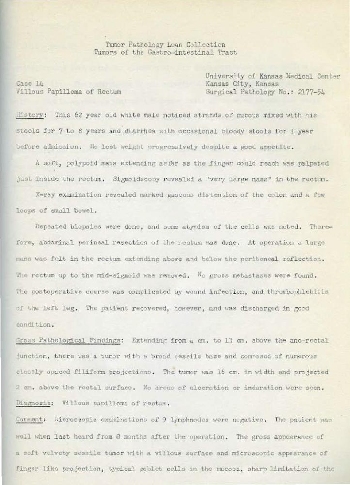
Tumor Patholo~y Loan Collection Tumors of tho Castro- intestinal Tract
Case 14 villous Papilloma of Rectum
IJniversity of Kansas l·iedica:L Center Kansas City, Kansas t-urr,ica:L ?atholop,y No .: 2177- 54
Jjstocy: Tbis 62 year old HhHe male noticed stt•onrls of mucous mi:xed with his
stools for 7 to 8 yertrs and diarrhea ~lith occasion.J.l bloody stoolt; f or l year
Jt.fore admission . He lost :.eight. nrogressively despite a good appetite .
A :>oft, polypoid mass extendinr a" ilir as the .finger could roach was palpated
just insi de tho rectum . Sigmoidscooy J'ovealed a "very l o.rge mass" in t he rectnrn .
X-ray examination re'fealed rnarked gaseous distention of the colon and a few
loops of small bowel .
ilepcated biopsies Nere dono, and some atyoisrn of the cells v:as noted. There-
fore, abdominal nerineal resection of ~he r ectum l!US done . At operation a larr,(,
rr.a.ss >tas felt in the rectum extending above and below the po:>rit.oneal reflect-ion .
Tll< rectum up to the mid- si.gmoid "'AS removed . l'lo pros~ metastases ~:ere found .
tf,c oostopera~ive course was complicated by wound infecti on, and tllrombophlebi:tls
?1: l. i1e lef t leg . Th" p;;.tient recovered, hOi·/ ever, nnd <tas ctischa.rgccl ;.n good
condition .
~ro~s Pa.thololtical Findin~<:s : Ex1.endinr from u em . to 13 err . above tile ano- rectal
iunct.ion, there t~o.s a tumor t·li th o br.oad sessiJ e ba.se and comr:onecl of numerous
c I o~oly spaced filiform projec~ion:; . !'he tumor t·ras 16 em . in ~1idth and orojected
- en . above t.he rectal S'~rface . rio arC«s of ulccrl'tion or induration lfere seen .
Di.'-ll)nOsis : Villous 0<1 pilloma of rectum .
Comflent : l·.icroscopic exa.lninatiom, of 9 4-.nlphnocle~ N\'<rc negative . 'I'M uatient \'Ia.
lieU uhen last heard from 8 montts after the oper:~t.ion . The gross ap!'learance of
a soft velvety sessile tur.;or >-lith a villous surface and microscoui.c appea:ranc~ of
ringer-like projection , tyoi ca1 goblet cells in ~ho mucosa, sharp limitati on of l.he

c -- 11. - page 2
~oit~elium and less glanduler elements nre tr.e characteristics of the vil1ous
~apillon~ (1) . The hyperplasia of ~he superficial cells is said to produce a
'lillous paoilloma, whereas t hat of the decpor cells pr oduces an adenomatous polyp
(1) . The adenoma tous ·L:ype is about, 10 timos as frequent a? the villous pll rrl.llomn .
Both show rndign;m~ transformation . I n one oeries, villous pauilloma 3ho~1ed
adenocarcinoma in about 35% of cases, 9ol'lt'times in the form of carcinoma in ~i t.1J 1
and cxtcrpation of the involved colon and it!! ly:nllha tic draina:>e ~<as reconr.:ended .
ilef.:rence : tl) Bacon, H.E ., LoHcll , E. J . l.nd Trimpi , H.rr. Villous Papilloma oi'
'thu Colon ~d Rvctum. Surgc~J . 35 :77, 1954.
Tumor Pathology Loan Colle ct i on Tumors of the Gastro-intesti.l'lal Tract
Case 15 University of Kansas lledical Center Kansas City, Kansas
Cl.clangio-carci noma of Liver Autopsy No . 1107
Histor.v: This 59-year-old col ored male had consumed about 1 pint of whisky a
day for at leas t 20 years and often n egl ected eating nr ouer meals . Ei ght weeks
~dore death he first noticed s~1elling of the .feet and ankl es, t h<m gradual a b-
dolLii1al distension, clay colored stools, int ermittent dark urine, and occasional
epi.staxis. No hell'ntemesi s , hemoptysis, or vomitin g Here noted.
Past and family hi s tory \·!ere non- cont ributory.
The F<>tient v:as markedly j aundiced and the abdomen vtas gr eatly distended .
':'he liver 11as nodtilar, tender on palpation, and. extended 6 finger b readths bcl01~
~h e costal margin .
Urinalysis v1as normal. VORL negati ve; RSC 2 .6 to 3 .3 million; NBC 13, 500;
<temoglobin 6 . 7 Jl)l'.S . Hematocrit 21..% . Bleeding time 1 1/2 Jlli nutes; clotting t jJne
11 rni.nutes . Glucose t olerance : 5 hours 1·!ere required for t he return to the base-
line leve.l. Prothrombin tilne 50-7~; of nol'l!l<ll activity . Serum bilirubin : tot.al
11 .8 , direct 11.3 JUg .;; ; alkaline phosnhat.ase l 4 .6 millim<;>l e unit s ; bhymol turbj dity
"4; serum a lbumin 2 .9:3; globulin ') . ?'( gm .%; t otal serum cholesterol 400 me;.:·.
Urine urobill.nogen 1.3 mg./99cc . Ascitic flui d : lia 136, K 3 .9 mE() ./liter; protein
1.7 -· I!Jfo .• J • Cytol ogical s t udy of t he asci tic f l ui d revealed no malignant cells .
X-ray examination revealed a non-fun ctioning gallbladder and d efor med ir-
r itabl e duodenal bul b .
The pat ient ~laS afebrile . Ci n·hosi s and tumor 11ere suspected . 'ith pallia~ive
tht:rapy t he patient f elt better' . Eight da,Vs befol'e death , explol'ato r y L?.oarot,omy
reveale d massive twnor invol vement of ~he liver . PostoperatiV<;ly the pati<=nt did

c~sc 15 - page 2
well untiJ. l day bef or e d!Jath ~;hen he vomited rJ.bout 300 cc . of darl< bloocl,y f l uid,
becmne comatose, and expired.
'ru:>::J F'athololtical Findin~<s : Three thousand cc . of serosanguinous ascitic flui d
•'c• found . l'he liver 1~s diffusely cirrhotic and in its mid,:ortion contained
m.:rr~rous tan and :,'hite nodules, 5- 10 nm. in dituneter . These involved not only t he
liver parenchyma but also invaded the portal vein complet ely obstructin(( ~he lumen
at ito confl.uence at the hilus of the live:r . t!,a~1ses of t urtior and orl(nnizing blood
clot Nero' f ound i n both t.he intra- and extra- hepatic bile ducts and p:allbladder
::i t!'l r<:trograde dilatation of the: bilian• tree . There was tumor over the: surface
~=- the eallbladder but no metastases Here found .
Hi.croscopically (besides l,he slide enclosed') , carcinoma was found i n the
coi'Jilon hepatic duct and portal vein; but not in t.he !'all bladder, t,hourh oxtcn-
cion from the liver t o the nerit.oneal surface of t'JC tTallbladder was noted .
~'i~l!:'losi:;: Pri.Jt.ary carcinoma of the liv<lr, •>rcdor.-.inantl,y cho 1 '\ . .o·w.ioma .
Comn~nt : The 1;istolop.icnl appearance i ndicates this nriw~ry car cinoma of t.he
liv<:>r to be ol' the cholangioma Lypc . IJascuJ.ar invaai on bv t hi s t:y-ne of' carc1noma.
i;; 9ro1~1inont . 1'. couple o.f surveys ( 1, 2,) renor t. that this tJ'l:>e is mucl·t rarer t han
the liver cell type, ~<ith the ratio about 1 :10 . llurger reoorts that tho cholan
erlolll:l lyp(' is quite .frequent arr.onF' "X!tner and Javanese (3) . This case sho1:s
abt:ru.tant bile t.luct proliferation resulting from the cirrhosis . In places pseudo
<1tcts of i.nfi l t rating carci.noma arise from t he I•IEil.l-formed car ci nomat ons nodules .
2tPiisrt reports that 51 per cenL of 1,9 cases o [' chola.ngioca r ci noma were associ<'~tcd
<i tn cirrhosis ( 4) .

Case 15 - page 3
<!efer cnces : (1) Cohn, J . and llaymond, A. H. Primary Cancer of tho Li·.rer . An Eighty
v_ar Surv<-y of N<M Orleans, Louisiana . Sur~::cr.v , J?, }56, 1955 .
(2) Zdmondson, H.A. and St~iner, P .E. , Primary Carcino~ of the Liver, ~nc~r
7:462, 1954 .
(3) Duggar, J .J.; ., Cholan(;ioll'a, Hcd . J . Aus~;ralia , 2:819, 1954.
(4) Stcl•art, t·i.J . , Pr .. canc"rous Lesions of the Alimentary Trac::. , Lancet 11 : 565,
1931 .

Tumor PatholotJY Loan Collection Tumors of the C-astro- intestinal Tract
G'lSe 16 Univ.ersHy of Kansas Eedical Center Kansas City, KahBas
!iepatoma ("adult type") in infant. Aut O!>SY ~!o . XXX\III- 1
;listonr: 'Ii1is one month old colored infant had an abdominal nass at bir th , •:Thi ch
rapidly i ncreased in size . The infant lest iJeight a nd became very irritable .
Hc"ev,~r, h<O continued t o feed a nd to ha,re normal bo>wl movements . t''o jaundice,
acholic -stool or hew ..... ·turia vtas noted.
Sendi ng of lo~Jer costo-chondral junctions, moderate c!'aniotabes, and protub-
~rant t ~"lsc a bdomon Here no ted . The veins of the abdominal. l·la;ll Her e di la.tecl. . A
moderatel y firm, Gmooth mass fi lled t he entire unper and lower right abdomen, ex-
tending past the micl- l ine to fill most of •lw '1eft side . A definite edge c • .,u1d be
1 ·aJ.pat<~d below t he right. iliac crest . The mas:. '#as not ballotable and no fluid
Laborat ory .findings were l'lithin normal limits . No liver function t est was
rlcme .
f'yelogrron :1as norll'.al .
1fhe patient wa~; acutel y ill 1-1h~ a dmitted, rapidly deter'i.orated • .. tith occas-
i onal anneic ')eriods, and OX'Jircd .
c.'ross ?atholo~:;; ca 1 Fi:ndinr.;s : The liver wei1~hed 590 !(rams (normal about 140 grar~s) .
IL 1.1easut>ec1 1 5 em . x 11 em . x 7 cm . I t >~as firm in consistency and the edges ~~ere
~mooth . The capsule anpeared to be t hickened but 1•1as smooth and transpP.rent .
J.oderate fj,ne atld toarse lobulations of the l i ver \'1ere noted . 'f.he biliarv passages
v1ere patent . On mt surface , the liver mass seemed to b~: almo:.t completely replaced
hy \iJl\l~\H!l ti$>;?ue 11hich appeare(l to have a tendency t.o form cord~ -to'·l<ll:'cl the peri-
p!ter .r of the liver mass . t~o normal liver tissue ,,,as notE>..d on any gross cut section.
Case 16 - page 2
;n aP.nosis: lhli f!llant ad•llt typv h~<matorna .
Co=cnt : This hepatoma i:; consid.,rud to be of the "adult t yp:;" (see case 17 for
1i~cussion, rcfor cnc.,s, and contrast.) and is charact-erized by bile 9rowction
"-nd ;>lugs . Incidental foci of o:ctramcdullary heJ:latopoi esis are elso seen .
References : (l) 1-lilli:;, iLA . , The Pathology of 1'UI1!or, pages 932-939, 9ut.<C!'H-'rth~,
~td . L,ndon, 1953 .
(2) \fcbster, R. , lialignanr. H-.;patom,., in I nf ancy, Hod . J . Australia, 11, 381,
19:38 .
(3) TomzykQSki , A.J . and Stt.vcns, R. C., Embryonal C(•ll Carcinoma of the Liver
in Infancy, J . Pod. . 4.3 :309, 1953 .
(4) Dicgolow, N. H. and ii1'ir,l1t., A .\f . , Primary Car cinoma of ~he Liver in Infancy
and Childhood , Cl.lncor 6 :!70, 1953 .
( 5) Chris tophcrson, \I .J. . , P1•im:Jry lcnign Liv~IT-coll Tumor i n Infancy and
Childhood, Cancer 6 :853, 195.3 .
(6) !!ells, H. G. , Occurr ... nc-: and Significance of Cong<...nitalJ:ali[!l=t llcoplasr.s ,
r,rch . Path . 30 : 535, 1940.
•
Tumor PatholoGY Loan Collection Tumors or Gastro- intestinal Tract
Cas•• 17 Uni versity of Kansas Hcdical Center l<anf.las City , Kansas
II -:llltOrlll ("embryonic t ypo") in Infant Surgical Pathology No . : 2672-55
· :i~tol·v : This l year old white malEo was born of a 27 year old gmvida 1 , para I
~ran . "1~10 pregnancy a.nc! deli very 1·•ere uncomPlicated . At the a('e of 9 J!IOnths, a
J.•ll<l decrea:;e in his appctil.c wts noted t·Ti thout other si gns . At the age of 1
year:, o mass v1as noted in ~he upper abdomen by 1,lw f e<mily oh.ysi cian . The parents
sca~0d Lhat they t hought the "stomach" had felt hard f o r s everal. weeks nrevious to
tt.is . The "eizht. 1·1as '21. pounds at both 9 r-.vntl"" and l year.
l'hys3 cal exru:Jirultion revealed a large mas!' in tl.c u~r.er mitlcllc nbclomen reach-
tnr .:>f:]oN the um0ilicus uhich was firm, non- tender :~n d moved •.rith respirations .
t'h~ contour \~as sn.oo~h and "as not a tt.uched to t.ho ~kin . The tip of the spl een ':la s
l"rl t tmd it. vtas separat<.d from the muss . The l.'klss Nas t hought to blond t·Ii.th the
_:_ver o.long the right side . T'lc liver t•I"'S 1 finr<'r breadth below thE' cost al 1r4rgin .
Urinalysis and bloocl counl 'fer" ~rithin normal limits . Prothrombin tj!;,~ o·:as
00~. or contro_l . C 1 li ch 1 t l ~ t · • epw n o cs er a ccs ua~ ne(:lll;lve . Tot~l serum ~ilirubin
Has 0 .) and direct 0 .2 mg .,; ; aJkaline pho5p11a~asn Vfas 3 .1 milJimoll!l Ltnits . 1'hymol
.•u·bidi ty t·las 7 units . Serum albumin ,.,as 4 . 75 and globulin l . 75 r,m . ·~ . Total chol-
' <.<.rol uas 334 lll.g .;; and 66~; e::ters. Retro"t"ade urogranhy was normal .
At exoloraLory laoo.rotorny, the tumor >~as founrl to have 1;roduced ext.rene
·' 1.1' .:ucnt. o f the left lobe of the liver . 1'hl' ri~t l obe of f,J-c, liver aopoar ed
to be normal. '!'he ki<lno;ys :~nd adrt>nttl,s were notl11ll.l. on •:oalpation . ThGre \~ere no
sl,cos of a primaT'J sitC3 ot.hE'r than the liver, o1· •Jf lntra- abdominul snrG,(ld or t·.umor •
.• ~ t.i' c loft. lobe of the li vet· was ren:oved, another ~:cll-Eoncansulatod pro.)ection or
• .. e t.umor in the right lob< 1•a" found, zo t.he unpcr medial third of Ll)e rilrl:t lobe
111~ :also r emoved . The tumor 1·1as cleaned ofC the hera tie nortion of the vc;na cava .

Ca s e 17 - page 2
Postoperatively t he ·patient did ver y Hell . 1'h e child r eceived 6 X-~'ay t r eat
ments amounti ng to 3000 r depth dosa.Re, f rom various ports and was dischar ged.
GrL>s.:J Pathol ol(i cal Findintts : One port i on of t he r esected specimen measured 1.3 x
10 .x 7 . 5 em. and >.•ei ghed 352 gms . The surface was lobulated and the lobule s va.ri ed
in oi ze f rom several em . up to a pproxi ma:;:,ely 6 em. i n di amet er. Cut section s hol!IG-d
numerous lorules of t umor t issue . The pieces of tu:mcJ" va r ied in size from 1 em .
to 5 em . in diameter . Fibr our st rands \•!ere seen r utming t hrough the tumor tissue .
The t,umor tis s ue va!"ied in color , some pinkis h whit e , a nd some fairly yell o•;! to
br own . It 1·1as al l soft . The other !)Ortion of the ~pecimen measur ed 8 x 7 :x 5 . 5 em .
al'ld '.'IC!ighed 173 gtns ., ~;as so ft and cui'. section _ro:wealed much "reyish- black n"'croti c
t,issu e . No r-egi onal lyin!'h node s were r emoved for exami nntion .
;:lbJmosis : I<:alignant embr~ronal hcp"'t oma . .
Comment : 'this case is offered to illustrate the histologic pattern of t he uncommr)n
''cm')r.vonic " t:,-·pe of bo~Jatoma as contrasted 1>1ith the more common "adult" ·~yoe of
Je)Ja.t.:>ma in i n fant s and childron (li . The diagnosi s of (;1/",bryonic hepatoma HM.: no t
,ma,limously ncce!'>ted by all observers in this case e ven t hough the !Jattem is
si milur to that in var i ous -ref er ences . Other di.a[PoStic oossib; 1; ties '~bich ':!8r<'>
~n·~ertained w:r e those of metast;,atic ne.u.robl astor::a (1, 2, 3) and metastatic carcinoid
IJu•,or' . No ncr,,e f iber s or silver positive ~ranules could be demonst r ated in the
'I ,-ion . Subsequent l y t he pat ient deter iorated and 4 mont hs after oper-ati on be
expired . Autopsy revealed icter i c ~ss~ of twnor in the liver ~li th meta~t•se.s to
the lungs and diaphl·agm . Ho other orr-;ans shol·!ed evidence of nr imary or metastatic
t w::or s .
In infants, r.ni xed t ype t umor s of U.ver have been r onorl;t:d (/4.). "enipn adeno
m;~Lous tumor of t he liver al.so oc curs i n infants ( 5) . These t;•o anrl met a s tatic

Case 17 page 3
nG1,U"ob1astoma , congenit al angiomatosi s and "adult 11-tyne hepatoma must be dif
f er entiated from the 11embryonic 11 hepatoma . Congeni tdl mal i gnant t umors in
general are excellentl y revi.eNed and discussed by 1/Jells . ( 6)
Rvferonces : (1 ) vJillis , R.A. : The Pathology of Tumor, peges 932-939 . Buttor
<·torths, Ltd ., london, 1953 .
(2) ;lobster, U. , halignant Hepat oma in Infancy, 11M. J . Australia , 11, 381,
1938 .
(3.) Tomsykoski , A. J . and Stevens, R.C., Einbryonal Cell Carcinoma of the
i.iver i n Infancy, J . Peel., lf3:309, 1953 .
(4) Bi cgelow, N. H. and '.!right, , A . \·1. , Pr imary Carcinoma of the Li ver in
Infancy and Childhood, Cru1cer 6:170, 1953 .
( 5) Ghrist op!1t.rso11, 11 .1'. . , Print<try Senigu Live-r-cell Tumor in Infa.ncy and
Childhood, Cancer 6: S53, 1953 .
(6 ) \<fells , H. G. , Occurrence and Significance o.f Congenital Halignant Neo
plasms , Arch . Path . 30:535, 1940 .

C11.sc 18 ~ngiosarcoma of Liver
'tumor Pathology Loan Collection Tumoro of the Gastro- intestin <tl Tract
Uni versi ty of Kansas 11cdi cal Center Kansas City, Kansas Autopsy No . : XXvi- 73
i'ist.ory : This 61 year old ••hite female began having excess weakness and tiredness
saveral mont he before death . About 2 months bef ore deat h , she had "infl uenza " at
uhich l;ime she i'i rst noticod pulling- tearing pains i n t he r ight flank . The pain
!)..,came ver y sever e, the patient ·.-~as bedri dden and vomited most of her food . She
l:'l.S aani.t.ted to this hospital 10 days before death ,
Pt1ysical examinati on r~vealcd a dys~neic , slightly cyanotic and extremely
·ca~. \oiOiolan . Becau!le of her oxtrerne obesity, t he examinati on was ve ry difficult ,
hoNt~ver,~ a ve ry large, fi.rm, slitthtly tencter ma:1s could be palpntad i.n the u~per
abdomen . The abdominal wa.U was edematous . No definite ascites 'll<tS obser ved.
Clinical :Lm:oression ~~as ovarian tllll'.or and cardiac deccm:oensation . The patient
wa:; s.i.:ig}ltly anemic 1tith r· d blood count of ) .9 million . No pertinent liver
f unc ~ion tests 1verc done .
Because of t;he edema and cyanosis, digi t a.Uza'l:;ion Has a ccomr>lished, but.
>~Hhout imtJr ovement , t·:ercurial diuratic:; 1~ere also ineff ective. Po '<- ray
-.ncrapy was given. She ,~radually grew \maker and cxnired .
Sross r'atho 1 o;;;i cal Findinr~ : The abdominal cavity contained 500 cc . of c l.ear strau
C"lO'""'d nu.id. The peril.nneal surface •·tas smooth and glist ening . The liv<;r ex-
tendod S em. belo~1 the costal margi.n . The t'ip;ht pleur al cavity contl!ine d 1000 cc .
of bloody fluid, ~~hile ~he, left contained 200 cc .
The liver weighed 5270 f7t · a'1d £leasured J3 x 31 x 15 e5 . 'll1e organ ~1as r ed
?rown in color . The capsule Has smooth , moist and glistening ':>ut the lobulati;)n
.-:w; indioti nct . The ric,llt lobe \•las almost enti rely re,..,laced by numerous blood

Case 18 - page 2
sr..:J.ces, the largest measuring 1 1/2 em . in diameter . These ~fere distended and
bet1~een them, in some areas, the tissue was greyish- y-ellol·t having a necrotic
a~oearancc ~thi1e in other area~ i t. ap·leared s·,mel<hat fibrous . !'>till other areas
'llere compos(';d of a grey white celh llal' a.ppear.Lne tissue. Small irregular foci of
eimilar strltcture l<ere sca;;tered throur,hou~ the left lobe . \olhere this ahnor!:lal
tissue ·ftaS not present , the l obulation of tre liver was distinct .
A massive idiopathic hem::>rrhagc 1·1as found in tl"e ri~l-!t occtoral reftion .
D:L~r:nos.i.s : HrmanP,iosarcoma of the liver .
Comment : Tl1e histological appearance suggest:; thi s to be a ll'Blil!nant vascular
tumor . ?.etastasis <!as str ongly suspected in the right pectoral rejtion ,.,ohcrc
masdv01 hcJnorrhages l<ere fcund, but microscopiC' "xcuni.naci on fail ed ~o show S!'Jread
in th i s or any other ar ea. lfalignant hemangioma in t. he liver i s r are. Stout;
re .. ,ortet.l ~~~o cases (1) . The ap!)earancc of Lhe cells and the presence of reticulum
:·i~<>r~ outside the tumor cells forming vascttlar structures suggest tris care to be
of the he1eangioendothelioma t;ype. Accordine to Stout , tw'O characteristics 'lre
:n.vrtant; on<. is the formation of atypical cells in p;reater number than arc- re
quired to line l,he vessels with a simple endothelial membrane and the other is
th<" formation of vascular tubes •flith a delic~Lt" fra.mework of ret iculin fibers and
a marked ~ond011cy for their lumens to anastomose .
?cd\Jrcnces : (l) Stout., ;, .P . Hcnungio-~o:ndoth, lioma . A Tumor of Blood 1Jcssols
Fv:..tturing \Tascul:·.r Endothdial Cells . Ann . Surg . 118: 1.1.5, 1943 .
(2) Stcut, •\.P . Hcmangiopericytoo..:~ . ,'.nn. Surg . 116, 26, · 1942 .

l'umor Fathology Loan Collection Tumors of the Gastro- intustinal Tract
Case 19 Ad.mocarcinoma and Squamous Cell
Carcinoma of ~he Gallbladder
Dr . l•'i n i am R . Pla. t.t. HiBsouri Baotist lfospi'Lal St . t.ouis, liissouri No . : S-54- 2800
-iistC'rv: This 'P-year old whte man l;<as admitted to the hospital on July 26
1951., ><i th a chief complaj nt of cram'Oing righ1 upncr and lower abdominal r.>ain, of
t'·IO mon t,lls 1 duration . This apparently had no 1'13 La tioo ~o food intake or specific
t.lJTk of t he day . I t "'ould last frol!! several r.;inutcs to sever al hours . llis aopc-
tHo had dic:in:.shed considerably ar,d he h;ld lost 15 nounds i n <;he two months'
r.1od . T'nere \:as no change in his bo~•el hll bit:; or color of stoo 1 and there had
?e.<. ~o mrlena .
Phy:sical exan1inah on r evt>aled a l·Iell rlcvclopt)d, 1~ell nourisl'>od "1hUe mal e ,
;1, i.•>hL 5' 8 11 , wei ght 150 lbs . , blood ore~sure lfl0/105 . A fi rr.:, enl.:ugcd l,ymnh
'!otle w·1s I)Q.).pabie in th"' ril')'lt su.,raclavicular region . T!!e th.vroid was not
o·lpable . The abdomen Has obese, soft, relaxed, and nontender. Tho liver edge
•,ta;~ roll. about 3 finrerbrt.:ndtho beloH ~he ri;'ilt co::>tal margin . The gallbladder
1-1a~ palpable as a globular mnsf. l•lhich descended •o~ell on ina l>iration .
Hc:t:JOglohin, 88 . 7;~, Rbc - If, 320,000 . t.cukocyte count and urinalysis ~1ere
n~nJal and V!hlL was neg~tivt.: . Thymol turbidity- 1 . 5 units; Cephalin-c~olcsterol
flocculation ·nas negati vo.: . 'J"J., gallbladder could not be visualhed by cholec:ysto-
;;~·. . There Has a calcanou:, rl~;nsity measurin•· :.1bout 2 em . in d:i.ameter, •:Ihich
awoarod l:.o be a gallbladder calcl\lus v;ith a calcified r i m. ::>tudi as of the r est
,;f tho gastrointest i nal tract by bariilln swaJ low and enema \·Iere negative .
At operation, the gallbladder ';as fo11ncl to oo enormously distended and tense,
containing many large stone:~ . The surface • oas blue-gray in color, al Lemating
•.~Hh patches of thicl~ gl·ay fi'Jrotic tissue . Irrer,uJ.ar masses of t.<ur.or ~iscuf:

Case 19 - page 2
,;.htended from the neck of the gallbladder to t:.he descending portion of the duodenum,
oinding i~ to the latter struct.ure. Induration ~ras mar ked a t thi s point, and ax
tended proXimally and dist ally alonr. the ch.1odenum for about 2 em. lrany largo,
firm node:> wera pal pated along the !)al'a-ao rtlc chain and a f i r m, oval ~hapod node
ltteasuring 2 X 3 X 3 em. was palpated along ~'"te anter io r edge of the ri~ht lobe of
,u livt..r, but not resected . Tbe gallbladder was removed .
Jiaenosi s : Adenocarcinoma and squamouo cell carcinoma of the gallbladder .
Co:rr;ent: Six months postoperative the patient expired t~ith symntoms of partial
·nt.e~tirol obstructi on, r:-.arked ,iaundice, Probable metastases to liver and lun.c:.,
:.nd :nrkect edema of the right leg . No autopsy l<a3 perforned .
Carcinoma of the gallbl a dder i s pr' edominuntly adenocarci noma . In Jones•
series (1) only 3 O\tt of 50 ~1ere squumou~J cell carcinoma . Thia case anpears to
cho'~ two types of carcinoma . ~Iillis says thut souamous metaplasia i.s present in
sorm dcgr"c in about lOS of the tumor:; and is sornetil:ies or ominent "hrour,hout.; ho~<
ever, it is never complet6 but is always as~ociated with ader.ocarcinor.13tous st.ruc
•ure . !Ia furth~ states that there is no evidence to sunport the conceot that
the rroHths showing epidermoid metaphsia have arise n in areas of mucosa in •,•hich
metapl asJ.a hac\ previousl y been pn1smt, but.. that the change takes olaco in tho
tr1alir,nant gro1~th Hself . He feels t hat ~~ouamoir< chr-mge i s not s een i n non-neo
,,J.-\::;tic l Clsions of the go.ll ':lladder ( 2) . Acco1·ding to ''Ii lli s , mucus- secroting
adenocarcinoma is quite com~on, but diffu~e~y infil~rating signet - ring- cell
c~rc:noma is rare (2) .
. ef rcnces : (.1) Jon.:.s, C . L. C.'lrcinoma of t.h .. G~llbladder . A Clini c:ll "\net P:>th
ological An~lys:is of f ift y C;->.scs . Ann . Surg. 1'32 : ll0, 1950 .
( .2 ) \1illi~ , R.,\ . Pat hology of Tumors . p .. 41+0 . Butt entorth & Co . London,
1953 .

Case 20
Tumor Pa t hology Loan Collect.ion Tumors of t he Gastro - intesti nal Tract
Uni vE>r s i ty of Kansas Uedical Center Kansa-s City> Kansas
Adenoacanti JOma of the body of Pancrea s Surgi<cal Pat hol ogy ~To .: 2- 6'38- 47
'"iistor .v: This 56 year ol d white J:ew.al e had t he gr a-dual onset of pr'ogressivel)!
incr easing dull pai n in 1,he er;igastrium about 9 months befor e ad1nission . The
r;aln •·;as unrelated to meals and .seemed to r adiate to the back f requent ly requiring
opiates f or reli ef . Examinat ions, includit1g x- ray, r evea lecl a normal eastr<:>-
intestinal ~raC'~ . She l ost about J 5 J:OUnds in t his period, became aui te 1-1eak a nd
• .. ;as una.ble co get about . !-To na1~sea, vomit ing, j aundice , changes i n bowel hahtt
or mr;lena 'ffere noted. Pas t OLl'ld famil y 11is·~orie.'5 ~rere non-contribut ory.
There vt<.s marke d t enderness of t he abdomen "'ith muscle guarding it; the
ep-igasttimn . i~o mass could be pal pat ed . Rectal examination v1as negative .
Ser um amylase \Vas S<-) mgat.~; and l i pase 0 . 5 units . Gastr ic analysis 1~as
rtor=l . Basic metabolic rate was -<29~; . Other laboratory finctings ><et' e Hithin
nor·mal limits .
X- ray examination t•evea l ecl only diver t i cula qf ,;he second portion of t he
·duo den urn .
Ex:oloratory laparotmny r evealed a firm tumor i n the bod;y of pancr eas, exten-
ding poster iorly into the l"etroperii:one1.1m, infe riorl;y- into t l•e ligament of T1·oitz
~,I anteriorly in t he posterior· Hall of the body of t.he stomach . No remote
meta.;;tases wer e f ound . Total r,astrectomy, splenect omy , partial ·oancreatectoll'.y,
•.Jn~ero-enterostonoy an.d esophagojejunos tomy t•rere done .
'l'he post-operative cour·se was satisfactory and the pat ient 1"as free of t.he
pr evious abdominal oa.in . She 1·tas discharged on the 16th posb- operati ve day. The
n'l.tient died about 14 months after discharge but no autopsy was done .
Case 20 - page 2
Groos Pathological Findinp,s : The tumor 1·1eiP.hcd )00 gr ams at•td was firm 1·r.Lth little
ul·1nt.ici'Ly . I ts s urface r esembled gl andtl l a r tiusuo but did not shaN the usua l
a•1pcarancc of pancreas , lacki ng orderly division into lobul es . It was traver sed
~'Y :.cv.:ral irrcgl!].ar yellow streaks . The cut surface disc losed a meaty a p_!>2arancu
and ,,as pale yello" 1-1i th do.rk ... r yellovt m:>ttline . Defini te capsulation or divi sion
Ltf.CJ lobul es ~tas not a pparent. The lesion ,,,a:; adherent t o thll stomach from t4hich
U v:a:;; dissected t·Jith case . :-!o intr insic gastr i c l e si ons \•icrc noted .
Dingnocis : Adcnoacanthorra of Pancreas .
Com: .... nt : ih.it<her norr..al !):mcr ... atic islet nor acin~r t-issue is rcco1,Uznble t.fl.thin
~r" tur.tor mass t·Jhich is folt to b ... a malignanL ductal ade noacanthoma . Squan>ous
cell carcinoma i s far r·a ror ~han adlinocarcinomn fiJ11ong ductal carcinomas of t he
pancreas . In Arki n ' s ser ies (1) ther e ar e) cases out of 75 and l n liiiler ' s (2 )
3 out of 165 cases . This co.sc is offered as a somc1~hat unusual his tologic<ll t.31Je .
The locuti on of the carcino~ of the pancreas po5cs L~~rt2nt problems in the
nanngomcnt and prognosis . In this r es:>ect ca1·cinoma of the J,cad of tho pancreas
n.ust. be, conside red tGeethor with por iampul lory carcinoma (sec case 7 ) ( 3) . Car-
cino~ in t he body a nd ~ij.il rostm diff er ent probh,ms b<:>caus<l of i ts r a t her C)UilJsccnt
proHth and usual widespread mct2.stasi s by the time of clinical recognition (4) .
tho sympt-om of t:>ain in such carcinomas i s •ioll demonstrated in this casc . The
form~tion of mult iple venous throl'lbi in cases of carcinoma i n the bod.v and t.ail.
of tho pancreas is •t~ell kn01m ( 5), a l t hough not nrominent in thi e cas•J .
Retur enccs : (1) Ar kin, A. and "cisber~, s :•., Carcino~ta oi' the Pl\ncrcas, A Clinicai
and i:'athologic Study of Sovcnty Five Cases, C-octror.mt orol. 1.3 : llA, 1949 .
( 2) liillcl' , J .iL , !3agr.cn~ross, A .ll . azvt r.omro rt, I,.' i. , Carcinom.'l. of tM: Pan-
crca3, Bffect of !l.i s t olor;ic.:l.l Type and Grade of '·"~li!Jnancy on its 3el1"-''ior, Cancer , ---h:233 ' 1951.

Case 20 - page 3
(3) Hutchinson, ',/ .B., Recent Status of Carcinoma of the Pancreas, ,\rch . Surr, . ,
6$:62, 1954.
(14) Du.ff, C .L. , The Cli nical and Pathological. Fea t ures of Carcinoma of the
Bo-ly and Tail of Pancreas .• 3ull . Jolms Hopkins Hosn . , 65:69, l939 .
( 5) Xenny, ·'. E., The Association of Carcinoma in the .9ody and Tail of the
PancrP.as , .. >ith Hulti!Jl c Venous. Thrombi, Surgery, 14:600, 1943 .

' T1.:.mor l'aLhology Loan Collection
T~~ors of the Gastro- intestinal Tract
Cast: 21 iom,vosarcom.-?. I nvolvine the l"'lncr<!nn
Univer sity ,,f Kansas Modi.cal Cent er I<o.nsas Ci t y, Kansas .~urgical Pathology No .: 8- 2643- 48 Autopsy ~fo . : XY.Y.IV - 162
.iistory: 'Mris 57 year ole\ ~lhi te rrale was follol!ed :.n the out !)atient. clinic ,
bccaunc of epigastric pains and vomiti ng after meals , f or one year bef ore death .
Ill h1vl hurl occasional eplf':c'lt1t ric dis tress for 10-.1. 5 years pr eviously and X- ray
studi cs shoHed a duodenal ulcer . His symptoms increased progr cssi vely and he was
acL:itted to the hospi tal about l r:onth before death .
Past and fm:-ily histories wet·e non- contributory . T'ne pt:ysical Cxat:'inati cn
.nrl lAborator y studie:; •t~ere ess(..>ntially negat.ive . Upr>er gastrointestinal series
otln/_;<ml.od a moderatel,r obstructi ng duodenal ulcer .
L.~mrotomy revealed 11. scarred duodenum, and n chronic duodenal peptic ulcer .
A tumor mass , 7 em . in diat:~eter, \otas found in the oancreas and a wide block dis-
~" ction of the !)ancreas and llOrtions of the loft adren,1.l gland, omentUM, spl een
a:ul stomach was done .
PoB1,oparati vel y the nat:Len t drained a considerable amount of Ht!ro-sanguineous
fl 1id h.e1ving a hi gh con tent of ali\Y"lasc . 'fhe outient Has al"ebr ilc and 01'1 tho lJt,h
JOSt.,.,t>rative cl<ly the dt·ain ~!as :ren<:~ved . He later bled r coeatedly f:ron the ab-
domin·tl v10und, and also hati hcrr.atemesis and rclcna . Des~ite application of oacks
:md t,,,c ligation of the Sl'llenic a r ter;,', his bleedin~ Has noi.. controlled and he
"xpl.rud 1 month a f ter the fir st laparotomy .
. :ro:..u PaLholo~ic F'indintts : Tht specimen consi!l ter\ of spleen, s ~.,mach , l eft ad renal
gland and a tumor ruzss . A portion of oancrcas measuring 2 1/2 x 2 em . in g:redest
ii:~<:nsion uas adhere:nt t.o the oyl oric norlion of the stomch . (Auto"SY later
1' vcalcd t he a bsence o f the body and tai l of the pancreas leaving the head m<'asuri ng

Case 21 - page 2
5 1/2 x 4 em. ) . A tumor extended directly f rom the pancreati c fraement t o fit the
sr,uce l)osterior to the gastric fundus . Tbe mass> 111hich measured 7 em. in diameter ,
was l oocely adhe rent to the stoll'..ach and to t,he adrenal gland l1-tt not to the
sploen. Its surface t·1as covered by £at . iii thin the pancreas the 11eor.:lasm 11as
sLarply dew~'lrcated f :rom tl'e surroundi.ng parenchyma . At one site i t complet ely
surrounded the splenic artery •~it.hout occluding its l umen . The neoplasm >~as very
i'ir!!l in · cvnsi.stency <.md its cut surf'e,ce wa:;; fibrous , fairly uniform and dense .
Intenninp,led here and theJ"e Nere a. f ew bright. yellow· streaks resembling f at .
The splee11 and adrenal v1ere unre~ru;trka ble . The stomach showed no intrinsic
abnormali t i es except for A tiny focus of sup·~ rfi ci3.l mucosal erosion and chronic
in.fl a.nauation a't the pylorus .
Autopsy r evealed ol d and r ecent hemorrhages in the l esser oer itoneal sac,
bilateral hydrothor ax and pulntonal'·y inf a rction . There uas no e,-'~dence of r esidual
prl m;;.ry t uD.or, although the l i ve r containe d sevoraJ. -.~bite firm metastatic nodules .
D.i.agnosis : Leiomyo~<arcoma involvinl! t ho pancr eas .
Comment : 1'hi s ca::;e poses c ertain problems . Thi! sarcoma i s cl assified as leiomyo
sarcoma be·caune o.t: the )'resence of characteristic myofibrils in some of t he tumo r
cells on PTAH stain ( 1 ) and desoi t<: t he abundant collagenous t issue . stout says
1~bat lciomyosarcomes easily los es t.he !JOt·TGr to make nyofi l:trils and one hrs t o
liep•=l'lr.i upon t he elongatc.,d s hape of t ho? cell Hith H.s blunt, en<\ed nuclei ( 2) . !1e
aloe stresses t.he wiscliagnosis of l ei.omyosarcoma as f i bro l':arcoma in soft t.i~sue
'bJI!Iol's due to t he abundance of accom\')an;ying fibr ous t i ssue ( J.) . According t o him
'tho mode of association of r eticulin to the cell is roui te hE>lpJul in diiferen
t.iotion of f ibroblasts f rom other soft t i.S$Ue elements ( 3 ) .
Tll6 origin of iJhi s tumor is debo,t<•bls because ~!le au tops;v findings do not

Case 21 - page 3
necessarily spec:.L.f,y the pancx·eas as T.he prin<try site, and nrimar y sar coma of the
p~mereas i s very rate (4, 5) . Adv.ancod arot·Jth is present within t he pancr eas, but
Auch a tmnor arising in bhe rctropcri tonoal r egion coul d produ ce Jmassi ve infi l
~;;ra.~ion i n t his or t;an . On ~he other hand among severaJ. seri es of retroper.itoneal
sarcomas, only the second case o.f Cunner shot·red i nfi.ltration into t he pancreas and
~tllcr adjacent or gans (6 , 7 , 8, 9) , Another inter esting opinion is t11at exo- enteric
leiomyosarcoma of the stomach may become cntircl~r separate from the origi nal site
and invade the pancr eas . The exo- enteric o r subserous for?l of smooth muscl e tumor
of ~ho gastro- i ntestinal tract i s rEt h0r uncolllJ:lon and Jt.ay grta' exclusivel y into
the rnJd toneal cavity. Sucl1 an (')Xample Has Hell photor.raph.;d i n case 2 of Lum.b
(10) . Although there i s no ~1ay t o deter min e exaet l:r the o rigin in thi s case, thi s.
bumor appears to b e one of the rarer sarcomas predom5.nantly involving the 1xmcrcas .
;1cfcrences : (1) 1·1allory, F . B. , Principles of Pn~hologic Histology, p ·. J05 ,
Saunders, PhiJ.adelph1.a, 1914.
(2) Stou-t, P. .P . , Sarcoma of the Soft Parts, J . t.!issouri r.t.A . , 44 :329, 19~-7 .
(3) Stout, A. P ., F:i.bros:.n·coma, The l{ali gnant Tumor of Fibrobl asts , Cancer, 1 :
30', 1948 .
(L,) KakoJ.s, J;t.S . , A Contribution to tho Study of Pril!lary Sar col!kq of t he Tail
of the Pancrous, Am. J . HGd. Sci., 123:£171, 1902 .
( 5) l'lorckcn, If on H. , Operat i on ci nes .Sarkoms des Pancreas , Zentbl. f . Chir .
78:503 , 1953 '
(6) Gunner, J .\'i.P . , Retroncritone~\l Sarcoma, \ofi.th a Repor t of Four Cases, !:k i t .
J . Surg ., 40:494, 1953 .
( ?) Hcllicoc•, l•i .H. , Pr imary Tumors of the fiet;.ropcritoneum, A Chnicon<\tholtJt.ic
/m<.lysis of 162 Cases, Review of ·the Literatur e and Tables of Classifi cation, J ,
International Col!. $urg . , 19 :1+01, 1953 .

Case 21 - page 4
(8) 1'1cNarno.ra, ':.L . , Smith, H. D. and 9osHcll, C.S., netrooori~oneal Fibro
surcomo, ~n . J . Cancer, 38 :6J, 1940 .
( 9) Colden, T . and Stout, A. P . , Smooth 1:usclo Tumors of the Gastro- intestinal
Tract and Retroperitoneal Tissue, Sur g . Cyn . Ob . , ?3 :?84, 1941 .
(10) Lumb, G. , Smooth ~:uscle Tulr.c:rs of tho Ga9r.ro- intest.inal Tract and Rctro
peritol,eill T-lssue Pro~vnting as Large Cystic Nnsscs, J . Path . Bact. ., 63:139, l 95L

Tumor Pathology Loan Collection Tumors of the Gastro-intestinal Tract
Case 22 Cyt; cadenor.>.a of Pancreas
Univer si t y of Kansas r·~edical Center 1(ansas City, Kansas Surgi cal Pathology i~o .: 6- 2li.J.- 48
i listcry: This 57 year old whi te f emale 1•as adT!li tted because of an epigastric mass
.•hich had been discover ed during rout ine physical OY.an>ination . She had not been
a·d~re o i' the ma,ss bttt had compl,aine d of wealmess, q.norexia and 1·1ei ght loss of 5
.no~mcls in the past year .
There v1as a firm r ounded mass of 12 em. in diamet er in the epi gastrium
ulightl.y to the left of the midline . It ~~as not t ender, slip;htly rnovable f rom
side to side but did not move wit h r espir ation .
Laborat ory finc\ings ,,,ore within norfllal liliiit s .
Z- ray r evealed fla ttening o:r the rurral fol<\s il1 the mi d oortion of the s t omach,
a crescenti c contour of the lesser curvature and anterior Clisnlacement of the stom-
ach . TJ,e emptying of the stomach Has no r llk'l.l.
Abdominal exploration revea l.cd a i·Iell encaJJStJlated mass attached to tho
antc·r ior surface of the body of th~' r;encreas . Large thin 1-1al1cd veins couracd over
its s urface and considerable bleeding 1•ras encountered in freeing t he lesion . No
distinct cleavage line was found bet;vec:n the mass and pancreatic tissue .
f'ost.op~rativel:r she did ~~ell except .for an i ncisiona l fis tula v1hich r equired
12 •:leeks to heal. She re gain">d her normal 1·1eight and t•Jas discharged .
: ~ross Patholof!.ical. Findings: The ttunor measured 1:?. :x 10 x 10 em . and l~oi~thed
~$4 gr.: . I t l ·fas oval i n shape, (!.ray pink i n col or and moderately firm in con-
sistcmcy . The surface ~~as lobular covered by a thin li brous capsule . Sectioning
sno·t~od numerous small cystic spa.ces surrounded by g ray ':lhi tc fibrous s tr.-mds . The
c.mtral portion of t.ha tumor had a white fibrous cora ;·Jith irregular bands of

Case 22 - page 2
fibrous tissue radiating to the per i pher y . lfo.ny cyst ic space~ containing mucoi d
mat or i nl produced a honeycomb appearance . Tho ma..jori ty of t ho cysts Here small
mf\ measured about l llllll . i n diameter . Tha occasional l arger ones on th<J surf a ce
m~asur .. d 4- 10 nm. i rt diameter. The v.all of tho cysts was thin and thei r inner
Eurfaco ~1as smooth .
Diagnosis : Cystadenoma of pancreas .
Conmc.nt : Thi s casa 1~as nublished and f ull y discussed by Johnson and Gibson ( 1 ) .
Cyntadcnoma of the pancr~as is a rare l esi on. It is a slowly gro~~nr, tumor,
l!ouctitn<;S causing pain and wcip.ht. loss and occasionally i s f ound 1rl.th diabetes
:rrcllitus . The i ncidenct. is :;ignificantly hi(llwr in ~romen than in men in a rati o
of 1:! : 1. Ev0n though tho t.umor i s benign, ~urgical removal l1as boon rocommondod
(2 ) .
!h.fc re>ncos : ( 1) John:;on, T . , 'md Gibson, i:l . : Cyst:~d ... n cr..:-. of thr. P-mcror.:J; South< 1'11
Surge en 16 : 878 , 1950 . .
( 2) BLnson , !t .E . nnd Gor don , !i . Cystad..:noro of t he P<!.ncrc.c..s . Sur~ory 21 , 353,
1947 .