Embed Size (px)
Citation preview

COMMUNICATION
Novel Xylene-linked Maltoside Amphiphiles (XMAs) for Membrane Protein StabilisationKyung Ho Cho,[a] Yang Du,[b] Nicola J Scull,[c] Parameswaran Hariharan,[d] Kamil Gotfryd,[e] Claus J Lolland,[e] Lan Guan,[d] Bernadette Byrne,[c] Brian K Kobilka,[b] and Pil Seok Chae*[a]
Dedication ((optional))
Abstract: Membrane proteins are key functional players in biological systems. These bio-macromolecules contain both hydrophilic and hydrophobic regions and thus amphipathic molecules are necessary to solubilize extract membrane proteins from their native lipid environment and stabilize them in aqueous environmentssolution. Conventional detergents are commonly used for membrane protein manipulation, but membrane proteins surrounded by these agents often undergo denaturation and aggregation. In this study, we developed a novel class of maltoside-bearing amphiphiles, with a xylene linker in the central region, designated xylene-linked maltoside amphiphiles (XMAs). When these novel agents were evaluated with a number of membrane proteins, we found that XMA-4 and XMA-5 have particularly favorable efficacy toward with respect to membrane protein stabilization, indicating that these agents hold significant potential for membrane protein structural study.
All cells, are surrounded by plasma membranes, comprising a lipid molecules and membrane proteins. Animal cells contain additional multiple intracellular compartments inside cells including the nucleus, endoplasmic reticulum, Golgi apparatus, lysosomes and mitochondria (or chloroplasts in plant cells). The individual functions of these organelles allow synchronized cellular activity in a controlled environment. Membrane proteins inserted in the lipid bilayers of these organelles or plasma membranesboth cells and organelles are involved in numerous biological processes. For instance, they transport a variety of versatile biomolecules ranging from small ions or molecules to large proteins and nucleic acids (DNAs and RNAs) or their complexes. Membrane proteins are also for mediatinge the transfer of information across the membrane, leading to cellular
responsesallowing cells to respond to a variety of environmental stimuli. Cell-to-cell communication mediated by membrane proteins is essential to arrange activity of a number of cells constituting individual tissues and organs. Due to these essential biological roles, membrane proteins form targets for about 50% of the currently available pharmaceuticals,[1] High resolution structural studies of membrane proteins are essential to both gain insight into the mechanisms of action of these important molecules and also to provide information for rational drug design.[2] However, membrane protein structural study lags far behind that of soluble proteins. Currently, only ~1% of proteins of known structure are membrane proteins,[3] highlighting that the structural study of membrane proteins is extremely challenging. This is mainly due to the incompatibility of the large hydrophobic protein surface with the polar environment of an aqueous medium.[4] Amphipathic agents, called detergents, are commonly used to overcome such incompatibility, as exemplified by the popular use of n–octyl––D–glucoside (OG), lauryldimethylamine–N–oxide (LDAO), and n-dodecyl––D–maltoside (DDM).[5] However, most detergent-solubilized membrane proteins exhibit limited structural stability.[6]
Therefore, it is of great interest and importance that novel classes of amphiphiles, with enhanced membrane protein stabilization efficacy, are developed in order to provide additional, improved tools for membrane protein research.[7] Conventional detergents are typically compriseed of a single flexible alkyl chain attached to a large hydrophilic head group.[5,8]
Limited structural diversity of conventional detergents results in a narrow scope of their micellar properties in terms of preventing protein aggregation and denaturation. Furthermore, despite more than 120 conventional detergents being available, only a handful of detergents are widely used for membrane protein study. and Mmembrane proteins encapsulated even by these popular detergents tend to undergo structural degradation, hampering advances in membrane protein structural study. [9] To cope with a large diversity in of membrane proteins with a range of tendencies to property in terms ofeither protein aggregateion and/or denaturateion, novel amphipathic agents with greater structural variations need to be developed. Over the last two decades, a number of novel agents have been described which can be divided into four categories; variants of conventional detergents, peptide-based amphiphiles, membrane-mimetic systems with an amphipathic polymer, and rigid hydrophobic group-bearing agents. Chae’s Glyco-Tritons (CGTs)[10a] and deoxycolate-based N-oxides (DCAOs),[10b] variants of conventional tritonTriton X-100 and 3-[(3-cholamindopropyl)dimethylammonio]-1-propanesulfonate (CHAPS), respectively, showed favorable behavior compared to the parent compounds for membrane protein solubilisation/stabilization. Peptide-based amphiphiles are represented by lipopeptide detergents (LPDs),[11a] short peptide
[a] K. H. Cho, Prof. P. S. ChaeDepartment of Bionanotechnology, Hanyang University, Ansan, 426-791 (Korea) E-mail: [email protected]
[b] Dr. Yang Du, Prof. B. K. KobilkaMolecular and Cellular Physiology, Stanford University Stanford, CA 94305 (USA)E-mail: [email protected]
[c] N. J. Scull, Dr. B. ByrneDepartment of Life Sciences, Imperial College LondonLondon, SW7 2AZ (UK)E-mail: [email protected]
[d] Dr. Parameswaran Hariharan, Prof. L. GuanDepartment of Cell Physiology and Molecular Biophysics Texas Tech University Health Sciences Center Lubbock, TX 79430 (USA)E-mail: [email protected]
[e] Dr. K. Gotfryd, Prof. C. J. LolandDepartment of Neuroscience and Pharmacology, University of Copenhagen, DK- 2200 Copenhagen (Denmark)E-mail: [email protected]
Supporting information for this article is given via a link at the end of the document.

COMMUNICATION designers,[11b] and beta-peptides (BPs).[11c] More complex systems utilizing amphiphilic polymers (e.g., amphipols (Apols),[12a,b] nanodiscs (NDs),[12c] nanolipodisqs[12d]) showed promising protein stabilization efficacy for a number of membrane protein systems. Apols and nanolipodisq particles have a flexible poly(acrylic acid) (PAA) and styrene-maleic acid (SMA) polymer backbone, respectively, while two amphipathic peptide chains derived from human high density lipoprotein apoA-1 protein were used to generate NDs. However, these peptide-based agents and polymer-based membrane-mimetic systems are yet to contribute to membrane protein structural study. The most encouraging results have been obtained by using rigid hydrophobic group-bearing agents, represented by the successful cases of facial amphiphiles (FAs) [13a,b] and neopentyl-glycol (NG) class of amphiphiles (glucose neopentyl-glycols (GNGs)[13c,d] and maltose neopentyl-glycols (MNGs) [13e,f]). The FAs, GNGs and MNGs have contributed to the high resolution structure determination various membrane proteins. MNG-3 (a.k.a., LMNG), has been particularly successful, with more than 20 new crystal structures of membrane proteins during the last four years.[14a-q] More representatives of rigid hydrophobic group-bearing agents include tripod amphiphiles (TPAs),[15a-c]
Chobimalt,[15d] glyco-diosgenin (GDN),[15e] and adamantane-based amphiphiles (ADAs).[15f] In this study, we designed and prepared a novel class of amphiphiles with a p-dimethylbenzene (i.e., p-xylene) linker, designated xylene-linked maltoside amphiphiles (XMAs) (Scheme 1). These novel agents with a rigid core and flexible tails were characterized in terms of their ability to stabilise membrane proteins using four different membrane proteins. The results showed that, XMA-4 and XMA-5, are comparable or superior to DDM and the other XMAs for most of the membrane proteins tested.
Scheme 1. Chemical structures of newly prepared xylene-linked maltoside amphiphiles (XMAs; XMA-1, XMA-2, XMA-3, XMA-4, and XMA-5).
The new agents bear two alkyl chains as the hydrophobic groups and four maltosides as the hydrophilic groups (Scheme 1). Two quaternary carbons located between the alkyl chain and the maltoside head groups were connected via a rigid linker, p-xylene group. Thus, these XMAs have a rather rigid core structure along with flexible alkyl chains. The new amphiphiles have variations in the alkyl chain length; XMA-1, XMA-2, XMA-3, XMA-4, and XMA-5 contain C8, C9, C10, C11 and C12 alkyl chains, respectively. These compounds were prepared in five synthetic steps; monoalkylation of diethylmalonate, coupling with p-bis(bromomethyl)benzene, reduction of the ester functional group, glycosylation and deprotection (see supporting information for details). Because of high efficiency of each
synthetic step, the final amphipathic compounds could be prepared with overall yields of ~60%, making preparation of multi-gram quantities of the materials feasible. All new agents were water-soluble up to 10%. In the case of XMA-5, generating a clear 10% aqueous solution required brief sonication. The critical micellar concentrations (CMCs) of the XMAs were determined by using a fluorescent dye, diphenylhexatriene (DPH),[16] and the hydrodynamic radius (Rh) of micelles formed by each agent was estimated through dynamic light scattering (DLS) experiments. The summarized results are presented in Table 1. The CMC values of the XMAs (from 1 M to 20 M) turned out be much smaller than that of DDM (170 M) and tended to decrease with increasing detergent alkyl chain length; XMA-1 and XMA-5 with the shortest and longest alkyl chain length (C8 and C12 estimated to give CMC values of ~20 M (~0.004 wt%) and 1 M (~ 0.0002 wt%), respectively. The relatively small CMC values of XMAs imply a strong tendency to self-aggregate and use ofmeans that smaller amounts of materials is needed for membrane protein handling than as compared to DDM when working with membrane proteins. The sizes of micelles formed by XMAs tend to increase with the alkyl chain length, giving the smallest and largest micelle sizes (2.7 nm and 3.7 nm) for XMA-1 (2.7 nm) and XMA-5 (3.7 nm), respectively. In terms of micelle size, XMA-1 and XMA-2 were smaller than DDM while XMA-3 and XMA-4 were comparable to DDM. When we investigated the size distribution for XMA micelles, XMA-1 and XMA-2 showed only one set ofpopulation of micelles, as does DDM, while XMA-3, XMA-4, and XMA-5 gave two set populations of micelles (Figure S1) with small and largevery different radii. The number ratios for the two sets of micelles were estimated to be more than 106 given the fact that the intensity of the scattered light is proportional to the sixth power of the micelle radius.[17] Thus, the set of micelles with smaller size is an almost exclusive entity present in an amphiphile solution containing XMA-3, XMA-4, or XMA-5.
Table 1. Molecular weights (MWs) and critical micelle concentrations (CMCs) of XMAs (XMA-1, XMA-2, XMA-3, XMA-4, and XMA-5) and a conventional detergent (DDM), and the hydrodynamic radii (Rh; n = 5) of their micelles
Detergent M.W.[a] CMC (M) CMC (wt%) Rh (nm)[b]
XMA-1 1775.9 ~20 ~0.004 2.7 ± 0.04
XMA-2 1803.9 ~10 ~0.002 3.2 ± 0.01
XMA-3 1832.0 ~7 ~0.001 3.5 ± 0.01
XMA-4 1860.0 ~3 ~0.0006 3.3 ± 0.03
XMA-5 1888.1 ~1 ~0.0002 3.7 ± 0.01
DDM 510.1 ~170 ~0.0087 3.4 ± 0.02
[a] Molecular weights of detergents. [b] Hydrodynamic radii of detergents measured at 1.0 wt% by dynamic light scattering.
O
O
O
O
OOHO
OH
OHOHO
HO OH
OH
O
OHO OH
HOO
HOHO OH
HO
OOHO
OH
OHO
HOHO OH
HO
O
OHO
OH
HOO
HOHO OH
HO R
R XMA-1 : R = n-C4H9
XMA-2 : R = n-C5H11XMA-3 : R = n-C6H13
XMA-4 : R = n-C7H15
XMA-5 : R = n-C8H17

COMMUNICATION
Figure 1. Thermal denaturation profile of Bor1 protein purified in DDM and then exchanged into in novel XMAs (XMA-1, XMA-2, XMA-3, XMA-4, and XMA-5) at two different detergent concentrations: CMC + 0.04 wt% (a) and CMC + 0.2 wt% (b). Bor1 in DDM at the two different concentrations was used as the control. Thermal stability of Bor1 protein was monitored by CPM assay performed at 40 °C for 120 minutes. The relative amounts of folded protein were normalized relative to the most destabilizing condition in this experiment, i.e., protein denaturation in DDM after 2 hours incubation. The data is representative of 3 independent experiments.
The new XMAs were first evaluated with Bor1, a boron transporter from Arabadopsis thaliana, expressed in Saccharomyces cerevisiae.[18] The protein was initially solubilised and purified in DDM and the isolated protein exchanged into the different detergents by a process of dilution (1:100) into solutions containing the individual XMAs. Protein unfolding (i.e., denaturation) was monitored by using a fluorescent dye, N-[4-(7-diethylamino-4-methyl-3-coumarinyl)phenyl]maleimide (CPM)[19] and this thermal denaturation assay was carried out at 40 °C for 120 minutes. Upon protein denaturation, the sulfhydryl group of cysteine amino acid residues become solvent-accessible and thus react with the maleimide group of CPM dye, increasing its fluorescence intensity. As DDM after two hours at 40 °C was the most destabilising condition for Bor1, the amounts of folded protein present in individual XMA solutions were normalized relative to that (Figure 1). In order to investigate the detergent concentration effect on protein stability, two concentrations were used for this assay; CMC+0.04 wt% and CMC+0.2 wt%. At both concentrations all XMAs were superior to DDM however it was difficult to identify the best XMA for Bor1. All the XMAs stabilised the protein to a similar extent and there was some interassay variability with respect to which of the detergents conferred the greatest stability.
Figure 2. SDS-PAGE and Western blotting analysis of MelBSt. Identical amounts ofSame amount of the membrane containing MelBSt was treated with each the individual detergents (XMA-1, XMA-2, XMA-3, XMA-4, XMA-5, and DDM) at 1.5 wt% for 90 minutes at the specified temperatures (0, 45, 55, or 65 °C), and the samples were analyzed by SDS-16% PAGE after following ultracentrifugation. The amount of soluble protein was detected using Western blotting with anti-His tag antibody. An untreated membrane sample (“Memb” ) was included as a control.
The new agents were further characterized with Salmonella typhimurium melibiose permease (MelBSt), which catalyzes the symport of a galactopyranoside and a coupling cation (H+, Li+, or Na+.[20] In order to investigate detergent efficacy for MelBSt
solubilization and stabilization, E. coli membranes expressing MelBSt were treated with 1.5 wt% individual detergent solutions at the four different temperatures (0, 45, 55, and 65 °C) for 90 minutes. After ultracentrifugation, the amounts of soluble protein were estimated by SDS-PAGE and Western blotting analysis. As shown previously,[13e] the conventional detergent, DDM, completely extracts MelBSt from membranes, yielding the highest solubilisation at both 0 °C and 45 °C, but a very little amount or none was observed when solubilised at 55 °C or 65 °C, respectively (Figure 2). This result indicates that the DDM-solubilized MelBSt tends to denature/aggregate with increasing temperature. With the XMAs, MelBSt solubilisation at 0 °C (Fig. 2, Fig. S2A) yielded reduced amount of the protein. At 45 °C, the amount of soluble MelBSt increased from less than 15% up to 50% or 80% with XMA-1 or XMA-5, respectively, and from 50% up to 70% or 90%, with XMA-2 or XMA-3, respectively. The temperature effect indicates that the reduced amount of soluble MelBSt with each of these new agents at 0 °C is due to poor solubilisation efficiency of these detergents but not due to aggregation/denaturation of MelBSt protein. When solubilisation temperature was further increased to 55 °C, the amounts of MelBSt solubilized by the XMAs were decreased; however, a significant amount of soluble MelBSt with was obtained with XMA-2, XMA-3, or XMA-5 was obtained. It is noteworthy that, even at 65 °C, there are still small amounts of MelBSt detected detectable following extraction with XMA-3 and XMA-5, in contrast to the complete absence of protein with solubilised with DDM at this temperature. Overall, these results indicate that
0 25 50 75 100 1250
20
40
60
80
100
Rel
ativ
e am
ount
of
fold
ed p
rote
in (%
)
Time (min)0 25 50 75 100 125
0
20
40
60
80
100
Rel
ativ
e am
ount
of
fold
ed p
rote
in (%
)Time (min)
a bCMC + 0.04 wt% CMC + 0.2 wt%
XMA-5DDM XMA-1 XMA-2 XMA-3 XMA-4
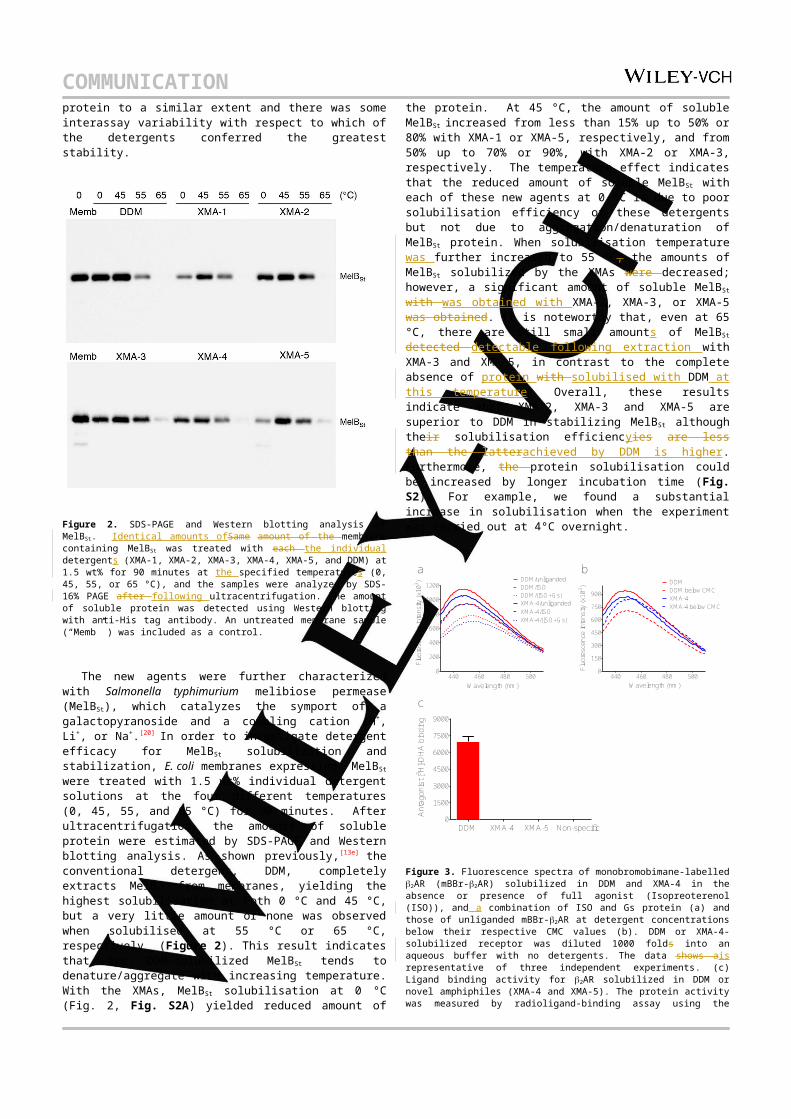
COMMUNICATION XMA-2, XMA-3 and XMA-5 are superior to DDM in stabilizing MelBSt although their solubilisation efficiencyies are less than the latterachieved by DDM is higher. Furthermore, the protein solubilisation could be increased by longer incubation time (Fig. S2). For example, we found a substantial increase in solubilisation when the experiment was carried out at 4°C overnight.
Figure 3. Fluorescence spectra of monobromobimane-labelled 2AR (mBBr-2AR) solubilized in DDM and XMA-4 in the absence or presence of full agonist (Isopreoterenol (ISO)), and a combination of ISO and Gs protein (a) and those of unliganded mBBr-2AR at detergent concentrations below their respective CMC values (b). DDM or XMA-4-solubilized receptor was diluted 1000 folds into an aqueous buffer with no detergents. The data shows ais representative of three independent experiments. (c) Ligand binding activity for 2AR solubilized in DDM or novel amphiphiles (XMA-4 and XMA-5). The protein activity was measured by radioligand-binding assay using the antagonist [3H]-dihydroalprenolol (DHA). Detergents were used at CMC + 0.04 wt% for these evaluations.
These promising results of the novel agents for membrane protein stabilization prompted us to test these agents with human 2 adrenergic receptor (2AR), a G-protein coupled receptor (GPCR).[21] In order to explore the effect of the novel agents on the conformational change of 2AR, we utilized measured fluorescence measurement in whichchanges in a bimane fluorophore can sense theassociated with alterations in receptor conformational changes of the receptor upon ligand and G protein binding[22]. The ; a bimane moiety is covalently attached to cysteine 265 located at the cytoplasmic end of transmembrane helix 6 (TM6). Thus, receptor conformation as well as conformational changes associated with inactive and active states of the receptor could be precisely detected by the changes of fluorescence emission spectrum of the monobromobimane–labelled 2AR (mBBr-2AR).[23] For this experiment, DDM-purified mBBr-2AR was diluted into individual detergent solutions at a concentration of CMC+0.04 wt%, and the bimane fluorescence spectra were taken in the absence or presence of a high affinity agonist, BI-167107, at the detergent concentration of CMC+0.04 wt% (Fig. S3). Among Of the five new agents, XMA-4 and XMA-5 resulted in bimane spectra similar to that of DDM, indicating effective preservation of the receptor activity by these two XMAs. Binding of a full agonist
(e.g., BI) to the receptor is known to be insufficient to fully activate the receptor, which further requires G protein binding.[14a]
A similar result was found in this study for mBBr-2AR in the presence of the full agonist, isoproterenol (ISO), although slight differences in the bimane spectra between obtained for DDM and XMA-4 or XMA-5 solubilised protein could bewas observed (Fig. 3a & S4). The ability of 2AR solubilized in XMA-4 or XMA-5 to properly activate Gs protein was characterized via G-protein coupling assay.[14a] As can be seen in Fig. 3a, the bimane spectra of XMA-4-solubilized receptor/G-protein complexes are similar to that of the DDM-solubilized complex. A similar trend was observed by using XMA-5-solubilized receptor (Fig. S4). These results indicate that these two agents behave well for receptor activation by G-protein coupling. The rReduction in fluorescence intensity and the shift of in maximal emission wavelength observed here is ascribed to conformational changes of 2AR associated with the from transition from the inactive to active state caused by the binding of both ISO and G-protein.[14a,n] Detergent efficacy (XMAs vs. DDM) was further compared by diluting these agents far below their respective CMC values. As shown in Fig. 3b, DDM-solubilized 2AR underwent an obvious conformational change by this dilution while XMA-4 and XMA-5-solubilized receptors underwent only minor changes, suggesting a slow off-rate for these new agents from the receptor compared to DDM (Fig. S5). These intriguing results brought us to carry out the ligand binding assay for 2AR after detergent exchange. The receptor activity purified in DDM and XMAs were assessed by the radio-ligand of [3H]-dihydroalprenolol ([3H]-DHA). Both Protein in both XMA-4 and XMA-5 showed a similar level of binding to the exhibited the receptor activity in a comparable level of that displayed by DDMradiolabel to protein in DDM, indicating that these two XMAs could be useful alternatives to DDM, the best conventional detergent for 2AR study.
Next, we moved to the leucine transporter (LeuT) from Aquifex aeolicus,[24] for the evaluation of the novel agents. Protein activity in aqueous solutions supplemented with individual XMAs and DDM was measured by a scintillation proximity assay (SPA) at the regular intervals over 12-day of incubation period at room temperature.[25] At a detergent concentration of CMC+0.04 wt%, all XMAs were inferior to DDM (Fig. S6a). When we increased detergent concentration to CMC+0.2 wt%, only XMA-4 and XMA-5 showed comparable efficacy to DDM in the latter part of the incubation period (i.e., from 5 day to 12 day), indicating that most XMAs have limited stabilizing effect on this particular membrane protein as compared to DDM (Fig. S6b).
Detergent efficacy is known to be membrane protein-specific. When we evaluated XMAs for four membrane proteins systems and compared with DDM, the most common conventional detergent for membrane protein study,[26] in terms of membrane protein stabilization efficacy, some new agents appeared to be superior to DDM for Bor1 and MelBSt while those agents were comparable to DDM for 2AR and inferior to DDM for LeuT. Of the XMAs, XMA-4 and XMA-5 showed generally favourable behaviour for protein stability; even for LeuT, these agents are the most comparable to DDM. We believe that these agents with C11 and C12 alkyl chains, respectively, are the most hydrophobic of XMAs and thus have optimal hydrophile-lipophile balance (HLB). In contrast, XMA-1 with the shortest alkyl chain is likely to be much more hydrophilic, explaining why in most cases this produces the least stable protein. Detergent HLB number has been shown to play a critical role in detergent
440 460 480 5000
200
400
600
800
1000
1200DDM/unligandedDDM/ISODDM/(ISO+Gs)
Wavelength (nm)
Fluo
resc
ence
inte
nsity
(x10
3 )
XMA-4/unligandedXMA-4/ISOXMA-4/(ISO+Gs)
440 460 480 5000
150
300
450
600
750
900XMA-4XMA-4 below CMC
Wavelength (nm)
DDMDDM below CMC
Fluo
resc
ence
inte
nsity
(x10
3 )
0
1500
3000
4500
6000
7500
9000
XMA-4 XMA-5DDM Non-specific
Ant
agon
ist [
3 H]-D
HA
bin
ding
a b
c

COMMUNICATION behaviour.[27] Of note, XMAs have a distinct architecture from conventional detergents but can be prepared in just five high yield synthetic steps, making those agents highly accessible. In addition some XMAs, particularly XMA-4 and XMA-5, conferred greater stability to a range of membrane protein, suggesting that these agents could be good alternatives to conventional detergents for membrane protein study.
Many important questions need to be addressed in the next study. For example, it would be valuable to determine micellar aggregation numbers formed by XMAs and how these numbers are affected by the chain length of the novel agents. More importantly, XMAs displayed favourable behaviours for protein stabilization efficacy, but these agents were suboptimal for LeuT. It is impossible to know a precise reason for this limited behaviour, but it may be related to the long distance between the two alkyl chains of XMAs. Because of this structural feature detergent packing around a target membrane protein may not be tight enough, leading to a decrease in both the detergent hydrophobic density and detergent affinity for the membrane protein. Currently we are developing new amphiphiles with increased hydrophobic density to address this topic. However, we believe that, based on the first characterization shown here, some XMAs will be useful tools for membrane protein study.
Experimental Section
Synthesis and characterization of novel amphiphiles, and membrane protein stability assays: Experimental details can be found in the Supporting Information.
Acknowledgements
This work was supported by the National Research Foundation of Korea (NRF) funded by the Korean government (MSIP) (grant number 2013R1A2A2A03067623 to P.S.C. and K.H.C). The work was also supported by Biotechnology and Biological Sciences Research Council grant BB/K017292/1 to B.B. NJS is in receipt of a BBSRC Doctoral Training Programme studentship awarded to BB. This work was supported by the National Science Foundation (grant MCB-1158085 to L.G.) and by the National Institutes of Health (grant R01 GM095538 to L.G.).
Keywords: amphiphile design • novel detergents • membrane protein • protein stabilisation • protein structure
[1] a) C. R. Sanders, J. K. Myers, Annu. Rev. Biophys. Biomol. Struct. 2004, 33, 25–51; b) J. P. Overington, B. Al-Lazikani, A. L. Hopkins, Nat. Rev. Drug Discovery 2006, 5, 993–996.
[2] a) H.-J. Nam, J. Jouhyun, K. Sanguk, BMB Rep. 2009, 42, 697–704; b) J. R. Deschamps, AAPS J. 2005, 7, 813–819; C. W. Murray, T. L. Blundell, Curr. Opin. Struct. Biol. 2010, 20, 497–507; d) T. L. Blundell, H. Jhoti, C. Abell, Nat. Rev. Drug Discovery 2002, 1, 45–54.
[3] http://blanco.biomol.uci.edu/Membrane_Proteins_xtal.html [4] a) S. H. White, W. C. Wimley, Annu. Rev. Biophys. Biomol. Struct.
1999, 28, 319–365; b) J. U. Bowie, Curr. Opin. Struct. Biol. 2001, 11, 397–402; c) J. J. Lacapere, E. Pebay-Peyroula, J. M. Neumann, C. Etchebest, Trends Biochem. Sci. 2007, 32, 259–270.
[5] a) G. G. Privé, Methods 2007, 41, 388–397; b) P. S. Chae, P. D. Laible, S. H. Gellman, Mol. BioSyst. 2010, 6, 89–94.
[6] a) M. J. Serrano-Vega, F. Magnani, Y. Shibata, C. G. Tate, Proc. Natl. Acad. Sci. U. S. A. 2008, 105, 877–882; b) S. Newstead, S. Ferrandon, S. Iwata, Protein Sci. 2008, 17, 466–472; c) Y. He, K.
Wang, N. Yan, Protein Cell 2014, 5, 658–672.[7] Q. Zhang, H. Tao, W.-X. Hong, Methods 2011, 55, 318–323; b) H.
J. Kang, C. Lee, D. Drew, Int. J. Biochem. Cell. Biol. 2013, 45, 636–644; c) I. Moraes, G. Evans, J. Sanchez-Weatherby, S. Newstead, P. D. S. Stewart, Biochem. Biophys. Acta 2014, 1838, 78–87.
[8] a) D. Marsh, Biochem. Biophys. Acta 2008, 1778, 1545–1575; b) J. L Parker, S. Newstead, Prot. Sci. 2012, 21, 1358–1365
[9] a) S. Newstead, S. Ferrandon, S. Iwata, Protein Sci. 2008, 17, 466–472; b) S. Newstead, J. Hobbs, D. Jordan, E.P. Carpenter, S. Iwata, Mol. Membr. Biol. 2008, 25, 631–638.
[10] a) P. S. Chae, M. J. Wander, K. H. Cho, P. D. Laible, S. H. Gellman, Mol. BioSyst. 2013, 9, 626–629; b) P. S. Chae, A. Sadaf, S. H. Gellman, Chem. Asian J. 2014, 9, 110–116.
[11] a) C.-L. McGregor, L. Chen, N. C. Pomroy, P. Hwang, S. Go, A. Chakrabartty, G. G. Privé, Nat. Biotechnol. 2003, 21, 171–176; b) X. Zhao, Y. Nagai, P. J. Reeves, P. Kiley, H. G. Khorana, S. Zhang, Proc. Natl. Acad. Sci. U. S. A. 2006, 103, 17707–17712; c) H. Tao, S. C. Lee, A. Moeller, R. S. Roy, F. Y. Siu, J. Zimmermann, R. C. Stevens, C. S. Potter, B. Carragher, Q. Zhang, Nat. Methods 2013, 10, 759–761.
[12] a) C. Tribet, R. Audebert, J.-L. Popot, Proc. Natl. Acad. Sci. U. S. A. 1996, 93, 15047–15050; b) J.-L. Popot, et al., Annu. Rev. Biophys. 2011, 40, 379–408; c) A. Nath, W. M. Atkins, S. G. Sligar, Biochemistry 2007, 46, 2059–-2069; d) M. Orwick-Rydmark, J.E. Lovett, A. Graziadei, L. Lindholm, M.R. Hicks, A. Watts, Nano Lett. 2012, 12, 4687–4692.
[13] a) S. C. Lee, B. C. Bennett, W.-X. Hong, Y. Fu, K. A. Baker, J. Marcoux, C. V. Robinson, A. B. Ward, J. R. Halpert, R. C. Stevens, C. D. Stout, M. J. Yeager, Q. Zhang, Proc. Natl. Acad. Sci. U. S. A. 2013, 110, E1203–1211; b) P. S. Chae, K. Gotfryd, J. Pacyna, L. J. W. Miercke, S. G. F. Rasmussen, R. A. Robbins, R. R. Rana, C. J. Loland, B. Kobilka, R. Stroud, B. Byrne, U. Gether, S. H. Gellman, J. Am. Chem. Soc. 2010, 132, 16750–16752; c) P. S. Chae, R. R. Rana, K. Gotfryd, S. G. F. Rasmussen, A. C. Kruse, K. H. Cho, S. Capaldi, E. Carlsson, B. K. Kobilka, C. J. Loland, U. Gether, S. Banerjee, B. Byrne, J. K. Lee, S. H. Gellman, Chem. Commun. 2013, 49, 2287–2289; d) K. H. Cho, H. E. Bae, M. Das, S. H. Gellman, P. S. Chae, Chem. Asian J. 2014, 9, 632–638; e) P. S. Chae, S. G. F. Rasmussen, R. R. Rana, K. Gotfryd, R. Chandra, M. A. Goren, A. C. Kruse, S. Nurva, C. J. Loland, Y. Pierre, D. Drew, J.-L. Popot, D. Picot, B. G. Fox, L. Guan, U. Gether, B. Byrne, B. Kobilka, S. H. Gellman, Nat. Methods 2010, 7, 1003–1008; f) K. H. Cho, B. Byrne, P. S. Chae, ChemBioChem 2013, 14, 452–455.
[14] a) S. G. F. Rasmussen, H.-J. Choi, J. J. Fung, E. Pardon, P. Casarosa, P. S. Chae, B. T. DeVree, D. M. Rosenbaum, F. S. Thian, T. S. kobilka, A. Schnapp, I. Konetzki, R. K. Sunahara, S. H. Gellman, A. Pautsch, J. Steyaert, W. I. Weis, B. K. Kobilka, Nature 2011, 469, 175–180; b) D. M. Rosenbaum, C. Zhang, J. Lyons, R. Holl, D. Aragao, D. H. Arlow, S. G. F. Rasmussen, H.-J. Choi, B. T. DeVree, R. K. Sunahara, P. S. Chae, S. H. Gellman, R. O. Dror, D. E. Shaw, W. I. Weis, M. Caffrey, P. Gmeiner, B. K. Kobilka, Nature 2011, 469, 236–240; c) A. C. Kruse, J. Hu, A. C. Pan, D. H. Arlow, D. M. Rosenbaum, E. Rosemond, H. F. Green, T. Liu, P. S. Chae, R. O. Dror, D. E. Shaw, W. I. Weis, J. Wess, B. K. Kobilka, Nature 2012, 482, 552–556; d) K. Haga, A. C. Kruse, H. Asada, T. Y. Kobayashi, M. Shiroishi, C. Zhang, W. I. Weis, T. Okada, B. K. Kobilka, T. Haga, T. Kobayashi, Nature 2012, 482, 547–551; e) A. Manglik, A. C. Kruse, T. S. Kobilka, F. S. Thian, J. M. Mathiesen, R. K. Sunahara, L. Pardo, W. I. Weis, B. K. Kobilka, S. Granier, Nature 2012, 485, 321–326; f) S. Granier, A. Manglik, A. C. Kruse, T. S. Kobilka, F. S. Thian, W. I. Weis, B. K. Kobilka, Nature 2012, 485, 400-404; g) J. F. White, N. Noinaj, Y. Shibata, J. Love, B. Kloss, F. Xu, J. Gvozdenovic-Jeremic, P. Shah, J. Shiloach, C. G. Tate, R. Grisshammer, Nature 2012, 490, 508–513; h) A. C. Kruse, A. M. Ring, A. Manglik, J. Hu, K. Hu, K. Eitel, H. Hubner, E. Pardon, C. Valant, P. M. Sexton, A. Christopoulos, C. C. Felder, P. Gmeiner, J. Steyaert, W. I. Weis, K. C. Garcia, J. Wess, B. K. Kobilka, Nature 2013, 504, 101–106; i) A. M. Ring, A. Manglik, A. C. Kruse, M. D. Enos, W. I. Weis, K. C. Garcia, B. K. Kobilka, Nature 2013, 502, 575–-579; j) P. S. Miller, A. R. Aricescu, Nature 2014, 512, 270–275; k) S. E. Rollauer, M. J. Tarry, J. E. Graham, M. Jaaskelainen,

COMMUNICATION F. Jager, S. Johnson, M, Krehenbrink, S. M. Liu, M. J. Lukey, J. Marcoux, M. A. McDowell, F. Rodriguez, P. Roversi, P. J. Stansfeld, C. V. Robinson, M. S. Sansom, T. Palmer, M. Hcgbom, B. C. Berks, S. M. Lea, Nature 2012, 492, 210–214; l) E. Karakas, H. Furukawa, Science 2014, 344, 992-997; m) H. Suzuki, T. Nishizawa, K. Tani, Y. Yamazaki, A. Tamura, R. Ishitani, N. Dohmae, S. Tsukita, O. Nureki, Y. Fujiyoshi, Science 2014, 344, 304-307; n) V. K. Dickson, L. Pedi, S. B. Long, Nature 2014, 516, 213-218; o) S. G. F. Rasmussen, B. T. DeVree, Y. Zou, A. C. Kruse, K. Y. Chung, T. S. Kobilka, F. S. Thian, P. S. Chae, E. Pardon, D. Calinski, J. M. Mathiesen, S. T. A. Shah, J. A. Lyons, M. Caffrey, S. H. Gellman, J. Steyaert, G. Skiniotis, W. I. Weis, R. K. Sunahara, B. K. Kobilka, Nature 2011, 477, 549–555; p) A. K. Shukla, et al., Nature 2014, 512, 218-222; q) J. Kellosalo, T. Kajander, K. Kogan, K. Pokharel, A. Goldman, Science 2012, 337, 473–476; r) A. Quigley, Y. Y. Dong, A. C. W. Pike, L. Dong, L. Shrestha, G. Berridge, P. J. Stansfeld, M. S. P. Sansom, A. N. Edwards, C. Bountra, F. Von Delft, A. N. Bullock, N. A. Burgess-Brown, E. P. Carpenter, Science 2013, 339 1604–1607; s) A. Frick, U. K. Eriksson, F. de Mattia, F. Oberg, K. Hedfalk, R. Neutze, W. J. Grip, P. M. T. Deen, S. Tornroth-Horsefield, Proc. Natl. Acad. Sci. U. S. A. 2014, 111, 6305–6310.
[15] a) D. T. McQuade, M. A. Quinn, S. M. Yu, A. S. Polans, M. P. Krebs, S. H. Gellman, Angew. Chem. Int. Ed. 2000, 39, 758–761; b) P. S. Chae, M. J. Wander, A. P. Bowling, P. D. Laible, S. H. Gellman, ChemBioChem 2008, 9, 1706–1709; c) P. S. Chae, K. H. Cho, M. J. Wander, H. E. Bae, S. H. Gellman, P. D. Labile, Biochim. Biophys. Acta 2014, 1838, 278–286; d) S. C. Howell, R. Mittal, L. Huang, B. Travis, R. M. Breyer, C. R. Sanders, Biochemistry 2010, 49, 9572–9583; e) P. S. Chae, S. G. F. Rasmussen, R. R. Rana, K. Gotfryd, A. C. Kruse, S. Nurva, U. Gether, L. Guan, C. J. Loland, B. Byrne, B. K. Kobilka, S. H. Gellman, Chem. Eur. J. 2012, 18, 9485–9490; f) P. S. Chae, H. E. Bae, M. Das, Chem Commun. 2014, 50, 12300–12303.
[16] A. Chattopadhyay, E. London, Anal. Biochem. 1984, 139, 408–412.[17] A. Hawe, W. L. Hulse, W. Jiskoot, R. T. Forbes, Pharm. Res. 2011,
28, 2302-2310.[18] J. Takano, K. Noguchi, M. Yasumori, M. Kobayashi, Z. Gajdos, K.
Miwa, H. Hayashi, T. Yoneyama, T. Fujiwara, Nature, 2002, 420, 337-340.
[19] A. Alexandrov, M. Mileni, E. Y. Chien, M. A. Hanson, R. C. Stevens, Structure 2008, 16, 351–359.
[20] a) L. Guan, S. Nurva, S. P. Ankeshwarapu, J. Biol. Chem. 2011, 286, 6367–6374; b) A. S. Ethayathulla, M. S. Yousef, A. Amin, G. Leblanc, H. R. Kaback, L. Guan, Nat. Commun. 2014;5:3009. doi: 10.1038/ncomms4009. c) A. Amin, A. S. Ethayathulla, L. Guan, J Bacterol. 2014, 196: 3134-3139.
[21] D. M. Rosenbaum, V. Cherezov, M. A. Hanson, S. G. Rasmussen, F. S. Thian, T. S. Kobilka, H. J. Choi, X. J. Yao, W. I. Weis, R. C. Stevens, B. K. Bobilka, Science 2007, 318, 1266–1273.
[22] S. E. Mansoor, H. S. McHaourab, D. L., Farrens, Biochemistry 2002, 41, 2475–2484.
[23] X. Yao, C. Parnot, X. Deupi, V. R. P. Ratnala, G. Swaminath, D. Farrens, Nat. Chem. Biol. 2006, 2, 417–422.
[24] G. Deckert, P. V. Warren, T. Gaasterland, W. G. Young, A. L. Lenox, D. E. Graham, R. Overbeek, M. A. Snead, M. Keller, M. Aujay, R. Huber, R. A. Feldman, J. M. Short, G. J. Olsen, R. V. Swanson, Nature 1998, 392, 353–358.
[25] a) H.E. Hart, E. B. Greenwald, Mol. Immunol. 1979, 16, 265–267; b) M. Quick, J. A. Javitch, Proc. Natl. Acad. Sci. USA 2007, 104, 3603–3608.
[26] a) Y. Sonoda, S. Newstead, N. J. Hu, Y. Alguel, E. Nji, K. Beis, S. Yashiro, C. Lee, J. Leung, A. D. Cameron, B. Byrne, S. Iwata, D. Drew, Structure 2011, 19, 17–25; b) M. Caffrey, D. Li, A. Dukkipati, Biochemistry 2012, 51, 6266–6288.
[27] P. S. Chae, A. C. Kruse, K. Gotfryd, R. R. Rana, K. H. Cho, S. G. F. Rasmussen, H. E. Bae, R. Chandra, U. Gether, L. Guan, B. K. Kobilka, C. J. Loland, B. Byrne, S. H. Gellman, Chem. Eur. J. 2013, 19, 15645–15651;

COMMUNICATION
COMMUNICATION
Structurally unique amphiphiles with a xylene linker were prepared via a convenient synthetic protocol. Some of these agents (e.g., XMA-5) conferred enhanced membrane protein stability relative to a conventional detergent (DDM), indicating the potential utility in membrane protein structure study.
Kyung Ho Cho, Yang Du, Nicola J Scull, Parameswaran Hariharan, Kamil Gotfryd, Claus J Lolland, Lan Guan, Bernadette Byrne, Brian K Kobilka, and and Pil Seok Chae*
Page No. – Page No.
Novel Xylene-linked Maltoside Amphiphiles (XMAs) for Membrane Protein Stabilisation
O
O
O
O
OOHO
OH
OHOHO
HO OH
OH
O
OHO OH
HOO
HOHO OH
HO
OOHO
OH
OHO
HOHO OH
HO
O
OHO
OH
HOO
HOHO
OH
HO
XMA-5


