Embed Size (px)
Citation preview

Provided by the author(s) and NUI Galway in accordance with publisher policies. Please cite the published
version when available.
Downloaded 2022-04-01T20:50:35Z
Some rights reserved. For more information, please see the item record link above.
Title The molecular characterization of ER stress-induced autophagyand cell death
Author(s) Deegan, Shane
PublicationDate 2012-12-21
Item record http://hdl.handle.net/10379/3695

The molecular characterization of ER
stress-induced autophagy and cell death
A thesis submitted to the National University of Ireland Galway in
fulfillment of the requirement for the degree of
Doctor of Philosophy
By
Shane Deegan
Discipline of Biochemistry, School of Natural Sciences,
National University of Ireland, Galway
Thesis Supervisors: Professor Afshin Samali
Head of School: Dr Heinz-Peter Nasheuer
November 2012

1
Table of Contents
Table of Contents ............................................................................................................. 1
Declaration ....................................................................................................................... 5
Acknowledgements .......................................................................................................... 6
Publications ...................................................................................................................... 9
Published ..................................................................................................................... 10
In preparation .............................................................................................................. 10
Abstract ........................................................................................................................... 11
Abbreviations ................................................................................................................. 14
Chapter 1: Introduction ................................................................................................... 20
1.1 Endoplasmic Reticulum ........................................................................................ 21
1.2 Endoplasmic Reticulum Stress ............................................................................. 21
1.3 Unfolded Protein Response Signaling .................................................................. 22
1.3.1 PERK .............................................................................................................. 22
1.3.2 ATF6 .............................................................................................................. 23
1.3.3 IRE1 ............................................................................................................... 24
1.4 Autophagy ............................................................................................................. 26
1.4.1 Autophagy Induction ...................................................................................... 27
1.4.2 Vesicle Nucleation ......................................................................................... 28
1.4.3 Origin of the phagophore ............................................................................... 29
1.4.4 Autophagosome Elongation ........................................................................... 30
1.4.5 Maturation of the Autophagosome ................................................................. 30
1.4.6 Selective Autophagy ...................................................................................... 31
1.5 Autophagy regulation by ER stress ...................................................................... 34
1.5.1 Autophagy Induction ...................................................................................... 34
1.5.2 Vesicle Nucleation ......................................................................................... 36
1.5.3 Elongation of the phagophore ........................................................................ 37
1.5.4 Negative Regulation of Autophagy by the UPR ............................................ 38
1.6 ER stress and Cell Death ...................................................................................... 41

2
1.7 Apoptosis .............................................................................................................. 42
1.7.1 Inhibitor of Apoptosis Proteins (IAPs) .......................................................... 42
1.7.2 Extrinsic Apoptotic Pathway .......................................................................... 44
1.7.3 Intrinsic Apoptotic Pathway ........................................................................... 46
1.7.4 Bcl-2 Family ................................................................................................... 46
1.7.5 Apoptosome ................................................................................................... 47
1.7.6 PIDDosome .................................................................................................... 47
1.7.7 Ripoptosome and other RIP1-containing complexes ..................................... 49
1.8 Necroptosis/Necrosis ............................................................................................ 50
1.9 Autophagic cell death ........................................................................................... 50
1.10 Aims and Objectives ........................................................................................... 52
Chapter 2: Materials and Methods ................................................................................. 53
2.1 Cell culture and treatments ................................................................................... 54
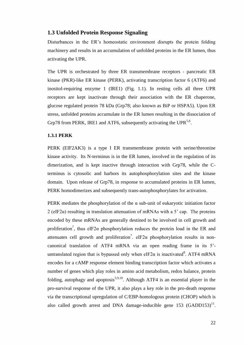
2.2 Plasmids ................................................................................................................ 55
2.3 Transfections ......................................................................................................... 57
2.4 Lentivirus Production ........................................................................................... 58
2.5 Lentivirus Transduction ........................................................................................ 58
2.6 Microscopy ........................................................................................................... 59
2.6.1 Fluorescent Confocal Microscopy: ................................................................ 59
2.6.2 Bright Field Microscopy: ............................................................................... 59
2.6.3 Transmission Electron Microscopy ................................................................ 60
2.7 RNA Extraction .................................................................................................... 60
2.8 cDNA synthesis .................................................................................................... 61
2.9 Conventional Polymerase Chain Reaction (PCR) ................................................ 61
2.10 Real Time PCR ................................................................................................... 62
2.11 Protein sample preparation and Sodium dodecyl sulphate-polyacrylamide gel
electrophoresis (SDS-PAGE) ..................................................................................... 63
2.12 Western Blotting ................................................................................................. 65
2.13 Immunoprecipitation ........................................................................................... 65
2.14 Enzyme-Linked Immunosorbent Assay (ELISA) ............................................... 66
2.15 Clonogenic Survival Assay ................................................................................. 69

3
2.16 Annexin V and Propidium Iodide Staining ......................................................... 69
2.17 Analysis of DEVDase Activity ........................................................................... 70
2.18 Statistical Analysis .............................................................................................. 70
Chapter 3: Unmasking of a Novel Death Pathway ......................................................... 71
3.1 Introduction and Objectives .................................................................................. 72
3.2 Results ................................................................................................................... 75
3.2.1 Mitochondrial-mediated apoptosis (MMA) compromised cells exhibit a
prolonged UPR ........................................................................................................ 75
3.2.2 MMA compromised cells undergo a delayed mode of cell death in response
to an array of cellular stresses ................................................................................. 79
3.2.3 MMA compromised cells display apoptosome-independent activation of
effector caspases in response to an array of cellular stresses .................................. 84
3.2.4 Processing of caspase-3 and PARP cleavage is Initiator caspase mediated .. 90
3.2.5 Inhibition of the intrinsic pathway unmasks a delayed caspase-8 mediated
apoptotic program in response to cell stress ........................................................... 91
3.2.6 Caspase-8 requires a RIP1-complex for its activation ................................... 94
3.2.7 Caspase-8 is activated by a RIP1-containing complex, caspase-8 knockdown
switches cell death to necroptosis ........................................................................... 96
3.2.8 C9-/-
MEFs mode of cell death is depicted by its cellular content ............... 101
3.2.9 TNF signaling is not involved in the formation of the RIP1-containing
complex ................................................................................................................. 106
3.2.10 Assessing the role of the adaptor proteins TRADD and FADD in assembly
of the caspase-8 activating complex ..................................................................... 111
3.2.11 The autophagy gene ATG5 is required for caspase-8 activation in MMA
compromised cells, however protects WT cells from ER stress induced cell death
............................................................................................................................... 115
3.2.12 Immunoprecipitation of ATG5 in C9-/-
MEFs co-precipitates with caspase-8,
RIP1 and FADD .................................................................................................... 124
3.3 Discussion ........................................................................................................... 126
Chapter 4: Transcriptional Regulation of the Autophagy Receptors by the UPR ........ 136
4.1 Introduction and Objectives ................................................................................ 137
4.2 Results ................................................................................................................. 138
4.2.1 Activation of the UPR in HCT116 cells results in the upregulation of
autophagy .............................................................................................................. 138

4
4.2.2 ER stress induces autophagy-mediated degradation of mitochondria and
ubiquitinated proteins ............................................................................................ 140
4.2.3 Microarray analysis of HCT116 cells reveals an array of autophagy-related
genes transcriptionally upregulated in response to ER stress ............................... 144
4.2.4 ER stress results in the transcriptional upregulation of the autophagy receptor
genes ...................................................................................................................... 147
4.2.5 The PERK arm of the UPR is required for the transcriptional upregulation of
the autophagy receptor genes ................................................................................ 151
4.3 Discussion ........................................................................................................... 153
Concluding Remarks and Future Prospects .................................................................. 156
References .................................................................................................................... 161

5
Declaration
This thesis is a presentation of my original research work. The Electron Microscopy
images presented in this work was carried out by Dr. David MacDonald. Wherever
contributions of others are involved, every effort is made to indicate this clearly,
with due reference to the literature, and acknowledgement of collaborative research
and discussions.
The work was done under the guidance of Professor Afshin Samali, at the National
University of Ireland Galway.

6
Acknowledgements

7
There are many people I would like to thank who have helped me over the last 4
years of my Ph.D, without their help and support this thesis would not have been
possible.
I would first of all like to express my deepest gratitude to my supervisor, Prof.
Afshin Samali, for his guidance, support and encouragement over the last 4 years.
You have helped open the door to many opportunities for me in my career and you
have always looked out for my best interests, I sincerely thank you for everything
you have done.
I would like to thank Dr. Sanjeev Gupta for his support and knowledge which
helped guide my project.
I would like to express many thanks to everyone in my lab; Karen Cawley, Donna
Kennedy, Svetlana Saveljeva, Patricia Cleary and Susan Logue. Thank you guys for
all your help and support both scientifically and personally.
Thank you Susan for your help with experiments over the last few months it really
helped me out.
A special thanks to Svetlana Saveljeva who has worked so hard over the last few
months to help me finish my Ph.D, it would not have been possible with out your
help, thank you.
I would like to thank Dr. Adrienne Gorman and Dr. Eva Szegezdi their time help
and expertise.
I would like to thank Dr. David MacDonald for carrying out the Electron
Microscopy presented in this thesis.
I want to thank Tak Mak for providing me with Caspase-9+/+
and Caspase-9-/-
mouse
embryonic fibroblasts (MEFs), Craig Thompson for providing Bax/Bak+/+
and
Bax/Bak-/-
MEFs, Bert Volgestein for providing Bax-/-
HCT116 cells and Alan Diehl
for providing MDA-MB-468 PERK shRNA and PLKO stable cell lines.
Thank you to Genetech for providing the BV-6 Smac-mimetic.

8
I would like to thank Professor Peter Vandenabeele for his collaboration in this
project and for welcoming me to his lab for 3 months. I would also like to thank him
for providing me with caspase-8 shRNA, TRADD shRNA, FADD shRNA, TNFR1
shRNA, RIP1 miRNA and RIP3 miRNA expression plasmids.
Not forgetting my boys who have helped me keep my manhood over the years of
working in a lab full of girls, especially Chris who has kept me on track when things
didn’t work, a few drinks solves everything. Not forgetting my non-science friends,
Liam, thanks for all the IT support over the years, you saved me a bomb.
Also, thank you to everyone in the apoptosis reseach group, I have made many
friends which has made the last 4 years a lot of fun.
None of this would be possible without the love and support of my amazing family;
Mam, Dad, Nana, Grandad, Louise, Dave and Laura. Thank you sooooo much Mam
(Caroline) and Dad (Aiden) for everything you have done to help me get to where I
am today, you are amazing parents and you have instilled your best attributes into
who I am today, I will never be able to thank you enough for everthing you have
done for me. Thank you Nana (Mary) and Grandad (Tim), for all your love and
support, you are the best grandparents anyone could ask for, you have always looked
after us like your own children and I will be forever gratefull.
Most of all I would like to thank my amazing and beautiful girlfriend and best
friend, Catriona. Thank you so much for all your love and support throughout this
Ph.D, you have had to put up with all my bad moods over the last 4 years, of which
their has been a few, but you were always able to get me out of them very quickly.
Without you to come home to at the end of the day would have made the last 4 years
very difficult, thank you sooo much for everything.

9
Publications

10
Published
Gupta, S., Z. Giricz, Natoni, A., Donnelly, N., Deegan, S., Samali, A. (2012).
"NOXA contributes to the sensitivity of PERK-deficient cells to ER stress." FEBS
Lett.
Deegan, S., S. Saveljeva, et al. (2012). "Stress-induced self-cannibalism: on the
regulation of autophagy by endoplasmic reticulum stress." Cell Mol Life Sci.
Cawley, K., S. Deegan, et al. (2011). Assays for Detecting the Unfolded Protein
Response. Methods in Enzymology. P. M. Conn, Academic Press. Volume 490: 31-
51.
Gupta, S., A. Deepti, S. Deegan, F. Lisbona, C. Hetz, A. Samali. (2010). "HSP72
protects cells from ER stress-induced apoptosis via enhancement of IRE1alpha-
XBP1 signaling through a physical interaction." PLoS Biol 8(7): e1000410.
Samali, A., U. Fitzgerald, S. Deegan, S. Gupta. (2010). "Methods for monitoring
endoplasmic reticulum stress and the unfolded protein response." Int J Cell Biol
2010: 830307.
In preparation
Deegan, S. et al. (2012). "Unmasking of a novel death pathway." Mol Cell.

11
Abstract

12
Autophagy is tightly regulated by the unfolded protein response (UPR). It plays an
important role in the removal of unfolded proteins, damaged mitochondria and
expanded endoplasmic reticulum (ER), to help relieve ER stress and reinstate
homeostasis. However, when persistent, ER stress responses can switch the
cytoprotective functions of UPR and autophagy into cell death promoting
mechanisms. Depending on the cellular context, autophagy can either serve as a cell
survival pathway, suppressing apoptosis, or it can lead to death itself. Although most
cells primarily use apoptosis as a mode of cell death it is often not an option in many
types of cancers and thus it is important to explore how the cell regulates other
forms of cell death. The objective of this project was to determine the mode of ER
stress-induced cell death in cells where the mitochondrial apoptotic pathway is
compromised, and investigate how autophagy influences cell death in these
conditions. For this we used caspase-9-/-
mouse embryonic fibroblasts (MEFs),
Bax/Bak-/-
MEFs and Bax-/-
HCT116 colon carcinoma cell line. Here we show that
ER stress induces two phases of cell death, the first of which is rapid, caspase-9-
dependent apoptosis and the second is a slow caspase-9-independent cell death.
Further we show that the caspase-9-independent cell death is executed through a
caspase-8 mediated activation of the executioner caspases. Inhibition of caspase-8
further delayed cell death however did not completely rescue the cells. Combination
of caspase-8 knockdown and addition of the RIP1 kinase inhibitor, necrostatin-1,
showed almost a complete rescue from cell death, further these cells were able to
proliferate following removal of the drug. Interestingly knockdown of ATG5 in
caspase-9-/-
MEFs inhibited the activation of the executioner caspases and reduced
the levels of cell death, implicating ATG5 as an essential component for caspase-8
activation in this model. To this end we carried out immunoprecipitation of
endogenous ATG5 and showed its interaction with caspase-8, RIP1 kinase and
FADD. For the first time we have identified a RIP1-containing death inducing
protein complex assembled in response ER stress which executes cell death in
conditions where the intrinsic pathway is compromised; furthermore we show that
the formation of this complex is dependent on the autophagy protein, ATG5.
In addition to this, we studied the interplay between the UPR and autophagy during
ER stress. We carried out a microarray analysis in HCT116 cells and identified the
transcriptional upregulation of an array of autophagy related genes. These genes
included the autophagy receptors NIX, NBR1 and p62 and were confirmed by real-

13
time RT-PCR. We further demonstrated that these genes were transcriptionaly
upregulated by the PERK arm of the UPR, instigating ER stress-induced autophagy
as a selective process.
The data presented in this thesis show that in conditions where the intrinsic
mitochondrial-mediated apoptotic pathway is compromised, exposure of cells to
multiple stress stimuli will execute death via alternative means. We show that this
caspase-9 independent cell death requires RIP1 kinase, FADD, ATG5 and caspase-
8. Further we show that depending on the cellular context, cell death can be
executed through apoptosis or necroptosis. Inhibiting components of the apoptotic
pathway or the necroptotic pathway independently does not completely inhibit cell
death; however, coperativly inhibiting these two processes results in a significant
resistance from cell death. In addition to these findings, a microarray analysis
showed that the autophagy process in reponse to ER stress is highly regulated by the
UPR. We focused our study on the autophagy receptor proteins and showed that
these proteins are transcriptionally upregulated in response to ER stress. Further we
show the PERK arm of the UPR is responsible for their transcriptional upregulation.

14
Abbreviations

15
3MA 3-Methyladenine
ACD Autophagic cell death
ADP Adenosine diphosphate
Alfy Autophagy-linked FYVE-domain protein
AMPK AMP-activated protein kinase
APAF1 Apoptotic protease activating factor 1
ARE AU-rich element
ASK1 Apoptosis signal-regulating kinase 1
ATF4 Activating transcription factor 4
ATF6 Activating transcription factor 6
ATG Autophagy-related Genes
ATP Adenosine triphosphate
Bak Bcl-2 homologous antagonist/killer
Bax Bcl-2–associated X protein
Bcl-2 B-cell lymphoma 2
Bfa Brefeldin A
BH3 Bcl-2 homology domain 3
BIF1 Bax interacting factor 1
BIR Baculovirus IAP repeat
BNIP3 BCL2/adenovirus E1B 19kD-interacting protein 3
BNIP3L BCL2/adenovirus E1B 19kD-interacting protein 3-like
Brtz Bortezomib
Ca2+
Calcium
CaMK Ca2+
/calmodulin-dependent protein kinases
CARD Caspase recruitment domains
cDNA Complementary DNA
cFLIP Cellular FLICE-like inhibitory protein
cFLIPL Cellular FLICE-like inhibitory protein long isoform
cFLIPs Cellular FLICE-like inhibitory protein short isoform

16
Chop C/EBP-homologous protein
COPII Coat protein 2
CQ Chloroquine
DAPK Death-Associated Protein kinase
DD Death domain
DED Death effector domain
DEPTOR DEP domain containing mTOR-interacting protein
DFCP1 Double FYVE domain-containing protein
DIABLO Direct IAP-Binding protein with Low PI
DISC Death inducing signalling complex
DMEM Dulbecco's Modified Eagle Medium
DMSO Dimethyl sulfoxide
DNA Deoxyribonucleic acid
dNTPs Deoxyribonucleotide triphosphate
DR Death receptor
DRAM Damage-regulated autophagy modulator
DTT Dithiothreitol
EDEM1 ER degradation enhancer, mannosidase alpha-like 1
eIF2 Eukaryotic initiation factor 2 alpha
ER Endoplasmic Reticulum
ERAD ER-associated degradation
ERO1 ER oxidoreductin 1 alpha
ERP72 Endoplasmic reticulum resident protein 72
ERSE ER stress response element
Etop Etoposide
FADD Fas-associated protein with death domain
FIP200 FAK family kinase-interacting protein of 200 kDa
FoxO1 Forkhead box protein O1
FYCO FYVE and coiled-coil domain containing

17
GABARAPS Gamma-aminobutyric acid receptor-associated protein
GADD153 Growth arrest and DNA damage-inducible gene 153
GCL Glutamate cysteine ligase
GRP78 Glucose regulated protein 78kDa
GST Glutathione S-transferase
HERP Homocysteine-inducible endoplasmic reticulum (ER) stress prote
HSPA5 Heat shock 70kDa protein 5
IAP Inhibitor of apoptosis
IB Immunoblotting
IBM IAP-binding motif
iDISC Intracellular death inducing signalling complex
IFN Interferon-gamma
IKK- Inhibitor of nuclear factor kappa-B kinase subunit gamma
IP Immunoprecipitation
IP3R Inositol trisphosphate receptor
IRE1 Inositol-requiring enzyme 1
JNK c-JUN NH2-terminal kinases
KEAP1 Kelch-like ECH-associated protein 1
LAMP Lysosomal-associated membrane protein
LC3 Microtubule-associated protein light chain 3
LIR LC3 interacting region
LUBAC Linear Ubiquitin Chain Assembly Complex
MAPK Mitogen-activated protein (MAP) kinases
MAPs Microtubule-associated proteins
MEFs Mouse embryonic fibroblasts
MMA Mitochondrial-mediated apoptosis
MOMP Mitochondrial outer-membrane permeabilization
mRNA Messenger Ribonucleic acid
mTOR mammalian target of rapamycin

18
NBR1 Neighbor of BRCA1 gene 1 protein
Nec-1 Necrostatin-1
NEMO NF-kappa-B essential modulator
NFkB Nuclear factor kappa-light-chain-enhancer of activated B cells
NLS Nuclear localization signal
NRF2 Nuclear factor erythroid 2-related factor 2
P58IPK 58-kilodalton inhibitor of protein kinase
PARP Poly (ADP-ribose) polymerase
PAS Pre-autophagosomal structure
PCR Polymerase chain reaction
PDI Protein disulphide isomerase
PE Phosphatidylethanolamine
PERK Pancreatic ER kinase (PKR)-like ER kinase
PI Propidium Iodide
PI3K Phosphatidylinositol 3-kinases
PI3P Phosphatidylinositol 3-phosphate
PIDD p53-induced protein with a death domain
PINK1 PTEN-induced putative kinase 1
PP2A Protein phosphatase 2A
PRAS40 Proline-rich Akt substrate, 40 kDa
RAIDD RIP-associated ICH-1/CED-3-homologous protein with a death
domain
Raptor Regulatory associated protein of mTOR
REDD1 Regulated in development and DNA damage responses 1
RING Really Interesting New Gene
RIP Receptor-Interacting Protein
RNA Ribonucleic acid
ROS Reactive oxygen species
S1P Site-1 protease
S2P Site-2 protease

19
SERCA Sarcoendoplasmic reticulum (SR) calcium transport ATPase
SIRT1 Sirtuin 1
SMAC Second Mitochondria-derived Activator of Caspases
SQSTM1 Sequestosome 1
Tg Thapsigargin
Tm Tunicamycin
TNF Tumor necrosis factor
TNFR1 TNF receptor 1
TRADD Tumor necrosis factor receptor (TNFR)1–associated death domain
protein
TRAF2 TNF receptor-associated factor 2
TRAIL The tumour necrosis factor (TNF) related apoptosis-inducing ligand
TRAILR The tumour necrosis factor (TNF) related apoptosis-inducing ligand
receptor
TRB3 Tribbles homolog 3
UBL Ubiquitin-like proteins
ULK Unc-51-like kinase
UPR Unfolded Protein Response
UPRE UPR element
UVRAG UV radiation resistance-associated gene protein
Vps34 Vacuolar protein sorting-associated protein 34
WIPI WD repeat domain phosphoinositide-interacting protein
XBP1 X-box binding protein 1
XBP1s X-box binding protein 1 spliced
XIAP X-linked inhibitor of apoptosis

20
Chapter 1: Introduction

21
1.1 Endoplasmic Reticulum
The endoplasmic reticulum (ER) is a very complex and elaborate cellular organelle.
It is composed of a single continuous phospholipid membrane that is comprised of
the outer nuclear envelope, flattened peripheral sheets with ribosomes (rough ER)
and a complex network of smooth tubules (smooth ER) that extend throughout the
cell. The ER has many different cellular functions which are accommodated by its
heterogeneous structures. While detoxification of drugs, fatty acid and steroid
biosynthesis and Ca2+
storage occurs in the smooth ER, most of the folding and
post-translational processing of membrane bound and secreted proteins takes place
in the rough ER. It contains an array of chaperone systems such as glycosidases,
Ca2+
-dependent chaperones and members of the protein disulphide isomerase (PDI)
family. These chaperones are responsible for the correct folding of proteins under
normal physiological conditions1. This process is highly sensitive and is dependent
on ER luminal factors such as Ca2+
concentration, redox homeostasis and oxygen
supply2. The processing of nascent proteins in the ER lumen requires an array of
chaperones and folding enzymes that depend on the ER’s rich oxidizing
environment and Ca2+
pools to function optimally. Physiological or pathological
conditions that disrupt this fine balanced, unique environment cripples the ER’s
protein folding machinery and results in a condition referred to as ER stress.
1.2 Endoplasmic Reticulum Stress
The cellular responses to ER stress are multifaceted and include the activation of a
set of signaling pathways termed the unfolded protein response (UPR), a catabolic
process termed autophagy, and cell death3. These processes are not mutually
exclusive, and there is significant cross-talk between these cellular stress responses.
The UPR’s primary aim is to sustain cell survival by attenuating protein synthesis
and restoring cellular homeostasis via the activation of a cascade of transcription
factors which regulate expression of genes encoding for chaperones, components of
the ER-associated degradation (ERAD) system and components of the autophagy
machinery4.
22
1.3 Unfolded Protein Response Signaling
Disturbances in the ER’s homeostatic environment disrupts the protein folding
machinery and results in an accumulation of unfolded proteins in the ER lumen, thus
activating the UPR.
The UPR is orchestrated by three ER transmembrane receptors - pancreatic ER
kinase (PKR)-like ER kinase (PERK), activating transcription factor 6 (ATF6) and
inositol-requiring enzyme 1 (IRE1) (Fig. 1.1). In resting cells all three UPR
receptors are kept inactivate through their association with the ER chaperone,
glucose regulated protein 78 kDa (Grp78; also known as BiP or HSPA5). Upon ER
stress, unfolded proteins accumulate in the ER lumen resulting in the dissociation of
Grp78 from PERK, IRE1 and ATF6, subsequently activating the UPR5,6
.
1.3.1 PERK
PERK (EIF2AK3) is a type I ER transmembrane protein with serine/threonine
kinase activity. Its N-terminus is in the ER lumen, involved in the regulation of its
dimerization, and is kept inactive through interaction with Grp78, while the C-
terminus is cytosolic and harbors its autophosphorylation sites and the kinase
domain. Upon release of Grp78, in response to accumulated proteins in ER lumen,
PERK homodimerizes and subsequently trans-autophosphorylates for activation.
PERK mediates the phosphorylation of the α sub-unit of eukaryotic initiation factor
2 (eIF2α) resulting in translation attenuation of mRNAs with a 5’ cap. The proteins
encoded by these mRNAs are generally destined to be involved in cell growth and
proliferation7, thus eIF2α phosphorylation reduces the protein load in the ER and
attenuates cell growth and proliferation7. eIF2α phosphorylation results in non-
canonical translation of ATF4 mRNA via an open reading frame in its 5’-
untranslated region that is bypassed only when eIF2α is inactivated8. ATF4 mRNA
encodes for a cAMP response element binding transcription factor which activates a
number of genes which play roles in amino acid metabolism, redox balance, protein
folding, autophagy and apoptosis5,9,10
. Although ATF4 is an essential player in the
pro-survival response of the UPR, it also plays a key role in the pro-death response
via the transcriptional upregulation of C/EBP-homologous protein (CHOP) which is
also called growth arrest and DNA damage-inducible gene 153 (GADD153)11
.

23
CHOP is reported to downregulate Bcl-212
and upregulate transcription of certain
BH3-only proteins13,14
. This event favors Bax/Bak activation which leads to
mitochondrial outer-membrane permeabilization (MOMP) and initiation of the
intrinsic apoptotic cascade15
. Furthermore, CHOP knockout mice show lower rates
of apoptosis in response to ER stress16
Although CHOP is thought to be a major
factor in determining cell fate in response to ER stress it is clear that other factors
are also involved (for review see 17).
Another PERK substrate is the transcription factor, nuclear factor erythroid 2-related
factor 2 (NRF2), required for free radical scavenging, detoxication of xenobiotics
and maintenance of redox potential17
. PERK phosphorylates NRF2 and causes its
nuclear translocation upon dissociation from KEAP118
. Known targets of NRF2 are
closely associated with redox homeostasis, and include glutamate cysteine ligase
(GCL), both catalytic and modulatory subunits, hemeoxygenase-1, and glutathione
S-transferase (GST) isoforms19
. These genes contain AU-rich elements (AREs) in
their promoter region which is recognized by NRF2, and are also believed to be
activated by ATF4 in response to ER stress, suggesting that these two transcription
factors can act in synergy20
.
1.3.2 ATF6
ATF6 is a type II transmembrane receptor and a member of the leucine zipper
protein family, that is synthetized as an ER membrane-tethered precursor, with its C
terminal domain located in the ER lumen and its N-terminal DNA-binding domain
facing the cytosol 21
. There are two isoforms of ATF6, ATF6α and β. Upon ER
stress Grp78 dissociates from ATF6, unmasking its two Golgi localization signals,
allowing ATF6 to interact with the protein trafficking complex COPII, which causes
translocation of ATF6 to the Golgi for processing22
. At the Golgi the 90 kDa ATF6
protein is cleaved by Site-1 protease (S1P) and Site-2 protease (S2P) into its active
50 kDa fragment which translocates to the nucleus where it acts as a transcription
factor23,24
.
Activated ATF6 is responsible for the transcriptional upregulation of XBP1 mRNA
which subsequently undergoes processing by IRE1 (see below) to produce a spliced
XBP1s mRNA which encodes an active transcription factor25
. ATF6, together with
XBP1, are capable of binding to the cis acting response elements, ER stress response

24
element (ERSE) and UPR element (UPRE), activating the expression of ER-
localized chaperones26
. Although, the expression of ATF6 alone is enough to fully
activate transcription from ERSE, in contrast to the ability of XBP1s to fully activate
the UPRE27
. Moreover, activation of ATF6 has also been described to regulate an
array of miRNAs to alleviate ER stress28
.
1.3.3 IRE1
IRE1 (ERN1) is a type I ER transmembrane protein containing a serine/threonine
kinase domain and an endoribonuclease. There are two IRE1 isomers in humans,
IRE1α and IRE1β. IRE1α is ubiquitously expressed, whereas IRE1β expression is
restricted to the epithelial cells of the intestine and the lungs. Most of our
understanding of IRE function is based on studies of IRE1α.
IRE1 is the most conserved branch of the UPR, and has been suggested to play a
role in processes such as development, metabolism, immunity, inflammation and
neurodegeneration29
. Upon activation, IRE1 is known to be oligomerize through
self-assembly of the cytosolic region, leading to RNase activation. It has been shown
that IRE1 oligomers consist of more than four molecules and upon attenuation of its
signaling during unmitigated ER stress IRE1 clusters dissociate, the kinase is
dephosphorylated and its endoribonuclease activity is decreased30
. Auto-
phosphorylation at serine724 and ADP binding are other events that can be observed
upon initiation of the signaling cascade31
. Activation of IRE1 is closely associated
with pro-survival pathway, providing cells an opportunity to readjust to unfavorable
conditions, that cause increase in the amount of unfolded proteins32,33
. IRE1
transmits the UPR signal through excision of a 26 base nucleotide intron from X-
box-binding protein 1 (XBP1) mRNA, which is then ligated by an uncharacterized
RNA ligase and translated to produce XBP1s31
. XBP1s is widely recognized as an
important pro-survival gene in the UPR artillery. XBP1s transcriptional activity
leads to the translation of stable transcription factors involved in the activation of
ER regulatory proteins34
. XBP1s has been shown to be crucially important for the
activation of unfolded protein response element (UPRE), controlling the expression
of the ER-associated degradation system, helping to degrade unfolded proteins in the
ER27
. Interestingly, acetylation of XBP1 by p300 and deacetylation by sirtuin 1

25
(SIRT1) have recently been shown to provide posttranslational modifications that
can enhance or inhibit XBP1 transcriptional activity35
.
In addition to its ribonuclease activity, the cytoplasmic part of IRE1 is known to
bind tumor necrosis factor-α (TNF-α) receptor-associated factor 2 (TRAF2),
resulting in activation of c-JUN NH2-terminal kinases (JNK)36
. This is one of the
mechanisms necessary for the activation of nuclear factor-κB (NF-κB) upon ER
stress37
.
Figure 1.1: The accumulation of unfolded protein in the ER lumen results in the
dissociation of Grp78 from the three UPR sensors PERK, ATF6 and IRE1. Following
Grp78 dissociation PERK dimerizes and autophosphorylates, activating its cytosolic kinase
domain. PERK phosphorylates EIF2α inhibiting general protein synthesis and
facilitating/permitting non canonical translation of ATF4 mRNA. Active PERK also
phosphorylates NRF2 resulting in its dissociation from KEAP1, allowing NRF2 to
translocate to the nucleus. Activation of ATF6 leads to its translocation to the Golgi where it
is processed by site 1 and site 2 proteases (S1P and S2P) into an active transcription factor
which results in the transcription of XBP1 mRNA. Activation of IRE1 results from its
dimerization and autophosphorylation in a manner similar to PERK. IRE1 contains an
endoribonuclease domain which processes unspliced XBP1 mRNA. Spliced XBP1 (XBP1s)
mRNA is translated into an active transcription factor. IRE1 also possesses a kinase domain
that recruits TRAF2 and ASK1 leading to the activation of JNK (from Deegan et al.38
).

26
1.4 Autophagy
Macroautophagy (hereafter referred to as autophagy), is an evolutionarily-
conserved, lysosomal-mediated system for bulk degradation of proteins, organelles
and cellular components. Autophagy was first coined in 1966 by Christian de
Deuve, who identified double membraned structures during his studies of
mammalian cells using electron microscopy39
. However, the molecular machinery
of autophagy was extensively characterized for the first time in yeast by Yoshinori
Ohsumi40
, and later was found to be evolutionarily conserved following the
identification of the mammalian orthologues of yeast autophagy genes41
.
Autophagy is characterized by the induction of a small isolation membrane which
elongates into a vacuole with a double membrane, capable of engulfing large
amounts of cytosolic components such as unfolded protein aggregates, damaged
organelles and invading pathogens such as bacteria42
. Autophagy is ongoing at basal
levels in eukaryotic cells allowing the cell to function optimally by removing
unwanted substrates which may otherwise lead to cellular toxicity43-45
. Eukaryotic
cells are continuously exposed to environmental changes which inflict minor
stresses on the cell, disrupting its homeostatic environment. These constant
fluctuations in the cell’s environment can result in the accumulation of misfolded
protein aggregates, reactive oxygen species (ROS) and damaged organelles.
While basal autophagy activity is important for general maintenance of cellular
homeostasis defective autophagy may lead to cellular transformation and subsequent
tumorigenesis. The exact mechanism of how defective autophagy results in
tumorigenesis is still unclear; however, mounting evidence implicates autophagy as
a tumor suppressor mechanism required for such events as cell death, senescence
and maintenance of metabolic stress, all of which are overcome for tumorigenesis to
progress (for review see 46
). Autophagy is also very important during various, more
acute, cellular stress responses41
. Autophagy is dramatically increased in response to
cellular stress such as starvation, hypoxia, heat shock, microbial infection and ER
stress44
. During cellular stress large quantities of proteins are damaged resulting in
their unfolding/misfolding and aggregation that accumulate and if they are not
rapidly dealt with they can ultimately induce apoptosis. Autophagy’s robust and

27
efficient removal of these toxic factors can help relieve the cell of the stress and
reinstate homeostasis45
.
The autophagic process requires the induction of a double membrane which is
subsequently elongated by two specialized ubiquitin-like conjugation systems. The
expanding double membrane is capable of engulfing large amounts of cytoplasmic
components such as unfolded proteins, protein aggregates and organelles. This
elongated double membrane encloses to form a cytosolic dense, double-membraned
vacuole termed an autophagosome. The mature autophagosome binds to a lysosome
forming an autolysosome, where the autophagosome’s contents are released into the
lysosomal lumen and degraded by resident cathepsins (Fig. 1.2)43,44
. The autophagic
pathway is a very complex process, involving over 34 known proteins to assemble
the machinery47
. Here we will discuss the different stages of the autophagy pathway
(autophagy induction, vesicle nucleation, origin of the phagophore, autophagosome
elongation and maturation of the autophagosome), and bring to light the complexity
of this unique process.
1.4.1 Autophagy Induction
1.4.1.1 ATG1/ULK Induction complex:
The induction of autophagy requires the activation of the ATG1/ULK Induction
complex, a complex which consists of four known proteins, ULK1/2, mATG13,
FIP200 and ATG10147
. This complex is essential for the induction of a small double
membrane known as a phagophore or an isolation membrane. The phagophore
eventually matures into a double-membraned vacuole termed an autophagosome via
an elongation step involving two conjugation systems48-50
. The induction complex is
regulated by two kinases, mammalian target of rapamycin (mTOR) complex
(mTORC) 1 and adenosine monophosphate-activated protein kinase (AMPK), via a
series of phosphorylation events51,52
. It has long been established that mTOR is a
key kinase in the regulation of autophagy48
. It exists in two different complex
forms, mTORC1 and mTORC2. mTORC1 is involved in autophagy regulation and
the complex is made up of mTOR, Regulatory Associated Protein of mTOR
(Raptor), mammalian LST8/G-protein β-subunit like protein (mLST8/GβL) and the
recently identified partners PRAS40 and DEPTOR53
. mTORC1 is incorporated into
the ATG1/ULK induction complex and phosphorylates mATG13 and ULK1/2,

28
maintaining the complex in an inactive state during normal resting conditions54
. The
phosphorylation of ULK1 by mTORC1 on serine757 has been shown to destabilize
AMPK binding. AMPK is the main sensor of intracellular energy under conditions
of starvation or environmental stresses52
. AMPK has been recently shown to play a
crucial role in the positive regulation of the induction complex. Six AMPK
phosphorylation sites have been identified on ULK1 (S467, S555, T574, S637,
S777, S317) which all result in the activation of ULK1. AMPK can also negatively
regulate mTORC1 via the tuberous sclerosis complex (TSC) to relieve mTORC1
inhibitory effects on ULK151,52
. Taken together, it is believed that ULK1 activation
occurs in a stepwise series of phosphorylation events. First mTORC1 is inactivated,
resulting in the dephosphorylation at serine757 which facilitates AMPK binding.
AMPK then activates ULK1 via a series of phosphorylation events. To add further
complexity to this process, active ULK1 is capable of relaying feedback messages to
both mTORC 1 and AMPK. It phosphorylates mTORC 1 resulting in its inactivation
and thus amplifying the positive regulation of ULK155
. In contrast, ULK1 has also
been shown to phosphorylate AMPK’s three subunits resulting in its inactivation
and thus resulting in a negative feedback loop to ULK156
. It is clear that there is
great complexity in the regulation of the induction complex, and different stress
responses may result in different phosphorylation events to activate ULK1 (for
review see57
)
1.4.2 Vesicle Nucleation
1.4.2.1 PI3K complex:
The induction of the isolation membrane via the ATG1/ULK induction complex
requires the activation of the PI3K complex (also known as the beclin1 complex) for
vesicle nucleation, expansion and curvature of the membrane. Mammalian cells
have two forms of the PI3K complex, PI3K complex I and II. The PI3K complex I
consists of the class III PI 3-kinase Vps34, p150, Beclin 1 and ATG14L. ATG14L
has been shown to increase stability of Beclin 1 and Vps34 and functions as the
mediator which recruits the PI3K complex I to the isolation membrane. PI3K
complex II consists of Vps34, p150, Beclin 1 and UVRAG (UV radiation resistance-
associated genes), ATG14L does not associate with this complex. UVRAG interacts
with BIF1 and localizes to the isolation membrane. BIF1 has an N-BAR domain
which has been shown to bind membranes and cause them to undergo curvature.

29
PI3K complex I regulates nucleation and PI3K complex II is involved in the
expansion and curvature of the membrane58
.
Inhibitors of PI3K complex such as 3-methyladenine (3MA), wortmannin and
LY294002 result in complete inhibition of autophagosome formation, thus
emphasising the importance of the PI3K complex in the autophagy process. A lot is
still unknown about how this complex regulates autophagy; however it is clear that
phosphatidylinositol 3-phosphates (PI3Ps) play an important role in the signalling
process. WD repeat proteins interacting with phosphoinositides (WIPI1 and WIPI2),
autophagy-linked FYVE protein (Alfy) and double FYVE domain-containing
protein (DFCP1) have all been reported to play important roles in the autophagy
process and all require phosphatidylinositol 3-phosphate signalling for their
recruitment to the phagophore. The exact function of these proteins is still to be fully
elucidated. However, it is likely that they play a role in scaffolding and signalling
for other autophagy machinery proteins59-62
.
1.4.3 Origin of the phagophore
The origin of the autophagic membrane has been a subject of debate for many years.
Many hypotheses have been formed to explain the origin of the phagophore (also
known as the isolation membrane), including suggestions that it originates from
already formed membrane structures, such as the ER, the Golgi and the
mitochondria, or that it is formed through de novo synthesis.
Early publications investigated the origin of the phagophore by fractionating the
autophagosomes from rat hepatocytes and carrying out biochemical assays such as
immunoblotting for protein markers of various membrane structures in the cell.
These studies did not identify any positive markers and thus hypothesized that the
autophagosome was in itself a unique organelle and thus was generated via de novo
synthesis63,64
. However, major advances in microscopy techniques and identification
of new autophagy markers led to new insights into the origin of the phagophore
which refuted earlier studies. Recent publications have convincingly described the
phagophore originating from structures in the ER membrane termed
‘omegasomes’61
, as well as from the mitochondria65
and the plasma membrane66
(reviewed in 66). It is likely that the phagophore’s origin is not from a unique
location in the cell, and that, depending on the stress, cell type, or the extent of

30
autophagy required all of these structures may contribute to the formation of the
phagophore.
1.4.4 Autophagosome Elongation
The elongation of the phagophore requires two ubiquitin-like conjugation systems,
the ATG12-ATG5 conjugation system and the ATG8 conjugation system.
1.4.4.1 Atg12-Atg5 conjugation system
The UBL proteins, ATG12 and ATG5, are essential players in the elongation of the
pre-autophagosomal structure (PAS). The covalent conjugation of ATG12 to ATG5
is mediated by the E1 enzyme ATG7 and the E2 enzyme ATG1047
. This complex
further interacts with ATG16L through ATG5. ATG16L function is not entirely
known, however because its C-terminal contains seven WD repeats it is believed to
serve as a platform for protein-protein interaction at the autophagosomal
membrane67
.
Recruitment of ATG12-ATG5-ATG16L complex to the autophagsomal membrane
requires the formation of phosphatidylinositol 3-phosphate by the PI3K complex,
however the exact mechanism for its recruitment is unknown.
1.4.4.2 ATG8 conjugation system
The UBL protein ATG8 is another major player in the elongation of the PAS. In
contrast to yeast which contains only one ATG8 protein, mammals express a family
of mATG8 proteins which is subdivided into LC3s and -aminobutyric acid
receptor-associated proteins (GABARAPS). The mATG8 family proteins are
translated as pro-forms which are subsequently cleaved at the C-terminal region by
the protease ATG4, exposing a glycine residue. The E1 enzyme, ATG7, and the E2
enzyme, ATG3, facilitate the binding of a phosphatidylethanolamine (PE) to
mATG8s exposed glycine residue via the PE amino group.
The lipidated form of ATG8 is recruited to the autophagosomal membrane, and is
thought to require the ATG12-ATG5-ATG16 complex as a platform.
1.4.5 Maturation of the Autophagosome
The final stage in the autophagy pathway is the transport and fusion of the mature
autophagosome to the lysosome. The trafficking of the autophagosome to the
lysosome is facilitated by the cytoskeleton, specifically the microtubule network.

31
The FYVE protein, FYCO, functions as an adaptor protein between autophagosomes
and the microtubule network to promote the trafficking of autophagosomes on the
lysosome. Multiple binding partners of FYCO have been identified at the
autophagosomal membrane. A complex between FYCO, phosphatidylinositol 3-
phosphate, Rab7 and LC3 is believed to be formed on the autophagosomal
membrane at the maturation stage. This adaptor complex is believed to bind to
kinesins through FYCO and facilitate microtubule plus end–directed transport of
autophagic vesicles47,68
.
It has been shown that fully formed autophagosomes can bind to early endosomes
forming structures known as amphisomes, before binding to lysosomes. However,
this stage of the autophagic pathway is not clearly understood and it remains unclear
whether the endocytic pathway is required for autophagsomal degradation47
.
The binding of the autophagosome to the lysosome is facilitated by Rab7, which
binds to LAMP1/2 on the lysosomal membrane. ATG9 is believed to be involved in
the transport of the SNARE machinery, VAM9, VAM7 and Vti1b to the
autophagosome to facilitate the fusion of the autophagosomal and lysosomal
membranes47
.
1.4.6 Selective Autophagy
Until recently autophagy of organelles and protein aggregates was considered a non-
selective process. However, identification of the autophagy receptor proteins
provides compelling evidence for the selective targeting of cargo for autophagy69
.
Publications reporting selective autophagy include evidence for selective
degradation of mitochondria (mitophagy), ER (ER-phagy/reticulophagy), ribosomes
(ribophagy), peroxisomes (pexophagy), Golgi (crinophagy), endosomes
(heterophagy), pathogens (xenophagy), aggresomes (aggrephagy) and lipids
(lipophagy)70-78
. It is becoming clear that autophagy is not as random as first
perceived and can in fact be a relatively selective and regulated process. Despite this
rising evidence, autophagy is still often described as a non-selective process,
primarily in response to starvation. However, a study recently showed that
autophagy due to starvation led to the degradation of proteins and organelles in a
systematic, selective way and not in a non-selective bulk degradation manner79
.

32
There are several proteins which have been identified to be required for the selective
removal of specific substrates, including the autophagy receptors p62, NBR1, NIX,
NDP52, Smurf1/optineurin and c-Cbl. What differentiates these from other proteins
involved in the selective removal of substrates is the presence of the LC3-interacting
region (LIR) which mediates the interaction with the autophagosome membrane
bound LC3 family members LC3/GABARAP/GATE-16, as well as a domain which
recognises the specific substrate to be targeted to the autophagosome69,80,81
.
The selective autophagosomal degradation of most substrates has been shown to
require p62 and NBR182
. However for the selective removal of invasive pathogens,
NDP52 and Smurf1/optineurin have been shown to be the important mediators for
their targeting to the autophagosome81,83
. Nix has only been shown to be involved in
the selective removal of mitochondria, most prominently shown during reticulocyte
differentiation84,85
. Damaged or depolarized mitochondria have also been shown to
be selectively removed by autophagy (mitophagy). This process also relies on Nix
for their removal; however, it also requires the kinase PINK1 and the E3 ligase
Parkin for the ‘priming’ of the mitochondria for their selective removal86,87
. More
recently c-Cbl has been described as an autophagy receptor protein following the
identification of an LIR. C-Cbl has been shown to selectively target Src to the
autophagosome in conditions where FAK is compromised, in turn preventing Src
toxicity and promote cancer cell survival80
.
Autophagy receptor proteins are quite well characterized both structurally and
functionally; however, very little is known about their regulation and the functional
consequence of their absence during different stress responses. Further discovery of
autophagy receptor proteins and new insights into the regulation of these proteins in
response to cellular stress responses will advance the field of selective autophagy.

33
Figure 1.2: The autophagy pathway is divided into different phases; induction, vesicle
nucleation, elongation, maturation, lysosomal fusion and degradation. Activation of the
ULK1/2 complex requires mTORC1 inhibition and AMPK mediated phosphorylation of
ULK1. This complex is essential for the initial induction of the phagophore. The PI3K
complex (see text and Fig. 1.3) is activated upon Bcl-2/Bcl-xL dissociation from beclin’s
BH3 domain. PI3K complex I is required for the induction and nucleation of the phagophore
whereas PI3K complex II is involved in the expansion and curvature of the autophagosomal
membrane (see text for details). The elongation phase of the autophagsome requires the
conversion of LC3I to LC3II and the formation of the ATG12-ATG5-ATG16 complex.
LC3II and ATG12-5-16 complex are required for substrate specificity and scaffolding roles
on the autophagosome. Upon maturation of the autophagosome, ATG12-5-16 and the outer
membrane bound LC3II are recycled back in the cytosol. The mature autophagosome fuses
with a lysosome where it is degraded by resident cathepsins (from Deegan et al.38
).

34
1.5 Autophagy regulation by ER stress
ER stress and autophagy are individualy very elaborate and complex systems. It is
well established that autophagy is upregulated in response to ER stress; however,
very little emphasis is put on its importance as a mediator in relieving ER stress.
Here we will describe what is known about how ER stress can affect various stages
of autophagy including autophagy induction, vesicle nucleation and elongation of
the phagophore and we will discuss/describe known autophagy machinery genes
which are transcriptionaly upregulated by UPR signaling (Fig. 1.3).
1.5.1 Autophagy Induction
1.5.1.1 Calcium release:
As discussed earlier, the ER is the site of the cells Ca2+
stores and is required for the
folding of nascent proteins by Ca2+
-requiring molecular chaperones in the ER
lumen. The release of Ca2+
from the ER lumen to the cytosol can be both an inducer
of ER stress or/and a result of ER stress. A commonly used pharmacological inducer
of ER stress is thapsigargin, an inhibitor of the sarco/endoplasmic reticulum Ca2+
ATPase (SERCA) pump . Thapsigargin inhibits the reuptake of Ca2+
into the ER
lumen and thus results in depletion of ER Ca2+
, resulting in malfunctioning ER
chaperones and accumulation of unfolded proteins in the ER lumen.
An increase in cytosolic Ca2+
has been shown to lead to initiation of autophagy.
Studies have also demonstrated that even Ca2(PO4)3 precipitates, that are introduced
to cells during transfections, are capable of specifically inducing autophagy in
cells88
. This process is mediated by Ca2+
/calmodulin-dependent kinase kinase-β
which is activated in response to increased cytosolic Ca2+
and subsequent activation
of AMPK89
. AMPK in turn is involved in autophagy activation through inhibition of
mTORC1 and direct phosphorylation of ULK18,90
.
ER stress has been shown to be important in the modulation of this particular Ca2+
flux through inhibition of ER-resident Bcl-2. Under resting conditions ER-localized
Bcl-2 is important for the maintainance of ER Ca2+
stores91
. JNK-mediated
phosphorylation of Bcl-2 affects the latter’s ability to control the ER Ca2+
stores92
.
Another factor which contributes to ER stress-induced Ca2+
release is CHOP-
mediated transcriptional upregulation of ER oxidoreductin 1 alpha (ERO1).

35
ERO1plays an essential role in the ER during resting conditions to provide an
oxidative environment to facilitate disulfide bond formation by enzymes such as
PDI during the folding of nascent proteins. Under these conditions ERO1 plays an
essential protective role at the ER; however, at high levels ERO1 stimulates
activity of the inositol 1,4,5-trisphosphate receptor (IP3R) resulting in the release of
Ca2+
into the cytosol. Tabas’ group have reported that CHOP-mediated
transcriptional upregulation of ERO1 is required for Ca2+
release in response to
ER stress and that knockdown of either CHOP or ERO1 prevents Ca2+
release and
delays ER stress induced cell death93
. ERO1activation of IP3Ris thought to be
mediated independently of its oxidative ability. It is hypothesised that
ERO1activates IP3R via the sequestration of Erp44, a negative regulator of
IP3R94
. These findings may support the evidence that autophagy induction in
response to fluctation in the cytosolic Ca2+
is directly regulated by ER stress in those
conditions89
.
It is possible that a feedback mechanism between these two processes also exists,
allowing autophagy to regulate the extent of ER stress-mediated autophagy through
Ca2+
signaling. It has been shown that in T lymphocytes, defective in autophagy, ER
Ca2+
stores are increased due to a defect in redistribution of stromal interaction
molecule-1, resulting in ER expansion upon defective Ca2+
flux to those cells95
.
1.5.1.2 REDD1
The expression of Regulated in development and DNA damage responses 1
(REDD1; also known as DDIT4) mRNA is upregulated in response to an array of
stress stimuli, including ER stress96
. During ER stress REDD1 expression is
regulated by the PERK-ATF4 arm of UPR. Experiments with PERK-/-
and ATF4-/-
MEFs demonstrated that ER stress failed to induce REDD1 mRNA in these cells96
.
In support of a role for PERK signalling in this process, overexpression of ATF4 in
HEK293T cells was sufficient to induce upregulation of REDD196
. Moreover,
further studies identified that activation of REDD1 during ER stress depends on
ATF4 and its downstream effector CCAAT/enhancer-binding protein-β (C/EBP-
β)97
. REDD1 transactivation leads to inhibition of mTOR in a TSC1/TSC2-
dependent manner, that will consequently activate the autophagic pathway upon the
release of ULK1 from inactive mTOR8,97,98
.

36
1.5.1.3 Akt
PI3K-Akt signaling pathway is a positive regulator of mTORC1 and has been well
described in a number of cell models and organisms99,100
. The PI3K-Akt pathway is
a pro-survival pathway involved in survival, cell growth and proliferation through
the positive regulation of mTORC1. As described earlier, the PI3K-Akt pathway
regulates mTORC1 activation101
.
ER stress results in the inactivation of the Akt pathway, contributing to the decrease
of mTOR activity and subsequent autophagy induction102,103
. The mechanism of
how ER stress and the UPR can inhibit the Akt pathway is still unclear, however a
few insights have been made.
The ER chaperone, Grp78 which is transcriptionally upregulated by ATF6, has been
demonstrated to prevent phosphorylation of Akt at serine 473 and thus preventing
Akt regulation of downstream kinases104
. Grp78 was shown to interact with Akt at
the plasma membrane in response to ER stress, however it is still unclear whether
there is a direct interaction or if other factors are required104,105
.
Tribbles homolog 3 (TRB3) is a negative regulator of the Akt signaling pathway.
TRB3 is transcriptionaly upregulated in response to ER stress through CHOP and
ATF4 working in concert106
. TRB3 has been shown to direcly bind to Akt and
inhibit its downstream signaling107
. Knockdown studies demonstrated that both
ATF4 and CHOP are required for the transcriptional upregulation of TRB3.
However, high protein levels of TBR3 result in a negative feedback loop by binding
to ATF4 and CHOP and targeting them for degradation106,107
.
1.5.2 Vesicle Nucleation
1.5.2.1 CHOP
PI3K complex is required for PAS induction and vesicle nucleation. As previously
discussed, Beclin 1 is a core component of the PI3K complex and can be tightly
regulated by anti-apoptotic Bcl-2 family members 108,109
. CHOP expression during
ER stress is tightly correlated with the inhibition of Bcl-2 expression both at the
protein and transcript level providing a direct link between ER stress and Beclin 1
activation110,111
.

37
1.5.2.2 JNK
In response to ER stress, IRE1 kinase domain recruits the adaptor molecule TRAF2.
Apoptosis-signal-regulating kinase (ASK1) is recruited by the IRE1-TRAF2 arm
where it mediates signaling by MAP kinases, JNK and p38112
. JNK activation results
in phosphorylation of pro-apoptotic proteins enhancing their activity and also
phosphorylates anti-apoptotic Bcl-2 inhibiting its activity113,114
. Under cellular
resting conditions PI3K remains in an inactive state due to the association of the
anti-apoptotic proteins Bcl-2 and Bcl-XL with Beclin’s BH3 domain. JNK-
dependent phosphorylation of Bcl-2 and Bcl-XL results in their dissociation from
Beclin’s BH3 domain and the activation of the PI3K complex115
.
Study of cells deficient in IRE1, ATF6 or PERK showed that IRE1 plays a
significant role in induction of autophagy, as measured by the intensity of LC3
puncta formation and LC3-I conversion to LC3-II116
. In this report, the importance
of transient JNK activation was highlighted in the promotion of pro-survival
autophagy against prolonged autophagy that leads to initiation of apoptosis117
.
Independent studies, using dihydrocapsaicin treatments, confirmed that ER stress-
induced autophagy relies heavily on transient activation of JNK signaling118
.
1.5.2.3 DAPK1
Death-associated protein kinase 1 (DAPK1) is a calcium/calmodulin (CaM)-
regulated serine/threonine kinase. DAPK1 is activated in response to ER stress
mediated via its dephosphorylation allowing CaMK to bind and positively regulate
it. It is believed that protein phosphatase 2A (PP2A) is involved in the
dephosphorylation of DAPK1 in response to ER stress; however the involvement of
other phosphatases also play a role in this process. DAPK1 is a positive regulator of
autophagy and it exerts its effects via the phosphorylation of Beclin 1119
. DAPK1-
mediated phosphorylation of Beclin 1 reduces its affinity for Bcl-2 and thus causes
dissociation of Beclin 1, relieving its inhibitory effects and allowing the formation
of the Vps34 complex120
.
1.5.3 Elongation of the phagophore
1.5.3.1 PERK-ATF4-CHOP
As discussed above the elongation of the phagophore requires two events to occur,
the conversion of LC3I to LC3II and the covalent binding of ATG12 to ATG5.

38
During prolonged stress-induced autophagy ATG5, ATG12 and LC3I are quickly
engaged in autophagosome formation, and thus these genes must be transcriptionally
upregulated in order to maintain flux through the pathway. PERK activation results
in the transcriptional upregulation of ATG5, ATG12 and LC3121,122
. ATG12 is
transcriptionally upregulated in response to ER stress in a PERK-eIF2-dependent
manner, however the transcription factor involved in its upregulation has yet to be
identified121
. LC3 and ATG5 are also transcriptionally upregulated through the
PERK-eIF2α arm; however, LC3 is upregulated by ATF4 whereas ATG5 is
upregulated by CHOP122
. Thus, during ER stress-induced autophagy PERK
replenishes cellular supplies of ATG5 ATG12 and LC3I allowing for sustained
autophagy flux.
1.5.4 Negative Regulation of Autophagy by the UPR
1.5.4.1 XBP1
FoxO1 has been described as a major regulator of autophagy in various cell lines,
both as a cytosolic protein and a transcription factor123,124
. For example, acting as a
transcription factor FoxO1 is involved in BNip3 expression and neuronal survival125
.
On the other hand, acetylated cytosolic FoxO1 binds to ATG7, can promote
autophagy in response to stress and leading to cell death126
. Studies in Hungtington’s
disease mouse models have demonstrated that XBP1 deficiency leads to increased
levels of macroautophagy in cells, and this was correlated with high expression of
FoxO1127
. This suggests that ER stress can also act as a negative regulator of
autophagy, being unfavourable for the physiological outcome in this particular
disease model.

39

40
Figure 1.3: The UPR can regulate autophagy at different stages in the process, induction,
vesicle nucleation, and elongation and maturation. Left hand panel: Induction of autophagy
by the UPR can occur through multiple pathways. Ca2+
release from the ER lumen via the
Inositol 1,4,5-trisphosphate receptor (IP3R) can activate calcium calmodulin kinase II
(CaMKII). CaMKII can subsequently phosphorylate and activate AMP kinase (AMPK)
which in turn phosphorylates and activates the tuberous sclerosis complex (TSC). TCS
inhibits mTORC1 and subsequently relieves mTORC1 inhibition on the ULK1/2 complex.
PERK activation results in the non-canonical translation of the transcription factor ATF4.
ATF4 can transcriptionally upregulate REDD1 which results in the activation of TSC and
subsequent inhibition of mTORC1. ATF4 can also transcriptionally upregulate another
transcription factor known as CHOP. CHOP increases the expression of tribbles-related
protein 3 (TRB3). TRB3 can directly inhibit Akt which relieves Akt’s inhibitory effects on
TSC, resulting in mTORC1 inhibition and subsequent activation of ULK1/2 complex.
CHOP also transcriptionally upregulates ERO1-α. ERO1-α has been shown to stimulate
IP3R-mediated Ca2+
release from the ER lumen resulting in activation of CaMKII-AMPK-
TSC arm leading to mTORC1 inhibition and subsequent activation of ULK1/2 complex.
Middle panel: The activation of the PI3K complex is an essential step for the induction,
nucleation and curvature of the phagophore. Multiple players are involved in the activation
of the PI3K complex in response to ER stress. DAPK1 remains in a phosphorylated inactive
state under resting conditions. In response to ER stress DAPK1 is dephosphorylated
resulting in the activation of its kinase domain. DAPK1 can phosphorylate Beclin’s BH3
domain preventing the inhibitory association of Bcl-2/Bcl-xL. The PERK-ATF4-CHOP arm
can also promote the activation of the PI3K complex. CHOP has been reportrd to
transcriptionally upregulate BH3-only proteins. BH3-only proteins can bind to Bcl-2/Bcl-xL
and displace them from Beclin’s BH3 domain. IRE1-mediated activation of JNK can also
result in activation of the PI3K complex. JNK has been shown to phosphorylate Bcl-2/Bcl-
xL and inhibit their association with Beclin1. Right hand panel: Elongation and maturation
of the phagophore requires two important processes to occur, the conversion of LC3I to
LC3II and the formation of the ATG12-5-16 complex (see text for details). ATF4 can
transcriptionally upregulate LC3 and ATG12, while CHOP can transcriptionally upregulate
ATG5. The transcriptional upregulation of these three proteins is essential for the formation
of the autophagosome (from Deegan et al.38
).

41
1.6 ER stress and Cell Death
The UPR and Autophagy both function to relieve cellular stress and reinstate
homeostatic environment. However prolonged or unresolved stress can result in
these responses to convert from pro-survival to a pro-death response.
The mechanism by which ER stress-induces apoptosis is not fully delineated, but
what is clear is that the intrinsic apoptosis pathway leading to mitochondrial damage
and mitochondrial outer membrane permeabilization (MOMP), release of
mitochondrial factors and activation of apoptosome and caspase-9 is central to the
process128
. This pathway is generally thought to be regulated by the balance between
anti-apoptotic and pro-apoptotic Bcl-2 family proteins. If this balance favours the
pro-apoptotic Bcl-2 family proteins mitochondrial permeabilization will occur and
lead to the activation of the intrinsic pathway. It has been well established that
caspase-9 is the apical caspase required to execute apoptosis in response to ER
stress; however, studies have also suggested that caspase-2 may play a role in
inducing MOMP in certain cell models upstream of caspase-9 activation128
.
Autophagy has also been shown to be a destructive process under certain cellular
stress conditions; however, the mechanism by which autophagy executes cell death
is still under investigation. Studies have shown that in conditions where the intrinsic
pathway is compromised ER stress can result in a form of autophagic cell death with
features of necrosis129
. More recent studies are suggesting that the autophagosome
may be acting as a platform for the formation of a death inducing signaling complex,
similar to that found at the plasma membrane, resulting in a form of autophagy
mediated apoptosis130
.
There are many types of cell death however they are generally subdivided under
three groups, apoptosis, necroptosis and autophagic cell death131
. Within these
groups various death inducing complexes have been identified depending on the
stress inflicted on the cell. With this multitude of death pathways, it is likely that in
conditions where the cells primary death pathway is compromised the cell will
switch to an alternative form of cell death.

42
1.7 Apoptosis
Apoptosis is a form of programmed cell death that occurs normally during
physiological events such as embryonic development, optimal functioning of the
immune system, turnover of old and damaged cells and in response to cellular
stresses. Apoptosis is characterized by morphological features such as cell
shrinkage, plasma membrane blebbing, formation of apoptotic bodies and chromatin
condensation (karyorrhexis)132
. The biochemical events that ultimately lead to these
morphological features require the activation of a family of proteolytic enzymes
known as caspases (cysteinyl aspartate specific proteases). Caspases are highly
specific proteases which cleave a multitude of proteins directly after aspartate
residues in short tetrapeptide motifs, these events ultimately lead to the cells
demise133
. There has been hundreds of caspase substrates identified including, pro-
survival proteins that are cleaved for inactivation, pro-death proteins that are cleaved
for activation and some are considered to be innocent bystanders134
. Caspases are
divided in two groups; apical caspases, which include caspase-8 caspase-10,
caspase-2 and caspase-9, and executioner caspases which include caspase-3,
caspase-6 and caspase-7135
. Apical caspase activation is generally a prerequisite for
executioner caspase activation. Apical caspases require the formation of a multi-
protein complex as a platform to be activated. These complexes include death
inducing signaling complex (DISC), Complex II, Apoptosome and PIDDosome135
and more recently the Ripoptosome136
. Apoptosis can be activated by both the
extrinsic and intrinsic pathways.
1.7.1 Inhibitor of Apoptosis Proteins (IAPs)
Inhibitors of Apoptosis Proteins (IAPs) were first identified in insect cells infected
with baculovirus. Baculovirus encodes these genes in its genetic makeup to prevent
the host cell dying upon infection137
. These proteins were later identified in a range
of species including mammals. The proteins were structurally characterized by the
presence of baculovirus IAP repeats (BIR) domains in the C-terminus of the protein.
IAPs contain between 1 and 3 BIR domains and are capable of supporting protein-
protein interactions137
. Some of the IAP family members contain a RING (Really
Interesting New Gene) finger domain in its C-terminal, capable of carrying out E3
ligase activity138
. Functionally, IAPs are important regulators of apoptosis as they
are capable of binding to and inhibiting the proteolytic activity of caspases. The

43
mammalian IAP family consists of eight members including, X-linked IAP (XIAP;
also known as ILP-1, MIHA and BIRC4), cellular IAP 1 (cIAP1; also known as
hIAP1, MIHB and BIRC2), cellular IAP 2 (cIAP2; also known as hIAP2, MIHC and
BIRC3), IAP-like protein 2 (ILP2; also known as BIRC8 and TS-IAP), Melanoma
IAP (ML-IAP; also known as livin, KIAP and BIRC7), neuronal apoptosis inhibitor
proteins (NAIP; also known as BIRC1), survivin (also known as TIAP and BIRC5),
BIR repeat-containing ubiquitin conjugate enzyme system (BRUCE; also known as
BIRC6 and apollon)139
.
The most extensively characterized of these IAPs are XIAP, c-IAP1 and c-IAP2.
While all IAPs are thought to have the potential to bind to caspases only XIAP has
found to be a direct inhibitor, with caspase-9, -3 and -7. XIAP binds to these
caspases via an IAP-binding motif (IBM), obstructing their substrate binding site140
.
c-IAP1 and c-IAP2 are more commonly known to interact with TRAF-1 and TRAF-
2 , playing a crucial in receptor mediated signaling pathways, most widely known in
regulating assembly and signaling of Complex I at the TNFR1 cytosolic tail.
Downregulation of cIAP1 and cIAP2 in response to TNF stimuli results in
formation of Complex II and massive sensitization to apoptosis, emphasizing the
importance of cIAPs in pro-survival signaling at the cell membrane receptors141
.
The downregulation of the IAP family members sensitizes cells to pro-apoptotic
stimuli. Mammalian cells contain natural IAP antagonists, Smac/DIABLO and
Omi/Htr2, which are released from the mitochondria following MOMP.
Smac/DIABLO and Omi/Htr2 are BIR-interacting proteins which contain an IAP
binding motif (IBM) capable of binding to BIR2 and BIR3 domains on IAPs,
inhibiting their function. Smac/DIABLO binds to cIAP1 and cIAP2 and induces IAP
ubiquitination and subsequent proteasomal degradation142
. Smac/DIABLO
inhibitory effects on XIAP seem to be more of a neutralization effect rather than
promoting its degradation143
. Omi/HtrA2 is a mitochondrial serine protease inhibitor
which is released into the cytosol following MOMP. Omi/HtrA2 binds to IAPs BIR
domain and subsequently cleaves the protein into inactive fragments and thus
promoting caspase activation and apoptosis144
.

44
1.7.2 Extrinsic Apoptotic Pathway
The extrinsic apoptotic pathway is mediated through a subclass of the TNF receptor
(TNFR) superfamily, collectively referred to as the death receptors. The TNFR
superfamily consists of 29 receptors, characterized by an extracellular cysteine-rich
domain. Eight of these receptors contain an 80 amino acid cytosolic region which is
capable of transducing apoptotic signals, these receptors are known as the death
receptors145
.
The death receptors include tumor necrosis factor receptor 1 (TNFR1), Fas (also
known as DR2, CD95, APO-1), APO-3 (also known as TRAMP, LARD, DR3,
WSL1), TNF-related apoptosis-inducing ligand receptor 1 (TRAILR1; also known
as APO-2 and DR4), TRAILR2 (also known as KILLER, TRICK2 and DR5),
ectodysplasin A receptor (EDAR), nerve growth factor receptor (NGFR1) and death
receptor 6 (DR6). The receptors are activated by the binding of a specific member of
the TNF ligand superfamily which includes TNF, FasL (also known as CD95L),
Tweak (also known as APO3L), TRAIL (also known as APO2L), EDA-A1 and
NGF respectively, the ligand for DR6 is still unknown. These receptors mediate
their signaling through the recruitment of an array of proteins to the cytosolic tail of
the receptor145
.
It is generally accepted that two types of protein complexes can accumulate at the
cytosolic tail of the death receptor; (1) The Death Inducing Signaling Complex
(DISC) which comprises of the adaptor protein Fas-Associated protein with Death
Domain (FADD) and caspase8/10, resulting in activation of downstream execution
caspases and subsequent apoptosis. (2) Alternatively it has been shown that a pro-
survival complex that activates NFB signaling can assembly, comprising of the
adaptor protein TNF Receptor type-1 Associated Death Domain protein (TRADD),
TNF Receptor Associated Factor-2 (TRAF-2) and Receptor Interacting Protein 1
(RIP1)146
.
The binding of the respective ligand to Fas, TRAILR1 and TRAILR2 primarily
result in the recruitment of FADD to the receptors death domain via its own C-
terminal death domain. FADD N-terminal death effector domain (DED) facilitates
the recruitment of other DED containing proteins, primarily caspase-8, but also
caspase-10 and cFLIP proteins. In the event of caspase-8 or caspase-10 binding, the

45
DISC complex facilitates there activation resulting in apoptosis147
. It was also
demonstrated in multiple B and T cell lines that Fas receptor activation can also
result in the formation of a secondary Complex, denoted Complex II148
. Complex II
is a cytosolic complex consisting of FADD, caspase-8 and cFLIPL, however does
not contain Fas receptor. It is not yet known how Complex II is formed however it is
most likely a secondary event to DISC formation and is believed to function as an
amplification complex to ensure cell death following stimulus. Conversely it was
shown in activated cytotoxic T Cells that Fas stimulation can activate NFB
signaling; however, it is unclear how it mediates NFB signaling, but seems to be
independent of its death domain149
. TRAILR activation can also result in the
recruitment of RIP1, TRADD, TRAF-2 and NEMO, which mediates a pro-survival
and proliferative effect due to activation of NFB; however the exact mechanism is
still unclear to how NFB signaling is activated150
.
In contrast to Fas and TRAIL receptors, TNFR1 activation initially results in the
recruitment of the pro-survival Complex I. Complex I is an NFB signaling
complex which consists of TNFR1, TRADD, TRAF2, TRAF5, LUBAC, RIP1 and
IAPs.TRAF2 and cIAP1/2 attach K-63 linked poly-ubiquitinated chains to RIP1,
promoting NFB signaling and the recruitment of linear ubiquitination chain
assembly complex (LUBAC). LUBAC stabilizes Complex I by attaching linear
ubiquitinated chains to NFB essential modulator (NEMO). Under conditions of
IAP1/2 depletion, Complex I convert to the death signaling Complex II. Complex II
consists of RIP1, TRADD, FADD and caspase-8, facilitating caspase-8 activation
and subsequent apoptosis151,152
.
The other members of the death receptor family are not as well characterized as Fas,
TRAILR and TNFR1. APO-3 Receptor is abundant in T cells and is believed to
behave in a similar way to TNFR1153
. EDAR has been shown to bind TRAF family
members and activate NFkB and JNK, however very little is known about its
capabilities of transmitting death signals154
. DR6 is not well characterized and its
mode of activation is still unknown; however, a recent studied showed that DR6
induces cell death in a Type I and Type II independent manner however is
dependent on Bax activation. Although DR6 is a member of the death receptor
family it does not behave in a similar manner155
.

46
1.7.3 Intrinsic Apoptotic Pathway
Activation of the intrinsic apoptotic pathway canonically requires mitochondrial
outer membrane permeabilization (MOMP) assembly of the apoptosome and
caspase-9 activation. Intrinsic cellular stresses converge at the mitochondrial
membrane resulting in MOMP and release of pro-apoptotic mitochondrial proteins
such as cytochrome c, Smac/DIABLO and OMI/HtA2, resulting in apoptosome
assembly, IAP depletion and subsequent apoptosis. MOMP requires activation and
homo-oligomerization of Bax and Bak, creating pores in the mitochondrial
membrane, releasing pro-apoptotic mitochondrial factors. The activation of Bax and
Bak is dependent on the ratio of pro-apoptotic Bcl-2 proteins to anti-apoptotic Bcl-2
proteins. In response to prolonged or acute cellular stresses the balance between pro-
apoptotic and anti-apoptotic proteins will generally determine the cells fate156
.
1.7.4 Bcl-2 Family
The B cell CLL/lymphoma-2 (Bcl-2) family are a conserved group of proteins which
are characterized by the presence of at least one of four helical motifs known as
Bcl-2 homology (BH) domain157
. The Bcl-2 family proteins are major regulators of
the intrinsic apoptotic pathway by controlling the outer mitochondrial membrane
(OMM) and the pro-apoptotic Bcl-2 proteins. The anti-apoptotic Bcl-2 proteins
include, A1, Bcl-2, Bcl-xl, Bcl-w and Mcl-1, all of which function to maintain the
integrity of the outer mitochondrial membrane by inhibiting the pro-apoptotic Bcl-2
proteins158
. The pro-apoptotic proteins can be divided into two groups, the multi-
domain effectors (Bax, Bak and Bok) which contain BH1-3 domains, and the BH3
only proteins which contain just a BH3 domain. The BH3 only proteins can be again
sub-grouped into sensitizers (BAD, BIK, BMF, HRK, Noxa and Puma) and direct
activators (Bid and Bim). The sensitizers can bind members of the anti-apoptotic
proteins and neutralize there activity. The direct activators can bind and neutralize
all the anti-apoptotic repertoire, in addition they can bind and directly activate Bax
and Bak158,159
. Upon activation, Bax and Bak homo-oligomerize to form proteolipid
pores in the mitochondrial outer membrane resulting in MOMP and release of pro-
apoptotic mitochondrial factors such as cytochrome c, Smac/DIABLO and
OMI/HtrA2160
. MOMP is an essential step in the intrinsic pathway. Cytochrome c
results in the assembly of the apoptosome161
while Smac/DIABLO and
OMI/HtrA2impede Inhibitor of Apoptosis Proteins (IAPS)144,162
.

47
1.7.5 Apoptosome
Caspase-9 is considered to be the primary apical caspase activated by the intrinsic
pathway in response to cellular stresses. Activation of caspase-9 requires the
assembly of a heptameric, wheel-like complex referred to as the apoptosome163
,
Apoptotic activating protease inhibitor-1 (APAF-1), a core component of the
apoptosome, contains three domains essential for its function, N-terminal CARD
domain, an expanded nucleotide-binding domain and a C-terminal domain
consisting of 13 WD40 repeats164
. During resting conditions APAF-1 takes a
conformation in which its CARD domain is masked by its C-terminal WD40
repeats. An ADP nucleotide is also bound to the nucleotide-binding domain which
stabilizes the complex in this conformation165
. Following MOMP, cytochrome c is
released into cytosol where it binds to the WD40 repeats in the C-terminal region of
APAF-1. In addition to cytochrome c binding, ADP to ATP substitution occurs on
the nucleotide-binding domain, an essential event to promote conformational
change, exposing APAF-1 CARD domain and facilitating APAF1 oligomerization.
The apoptosome recruits pro-caspase-9 zymogen, facilitating its dimerization and
auto-processing into active caspase-9161,165
. Active caspase-9 directly activates the
executioner caspases-3,6 and 7, ensuring apoptotic cell death.
1.7.6 PIDDosome
PIDD (p53-induced protein with a death domain) is structurally similar to that of
other caspase activating molecules such as APAF-1. Like APAF-1 in the
apoptosome, PIDD forms the core of a hierarchy complex with a wheel-like
structure known as the PIDDosome. As its name suggests PIDD is a p53 inducible
protein which is upregulated in response to DNA damage to form the
PIDDosome166
. Although the PIDDosome is primarily assembled in response to
DNA damage167
, studies have showed PIDDosome assembly in response to other
cellular stresses such as ER stress168
and heat shock169,170
.
There are two distinct forms which the PIDDosome complex can take, a pro-
survival NFB signaling complex (PIDD-RIP1-NEMO)171
and a pro-apoptotic
signaling complex (PIDD-RAIDD-Caspase-2)172
.
The pro-form of PIDD consists of seven leucine-rich repeats (LRR) at its N-
terminal, followed by two ZU-5 domains and a C-terminal death domain (DD)173
.

48
PIDD must undergo auto-processing before recruitment of its interacting partners.
PIDD can cleave itself at two sites, S446 and S588, producing an N-terminal
fragment (48kDa, PIDD-N), and two C-terminal fragments (55kDa, PIDD-C and
37kDa PIDD-CC). The complex which the PIDDosome forms depends on the C-
terminal fragment that is produced174
. PIDD-C has been shown to translocate to the
nucleus where it recruits RIP1 and NEMO. In response to DNA damage, NEMO is
sumoylated to signal its translocation to the nucleus. NEMO is then phosphorylated
and ubiquitinated in an ATM depending manner, resulting in its translocation to the
cytosol where it activates the IKK complex, activating NFB and exerting a pro-
survival response. However if the stress persists or becomes too severe, PIDD will
undergo a second cleavage event at S588, producing PIDD-CC. PIDD-CC moves to
the cytosol where it recruits the adaptor protein RAIDD (RIP-associated ICH-
1/CAD3 homologous protein with a death domain). RAIDD consists of an N-
terminal CARD domain and a C-terminal death domain. RAIDD interacts with
PIDD-CC via their individual death domains and facilitates the recruitment of
caspase-2 via its CARD domain. Once recruited, pro-caspase-2 dimerizes and
undergoes auto-processing to form an active caspase174,175
.
The pro-apoptotic potential of caspase-2 is highly speculated throughout the
literature. It is generally considered that caspase-2 potentiates its apoptotic effect
through the cleavage of the Bcl-2 family protein Bid to its active form, tBid176
. tBid
is classed as a direct activator and can directly activate Bax and Bak homo-
oligomerization, inducing MOMP and activating the apoptosome177
. Although this is
the most likely route in which caspase-2 initiates apoptosis, it has been demonstrated
that caspase-2 is much less efficient than caspase-8 at cleaving Bid and thus an
exceptionally large amount of active caspase-2 is required to cleave enough Bid for
it to activate Bax and BAK176
.
The exact function of caspase-2 in the cell is still unclear. Caspase-2 contains a
nuclear localizing signal (NLS) and is basally located at the Golgi and in the
nucleus178
. It has been implicated in playing roles in DNA damage179
and in cell
cycle arrest180
. Studies to date suggest that caspase-2 is not a dominant apical
caspase in most cellular stress responses and thus it would seem likely that caspase-
2 may play a more important and prominent role independent of apoptosis.

49
1.7.7 Ripoptosome and other RIP1-containing complexes
The recently discovered Ripoptosome is a 2MDa, intracellular death signaling
complex which can induce cell death through either apoptosis or necroptosis,
depending on the cellular content. The core component of this complex includes
RIP1K, FADD, Caspase-8 and most likely RIP3K136
.
The form of cell death the ripoptosome executes is dependent on the levels of the
caspase inhibitor cFLIP isoforms, cFLIPs and cFLIPL, which heterodimerize with
caspase-8, either inhibiting Ripoptosome formation in the case of cFLIPL or
promoting formation of the Necrosome, a RIP1-RIP3 complex which induces
necroptosis, in the case of cFLIPs181
.
The current working model of the Ripoptosome and how the dynamics between
apoptosis and necroptosis transpire were proposed as follows. Upon Ripoptosome
formation, caspase-8 is recruited to RIP1-FADD platform resulting in homo-
dimerization and auto-processing to generate an active caspase-8 heterotetramer.
Active caspase-8 subsequently cleaves RIP1 in the complex, preventing RIP1
mediated necroptosis. Under these conditions the ripoptosome primarily executes
cell death through apoptosis. Conversely, high levels of cFLIPs will result in
caspase-8-cFLIPs heterodimer and thus inhibition of caspase-8 activity. Under these
circumstances RIP1 does not undergo cleavage in the complex. This will result in
RIP1 mediated phosphorylation of RIP3 and subsequent Necrosome formation,
inducing necroptosis. Finally caspase-8-cFLIPL heterodimers will result in proximal
enzymatic activity of caspase-8 allowing for RIP1 cleavage, however insufficient to
initiate apoptosis resulting in cell survival181
.
The inhibitor of apoptosis protein (IAPS) cIAP1, cIAP2 and XIAP also negatively
regulate the assembly of the Ripoptosome, most likely through ubiquitination of
essential components of the Ripoptosome, inactivating there activity or targeting
them for proteasomal degradation. It was shown that knockdown of all three, cIAP1,
cIAP2 and XIAP, was necessary for Ripoptosome formation, whereas individual
knockdown of them showed no effect136,181
.
The Ripoptosome has many similarities to TNF stimulated Complex IIB formation,
however the use of blocking antibodies for TNFR1, CD95 and TRAIL did not
inhibit the formation of this complex or its effect on cell death. Also the

50
overexpression of Bcl-2 to block the mitochondrial apoptotic pathway had no effect
on Ripoptosome formation indicating that this is both independent of extrinsic and
canonical intrinsic apoptotic signaling136
.
1.8 Necroptosis/Necrosis
Necrosis is a form of cell death which was morphologically characterized by the
rounding of the cell, enhanced cell volume, swelling of organelles, lack of inter-
nucleosomal fragmentation, and plasma membrane rupture182
. Necrosis was
originally defined as a caspase independent, uncontrolled form a cell death usually
observed in response to severe trauma. From extensive work carried out on TNF
mediated necrosis, receptor-interacting protein kinase 1 (RIP1K) was identified. It
was later shown that RIP1 and RIP3 could form a complex referred to a Necrosome,
which could induce cell death with all the features of necrosis. Inactivation of these
kinases blocked necrosis and for the first time showed that at least not all forms of
necrosis were uncontrolled processes; this form of programmed necrosis was
referred to as necroptosis183
.
1.9 Autophagic cell death
As previously discussed autophagy is a “bulk” degradation system which occurs
basally for the turnover of long lived protein and organelles. Autophagy is
significantly upregulated in response to multiple stress stimuli for the degradation of
unfolded proteins, protein aggregates and damaged organelles. Autophagy is
generally referred to as a protective response initiated by the cell during adverse
conditions, however under certain conditions autophagy has been shown to induce
cell death denoted as autophagic cell death or type II cell death.
The term autophagic cell death has become very controversial over recent years with
no defined mechanism of how it executes cell death. Characterization of autophagic
cell death is simply conversion of LC3I to LC3II and knockdown of an essential
autophagy component, observing a rescue in cell death.
It was initially hypothesized that autophagic cell death was simply an uncontrolled
bulk degradation of the cell; however, recent findings suggest that the
autophagosome may act as a molecular platform for assembly of signaling complex
that can lead to caspase activation e.g., intracellular death inducing signaling

51
complex (iDISC)130
. One of the first indications that the autophagosome may act as
a signaling platform for a caspase activating complex was described in 2005 by
Yong-Keun Jung184
. For the first time Jung showed a direct interaction between the
C-Terminal region of the autophagy gene ATG5 and the death domain of the
adaptor protein FADD by immunoprecipitation (IP). He further showed that HeLa
cells exposed to IFN- underwent cell death which could be inhibited by down-
regulation of ATG5 or by using the caspase inhibitor z-VAD-FMK.
Further confirmation that DISCs can use autophagosomes as signaling platforms
was shown during T cell proliferation. Previous studies have demonstrated the
importance of the DISC components FADD, cFLIP and caspase-8 in mature T cell
proliferation185-187
. Furthermore, independent studies monitoring the effects of
autophagy in T cell maintenance and proliferation demonstrated that autophagy was
essential in these processes188,189
. The group of Craig M. Walsh was the first to
connect the DISC proteins and autophagy. They confirmed that knockdown of
autophagy genes, caspase-8 or FADD during T-cell expansion resulted in
necroptosis which could be blocked by necrostatin-1, an inhibitor of RIP1 kinase.
There model proposed a complex containing ATG5-ATG12, RIP1, FADD and
Caspase-8, forming on autophagosomes during T-cell expansion. Minimal caspase-8
activity controls RIP1 kinase activity by cleaving and inactivating its function,
subsequently inhibiting necroptosis and reducing levels of autophagy during T-cell
expansion. However if caspase-8 or FADD is removed from the system RIP1 kinase
remains active and induces necroptosis. Moreover if autophagy is knocked down the
DISC like complex has no platform to assemble and thus RIP1 kinase remains
active. They further confirmed the interaction between the components of the
complex by overexpressing HA-ATG5 in mouse embryonic fibroblasts (MEFs) and
immunoprecipitating (IP) HA, pulling down with it RIP1 kinase, caspase-8 and
FADD. Although this was an overexpression system it confirmed that these
components were capable of interacting.
Recent publications by the group of Marcus Rehm190
and the group of Hong-Gang
Wang130
further enforced the idea that DISC like complexes can signal from
autophagosomes; however, it is proving difficult to show that an actual DISC
complex can form on an autophagosome and that these are not two independent
processes. Furthermore this only explains cases where autophagy mediates cell

52
death via apoptosis; however many studies show that autophagic cell death is
caspase independent with morphological features of necrosis129,191-193
. The
conclusion drawn from autophagy mediated necrotic-like cell death suggests a
possible over degradation of the cell by the autophagosome; however these
conclusions were drawn without examining if the necrosis that occurred was
uncontrolled or if it was regulated by RIP kinase.
A Recent study by Jean-Pierre Bourquin194
showed that the upregulation of
autophagy sensitized acute lymphoblastic leukemia (ALL) to glucocorticoids and a
range of chemotherapeutic drugs. Autophagy upregulation was stimulated by
inhibition of mTOR, using rapamycin or with obatoclax, a Bcl-2 antagonist which
displaced Bcl-2 from beclin1 complex. Moreover they showed that the cell death
bypassed the mitochondrial block and induced autophagy-dependent necroptosis,
which could be completely blocked using necrostatin-1, an inhibitor of RIP1 kinase
or 3MA an inhibitor of the beclin1 complex.
To date the mechanism of autophagic cell death still remains undefined; however
with the extensive research that is been carried out on autophagy-mediated death it
is slowly being unraveled.
1.10 Aims and Objectives The aim of this thesis is to investigate the mechanism of ER stress induced cell
death in conditions where the intrinsic apoptotic pathway is functional and in
conditions where it is compromised. Furthermore, we aim to delineate the role of
autophagy in these conditions and provide further mechanistic insights into the
relationaship between the UPR, autophagy and cell death.

53
Chapter 2: Materials and Methods

54
2.1 Cell culture and treatments
Caspase-9+/+
and Caspase-9-/-
Mouse embryonic fibroblasts (MEFs) were a kind gift
from “Tak Mak”, Bax/Bak+/+
and Bax/Bak-/-
MEFs were a kind gift from “Craig
Thompson”, wild-type and Bax-/-
HCT116 cells were a kind gift from “Bert
Volgestein”, MDA-MB-468 PLKO and MDA-MB-468 PERK shRNA cells were a
kind gift from “Alan Diehl”, HEK 293T/17 cells were purchased from ATCC (CRL-
11268). MEFs and human embryonic kidney (HEK) 293T cells were cultured in
Dulbecco’s modified Eagle’s medium (DMEM) from Sigma (D6429) supplemented
with 10% heat inactivated fetal bovine serum (FBS), 100 U/ml penicillin and 100
mg/ml streptomycin. HCT116 colon cancer cells were cultured in McCOYs 5A
medium modified from Sigma (M9309) supplemented with 10% heat inactivated
FBS, 100 U/ml penicillin and 100 mg/ml streptomycin. MDA-MB-468 breast cancer
cells were cultured in DMEM from Sigma (D6429) supplemented with 10% heat
inactivated FBS, 100 U/ml penicillin and 100 mg/ml streptomycin and 10 g/ml of
insulin. All cells were cultured at 37 °C, 5% CO2 in a humidified incubator. Cells
were seeded at a 60-70% density 24 h prior to treatment. To induce apoptosis, cells
were treated with thapsigargin (Tg), tunicamycin (Tm), brefeldin A (Bfa), etoposide
(Etop) and bortezomib (Brtz) at the indicated concentrations for the indicated time.
To inhibit autophagosome degradation, cells were treated with 60 µM of
chloroquine. To inhibit caspases, cells were treated with 20 µM of Boc-D-Fmk. To
inhibit RIP1 kinase, cells were treated with necrostatin-1 (Nec1). All reagents were
purchased from Sigma-Aldrich unless otherwise stated.
Thapsigargin is an irreversible inhibitor of the class of enzymes called
sarco/endoplasmic reticulum Ca2+
ATPase (SERCA), which is extracted from the
roots of the plant Thapsia Garganica. Thapsigargin inhibits SERCA mediated
uptake of calcium into the endoplasmic reticulum (ER), resulting in ER calcium
pool depletion195
.
Tunicamycin is a mixture of homologous nucleoside antibiotics which inhibit N-
acetylglucosamine transferases, thus inhibiting the first step of N-glycosylation of
newly synthesized glycoproteins in the ER. This results in extensive misfolding of
proteins in the ER, activating the UPR. There are about 10 different homologues of

55
tunicamycins produced by the bacteria Streptomyces Lysosuperficus, a combination
of these homolouges are used in the commercially available drugs196
.
Brefedin A is a lactone antibiotic produced by the fungus Eupenicillium
brefeldianum. Brefeldin A inhibits transport of proteins from ER to the Golgi and
promotes retrograde protein transport from the Golgi to the ER. This results in the
accumulation of protein in the ER, resulting in activation of the UPR.
Etoposide was synthesised based on the chemical makeup of a toxin derived from
the plant Podophyllum peltatum. Etoposide is a topoisomerase inhibitor which forms
a complex with topoisomerase II, inhibiting its function to ligate cleaved DNA
molecules resulting in the accumulation of single and double strand DNA breaks.
Bortezomib is a proteasome inhibitor which binds to the catalytic domain of the
26S proteasome with high affinity, inhibiting the degradation of incoming
ubiquitinated proteins.
Boc-D-FMK is a synthetic peptide that is used a broad spectrum caspase inhibitor.
Boc-D-FMK irreversibly binds to the catalytic site of caspases inhibiting there
proteolytic activity.
BV-6 is a Smac-mimetic that binds to the IAP family proteins, cIAP1, cIAP2 and
XIAP, inducing autoubiquitination and subsequent degradation of these protiens.
BV-6 was supplied to us by Genentech through a material transfer agreement.
Chloroquine is classed as a lysosomotropic agent and thus accumulates in
lysosomes increasing lysosomal pH and inactivating cathepsins catalytic properties.
Necrostatin-1 is a member of a family of synthetic drugs known as necrostatins.
Necrostatin-1 is a potent inhibitor of the RIP1 kinase activity by inhibiting an
essential conformational change required for RIP1 kinase activity.
All the above drugs were prepared in dimethysulfoxide (DMSO) and stored at -20
°C.
2.2 Plasmids
Plasmids (Table 1) were transformed into competent Escherichia coli strain
DH5cells. In an eppendorf, 1l of plasmid was added to 25 l of DH5cells and

56
incubated on ice for 30 min. The eppendorf was placed on a heat block at 42 °C and
the cells were heat-shocked for 30 sec. Cells were placed directly back onto ice and
left to recover for 2 min. 250 l of LB Broth was added to the cells and placed into a
37 °C shaker for 1 h at 200 rpm. Following incubation, 50 l of the transformation
mixture was spread onto an agar plate containing the appropriate antibiotic, 100
g/ml of ampicillin or 30 g/ml of kanamycin. The plate was incubated overnight at
37 °C to allow for colony formation. A single colony was inoculated in 5 ml of LB
broth containing the appropriate antibiotic (starter culture) and placed into a 37 °C
shaker for 6 h. Following incubation, 1 ml of starter culture was added to 100 ml of
LB broth containing the appropriate antibiotic, and placed into a 37 °C shaker
overnight. The following day the culture was centrifuged at 5,000 × g for 20 min to
pellet the bacterial cells containing the plasmid of interest. Plasmid extraction was
carried out using Qiagen maxi-prep kit (Cat. No. 12163).
Vector Insert Selection Species Obtained from
PGIPZ Empty GFP/Puro M Open biosystems
PGIPZ ATG5 shRNA GFP/Puro M Open biosystems
PLKO FADD shRNA Puro M Peter Vandenabeele
PLKO TRADD shRNA Puro M Peter Vandenabeele
PLKO Caspase-8 shRNA Puro M Peter Vandenabeele
PLKO TNFR1 shRNA Puro M Peter Vandenabeele
PEGFP-C2 GFP-LC3 cDNA GFP-Fusion H Addgene (#24920)
Plenti RIP1 shRNA GFP M Peter Vandenabeele
Plenti RIP3 shRNA GFP M Peter Vandenabeele
Plenti Empty GFP M Peter Vandenabeele
PMD2.G VSV G None HIV Packaging Addgene (#12259)
PRSV Rev None HIV Packaging Addgene (#12253)
57
psPAX2 none None HIV Packaging Addgene (#12260)
Table 1: Plasmids
2.3 Transfections
Transfection of GFP-LC3 expression plasmid into MEF cells was carried out using
the transfection reagent Turbofect (Fermentas; Cat No. R0531). MEFs were seeded
at 100,000 cells/well in a 6-well plate 24 h prior to transfection. Transfection of
MEFs with Turbofect requires a 1:1 ratio of DNA to lipid. Next, 2 g of DNA per
well, and 2 l of Turbofect per well, were directly added to empty DMEM medium
and incubated for 25 min at RT. During the incubation period the medium from the
cells was replaced with 1 ml of fresh DMEM medium containing only 10% FBS.
Following incubation of DNA and lipids, the DNA-lipid complexes were added drop
wise to the dish and returned to the incubator for 3 h. The transfection mixture was
removed after the 3 h incubation and replaced with fresh growth medium. The next
day the transfection efficiency was determined by fluorescent microscopy and the
cells were treated as discussed in the results section.
Transfection of plasmids into 293T cells was carried out using the transfection
reagent JET PEI (Polyplus; Cat No. 101-01N). 293T cells were primarily transfected
for the production of lentiviruses. The day before transfection, 10×106 293T cells
were seeded into a T175 flask. The following day the 293T cells were 60-70%
confluent. The cells were transfected with a mixture of 4 plasmids, 3 lentiviral
packaging plasmids (6 g of pMD2.G, 14 g of psPAX2 and 6 g of pRSV-Rev)
and 14 g of the transfer plasmid. In total the cells were transfected with 40 g of
DNA. In an eppendorf, the DNA was pooled and diluted in 500 l of sodium
chloride solution and in a separate eppendorf 80 l of JET PEI was added to 500 l
of sodium chloride solution, these were incubated at RT for 5 min. Following
incubation the JET PEI mixture was added drop wise into the DNA mixture,
pipetted up and down, and left to incubate for 25 min.
During incubation the media was removed from the 293T cells and replaced with 15
ml of DMEM containing just 10% FBS and returned to the incubator for 20 min.
After the incubation period the media was again removed from the cells and

58
replaced with 15 ml of fresh DMEM 10% FBS. The transfection mixture was then
added drop wise to the cells and returned to the 37 °C incubator.
2.4 Lentivirus Production
Lentivirus for pGIPZ-ATG5 shRNA (Open Biosystems Cat# RMM4431-99342719),
pGIPZ-empty (Open Biosystems Cat# RHS4349), PLKO-empty, PLKO-caspase-8
shRNA, PLKO-TRADD shRNA, PLKO-FADD shRNA, PLKO-TNFR1 shRNA,
pLENTI6-empty, pLENTI6-RIP3K miRNA, pLENTI6-RIP1K miRNA, was
generated by co-transfecting the above transfer plasmids with a 2nd
generation
lentivirus packaging system (Addgene, pMD2.G Cat#12259, psPAX2 Cat#12260,
pRSV-Rev Cat#12253) using JET PEI transfection reagent (Polyplus Transfection,
Cat#101-01N) into HEK 293T cells as discussed above. 24 h post transfection the
supernatant (containing lentivirus) of the transfected cells was harvested and
replaced with 15 ml of fresh growth medium. The second round of lentivirus harvest
was carried out 24 h later. The supernatant containing the lentivirus was filtered
through a 0.45 nm Nalgene filter to remove any cells. The virus can be used
immediately for transduction of cells, stored at 4 °C for 1 week or aliquoted into
cryotubes and stored in -80 °C freezer.
2.5 Lentivirus Transduction
Lentivirus was used to generate stable subclones of wild-type and caspase-9-/-
MEFs
(Table 2). MEFs were seeded at a low density (30-40% density) in a 6-well plate the
day before transduction. On day of transduction, lentivirus was incubated with 5
g/ml of polybrene for 5 min. Media was removed from the cells in the 6-well plate
and 1.5 ml of lentiviral-polybrene mixture was added per well. The plates were then
centrifuged for 90 min at 1500 rpm (400 g) at room temperature. Following
centrifugation cells were placed directly into the incubator and left overnight. The
next day, virus was removed and replaced with fresh complete medium.
Once the cells recovered and were proliferating normally they were selected with the
addition of 5 g/ml of puromycin or 5 g/ml of blasticidin for 5 days. After
selection cells were left recover for 2 passages before seeded for experiments.

59
Cell line Stably expressing Ctrl Vector Resistance marker
WT MEF ATG5 shRNA/GFP PGIPZ-GFP PURO
WT MEF Caspase-8 shRNA PLKO PURO
C9-/- MEF Caspase-8 shRNA PLKO PURO
C9-/- MEF FADD shRNA PLKO PURO
C9-/- MEF TRADD shRNA PLKO PURO
C9-/- MEF TNFR1 shRNA PLKO PURO
C9-/- MEF RIP1K miRNA/GFP PLENTI-GFP Blasticidin
C9-/- MEF RIP3K miRNA/GFP PLENTI-GFP Blasticidin
C9-/- MEF ATG5 shRNA/GFP PGIPZ-GFP PURO
Table 2: Generated stable cell lines
2.6 Microscopy
2.6.1 Fluorescent Confocal Microscopy:
To monitor autophagosome formation, WT and C9-/-
MEFs were transfected using
Turbofect, as discussed above, with a vector expressing a GFP-LC3 fusion protein.
24 h post-transfection MEFs were treated with Tg for 6, 12 and 24 h. Following
treatment cells were fixed using 10% Formalin. Cell nucleus was stained using 0.5
g/ml solution of 4-6-Diamidino-2-phenylindote (DAPI), a DNA Intercalating
fluorophore with an emission max of 460 nm. The cells were visualized using
confocal microscopy at 40X magnification.
2.6.2 Bright Field Microscopy:
WT and C9-/-
MEFs were treated with either Tg, Tm or BFA for the indicated
timepoints. The cells were fixed using 10% Formalin and stained with hematoxylin
and eosin stain to visualize the cells. Images of the cells were taken in bright field
using Olympus microscope at 20X magnification.

60
2.6.3 Transmission Electron Microscopy
WT and C9-/-
MEFs were treated with Tg for 24 h. Cells were fixed using 2%
glutaraldehyde for 1 h. Cells were washed for 3x5 min in phosphate buffered saline
(PBS). Cells were then subjected to a secondary fixation buffer of 1% Osmium
tetraoxide for 1 h. Cells were then dehydrated by sequential washes in 25% ethanol
(10 min), 50% ethanol (10 min) 70% ethanol (10 min) 90% ethanol (10 min) 100%
ethanol (10 min). Cells were then embedded in 50% resin for 1 h followed by 100%
resin for 1 h. Polymerization was carried out at 70 °C for 8 h.
2.7 RNA Extraction
Cells were seeded in a 6-well plate at 70-80% density. After experimental treatment,
the 6-well plate was placed on ice; cells were scraped into the media and transferred
to an eppendorf. Samples were centrifuged at 1,000 g for 5 min at 4 °C to pellet the
cells. Cells were resuspended and lysed in 500 l of TRI reagent (Invitrogen; Cat
No. #9738) and incubated for 5 min at room temperature (RT) to allow full
dissociation of any nucleoprotein complexes. Next, 100 l of chloroform was added
to each tube and vortexed for 10 secs. Samples were incubated at RT for 3 min
before centrifugation at 12,000 g for 15 min at 4 °C. Following centrifugation the
sample will have separated into 3 phases, a lower red phenol-chloroform phase, a
white interphase and an upper colourless aqueous phase. The lower and interphase
contain a mixture of proteins and DNA, whereas the aqueous phase contains RNA.
The aqueous phase was carefully pipetted out and transferred into a sterile
eppendorf. Next, 250 l of isopropanol was added to the aqueous phase and inverted
6 times followed by incubation at -70 °C for 1 h. Following incubation, samples
were removed from the freezer and centrifuged at 12,000 g for 15 min at 4 °C.
Supernatant was removed and the RNA pellet was washed in 500 l of 75% ethanol.
Sample was centrifuged at 7,000 g for 8 min at 4 °C. Supernatant was removed and
RNA pellet was left to dry at RT for 5-10 min (important not to let dry completely
or it will not dissolve in water efficiently). RNA was resuspended in 30 l of sigma
clean water and heated at 60 °C for 10 min followed by pipetting up and down to
resuspend RNA. RNA concentration was determined by measuring absorbance at
260 nm (A260 nm) using spectrophotometer (Nanodrop). RNA purity was assessed
using A260 nm/A280 nm ratio, ensuring the ratio of nucleic acid/protein was
between 1.7 and 2.1. RNA was stored in the -80 °C freezer.

61
2.8 cDNA synthesis
To monitor gene expression, the isolated RNA was reverse transcribed into cDNA.
For this, 2 μg of RNA was transferred to a PCR tube with 1 U DNase-1, 1 l of 10X
DNase-1 buffer, made up to 8 l with sterile ddH2O. The sample was incubated at
room temperature (RT) for 15 min to allow degradation of any DNA contamination.
The DNase was inactivated with 1 l of 25 mM EDTA incubated at 65 °C for 8 min.
1 l of Oligo dT (cDNA primer) was added to the reaction mixture and incubated at
65 °C for 2 min (denaturation) followed by 42 °C for 2 min to allow the primer to
anneal to the poly(A) tail of the mRNA. To the reaction mixture, 10 l of master
mix was added which contained 2.6 l sterile ddH2O, 4 l of 5X 1st strand buffer, 2
l of a 100 mM DTT (dithiothreitol), 1 l of 10 mM dNTPs, 0.4 l superscript II.
Mixture was incubated at 42 °C for 50 min for cDNA synthesis followed by 75 °C
for 10 min to inactivate superscript reverse transcriptase. (Reagents purchased from
Invitrogen).
2.9 Conventional Polymerase Chain Reaction (PCR)
For conventional-PCR, a reaction mixture was made up, containing 2 l of cDNA
product, 2 l of a 100 nM forward and reverse primer(Table 3), 4 l ddH2O and 10
l 2X PCR master mix (Promega #M7122). The reactions were placed in a
thermocycler and cDNA was amplified under the following conditions. The reaction
mixtures underwent 25- 35 cycles, depending on the product, for 1 min denaturation
at 95°C, 1 min annealing at 60 °C and 1 min extension at 72 °C. (Primers were
designed and purchased from Sigma).
Species gene Forward Primer Reverse Primer
M ATG5 5’-ATGAAGGCACACCCCTGAAATG-3’ 5’-ATTCTGCAGTCCCATCCAGAGC-3’
M TNFR1 5’-CAGTCTGCAGGGAGTGTGAA-3’ 5’-CACGCACTGGAAGTGTGTCT-3’
M/H/R GAPDH 5’-ACCACAGTCCATGCCATC-3’ 5’-TCCACCACCTGTTGCTG-3’
Table 3: Primers

62
2.10 Real Time PCR
Assays for EDEM1, ERP72, ATF4, HERP, CHOP, p58IPK and GRP78 were
purchased from Applied Biosystems. These probes are in Applied Biosystems
inventories and are assigned an assay code for reordering; however details of the
primers and probe sequences are not supplied (Table 4). The assays for PERK, p62,
NBR1 and NIX were supplied from Integrated DNA technologies (IDT) (Table 4).
cDNA was diluted 1:5 ratio in ddH2O, the assay probes (1 l/well) were diluted in
the supplied 2X master mix (10 l/well) and ddH20 (4 l/well). Next, 5 l of cDNA
was pipetted into each well of the 96 well reaction plate (Applied Biosystems; cat
no. N8010560). To this, 15 l of the master mix containing the probe of interest was
added to each well. The plate was sealed and centrifuged 400 g for 1 min to mix the
reaction. The plate was placed into the thermocycler and subjected to 40 cycles of
PCR. Relative expression was evaluated with CT method and GAPDH was used
as the house keeping gene to normalize gene expression.
Gene Accession No. Assay code Species
EDEM1 NM_138677.2 Mm00551797_m1 Mouse
ERP72 NM_009787.2 Mm00437958_m1 Mouse
ATF4 NM_009716.2 Mm01298604_g1 Mouse
HERP NM_022331.1 Mm01249592_m1 Mouse
CHOP NM_007837.2 Mm00492097_m1 Mouse
P58ipk NM_008929 Mm00515299_m1 Mouse
GRP78 NM_005347.2 Hs00607129_gH Human
GAPDH NM_008084.2 Mm99999915_g1 Mouse
Applied Biosystems
Gene Primer1 Primer2 Probe Species
PERK 5'-
GAACCAGACGATGAG
5'-
GGATGACACCAAGG
5'-/56-
FAM/AGCAGTGGG/ZEN/ATTTHuman

63
ACAGAG-3' AACCG-3' GGATGTGGGAT/3IABkFQ/-3'
NBR1
5'-
AGTAGATCCTTTCCCCT
CCG-3'
5'-
TGCTCAGTCACTTCA
CCAAG-3'
5'-/56-
FAM/AGTTTCCTC/ZEN/TTGCT
GGCAGGTGA/3IABkFQ/-3' Human
p62
(SQSTM1)
5'-
AAGAACTATGACATCG
GAGCG-3'
5'-
AGAGGTGGGCAAAA
GTGG-3'
5'-/56-
FAM/ACACCATCC/ZEN/AGTAT
TCAAAGCATCCCC/3IABkFQ/-3' Human
NIX
(BNIP3L)
5'-
CTCTTCCTTTCTCATGT
TTTGGC-3'
5'-
ACTTCACAGGTCACA
CGC-3'
5'-/56-
FAM/TGCTCAGTC/ZEN/GCTTT
CCAATATAGATGCC/3IABkFQ/-
3' Human
GAPDH
5'-
TCTTCCTCTTGTGCTCT
TGC-3'
5'-
CTTTGTCAAGCTCAT
TTCCTGG-3'
5'-/56-
FAM/CACCCTGTT/ZEN/GCTGT
AGCCAAATTCG/3IABkFQ/-3' Human
Integrated DNA Technologies (IDT)
Table 4: Real -time PCR Assays
2.11 Protein sample preparation and Sodium dodecyl sulphate-
polyacrylamide gel electrophoresis (SDS-PAGE)
Cells were seeded in a 6-well plate for protein sample preparation. Following
treatment, the 6-well plate was placed on ice. Media and floating cells were
transferred into an eppendorf on ice. Cells on the plate were washed once with PBS,
PBS was transferred from the cells to the corresponding eppendorf containing media
and floating cells. Eppendorfs were centrifuged at 1000 g for 5 min at 4 °C and the
supernatant was removed from cell pellet. The pellet was washed with PBS,
centrifuged and PBS was removed from the cell pellet. In parallel the cells
remaining on the plate were lysed with 100 µl of 1X sample buffer (Table 6) and
transferred to the corresponding eppendorf containing the pellet of floating cells
from that sample which were subsequently lysed. This method ensured that all dead
cells were collected and included in the protein lysate and that minimum stress was
applied to the live cells still attached to the plate. Scraping of the live cells and
centrifugation steps can result in stress markers being expressed and may obscure
results.

64
The protein samples were boiled at 95 °C for 5 min and either stored at -20 °C or
directly loaded on to an SDS-PAGE alongside a protein molecular weight marker.
The constituents and volumes used to make these Gels are described in table 5. The
SDS-Gels were placed in a tank containing running buffer and electrophoresis was
carried out at 80 V for 30 min followed by 100 V for 60 min or until the samples
reach the bottom of the gel.
Reagents 8 % Resolving gel (ml) 12 % Resolving gel (ml) 5 % Stacking gel (ml)
H20 4.6 3.4 3.4
30% Acrylamide 2.6 4 0.85
1.5M Tris, pH 8.8 2.6 2.6 0.65
1.0M Tris, pH 6.8 - - 0.05
10% SDS 0.1 0.1
10% APS 0.1 0.1 0.05
TEMED 0.006 0.004 0.005
Total Volume 10 10 5
Table 5: SDS-PAGE
Reagents 2X Sample buffer (IP) 1X Sample buffer (WB)
20% SDS 2 ml 1 ml
1 M Tris pH 6.8 1.2 ml 0.6 ml
Glycerol 1 ml 0.5 ml
Bromophenol blue Dash Dash
DTT 0.154 g 0.308 g
dH20 up to 10 ml up to 10 ml
Table 6: Protein sample buffer

65
2.12 Western Blotting
For blotting of the proteins, the SDS-PAGE gel was sandwiched between a sponge,
filter paper, gel, nitrocellulose paper, filter paper, and a sponge. The proteins were
electrophoretically transferred onto the nitrocellulose paper for 90 min at 110 V in
transfer buffer (10 mM CAPS at pH 11 and 20% methanol). Following transfer, the
nitrocellulose membrane was blocked with 5% non-fat milk in PBS containing
0.05% Tween. The membranes were incubated with the primary antibody under the
antibodies optimum conditions, described in table 7. Membrane were then washed 3
times with PBS-T (0.05% Tween in PBS) and further incubated in the appropriate
horseradish peroxidase-conjugated secondary antibody for 2 h at room temperature
(Table 7). Antibodies were visualized using Millipore detection reagent horseradish
peroxidase substrate chemiluminescent (Millipore #11556345).
2.13 Immunoprecipitation
Cells were seeded at the appropriate density in 100 mm dishes 24 h prior to
treatment. On day 0 (harvesting samples), the plates were placed on ice and the cells
were washed with ice cold PBS. Next, 1 ml of ice cold NP-40 buffer (150 mM
NaCl, 1% NP-40, 10% glycerol, 10 mM Tris pH8) containing protease inhibitors
(Roche #11836170001, 1 tablet/10 ml) was added to the plates, on ice (Everything
was kept on ice from this moment on). Plates were put on the shaker at 4 °C for 15
min. Cell lysates were scraped and added to an eppendorf, and centrifuged at 15,000
g for 10 min at 4 °C. Next, 60 l of the supernatant was added to 60 µl of 2X
Sample Buffer (Table 6); this was boiled at 95 °C for 5 min, and then put on ice.
This was the pre-IP. The remainder of the lysate was subjected to the addition of 2.5
µl of antibody and overnight rotation at 4 °C. The next day, 40 μl of 50% beads
slurry was added to the lysate and rotated at 4 ºC for 3 h. After incubation the beads
were centrifuged at 3,000 g for 1 min at 4 °C, the supernatant was discarded. The
beads were washed with 1 ml of ice cold NP-40 buffer, and then centrifuged at
3,000 g, 4 °C, for 1min. Supernatant was discarded using a vacuum and a protein tip.
The washing step was repeated 10 times. After the last wash, the beads were
centrifuged at 10,000 g for 30 sec. All the supernatant was discarded. 60 µl of 2X
SB was added to the bead pellet and vortex briefly. Samples were boiled at 95 °C for
10 min, and then put back on ice. Samples were centrifuged at 10,000 g for 1 min.
18 µl was loaded on SDS-gel or frozen at -20 °C.

66
2.14 Enzyme-Linked Immunosorbent Assay (ELISA)
The ELISA plate was coated with 50 µl/well of TNF antibody (capture antibody)
diluted in coating buffer (PBS), sealed and incubated overnight at 4 °C. The next
day the coating buffer was aspirated off and the wells were washed 3 times for 1 min
with wash buffer (PBS-T-0.05%). Excess liquid was removed by blotting plate on
tissue paper. The wells were blocked with 200 µl/well of 1X Assay diluent (5%
BSA in PBS-T-0.05% Tween) and incubated at room temperature (RT) for 1 h.
After incubation, assay diluent was aspirated off and the wells were washed 3 times
as previously described. Standards were prepared ranging from 250-1,000 pg/ml of
recombinant mouse TNF. Next, 50 µl of the standards and 50 µl of the samples were
added to the plate and incubated on the shaker at RT for 2 h. After incubation, wells
were aspirated off and washed 3 times as previously described. The detection
antibody (Avidin-HRP) was diluted in 1x assay dilutent. 50 µl of detection antibody
was added to each well and incubated for 30 min at RT in a dark area (light
sensitive). Following incubation, the wells were aspirated off and washed 7 times
with a soak period of 1-2 min for every wash. 50 µl of substrate solution
(Tetramethylbenzidine) was added to each well and incubated at RT for 15 min. The
reaction was stopped after incubation with 25 µl of stop solution (1M H3PO4). The
plate was read at 450 nm.

67
Antibody Company Cat No. Poly/Mono Species Reactivity Raised In Primary Dilution
Primary
Incubation
Anti-Goat IgG (H+L) Jackson 705-035-003 poly G donkey 1:5000 5% Milk 1 hour RT
Anti-Mouse IgG (H+L) Thermo #31430 poly M goat 1:5000 5% Milk 1 hour RT
Anti-Rabbit IgG (CS) CST #3678 mono R mouse 1:5000 5% Milk 2 hour RT
Anti-Rabbit IgG (H+L) Jackson 111-035-003 poly R goat 1:5000 5% Milk 1 hour RT
ATG5 CST #8540 mono H, M, R, Mk rabbit 1:1000 5% Milk 0/N 4°C
Beclin 1 Santa Cruz sc-11427 poly H, M, R rabbit 1:500 5% Milk 0/N 4°C
Beta-actin Sigma A-5060 poly H, M, R rabbit 1:5000 5% Milk 1 hour RT
Caspase-3 CST #9662 S poly H, M, R, Mk rabbit 1:1000 5% Milk 0/N 4°C
Caspase-7 CST #9492 poly H, M, R, Mk rabbit 1:1000 5% Milk 0/N 4°C
Caspase-8 CST #4790 S mono M rabbit 1:1000 5% Milk 0/N 4°C
Caspase-8 CST #9746 mono H mouse 1:1000 5% Milk 0/N 4°C
Caspase-9 CST #9502 poly H rabbit 1:1000 5% Milk 0/N 4°C
Caspase-9 CST #9508 S mono H, M, R mouse 1:1000 5% Milk 0/N 4°C
Chop Santa Cruz sc-793 poly H, M, R rabbit 1:1000 5% Milk 0/N 4°C
cIAP1/2 Santa Cruz sc-12410 poly H, M, R goat 1:1000 5% Milk 0/N 4°C

68
Table 7: Antibodies for Western blotting and Immunoprecipitation
cleaved caspase-3 CST #9664 L mono H, M, R, Mk, Dg rabbit 1:1000 5% Milk 0/N 4°C
cleaved-PARP CST #9544 S poly M rabbit 1:1000 5% Milk 0/N 4°C
COX IV CST #4844 poly H, M, R, Mk, B rabbit 1:1000 5% Milk 0/N 4°C
FADD Santa Cruz sc-5559 poly H, M, R rabbit 1:1000 5% Milk 0/N 4°C
LC3 Sigma L7543 poly H, M, R rabbit 1:1000 5% Milk 1 hour RT
p62 (SQSTM1) ENZO PW9869 poly H rabbit 1:1000 5% Milk 0/N 4°C
PARP CST #9542 L poly H, M, R, Mk rabbit 1:1000 5% Milk 0/N 4°C
phospho-EIF2- CST #9721 S poly H, M, R, Mk rabbit 1:1000 5% BSA 0/N 4°C
RIP1K BD biosciences 610459 mono M, R, Dg, Ch mouse 1:2000 5% Milk 0/N 4°C
RIP3K Sigma R4277 poly M, R rabbit 1:1000 5% Milk 0/N 4°C
sXBP1 Biolegend 619502 poly H, M, R rabbit 1:1000 5% Milk 0/N 4°C
Total-EIF2-a CST #9722 poly H, M, R, Mk rabbit 1:1000 5% BSA 2 hour RT
TRADD Santa Cruz sc-7868 poly H, M, R rabbit 1:1000 5% Milk 0/N 4°C
XIAP ENZO AD1-AAM-050 mono H, M mouse 1:1000 5% Milk 0/N 4°C

69
2.15 Clonogenic Survival Assay
C9-/-
MEFs PLKO and C9-/-
MEFs caspase-8 shRNA were seeded at three different
densities in a 6-well plate and subjected to treatment with either brefeldin A or brefeldin
A and necrostatin-1, as described in the results section. After 72 h treatment the media
was removed from the cells and replaced with fresh growth medium, however the cells
which contained treatment in combination with necrostatin-1 continued to be
maintained with necrostatin-1 in the fresh growth medium without Bfa for the first 2
days of recovery. The remaining live cells on the plate were allowed to recover and
grow for 7 days. Following 7 day recovery the media was removed from the cells, the
cells were washed with PBS and then stained with crystal violet solution (2.5%
methylene blue, 20% methanol 77.5% dH2O) for 5 min at room temperature. Excess
stain was removed by washing with dH2O.
2.16 Annexin V and Propidium Iodide Staining
Externalization of phosphatidylserine (PS) to the outer leaflet of the plasma membrane
is a marker of early apoptotic cells and can be assessed using annexin V-FITC which
binds to the exposed PS in the presence of calcium. Propidium Iodide (PI) is a
fluorescent DNA stain which is excluded from viable cells; however, if the cell has lost
its plasma membrane integrity the PI will enter the cell and bind to the DNA. PI
positive cells are a marker of late apoptotic or necrotic cells.
Following treatment, cells were trypsinized and transferred to an eppendorf. Cells were
let recover from trypsinization for 10 min at 37 °C. The cells were collected by
centrifugation at 1000 g for 5 min at 4 °C. The supernatant was removed and the cells
were resuspended in 50 µl of ice-cold calcium buffer (10 mM HEPES/NaOH, pH 7.4,
140 mM NaCl, 2.5 mM CaCl2), containing annexin V-FITC (1l sample) and incubated
in the dark for 15 min on ice. Prior to analysis 300 µl of ice-cold calcium buffer
containing 4 µl of PI (50 µg/ml) was added to the cells immediately before analysis,
which was performed using FACS Calibur flow cytometer (Becton Dickinson).

70
2.17 Analysis of DEVDase Activity
The activity of effector caspases, DEVDases, was determined fluorometrically.
Caspases recognize their substrates by the presence of a DEVD* peptide sequence.
Effector caspases cleave their protein substrates after D*. The DEVDase assay exploits
this characteristic of effector caspases in order to quantify the caspase activity in a cell
population. The tetrapeptide motif DEVD has been used to develop caspase-specific
fluorogenic peptide substrate Acetyl-L-Aspartyl-L-Glutamyl-L-Valyl-L-Aspartic Acid
-(4-Methyl-Coumaryl-7-Amide) (Ac-DEVD-MCA). When the effector caspases
cleave the peptide substrate, AMC is released which fluoresces at 460nm.
Following treatment, cells were scraped into the media and pelleted by centrifugation at
1000 g for 5 min at 4 °C. The cell pellet was washed in PBS and centrifuged as before.
The cells were re-suspended in 50 µl of PBS and 25 µl was transferred to duplicate
wells of a microtiter plate and snap-frozen in liquid nitrogen. To initiate the reaction, 50
µM of the caspase substrate Ac-Asp-Glu-Val-Asp-α-(4-methyl- coumaryl- 7- amide)
(DEVD-AMC, Peptide Institute Inc Cat# 3171-v) in assay buffer (100 mM HEPES, pH
7.5, 10% sucrose, 0.1% CHAPS, 5 mM DTT and 0.0001% Igepal-630, pH 7.25) was
added to the cell lysates. Liberated free AMC was measured by a Wallac Victor 1420
Multilabel counter (Perkin Elmer Life Sciences) using 355 nm excitation and 460 nm
emission wavelengths at 37 °C at 60 sec intervals for 25 cycles. The data was analysed
by linear regression and enzyme activity was expressed as nM of AMC released ×
min−1
×mg−1
total cellular protein.
2.18 Statistical Analysis
Cell death data is expressed as mean SD for at least three independent experiments.
Real-Time data is expressed as mean SEM for three independent experiments.
Differences between the treatment groups were assessed using graph pad’s Two-tailed
unpaired student’s t-tests. The p-values with pV<0.05 is considered statistically
significant, values with pV<0.01 is considered very statistically significant and values
with pV<0.001 is considered extremely statistically significant.

71
Chapter 3: Unmasking of a Novel
Death Pathway

72
3.1 Introduction and Objectives
The ER is the site of cellular Ca2+
storage and maintains a rich oxidative environment
which is required for the synthesis, folding and maturation of most secreted and
transmembrane proteins. Physiological or pathological processes that disturb this
unique environment cripple the ER’s protein folding machinery, a condition referred to
as ER stress 197
. The initial cellular responses to ER stress are collectively referred to as
Unfolded Protein Response (UPR) 198
. The UPR sustains cell survival by attenuating
protein synthesis, and restoring cellular homeostasis via the activation of a cascade of
transcription factors which upregulate genes encoding chaperones, components of the
ER associated degradation (ERAD) machinery 199
and components of the autophagy
machinery 200
. If these cellular adaptive mechanisms fail to re-establish proper ER
homeostasis, persistent ER stress induces cell death 112
.
The molecular details of how the UPR switches from a pro-survival response to a pro-
death response are still unclear, but it does appear to be dependent on the regulation of
the Bcl-2 family proteins. The role of the Bcl-2 family in ER stress-induced apoptosis is
emphasized by the fact that overexpression of Bcl-2, Bcl-xL and Mcl-1, the anti-
apoptotic members of the family can repress ER stress-induced apoptosis 201,202
.
Furthermore, the expression levels of the BH3-only proteins, Noxa, Puma and Bim, are
increased in cells undergoing ER stress-induced apoptosis, thereby coupling ER stress
to Bax/Bak mediated MOMP 201,202
. It is likely that the upstream events of the
mitochondria differ in response to ER stress depending on the ER stress inducing
stimuli, the cell type or the species under investigation. However, the mode of cell death
executed by the cell in response to ER stress is generally quite consistent. Following
Bax and Bak activation, mitochondrial outermembrane permeabilization results in the
release of the mitochondrial factor, cytochrome c, stimulating the assembly of the
apoptosome. The apoptosome provides a platform for caspase-9 activation, resulting in
subsequent processing and activation of executioner caspases, ensuring apoptotic cell
death. Although ER stress primarily uses the mitochondrial-mediated apoptotic pathway
to execute cell death, conditions where this pathway is compromised does not inhibit
cell death rather it only delays cell death.

73
Apoptosis is one of most well characterized forms of cell death; however, other forms
of cell death also exist, namely necroptosis and autophagy-mediated cell death. These
processes are not so well defined, therefore determining their activation can prove
difficult203
.
Necroptosis is a programmed form of necrotic cell death. Like necrotic cell death,
necroptosis shows similar morphological hallmarks such as rounding of the cell,
enhanced cell volume, swelling of organelles, lack of inter-nucleosomal fragmentation,
and plasma membrane rupture182
. Unlike necrotic cell death, necroptosis can be
regulated by the receptor-interacting protein kinase 1 (RIP1) and receptor-interacting
protein kinase 3 (RIP3). RIP1 and RIP3 form a death complex referred to as the
Necrosome. The assembly of the complex requires RIP1 kinase activity, thus chemical
inhibition of RIP1 kinase using necrostatin-1 can inhibit the formation of the
Necrosome and thus inhibit necroptosis and cell death. Although RIP1 and RIP3 act in
concert to form the Necrosome, studies have shown that necroptosis can be executed by
RIP3 independent of RIP1204,205
. It is evident that RIP1 and RIP3 are major regulators
of necroptosis; however, it is unclear what the downstream targets of the Necrosome
are, or how the Necrosome actually results in cell death.
As discussed in the introduction, autophagic cell death (ACD) is still a controversial
topic, there is no clear mechanism of ACD nor are there any hallmarks that can
differentiate ACD from pro-survival autophagy signaling. The term autophagic cell
death is generally not used as the resulting cell death usually shows features of
apoptosis or necrosis, because of this we refer to ACD as autophagy-mediated cell
death. What is clear is that in certain conditions inhibition of the autophagic pathway
can result in reduced cell death. From the literature morphological descriptions of
autophagy-mediated cell death include excessive levels of autophagosome formation
and subsequent necrotic-like cell death or apoptotic-like cell death. Inhibition of
autophagy reduces cell death in these systems. The studies described in the introduction
insinuate that there seems to be no clear mechanism describing how autophagy induces
cell death and indeed it may vary depending on the cellular context; however, what is

74
evident is that in certain conditions autophagy can act as a destructive process,
enhancing the levels of cell death.
Exposure of cells to cytotoxic chemotherapeutic drugs or severe adverse conditions will
eventually result in cell death. Although the dose of the drug and the time of exposure
to the drug may vary depending on the cell, death will generally ensure, at least in
culture conditions. The cell displays a very dynamic system; if its primary mode of cell
death is compromised it will most likely use an alternative pathway to execute death.
Alternative modes of cell death may not be as efficient as the primary route to cell
death; however, if we can understand how the cell regulates these alternative pathways
and elucidate the major components and inhibitors involved, it will make it possible to
develop drugs to specifically enhance the sensitivity of highly resistant cells to these
death pathways.
The aim of this study is to investigate the mode of stress induced cell death in cells
whose intrinsic apoptotic machinery is compromised. We primarily investigate the
effect of ER stress induced cell death in WT and C9-/-
mouse embryonic fibroblasts
(MEFs); however we extend the study using human cancer cells, different cellular stress
inducing agents and different knockout models to inhibit the intrinsic pathway. Our
study revealed a novel death inducing pathway which requires components of the
autophagy machinery, the apoptotic machinery and the necroptosis machinery.
Furthermore, inhibiting these systems individually provides increased resistance to the
stress stimuli exerted on the cell; however the cell still dies, albeit at a slower rate,
emphasizing how dynamic the cell can be in executing death. Only when we inhibit
multiple pathways at a time do we observe a dramatic resistance to cell death.

75
3.2 Results
3.2.1 Mitochondrial-mediated apoptosis (MMA) compromised cells exhibit a
prolonged UPR
It is well established that prolonged ER stress will result in mitochondria-mediated
apoptosis (MMA) via activation of the apoptosome206
; however, no studies have
monitored the UPR when this route of cell death is compromised. Here we monitor the
effect of caspase-9 deficiency on the UPR. To test whether caspase-9 deficiency
resulted in a differential UPR we treated WT and C9-/-
MEFs with the ER stress
inducing agents thapsigargin (Tg), A SERCA pump inhibitor, or tunicamycin (Tm), an
inhibitor of N-glycosylation, and monitored an array of UPR target genes, both at the
RNA level by real time PCR (Fig 3.1) and at the protein level by Immunoblotting (Fig
3.2).
In order to obtain a direct read out of UPR signaling kinetics in WT and C9-/-
MEFs we
monitored an array of UPR target genes (EDEM1, ERP72, ATF4, Chop, HERP and
P58IPK) using real time PCR (Fig 3.1). Basal transcript pools in WT and C9-/-
MEFs
were similar; however, upon Tg or Tm treatment WT MEFs showed an increase of
transcript pools over the first 12 h followed by a decline over the course of 12 to 24 h.
In contrast, C9-/-
MEF transcript levels increased more gradually and levels were
retained up to 24 h.

76
Figure 3.1: Differential kinetic of UPR induction in WT vs C9-/-
MEFs. WT and C9-/-
MEFs
were treated with 0.5 M Tg or 0.5 g/ml Tm for the indicated time points. Cells were
harvested in TRIzol at the indicated time points. RNA was extracted as described in the
materials and methods section. 2 g of RNA was reverse transcribed and the cDNA was diluted
in ddH2O to 1:5 ratios. Real time PCR was carried out on the cDNA using Taqman probes for
EDEM1, ERP72, ATF4, Chop, HERP, P58IPK. Genes were normalized to GAPDH levels.
Graphs were plotted as relative fold induction in respect to WT 0 h. Graphs represent the mean
± SEM of three independent experiments.

77
To ensure that this kinetic response observed at the RNA level translated at the protein
level we carried out immunoblots for some of the most well established markers of the
UPR, including Chop, total and phosphorylated eIF2and spliced XBP1 (XBP1s) (Fig
3.2). Interestingly there was no attenuation of eIF2 phosphorylation in the C9-/-
MEFs
upon Tg treatment throughout the 72 h time course, and only a negligible decrease at 72
h upon Tm treatment. This also correlated with prolonged expression of Chop, a
downstream transcription factor of the PERK arm of the UPR. Spliced XBP1 also
exhibited a prolonged activation similar to that of Chop and eIF2 In contrast, WT
MEFs showed early attenuation of eIF2 phosphorylation and Chop expression. Studies
have shown that prolonged phosphorylation of eIF2protects cells during ER stress
induced cell death which suggests that eIF2 dephosphorylation may act as a
determining factor for the switch between protective and pro-death UPR signaling207
.
Our results suggest that inhibition of caspase-9 may result in prolonged
eIF2phosphorylation as well as an overall prolonged UPR response. Of course, these
experiments were primarily executed to ensure that these cells actually exhibited a UPR
response upon treatment with ER stress inducing drugs, as well as ensuring that the
drugs remained active over the long time course of 72 h, of which we have shown in
both cases; however, some interesting insights into how the UPR behaves in conditions
where cell death is compromised were also revealed.

78
Figure 3.2: WT and C9-/-
MEFs exhibit a functional UPR response.WT and C9-/-
MEFs were
treated with 0.5 M Tg or 0.5 g/ml Tm for the indicated time points. Cells were harvested in
1X sample buffer following treatment. Whole cell lysates were subjected to SDS-PAGE
followed by immunoblotting. Cell lysates were assessed using antibodies against Actin, Chop,
Total-eIF2(T-eIF2), phosphorylated-eIF2(P-eIF2) and spliced XBP1 (XBP1s). * is
indicative of non-specific band.

79
3.2.2 MMA compromised cells undergo a delayed mode of cell death in
response to an array of cellular stresses
Cellular stress stimuli activate an intrinsic apoptotic pathway that is mediated largely by
the mitochondria and MOMP208
. Mitochondrial release of cytochrome c into the
cytoplasm induces the formation of a multiprotein complex called the apoptosome163
.
The apoptosome contains, among others, cytochrome c, pro-caspase-9 and the adaptor
protein APAF-1, which supports the auto-activation of caspase-9 through enforced
multimerization163
. Caspase-9 then in turn cleaves and activates the effector caspase-3
resulting in the subsequent degradation of cellular substrates and dismantling of the
cell208
.
To determine the sensitivity of WT and C9-/-
MEFs to ER stress-induced cell death we
treated the cells with the ER stress inducing drugs Tg and Tm. Bright phase microscopy
images were analyzed at the indicated time points (Fig 3.3A). Moreover, following
treatment, cells were stained with PI and cell viability was analyzed by flow cytometry
(Fig 3.3B). We observed that when treated with Tg or Tm for a prolonged period, C9-/-
MEFs progressively died and there was a significant loss in viability of C9-/-
MEF
cultures at 72 h. In contrast WT MEFs died much more rapidly with cell death observed
as early as 12 h and almost complete loss in viability at 36 h. Thus, ER stress induces
two phases of cell death, the first of which is caspase-9-dependent rapid cell death; the
other is caspase-9-independent cell death that occurs at a slower rate.

80

81
Figure 3.3: Loss of caspase-9 provides resistance to ER stress-induced cell death. (A) WT
and C9-/-
MEFs were treated with 0.5 M Tg or 0.5 g/ml Tm for the indicated time points and
assessed by bright field microscopy. (B) WT and C9-/-
MEFs were treated with 0.5 M Tg or
0.5 g/ml Tm for the indicated time points. Following treatment, loss of viability was
determined with PI staining followed by FACS analysis. Graphs represent the mean ± SD of
three independent experiments.

82
We further subjected WT and C9-/-
MEFs to etoposide and bortezomib treatment to
ensure that the loss of caspase-9 had the same effect in response to other cellular stress
stimuli that use the mitochondrial intrinsic pathway as the primary mode of cell death.
Loss of viability was monitored over a prolonged time course using PI staining and
FACs analysis. Similar to the ER stress inducing agents Tg and Tm, both DNA damage,
mediated by etoposide treatment and proteasome inhibition by bortezomib treatment
resulted in a caspase-9 dependent rapid cell death and a delayed caspase-9 independent
mode of cell death (Fig 3.4).

83
Figure 3.4: Resistance to cell death in caspase-9 deficient cells is independent of stress
stimulus. WT and C9-/-
MEFs were treated with 50 M etoposide (Etop) or 1 M of bortezomib
(Brtz) for the indicated time points. Following treatment, loss of viability was determined with
PI staining followed by FACS analysis. Graphs represent the mean ± SD of three independent
experiments.

84
3.2.3 MMA compromised cells display apoptosome-independent activation of
effector caspases in response to an array of cellular stresses
Although MMA is the primary mode of cell death executed by cells in response to
intracellular stresses such as ER stress, DNA damage or proteasomal inhibition, it is
well established that other forms of cell death exist, including necroptosis and
autophagy-mediated cell death. It is clear that the cell is an exceptionally dynamic
system and has the ability to adapt when functions fail, activating alternative signaling
pathways. Insight of this we wanted to determine the type of cell death by which WT
and C9-/-
MEFs endured. One of the most well established features of apoptosis is
caspase activation and subsequent cleavage of essential proteins such as PARP. As
mentioned earlier, autophagy-mediated cell death and necroptosis are not well defined
and thus markers for these processes are not well established. Monitoring the
conversion of LC3I to LC3II will allow us to determine if autophagy is induced in our
system; however, inhibition of autophagy and subsequent functional studies will be
required to determine if autophagy is functioning as a protective response or if it is
contributing towards cell death.
We subjected WT and C9-/-
cells to an array of cellular stresses by treating with either
Tg, Tm, Bfa, Etop or Brtz. Whole cell lysates were harvested at the indicated time
points and assessed by Immunoblotting for caspase-9 activation, caspase-3 processing,
cleavage of PARP and LC3I to LC3II conversion. WT MEFs displayed characteristic
mitochondrial-mediated apoptotic cell death, showing early caspase-9 cleavage
followed by processing of the effector caspases-3 as well as PARP cleavage in response
to multiple stress inducing stimuli. Interestingly, the C9-/-
MEFs showed processing of
caspase-3 and PARP cleavage at 48-72 h time points (Fig 3.5). Since these cells have
no caspase-9, processing of caspase-3 in these cells is independent of apoptosome.
Induction of autophagy in response to these treatments is not surprising; however, the
levels of LC3II at later time points in the C9-/-
MEFs indicates excessively high levels of
autophagy is occurring in the system. Further investigation is required to determine the
role of autophagy in this system.

85
Figure 3.5: C9-/-
MEFs display delayed caspase activation in response to multiple cellular
stresses. WT and C9-/-
MEFs were treated with 0.5 M Tg, 0.5 g/ml Tm, 0.5 g/ml Bfa, 50
M Etop or 1 M Brtz for the indicated time points. Cells were harvested in 1X sample buffer
following treatment. Whole cell lysates were subjected to SDS-PAGE followed by
immunoblotting. Cell lysates were assessed using antibodies against Actin, caspase-9, cleaved
caspase-3, PARP and LC3.

86
To ensure that the delayed cell death with features of apoptosis observed in the C9-/-
MEFs was not a specific feature of caspase-9 deficiency and is rather a feature of a
compromised mitochondrial-mediated apoptotic pathway, we used Bax/Bak double
knockout (BB DKO) MEFs. A prerequisite for MOMP is Bax and Bak homo-
oligomerization, resulting in formation of pores which assist cytochrome c release and
subsequent apoptosome assembly. Thus BB DKO MEFs also inhibit apoptosome
assembly in response to cellular stresses; however, whereas C9-/-
MEFs inhibit
apoptosome formation downstream of the mitochondria and MOMP, BB DKO MEFs
inhibit upstream of the mitochondria.
We show that BB DKO MEFs display a similar response to the C9-/-
MEFs. BB DKO
MEFs underwent a delayed cell death compared to their WT counterpart; however,
although delayed, similar to that of their WT counterpart, BB DKO MEFs displayed
features of apoptosis assessed by DEVDase activity of whole cell lysates (Fig 3.6A) as
well as caspase-3 processing and PARP cleavage determined by Immunoblotting (Fig
3.6B). Again autophagy levels seem to be quite excessive at later time points in the BB
DKO MEFs, indicated by the levels of LC3II. Chop is used here as a marker of ER
stress in this system, ensuring that Tm remained active over the prolonged time course.

87
Figure 3.6: Delayed caspase activation is displayed in Bax/Bak-/-
MEFs in response to ER
stress. WT and Bax/Bak-/-
MEFs were treated with 0.5 M Tg, or 0.5 g/ml Tm for the
indicated time points. (A) Caspase activity was determined by DEVD-AMC cleavage activity
which was measured in whole cell extracts. The rate of DEVDase activity was normalized to the
amount of protein. Graphs represent the mean ± SD of three independent experiments. (B) WT
and Bax/Bak-/-
MEFs were harvested in 1X sample buffer at the indicated time points. Whole
cell lysates were subjected to SDS-PAGE followed by immunoblotting. Cell lysates were
assessed using antibodies against tubulin, caspase-9, caspase-3, cleaved PARP, LC3 and Chop.

88
To further confirm that this delayed mode of cell death in MMA compromised cells is
conserved, we used human derived WT and Bax-/-
HCT116 colon cancer cell line. WT
and Bax-/-
HCT116 cells were treated with the ER stress inducing agent Bfa, DNA
damage inducer etoposide and proteasome inhibitor bortezomib for the indicated time
points. Whole cell lysates were assessed by immunoblotting for caspase-9 and caspase-
3 processing as well as PARP cleavage. Analogous to the MEFs, the WT HCT116 cells
demonstrated a characteristic mitochondrial-mediated cell death showing early caspase-
9 activation, caspases-3 processing and PARP cleavage in response to multiple stress-
inducing stimuli (Fig 3.7). Yet again the Bax-/-
HCT116 cells were less sensitive to the
stress stimuli, however at later time points caspase-3 was processed and PARP cleavage
was observed. Bax-/-
HCT116 also showed some cleavage of caspase-9, more evident in
Brtz and Etop treatment; however, caspase-9 was not processed as evidently as in the
WT model. The caspase-9 processing is most likely a result of other caspases; thus,
although caspase-9 may contribute to caspase-3 processing, it is not the apical caspase
in this cascade of events.

89
Figure 3.7: Bax-/-
HCT116 colon cancer cells
display MOMP independent caspase
activation in response to cellular stresses. WT and Bax
-/- HCT116 cells were treated 0.5
g/ml Bfa, 50 M Etop or 1 M Brtz for the
indicated time points. Cells were harvested in
1X sample buffer following treatment. Whole
cell lysates were subjected to SDS-PAGE
followed by immunoblotting. Cell lysates
were assessed using antibodies against actin,
caspase-9, caspase-3 and PARP.

90
3.2.4 Processing of caspase-3 and PARP cleavage is Initiator caspase
mediated
To determine if caspase-3 processing and PARP cleavage was mediated by an initiator
caspase and not through other proteolytic enzymes such as calpains, we used the
caspase-specific inhibitor Boc-D-FMK. The C9-/-
MEFs were treated with Tg alone for
the first 36 h which was followed by the addition Boc-D-FMK for the remaining 36 h
with harvests at 48 and 72 h post Tg treatment. The addition of Boc-D-FMK at 36 h was
to ensure that the drug remained active at the late time points of 48 and 72 h. The
addition of Boc-D-fmk at 36 h was decided due to the fact that caspase activity was not
evident in the C9-/-
MEFs until 48 h. We observed that Boc-D-FMK prevented ER
stress-induced caspase-3 processing and PARP cleavage in C9-/-
MEFs (Fig 3.8). Taken
together these results suggest that ER stress induced activation of caspase-3 in C9-/-
MEFs is dependent on Boc-D-FMK-inhibited cysteine proteases.
Figure 3.8: Caspase-3 processing and PARP cleavage is impeded by caspase inhibitor. C9-/-
MEFs were treated with 0.5 M Tg or 0.5 M Tg in combination with 20 M Boc-D-Fmk for
the indicated time points. Tg was added at 0 h whereas Boc-D-FMK was added 36 h post Tg
treatment. Cells were harvested in 1X sample buffer following treatment. Whole cell lysates
were subjected to SDS-PAGE followed by immunoblotting. Cell lysates were assessed using
antibodies against actin, caspase-3 and PARP

91
3.2.5 Inhibition of the intrinsic pathway unmasks a delayed caspase-8
mediated apoptotic program in response to cell stress
Thus far we have demonstrated that blocking the mitochondrial intrinsic pathway both
upstream and downstream of the mitochondria results in the activation of an alternative
apoptotic pathway characterized by the activation of effector caspases which lead to
PARP cleavage. We further demonstrate that this event is not cell type or cell stress
specific and that it requires the activation of an apical caspase as determined from the
Boc-D-FMK study.
With this in mind, we next wanted to determine which apical caspase was involved in
activating caspase-3. With the exception of caspase-9, caspase-8 has been the only other
apical caspase which has been convincingly shown to result in caspase-3 processing. To
determine the role of caspase-8 in our system we generated lentivirus expressing either
caspase-8 shRNA or a PLKO control (ctrl) vector, and generated stable WT and C9-/-
MEFs expressing caspase-8 shRNA. Knockdown of caspase-8 was confirmed by
immunoblotting.
C9-/-
MEFs PLKO (ctrl transduced cell line) and C9-/-
MEFs caspase-8 shRNA were
treated with the ER stress inducing agents Tm and Bfa for the indicated time points.
Whole cell lysates were assessed by immunoblotting for caspase-3 processing and
PARP cleavage. As predicted, caspase-8 knockdown resulted in the complete inhibition
of caspase-3 processing and PARP cleavage (Fig 3.9A). This confirmed that the effector
caspase-3 was activated by caspase-8 in the C9-/-
MEFs; however, to show that these
caspases actually contributed towards the death of the cell we carried out PI staining
and FACs analysis of C9-/-
MEFs PLKO and C9-/-
MEFs caspase-8 shRNA at the
indicated time points following treatment of Tm or Bfa (Fig 3.9B). Knockdown of
caspase-8 in the C9-/-
MEFs resulted in an extremely significant rescue from cell death
indicating that the activation of caspases are essential for catalyzing this delayed form
of cell death.

92
Figure 3.9: Caspase-8 knockdown reduces cell death in C9-/-
MEFs. C9-/-
MEFs were
transduced with a caspase-8 shRNA containing lentivirus or the corresponding PLKO empty
vector containing lentivirus as a control. C9-/-
MEFs PLKO and C9-/-
MEFs caspase-8 shRNA,
were treated with 0.5 g/ml Tm or 0.5 g/ml Bfa for the indicated time points. (A) Following
treatment, cells were harvested in 1X sample buffer. Whole cell lysates were subjected to SDS-
PAGE followed by immunoblotting. Cell lysates were assessed using antibodies against actin,
caspase-8, caspase-3, PARP and LC3. (B) Following treatment for the indicated time points,
loss of viability was determined with PI staining followed by FACS analysis. Graphs represent
the mean ± SD of three independent experiments. Unpaired student t test was used to determine
the P-value (pV<0.05*, pV<0.01**, pV<0.001***)
We also wanted to determine the role of caspase-8 in conditions where the intrinsic
pathway was not compromised. We used WT MEFs transduced with either control
PLKO vector or with caspase-8 shRNA to generate stable cell lines, knockdown was
validated by immunoblotting. The transduced WT MEFs were treated with ER stress
inducing agents, Tm and Bfa. Whole cell lysates were assessed by immunoblotting for
the processing of caspase-9, caspase-3 and PARP cleavage (Fig 3.10). In this system

93
caspase-8 knockdown had no obvious effect on the processing of caspase-9, caspase-3
or PARP cleavage. It is likely that the effects of caspase-8 on cell death are concealed
by the potent effect of the apoptosome mediated processing of caspase-3 and apoptosis.
The MMA pathway is an exceptionally efficient mode of effector caspase activation and
cell death, for this reason cells primarily execute death via this pathway. Thus caspase-8
mediated apoptosis becomes most apparent in conditions where the MMA pathway is
compromised.
Figure 3.10: Caspase-8 is not required for the processing of caspase-3 and cleavage of
PARP in WT MEFs. WT MEFs were transduced with a caspase-8 shRNA containing
lentivirus or the corresponding PLKO empty vector containing lentivirus as a control. WT
MEFs PLKO and WT MEFs caspase-8 shRNA were treated with 0.5 g/ml of Tm or 0.5 g/ml
of Bfa for the indicated time points. Following treatments, cells were harvested in 1X sample
buffer. Whole cell lysates were subjected to SDS-PAGE followed by immunoblotting. Cell
lysates were assessed using antibodies against actin, caspase-8, caspase-9, caspase-3 and PARP.

94
Next, we wanted to investigate how caspase-8 gets activated in response to intracellular
stress stimuli. Caspase-8 is an initiator caspase and thus requires a platform for its
activation. Generally caspase-8 activation is observed at the plasma membrane in
response to death ligand-mediated activation of death receptors, resulting in the
formation of the DISC and recruitment of caspase-8. Less well studied forms of
caspase-8 activation include the formation of the ripoptosome as well as the formation
of the iDISC on the autophagosome (described in the introduction). To elucidate the
mechanism of caspase-8 activation in our system we investigated these different modes
of caspase-8 activation.
3.2.6 Caspase-8 requires a RIP1-complex for its activation
We first investigated the possibility of a RIP1-containing complex forming in our
system. As discussed in the introduction, the ripoptosome is a newly discovered death
inducing signaling complex, which has been described to assemble in response to
genotoxic stress or IAP depletion. The unraveling of this complex is still in the early
stages. We have yet to understand how cellular stress results in the formation of this
complex and the fact that this complex was described to be 2 MDa in size suggests that
there are of yet many interacting partners to be discovered. However, what is clear
about this complex is that it requires the core proteins RIP1, FADD and caspase-8 for
its formation and it forms independent of death receptor activation. It has also been well
demonstrated that the complex is regulated by both the inhibitor of apoptosis (IAP)
protein family and the FLICE-inhibitory proteins (FLIP) family; depending on the
cellular content, the ripoptosome can result in apoptosis, survival or necroptosis (see
introduction for more detail).
To determine if a RIP1-containing complex is required for caspase-8 activation in our
system we generated lentivirus containing a RIP1 miRNA. C9-/-
MEFs were transduced
with either pLENTI ctrl lentivirus or RIP1 miRNA containing lentivirus to generate
stable cell lines. Knockdown of RIP1 was demonstrated by immunoblotting. C9-/-
MEFs
pLENTI ctrl and C9-/-
MEFs RIP1 miRNA were treated with the ER stress inducing
compound Bfa for the indicated time points. Our results show that the knockdown of

95
RIP1 inhibits caspase-3 processing and PARP cleavage, instigating RIP1 as a major
player for caspase-8 activation in our system (Fig 3.11).
Figure 3.11: RIP1 is required for caspase-3 processing in C9-/-
MEFs. C9-/-
MEFs were
transduced with a RIP1 miRNA containing lentivirus or the corresponding pLENTI empty
vector containing lentivirus as a control. C9-/-
MEFs pLENTI and C9-/-
MEFs RIP1 miRNA,
were treated with 0.5 g/ml of Bfa for the indicated time points. Following treatment, cells were
harvested in 1X sample buffer. Whole cell lysates were subjected to SDS-PAGE followed by
immunoblotting. Cell lysates were assessed using antibodies against actin, caspase-8, caspase-3,
PARP and LC3.

96
3.2.7 Caspase-8 is activated by a RIP1-containing complex, caspase-8
knockdown switches cell death to necroptosis
Next we wanted to determine whether RIP1 functioned as a scaffolding protein in the
caspase-8 activating complex or if it also required its kinase activity for the activation of
caspase-8 and subsequent caspase-3 processing. To differentiate between RIP1
scaffolding function and RIP1 kinase function we specifically inhibited RIP1 kinase
with necrostatin-1. C9-/-
MEFs were treated with Bfa in the absence or presence of
necrostatin-1 for the indicated time points. Whole cell lysates were assessed by
Immunoblotting for caspase-3 processing and PARP cleavage (Fig 3.12). Our results
show that there is only a negligible reduction in caspase-3 processing and PARP
cleavage. These results indicate that RIP1 is required as a scaffold protein for the
activation of caspase-8 and that its kinase domain is not essential for complex formation
however may catalyze its formation as indicated by the slight delay in caspase-3
processing and PARP cleavage.

97
Figure 3.12: C9-/-
MEFs requires RIP1 kinase activity for optimal caspase-3 processing in
response to ER stress. C9-/-
MEFs were treated with 0.5 g/ml of Tm, 0.5 g/ml of Bfa, 50 M
of necrostatin-1 (Nec-1), Tm+Nec1 and Bfa+Nec-1 for the indicated time points. Following
treatment, C9-/-
MEFs were harvested in 1X sample buffer. Whole cell lysates were subjected to
SDS-PAGE followed by immunoblotting. Cell lysates were assessed using antibodies against
actin, caspase-3, PARP and LC3.

98
We furthered this study to monitor the effects of necrostatin-1 on cell death in C9-/-
MEFs PLKO ctrl and C9-/-
MEF caspase-8 shRNA. While we showed that RIP1 kinase
domain only had a negligible effect on caspase-3 processing, it did however have a
significant effect on cell viability. We further demonstrated that the combination of
caspase-8 and necrostain-1(nec-1) resulted in almost complete inhibition from cell death
(Fig 3.13).
These results suggest a model similar to that of the ripoptosome or complex II. In
conditions where caspase-8 is removed or down regulated the cells can no longer
undergo apoptotic cell death; however, because caspase-8 can no longer cleave RIP1in
complex, RIP1 phosphorylates RIP3 and subsequently inducing necroptosis. However if
in addition to inhibiting caspase-8, we inhibit RIP1 kinase activity we are able to block
both modes of cell death.
The effect that nec-1 and caspase-8 knockdown exerts on cell viability was very
evident; however, for this effect to have relevance we wanted to demonstrate that the
surviving cells could survive and proliferate if treatment was removed. To this end we
carried out a clonogenic survival assay on C9-/-
MEFs PLKO and C9-/-
MEFs caspase-8
shRNA, treated with Bfa or Bfa and nec-1 for 72 h (Fig 3.14). Following the 72 h
treatment the drugs were removed and replaced with fresh growth medium. However, in
addition to growth medium nec-1 was added in the case of the cells that had been
treated with Bfa and nec-1. Nec-1 was only added for the first 2 days of recovery to
allow the cell to reinstate a homeostatic environment before RIP1 inhibition was
relieved. The cells were given a further 5 day recovery subsequent to nec-1 removal.
The rationale for the addition of nec-1 for the first 2 days of recovery came from the
fact that although the cells death pathway was blocked with nec-1over the period of Bfa
treatment the cells would still be quite stressed upstream of necroptosis or apoptosis.
Thus removal of nec-1 would allow activation of RIP1 kinase activity, catalyzing the
formation of the RIP1-containing death complex.

99
Figure 3.13: C9-/-
MEFs undergo a caspase-8 and RIP1 kinase dependent cell death in
response to ER stress. C9-/-
MEFs PLKO and C9-/-
MEFs caspase-8 shRNA were treated with
0.5 g/ml Tm, 0.5 g/ml Bfa, 50 M necrostatin-1 (Nec-1), Tm+Nec1 and Bfa+Nec-1 for the
indicated time points. Following treatment, loss of viability was determined with PI staining
followed by FACS analysis. Graphs represent the mean ± SD of three independent experiments.
Unpaired student t test was used to determine the P-value (pV<0.05*, pV<0.01**,
pV<0.001***).

100
Our results confirm what was observed in the viability assays. Caspase-8 knockdown
resulted in a significant protection in the C9-/-
MEFs and showed a significant amount
of colony formation compared to C9-/-
MEFs PLKO which resulted in virtually no
colony formation. The addition of nec-1 to C9-/-
MEFs resulted in an even more
pronounced recovery suggesting that RIP1 kinase also significantly contributed to cell
death in our system. However addition of nec-1 to the C9-/-
MEFs caspase-8 shRNA
resulted in even further colony formation.
Figure 3.14: Clonogenic survival of C9-/-
MEFs is observed subsequent to ER stress
inducing treatment. C9-/-
MEFs PLKO and C9-/-
MEFs caspase-8 shRNA were seeded in a 6
well plate at the indicated densities. Cells were treated with 0.5 g/ml Bfa or 0.5 g/ml Bfa+ 50
M of Nec-1 for 72 h. Following treatment, culture medium was removed and replaced with
fresh growth medium or growth medium+50 M of Nec-1. Cells were left to proliferate for 7
days. After 7 days of recovery cells were stained with crystal violet dye.

101
Together, these results suggest that in our system we get the formation of a RIP1-
containing complex which reflects dynamics similar to that of the ripoptosome and
complex II. As described earlier these RIP1-containing death complexes such as the
ripoptosome and complex II are quite dynamic, depending on the cellular content they
can either induce cell death through apoptosis or necroptosis. In our system it may be
the case that a population of the cell culture is undergoing apoptosis as this population
may have low FLIP protein levels allowing for caspase-8 activation, conversely the
culture may also contain a population of cells that have high FLIP protein levels and
thus the RIP1-containing complex signals cell death through necroptosis. Cells with in
the same culture having different protein ratios and concentrations is well accepted, it
may be for reasons such as stage of cell cycle or possibly sites where cells have more
contact with each other in the culture dish, it is for these reasons why cells gradually die
in response to a drug over a time course and not all at the same time point.
Alternatively the cells could be dying primarily through apoptosis, however when we
knockdown caspase-8 the RIP1-containing complex now switches to necroptosis.
3.2.8 C9-/- MEFs mode of cell death is depicted by its cellular content
To try and determine the primary mode of cell death in C9-/-
MEF we treated the cells
with Tm or Bfa for the indicated time points followed by co-staining of the cells with
Annexin V and PI (Fig 3.15). Annexin V binds to externalized phosphatidylserine on
the cell membrane. Positive Annexin V cells indicate early apoptotic cell, at this stage
the cells still maintain their plasma membrane integrity and thus PI cannot penetrate.
Cells stained Annexin V and PI positive indicates either late apoptotic or secondary
necrotic cells, and PI alone indicates late necrotic.
Our results indicate that the C9-/-
MEFs seem to be primarily undergoing apoptotic cell
death. The dying cells are migrating into the Annexin V + channel (early apoptosis) and
up into the Annexin V+/PI+ channel (secondary necrosis). However, because Annexin
V+/PI+ channel can be indicative of late apoptosis or necrotic cells it is still possible
that some cells are undergoing necroptosis. The cell death profile of the C9-/-
MEFs is
similar to the WT MEFs, although with slower kinetics, suggesting that the C9-/-
MEFs
are dying by similar means, in contrast, the H2O2 treated cells are a positive marker for

102
necrotic cell death which displays a completely different profile. However, because this
experiment only catches cell death at defined time points, it is impossible to determine
if cells in the Annexin V+/PI+ channel migrated from the Annexin V+ channel or
directly from the Annexin V- /PI – channel.
Figure 3.15: Annexin V/PI staining of WT and C9-/- MEFs. WT and C9-/-
MEFs were
treated with 0.5 g/ml of Tm or 0.5 g/ml of Bfa for the indicated time points. Following
treatment, cell death was determined with Annexin V/PI staining followed by FACS analysis.
Graphs represent the mean of three independent repeats.

103
To try and resolve this question, we attempted to uncouple apoptosis and necroptosis in
the C9-/-
MEFs. Necroptosis occurs through the activation of the Necrosome, a complex
which consists of the core proteins RIP1 and RIP3. If caspase-8 is inhibited in the
ripoptosome or complex II, RIP1 phosphorylates RIP3 to induce necroptosis. With this
in mind, knocking down RIP3 should inhibit necroptosis and not affect the apoptotic
signaling of the RIP1-containing, caspase-8 activating complex in our system. To this
end we generated lentivirus containing RIP3 miRNA and the corresponding control
vector pLENTI. C9-/-
MEFs were transduced with either pLENTI ctrl lentivirus or RIP3
miRNA containing lentivirus to generate stable cell lines. Knockdown of RIP3 was
confirmed by Immunoblotting (Fig 3.16). C9-/-
MEFs pLENTI ctrl and C9-/-
MEFs RIP3
miRNA were treated with the ER stress inducing compound Bfa or Tm for the indicated
time points. Our results show that the knockdown of RIP3 enhanced the levels of
caspase-3 processing suggesting that the knockdown of RIP3 pushes the cells into
apoptosis (Fig 3.16).

104
Figure 3.16: Knockdown of RIP3 in C9-/-
MEFs enhances caspase-3 processing. C9-/-
MEFs
were transduced with a RIP3 miRNA containing lentivirus or the corresponding pLENTI empty
vector containing lentivirus as a control. C9-/-
MEFs pLENTI and C9-/-
MEFs RIP3 shRNA,
were treated with 0.5 g/ml of Tm or 0.5 g/ml of Bfa for the indicated time points. Following
treatment, cells were harvested in 1X sample buffer. Whole cell lysates were subjected to SDS-
PAGE followed by immunoblotting. Cell lysates were assessed using antibodies against actin,
RIP3, caspase-3, PARP and LC3.
To determine if RIP3 had any effect on cell viability we treated the cells with Bfa or
Tm. At the indicated time points, the cells were stained with PI and cell viability was
analyzed by flow cytometry (Fig 3.17). Our results indicate that the knockdown of RIP3
and thus the inhibition of necroptosis has no effect on overall cell death (Fig 3.17),

105
however it does enhance caspase-3 processing (Fig 3.16), thus indicating that C9-/-
MEFs may undergo both apoptosis and necroptosis in response to cellular stress;
however, knockdown of RIP3 in cells undergoing necroptosis switches their mode of
cell death to apoptosis.
Figure 3.17: Knockdown of RIP3 in C9-/-
MEFs has no effect on cell death. C9-/-
MEFs were
transduced with a RIP3 miRNA containing lentivirus or the corresponding pLENTI empty
vector containing lentivirus as a control. C9-/-
MEFs pLENTI and C9-/-
MEFs RIP3 miRNA,
were treated with 0.5 g/ml of Tm or 0.5 g/ml of Bfa for the indicated time points. Following
treatment for the indicated time points, loss of viability was determined with PI staining
followed by FACS analysis. Graphs represent the mean ± SD of three independent experiments.

106
3.2.9 TNF signaling is not involved in the formation of the RIP1-containing
complex
Although the complex in our system meets many of the characteristics of the
ripoptosome, the ripoptosome tends to have the same dynamic characteristic of the TNF
mediated Complex II (discussed in the introduction). To rule out the possibility of TNF
secretion and subsequent autocrine signaling we carried out an ELISA assay for the
secretion of TNF in response to Tm, Eto and Brtz for the indicated time points in both
WT and C9-/-
MEFs (Fig 3.18). Our results showed that there were no detectable levels
of TNF secreted into the media in response to multiple cellular stresses.

107
Figure 3.18: WT and C9-/-
MEFs do not secrete TNFin response to cellular stress. WT
and C9-/-
MEFs were treated with 0.5 g/ml Tm, 50 M Etop or 1 M Brtz for the indicated
time points. Following treatment, the medium was removed from the cells, centrifuged to
remove dead cells and debris, supernatant was transferred to new eppendorf and stored at -20 °C
until required. ELISA was carried out on the harvested medium for the detection of secreted
TNFSecretion of TNFfrom the MEFs was measured against a standard generated from a
serial dilution of recombinant TNF

108
Studies have demonstrated that TNFR1 can also signal from IRE1 at the ER209
. To be
confident that TNFR1 signaling was playing no role in the activation of the caspase-8
activating complex in our system, we generated a lentivirus containing TNFR1 shRNA
and a lentivirus containing PLKO ctrl vector. We transduced C9-/-
MEFs with these
lentiviruses to generate stable cell lines. TNFR1 knockdown was validated by
conventional PCR against a small sequence of TNFR1 mRNA (Fig 3.18A). To confirm
that the knockdown of TNFR1 was sufficient enough to have a functional consequence
we treated C9-/-
MEFs PLKO and C9-/-
MEFs TNFR1 shRNA with the SMAC mimetic
(BV6) alone and with BV6 and TNF-in combination. Addition of BV6 is required for
IAP depletion, If IAPs are not depleted TNF-will only stimulate the activation of the
NFB pathway. Following treatment, cell death was assessed by PI staining and FACS
analysis (Fig 3.18B). Our results confirm that TNFR1 knockdown inhibits TNF-
mediated cell death.

109
Figure 3.18: Knockdown of TNF Receptor 1 in C9-/-
MEFs provides resistance against
TNF stimulation. C9-/-
MEFs were transduced with a TNFR1 shRNA containing lentivirus or
the corresponding PLKO empty vector containing lentivirus as a control (A) RNA was
extracted from the generated stable cell lines. cDNA synthesis was performed on 2 g of
extracted RNA. Knockdown was determined by conventional PCR using primers designed
against a small region of TNFR1 cDNA, GAPDH was used as control. (B) C9-/-
MEFs PLKO
and C9-/-
MEFs TNFR1 shRNA were treated with 10 M of the SMAC mimetic (BV6) or 200
ng/ml of TNF + 10 M BV6 for 24 h. Cell viability was assessed by PI staining and FACS
analysis.

110
To determine if TNFR1 knockdown had any effect on caspase-3 processing in response
to ER stress in the C9-/-
MEFs, cells were treated with Tm or Bfa for the indicated time
points. Whole cell lysates were assessed by Immunoblotting for caspase-3 processing
and PARP cleavage (Fig 3.19A). Furthermore we carried out PI staining and FACS
analysis to determine if there was any effect on overall cell death (Fig 3.19B). Our
results confirm that TNFR1 has no contribution to the processing of capase-3 or PARP
cleavage in our system, nor does it affect the viability of the cells in response to ER
stress.
Figure 3.19: Knockdown of TNF Receptor 1 in C9-/-
MEFs has no effect on caspase-3
processing or cell viability. C9-/-
MEFs PLKO and C9-/-
MEFs TNFR1 shRNA were treated
with 0.5 g/ml of Tm or 0.5 g/ml of Bfa for the indicated time points. (A) Following treatment
for the indicated time points, C9-/-
MEFs PLKO and C9-/-
MEFs TNFR1 shRNA were harvested
in 1X sample buffer. Whole cell lysates were subjected to SDS-PAGE followed by
immunoblotting. Cell lysates were assessed using antibodies against actin, caspase-3 and PARP.
(B) Following treatment for the indicated time points, loss of viability was determined with PI
staining followed by FACS analysis. Graphs represent the mean ± SD of three independent
experiments. Unpaired student t test was used to determine the P-value (pV<0.05*, pV<0.01**,
pV<0.001***).

111
3.2.10 Assessing the role of the adaptor proteins TRADD and FADD in
assembly of the caspase-8 activating complex
The assembly of a caspase activating platform requires adaptor proteins for the
recruitment of the initiator caspase. In our case, caspase-8 requires an adaptor protein
which contains a death effector domain (DED) for its recruitment to the complex. The
most well established platforms which recruit caspase-8 require the adaptor protein
FADD for its recruitment, i.e. DISC formation in response to Fas ligand stimulation of
the Fas receptor or TRAIL stimulation of the TRAIL receptors. In some cases TRADD
acts as an intermediate adaptor protein, binding FADD via its death domain (DD) and
FADD subsequently recruiting caspase-8 via its DED, i.e. formation of complex II in
response to TNF stimulation.
To determine if FADD was required for caspase-3 processing in our system we again
generated lentivirus containing either PLKO or FADD shRNA. Although we obtained a
substantial knockdown of FADD, determined by immunoblotting, we found that there
was no inhibition of caspase-3 processing in the C9-/-
MEFs FADD shRNA compared to
PLKO ctrl in response to Bfa treatment (Fig 3.20A). To ensure that the amount of
FADD knocked down was sufficient we carried out a functional test similar to that of
C9-/-
MEF TNFR1 shRNA (Fig 3.18B). C9-/-
MEFs PLKO ctrl and C9-/-
MEF FADD
shRNA were treated with the SMAC mimetic (BV6) alone and with BV6 and TNF-in
combination. C9-/-
MEFs PLKO and C9-/-
MEFs FADD shRNA showed no difference in
sensitivity to treatments, thus indicating that the amount of FADD knockdown obtained
was not sufficient (Fig 3.20B).
Upon discussions with other groups we learned that FADD knockdown required almost
undetectable levels by Immunoblotting as it can still carry out its function at low levels.
To this end we tried a further round of lentivirus transduction; however the shRNA was
not capable of lowering the levels of FADD any further.

112
Figure 3.20: Knockdown of FADD in C9-/-
MEFs was insufficient to provide a functional
effect. C9-/-
MEFs were transduced with a FADD shRNA containing lentivirus or the
corresponding PLKO empty vector containing lentivirus as a control. Knockdown was
determined by immunoblotting. (A) C9-/-
MEFs PLKO and C9-/-
MEFs FADD shRNA were
treated with 0.5 g/ml of Tm or 0.5 g/ml of Bfa for the indicated time points. Following
treatment, cells were harvested in 1x sample buffer. Whole cell lysates were subjected to SDS-
PAGE followed by Immunoblotting. Cell lysates were assessed using antibodies against actin,
caspase-3, PARP and FADD. (B) C9-/-
MEFs PLKO and C9-/-
MEFs TRADD shRNA were
treated with 10M of the SMAC mimetic (BV6) or 200 ng/ml of TNF + 10 M BV6 for 24 h.
Cell viability was assessed by PI staining and FACS analysis.

113
To evaluate the role of TRADD in our system we generated lentivirus containing either
PLKO ctrl vector or TRADD shRNA. C9-/-
MEFs were transduced with these
lentiviruses to generate stable cell lines. TRADD knockdown was confirmed by
immunoblotting. C9-/-
MEFs PLKO ctrl and C9-/-
MEFs TRADD shRNA were treated
with Tm or Bfa for the indicated time points. Whole cell lysates were assessed by
Immunoblotting for the processing of caspase-3 and PARP cleavage (Fig 3.21A).
Furthermore, following treatment with Tm or Bfa at the indicated time points, we
carried out PI staining and FACS analysis to determine if there was any effect on
overall cell death (Fig 3.21B).
Our results are somewhat confusing. TRADD seems to have only a negligible effect on
caspase-3 processing and PARP cleavage; however, TRADD shows a significant
protection against cell death. This may suggest that TRADD knockdown is affecting
both apoptosis and necroptosis in this system.

114
Figure 3.21: Loss of TRADD in C9-/-
MEFs has a negligible effect on caspase-3 processing,
but a pronounced effect on cell viability. C9-/-
MEFs were transduced with a TRADD shRNA
containing lentivirus or the corresponding PLKO empty vector containing lentivirus as a
control. Knockdown was determined by immunoblotting. C9-/-
MEFs PLKO and C9-/-
MEFs
TRADD shRNA were treated with 0.5 g/ml of Tm or 0.5 g/ml of Bfa for the indicated time
points. (A) Following treatment, cells were harvested in 1X sample buffer. Whole cell lysates
were subjected to SDS-PAGE followed by immunoblotting. Total protein was assessed using
antibodies against actin, caspase-3 and PARP, LC3 and TRADD. (B) Following treatment for
the indicated time points, loss of viability was determined with PI staining followed by FACS
analysis. Graphs represent the mean ± SD of three independent experiments. Unpaired student t
test was used to determine the P-value (pV<0.05*, pV<0.01**, pV<0.001***)
115
3.2.11 The autophagy gene ATG5 is required for caspase-8 activation in MMA
compromised cells, however protects WT cells from ER stress induced cell
death
As described in the introduction, accumulating evidence is building to suggest that the
autophagosome may act as a signaling platform for assembly of a caspase activating
complex. Also the connection between autophagy, FADD, caspase-8 and RIP1 in T-cell
proliferation insinuates that these proteins can communicate amongst each other in the
same pathway or possibly the same complex210
. Furthermore it is clear that autophagy
and ER stress are tightly connected processes, and like the UPR, autophagy can have
pro-survival and pro-death consequences if the stress exerted on the cell becomes too
extreme.
To demonstrate that autophagy was induced in WT and C9-/-
MEFs in response to ER
stress, cells were treated with Tm or Tg with or without chloroquine (CQ). Cells were
harvested at the indicated time points and whole cell lysates were assessed by
Immunoblotting for LC3I and LC3II (Fig 3.22). As described in the material and
methods CQ inhibits the final degradation step of the autophagosome at the lysosome.
CQ is used to provide evidence to suggest that autophagy is in constant flux in the cell.
Thus, if Tm or Tg is inducing autophagosome biogenesis then inhibition of lysosomal
degradation should result in enhanced levels of autophagosomes in the cell. The
conversion of LC3I to LC3II is used as a marker of autophagosome biogenesis.

116
Figure 3.22: ER stress induces autophagy flux in WT and C9-/-
MEFs. (A) WT and C9-/-
MEFs were treated with 0.5 M Tg or 0.5 g/ml Tm, in the presence or absence of 60 M of
chloroquine (CQ) to monitor the autophagy flux at indicated time points. Following treatment,
WT MEFs and C9-/-
MEFs were harvested in 1X sample buffer. Whole cell lysates were
subjected to SDS-PAGE followed by immunoblotting. Cell lysates were assessed using
antibodies against actin and LC3. * is indicative of non-specific band.

117
To further provide evidence that autophagosome biogenesis was occurring, we
transfected the MEFs with GFP-LC3 expression construct. During resting condition
GFP-LC3 remains dispersed throughout the cell; however, in response to ER stress
GFP-LC3 is recruited to the autophagosome. This results in the accumulation of GFP-
LC3 to a distinct site (the autophagosomes) which was then observed by confocal
microscopy (Fig 3.23A).
We also carried out transmission electron microscopy (TEM). We observed cytosolic
dense double membrane vacuoles in WT and C9-/-
MEFs in response to Tm treatment.
The autophagosomes contained mitochondrial shaped structures as well as other
unidentifiable substrates, most likely protein aggregates and damaged organelles (Fig
3.23B).
Our results confirm that autophagy is functional and is capable of engulfing large
cytosolic substrates, confirming the role for autophagy in response to cellular stress.

118
Figure 3.23: Monitoring autophagosome formation in WT and C9-/-
MEFs in response to
ER stress. (A) WT and C9-/-
MEFs were transfected with GFP-LC3 plasmid. 24 h post-
transfection, WT and C9-/-
MEFs were treated with 0.5 M Tg for the indicated time points. The
punctation of GFP-LC3 was assessed by confocal microscopy. (B) WT and C9-/-
MEFs were
treated with 0.5 M Tg for 24 h and subjected to electron microscopy after fixing. A
representative morphology of autophagic vacuoles is shown.

119
To determine if autophagy contributes to cell death in C9-/-
MEFs we generated
lentivirus containing ATG5 shRNA and a corresponding pGIPZ ctrl lentivirus.
Autophagy is well established in providing a protective role during cellular stress in
conditions where mitochondrial-mediated apoptosis is functional; thus, we also
inhibited the autophagy process in WT MEFs as a control to demonstrate that
autophagy is functioning normally in these cells. WT and C9-/-
MEFs were transduced
with ATG5 shRNA lentivirus or pGIPZ empty lentivirus. To validate knockdown of
ATG5 in the MEFs we carried out RT-PCR with primers specific for mouse ATG5
mRNA (Fig 3.24A). To determine the role of ATG5 in ER stress induced cell death we
carried out PI staining on WT and C9-/-
MEFs expressing ATG5 shRNA or pGIPZ
control subjected to Tg treatment (Fig 3.24B). In WT MEFs ATG5 knockdown
enhanced the rate of cell death, confirming autophagy as a protective response during
ER stress under normal conditions; however, in C9-/-
MEFs ATG5 knockdown
enhanced cell survival.
We further validated this result using multiple ER stress-inducing agents. To monitor
cell survival we fixed the cells with formalin and stained them with hematoxylin and
eosin for visualization under bright field microscopy (Fig 3.24C). In response to all
three ER stress-inducing agents, Bfa, Tg and Tm we observed that ATG5 knockdown
increased cell death in WT MEFs and decreased the rate of cell death in C9-/-
MEFs.

120

121

122
Figure 3.24: Opposite effect of Atg5 Knockdown on ER stress-induced cell death in WT
MEFs vs C9-/-
MEFs. WT and C9-/-
MEFs were transduced with an ATG5 shRNA containing
lentivirus or the corresponding pGIPZ empty vector containing lentivirus as a control. (A) WT
and C9-/-
MEFs expressing Atg5 shRNAs or empty pGIPZ vector were treated with 0.5 M Tg
for 12 h. RT-PCR analysis was carried out to validate ATG5 knockdown. Total RNA was
reverse transcribed and PCR of the cDNA was carried out to detect Atg5 and GAPDH. (B) WT
and C9-/-
MEFs expressing Atg5 shRNAs were treated with 0.5 M Tg for indicated time
points. The reduction in cell viability was determined with PI staining followed by FACS
analysis. Graphs represent the mean ± SD of three independent experiments. Unpaired student t
test was used to determine the P-value (pV<0.05*, pV<0.01**, pV<0.001***). (C) WT and C9-
/- MEFs were treated with 0.5 M Tg, 0.5 g/ml Tm or 0.5 g/ml of Bfa. The WT and C9
-/-
MEFs were fixed at the indicated time points in 10% formalin and stained with Hematoxylin
and Eosin stain. Images of the cells were taken using bright phase microscopy
These results confirmed that ATG5 contributed to cell death in C9-/-
MEFs; however,
this could be an independent event to the formation of the caspase-8 activating complex.
Therefore, to determine if compromised autophagy affected caspase-3 processing in C9-
/- MEFs we treated C9
-/- MEF pGIPZ and C9
-/- MEF ATG5 shRNA with Tm or Bfa for
the indicated time points and assessed whole cell lysates by Immunoblotting for the
processing of caspase-3 and the conversion of LC3 I to LC3 II. Interestingly ATG5
knockdown resulted in no caspase-3 processing in response to Bfa and Tm, and LC3
blots confirmed autophagy knockdown (Fig 3.25).
These results suggest that ATG5 is essential for the activation of caspase-8 and the
subsequent processing of caspase-3.

123
Figure 3.25: ATG5 is essential for caspase-3 processing in C9-/-
MEFs. C9-/-
MEFs pGIPZ
and C9-/-
MEFs ATG5shRNA were treated with 0.5 g/ml of Tm or 0.5 g/ml of Bfa for the
indicated time points. Following treatment, cells were harvested in 1X sample buffer. Whole
cell lysates were subjected to SDS-PAGE followed by Immunoblotting. Cell lysates were
assessed using antibodies against actin, caspase-3 and LC3.

124
3.2.12 Immunoprecipitation of ATG5 in C9-/- MEFs co-precipitates with
caspase-8, RIP1 and FADD
To demonstrate that an actual complex exists in our system consisting of RIP1, caspase-
8 and ATG5, and that these are not just individual events that converge on caspase-3
processing, we had to IP one of the proteins involved and show that the other potential
interacting partners co-IP.
C9-/-
MEFs were treated with Bfa in combination with Boc-D-Fmk. Boc-D-Fmk is
required to stabilize the complex by preventing caspase-8 processing. Once caspase-8 is
processed the complex will most likely dissociate. We immunoprecipitated ATG5 at 0,
24 and 48 hours and probed for the RIP1, ATG5, caspase-8 and FADD. We used IgG
conjugated to beads as a control to ensure that there was no non-specific binding to the
beads (Fig 3.26). Our results show that RIP1, FADD and caspase-8 co-precipitated with
ATG5. Moreover these results suggest that this complex associates over time as there is
an accumulation of these proteins interacting with one another over the time course.

125
Figure 3.26: Immunoprecipitation of ATG5 results in the co-precipitation of FADD, RIP1
and Caspase-8. C9-/-
MEFs were treated with 0.5 g/ml of Bfa for the indicated time points.
Following treatment, cells were lysed in NP40 IP buffer. Lysates were incubated with beads and
antibody as described in materials and methods. Following incubation, beads were centrifuged
and washed. The beads (IP) was lysed in 60l of 2X sample buffer. The remainder of the cell
extract was used as the input. 60l of 2X sample buffer was added to 60l of input. Lysates
were subjected to SDS-PAGE and assessed by Immunoblotting for caspase-8, RIP1, FADD and
ATG5.

126
3.3 Discussion
Cell death is an essential part of an organism’s survival. The generation of new cells
requires old cells to die for homeostasis to be met in the body. If old or damaged cells
do not undergo cell death mutations can arise resulting in increased resistance to cell
death stimuli and uncontrolled proliferation in the body resulting in the formation of
carcinomas. In light of this it is important that the cell can execute death through
multiple pathways. The most well used route of programmed cell death in organisms is
apoptosis as this is a clean and highly efficient program in the removal of old or
damaged cells without initiating an immune response.
Although apoptosis is the most widely used form of programmed cell death other forms
of programmed cell death also exist such as autophagic cell death (ACD) and
necroptosis. Unlike apoptosis, these pathways are not well characterized and often only
activated in conditions where apoptosis is compromised. To better understand these
alternative modes of cell death we used cells compromised in the mitochondrial-
mediated intrinsic apoptotic pathway. The results in this chapter show that the
mitochondrial mediated apoptotic pathway is the primary and most efficient mode of
cell death in response to multiple stress inducing stimuli; however, in conditions where
this pathway is compromised cells can use an alternative death pathway albeit at a
slower, less efficient rate. The mode of cell death used when the mitochondria-mediated
apoptotic (MMA) pathway is compromised shows features of all three forms of
programmed cell death, apoptosis, autophagic cell death and necroptosis. Furthermore,
individual inhibition of these pathways only delays cell death but does not completely
inhibit the process insinuating that the cell will be pushed in the direction of cell death
that it is most effectively able to execute. Only in conditions where we knockdown
multiple pathways do we observe a considerable protection against cell death that would
be worthy of using the term ‘resistant to cell death’.
ER stress inducing agents were used as the primary stimuli in this study; however it was
demonstrated that similar results were obtained using chemotherapeutic drugs such as
etoposide and bortezomib. The models used in this study were primarily WT and C9-/-
MEFs as caspase-9 is the essential apical caspase in the intrinsic apoptotic pathway.

127
Furthermore, our studies in Bax-/-
/Bak-/-
double knockout MEFs and Bax-/-
HCT116
human colon carcinoma cells confirmed that our observations were not caspase-9, cell
type or species specific.
Exposure of WT and C9-/-
MEFs to the ER stress inducing compounds tunicamycin
(0.5mg/ml Tm) and thapsigargin (0.5Mm Tg) resulted in cell death, measured, by
Propidium Iodide (PI) staining and FACs analysis. However, although cell death
occurred in WT and C9-/-
MEFs, WT MEFs underwent cell death as early as 12 h and
almost 100% cell death was observed at 36-42 h. In contrast, C9-/-
MEFs only began to
show features of cell death at 36-48 h and considerable cell death was observed at 72 h.
The results suggested that ER stress induces two phases of cell death, a rapid caspase-9
dependent cell death and acaspase-9 independent cell death, albeit at a slower rate.
These results were also observed in response to the DNA damage inducing agent
etoposide (Etop) and the proteasome inhibitor bortezomib (Brtz), suggesting that this is
not an ER stress specific effect.
To elucidate the mode of cell death being executed by WT and C9-/-
MEFs in response
to the stress inducing agents Tm, Bfa, Tg, Etop and Brtz, cells were treated for 3-4 days
and whole cell lysates were assessed by immunoblotting for processing of caspases. Our
results demonstrated that WT MEFs underwent rapid caspase-9 processing, caspase-3
processing and PARP cleavage, indicating that these cells executed cell death through
the canonical intrinsic apoptotic pathway in response to the different stress inducing
agents. C9-/-
MEFs surprising underwent caspase-3 processing and PARP cleavage at
later time points where cell death is observed. As these cells were deficient in the apical
caspase-9, the processing of caspase-3 was a result of another protease.
These results were validated in Bax and Bak double knockout MEFs (BB-/-
MEFs) to
confirm that this was not an effect only observed when the intrinsic pathway is inhibited
post-MOMP but also pre-MOMP. WT and BB-/-
MEFs demonstrated a similar caspase
profile as WT and C9-/-
MEFs, as demonstrated by immunoblot, in response to Tm
treatment. Moreover, DEVDase activity assay confirmed that WT and BB-/-
MEFs
displayed caspase activity in response to ER stress; however, BB-/-
MEF caspase
activation was delayed compared to its WT counterpart. Furthermore, to demonstrate

128
that this effect also translated to a human cell line we used WT and Bax-/-
HCT116
colon cancer cells. Exposure of WT and Bax-/-
HCT116 to Bfa, Eto and Brtz, again
correlated with previous data obtained in WT and C9-/-
MEFs. WT HCT116 cells
underwent rapid processing of caspase-9 and caspase-3 as well as PARP cleavage,
indicative of mitochondrial-mediated apoptosis; however, Bax-/-
HCT116 cells also
showed processing of caspase-3 and PARP cleavage, although at a later time point.
Minimal levels of caspase-9 cleavage were evident in the Bax-/-
HCT116 cells. The
caspase-9 processing observed was not as extensive as in the WT cells and is most
likely a downstream event in the apoptotic cascade due to activation of the executioner
caspases.
Together, our data confirmed that the late caspase-3 processing and PARP cleavage
observed in cells compromised in intrinsic apoptosis, was not MOMP dependent, nor
was it stimulus, cell type or species specific.
We confirmed that the processing of caspase-3 in the C9-/-
MEFs was a result of an
initiator caspase, and not due to the activation of other proteases such as calpains. C9-/-
MEFs treated with the broad spectrum caspase inhibitor, Boc-D-Fmk, resulted in
attenuation of caspase-3 processing and PARP cleavage in response to the ER stress
inducing agent Tg, suggesting that this processing of caspase-3 is mediated by an apical
caspase.
Our results suggested that in response to death inducing stimuli in conditions where the
intrinsic apoptotic pathway is compromised, another apical caspase is activated to
compensate for caspase-9 deficiency. Although there are a number of apical caspases,
caspase-8 is the only one that has been well established to lead to the processing of
caspase-3 independent of the intrinsic apoptotic pathway. To determine if caspase-8 was
involved in this MOMP independent caspase-3 processing, C9-/-
MEFs were transduced
with caspase-8 (C-8) shRNA containing lentivirus. Knockdown of caspase-8 completely
blocked caspase-3 processing in the C9-/-
MEFs and reduced the amount of ER stress
induced cell death. Knockdown of caspase-8 in WT MEFs had no obvious effect on
caspase-9 and caspase-3 processing, confirming that caspase-9 is the dominant apical

129
caspase activated in response to ER stress in conditions where the apoptotic machinery
is intact.
Our results confirmed that caspase-3 processing in C9-/-
MEFs is a result of caspase-8
activation; however, for apical caspases to be activated the formation of a death
inducing signaling platform is required211
. Studies show, many types of caspase-8
activating platforms exist, i.e. Fas ligand (Fas L) and TRAIL ligand, mediate the
activation of their respective receptors, resulting in the formation of the death inducing
signaling complex (DISC) at the plasma membrane212-214
, TNF induced activation of
the TNF receptor 1(TNFR1) in the absence of IAPs results in complex II formation215
,
genotoxic stress or IAP depletion results in the spontaneous assembly of the
ripoptosome136,181
and treatment of cells with sphingosone kinase inhibitor (SKI-I) leads
to the formation of the intracellular DISC (iDISC)130
, an intracellular death inducing
signaling complex that has been proposed to signal from the autophagosomal
membrane.
The ripoptosome is a relatively new complex which was identified through the
collaboration of Pascal Meiers group136
and Martin Leverkus group181
. As described in
the introduction, the current studies have shown that the ripoptosome forms
spontaneously in response to IAP depletion or upon genotoxic stress as demonstrated by
etoposide treatment. The complex is a massive 2 MDa structure, and consists of the core
components, RIP1, FADD and caspase-8. This complex is negatively regulated by the
IAP family cIAP1, cIAP2 and XIAP, most likely through the ubiquitination and
subsequent degradation of important components of the complex. Furthermore the
caspase inhibitor cFLIP isoforms are involved in the regulation of the ripoptosome, with
cFLIPL preventing ripoptosome assembly, whereas cFLIPs promotes ripoptosome
assembly and favors necroptosis signaling (see introduction).
Knockdown of RIP1 in C9-/-
MEFs completely abrogated caspase-3 process providing
evidence that RIP1 is required for the activation of caspase-8. Furthermore inhibition of
RIP1 kinase activity using necrostatin-1 rescued C9-/-
MEFs PLKO from cell death;
however, only a negligible decrease in caspase-3 processing was observed. Furthermore
the combination of caspase-8 knockdown and RIP1 kinase inhibition in C9-/-
MEFs
130
resulted in a substantial inhibition from cell death in response to ER stress inducing
agents. Moreover, clonogenic survival assays confirmed that the surviving cells
subsequent to treatment were able to proliferate. The results obtained from these
experiments resembled the dynamics of both complex II and the ripoptosome. Our data
shows that RIP1 is required for the formation of the caspase-8 activating complex;
however, RIP1 kinase activity is not essential but does enhance its formation. RIP1
kinase activity may be required to bring multiple complexes together forming a
hierarchy structure which is more active due to the close proximity of caspase-8 and
RIP proteins. Furthermore, these results indicate that the cell may be undergoing either
apoptosis or necroptosis depending on the cellular context. If caspase-8 is knocked
down, RIP1 can still form the Necrosome and signal cell death through necroptosis. On
the other hand, inhibition of RIP1 kinase activity can still result in ripoptosome
formation, although less efficient, and induce apoptotic cell death. Moreover, RIP1 and
RIP1 kinase activity has been shown in multiple studies to be dispensable for
necroptosis in response to TNFstimulation; however, RIP3 is essential204,205
.
Although we observe caspase activation in C9-/-
MEFs, it is difficult to differentiate
whether the cells are primarily undergoing apoptosis in response to treatment, or if
some cells are undergoing apoptosis and some are undergoing necroptosis, depending of
their cellular context. As mentioned in the introduction, if the cell contains high levels
of FLIP proteins, the ripoptosome will primarily signal through necroptosis however if
FLIP levels are low the ripoptosome will recruit caspase-8 and signal through apoptosis.
Because there is no direct read out of necroptosis it is not possible to say if necroptosis
is occurring; however we have carried out a couple of strategies to try and address this
question.
Annexin V/PI staining showed that C9-/-
MEFs treated with ER stress inducing drugs,
Tm and Bfa, migrated into the Annexin V positive channel (early apoptosis) and
subsequently into the Annexin V/PI positive channel (late apoptosis, secondary
necrosis). The results indicate that at the onset of cell death in the C9-/-
MEF, cells are
showing markers of apoptosis, as the treatment progresses the cells migrate into the
Annexin V/PI positive channel suggesting that the cells are in late stages of apoptosis;

131
however, cells undergoing necrosis also migrate to the Annexin V/PI positive channel
and subsequently into the PI only channel. Hydrogen peroxide treatment was used as a
positive control for necrosis in the C9-/-
MEFs. The dot plots of the C9-/-
MEFs are
similar to that of WT MEFs, although with delayed kinetics of cell death, suggesting
that the cells seem to be, at least in part, primarily undergoing apoptosis. However,
because the Annexin V/PI positive channel can be indicative of late apoptosis and early
necrosis it is still possible that within the same culture some cells are undergoing
apoptosis and other may be dying through necroptosis.
To try and resolve this issue RIP3 was knocked down in C9-/-
MEFs. RIP3 is required
for the formation of the Necrosome and is essential in the execution of
necroptosis204,205,216
. This strategy will not affect RIP1s involvement in the formation of
the ripoptosome and thus only inhibits ripoptosome-mediated necroptosis signaling but
not ripoptosome-mediated apoptosis signaling. The data revealed that knockdown of
RIP3 did not have a significant effect on ER stress induced cell death in C9-/-
MEFs;
however, it did seem to enhance the levels of caspase-3 processing suggesting that
knockdown of RIP3 pushes the cells into apoptosis. This data suggests that there may
indeed be some cells undergoing apoptosis and some cells undergoing necroptosis
within the same culture; however, in conditions where RIP3 is not available to assemble
the Necrosome, increased free RIP1protein is available to form the ripoptosome,
providing more surfaces for caspase-8 to signal from. This dynamic effect of the
ripoptosome allows for the cell to compensate cell death in condition where one
pathway may be compromised.
Although the system under investigation correlates with the dynamics of the
ripoptosome, Complex II also displays similar kinetics. What differentiates these two
complexes is that the ripoptosome forms independently of TNF stimulation, whereas
TNF stimulation and formation of complex I is a pre-requisite for complex II
formation136,181
. Reports have shown that in response to ER stress, NFB activation can
result in TNF secretion and subsequent autocrine signaling to induce TNF mediated
complex II formation217
. ELISA assays confirmed that WT and C9-/-
MEFs do not
secrete TNF in response to ER stress, DNA damage or proteasome inhibition. To add

132
further complexity, studies of TNF receptor-associated periodic syndrome (TRAPS),
have shown that due to a mutation in the TNFR1 gene, TNFR1 proteins are unable to
translocate from the ER to the golgi and subsequently to the surface of the cell
membrane. Consequently, TNFR1 can accumulate in the ER, oligomerize through
disulphide bonds and signal from the ER membrane independent of TNF. Because ER
stress can disrupt ER trafficking to the golgi it has been speculated that a similar event
could occur. To ensure that similar events as described in TRAPS models did not occur,
TNF receptor 1was stably knocked down in C9-/-
MEFs using lentivirus. TNFR1
knockdown in C9-/-
MEFs had no effect on ER stress induced caspase-3 processing or
on cell death as monitored by immunoblotting and PI staining respectively. These
results suggest that caspase-8 activation in our system is independent of TNFR1
signaling.
To further identify other crucial partners, such as adaptor proteins, involved in the
assembly of the caspase-8 activating platform, both FADD and TRADD were knocked
down in C9-/-
MEFs using lentivirus containing FADD or TRADD shRNA.
Knockdown of FADD showed no effect on caspase-3 processing. This was a surprising
result as FADD is required in all well-established caspase-8 activating platforms.
However, following a TNF stimulation functional test, it was confirmed that the
knockdown of FADD was not sufficient to provide a functional effect. Further rounds
of lentiviral transduction did not improve the knockdown thus the rate limiting effect
was the efficiency of the shRNA not the lentivirus. Two studies examining the
stoichiometry of the DISC were released this year by the MacFarlane group218
and the
Lavrik group219
demonstrating that small amount of FADD is capable of activating
substantial amounts caspase-8. They proposed that unlike the hypothetical model of a
FADD and caspase-8 interaction at a 1:1 ratio, that in fact 1 FADD molecule can recruit
chains of caspase-8 molecules through a stacking effect via the DED of the caspase-8
molecules. This could explain why no effect was observed with what seems to be quite
an adequate knockdown in our system.
TRADD is another adaptor protein most well known to be required for the formation of
complex II, however; it has been described to play a role in the formation of the

133
ripoptosome in certain conditions, although no published data confirms this stipulation.
Knockdown of TRADD in C9-/-
MEFs caused a negligible reduction in caspase-3
processing and PARP cleavage. Furthermore TRADD knockdown led to a considerable
amount of protection in response to ER stress induced cell death. It is unclear what
TRADDs role is in the complex. Caspase-3 reduction did not seem to correlate with the
highly efficient knockdown of TRADD, indicating that it may also contribute towards
necroptosis. Our results clearly demonstrate that TRADD contributes to cell death and
caspase-3 processing but it is not clear what role TRADD plays in the formation of the
caspase-8 activating complex or how it enhances its activity. It would be plausible that
there may be more than just one type of caspase-8 activating platform, there may be
multiple platforms which consist of similar core components; however, they may recruit
different interacting partners required for their activity. Alternatively TRADD may
result in a higher order assembly of these caspase-8 activating complexes, enhancing
their activity, be it though caspase-8 mediated apoptosis or RIP1-RIP3 mediated
necroptosis.
Finally we tested the hypothesis that the autophagosome may be acting as a signaling
platform for the formation of a death inducing signaling complex. As described in the
introduction links between autophagy and apoptosis have been proposed over the last
decade. Publications throughout the years have suggested that autophagy can mediate
cell death through apoptosis. In 2005, the Jung group identified a direct interaction
between FADD and ATG5184
. Treatment of HeLa cells with IFN resulted in a caspase
dependent cell death which was dependent on an interaction between FADD and ATG5.
This data was the first to suggest that the autophagosome could act as a signaling
platform for caspase-8 activation. The following years resulted in only a few
publications which strengthened this hypothesis190,210
. It was only in 2012 that the
Wang group coined this complex the iDISC130
, a structure capable of activating
caspase-8 from the autophagosomal membrane and inducing apoptotic cell death.
ER stress and in fact most cellular stresses are potent inducers of autophagy,
demonstrated by the conversion of LC3I to LC3II. To determine the role of autophagy
in our system we took a genetic approach to knockdown the autophagic pathway.

134
Knockdown of the autophagy gene ATG5 inhibited the conversion of LC3 I to LC3 II, a
key marker of autophagy. Furthermore this also abrogated caspase-3 processing and
reduced cell death, as determined by PI staining and FACs analysis, in response to ER
stress induced cell death. Knockdown of ATG5 in WT MEFs provided the opposite
effect as seen in C9-/-
MEFs. ATG5 knockdown in WT MEFs enhanced cell death as
determined by PI staining and FACs analysis. These results provide evidence to suggest
that the autophagosome may act as a platform for the formation of a ripoptosome-like
death inducing signaling complex. Alternatively ATG5 in itself could be playing an
independent role to autophagy and be an essential partner in the formation of the
caspase-8 activating platform.
To provide evidence that there is indeed a complex formed in our system that comprises
of RIP1, caspase-8 and ATG5, an immunoprecipitation was performed. ATG5 was
precipitated from C9-/- MEF cell extract in non-stressed conditions and in response to
Bfa treatment for 24 and 48 h. The addition of the caspase inhibitor Boc-D-Fmk was
required along with the treatment to stabilize the complex once assembled. Once
caspase-8 is activated and processed it is likely the signaling complex will disassemble.
Our result confirmed the co-precipitation of RIP1, FADD and caspase-8 with ATG5.
The formation of this complex was enhanced throughout the treatment, showing
increased binding of the interacting proteins over time.
Together our results provide evidence of a novel complex which contains components
of the apoptotic, necroptotic and autophagic machineries (Fig. 3.27). In conditions
where the intrinsic apoptotic pathway is compromised cell death is executed through a
ripoptosome-like death complex which requires ATG5 for its assembly. This is the first
time a complex consisting of these components has been shown to induce cell death in
response to ER stress. We show that this complex can signal cell death through
apoptosis or necroptosis depending on its cellular context. Furthermore the formation of
this complex requires the autophagy protein ATG5. Several studies investigating
autophagic cell death show different read outs of death. Some studies show that
autophagy results in a necrotic-like cell death, while others show that autophagy
mediates apoptotic like cell death. This complex may be able to explain these

135
variations. Depending on the cellular context or the stress stimulus this complex can
execute cell death through apoptosis or necroptosis.
Figure 3.27: Here we present a hypothetical model of a complex which we predict assembles in
response to cellular stress. In conditions where the intrinsic apoptotic pathway is compromised
cell death is executed through a ripoptosome-like death complex which requires the
autophagosomal membrane for its assembly. We predict that in response to cellular stress, the
upregulation of autophagy provides a platform for the assembly of a caspase-8 activating
complex. ATG5 interacts with the adaptor protein FADD which subsequently recruits caspase-8
and RIP1 kinase to the complex. Similar to the dynamics of the ripoptosome, depending on the
cellular content, this complex can induce caspase-8 mediated apoptosis or RIP1 mediated
necroptosis.

136
Chapter 4: Transcriptional
Regulation of the Autophagy
Receptors by the UPR

137
4.1 Introduction and Objectives
In the previous chapter we have demonstrated a pro-death role for autophagy; however,
as discussed this is often only seen in conditions where the mitochondrial-mediated
apoptotic pathway is compromised. Autophagy, in general, is considered to aid in the
relief of cellular stress by degrading unfolded proteins, large protein aggregates and
damaged organelles such as ER and mitochondria87,220
. ER stress can lead to all of the
above mentioned conditions. With this in mind it would seem reasonable that ER stress-
induced autophagy would aid in the removal of these substrates and contribute to
reinstating homeostasis. Although it is well established that autophagy is induced in
response to ER stress116,221
there is a lack of data on the function of autophagy in
response to ER stress, or how autophagy is regulated by ER stress and the UPR.
There are many indirect and some direct connections that can link ER stress and
autophagy, yet there are many questions that need to be addressed38
. With over 34
proteins required for the autophagy pathway to function correctly it would seem likely
that for autophagy to be upregulated in response to cellular stress a massive
transcriptional cascade would need to be executed.
The objective of this study was to investigate the connection between the UPR and how
it regulates the autophagy machinery. To do this, we carried out a microarray analysis in
HCT116 cells treated with the ER stress inducing agents, brefeldin A and tunicamycin.
The screen identified the transcriptional upregulation of a number of autophagy-related
genes involved in many aspects of the autophagy pathway. Of these genes we identified
the upregulation of the recently characterized autophagy receptors, NIX/BNIP3L, p62
and NBR1, which specifically target damaged organelles and ubiquitinated proteins and
aggregates to the autophagosome47
. Although these autophagy receptors have been well
characterized both structurally and functionally, not much is known about their
regulation. Furthermore, whether ER stress induced autophagy is a selective process or
a non-selective process remains a question of debate. In light of this we wanted to first
confirm the regulation of the autophagy receptors by the UPR and determine which
UPR arm is involved in there regulation.

138
4.2 Results
4.2.1 Activation of the UPR in HCT116 cells results in the upregulation of
autophagy
To confirm that both the UPR and the autophagy pathway were functional in HCT116
cells, we treated the cells with the ER stress inducing agent brefeldin A (Bfa, 0.5 g/ml)
or with 0.5 g/ml Bfa in combination with the lysosomotropic agent chloroquine (60
M CQ) over an extensive time course of 0, 6, 12, 24, 32, 36 hours. Cells were
harvested in 1X sample buffer and whole cell lysates were assessed by Immunoblotting
for markers of the UPR (Chop, spliced XBP1, total and phosphorylated eIF2 and
autophagy (the conversion of LC3I to LC3II).
The results show that in response to Bfa treatment, HCT116 cells induce a typical UPR
response, with target genes of all three UPR sensors being induced, including Chop,
spliced XBP1, total and phosphorylated eIF2(Fig 4.1A)
To confirm that autophagy is induced in HCT116 cells in response to ER stress, and that
it is in a constant state of flux, i.e., a constant state of autophagosome biogenesis and
autophagosome degradation, we treated cells with Bfa or Bfa in combination with CQ.
Chloroquine inhibits the degradation of the autophagosome at the lysosomal
compartment, thus if autophagy is in a constant flux of biogenesis and degradation,
blocking the degradation step should result in the accumulation of autophagosomes in
the cytosol. Our read out for autophagosome biogenesis is the immunoblot detection of
LC3I to LC3II conversion. Cells were harvested at the indicated time points, lysed and
whole cell lysates were assessed by immunoblotting for the conversion of LC3I to
LC3II. Our results show that autophagy is induced in response to Bfa treatment;
however, the addition of CQ to Bfa dramatically increases the levels of LC3I and
LC3II, confirming that autophagosomes are in a constant flux of biogenesis and
degradation (Fig 4.1B).
Together the results here confirm that in response to ER stress inducing agents,
HCT116 cells activate the UPR and enhances autophagy flux in the cell.

139
Figure 4.1: HCT116 cells exhibit a functional UPR and enhanced autophagy flux in
response to ER stress. (A) HCT116 cells were treated with 0.5 g/ml of Bfa for the indicated
time points. Cells were harvested in 1X sample buffer following treatment. Whole cell lysates
were subjected to SDS-PAGE followed by immunoblotting. Total protein was assessed using
antibodies against Chop, spliced XBP1, total-eIF2, phospho-eIF2. (B) HCT116 cells were
treated with 0.5 g/ml of Bfa, in the presence or absence of 60 M of chloroquine (CQ) to
monitor autophagy flux at the indicated time points. Following treatment, cells were harvested
in 1X sample buffer. Whole cell lysates were subjected to SDS-PAGE followed by
Immunoblotting. Cell lysates were assessed using antibodies against actin and LC3.

140
4.2.2 ER stress induces autophagy-mediated degradation of mitochondria
and ubiquitinated proteins
ER stress induced autophagy is generally thought of as a protective response, required
for the degradation of unfolded proteins, protein aggregates and damaged organelles
such as the ER and the mitochondria. Although autophagy has been shown to be
capable of all these functions, very little evidence exists to support that autophagy is
actually required for these processes in response to ER stress. Here we used wild-type
mouse embryonic fibroblasts (WT MEFs) co-transfected with GFP-LC3 and Mito-RFP
to monitor mitochondria and autophagosome co-localization as a marker of mitophagy
in response to ER stress. Furthermore, we carried out transmission electron microscopy
(TEM) on WT MEFs treated with Tm to observe mitochondria containing
autophagosomes. To monitor the effect of autophagy on the degradation of
ubiquitinated proteins we used HCT116 cells treated with 0.5 g/ml of Bfa or with 0.5
g/ml of Bfa in combination with 60 M CQ. If autophagy is required for the
degradation of ubiquitinated proteins, the inhibition of autophagosome degradation
should result in the accumulation of ubiquitinated substrates.
To monitor autophagy-mediated mitochondrial degradation “Mitophagy” in response to
ER stress, WT MEFs were co-transfected with a GFP-LC3 expression construct as a
marker for autophagosomes and a Mito-RFP expression construct as a marker for
mitochondria. 24 h post transfection, the cells were treated with 0.5 M thapsigargin
(Tg) for the indicated time points. Following treatment, cells were fixed in a 10%
formalin solution and images were taken with a confocal microscope at a 40x
magnification (Fig 4.2A).
Here we show that during resting conditions GFP-LC3 is dispersed in the cell and
resides mainly in the nucleus222
. Upon ER stress stimulation GFP-LC3 gets processed to
GFP-LC3 II which binds to the autophagosomes resulting in punctate formation. Mito-
RFP is a fusion protein which binds specifically to the mitochondria irrespective of
membrane potential. In response to ER stress inducing agent Tg, GFP-LC3 translocates
to the cytosol and is incorporated into the autophagosome, forming bright green
punctates. Some co-localization of the mitochondria and the autophagosomes is

141
observed in response Tg treatment (Fig 4.2A). However this was by no means
extensive, nor was there a time dependent increase in the amount of co-localization of
the mitochondria and the autophagosomes. Although the amount of co-localization was
minimal, it still may be enough to delay cell death. Furthermore electron microscopy
images of WT MEFs treated with Tm confirmed that mitochondrial degradation occurs
in response to ER stress, observing mitochondrial structures in double membrane
vacuoles (Fig 4.2B).

142
Figure 4.2: Monitoring mitochondrial degradation in response to ER stress. (A)WT MEFs
were co-transfected with GFP-LC3and Mito-RFP plasmid. 24 h post-transfection, WT MEFs
were treated with 0.5 M Tg for the indicated time points. Following treatment cells were fixed
in a 10% formalin solution and the co-localization of GFP-LC3 and Mito-RFP was assessed by
confocal microscopy. (B) WT MEFs were treated with 0.5 M Tg for 24 h and subjected to
electron microscopy after fixing. Images show morphology of autophagic vacuoles.

143
To determine if autophagy was involved in the removal of ubiquitinated proteins from
the cell in response to ER stress we treated WT HCT116 cells with either Bfa or Bfa in
combination with CQ. As discussed earlier CQ blocks autophagosome degradation in
turn preventing the degradation of its contents. Following treatment, cells were lysed in
1X sample buffer. Whole cell lysates were assessed by Immunoblotting for
ubiquitinated proteins (Fig 4.3).
Our results demonstrated that in response to ER stress, ubiquitinated protein levels do
not increase over time; however, blocking autophagosome degradation by means of CQ
treatment resulted in the accumulation of ubiquitinated proteins throughout treatment
indicating that autophagy mediates the degradation of ubiquitinated proteins in response
to ER stress. The ubiquitinated proteins appeared quite high on the membrane,
indicating that these substrates are large protein aggregates. Smaller soluble
ubiquitinated proteins did not appear and are most likely removed via the proteasome.
Figure 4.3: Inhibition of autophagosome degradation results in the accumulation of
ubiquitinated proteins in response to ER stress. HCT116 cells were treated with 0.5 g/ml of
Bfa in the presence or absence of 60 M of chloroquine (CQ) for the indicated time points.
Cells were harvested in 1X sample buffer following treatment. Whole cell lysates were
subjected to SDS-PAGE followed by Immunoblotting. Cell lysates were assessed using
antibodies against actin and ubiquitin.
144
4.2.3 Microarray analysis of HCT116 cells reveals an array of autophagy-
related genes transcriptionally upregulated in response to ER stress
As discussed previously the connection between the UPR and its regulation of the
autophagy pathway remains vague. Here we aimed to shine some light on this
connection by carrying out a microarray analysis of HCT116 cells treated with ER
stress inducing agents brefeldin A and tunicamycin to identify autophagy related genes
transcriptionally upregulated in response to ER stress.
Wild-type (WT), p53-/-
and dicer-/-
HCT116 colon cancer cells were treated with 0.5
g/ml Bfa or 2 g/ml Tm for 24 h. Following treatment, cells were lysed in TRIzol and
RNA was extracted. The RNA samples were sent to DNA vision, a company which
specializes in performing microarrays. The p53-/-
and dicer-/-
HCT116 cells were
included in this study to ensure that the transcriptional upregulation of the autophagy
genes were a direct target of the UPR. The transcription factor p53 has been described
to be upregulated at later stages during ER stress of which we have also observed in our
hands223
. Many autophagy related genes which are involved in modulating the induction
of autophagy, such as Sestrin2224
, REDD1225
, DRAM226
and NIX/BNIP3L227
, have been
described to be a target of p53, thus p53-/-
cells should be able to differentiate between
UPR indirect transcriptional upregulation of autophagy-related genes by p53. There is
mounting evidence to suggest that micro-RNAs (miRNAs) are regulated by the UPR228
;
therefore, to rule out miRNAs playing a role in the regulation of the autophagy-related
genes at the mRNA level we used dicer-/-
cells.
The data obtained from the microarray analysis was immense; however, as my interest
lied in the connection between ER stress and autophagy, I screened the thousands of
hits for any autophagy related gene upregulated greater than a 3 fold induction
compared to the untreated sample. Furthermore, these genes were also transcriptionally
upregulated to a similar extent in all three cell lines, WT, p53-/-
and dicer-/-
HCT116
cells, therefore indicating that there upregulation was not an artifact as it was essentially
repeated under three different conditions, nor was it dependent on p53 or microRNA
regulation. From this we identified an array of autophagy related genes upregulated in
response to ER stress (Fig 4.4).

145
Figure 4.4: Microarray analysis of HCT116 identifies the transcriptional upregulation of
an array of autophagy-related genes in response to ER stress. WT, p53-/-
and dicer-/-
HCT116 cells were treated with 0.5 g/ml of Bfa or 2 g/ml of Tm for 24 h. Following
treatment cells were harvested and lysed in TRIzol reagent and RNA was extracted. RNA was
sent to DNA vision, a company that specializes in microarray analysis. The data received was
screened for the transcriptional upregulation of autophagy-related genes in response to both Tm
and Bfa in all 3 cell lines which had a relative fold induction of 3 or more compared to
untreated sample.

146
Gene Description
DRAM (DAMAGE-REGULATED AUTOPHAGY MODULATOR): DRAM is a p53 target gene involved in autophagy, activates autophagy via beclin1.
DDIT4(REDD1)
(REGULATED IN DEVELOPMENT AND DNA DAMAGE RESPONSES 1): REDD1 is a transcriptional target of p53 induced following DNA damage, activates TSC and thus inhibits mTOR.
Sestrin2 SEST2: is a transcriptional target of p53 induced following hypoxia, activates AMPK and thus inhibits mTOR.
BNIP3 (ADENOVIRUS E1B 19-KD PROTEIN-INTERACTING PROTEIN 3) upregulated in response to hypoxia, required for hypoxia induced autophagy.
ATG4A Protease required for the cleavage of LC3I.
ATG5 Binds ATG12 and ATG16 to form a complex involved in the elongation and maturation of the autophagosome.
ATG12 Binds ATG5 and ATG16 to for a complex involved in the elongation and maturation of the autophagosome
Ubiquilin
(PLIC) Ubiquilins (UBQLNs) are adaptor proteins, they bind to protein aggrgates and generate aggresomes, they then act as adaptor proteins for autophagy receptors to target the aggresome to the autophagosome.
SQSTM1(p62)
Sequestosome 1/p62 Is a Polyubiquitin Chain Binding Protein, can bind to ubiquitinated substrates and targets them to the autophagosme via interaction with LC3 family proteins.
BNIP3L/NIX
BCL2/ADENOVIRUS E1B 19-KD PROTEIN-INTERACTING PROTEIN 3-LIKE: binds to mitochondria and act as an autophagy receptor for the selective degradation of mitochondria (mitophagy)
SH3GLB1 (Bif-1)
Bif-1 (also known as Endophilin B1) interacts with Beclin 1 through ultraviolet irradiation resistance-associated gene (UVRAG) and functions as a positive mediator of the class III PI(3) kinase (PI(3)KC3).
DNM1L Dynamin-1-like protein is a member of the dynamin superfamily of GTPases, required for mitochondrial fission, a pre-requisite for mitophagy.
GOPC(PIST) PIST is a PDZ domain-containing Golgi protein. It has been shown to directly bind to beclin1 and modulate its activity.
Beclin1
Beclin-1 is also known as autophagy-related gene (Atg) 6. Beclin-1 and its binding partner class III phosphoinositide 3-kinase (PI3K), also named Vps34, are required for the initiation and the formation of the autophagosome in autophagy.
MAP1LC3B
Microtubule-associated proteins 1A/1B light chain 3B, required for autophagosome elongation, autophagosome translocation and binding of autophagy receptors.
MAP1LC3B2
Microtubule-associated proteins 1A/1B light chain 3B 2, required for autophagosome elongation, autophagosome translocation and binding of autophagy receptors.
MAP2 Microtubule-associated protein 2, required for autophagosome translocation.
NBR1 Next to BRCA1 gene 1 protein, binds ubiquitinated proteins and aggresomes

147
and acts as an autophagy receptor for the selective degradation of protein aggregates.
GABARAPL1 Member of the LC3 family, component of the autophagosome, binds to NIX mediating mitochondrial specific autophagy (mitophagy)
GABARAPL3 Member of the LC3 family, component of the autophagosome, binds to NIX (less affinity then L1) mediating mitochondrial specific autophagy (mitophagy)
Alfy (WDFY3)
WDFY3 is also known as autophagy-linked FYVE protein (Alfy). Acts as a scaffolding protein for p62 and LC3, ATG5, ATG12, ATG16 to mediate selective degradation of aggregates.
4.2.4 ER stress results in the transcriptional upregulation of the autophagy
receptor genes
The microarray analysis identified an array of autophagy-related genes upregulated in
response to ER stress. Many of these genes are involved in the induction of autophagy
or are involved in the biogenesis of the autophagosome. These genes provide interesting
insights into the close connection between the UPR and the autophagy pathway;
furthermore it indicates that many factors can result in autophagy induction in response
to ER stress. Investigation of these genes will further help unravel how the UPR
regulates the induction of autophagy and how it contributes to the biogenesis of the
autophagosome. Although investigation into these genes will further our understanding
of ER stress induced autophagy and autophagy biogenesis in response to ER stress, it
still however remains unclear whether autophagy is a selective or non-selective process
during ER stress. The idea that autophagy is a non-selective process seems
uncharacteristic of such a complex and highly regulated pathway. The identification of
the autophagy receptor proteins shed new light on the whole concept of autophagy
degradation. These receptors clearly demonstrate that for a substrate to be taken up by
the autophagosome it must undergo some modification that is recognized by these
autophagy receptors and thus targeting them to the autophagosome47
. Although the
autophagy receptor proteins have been well characterized both structurally and
functionally, not much is known about their regulation.
The HCT116 cells were exposed to Tm and Bfa for 24 h before RNA was harvested and
sent for microarray analysis. The treatments, Tm and Bfa, resulted in different severities
of stress, where Tm was only resulting in 20% cell death; Bfa resulted in around 60%
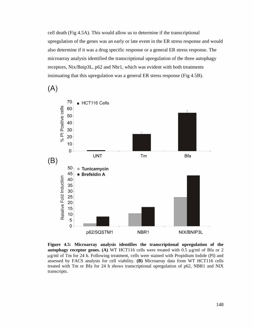
148
cell death (Fig 4.5A). This would allow us to determine if the transcriptional
upregulation of the genes was an early or late event in the ER stress response and would
also determine if it was a drug specific response or a general ER stress response. The
microarray analysis identified the transcriptional upregulation of the three autophagy
receptors, Nix/Bnip3L, p62 and Nbr1, which was evident with both treatments
insinuating that this upregulation was a general ER stress response (Fig 4.5B).
Figure 4.5: Microarray analysis identifies the transcriptional upregulation of the
autophagy receptor genes. (A) WT HCT116 cells were treated with 0.5 g/ml of Bfa or 2
g/ml of Tm for 24 h. Following treatment, cells were stained with Propidium Iodide (PI) and
assessed by FACS analysis for cell viability. (B) Microarray data from WT HCT116 cells
treated with Tm or Bfa for 24 h shows transcriptional upregulation of p62, NBR1 and NIX
transcripts.

149
To confirm the microarray data, we treated WT HCT116 cells with 0.5 g/ml of Bfa
and carried out a time course of 0, 6, 12, 24, 32 and 36 h (Fig 4.6A). Furthermore we
confirmed the induction of the autophagy receptors with additional ER stress inducing
agents, tunicamycin (0.5 g/ml Tm) and thapsigargin (0.5 M Tg) (Fig 4.6B). To
confirm that this was not a cell-type specific effect we treated MDA-MB-468 breast
cancer cells with 0.5 g/ml of Bfa and monitored the transcriptional upregulation of the
autophagy receptor genes at the indicated time points (Fig 4.6C). Following treatment,
cells were harvested and lysed in TRIzol reagent. RNA was extracted from the lysate
and revere transcribed into cDNA. The cDNA was diluted to a 1:5 ratio. Realtime PCR
was carried out using probes specific for NIX/BNIP3L, p62 and NBR1cDNA.
Our results confirm the transcriptional upregulation of the autophagy receptor genes,
NIX/BNIP3L, p62 and NBR1, in response to ER stress. Furthermore we showed that
this was not a cell type specific event, using both HCT116 colon cancer cells and MDA-
MB-468 breast cancer cells.

150
Figure 4.6: Real-Time PCR confirms the transcriptional upregulation of the autophagy
receptor genes. Real-Time PCR was carried out on HCT116 cells and MDA-MB-468 cells to
monitor autophagy receptor gene expression in response to ER stress. (A) WT HCT116 cells
were treated with 0.5 g/ml of Bfa for over an extensive time course to monitor the gene
expression kinetics. Cells were harvested at the indicated time points and lysed in TRIzol
reagent. RNA was extracted and reverse transcribed into cDNA. cDNA was diluted to a 1:5
ratio and real-time PCR was carried out to monitor the expression of p62, NBR1 and NIX over
the time course. (B) WT HCT116 cells were treated with 2 g/ml Tm or 0.5 Tg for 24 h.
Following treatment cells were harvested and lysed in TRIzol reagent. Real-time PCR was
carried out as in (A). (C) MDA-MB-468 cells were treated with 0.5 g/ml Bfa for 24 h.
Following treatment cells were harvested and lysed in TRIzol reagent. Real-time PCR was
carried out as in (A).
151
4.2.5 The PERK arm of the UPR is required for the transcriptional
upregulation of the autophagy receptor genes
Next we wanted to determine which arm of the UPR was involved in the upregulation
of the autophagy receptor genes. Our preliminary data suggests that the transcriptional
upregulation of the autophagy receptor genes is dependent on the PERK arm of the
UPR. PERK has been previously described as an essential factor for the regulation of
the autophagy pathway, involved in ATF4 mediated upregulation of LC3, and Chop
mediated upregulation of ATG5. Our data suggests that PERK is also involved in the
upregulation of the autophagy receptor genes.
MDA-MB-468 breast cancer cell lines stably expressing PERK shRNA or PLKO ctrl,
were treated with 0.5 g/ml of Bfa for 24 h. Following treatment, cells were harvested
and lysed in TRIzol reagent and RNA was extracted from the lysate. The RNA was
revere transcribed and the cDNA was diluted to a 1:5 ratio. Realtime PCR was carried
out using probes specific for NIX/BNIP3L, p62, NBR1, PERK and GRP78 cDNA (Fig
4.7).
Our results confirm that PERK is indeed knocked down; subsequently, knockdown of
PERK resulted in reduced levels of GRP78. Furthermore we also observe reduced
induction of the autophagy receptor genes, indicating that a downstream transcription
factor of the PERK arm is involved in the regulation of the autophagy receptor genes in
response to ER stress. Although these results are convincing they are preliminary and
thus most be confirmed with different ER stress inducing drugs and different cell lines.

152
Figure 4.7: Preliminary data suggest that PERK is involved in the regulation of the
autophagy receptor genes in response to ER stress. MDA-MB-468 PLKO control and MDA-
MB-468PERK shRNA expressing cells were treated with 0.5 g/ml of Bfa for 24 h. Following
treatment, Cells were harvested and lysed in TRIzol reagent. RNA was extracted and reverse
transcribed into cDNA. cDNA was diluted to a 1:5 ratio and real-time PCR was carried out to
monitor the expression of p62, NBR1, NIX, PERK and GRP78. Graphs represent the mean ±
SEM of three independent experiments

153
4.3 Discussion The UPR and autophagy are independent processes; however, although the UPR and
autophagy can act independently, it is clear that these processes are quite tightly
interlinked in response ER stress. As mentioned, there are many indirect and direct
connections between the UPR and the autophagy pathway demonstrated throughout the
literature, of which our group has recently reviewed38
.
The Microarray analysis performed here, emphasizes this connection with the
identification of over 20 genes involved in the regulation of autophagy in response to
ER stress, most of which have not yet been connected to the UPR. Of these genes we
have identified the upregulation of the autophagy receptor genes NIX/BNIP3L, p62 and
NBR1. Experts in the field of ER stress and autophagy agree that ER stress is a potent
inducer of autophagy; however, whether autophagy is a selective or non-selective
process in response to ER stress becomes a matter of debate. Here we investigated the
regulation of the autophagy receptor genes by the UPR to try and shed some light on
this controversial topic.
Autophagy has long been considered a non-selective bulk degradation process. In the
last few years the identification of the autophagy receptor proteins has re-established the
importance of this process in many cellular events for the selective removal of
unwanted substrates such as invading pathogens, protein aggregates and old or damaged
organelles47
. There have been many autophagy receptor proteins identified,
characterized by the presence of an LC3 interacting region (LIR); however of these
proteins the best characterized is p62 and more recently NBR1, a protein shown to
behave and function similar to that of p62. Both p62 and NBR1 are involved in the
selective degradation of ubiquitinated substrates and aggregates. p62 and NBR1 contain
a UBA domain which facilitates binding of ubiquitinated substrates, and a LC3
interacting region (LIR) which targets the ubiquitin substrates to the autophagosome via
direct interaction with LC3 family members229-231
Although it is proposed that p62 and
Nbr1 can target soluble single ubiquitinated proteins to the autophagosome it has not
yet been clearly demonstrated. However p62 and NBR1 are capable of oligomerizing
via an N-terminal Phox and Bem1p (PB1) domain resulting in the formation of

154
unfolded protein aggregates in the cytosol. These aggregates have been clearly shown to
co-localize with autophagosomal structures232
. The results in this chapter show that both
p62 and NBR1 are transcriptionally upregulated in response to multiple ER stress
inducing agents in both HCT116 colon cancer cells and MDA-MB-468 breast cancer
cells. Inhibition of autophagy degradation with the addition of CQ resulted in the
accumulation of ubiquitinated substrates in response to ER stress. This data confirms
that ubiquitinated substrates require autophagy for their removal in response to ER
stress; however single and co-knockdown studies of p62 and NBR1 will be required to
confirm that these proteins are required for autophagy mediated degradation of
ubiquitinated substrates.
Mitochondrial degradation or “Mitophagy” is a process that is characterized by
autophagy-mediated degradation of mitochondria. NIX/BNIP3L has been identified as
an important adaptor for the removal of mitochondria. Nix was first established as an
important mediator for the removal of mitochondria in reticulocyte development233
,
however further studies demonstrate that damaged or depolarized mitochondria can also
be removed by autophagy and this process is dependent on NIX87
. In this chapter we
show co-localization of mitochondria and autophagosomes in response to ER stress
both by fluorescent tagging experiments and electron microscopy. Furthermore we
show that NIX is transcriptionally upregulated in response to ER stress both in HCT116
cells and MDA-MB-468 cells. Knocking down NIX in this system will be crucial in
determining if (a) the mitophagy observed in response to ER stress requires NIX and (b)
if mitophagy plays a role in protecting cells against ER stress.
PERK knockdown in MDA-MB-468 cells demonstrated that the transcriptional
upregulation of NIX, p62 and NBR1 are dependent on transcription factors downstream
of this arm. However, this data is preliminary and thus knockdown of IRE1 and ATF6
would need to be done to demonstrate that it is a PERK dependent effect. Because the
UPR is such a complex and dynamic system, knockdown of one arm of the UPR can
result in an enhancement or differential regulatory effect on the other UPR arms which
may result in obscured results.

155
This study has provided a lot of information and thus a lot of questions which need to
be addressed. Here I specifically concentrate on the hypothesis that the UPR may
regulate the autophagy receptors to aid in the removal of damaged organelles and
ubiquitinated substrates. In this study I show that ER stress induced autophagy results in
the degradation of mitochondria and ubiquitinated substrates. Furthermore I show that
the UPR can transcriptional induce the autophagy receptor genes which have been
shown, in different contexts, to be play a crucial role in there degradation. Moreover, I
show that the transcriptional regulation of the autophagy receptors may be dependent on
the PERK arm of the UPR.

156
Concluding Remarks and Future
Prospects

157
In this thesis we have examined the dynamics of cell death and the close relationship
that exists between ER stress, the UPR and autophagy; furthermore, we show the
integral crosstalk that occurs between these processes.
In the first results section (chapter 3) we demonstrate the ability of the cell to use
alternative mechanisms to induce cell death when the intrinsic apoptotic pathway is
compromised. To understand these alternative forms of cell death we use cells that are
resistant to mitochondrial-mediated apoptosis and expose them to cellular stress stimuli
such as ER stress inducing agents and chemotherapeutic drugs. Although we use quite
an artificial system, it is required for primarily elucidating these mechanisms without
speculating what level of death is being contributed from the intrinsic pathway and what
is being contributed from the death pathway under investigation. Once the intrinsic
pathway is initiated and caspase-9 becomes activated a cascade of cleavage events
occurs, including cleavage of other apical caspases, further amplifying cell death. If we
are to monitor other forms of cell death independent to this process it is essential that
our observations aren’t obscured by even negligible amounts of apoptosome activation.
For this reason we use C9-/-
MEFs; however, by using different cell lines and knockout
models we confirm that our observations are not an artifact of these cells. To date
apoptosis is the king of programmed cell death, there is an immense amount of
literature describing how apoptosis is initiated and how it executes cell death in many
contexts. It is clear that other forms of cell death also exist; however, because these
pathways are not frequently observed under culture conditions they tend to be ignored
and consequently are poorly defined. It is important to elucidate how other forms of cell
death are activated and how they execute cell death. If these other forms of cell death
are characterized as well as apoptosis it could revolutionize therapeutic approaches
against diseases such as cancer. Intrinsic apoptosis is clearly a very powerful system for
inducing cell death; however for most cancer cells this mechanism of death is
abrogated. In this section we show that although the intrinsic pathway is compromised
these cells can still undergo cell death, albeit at a slower, less efficient rate. The slow,
less efficient characteristic of these pathways is clearly a disadvantage; however if we
can understand how these pathways are activated and identify the regulatory

158
components involved, pharmacological manipulation could expose these pathways to be
as efficient if not more efficient than the intrinsic apoptotic pathway.
In this section (chapter 3) we show that cells compromised in the MMA pathway,
treated with ER stress inducing agents or chemotherapeutic drugs, undergo cell death
with features of caspase activation, necroptosis and a requirement for ATG5.
Furthermore, we show this pathway is quite dynamic in determining cell death,
depending on the cellular context this pathway can execute cell death through either
caspase-8 mediated apoptosis or RIP mediated necroptosis. We show that this death
pathway requires RIP1, caspase-8 and ATG5. Furthermore we show that both TRADD
and RIP1 kinase activity are required for the optimal functioning of this pathway.
Immunoprecipitation of ATG5 suggests that at least RIP1, FADD, ATG5 and caspase-8
are all components of one complex.
In this study we propose the formation of an ATG5-FADD-RIP1-caspase-8 containing
complex that is capable of signaling cell death through either caspase-8 mediated
activation of executioner caspases and subsequent apoptosis or through RIP1 mediated
activation of the Necrosome and subsequent necroptosis. As this complex resembles the
dynamics of other RIP1-containing complexes, such as complex II and the ripoptosome,
we assume that similar regulators, such as FLIP proteins, are capable of having similar
effects on our complex. With this in mind we can assume that depending on the levels
of FLIP proteins in the cell this complex will signal through either apoptosis or
necroptosis. Knockdown of ATG5 and subsequently autophagy inhibited caspase-3
processing and subsequently reduced the levels of cell death in the C9-/-
MEFs. This
may implicate the autophagosome acting as a platform for the formation of this
complex; alternatively, ATG5 may be an essential component of this caspase-8
activating complex independent of its role in autophagy. Studies have shown a direct
interaction between ATG5 and FADD of which were very convincing. Subsequent
studies have presumed that this interaction correlates directly with autophagy and
hypothesize that the autophagosome acts as a signaling platform for the formation of an
iDISC; however, although this seems logical, as of yet, this has not been adequately
demonstrated.

159
Future investigation is required to address if ATG5 is acting independent of autophagy
in this complex or if the autophagosome is required as a signaling platform. To address
this question, proteins essential for the induction of autophagy need to be targeted for
knockdown, such as Beclin1 or ULK1, to demonstrate that the autophagosome and not
specifically ATG5 is required for this complex to form. Furthermore upon knockdown
of these genes, ATG5 must be monitored within the same experiment to ensure that
inhibition upstream of autophagy has no effect on the stability of downstream
components in the autophagy pathway. To try and further address this issue, Gold
particle labeling of active caspase-8 followed by electron microscopy would be the most
convincing, observing the outer membrane of the autophagosomes for gold dotted
caspase-8. One possible method would be to add biotin labeled zVAD to the cells
during treatment, followed by the addition of a gold labeled anti-biotin antibody. In
addition to determining the role of ATG5 and autophagy in this complex, it is important
to understand how this complex if regulated. Depicting the role of the different FLIP
isoforms and the IAP family proteins in the regulation of this complex will also be
essential, this will determine if its regulation is similar to that of complex II and the
ripoptosome or if it requires completely different regulatory partners. Finally to confirm
that all these components are indeed in complex, reciprocal IPs for some of the other
partners would be required followed by mass spectrometry to determine other
interacting partners.
In the second result section (chapter 4) we examine the close relationship between the
UPR and its regulation of the autophagy pathway. Although these processes are known
to be tightly connected not much evidence exists to directly relate these two processes.
Treatment of HCT116 cells with ER stress inducing agents followed by microarray
analysis revealed an array of autophagy-related genes transcriptionally upregulated. Of
these genes we identified the transcriptional upregulation of the autophagy receptors
p62, NIX and NBR1.
Autophagy is an exceptionally complex system and plays many essential roles in the
cell. The early characterization of this process as being a non-selective process is slowly
being shunned with mounting evidence to suggest that the removal of substrates by

160
autophagy requires selective labeling with an autophagy receptor protein. Here we show
that in response to ER stress p62, NIX and NBR1 are transcriptionally upregulated.
Furthermore we show that the knockdown of the UPR arm, PERK, abrogated the
upregulation of these genes.
Further investigation in to the functional relevance of these proteins in response to ER
stress is required. Theoretically if ER stress induced autophagy is a selective process the
inhibition of these receptors should show similar functional defects to the knockdown of
the autophagy pathway. Moreover, inhibition of the other UPR arms will be required to
determine if PERK is the main arm involved in their regulation. Commercially available
shRNAs for the knockdown of the UPR arms in human cell lines have, in our
experience, been very problematic; however, the recently available PERK inhibitor and
IRE1 inhibitors have demonstrated exceptionally high specificity for the inhibition of
these proteins and will make addressing questions such as this very easy.
On a broader scale, this thesis demonstrates the close connection between ER stress,
autophagy and cell death. We show that the cell is a very dynamic system which is
capable of using multiple processes to deliver different outcomes. In the context of the
UPR and autophagy, both are induced by ER stress as a survival response; however, if
the stress persists the same processes are capable of inducing cell death. Furthermore
inhibition of the cells primary mode of cell death in response to severe cellular stresses
will result in the activation of a non-canonical cell death pathway to carry out the job.
Elucidating the players involved in these processes and understanding how these
switches are regulated will further our understanding of the cell and its behavior in
response to cellular stress.

161
References

162
1 McLaughlin, M. & Vandenbroeck, K. The endoplasmic reticulum protein
folding factory and its chaperones: new targets for drug discovery? British
Journal of Pharmacology 162, 328-345, doi:10.1111/j.1476-5381.2010.01064.x
(2011).
2 Gotoh, T. et al. Endoplasmic Reticulum Stress-Related Inflammation and
Cardiovascular Diseases. International Journal of Inflammation 2011 (2011).
3 Gorman, A. M. et al. Stress management at the ER: Regulators of ER stress-
induced apoptosis. Pharmacology & Therapeutics,
doi:10.1016/j.pharmthera.2012.02.003 (2012).
4 Cawley, K.. et alAssays for detecting the unfolded protein response. Methods in
Enzymology Vol. Volume 490 (ed P. Michael Conn) 31-51 (Academic Press,
2011).
5 Szegezdi, E. et al. Mediators of endoplasmic reticulum stress-induced apoptosis.
EMBO reports 7, 880-885 (2006).
6 Reddy, R. K. et al. Endoplasmic Reticulum Chaperone Protein GRP78 Protects
Cells from Apoptosis Induced by Topoisomerase Inhibitors. Journal of
Biological Chemistry 278, 20915-20924, doi:10.1074/jbc.M212328200 (2003).
7 Goldson, T. M. et al. Eukaryotic initiation factor 4E variants alter the
morphology, proliferation, and colony-formation properties of MDA-MB-435
cancer cells. Molecular Carcinogenesis 46, 71-84, doi:10.1002/mc.20276
(2007).
8 Saito, A. et al. Endoplasmic Reticulum Stress Response Mediated by the PERK-
eIF2α-ATF4 Pathway Is Involved in Osteoblast Differentiation Induced by
BMP2. Journal of Biological Chemistry 286, 4809-4818,
doi:10.1074/jbc.M110.152900 (2011).
9 Harding, H. P. et al. Perk Is Essential for Translational Regulation and Cell
Survival during the Unfolded Protein Response. Molecular Cell 5, 897-904
(2000).
10 Harding, H. P. et al. An Integrated Stress Response Regulates Amino Acid
Metabolism and Resistance to Oxidative Stress. Molecular Cell 11, 619-633,
doi:10.1016/s1097-2765(03)00105-9 (2003).
11 Woo, C. W. et al. Adaptive suppression of the ATF4-CHOP branch of the
unfolded protein response by toll-like receptor signalling. Nature Cell Biology
11, 1473-1480,
doi:http://www.nature.com/ncb/journal/v11/n12/suppinfo/ncb1996_S1.html
(2009).
12 McCullough, K. D. et al. Gadd153 sensitizes cells to endoplasmic reticulum
stress by down-regulating Bcl2 and perturbing the cellular redox state.
Molecular and Cellular Biology 21, 1249-1259 (2001).
13 Cazanave, S. C. et al. CHOP and AP-1 cooperatively mediate PUMA expression
during lipoapoptosis. Am J Physiol Gastrointest Liver Physiol 299, 29 (2010).
14 Puthalakath, H. et al. ER stress triggers apoptosis by activating BH3-only
protein Bim. Cell 129, 1337-1349 (2007).
15 Kim, H. et al. Stepwise activation of BAX and BAK by tBID, BIM, and PUMA
initiates mitochondrial apoptosis. Molecular Cell 36, 487-499 (2009).

163
16 Oyadomari, S. et al. Targeted disruption of the Chop gene delays endoplasmic
reticulum stress–mediated diabetes. The Journal of Clinical Investigation 109,
525-532 (2002).
17 Chan, J. Y. & Kwong, M. Impaired expression of glutathione synthetic enzyme
genes in mice with targeted deletion of the Nrf2 basic-leucine zipper protein.
Biochimica et Biophysica Acta 15, 19-26 (2000).
18 Cullinan, S. B. et al. Nrf2 Is a Direct PERK Substrate and Effector of PERK-
Dependent Cell Survival. Molecular and Cellular Biology. 23, 7198-7209,
doi:10.1128/mcb.23.20.7198-7209.2003 (2003).
19 Ho, H. K. et al. Nrf2 Activation Involves an Oxidative-Stress Independent
Pathway in Tetrafluoroethylcysteine-Induced Cytotoxicity. Toxicological
Sciences 86, 354-364, doi:10.1093/toxsci/kfi205 (2005).
20 Cullinan, S. B. & Diehl, J. A. Coordination of ER and oxidative stress signaling:
The PERK/Nrf2 signaling pathway. The International Journal of Biochemistry
& Cell Biology 38, 317-332, doi:10.1016/j.biocel.2005.09.018 (2006).
21 Haze, K.. et al. Mammalian Transcription Factor ATF6 Is Synthesized as a
Transmembrane Protein and Activated by Proteolysis in Response to
Endoplasmic Reticulum Stress. Molecular Biology of the Cell 10, 3787-3799
(1999).
22 Shen, J. et al. ER Stress Regulation of ATF6 Localization by Dissociation of
BiP/GRP78 Binding and Unmasking of Golgi Localization Signals.
Developmental Cell 3, 99-111 (2002).
23 Ye, J. et al. ER Stress Induces Cleavage of Membrane-Bound ATF6 by the
Same Proteases that Process SREBPs. Molecular Cell 6, 1355-1364,
doi:10.1016/s1097-2765(00)00133-7 (2000).
24 Li, M. et al. ATF6 as a Transcription Activator of the Endoplasmic Reticulum
Stress Element: Thapsigargin Stress-Induced Changes and Synergistic
Interactions with NF-Y and YY1. Molecular and Cellular Biology 20, 5096-
5106, doi:10.1128/mcb.20.14.5096-5106.2000 (2000).
25 Haze, K. et al. Mammalian transcription factor ATF6 is synthesized as a
transmembrane protein and activated by proteolysis in response to endoplasmic
reticulum stress. Molecular Biology of the Cell 10, 3787-3799 (1999).
26 Yamamoto, K.. et al . Differential contributions of ATF6 and XBP1 to the
activation of endoplasmic reticulum stress-responsive cis-acting elements ERSE,
UPRE and ERSE-II. Journal of Biochemistry 136, 343-350 (2004).
27 Yamamoto, K. et al. Differential Contributions of ATF6 and XBP1 to the
Activation of Endoplasmic Reticulum Stress-Responsive cis-Acting Elements
ERSE, UPRE and ERSE-II. Journal of Biochemistry 136, 343-350,
doi:10.1093/jb/mvh122 (2004).
28 Belmont, P. J. et al. Regulation of microRNA expression in the heart by the
ATF6 branch of the ER stress response. Journal of Molecular and Cellular
Cardiology, doi:10.1016/j.yjmcc.2012.01.017 (2012).
29 Kaufman, R. J. & Cao, S. Inositol-requiring 1/X-box-binding protein 1 is a
regulatory hub that links endoplasmic reticulum homeostasis with innate
immunity and metabolism. EMBO Molecular Medicine 2, 189-192,
doi:10.1002/emmm.201000076 (2010).

164
30 Hocking, L. J. et al. Functional interaction between Sequestosome-1/p62 and
Autophagy-Linked FYVE-containing protein WDFY3 in human osteoclasts.
Biochemical and Biophysical Research Communications 402, 543-548,
doi:10.1016/j.bbrc.2010.10.076 (2010).
31 Korennykh, A. V. et al. The unfolded protein response signals through high-
order assembly of Ire1. Nature 457, 687-693,
doi:http://www.nature.com/nature/journal/v457/n7230/suppinfo/nature07661_S1
.html (2009).
32 Lin, J. H. et al. IRE1 Signaling Affects Cell Fate During the Unfolded Protein
Response. Science 318, 944-949, doi:10.1126/science.1146361 (2007).
33 Trevino, L. R. et al. Germline genomic variants associated with childhood acute
lymphoblastic leukemia. Nature Genetics 41, 1001-1005,
doi:http://www.nature.com/ng/journal/v41/n9/suppinfo/ng.432_S1.html (2009).
34 Drogat, B. et al. IRE1 Signaling Is Essential for Ischemia-Induced Vascular
Endothelial Growth Factor-A Expression and Contributes to Angiogenesis and
Tumor Growth In vivo. Cancer Research 67, 6700-6707, doi:10.1158/0008-
5472.can-06-3235 (2007).
35 Wang, F. M. et al. Regulation of unfolded protein response modulator XBP1s by
acetylation and deacetylation. Biochemical Journal 433, 245-252,
doi:10.1042/bj20101293 (2010).
36 Urano, F. et al. Coupling of Stress in the ER to Activation of JNK Protein
Kinases by Transmembrane Protein Kinase IRE1. Science 287, 664-666,
doi:10.1126/science.287.5453.664 (2000).
37 Kaneko, M. et al. Activation Signal of Nuclear Factor-κB in Response to
Endoplasmic Reticulum Stress is Transduced via IRE1 and Tumor Necrosis
Factor Receptor-Associated Factor 2. Biological & Pharmaceutical Bulletin 26,
931-935 (2003).
38 Deegan, S. et al. Stress-induced self-cannibalism: on the regulation of
autophagy by endoplasmic reticulum stress. Cellular and molecular life sciences
: Cellular and Molecular Life Sciences, doi:10.1007/s00018-012-1173-4 (2012).
39 De Duve, C. & Wattiaux, R. Functions of lysosomes. Annual Review of
Physiology 28, 435-492, doi:10.1146/annurev.ph.28.030166.002251 (1966).
40 Ohsumi, Y. Molecular mechanism of autophagy in yeast, Saccharomyces
cerevisiae. Philosophical transactions of the Royal Society of London. Series B,
Biological sciences 354, 1577-1580; discussion 1580-1571,
doi:10.1098/rstb.1999.0501 (1999).
41 Rubinsztein, D. C. Autophagy: where next? EMBO reports 11, 3-3 (2010).
42 Kondo, Y. et al. The role of autophagy in cancer development and response to
therapy. Nature Reviews Cancer 5, 726-734, doi:nrc1692 [pii]10.1038/nrc1692
(2005).
43 Moretti, L. et al. Switch between apoptosis and autophagy: radiation-induced
endoplasmic reticulum stress? Cell Cycle 6, 793-798, doi:4036 [pii] (2007).
44 Levine, B. Cell biology: autophagy and cancer. Nature 446, 745-747,
doi:446745a [pii]10.1038/446745a (2007).
165
45 Kroemer, G. et al. Autophagy and the integrated stress response. Molecular Cell
40, 280-293, doi:S1097-2765(10)00751-3 [pii]10.1016/j.molcel.2010.09.023
(2010).
46 Rosenfeldt, M. T. & Ryan, K. M. The role of autophagy in tumour development
and cancer therapy. Expert Reviews in Molecular Medicine 2 2009;11:e36. doi:
10.1017/S1462399409001306.
47 Weidberg, H. et al. Biogenesis and cargo selectivity of autophagosomes. Annual
Review of Biochemistry 80, 125-156, doi:10.1146/annurev-biochem-052709-
094552 (2011).
48 Mizushima, N. Autophagy: process and function. Genes & Development 21,
2861-2873, doi:21/22/2861 [pii]10.1101/gad.1599207 (2007).
49 Maiuri, M. C. et al. Self-eating and self-killing: crosstalk between autophagy
and apoptosis. Nature Reviews Molecular Cell Biology 8, 741-752, doi:nrm2239
[pii]10.1038/nrm2239 (2007).
50 Jung, C. H. et al. ULK-Atg13-FIP200 complexes mediate mTOR signaling to
the autophagy machinery. Molecular Biology of the Cell 20, 1992-2003,
doi:E08-12-1249 [pii]10.1091/mbc.E08-12-1249 (2009).
51 Egan, D. F. et al. Phosphorylation of ULK1 (hATG1) by AMP-activated protein
kinase connects energy sensing to mitophagy. Science 331, 456-461,
doi:10.1126/science.1196371 (2011).
52 Kim, J. et al. AMPK and mTOR regulate autophagy through direct
phosphorylation of Ulk1. Nature Cell Biology 13, 132-141,
doi:10.1038/ncb2152 (2011).
53 Shackelford, D. B. & Shaw, R. J. The LKB1-AMPK pathway: metabolism and
growth control in tumour suppression. Nature Reviews Cancer 9, 563-575,
doi:nrc2676 [pii]10.1038/nrc2676 (2009).
54 Hosokawa, N. et al. Nutrient-dependent mTORC1 association with the ULK1-
Atg13-FIP200 complex required for autophagy. Molecular Biology of the Cell
20, 1981-1991, doi:10.1091/mbc.E08-12-1248 (2009).
55 Dunlop, E. A. et al. ULK1 inhibits mTORC1 signaling, promotes multisite
Raptor phosphorylation and hinders substrate binding. Autophagy 7, 737-747
(2011).
56 Loffler, A. S. et al. Ulk1-mediated phosphorylation of AMPK constitutes a
negative regulatory feedback loop. Autophagy 7, 696-706 (2011).
57 Chan, E. Y. Regulation and Function of Uncoordinated-51 Like Kinase Proteins.
Antioxidants & Redox Signaling, doi:10.1089/ars.2011.4396 (2012).
58 Simonsen, A. & Tooze, S. A. Coordination of membrane events during
autophagy by multiple class III PI3-kinase complexes. The Journal of Cell
Biology 186, 773-782, doi:jcb.200907014 [pii]10.1083/jcb.200907014 (2009).
59 Polson, H. E. et al. Mammalian Atg18 (WIPI2) localizes to omegasome-
anchored phagophores and positively regulates LC3 lipidation. Autophagy 6
(2010).
60 Proikas-Cezanne, T. et al. WIPI-1alpha (WIPI49), a member of the novel 7-
bladed WIPI protein family, is aberrantly expressed in human cancer and is
linked to starvation-induced autophagy. Oncogene 23, 9314-9325,
doi:10.1038/sj.onc.1208331 (2004).

166
61 Axe, E. L. et al. Autophagosome formation from membrane compartments
enriched in phosphatidylinositol 3-phosphate and dynamically connected to the
endoplasmic reticulum. The Journal of Cell Biology 182, 685-701,
doi:10.1083/jcb.200803137 (2008).
62 Simonsen, A. et al. Alfy, a novel FYVE-domain-containing protein associated
with protein granules and autophagic membranes. Journal of Cell Science 117,
4239-4251, doi:10.1242/jcs.01287 (2004).
63 Stromhaug, P. E. et al. Purification and characterization of autophagosomes
from rat hepatocytes. Biochemical Journal 335 ( Pt 2), 217-224 (1998).
64 Fengsrud, M. et al. Ultrastructural and immunocytochemical characterization of
autophagic vacuoles in isolated hepatocytes: effects of vinblastine and
asparagine on vacuole distributions. Experimental Cell Research 221, 504-519,
doi:10.1006/excr.1995.1402 (1995).
65 Hailey, D. W. et al. Mitochondria supply membranes for autophagosome
biogenesis during starvation. Cell 141, 656-667, doi:10.1016/j.cell.2010.04.009
(2010).
66 Ravikumar, B. et al. Plasma membrane contributes to the formation of pre-
autophagosomal structures. Nature Cell Biology 12, 747-757,
doi:10.1038/ncb2078 (2010).
67 Mizushima, N. et al. Mouse Apg16L, a novel WD-repeat protein, targets to the
autophagic isolation membrane with the Apg12-Apg5 conjugate. Journal of Cell
Science 116, 1679-1688 (2003).
68 Pankiv, S. et al. FYCO1 is a Rab7 effector that binds to LC3 and PI3P to
mediate microtubule plus end-directed vesicle transport. The Journal of Cell
Biology 188, 253-269, doi:10.1083/jcb.200907015 (2010).
69 Knaevelsrud, H. & Simonsen, A. Fighting disease by selective autophagy of
aggregate-prone proteins. FEBS Letters 584, 2635-2645, doi:S0014-
5793(10)00325-X [pii]10.1016/j.febslet.2010.04.041 (2010).
70 Hirota, Y. et al. [Mitophagy: selective degradation of mitochondria by
autophagy]. Seikagaku. The Journal of Japanese Biochemical Society 83, 126-
130 (2011).
71 Bernales, S. et al. ER-phagy: selective autophagy of the endoplasmic reticulum.
Autophagy 3, 285-287 (2007).
72 Kraft, C. et al. Mature ribosomes are selectively degraded upon starvation by an
autophagy pathway requiring the Ubp3p/Bre5p ubiquitin protease. Nature Cell
Biology 10, 602-610, doi:10.1038/ncb1723 (2008).
73 Sakai, Y. et al. Pexophagy: autophagic degradation of peroxisomes. Biochimica
et Biophysica Acta 1763, 1767-1775, doi:10.1016/j.bbamcr.2006.08.023 (2006).
74 Glaumann, H. Crinophagy as a means for degrading excess secretory proteins in
rat liver. Revisiones Sobre Biologia Celular : RBC 20, 97-110 (1989).
75 Mousavi, S. A. et al. Effects of inhibitors of the vacuolar proton pump on
hepatic heterophagy and autophagy. Biochimica et Biophysica Acta 1510, 243-
257 (2001).
76 Knodler, L. A. & Celli, J. Eating the strangers within: host control of
intracellular bacteria via xenophagy. Cellular Microbiology 13, 1319-1327,
doi:10.1111/j.1462-5822.2011.01632.x (2011).

167
77 Yamamoto, A. & Simonsen, A. The elimination of accumulated and aggregated
proteins: a role for aggrephagy in neurodegeneration. Neurobiology of Disease
43, 17-28, doi:10.1016/j.nbd.2010.08.015 (2011).
78 Weidberg, H. et al. Lipophagy: selective catabolism designed for lipids.
Developmental Cell 16, 628-630, doi:10.1016/j.devcel.2009.05.001 (2009).
79 Kristensen, A. R. et al. Ordered organelle degradation during starvation-induced
autophagy. Molecular & Cellular Proteomics : MCP 7, 2419-2428,
doi:10.1074/mcp.M800184-MCP200 (2008).
80 Sandilands, E. et al. Autophagic targeting of Src promotes cancer cell survival
following reduced FAK signalling. Nature Cell Biology 14, 51-60,
doi:10.1038/ncb2386 (2012).
81 Wild, P. et al. Phosphorylation of the autophagy receptor optineurin restricts
Salmonella growth. Science 333, 228-233, doi:10.1126/science.1205405 (2011).
82 Kirkin, V. et al. A role for ubiquitin in selective autophagy. Molecular Cell 34,
259-269, doi:10.1016/j.molcel.2009.04.026 (2009).
83 Ivanov, S. & Roy, C. R. NDP52: the missing link between ubiquitinated bacteria
and autophagy. Nature Immunology 10, 1137-1139, doi:10.1038/ni1109-1137
(2009).
84 Novak, I. et al. Nix is a selective autophagy receptor for mitochondrial
clearance. EMBO Reports 11, 45-51, doi:embor2009256
[pii]10.1038/embor.2009.256 (2010).
85 Tolkovsky, A. M. Mitophagy. Biochim Biophys Acta 1793, 1508-1515,
doi:S0167-4889(09)00060-3 [pii]10.1016/j.bbamcr.2009.03.002 (2009).
86 Narendra, D. P. & Youle, R. J. Targeting mitochondrial dysfunction: role for
PINK1 and Parkin in mitochondrial quality control. Antioxidants & Redox
Signaling 14, 1929-1938, doi:10.1089/ars.2010.3799 (2011).
87 Ding, W. X. et al. Nix is critical to two distinct phases of mitophagy, reactive
oxygen species-mediated autophagy induction and Parkin-ubiquitin-p62-
mediated mitochondrial priming. The Journal of Biological Chemistry 285,
27879-27890, doi:10.1074/jbc.M110.119537 (2010).
88 Gao, W. et al. Induction of macroautophagy by exogenously introduced
calcium. Autophagy 4, 754-761 (2008).
89 Høyer-Hansen, M. et al. Control of Macroautophagy by Calcium, Calmodulin-
Dependent Kinase Kinase-β, and Bcl-2. Molecular Cell 25, 193-205,
doi:10.1016/j.molcel.2006.12.009 (2007).
90 Egan, D. F. et al. Phosphorylation of ULK1 (hATG1) by AMP-Activated
Protein Kinase Connects Energy Sensing to Mitophagy. Science,
doi:10.1126/science.1196371 (2010).
91 Ferrari, D. et al. Endoplasmic reticulum, Bcl-2 and Ca2+ handling in apoptosis.
Cell Calcium 32, 413-420, doi:10.1016/s0143416002002014 (2002).
92 Bassik, M. C. et al. Phosphorylation of BCL-2 regulates ER Ca2+ homeostasis
and apoptosis. EMBO Journal 23, 1207-1216,
doi:http://www.nature.com/emboj/journal/v23/n5/suppinfo/7600104a_S1.html
(2004).

168
93 Li, G. et al. Role of ERO1-alpha-mediated stimulation of inositol 1,4,5-
triphosphate receptor activity in endoplasmic reticulum stress-induced apoptosis.
The Journal of Cell Biology 186, 783-792, doi:10.1083/jcb.200904060 (2009).
94 Ramming, T. & Appenzeller-Herzog, C. The Physiological Functions of
Mammalian Endoplasmic Oxidoreductin 1 (Ero1): On Disulfides and More.
Antioxidants & Redox Signaling, doi:10.1089/ars.2011.4475 (2012).
95 Jia, W. et al. Autophagy Regulates Endoplasmic Reticulum Homeostasis and
Calcium Mobilization in T Lymphocytes. The Journal of Immunology 186,
1564-1574, doi:10.4049/jimmunol.1001822 (2011).
96 Whitney, M. L. et al. ATF4 is necessary and sufficient for ER stress-induced
upregulation of REDD1 expression. Biochem Biophys Res Commun 379, 451-
455 (2009).
97 Jin, H.-O. et al. Activating transcription factor 4 and CCAAT/enhancer-binding
protein-β negatively regulate the mammalian target of rapamycin via Redd1
expression in response to oxidative and endoplasmic reticulum stress. Free
Radical Biology and Medicine 46, 1158-1167,
doi:10.1016/j.freeradbiomed.2009.01.015 (2009).
98 Brugarolas, J. et al. Regulation of mTOR function in response to hypoxia by
REDD1 and the TSC1/TSC2 tumor suppressor complex. Genes & Development
18, 2893-2904, doi:10.1101/gad.1256804 (2004).
99 Zhang, H. et al. Loss of Tsc1/Tsc2 activates mTOR and disrupts PI3K-Akt
signaling through downregulation of PDGFR. The Journal of Clinical
Investigation 112, 1223-1233 (2003).
100 Gingras, A.-C. et al. 4E-BP1, a repressor of mRNA translation, is
phosphorylated and inactivated by the Akt(PKB) signaling pathway. Genes &
Development 12, 502-513 (1998).
101 Manning, B. D. a. C., L.C. United at last: the tuberous sclerosis complex gene
products connect the phosphoinositide 3-kinase/Akt pathway to mammalian
target of rapamycin (mTOR) signalling. Biochemical Society Transactions 31,
573-578 (2003).
102 Qin, L. et al. ER stress negatively regulates AKT/TSC/mTOR pathway to
enhance autophagy. Autophagy 6, 239-247 (2010).
103 Salazar, M. et al. Cannabinoid action induces autophagy-mediated cell death
through stimulation of ER stress in human glioma cells. The Journal of Clinical
Investigation 119, 1359-1372 (2009).
104 Yung, H. W. et al. Regulation of AKT phosphorylation at Ser473 and Thr308 by
endoplasmic reticulum stress modulates substrate specificity in a severity
dependent manner. PLoS One 6 (2011).
105 Baumeister, P. et al. Endoplasmic Reticulum Stress Induction of the Grp78/BiP
Promoter: Activating Mechanisms Mediated by YY1 and Its Interactive
Chromatin Modifiers. Molecular and Cellular Biology 25, 4529-4540,
doi:10.1128/mcb.25.11.4529-4540.2005 (2005).
106 Ohoka, N. et al. TRB3, a novel ER stress-inducible gene, is induced via ATF4-
CHOP pathway and is involved in cell death. The EMBO Journal 24, 1243-
1255, doi:10.1038/sj.emboj.7600596 (2005).

169
107 Du, K.. et al. TRB3: a tribbles homolog that inhibits Akt/PKB activation by
insulin in liver. Science 300, 1574-1577, doi:10.1126/science.1079817 (2003).
108 Sinha, S. & Levine, B. The autophagy effector Beclin 1: a novel BH3-only
protein. Oncogene 27, S137-S148 (0000).
109 Boya, P. & Kroemer, G. Beclin 1: a BH3-only protein that fails to induce
apoptosis. Oncogene 28, 2125-2127 (2009).
110 McCullough, K. D. et al. Gadd153 Sensitizes Cells to Endoplasmic Reticulum
Stress by Down-Regulating Bcl2 and Perturbing the Cellular Redox State.
Molecular and Cellular Biology 21, 1249-1259, doi:10.1128/mcb.21.4.1249-
1259.2001 (2001).
111 Szegezdi, E. et al. Bcl-2 family on guard at the ER. American Journal of
Physiology - Cell Physiology 296, C941-C953, doi:10.1152/ajpcell.00612.2008
(2009).
112 Szegezdi, E. et al. Mediators of endoplasmic reticulum stress-induced apoptosis.
EMBO Reports 7, 880-885, doi:7400779 [pii]10.1038/sj.embor.7400779 (2006).
113 Yamamoto, K.. et al. BCL-2 is phosphorylated and inactivated by an ASK1/Jun
N-terminal protein kinase pathway normally activated at G(2)/M. Molecular and
Cellular Biology 19, 8469-8478 (1999).
114 Lei, K. & Davis, R. J. JNK phosphorylation of Bim-related members of the Bcl2
family induces Bax-dependent apoptosis. Proceedings of the National Academy
of Sciences of the United States of America 100, 2432-2437 (2003).
115 Wei, Y. et al. JNK1-mediated phosphorylation of Bcl-2 regulates starvation-
induced autophagy. Molecular Cell 30, 678-688, doi:S1097-2765(08)00389-
4[pii]10.1016/j.molcel.2008.06.001 (2008).
116 Ogata, M. et al. Autophagy is activated for cell survival after endoplasmic
reticulum stress. Molecular and Cellular Biology 26, 9220-9231,
doi:10.1128/MCB.01453-06 (2006).
117 Ogata, M. et al. Autophagy Is Activated for Cell Survival after Endoplasmic
Reticulum Stress. Molecular and Cellular Biology 26, 9220-9231,
doi:10.1128/mcb.01453-06 (2006).
118 Oh, S.-H. & Lim, S.-C. Endoplasmic Reticulum Stress-Mediated
Autophagy/Apoptosis Induced by Capsaicin (8-Methyl-N-vanillyl-6-
nonenamide) and Dihydrocapsaicin is Regulated by the Extent of c-Jun NH2-
Terminal Kinase/Extracellular Signal-Regulated Kinase Activation in WI38
Lung Epithelial Fibroblast Cells. Journal of Pharmacology and Experimental
Therapeutics 329, 112-122, doi:10.1124/jpet.108.144113 (2009).
119 Gozuacik, D. et al. DAP-kinase is a mediator of endoplasmic reticulum stress-
induced caspase activation and autophagic cell death. Cell Death and
Differentiation 15, 1875-1886, doi:10.1038/cdd.2008.121 (2008).
120 Zalckvar, E. et al. DAP-kinase-mediated phosphorylation on the BH3 domain of
beclin 1 promotes dissociation of beclin 1 from Bcl-XL and induction of
autophagy. EMBO Reports 10, 285-292, doi:10.1038/embor.2008.246 (2009).
121 Kouroku, Y. et al. ER stress (PERK/eIF2alpha phosphorylation) mediates the
polyglutamine-induced LC3 conversion, an essential step for autophagy
formation. Cell Death and Differentiation 14, 230-239,
doi:10.1038/sj.cdd.4401984 (2007).

170
122 Rouschop, K. M. et al. The unfolded protein response protects human tumor
cells during hypoxia through regulation of the autophagy genes MAP1LC3B and
ATG5. The Journal of Clinical Investigation 120, 127-141, doi:40027
[pii]10.1172/JCI40027 (2010).
123 Salih, D. A. M. & Brunet, A. FoxO transcription factors in the maintenance of
cellular homeostasis during aging. Current Opinion in Cell Biology 20, 126-136,
doi:10.1016/j.ceb.2008.02.005 (2008).
124 Medema, R. H. & Jaattela, M. Cytosolic FoxO1: alive and killing. Nature Cell
Biology 12, 642-643 (2010).
125 Xu, P. et al. JNK regulates FoxO-dependent autophagy in neurons. Genes &
Development 25, 310-322, doi:10.1101/gad.1984311 (2011).
126 Zhao, Y. et al. Cytosolic FoxO1 is essential for the induction of autophagy and
tumour suppressor activity. Nature Cell Biology 12, 665-675,
doi:http://www.nature.com/ncb/journal/v12/n7/suppinfo/ncb2069_S1.html
(2010).
127 Vidal, R. L. et al. Targeting the UPR transcription factor XBP1 protects against
Huntington's disease through the regulation of FoxO1 and autophagy. Human
Molecular Genetics, doi:10.1093/hmg/dds040 (2012).
128 Gupta, S. et al. Mechanisms of ER Stress-Mediated Mitochondrial Membrane
Permeabilization. International Journal of Cell Biology 2010 (2010).
129 Ullman, E. et al. Autophagy promotes necrosis in apoptosis-deficient cells in
response to ER stress. Cell Death and Differentiation 15, 422-425,
doi:10.1038/sj.cdd.4402234 (2008).
130 Young, M. M. et al. Autophagosomal membrane serves as platform for
intracellular death-inducing signaling complex (iDISC)-mediated caspase-8
activation and apoptosis. The Journal of Biological Chemistry 287, 12455-
12468, doi:10.1074/jbc.M111.309104 (2012).
131 Kroemer, G. et al. Classification of cell death: recommendations of the
Nomenclature Committee on Cell Death 2009. Cell Death and Differentiation
16, 3-11, doi:10.1038/cdd.2008.150 (2009).
132 Kerr, J. F. et al. Apoptosis: a basic biological phenomenon with wide-ranging
implications in tissue kinetics. British Journal of Cancer 26, 239-257 (1972).
133 Taylor, R. C. et al. Apoptosis: controlled demolition at the cellular level. Nature
Reviews Molecular Cell Biology 9, 231-241,
doi:http://www.nature.com/nrm/journal/v9/n3/suppinfo/nrm2312_S1.html
(2008).
134 Timmer, J. C. & Salvesen, G. S. Caspase substrates. Cell death and
differentiation 14, 66-72, doi:10.1038/sj.cdd.4402059 (2007).
135 Li, J. & Yuan, J. Caspases in apoptosis and beyond. Oncogene 27, 6194-6206,
doi:10.1038/onc.2008.297 (2008).
136 Tenev, T. et al. The Ripoptosome, a signaling platform that assembles in
response to genotoxic stress and loss of IAPs. Molecular Cell 43, 432-448,
doi:10.1016/j.molcel.2011.06.006 (2011).
137 Crook, N. E. et al. An apoptosis-inhibiting baculovirus gene with a zinc finger-
like motif. Journal of Virology 67, 2168-2174 (1993).

171
138 Lorick, K. L. et al. RING fingers mediate ubiquitin-conjugating enzyme (E2)-
dependent ubiquitination. Proceedings of the National Academy of Sciences of
the United States of America 96, 11364-11369 (1999).
139 Wei, Y. et al. Inhibitor of apoptosis proteins and apoptosis. Acta Biochimica et
Biophysica Sinica 40, 278-288 (2008).
140 Eckelman, B. P. et al. Human inhibitor of apoptosis proteins: why XIAP is the
black sheep of the family. EMBO Reports 7, 988-994,
doi:10.1038/sj.embor.7400795 (2006).
141 Varfolomeev, E. et al. Cellular inhibitors of apoptosis are global regulators of
NF-kappaB and MAPK activation by members of the TNF family of receptors.
Science Signaling 5, ra22, doi:10.1126/scisignal.2001878 (2012).
142 Yang, Q. H. & Du, C. Smac/DIABLO selectively reduces the levels of c-IAP1
and c-IAP2 but not that of XIAP and livin in HeLa cells. The Journal of
Biological Chemistry 279, 16963-16970, doi:10.1074/jbc.M401253200 (2004).
143 Srinivasula, S. M. et al. A conserved XIAP-interaction motif in caspase-9 and
Smac/DIABLO regulates caspase activity and apoptosis. Nature 410, 112-116,
doi:10.1038/35065125 (2001).
144 Yang, Q. H. et al. Omi/HtrA2 catalytic cleavage of inhibitor of apoptosis (IAP)
irreversibly inactivates IAPs and facilitates caspase activity in apoptosis. Genes
& Development 17, 1487-1496, doi:10.1101/gad.1097903 (2003).
145 Aggarwal, B. B. Signalling pathways of the TNF superfamily: a double-edged
sword. Nature Reviews. Immunology 3, 745-756, doi:10.1038/nri1184 (2003).
146 Dickens, L. S. et al. The 'complexities' of life and death: death receptor
signalling platforms. Experimental Cell Research 318, 1269-1277,
doi:10.1016/j.yexcr.2012.04.005 (2012).
147 Peter, M. E. et al. The CD95 receptor: apoptosis revisited. Cell 129, 447-450,
doi:10.1016/j.cell.2007.04.031 (2007).
148 Lavrik, I. N. et al. CD95 stimulation results in the formation of a novel death
effector domain protein-containing complex. The Journal of Biological
Chemistry 283, 26401-26408, doi:10.1074/jbc.M800823200 (2008).
149 Ponton, A. et al. The CD95 (APO-1/Fas) receptor activates NF-kappaB
independently of its cytotoxic function. The Journal of Biological Chemistry
271, 8991-8995 (1996).
150 Cao, X. et al. The role of TRADD in TRAIL-induced apoptosis and signaling.
FASEB journal : official publication of the Federation of American Societies for
Experimental Biology 25, 1353-1358, doi:10.1096/fj.10-170480 (2011).
151 Wajant, H., Pfizenmaier, K. & Scheurich, P. Tumor necrosis factor signaling.
Cell Death and Differentiation 10, 45-65, doi:10.1038/sj.cdd.4401189 (2003).
152 Vucic, D. et al. Ubiquitylation in apoptosis: a post-translational modification at
the edge of life and death. Nature Reviews Molecular Cell Biology 12, 439-452,
doi:10.1038/nrm3143 (2011).
153 Meylan, F. et al. The TNF-family receptor DR3 is essential for diverse T cell-
mediated inflammatory diseases. Immunity 29, 79-89,
doi:10.1016/j.immuni.2008.04.021 (2008).
154 Kumar, A. et al. The ectodermal dysplasia receptor activates the nuclear factor-
kappaB, JNK, and cell death pathways and binds to ectodysplasin A. The

172
Journal of Biological Chemistry 276, 2668-2677, doi:10.1074/jbc.M008356200
(2001).
155 Zeng, L. et al. Death Receptor 6 Induces Apoptosis Not through Type I or Type
II Pathways, but via a Unique Mitochondria-dependent Pathway by Interacting
with Bax Protein. The Journal of Biological Chemistry 287, 29125-29133,
doi:10.1074/jbc.M112.362038 (2012).
156 Tait, S. W. & Green, D. R. Mitochondria and cell death: outer membrane
permeabilization and beyond. Nature Reviews Molecular Cell Biology 11, 621-
632, doi:10.1038/nrm2952 (2010).
157 Reed, J. C. et al. Structure-function analysis of Bcl-2 family proteins. Regulators
of programmed cell death. Advances in Experimental Medicine and Biology 406,
99-112 (1996).
158 Youle, R. J. & Strasser, A. The BCL-2 protein family: opposing activities that
mediate cell death. Nature Reviews Molecular Cell Biology 9, 47-59,
doi:10.1038/nrm2308 (2008).
159 Chipuk, J. E. et al. The BCL-2 family reunion. Molecular Cell 37, 299-310,
doi:10.1016/j.molcel.2010.01.025 (2010).
160 Martinez-Caballero, S. et al. Assembly of the mitochondrial apoptosis-induced
channel, MAC. The Journal of Biological Chemistry 284, 12235-12245,
doi:10.1074/jbc.M806610200 (2009).
161 Li, P. et al. Cytochrome c and dATP-dependent formation of Apaf-1/caspase-9
complex initiates an apoptotic protease cascade. Cell 91, 479-489 (1997).
162 Shiozaki, E. N. & Shi, Y. Caspases, IAPs and Smac/DIABLO: mechanisms
from structural biology. Trends in Biochemical Sciences 29, 486-494,
doi:10.1016/j.tibs.2004.07.003 (2004).
163 Acehan, D. et al. Three-dimensional structure of the apoptosome: implications
for assembly, procaspase-9 binding, and activation. Molecular Cell 9, 423-432
(2002).
164 Shi, Y. Apoptosome assembly. Methods in Enzymology 442, 141-156,
doi:10.1016/S0076-6879(08)01407-9 (2008).
165 Reubold, T. F. et al. Crystal structure of full-length Apaf-1: how the death signal
is relayed in the mitochondrial pathway of apoptosis. Structure 19, 1074-1083,
doi:10.1016/j.str.2011.05.013 (2011).
166 Lin, Y. et al. Pidd, a new death-domain-containing protein, is induced by p53
and promotes apoptosis. Nature Genetics 26, 122-127, doi:10.1038/79102
(2000).
167 Tinel, A. & Tschopp, J. The PIDDosome, a protein complex implicated in
activation of caspase-2 in response to genotoxic stress. Science 304, 843-846,
doi:10.1126/science.1095432 (2004).
168 Gu, H. et al. Caspase-2 functions upstream of mitochondria in endoplasmic
reticulum stress-induced apoptosis by bortezomib in human myeloma cells.
Molecular Cancer Therapeutics 7, 2298-2307, doi:10.1158/1535-7163.MCT-
08-0186 (2008).
169 Tu, S. et al. In situ trapping of activated initiator caspases reveals a role for
caspase-2 in heat shock-induced apoptosis. Nature Cell Biology 8, 72-77,
doi:10.1038/ncb1340 (2006).

173
170 Tinel, A. et al. Regulation of PIDD auto-proteolysis and activity by the
molecular chaperone Hsp90. Cell Death and Differentiation 18, 506-515,
doi:10.1038/cdd.2010.124 (2011).
171 Janssens, S. et al. PIDD mediates NF-kappaB activation in response to DNA
damage. Cell 123, 1079-1092, doi:10.1016/j.cell.2005.09.036 (2005).
172 Bouchier-Hayes, L. et al. Characterization of cytoplasmic caspase-2 activation
by induced proximity. Molecular Cell 35, 830-840,
doi:10.1016/j.molcel.2009.07.023 (2009).
173 Wang, R. et al. Autoinhibition of UNC5b revealed by the cytoplasmic domain
structure of the receptor. Molecular Cell 33, 692-703,
doi:10.1016/j.molcel.2009.02.016 (2009).
174 Tinel, A. et al. Autoproteolysis of PIDD marks the bifurcation between pro-
death caspase-2 and pro-survival NF-kappaB pathway. The EMBO Journal 26,
197-208, doi:10.1038/sj.emboj.7601473 (2007).
175 Ando, K. et al. PIDD Death-Domain Phosphorylation by ATM Controls
Prodeath versus Prosurvival PIDDosome Signaling. Molecular Cell 47, 681-
693, doi:10.1016/j.molcel.2012.06.024 (2012).
176 Bonzon, C. et al. Caspase-2-induced apoptosis requires bid cleavage: a
physiological role for bid in heat shock-induced death. Molecular Biology of the
Cell 17, 2150-2157, doi:10.1091/mbc.E05-12-1107 (2006).
177 Korsmeyer, S. J. et al. Pro-apoptotic cascade activates BID, which oligomerizes
BAK or BAX into pores that result in the release of cytochrome c. Cell Death
and Differentiation 7, 1166-1173, doi:10.1038/sj.cdd.4400783 (2000).
178 O'Reilly, L. A. et al. Caspase-2 is not required for thymocyte or neuronal
apoptosis even though cleavage of caspase-2 is dependent on both Apaf-1 and
caspase-9. Cell Death and Differentiation 9, 832-841,
doi:10.1038/sj.cdd.4401033 (2002).
179 Oliver, T. G. et al. Caspase-2-mediated cleavage of Mdm2 creates a p53-
induced positive feedback loop. Molecular Cell 43, 57-71,
doi:10.1016/j.molcel.2011.06.012 (2011).
180 Kitevska, T. et al. Caspase-2: controversial killer or checkpoint controller?
Apoptosis : an international journal on programmed cell death 14, 829-848,
doi:10.1007/s10495-009-0365-3 (2009).
181 Feoktistova, M. et al. cIAPs block Ripoptosome formation, a RIP1/caspase-8
containing intracellular cell death complex differentially regulated by cFLIP
isoforms. Molecular Cell 43, 449-463, doi:10.1016/j.molcel.2011.06.011
(2011).
182 Laster, S. M. et al. Tumor necrosis factor can induce both apoptic and necrotic
forms of cell lysis. Journal of Immunology 141, 2629-2634 (1988).
183 Vanlangenakker, N. et al. Many stimuli pull the necrotic trigger, an overview.
Cell Death and Differentiation 19, 75-86, doi:10.1038/cdd.2011.164 (2012).
184 Pyo, J. O. et al. Essential roles of Atg5 and FADD in autophagic cell death:
dissection of autophagic cell death into vacuole formation and cell death. The
Journal of Biological Chemistry 280, 20722-20729,
doi:10.1074/jbc.M413934200 (2005).

174
185 Newton, K.. et al. A dominant interfering mutant of FADD/MORT1 enhances
deletion of autoreactive thymocytes and inhibits proliferation of mature T
lymphocytes. The EMBO Journal 17, 706-718, doi:10.1093/emboj/17.3.706
(1998).
186 Salmena, L. et al. Essential role for caspase 8 in T-cell homeostasis and T-cell-
mediated immunity. Genes & Development 17, 883-895,
doi:10.1101/gad.1063703 (2003).
187 Chau, H. et al. Cellular FLICE-inhibitory protein is required for T cell survival
and cycling. The Journal of Experimental Medicine 202, 405-413,
doi:10.1084/jem.20050118 (2005).
188 Li, C. et al. Autophagy is induced in CD4+ T cells and important for the growth
factor-withdrawal cell death. Journal of Immunology 177, 5163-5168 (2006).
189 Pua, H. H. et al. A critical role for the autophagy gene Atg5 in T cell survival
and proliferation. The Journal of Experimental Medicine 204, 25-31,
doi:10.1084/jem.20061303 (2007).
190 Laussmann, M. A. et al. Proteasome inhibition can induce an autophagy-
dependent apical activation of caspase-8. Cell Death and Differentiation 18,
1584-1597, doi:10.1038/cdd.2011.27 (2011).
191 Shao, Y. et al. Apoptotic and autophagic cell death induced by histone
deacetylase inhibitors. Proceedings of the National Academy of Sciences of the
United States of America 101, 18030-18035, doi:10.1073/pnas.0408345102
(2004).
192 Ding, W. X. et al. Differential effects of endoplasmic reticulum stress-induced
autophagy on cell survival. The Journal of Biological Chemistry 282, 4702-
4710, doi:10.1074/jbc.M609267200 (2007).
193 Ohmuraya, M. et al. Autophagic cell death of pancreatic acinar cells in serine
protease inhibitor Kazal type 3-deficient mice. Gastroenterology 129, 696-705,
doi:10.1016/j.gastro.2005.05.057 (2005).
194 Bonapace, L. et al. Induction of autophagy-dependent necroptosis is required for
childhood acute lymphoblastic leukemia cells to overcome glucocorticoid
resistance. The Journal of Clinical Investigation 120, 1310-1323,
doi:10.1172/JCI39987 (2010).
195 Lytton, J. et al. Thapsigargin inhibits the sarcoplasmic or endoplasmic reticulum
Ca-ATPase family of calcium pumps. The Journal of Biological Chemistry 266,
17067-17071 (1991).
196 Bassik, M. C. & Kampmann, M. Knocking out the door to tunicamycin entry.
Proceedings of the National Academy of Sciences of the United States of
America 108, 11731-11732, doi:10.1073/pnas.1109035108 (2011).
197 Lin, J. H. et al. Endoplasmic reticulum stress in disease pathogenesis. Annual
Review Pathology 3, 399-425,
doi:10.1146/annurev.pathmechdis.3.121806.151434 (2008).
198 Schroder, M. & Kaufman, R. J. The mammalian unfolded protein response.
Annual Review of Biochemistry 74, 739-789,
doi:10.1146/annurev.biochem.73.011303.074134 (2005).

175
199 Ron, D. & Walter, P. Signal integration in the endoplasmic reticulum unfolded
protein response. Nature Review Molecular Cell Biology 8, 519-529,
doi:nrm2199 [pii]10.1038/nrm2199 (2007).
200 Ding, W. X. et al. Linking of autophagy to ubiquitin-proteasome system is
important for the regulation of endoplasmic reticulum stress and cell viability.
American Journal Pathology 171, 513-524, doi:ajpath.2007.070188
[pii]10.2353/ajpath.2007.070188 (2007).
201 Giam, M. et al. BH3-only proteins and their roles in programmed cell death.
Oncogene 27, S128-S136 (0000).
202 Szegezdi, E. et al. Bcl-2 family on guard at the ER. Am erican Journal
Physiology Cell Physiology, doi:00612.2008 [pii]10.1152/ajpcell.00612.2008
(2009).
203 Fuchs, Y. & Steller, H. Programmed cell death in animal development and
disease. Cell 147, 742-758, doi:10.1016/j.cell.2011.10.033 (2011).
204 Vanlangenakker, N. et al. TNF-induced necroptosis in L929 cells is tightly
regulated by multiple TNFR1 complex I and II members. Cell Death & Disease
2, e230, doi:10.1038/cddis.2011.111 (2011).
205 Zhang, D. W. et al. RIP3, an energy metabolism regulator that switches TNF-
induced cell death from apoptosis to necrosis. Science 325, 332-336,
doi:10.1126/science.1172308 (2009).
206 Di Sano, F. et al. Endoplasmic reticulum stress induces apoptosis by an
apoptosome-dependent but caspase 12-independent mechanism. Journal of
Biological Chemistry 281, 2693-2700, doi:M509110200
[pii]10.1074/jbc.M509110200 (2006).
207 Boyce, M. et al. A selective inhibitor of eIF2alpha dephosphorylation protects
cells from ER stress. Science 307, 935-939, doi:307/5711/935
[pii]10.1126/science.1101902 (2005).
208 Gupta, S. et al. The mitochondrial death pathway: A promising therapeutic
target in Diseases. Journal of Cellular and Molecular Medicine, doi:JCMM697
[pii]10.1111/j.1582-4934.2009.00697.x (2009).
209 Yang, Q. et al. Tumour necrosis factor receptor 1 mediates endoplasmic
reticulum stress-induced activation of the MAP kinase JNK. EMBO Reports 7,
622-627, doi:10.1038/sj.embor.7400687 (2006).
210 Bell, B. D. et al. FADD and caspase-8 control the outcome of autophagic
signaling in proliferating T cells. Proceedings of the National Academy of
Sciences of the United States of America 105, 16677-16682,
doi:10.1073/pnas.0808597105 (2008).
211 Mace, P. D. & Riedl, S. J. Molecular cell death platforms and assemblies.
Current Opinion in Cell Biology 22, 828-836, doi:10.1016/j.ceb.2010.08.004
(2010).
212 Bodmer, J. L. et al. TRAIL receptor-2 signals apoptosis through FADD and
caspase-8. Nature Cell Biology 2, 241-243, doi:10.1038/35008667 (2000).
213 Kischkel, F. C. et al. Apo2L/TRAIL-dependent recruitment of endogenous
FADD and caspase-8 to death receptors 4 and 5. Immunity 12, 611-620 (2000).
214 Peter, M. E. & Krammer, P. H. Mechanisms of CD95 (APO-1/Fas)-mediated
apoptosis. Current Opinion in Immunology 10, 545-551 (1998).
176
215 Micheau, O. & Tschopp, J. Induction of TNF receptor I-mediated apoptosis via
two sequential signaling complexes. Cell 114, 181-190 (2003).
216 Feoktistova, M. et al. Pick your poison: the Ripoptosome, a cell death platform
regulating apoptosis and necroptosis. Cell Cycle 11, 460-467,
doi:10.4161/cc.11.3.19060 (2012).
217 Hu, P. et al. Autocrine tumor necrosis factor alpha links endoplasmic reticulum
stress to the membrane death receptor pathway through IRE1alpha-mediated
NF-kappaB activation and down-regulation of TRAF2 expression. Molecular
and Cellular Biology 26, 3071-3084, doi:10.1128/MCB.26.8.3071-3084.2006
(2006).
218 Dickens, L. S. et al. A death effector domain chain DISC model reveals a crucial
role for caspase-8 chain assembly in mediating apoptotic cell death. Molecular
Cell 47, 291-305, doi:10.1016/j.molcel.2012.05.004 (2012).
219 Schleich, K. et al. Stoichiometry of the CD95 death-inducing signaling
complex: experimental and modeling evidence for a death effector domain chain
model. Molecular Cell 47, 306-319, doi:10.1016/j.molcel.2012.05.006 (2012).
220 Bernales, S. et al. Autophagy counterbalances endoplasmic reticulum expansion
during the unfolded protein response. PLoS Biology 4, e423,
doi:10.1371/journal.pbio.0040423 (2006).
221 Yorimitsu, T. et al. Endoplasmic reticulum stress triggers autophagy. The
Journal of Biological Chemistry 281, 30299-30304,
doi:10.1074/jbc.M607007200 (2006).
222 Drake, K. R. et al. Nucleocytoplasmic distribution and dynamics of the
autophagosome marker EGFP-LC3. PloS One 5, e9806,
doi:10.1371/journal.pone.0009806 (2010).
223 Lin, W. C. et al. Endoplasmic reticulum stress stimulates p53 expression
through NF-kappaB activation. PloS One 7, e39120,
doi:10.1371/journal.pone.0039120 (2012).
224 Maiuri, M. C. et al. Stimulation of autophagy by the p53 target gene Sestrin2.
Cell Cycle 8, 1571-1576 (2009).
225 Ellisen, L. W. et al. REDD1, a developmentally regulated transcriptional target
of p63 and p53, links p63 to regulation of reactive oxygen species. Molecular
Cell 10, 995-1005 (2002).
226 Crighton, D. et al. DRAM, a p53-induced modulator of autophagy, is critical for
apoptosis. Cell 126, 121-134, doi:10.1016/j.cell.2006.05.034 (2006).
227 Fei, P. et al. Bnip3L is induced by p53 under hypoxia, and its knockdown
promotes tumor growth. Cancer Cell 6, 597-609, doi:10.1016/j.ccr.2004.10.012
(2004).
228 Belmont, P. J. et al. Regulation of microRNA expression in the heart by the
ATF6 branch of the ER stress response. Journal of Molecular and Cellular
Cardiology 52, 1176-1182, doi:10.1016/j.yjmcc.2012.01.017 (2012).
229 Vadlamudi, R. K. et al. p62, a phosphotyrosine-independent ligand of the SH2
domain of p56lck, belongs to a new class of ubiquitin-binding proteins. The
Journal of Biological Chemistry 271, 20235-20237 (1996).

177
230 Waters, S. et al. Interactions with LC3 and polyubiquitin chains link nbr1 to
autophagic protein turnover. FEBS Letters 583, 1846-1852,
doi:10.1016/j.febslet.2009.04.049 (2009).
231 Pankiv, S. et al. p62/SQSTM1 binds directly to Atg8/LC3 to facilitate
degradation of ubiquitinated protein aggregates by autophagy. The Journal of
Biological Chemistry 282, 24131-24145, doi:10.1074/jbc.M702824200 (2007).
232 Bjorkoy, G. et al. p62/SQSTM1 forms protein aggregates degraded by
autophagy and has a protective effect on huntingtin-induced cell death. The
Journal of Cell Biology 171, 603-614, doi:10.1083/jcb.200507002 (2005).
233 Schweers, R. L. et al. NIX is required for programmed mitochondrial clearance
during reticulocyte maturation. Proceedings of the National Academy of
Sciences of the United States of America 104, 19500-19505,
doi:10.1073/pnas.0708818104 (2007).