Embed Size (px)
Citation preview

Title Caenorhabditis elegans as a whole organism screening systemfor isoquinoline alkaloid bioactivities( Dissertation_全文 )
Author(s) Chow, Yit Lai
Citation Kyoto University (京都大学)
Issue Date 2014-03-24
URL http://dx.doi.org/10.14989/doctor.k18421
Right 許諾条件により本文は2015-03-01に公開
Type Thesis or Dissertation
Textversion ETD
Kyoto University

Caenorhabditis elegans as a whole organism screening system
for isoquinoline alkaloid bioactivities
Yit Lai Chow

Content Page
Abstract
Abbreviations
Introduction 1
Chapter I 4
– Screening of isoquinoline alkaloids for potent lipid metabolism modulator
Chapter II 24
– Application of the whole-worm model to screen for lipid reducing activities
of metabolically-engineered plant cell cultures
Chapter III 35
– RNAi knockdown of nhr-8 enhanced worms’ sensitivity to bioassay
Overall Summary 47
References 50
Acknowledgement 56
Publication 57

Abstract
The presence of valuable bioactive molecules in plant alkaloids renders isoquinoline
alkaloid biosynthesis one of the most focused targets for metabolic engineering as well
as chemical synthesis. However, due to the lack of an effective, rapid and broad-based
bioactivity screening tool, many of these alkaloids remained unexplored for the
development of novel pharmaceutical compounds. Research into the molecular and
developmental biology of Caenorhabditis elegans was begun in 1965 by Sydney
Brenner and it has since been used extensively as a model organism. This worm model
offers several distinct advantages including low running cost, ease of manipulation and
short generation time which allows rapid experimentation. The completion of C.
elegans genome sequencing revealed that many important biological processes are
evolutionarily conserved between human and the worm; thus making this worm a
popular model to study drug-target interaction and target validation.
In this study, C. elegans was used as a whole organism model to screen for
biological activities of isoquinoline alkaloids. Chapter I describes the screening of
isoquinoline alkaloids for lipid reducing activities. Using berberine as a reference
compound which was reported to exhibit triglycerides- and cholesterol-lowering
properties, worms were stained with Nile Red and Oil Red O stain to observe changes
in lipid accumulation in vivo. Among the several classes of isoquinoline alkaloids tested,
only berberine and sanguinarine were found to be lipid metabolism modulators, with
sanguinarine showing more potent lipid-reducing activity at 20-fold lower concentration.
Further investigation was carried out on the involvement of AMPK and transcription
factors in the lipid metabolism pathway responsible for the lipid reduction activity upon
berberine and sanguinarine treatment. Immunoblotting revealed AMPK activation in
berberine- and sanguinarine-treated worms. RNAi experiments further showed
involvement of AAK-2, the C. elegans homologs of the catalytic -subunits of AMPK,
in the lipid-reducing effect of berberine and sanguinarine. On the other hand, this effect
was found to be independent on NHR-49, a hormone nuclear receptor gene that
functions as key regulator of fat consumption. These results suggest that the alkaloids’
lipid reducing effect predominantly affect lipid synthesis, rather than fatty acid -
oxidation.

In Chapter II, the worm model was applied to screen extracts from metabolically
engineered plant cell cultures for synergistic bioactivities in plant alkaloids. As known
in the case of most ethnobotanical or traditional medicine applications, it is a concoction
of plant extracts that produced therapeutic effects. The lipid reducing effect was
compared to that observed in single compounds. The lipid reducing effect of Coptis
japonica cell culture extract showed higher activity than berberine at similar
concentration. However, extract from an Eschscholzia californica cell culture required
higher concentration of sanguinarine than in single compound treatment. This shows
that cell extracts contain both synergistic and antagonistic components that may lead to
differences in the bioactivity strength compared to single alkaloids.
The function of plant alkaloids has been suggested as defense against herbivores and
pathogens, therefore they are likely to induce xenobiotic response and consequently
their bioavailability might be reduced in vivo. Accordingly, the isoquinoline alkaloids,
berberine and sanguinarine, were also expected to induce xenobiotics response in the
worms. Chapter III describes an attempt to enhance the sensitivity of the worm
screening system and to minimize the effective concentration of test compounds needed
for bioactivity assays. The detoxification response in C. elegans was impeded by
knocking down nhr-8, a gene encoding a nuclear hormone receptor which is involved in
xenobiotic resistance response. nhr-8 RNAi worms were found to be more susceptible
to the lipid reduction effects of some isoquinoline alkaloids, especially berberine which
could reduce lipid accumulation at about 8-fold lower concentration than in wild-type
worms. The nhr-8 RNAi worms were expected to accumulate more alkaloids when the
detoxification response was knocked down but the results showed that it occurred only
at low dosages. It is plausible that the lower, sub-lethal alkaloids dosages would trigger
the defense response genes to a moderate level that NHR-8 knockdown was effective to
suppress such response and resulted in higher alkaloid accumulation in the nhr-8 RNAi
worms. Although enhanced sensitivity of the nhr-8 RNAi worms was expected to have
suppressed detoxification mechanism, the major upregulated detoxification genes were
found unimpeded, except for ugt-21 which could be a target of NHR-8.

Abbreviations
ACC - acetyl-CoA carboxylase
AICAR - 5-aminoimidazole-4-carboxamide ribotide
AMP - adenosine monophosphate
AMPK - AMP-activated protein kinase
ATP - adenosine triphosphate
BSA - bovine serum albumin
CYP - cytochrome P450
GST - glutathione-S-transferase
HMG-CoA - 3-hydroxy-3-methylglutaryl-CoA
HPLC - liquid chromatography-mass spectrometry
HRP - horseradish peroxidase
IPTG - isopropyl--D-thiogalactopyranoside
IQA - isoquinoline alkaloid
KPB - potassium phosphate buffer
mTOR - mammalian target of rapamycin
NGM - nematode growth medium
NHR - nuclear hormone receptor
PBS - phosphate buffered saline
PBST - phosphate buffered saline with Tween-20
P-gp - p-glycoprotein
PVDF - polyvinylidene difluoride
qRT-PCR - quantitative real-time polymerase chain reaction
RNAi - RNA interference
SDS-PAGE - sodium dodecyl sulfate-polyacrylamide gel electrophoresis
TBST - tris buffered saline with Tween-20
TFA - trifluoroacetic acid
Tris - tris (hydroxymethyl) aminomethane
UGT - uridine diphosphate glucuronosyltransferase

1
Introduction
Alkaloids are natural occurring, low molecular weight, nitrogenous secondary
metabolites found in about 20% of flowering plant species and many among them were
reported to exhibit potent pharmacological activities. There are increasingly extensive
investigations on the therapeutic effects of these plant natural chemicals for drug
applications. For this purpose, a rapid and broad-based bioassay system is ideal for the
preliminary screen.
In this study, Caenorhabditis elegans was used as a screening model to detect
biological activities of isoquinoline alkaloids since it has many advantageous features as
preliminary screen in terms of cost, time and ease of manipulation (Corsi, 2006). It is
relatively inexpensive to cultivate worms for use in the lab. Due to its small size, the
worm is easy to physically manipulate and most assays can be carried out in microtitre
or agar plates. Its average lifespan of three weeks is advantageous for lifespan and
ageing studies. About 40% of the genes associated with human diseases have homologs
in the C. elegans genome, making it a good model for drug development research. In
fact, the popularity of C. elegans as a model for drug discovery is evident by the amount
of published work on chemistry-to-gene, gene-to-drug screen using this nematode (Link
et al., 2000; Kaletta and Hengartner, 2006; Burns et al., 2010). Genes do not work in
isolation to generate particular phenotypes, rather they interact with other genes and are
influenced by the environment, therefore using whole-animal invertebrate models, such
as C. elegans, have proven to be useful in these endeavours. Besides, an intact animal
has advantage over in-vitro or cell-based assays as many compounds that exhibit
bioactivities fail to show efficacy when applied to whole animal (Stelling et al., 2004;
Knight et al., 2007). As model organism, C. elegans also offers physiological
advantages such as rapid reproduction cycle and transparent body wall which enable the
use of in vivo fluorescence markers and stains to study processes such as axon growth,
embryogenesis and fat metabolism in the worm.
In the preliminary experiment, worms were treated with isoquinoline alkaloids to
screen for lipid-modulating activity. Although the mammalian regulatory pathways
involving energy homeostasis are more sophisticated than those of C. elegans, the core
mechanisms are largely conserved. Lemieux et al. (2011) also indicated the usefulness

2
of C. elegans in screening small molecules to identify new regulators of fat storage.
Berberine, a benzylisoquinoline alkaloid commonly used as an antibacterial agent for
gut infection and diarrhea, has recently been reported to show triglycerides and plasma
cholesterol-lowering effects and improved insulin action in high-fat-fed rats. Using
berberine as a reference compound, the lipid reduction effect of alkaloids in the worm
was examined using staining method with Nile Red and Oil Red O. Potent lipid
metabolism modulators identified from the preliminary screen were further investigated
on their action mechanisms. Recent reports suggested activation of AMP-activated
protein kinase (AMPK) by berberine is responsible for its lipid-reducing effect. As there
are various types of AMPK activator and also various mechanisms for AMPK
activation (Hawley et al., 2010), further investigation was carried out on the
involvement of AMPK and transcription factors in the lipid metabolism pathway
responsible for the lipid reduction of alkaloids using immunoblotting and RNA
interference methods.
In traditional medicine applications, extracts that contain a mixture of active
components are usually used to yield therapeutic effect. Unlike harvesting medicinal
plants from the field which is limited in supply and time-consuming, one possible way
to enhance production is through metabolic engineering. Metabolic engineering of plant
secondary metabolites biosynthesis pathway could improve quality of metabolites by
reducing undesired pathways, introducing new pathways to produce novel compounds,
or by completely blocking a pathway to accumulate intermediates. In addition to
screening single alkaloids, the established C. elegans screening system was next applied
to screen for synergistic lipid modulating activity of metabolically engineered plant cell
culture extracts.
Recent studies on C. elegans have identified many genes encoding sensors and
enzymes that the worms may use in their xenobiotics responses such as a large number
of four main classes of detoxification enzymes including cytochromes P450, short-chain
dehydrogenases, UDP-glucuronosyl or glycosyl transferases, and glutathione-S-
transferases (Lindblom and Dodd, 2006). In order to enhance the sensitivity of the
worm screening system and to minimize the effective concentration of test compounds
needed for bioactivity assays, the detoxification response in C. elegans was impeded by
knocking down a gene encoding a nuclear hormone receptor which is involved in

3
xenobiotic resistance response, nhr-8. Although nhr-8 RNAi worm had been used to
investigate its sensitivity to toxic compounds that act as nematicide, there is no report
on studies done to explore nhr-8 RNAi worms’ response to the bioactivities of the
compounds they were treated with. Further analysis of alkaloids accumulation in the
worms was done to find out if the increased sensitivity to alkaloid treatment could be
due to higher bioavailability in the worms. In addition, the differences in detoxification
genes induction between the alkaloid-treated RNAi control and nhr-8 RNAi worms
were also analyzed.

4
CHAPTER I
Screening of isoquinoline alkaloids for potent lipid metabolism modulator
Metabolic syndrome is becoming more prevalent in our modern society, posing
health risks such as cardiovascular disease, stroke and diabetes. Extensive studies are
being done to search for potent lipid-reducing agents as obesity is linked to the risk
factors of metabolic syndrome. Currently, the drugs used clinically to control metabolic
disorders are mainly synthetic compounds such as metformin, thiazolidinediones to treat
insulin resistance in Type-2 diabetic patients, and lovastatin, alpha lipoic acid to treat
hyperlipidemia. The search for potent lipid-reducing agents among plant natural
chemicals is gaining more interest. Several natural compounds have been identified as
good candidates such as catechin found in green tea (Lee et al., 2009), capsaisin in chilli
pepper (Iwasaki et al., 2011), and resveratrol in grape seeds and peanut roots (Ahn et al.,
2008).
In this study, the effects of several isoquinoline alkaloids (Fig. 0-1) on lipid
accumulation were examined using a vital dye, Nile Red, which stains lipid droplets in
C. elegans. Nile Red has been widely adopted in C. elegans experiments (McKay et al,
2003; Ashrafi et al., 2003; Lemieux et al., 2011). However, recent studies reported that
Oil Red O stain is a more accurate proxy for major fat stores in the subcellular
compartments of the worm (O’ Rourke et al., 2009). Thus Oil Red O was used to
validate changes in lipid storage for chemical treatment that showed lipid reduction
effect after the preliminary screening of chemicals with Nile Red staining.
Berberine, a benzylisoquinoline alkaloid obtained from Berberis (Berberidaccae)
and Coptis rhizomes has been in use traditionally for intestinal infection based on its
antibacterial property. Recently, investigation on its effect on metabolic syndromes such
as obesity and diabetes are increasing. Since effects such as decrease in plasma
cholesterol and triglycerides in hypercholesterolemic patients, reduced body weight and
plasma triglycerides and improved insulin action in high-fat-fed rats have been reported,
it was suggested as a new cholesterol-lowering drug (Kong et al, 2004). Those
bioactivities of berberine were reported to be associated with the activation of AMP-
activated protein kinase (AMPK) via the modulation of downstream molecules (Lee et

5
al., 2006; Hwang et al., 2009; Hawley et al., 2010); for instance, the inhibition of ACC
in HepG2 cells inhibits cholesterol and triglyceride synthesis (Brusq et al., 2006). The
insulin-sensitizing effect of berberine is reported to have resulted from its activation of
AMPK which promoted the assembly of high molecular weight (HMW) adiponectin
and increased the HMW/Total adiponectin ratio in adipocytes (Li et al., 2011).
Sanguinarine is a benzophenanthridine alkaloid present in the Papaveraceae,
Fumariaceae, and Rutaceae families of plants and has been reported to exhibit
antimicrobial, anti-inflammatory and antitumor activities. It is being used as
antimicrobial component in oral hygiene products and animal feed additive.
Sanguinarine was recently found to be a direct activator of AMPK (Choi et al., 2011),
but its application as a lipid modulating agent has not been reported so far.
AMPK functions like a biological “fuel gauge” (Beale, 2008). It is activated under
conditions that deplete cellular ATP and elevate AMP levels, such as glucose
deprivation, heat shock, hypoxia, and ischemia, and also by hormones like leptin,
adiponectin, catecholamine, and interleukin-6. Recent studies have demonstrated that
AMPK can also be activated by other stimuli that do not cause a detectable change in
the AMP/ATP ratio, like hyperosmotic stress and pharmacological agents like
thiazolidinediones (TZD), metformin, and AICAR. Upon activation, AMPK
phosphorylates and inactivates a number of metabolic enzymes involved in ATP-
consuming pathways including acetyl CoA carboxylase (ACC), fatty acid synthase, 3-
hydroxy-3-methylglutaryl-CoA (HMG-CoA) reductase, mammalian target of rapamycin
(mTOR), and activates ATP-generating process like fatty acid oxidation and glucose
uptake (Fig. 0-2). AMPK signaling is thus considered a prime target for new therapies
for metabolic disorders such as insulin resistance and type 2 diabetes (Hardie, 2011).
Here, the effects of berberine and sanguinarine on lipid reduction through the lipid
synthesis and fatty acid oxidation via NHR-49, a key regulator of fat consumption in C.
elegans, were examined.

6
Fig. 0-1 Molecular structures of test compounds

7
Materials and Methods
Chemicals and Reagents
Berberine sulfate was purchased from Tokyo Chemical Industry Co., Ltd. (Tokyo,
Japan); AICAR from Wako Pure Chemicals (Osaka, Japan); sanguinarine chloride, -
Figure 0-2 Scheme showing the effects of AMPK activation on the lipid metabolism
pathway.
When AMPK is activated, it phosphorylates and inactivates several metabolic
enzymes involved in ATP-consuming pathways such as acetyl-CoA carboxylase
(ACC), fatty acid synthase, and 3-hydroxy-3-methylglutaryl-CoA (HMG-CoA)
reductase. The inhibition of ACC leads to the decreased conversion of acetyl-CoA to
malonyl-CoA, which subsequently activates carnitine palmitoyl transferase (CPT).
CPT transports long-chain acyl-CoAs into the mitochondrial matrix for -oxidation,
which eventually enters the citric acid cycle and generates ATP molecules.
The C. elegans genes included in this study are shown in blue italics. aak-1 and
aak-2 are homologs of the catalytic -subunits of AMPK in the worm. nhr-49
encodes a C. elegans nuclear hormone receptor which serves as a key regulator of fat
consumption by targeting several enzymes that are involved in mitochondrial -
oxidation, including ech-1, acs-2, and cpt-5.

8
lipoic acid and cholesterol from Sigma-Aldrich (St. Louis, MO, U.S.A.); and
magnoflorine, aromoline, tetrandrine, and isotetrandrine were kind gifts from Dr. K.
Iwasa (Kobe Pharmaceutical University). Phospho-Thr172-AMPK, AMPK, phospho-
Ser79-ACC and ACC antibodies were from Cell Signaling Technology (Beverly, MA,
U.S.A.) and horseradish peroxide (HRP)-conjugated donkey anti-rabbit IgG from GE
Healthcare (Buckinghamshire, U.K.). All other reagents were purchased from Wako
Pure Chemicals unless otherwise stated.
Nematode Strains
Wild-type: N2 (Bristol) were maintained on nematode growth media (NGM) at
20C according to standard culture methods (Stiernagle, 2006).
Nematode Treatment
In the preliminary screen using Nile Red staining, 3-day-old worms were used.
When larger amount of worms were required for Oil Red staining, protein and RNA
extraction, 2-day-old worms were used for treatment in subsequent assays to avoid a
mixed population of adult worms, eggs, and larval L1 at the end of 24h treatment.
Nile Red Staining
Synchronized 3-day-old C. elegans were treated with various chemicals for 24 hours
on NGM medium plated with E. coli OP50 as a food source, 50 ng ml-1
Nile Red
(Sigma Chemicals) and test compounds at the indicated final concentrations. Ten to 12
worms were treated in duplicate for each test compound in three independent
experiments. After 24 hours of culture at 20C, 10 to 12 worms were randomly selected
for observation under bright field, and red fluorescence images were obtained using a
Keyence BIOREVO BZ-9000 Imaging System (Keyence Corporation, Osaka, Japan).
Hazy images were omitted and the rest were used to quantify the fluorescence intensity
of lipid droplets using ImageJ software (http://rsbweb.nih.gov/ij/). The results were
verified by reproducibility in at least two of three independent experiments. The figures
represent the results from one experiment.

9
Oil Red O Staining
Two-day-old worms were treated with and without alkaloids (as a control) for 24
hours as in the Nile Red staining experiments. About 200-300 worms were collected
and washed three times with 1X PBS pH 7.4 buffer. Oil Red O staining was performed
as previously reported by O’ Rourke et al. (2009). After staining, 10 to 12 worms were
randomly selected for observation under bright field, and images were captured using a
Keyence BIOREVO BZ-9000 Imaging System. Hazy images were omitted and the rest
were used to quantify the intensity of Oil Red O staining using ImageJ software. Each
color image was separated into its RGB channel components and the green channel was
used for further analysis following previous method by Yen et al. (2010). The results
were verified by reproducibility in at least two of three independent experiments. The
figures represent the results from one experiment.
RNA Interference
An RNAi feeding method (Lehner et al., 2006) was used. Part of the nucleotides of
the coding regions of aak-1, aak-2, and nhr-49 complementary DNA were used for
RNAi. The following primers were used:
aak-1 forward, 5'-ATGCCTCCAAGTGGACGTTTCGATA-3'; aak-1 reverse, 5'-
CAGCAAGTAGAGCTCCAGTTACATC-3';
aak-2 forward, 5'-ATGTTTTCTCATCAAGATCGAGACCG -3'; aak-2 reverse, 5'-
TACAACTTTCCGCTAATAACCTCAGG-3';
nhr-49 forward, 5'-ATGGACCTAGTAGATCCTCTTG-3'; nhr-49 reverse, 5'-
AGAGGATGAATTGCCAATGGAG-3'.
Primers were fused with restriction enzyme sites: Xba1, Xho1 for aak-1 and aak-2;
Xba1, Xma1 for nhr-49 and were used to amplify the genes of interest. Each cDNA
segment was cloned into feeding vector pL4440 (A. Fire, Stanford University, CA,
U.S.A.) with respective restriction sites and transformed into HT115 bacterial cells.
Colonies were screened and positive transformants were confirmed by PCR using the
original primer pairs. RNAi control worms were fed bacteria carrying an empty pL4440
vector.
HT115 bacteria containing an RNAi vector or control vector were grown in Luria-
Bertani (LB) broth with 25 g ml-1
carbenicillin overnight at 37C, and induced with 1

10
mM isopropyl--D-thiogalactopyranoside (IPTG) for the final 3 hours. The bacterial
culture was pelleted and resuspended in the same volume of S-medium with added 1
mM IPTG and 25 g ml-1
carbenicillin, and 500 l was dispensed into each well of a
24-well plate. Two synchronized L3 larval worms were added to each well. Plates were
incubated at 20C for 3 days, during which period the worms would have laid eggs that
then hatched and grew to the L3 larval stage. 2-day-old worms were treated with the test
compounds in 500 l of S-medium with fresh HT115 RNAi bacteria culture, 1 mM
IPTG and 25 g ml-1
carbenicillin per well. The plates were incubated at 20C for 24
hours and RNAi worms that had been treated with the same test compound were
combined (24-wells). Worms were washed in M9 1X buffer and subjected to the
measurement of Oil Red O staining intensity, or protein or RNA extraction.
Quantitative RT–PCR
Total RNA was extracted with Sepasol-RNA I Super G (Nacalai Tesque, Kyoto,
Japan), purified with an RNeasy Mini Kit (Qiagen, Hilden, Germany), and reverse-
transcribed into cDNA using SuperScript III reverse transcriptase (Invitrogen, Carlsbad,
CA, U.S.A.) with oligo(dT) primer. cDNA (final concentration of 500 pg l-1
) was
subjected to qRT-PCR analysis using the CFX96 Real-Time PCR System (Bio-Rad
Laboratories, Inc., Foster City, CA, U.S.A.) with IQ SYBR Green Super Mix (Bio-Rad,
Hercules, CA, U.S.A.). The conditions for PCR reactions were 95C for 15 min,
followed by 40 cycles of 95C for 10s, 60C for 20s and 72C for 20s. Melting curve
analysis was performed after each run at 72C to 95C to check the specificity of
amplification. Data were analyzed using Bio-Rad CFX Manager (Bio-Rad) Version 1.5.
The number of transcripts in a sample was determined by comparing the number of
cycles (c) required for the reaction to reach a common threshold (t). The relative amount
of transcript between samples was further standardized by the amplification of cdc-42 as
an internal control.
Sequences of forward and reverse primers used in quantitative RT-PCR analysis:
ech-1 forward, 5'-GAGGCTAAGGCATTTGGTGA-3'; ech-1 reverse, 5'-
CGATTTCATTGACCGGAAGT-3';

11
acs-2 forward, 5'-TGACGTGCTCAAGTCTCCAC-3'; acs-2 reverse, 5'-
CTTCACCATCTTCTCGCACA-3';
cpt-5 forward, 5'-TGCGATGGAGCTGAGTTAGA-3'; cpt-5 reverse, 5'-
GTGACAGTCGCAATCTCCAA-3';
cdc-42 forward, 5'-AGCTTCATTCGAGAATGTCC-3'; cdc-42 reverse, 5'-
CTCGAGCATTCCTGGATCAT-3'.
Immunoblot Analysis
Two-day-old C. elegans were treated with 1 mM of various chemicals for 24 hours
on NGM medium plated with E. coli OP50 as a food source. The nematodes were
collected and washed with M9 1X buffer. After M9 buffer was removed, 50 l of
nematode sample was dispensed into Eppendorf tubes, protease inhibitor cocktail
(Calbiochem, Billerica, MA, U.S.A.) was added, and protein was extracted with 1X
Sample buffer (50 mM Tris-HCl pH 6.8, 2% SDS, 6% 2-mercaptoethanol, 10% glycerol,
0.01% bromophenol blue, distilled water). Protein concentrations were determined by
the Bradford method and, for each sample, 20 g protein was loaded and separated by
SDS-PAGE. Proteins were electro-transferred onto a polyvinylidene difluoride
membrane (PVDF) (Millipore Immobilon-P, Billerica, MA, U.S.A.) and probed with
phospho-Thr172-AMPK or phospho-Ser79-ACC [1/2000x dilution in 5% BSA/Tris
buffered saline with Tween-20 (TBST)], followed by HRP-conjugated secondary
donkey anti-rabbit IgG (1/5000x dilution in 5%BSA/TBST) and chemiluminescence
was detected using ImageQuant LAS4010 (GE Healthcare Life Sciences,
Buckinghamshire, U.K.). Blots were then stripped and reprobed with AMPK or ACC
(1/2000x dilution in 5%BSA/TBST) and subjected to chemiluminescence detection.
Results and Discussion
Identification of alkaloids that affect lipid accumulation in worm using Nile Red and
Oil Red O stain
C. elegans were treated with isoquinoline alkaloids such as berberine
(protoberberine-type), sanguinarine (benzophenanthridine-type), magnoflorine

12
(aporphine-type), aromoline, tetrandine, and isotetrandine (bisbenzylisoquinoline-type)
in the presence of Nile Red for 24 hours. 5-Aminoimidazole-4-carboxamide ribotide
(AICAR) and alpha lipoic acid were selected as reference of lipid reducing agents (Fig.
0-1). AICAR, a nucleoside converted within cell to an AMP mimetic, is commonly used
as AMPK activator which affects lipid metabolism resulting in lipid reduction (Fryer et
al., 2002; Lee et al., 2006; Hawley et al., 2010). Alpha lipoic acid has known
pharmacological effect to improve hypertriglyceridemia by stimulating triacylglycerol
clearance and down-regulating liver triacylglycerol secretion.
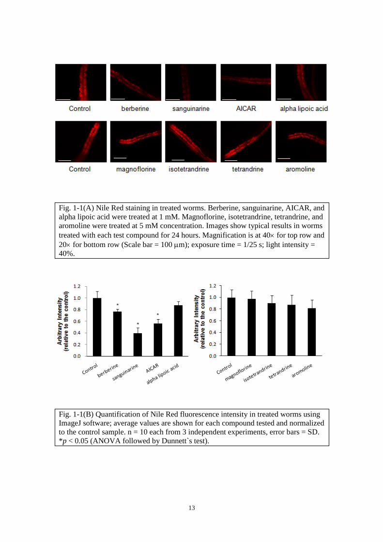
After 24 hours, AICAR, berberine and sanguinarine treated worms showed marked
reduction of lipid accumulation as reflected in weaker fluorescence intensity compared
to untreated worms (Fig. 1-1A, B), whereas other alkaloids showed little or no reduction
of fluorescence up to 5 mM concentration. At 1 mM concentration, alpha lipoic acid
showed weak reduction of lipid. It is noted that with 24-hour treatment in this
experiment set-up, no abnormality in the worms` growth or mobility associated with the
lipid accumulation changes was observed.
13
Fig. 1-1(A) Nile Red staining in treated worms. Berberine, sanguinarine, AICAR, and
alpha lipoic acid were treated at 1 mM. Magnoflorine, isotetrandrine, tetrandrine, and
aromoline were treated at 5 mM concentration. Images show typical results in worms
treated with each test compound for 24 hours. Magnification is at 40 for top row and
20 for bottom row (Scale bar = 100 m); exposure time = 1/25 s; light intensity =
40%.
Fig. 1-1(B) Quantification of Nile Red fluorescence intensity in treated worms using
ImageJ software; average values are shown for each compound tested and normalized
to the control sample. n = 10 each from 3 independent experiments, error bars = SD.
*p < 0.05 (ANOVA followed by Dunnett`s test).

14
The lipid reduction effects of berberine and sanguinarine were verified using Oil
Red O stain (Fig. 1-1C, D). Among the non-effective compounds tested with Nile Red
stain, only magnoflorine and tetrandrine were confirmed using Oil Red O stain due to
limited availability of chemicals (Fig. 1-1 E, F).
(C) (D)
(E) (F)
Fig. 1-1(C) Oil Red O staining in treated worms. Images show typical results in
worms treated with berberine or sanguinarine for 24 hours. Magnification at 10
(Scale bar = 100 m). (D) Quantification of Oil Red O staining intensity in treated
worms using ImageJ; average values are shown for each compound tested and
normalized to the control sample, error bars = SD. *p < 0.05 (ANOVA followed by
Dunnett`s test).
Fig. 1-1(E) Oil Red O staining in treated worms. Images show representative results
in worms after treatment with magnoflorine or tetrandrine for 24 hours.
Magnification at 20x (scale bar = 100 m). n = 10-12 each from 3 independent
experiments. (F) Quantification of the intensity of Oil Red O staining in treated
worms using ImageJ. Values are the average of 10-12 images for each compound
tested and are normalized to the control sample, error bars = SD.

15
The lipid-reducing effects of berberine and sanguinarine were found to be dose-
dependent. Interestingly, sanguinarine exhibited more potent lipid reduction activity
than berberine. When tested with lower concentration of sanguinarine, 25 M still
showed significant reduction of lipid accumulation, at almost 20-fold lower than the
effective concentration of berberine (Fig. 1-2A, B). Further experiments were carried
out to elucidate the difference in lipid reduction efficacy between berberine and
sanguinarine based on the postulated pathways they act upon.
(A) (B)
Enhanced phosphorylation of AMPK in berberine- and sanguinarine-treated worms
The involvement of AMPK pathway in berberine-induced fat reduction has been
reported, whereby the inhibition of ACC in HepG2 cells resulted in the inhibition of
cholesterol and triglyceride synthesis (Hawley et al., 2010; Brusq et al., 2006). Here, the
effect of sanguinarine on AMPK and its downstream substrate, ACC, was further
characterized in comparison with berberine. To examine the activation conditions of
AMPK and ACC, their phosphorylation conditions were checked using anti-phospho
AMPK (pAMPK)- and anti-phospho ACC (pACC)-specific antibodies. Sanguinarine
and AICAR treatment for 24 h induced phosphorylation of AMPK but phosphorylation
of ACC was insignificant. Meanwhile, berberine had only a marginal effect on AMPK
Fig. 1-2 Oil Red O staining in worms treated with (A) berberine and (B) sanguinarine
for 24 hours. Magnification at 20x (scale bar = 100 m). n = 10-12 each from 3
independent experiments, error bars = SD. *p < 0.05 (ANOVA followed by
Dunnett`s test). C is control.

16
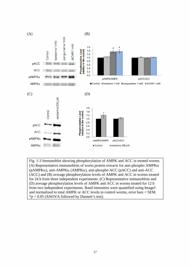
and ACC (Fig. 1-3A, B). Transient AMPK activation could be a possible reason for the
weak phosphorylation detected. For instance, experiments using AICAR to activate
AMPK had been reported whereby maximum phosphorylation activity was observed at
60 min (Pang et al., 2010) and at 2 h (Anilkumar et al., 2013) after which it gradually
reduced to base level. Han et al. (2010) reported that berberine increased
phosphorylation of AMPK and ACC in a dose- and time-dependent manner which
peaked at 60 min after addition of 10 M berberine to bovine aortic endothelial cells.
Since lipid reducing effect in worms using staining method was observed after 24 h
alkaloid treatment, protein samples from 24 h-treated worms were used in earlier
experiments. As follow-up, worms were treated with 500 M berberine for 12 h and
checked for AMPK and ACC activation conditions. Although statistically insignificant,
increased AMPK phosphorylation was obvious when compared to 24 h treated worms
(Fig. 1-3C, D). These results imply that there is a time lag between the peak of AMPK
activation and the observable lipid-reducing effect. Therefore, time-course experiment
would be necessary in future experiments to detect maximal AMPK activation by each
test compound.
17
(A) (B)
(C) (D)
Fig. 1-3 Immunoblot showing phosphorylation of AMPK and ACC in treated worms.
(A) Representative immunoblots of worm protein extracts for anti-phospho AMPK
(pAMPK), anti-AMPK (AMPK), anti-phospho ACC (pACC) and anti-ACC
(ACC) and (B) average phosphorylation levels of AMPK and ACC in worms treated
for 24 h from three independent experiments. (C) Representative immunoblots and
(D) average phosphorylation levels of AMPK and ACC in worms treated for 12 h
from two independent experiments. Band intensities were quantified using ImageJ
and normalized to total AMPK or ACC levels in control worms, error bars = SEM.
*p < 0.05 (ANOVA followed by Dunnett’s test).

18
Knockdown of the C. elegans homologs of catalytic -subunits of AMPK revealed
involvement of aak-2 in the lipid modulation mechanisms for berberine and
sanguinarine
To investigate the mechanism through which AMPK activation might have induced
lipid reduction in treated worms, the genes encoding the two C. elegans homologs of
the catalytic -subunits of AMPK, aak-1 and aak-2 were knocked down. AAK-1 and
AAK-2 are 40% and 52% identical to, and they share 71% and 80% amino acid identity
respectively with the kinase domain of the human AMPKα subunits, including
conservation of a critical threonine residue whose phosphorylation is required for
AMPK activation (Apfeld et al., 2004). The function of AAK-1 was much
uncharacterized until a recent study by Lemieux et al. (2011) through which they
identified an undisclosed compound F17 that activated AMPK signaling in C. elegans
via aak-1.
Although phosphorylation of AMPK was observed, further RNAi experiments
showed that lipid reduction activity of berberine and sanguinarine was still effective in
aak-1 but suppressed in aak-2 RNAi worms (Fig. 1-4A, B).
(A)

19
(B)
Although less viable compared to wild-type worms, aak-1(tm1944) mutants was
reported to survive better than aak-2(ok524) mutants during larval L1 diapause
(Fukuyama et al., 2012). It suggests that aak-2 has a more critical function in promoting
survival under nutritional stress condition. According to the findings of Li et al. (2006),
the 1 and 2 isoforms of the catalytic AMPK subunits are present in most tissues but
are differentially activated. Salt et al. (1998) reported that the α1-isoform was less
activated than the α2-isoform in the rat’s heart and skeletal muscle suggesting the lower
sensitivity of the α1- than the α2-isoform complexes to smaller increases in AMP
concentration.
In this study, quantitative real-time PCR analysis of the transcripts level of aak-1
and aak-2 in aak-1 RNAi worms showed that aak-2 expression was induced to a ratio
comparable to the reduction of aak-1 whereas the aak-1 expression level was unaffected
in aak-2 RNAi worms (Fig. 1-5A, B). This result suggests that the worms’ AMPK -
subunit expressions are regulated mainly through aak-2 to maintain endogenous AMPK
Fig. 1-4 Knockdown of the C. elegans homologs of catalytic -subunits of AMPK
revealed that aak-2 was involved in the mechanisms of lipid-modulation for berberine
and sanguinarine. The relative intensity of Oil Red O-stained lipid droplets quantified
using ImageJ in (A) aak-1 and (B) aak-2 RNAi worms treated for 24 hours with
berberine, B at 200 M or 400 M and sanguinarine, S at 25 M. n = 10-12 for each
compound tested; error bars = SD; *p < 0.05 (ANOVA followed by Dunnett`s test).
pL4440 – RNAi vector control, aak-1i – aak-1 RNAi worms , aak-2i – aak-2 RNAi
worms.

20
level and it accounts for most of the basal activity and activation by berberine and
sanguinarine. It also implies that, in this worm, the aak-1 isoform of the AMPK α-
subunit is less responsive to activation by berberine and sanguinarine.
(A) (B)
Lipid- reducing effects of berberine and sanguinarine are independent of the nhr-49
regulated fatty-acid beta-oxidation pathway
Nuclear receptors (NRs) are a diverse class of transcription factors that mediate
hormonal signaling processes in vertebrates and insects and now known to extend
beyond direct transduction of endocrine signals to include responses to a variety of
signaling molecules, participation in multiple signal transduction pathways, and
regulation of diverse physiological and developmental processes. While many of the
mechanisms by which ligand-regulated, hormone-responsive NRs activate or repress the
transcription of target genes have been well characterized, the remaining orphan NRs
lack known cognate ligands (Gissendanner et al., 2004). The C. elegans genome
sequence contains 284 confirmed or predicted NR genes which is over 5-fold more than
Fig. 1-5 Quantitative RT-PCR of aak-1 and aak-2 transcript levels in (A) aak-1 and
(B) aak-2 RNAi worms. mRNA abundance values represent the average of triplicate
cDNA templates applied to run qRT-PCR and the results were normalized against
cdc-42 as an internal control. Results were verified by reproducibility in at least two
of three independent experiments and representative results from one experiment are
shown, error bars = SD. *p < 0.05, **p < 0.005, two-tailed t-test. pL4440 – RNAi
vector control, aak-1i – aak-1 RNAi worms, aak-2i – aak-2 RNAi worms.

21
the number found in the human genome (Maglich et al., 2001), with a few of them
exhibiting conserved physiological functions across taxa.
The C. elegans nuclear hormone receptor gene nhr-49 has significant homology
with hepatocyte nuclear factor 4 (HNF4) in mammals but its function is closer to the
mammalian peroxisome proliferator-activated receptors (PPARs). nhr-49 serves as a
key regulator of fat usage by modulating fat consumption pathways and maintaining
fatty acid saturation in balance. It targets multiple enzymes involved in transporting
fatty acids across the mitochondrial membrane for -oxidation. nhr-49 knockout
exhibited high-fat phenotype that was attributed to the deficiencies in two metabolic
pathways, fatty acid -oxidation and fatty acid desaturation. nhr-49 was found to
stimulate expression of a carnitine palmitoyl transferase (F09F3.9) which acts
downstream of acyl-CoA synthetase in shuttling activated acyl-CoAs into the
mitochondrial matrix (Van Gilst et al., 2005).
The involvement of -oxidation genes targeted by NHR-49 in lipid reducing effects
of berberine and sanguinarine was investigated through RNAi experiments. As
mitochondrial -oxidation process degrades stored fats for the production of energy,
reduced expression of the key -oxidation enzymes would result in high-fat storage.
Consistent with the reported high-fat phenotype, nhr-49 RNAi worms were found to be
slightly larger in size and had increased fat storage with Oil Red O stain (Fig. 1-6A, B).
They also had significantly lower expressions of mitochondrial -oxidation genes, i.e.,
ech-1 (encodes a mitochondrial -oxidation trifunctional enzyme), cpt-5 (encodes a
carnitine palmitoyl transferase), and acs-2 (encodes a mitochondrial acyl-CoA
synthetase) (Fig. 1-7).

22
(A) (B)
Fig. 1-6 Effects of alkaloid treatment on nhr-49 RNAi worms. (A) Relative intensity
of Oil Red O-stained lipid droplets quantified using ImageJ in nhr-49 RNAi worms
treated with berberine, B (400 M) or sanguinarine, S (25 M) for 24 hours. C is
non-treated control; n = 10-12 for each compound tested, error bars = SD. *p < 0.05
(ANOVA followed by Dunnett`s test). (B) Oil Red O staining of RNAi control worm
and nhr-49 RNAi worms. Images show typical results with worms at 20
magnification (Scale bar = 100 m).
Fig. 1-7 Quantitative RT-PCR analysis on the expression of fatty-acid oxidation
genes. The mRNA abundance value represents the average of triplicate cDNA
template applied to run qRT-PCR and the results were normalized against cdc-42,
error bars = SD. *p < 0.05 (ANOVA followed by Dunnett`s test). pL4440 – RNAi
vector control, nhr-49i – nhr-49 RNAi worm. Results were verified by
reproducibility in at least two of three independent experiments.

23
Despite their high fat accumulation, nhr-49 RNAi worms still retained sensitivity to
berberine and sanguinarine (Fig. 1-6A), suggesting that both alkaloids act independently
of nhr-49 and their fat reducing activity would be on fat biosynthesis rather than its
catabolism. However, analysis of nhr-49 transcript level in RNAi control worms treated
with berberine and sanguinarine showed reduced nhr-49 expression (Fig. 1-8). These
results suggest that they have antagonistic action on the lipid synthesis and degradation
processes. While the details of the antagonistic action of berberine and sanguinarine on
lipid synthesis (via AMPK) and degradation (via NHR-49) remains to be elucidated, the
inhibition of lipid biosynthesis through AMPK activation showed greater effect on lipid
accumulation.
Fig. 1-8 Quantitative RT-PCR analysis on the expression of nhr-49 gene. The mRNA
abundance value represents the average of triplicate cDNA template applied to run
qRT-PCR and the results were normalized against cdc-42, error bars = SD. *p < 0.05
(ANOVA followed by Dunnett`s test). pL4440 – RNAi vector control, nhr-49i – nhr-
49 RNAi worm.

24
CHAPTER II
Application of the whole-worm model to screen for lipid reducing activities of
metabolically-engineered plant cell cultures
Although many of the phytochemicals exhibit therapeutic effects, a lot of them still
cannot be synthesized efficiently in the laboratory. One possible way to enhance
production involves combination of biochemical knowledge, cloning, and
transformation techniques, such as increasing the supply of bioactive compounds
through metabolic engineering. The identification of many biosynthetic genes and
characterization of the spatial and developmental regulation of their expression had
proven their importance in the biosynthesis of secondary metabolites and revealed
bottlenecks for their production in plant cells. Metabolic engineering of plant secondary
metabolites biosynthesis pathway could improve quality of metabolites by reducing
undesired pathways, introducing new pathways to produce novel compounds, or by
completely blocking a pathway to accumulate intermediates (Chow and Sato, 2013). In
the metabolic engineering of alkaloids, isoquinoline alkaloid biosynthesis has been most
intensively investigated for production of bioactive compounds with pharmaceutical
values.
Eschscholzia californica (California poppy) accumulates pharmacologically active
alkaloids biosynthetically related to the morphinan alkaloids of Papaver somniferum.
This, in combination with the relative ease with which it is propagated in vitro, makes it
a key model for benzylisoquinoline biosynthesis (Loyola-Vargas and Vázquez-Flota,
2006). In this study, transgenic cell lines produced by heterologous expression of
CYP80G2, a cytochrome P450 gene, from Coptis japonica in E. californica cells were
screened for lipid-reducing effect in comparison with wild-type and vector control E.
californica cell cultures. In addition, effect of C. japonica cell culture extract which
contains main metabolite, berberine, was analyzed.

25
Materials and Methods
Chemicals and reagents
Sanguinarine (Sigma), magnoflorine (a gift from R. Nishida, Kyoto University), and
scoulerine ( a gift from Mitsui Chemicals, Inc., Japan), berberine sulfate (Tokyo
Chemical Industry Co., Ltd.), palmatine chloride (Mitsui Petrochemical Industries),
coptisine chloride (Wako Pure Chemicals, Osaka, Japan), columbamine (prepared in our
laboratory; Ikezawa et al.,2007). All other reagents were purchased from Wako Pure
Chemicals unless otherwise stated.
Plant cell cultures
Wild-type, vector control and transgenic CYP80G2-overexpression E. californica
cell lines were established and are maintained by our laboratory. The integration of the
CYP80G2 expression construct in transgenic cells was confirmed by genomic PCR.
Expression level of CYP80G2 in those transgenic cells was further validated by qRT-
PCR. The C. japonica (156-S) cell line was established by Sato et al. (2001) and is
maintained in our laboratory.
Fig. 2-0 Partial schematic of the isoquinoline biosynthesis pathway

26
LC-MS analysis of metabolites in E. californica cultured cells extracts
Constantly growing wild-type, vector control and transgenic CYP80G2
overexpression cells were used for the alkaloid analysis. Cultured cells were harvested
after 14 d of culture and alkaloids were extracted from 2.0 g of fresh weight cells with 8
ml of methanol for 24 h. After homogenization and centrifugation, the alkaloids in
supernatants were concentrated using rotary evaporator. The extracts were dissolved in
distilled water and used for bioassay. The contents were determined by LC-MS 2010
(Shimadzu) with isocratic 40% (v/v) acetonitrile/H2O solvent containing 0.05% (v/v)
trifluoroacetic acid (TFA) at constant flow rate of 0.8 ml/min. Column = TOSOH TSK-
Gel ODS 80-Tm silica-based, reversed phase 4.6 x 250 mm; column temperature =
40C. UV absorbance was measured at 280 nm; mass range (m/z 100 to 800) by both
single-ion and scan modes. Sanguinarine, magnoflorine, and scoulerine were identified
by direct comparison with standard chemicals in LC-MS analysis. Identification of
other main peaks was deduced from their m/z values based on previous study
(Takemura et al., 2010). Alkaloids concentrations were quantified relative to the peak
area of standards.
LC-MS analysis of metabolites in C. japonica cultured cells extracts
10.0 g of fresh weight C. japonica (156-S) cells were soaked in 100 ml methanol for
48 h. The filtered extract was concentrated using rotary evaporator and dissolved in
distilled water for bioassay and analysis by LC-MS 2010 (Shimadzu).
H2O(A)/acetonitrile (B) solvent containing 0.05% (v/v) TFA was used at constant flow
rate of 0.5 ml/min. After initial hold at 35% B for 15 min, linear gradient was applied
from 35% to 70% B in 4 min, hold at 70% B for 3 min, 70% to 35% B in 4 min and
finally hold at 35% B for 4 min. Column type, column temperature and detection
parameters were same as above. Peaks were identified by direct comparison with
standard chemicals and alkaloids concentrations were quantified relative to the peak
area of standards.
C. elegans bioassay
Two-day-old worms were treated with distilled water (as control), cell extracts, or
alkaloids for 24 hours. About 200-300 worms were collected and washed three times

27
with 1X PBS pH 7.4 buffer. Oil Red O staining and its quantification were performed as
described in Chapter I.
Results and Discussion
Alkaloid profiles of E. californica cultured cells extracts
Since CYP80G2 has been identified as corytuberine synthase (Ikezawa et al., 2008)
acting on substrate, (S)-reticuline, this infers that heterologous expression of CYP80G2
gene from C. japonica in E. californica cells may alter its alkaloid biosynthesis pathway
to branch off at (S)-reticuline and produce (S)-corytuberine and/or magnoflorine as
intermediate or end-products. It could also concurrently assume its inherent pathway to
produce (S)-scoulerine, sanguinarine, or accumulate other intermediates (Fig. 2-0).
LC-MS analysis of the cell extracts (Fig. 2-1) revealed similar alkaloids
composition in wild-type (W), vector control (G) and CYP80G2-overexpression (C)
culture cells. The main peaks identified based on known m/z values are magnoflorine
(m/z 342), scoulerine (m/z 328), sanguinarine (m/z 332), protopine (m/z 354),
allocryptopine (m/z 370), 10-hydroxychelerythrine (m/z 364), chelerythrine (m/z 348),
chelirubine (m/z 362). The CYP80G2-overexpression culture cell lines have similar or
higher magnoflorine content compared to the wild-type or vector control lines. However,
accumulation of scoulerine and sanguinarine were also found. It is noted that line C9
had remarkably higher alkaloid content compared to other transgenic cell lines with the
same mass (Fig. 2-2).

28

29
Fig. 2-1 Liquid chromatogram of wild-type (W), vector control (G) and CYP80G2-
overexpression (C) E. californica cell extracts. Labeled peaks indicate m/z 342
(magnoflorine), m/z 328 (scoulerine), m/z 332 (sanguinarine ), m/z 354 (protopine),
m/z 370 (allocryptopine), m/z 364 (10-hydroxychelerythrine), m/z 348
(chelerythrine), m/z 362 (chelirubine).

30
Fig. 2-2 (A) Total concentration of three alkaloids and the respective concentration
for (B) magnoflorine (m/z 342), (C) scoulerine (m/z 328) and (D) sanguinarine (m/z
332) in E. californica cell extracts. Semi-quantitative values are presented here as M
equivalence of the respective alkaloid calculated relative to the peak area of each
standard.

31
Lipid-reducing effect of E. californica cell extracts in C. elegans
Extracts from wild-type (W), vector control (G) and CYP80G2-overexpression (C)
culture cells with the alkaloids profile and concentration shown in Fig. 2-1 and 2-2 were
fed to the worms for 24 h and effects on lipid accumulation was observed using Oil Red
O stain. Among the extracts from 10 culture cell lines tested, only C9 showed
significant lipid reduction in worms (Fig. 2-3). One obvious factor for the effectiveness
of C9 is its high alkaloids content (244 M magnoflorine, 85 M scoulerine and 191
M sanguinarine) compared to other cell line extracts (Fig. 2-2A). By comparison of the
alkaloids composition of various cell line extracts, the profile of G9 resembles that of
C9 with similar concentration of scoulerine (80 M) and high sanguinarine(72 M)
content. Although G9 contained only 22 M magnoflorine, it is known from earlier
results that magnoflorine did not have lipid-reducing effect in the worms (Chapter I, Fig.
1-1F). This ruled out the contribution of magnoflorine in the lipid-reducing effect in
treated worms. Since both lines contained similar amount of scoulerine at 85 M (C9)
and 80 M (G9) but G9 extract did not have significant lipid-reducing effect in treated
worms, it showed that scoulerine was ineffective at lipid-reduction at those
concentrations. Moreover, C1 extract, with relatively high content of magnoflorine (135
M) and scoulerine (38 M) but low content of sanguinarine (2 M) as compared to C9,
did not reduce lipid accumulation in the worms. These results suggest that sanguinarine
is the main contributor to the lipid-reduction activity observed with C9 extract.
Although the effective concentration of sanguinarine was not determined here, it
was noted that its lipid-reducing effect was weaker in the presence of other metabolites
contained in plant cell culture extracts. For example, G9 which contained 72 M
sanguinarine did not yield lipid-reducing effect whereas 25 M sanguinarine was
sufficient to yield such effect when applied alone (Fig. 1-1D).
32
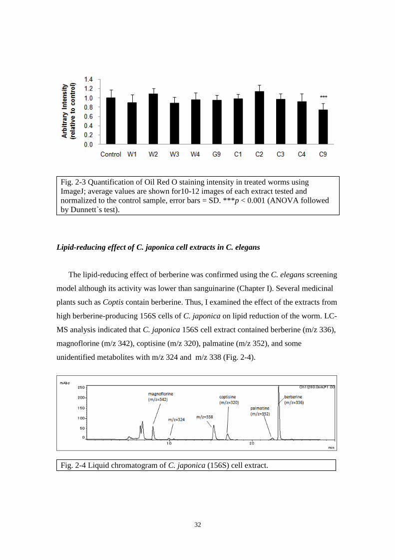
Lipid-reducing effect of C. japonica cell extracts in C. elegans
The lipid-reducing effect of berberine was confirmed using the C. elegans screening
model although its activity was lower than sanguinarine (Chapter I). Several medicinal
plants such as Coptis contain berberine. Thus, I examined the effect of the extracts from
high berberine-producing 156S cells of C. japonica on lipid reduction of the worm. LC-
MS analysis indicated that C. japonica 156S cell extract contained berberine (m/z 336),
magnoflorine (m/z 342), coptisine (m/z 320), palmatine (m/z 352), and some
unidentified metabolites with m/z 324 and m/z 338 (Fig. 2-4).
Fig. 2-3 Quantification of Oil Red O staining intensity in treated worms using
ImageJ; average values are shown for10-12 images of each extract tested and
normalized to the control sample, error bars = SD. ***p < 0.001 (ANOVA followed
by Dunnett`s test).
Fig. 2-4 Liquid chromatogram of C. japonica (156S) cell extract.

33
The C. japonica 156S cell extract was diluted to a final concentration of about 109
M berberine, 5 M palmatine and 23 M coptisine and applied to the worms as in
Chapter I. Oil Red O staining of worms after 24 h treatment showed that C. japonica
cell culture extract had stronger lipid-reducing effect than 100 M berberine applied
alone (Fig. 2-5).
Mixed compositions in extracts of E. californica and in C. japonica cell cultures
affect lipid-reducing activity differently
Plant extracts with a mixed composition of secondary metabolites are commonly
applied as traditional therapeutics. The combination of active components is believed to
produce synergistic effect for treatment of different ailment. The results here showed
that the C. japonica cell extract have a stronger lipid-reducing effect than berberine
alone. This implies a synergistic effect between berberine and other components in the
extract. In fact, some of the protoberberine alkaloids showed similar effects on lipid
accumulation as berberine (Kawasaki, unpublished results). On the contrary, results
from treatment with E. californica cell extract suggest other components had an
antagonistic effect on sanguinarine which is more effective when applied alone.
Besides the major alkaloids identified here by LC-MS analysis, there are other
bioactive components present in the cell extracts including phenolic compounds such as
flavonoids, polyphenols and phenolic acids yet to be analyzed. Phenolic compounds are
reported to have antioxidant, antimicrobial, and antitumor properties (Guimarães et al.,
Fig. 2-5 Quantification of Oil Red O staining intensity in treated worms using
ImageJ; average values are shown for10-12 images of each treatment and normalized
to the control sample, error bars = SD. **p < 0.01, ***p < 0.001, two-tailed t-test.
B – berberine, CE – cell extract.

34
2013; Wang et al., 2013; Dias et al., 2013) and their presence in crude extracts could
affect the overall bioactivity. In this study, the results indicate that the interactions
between protoberberine-type alkaloid and other metabolites in the C. japonica cell
extracts yield different bioactivity strength than that of the benzophenanthridine-type
alkaloids found in E. californica cell extract. Results of C9 suggests that its bioactivity
is more related to the constituents profile than to the amounts present in the extract since
the effective concentration of sanguinarine was determined earlier to be 25 M.
It is interesting that the non-alkaloid components might be synergistic in one extract
and antagonistic in another. While synergism of alkaloids with other plant secondary
metabolites had been reported (Eid et al., 2012a), antagonistic effects were found in
other studies. Dias et al. (2013) reported that purified extracts of wild fruits, Arbutus
unedo, Prunus spinosa, Rosa micrantha and Rosa canina, exhibited higher antioxidant
properties than the crude extracts in which antagonistic effects between the compounds
present resulted in a decrease in the antioxidant activity. While further investigations are
needed to identify and compare the other non-alkaloid chemical constituents between C.
japonica and E. californica cell cultures and to evaluate their effects on the overall
bioactivity of extracts, this worm model is useful in the preliminary screening as it
reflects a more comprehensive response of an organism as a whole compared to in-vitro
or cell-based assays. Such studies will help elucidate the interaction of bioactive
chemical constituents in cell extracts for therapeutical applications in the future.

35
CHAPTER III
RNAi knockdown of nhr-8 enhanced worms’ sensitivity to bioassay
As described in previous chapters, C. elegans is a good model organism for
isoquinoline alkaloids bioassay. However, this worm is less sensitive to chemicals
compared to other assay systems such as mammalian cell cultures (Broeks et al., 1995;
Kong et al., 2004; Burns et al., 2010). Under natural conditions, C. elegans are exposed
to large range of chemical assaults found in soil. Besides feeding on microbes, it
behaves like a scavenger in the soil environment, consuming compounds from other
animals, microorganisms, plants, and xenobiotics. As a result, it requires efficient
detoxification ability of organic chemicals for survival. Recent studies have identified
many genes encoding sensors and enzymes that the worms may use in their xenobiotics
responses such as a large number of four main classes of detoxification enzymes
including cytochromes P450 (CYP), short-chain dehydrogenases (SDR), UDP-
glucuronosyl or glycosyl transferases (UGT), and glutathione-S-transferases (GST)
(Lindblom and Dodd, 2006).
Most studies on C. elegans detoxification mechanism were attempted to circumvent
its xenobiotic resistance in screening for more effective nematicide candidates among
compounds that exhibit toxic property, including plant secondary metabolites. nhr-8 is a
gene which encodes a nuclear hormone receptor involved in xenobiotic resistance
response of the C. elegans. Although the actual target genes of nhr-8 have not been
identified yet, the precedent PXR (pregnane X receptor) and CAR (constitutive
androstane receptor) activities in vertebrates suggested that NHR-8 may regulate the
expression of the cytochrome P450 genes in C. elegans. The toxin sensitivity of nhr-8 is
specific as nhr-8 RNAi worms were found to be more sensitive than wild-type worms to
the toxins cholchicine and chloroquine but not to the pathogenic bacterium
Pseudomonas aeruginosa (Lindblom et al., 2001).
The function of plant alkaloids has been suggested as defense against herbivores and
pathogens, therefore they are likely to induce xenobiotic response and consequently
their bioavailability might be reduced in vivo. Accordingly, the isoquinoline alkaloids,
berberine and sanguinarine, were also expected to induce xenobiotics response in the

36
worms. The genome-wide response of C. elegans to berberine and sanguinarine
treatment was previously investigated by microarray analysis and the results indicated
that defense-response and detoxification genes including F08G5.6 (a defense response
gene), cyp-35C1, gst-5, ugt-21, and ugt-25 were up-regulated (data not shown).
Although nhr-8 RNAi worm had been used to investigate its sensitivity to toxic
compounds that act as nematicide, there is no report on studies done to explore nhr-8
RNAi worms’ response to the bioactivities of the compounds being treated with. In an
attempt to enhance the sensitivity of the worm screening system and to minimize the
effective concentration of test compounds needed for bioactivity assays, the
detoxification response in C. elegans was impeded by knocking down nhr-8 and
treating the nhr-8 RNAi worms with lower alkaloid concentration to check if this could
increase the alkaloid bioavailability in those worms. Next, alkaloids accumulation in the
worms was analyzed to investigate if the increased sensitivity to alkaloid treatment
could be due to higher bioavailability in the RNAi worms. In addition, the differences in
detoxification genes induction between the alkaloid-treated RNAi control and nhr-8
RNAi worms were also analyzed.
Materials and Methods
Chemicals and reagents
As described in Chapter I.
Nematode treatment
As described in Chapter I.
Oil Red O staining
As described in Chapter I.
RNA interference of nhr-8
An RNAi feeding method (Lehner et al., 2006) was used as described in Chapter I.
Part of the nucleotides of the coding regions of nhr-8 complementary DNA was used
for RNAi.

37
nhr-8 forward, 5'-ATGCCTTCGTCTTCTCCATC-3'; nhr-8 reverse, 5'-
CTCCCCAAATCCACTTTTCA-3'
nhr-8 primers were fused with Xma1 and Xho1 restriction enzyme sites and were
used to amplify the genes of interest. Each cDNA segment was cloned into feeding
vector pL4440 (A. Fire, Stanford University) with respective restriction sites and
transformed into HT115 bacterial cells.
Quantitative RT–PCR
To measure the expression levels of xenobiotic response genes, qRT-PCR was done
as described in Chapter I. The relative amount of transcript between samples was
standardized by the amplification of cdc-42 as an internal control.
Sequences of forward and reverse primers used in quantitative RT-PCR analysis:
cyp-35C1 forward, 5'-AATTGGAGGACATCCTGTCG-3'; cyp-35C1 reverse, 5'-
AAATACAGCTCGGCTCTTGC-3';
gst-5 forward, 5'-TCAAGCTCAACGGAAAAACC-3'; gst-5 reverse, 5'-
CCGAAGCCTTCAAGAAGTTG -3';
ugt-21 forward, 5'-AGGGAGAAATGCACAAATGC-3'; ugt-21 reverse, 5'-
CTTGCTGCAAATTCCACGTA-3';
ugt-25 forward, 5'-AAATCCGAGCCAAATGTCAC-3'; ugt-25 reverse, 5'-
TGCAAGCATATTCGCATTTC-3';
F08G5.6 forward, 5'-GTCCCACTGTCACAAGCTCA-3'; F08G5.6 reverse, 5'-
GTTTCGACCGAGAAATCGAG-3';
cdc-42 forward, 5'-AGCTTCATTCGAGAATGTCC-3'; cdc-42 reverse, 5'-
CTCGAGCATTCCTGGATCAT-3'.
LC-MS analysis of alkaloids and their metabolites accumulation in worms
Worms were washed with 0.1% SDS solution and then with M9 buffer to remove
compounds stuck to the worm cuticle. After washing, worms were homogenized in 2X
lysis solution (100 mM KCl, 20 mM Tris pH8.3, 0.4% SDS, 120 g/ml proteinase K)
with coptisine chloride added as internal standard. The lysis samples were incubated
60C for 1 h and then kept on ice. 100 l cold acetonitrile was added to each lysate,
38
mixed by pipeting, and centrifuged at 800 x g for 5 min. 40 l of sample was injected
for analysis by LCMS2010 system (Shimadzu) with the following parameters:
Column = Cosmosil 4.6 x 250 mm 5C18-AR-300; Column temperature = 27C;
Solvent A = 4.9:95:0.1 (acetonitrile/H2O/TFA);
Solvent B = 95:4.9:0.1 (acetonitrile/H2O/TFA) using gradient elution as shown below.
Time (min) Solvent Flow rate (ml/min)
0.00 15% B 0.7
0.15 15% B 0.7
16.75 70% B 0.7
20.00 100% B 1.0
23.00 100% B 1.0
24.00 15% B 0.7
25.00 15% B -
UV absorbance was measured at 280 nm; mass range (m/z 100 to 800) by both
single-ion and scan modes.
Accumulation of alkaloid and metabolites were calculated based on LC peak area
relative to the internal standard peak of coptisine. This value is normalized to worm
protein content in each sample. Protein was extracted with 1X Sample buffer (50 mM
Tris-HCl pH 6.8, 2% SDS, 6% 2-mercaptoethanol, 10% glycerol, 0.01% bromophenol
blue, distilled water). Protein concentrations were determined by the Bradford method.
Results and Discussion
RNAi knockdown of nhr-8 enhanced lipid reduction activity of berberine
In previous experiments with wild-type N2 worms (Chapter I), treatment with 500
M berberine and 25 M sanguinarine consistently yielded detectable lipid reduction
activity through Oil Red O staining, but lower concentration such as 50 M berberine
and 10 M sanguinarine were ineffective. In this study, the worms’ detoxification
process was impeded by knocking down nhr-8 and the effects of alkaloid treatment on
those worms were examined. Results of nhr-8 RNAi worms showed that 50 M

39
berberine was effective for lipid reduction at statistically significant level (p < 0.005)
after 24 hour treatment (Fig. 3-1A), whereas this concentration was ineffective in wild-
type worms. Although lipid reduction effect was also observed at 10 M sanguinarine
in RNAi control worms, the difference between nhr-8 and the control RNAi worms
treated with sanguinarine was significant (p < 0.05) (Fig. 3-1B).
(A) (B)
In this experiment, I used only berberine (protoberberine-) and sanguinarine
(benzophenanthridine)-type alkaloids to evaluate the susceptibility of nhr-8 RNAi
worms to the alkaloids’ lipid-reducing effect. The results showed berberine`s effect was
more enhanced than sanguinarine’s effect on the nhr-8 RNAi worms. This enhanced
sensitivity to berberine suggests that detoxification process was induced in the worms
and may have affected their sensitivity to chemical treatment. Therefore, nhr-8 RNAi
worms would be useful for future screening experiments to evaluate the full activity of
test compounds and their sensitivity to detoxification. Moreover, the amount of complex
plant alkaloids isolated for bioassay could also be reduced.
Fig. 3-1 Effects of (A) berberine and (B) sanguinarine on lipid accumulation in RNAi
control worm and nhr-8 RNAi worms. Quantification of Oil Red O staining intensity
using ImageJ. Values are the averages of 10-12 images for each compound tested and
normalized to the RNAi control sample; error bar = SD; **p < 0.005, ***p < 0.001; #p < 0.05,
###p < 0.001 (ANOVA followed by Dunnett`s test). B – berberine, S –
sanguinarine.

40
Accumulation of alkaloids in nhr-8 RNAi worms at low dosage
Next, to investigate the enhanced sensitivity of nhr-8 RNAi worms to berberine and
sanguinarine treatment, the accumulation of alkaloids and their metabolites in the
worms was analyzed to examine whether the bioavailability of alkaloids was modified
by nhr-8 knockdown.
In berberine-treated worm samples, four major metabolites of berberine were
monitored based on the metabolic analyses reported for mammalian cells, i.e.,
berberrubine (m/z 322), thalifendine (m/z 322), demethyleneberberine (m/z 324) and
jatrorhizine (m/z 338) in rat plasma, human liver microsomes, and in rat and human
urine (Zuo et al., 2006; Qiu et al., 2008; Li et al., 2011; Ma et al., 2013).
On the other hand, study on the metabolism of sanguinarine is rather limited.
Besides dihydrosanguinarine (m/z 334), which was reported as the main metabolite of
sanguinarine, some metabolites such as m/z 334 after ring cleavage, m/z 320 after
successive O-demethylation or m/z 336 after ring-cleavage of m/z 334 were also
detected in human, rat and pig liver microsomes (Deroussent et al., 2010; Zhang et al.,
2013).
Using these metabolic analyses data for berberine and sanguinarine, the alkaloid
metabolites in treated worms were analyzed based on the reported m/z values using LC-
MS (Fig. 3-2, 3-3), since standards of those metabolites were unavailable. Preliminary
experiments using wild-type worms in 6 h and 24 h treatment showed no distinct peak
difference in metabolite profiles between 6 h and 24 h. The culture media samples also
showed no difference between 6 h and 24 h. Therefore, in nhr-8 RNAi experiment, 24 h
worm samples were analyzed.
In berberine treatment, only accumulation of berberine was detected and no
molecular ion peak that corresponds to the 4 major metabolites (berberrubine,
thalifendine, demethyleneberberine and jatrorhizine) nor the glucuronide conjugates
was found. Thus, the change in berberine content was analyzed in nhr-8 RNAi
experiment. On the other hand, in sanguinarine treatment, accumulation of sanguinarine
in the worms was low and two metabolites of sanguinarine (m/z 334, m/z 336) were
detected. Thus, these metabolites contents were analyzed.

41

42
Fig. 3-2 Liquid chromatogram of (A) coptisine (IS), and alkaloids extracted from
worms, i.e., (B) pL4440 control, (C) nhr-8i control, (D) pL4440 B500 M, (E) nhr-8i
B500 M, (F) pL4440 B50 M, (G) nhr-8i B50 M, (H) pL4440 S25 M, (I) nhr-8i
S25 M, (J) pL4440 S10 M, (K) nhr-8i S10 M.
pL4440 – RNAi vector control, nhr-8i – nhr-8 RNAi worm. IS – internal standard, B
– berberine (m/z 336), S – sanguinarine (m/z 332).

43
Although I expected that the nhr-8 RNAi worms would have accumulated more
alkaloids when the detoxification response was knocked down, such increases in
alkaloid contents were only detected at low dosages, i.e. 50 M berberine and 10 M
sanguinarine but not at high dosages, i.e. 500 M berberine (Fig. 3-4A) and 25 M
sanguinarine (Fig. 3-4B). Since the availability of alkaloids in vivo would be critical for
biological activity, the increase in alkaloids accumulation in nhr-8 RNAi worms at low
concentration is an important finding. The genetic analyses of cytoprotective
mechanisms in C. elegans suggest that such mechanisms may function efficiently at low
doses but are overwhelmed at high doses (Shore and Ruvkun, 2013). This correlates
with higher alkaloid retention in the worms at higher dosages in current results (Fig. 3-
4A, B). However, it is unclear why nhr-8 RNAi worms accumulated less alkaloid than
RNAi control worms at high dosage. As discuss later, there are other orphan receptor
genes involved in xenobiotic metabolism and nhr-8 knockdown might enhance the other
detoxification pathways.
Fig. 3-3 Accumulation of berberine, sanguinarine and their metabolites in RNAi
vector control and nhr-8 RNAi worms. Semi-quantitative values are presented here as
M quivalence of coptisine, (internal standard used in worm extraction process).
These values represent the one of three independent experiments. Results were
verified by reproducibility in at least two of three independent experiments. L4–
RNAi vector control, N8 – nhr-8 RNAi worm. B – berberine, S – sanguinarine.

44
(A) (B)
The major detoxification response genes induced by berberine and sanguinarine are
mostly NHR-8 independent
Enhanced sensitivity of the nhr-8 RNAi worms to the lipid reduction effects of
berberine and sanguinarine and higher accumulation of alkaloids in the worms suggest
that the xenobiotic response in NHR-8 knockdown worms may have been impeded. To
clarify the actual target of NHR-8, the expressions of the major detoxification genes
which were up-regulated after berberine and sanguinarine treatment in wild-type worms,
were analyzed in nhr-8 RNAi worms. However, qRT- PCR analysis showed similar
increase of gene expression in nhr-8 RNAi worms after berberine and sanguinarine
treatment as in wild-type worms (Fig. 3-5).
Fig. 3-4 Relative concentration of the accumulation of (A) berberine, (B)
sanguinarine and their metabolites (m/z 334, 336) in RNAi control and nhr-8 RNAi
worms. Values are presented relative to the RNAi vector control which was set at 1
for each concentration tested. These values represent the average of three independent
experiments. Error bars = SD. L4– RNAi vector control, N8 – nhr-8 RNAi worm, B –
berberine, S – sanguinarine.

45
Differences in induction of xenobiotics response by a wide range of chemicals have
been found in C. elegans indicating its complexity as well as specificity. Such complex
response is likely to be regulated by more than a single PXR/CAR-like regulator (NHR-
8). The mediator subunit MDT-15, a transcriptional coregulator in RNA polymerase II
dependent transcription, appears to regulate a number of nhr-8 independent genes
encoding cellular metabolic enzymes including CYPs, UGTs and GSTs in response to
fluoranthene but not -naphthoflavone (Kimura et al, 1997). Taubert et al. (2008)
reported that the expressions of CYP35C1, gst-5, and ugt-25, among several other
detoxification genes, are MDT-15 dependent and nhr-8 was dispensable for MDT-15
Fig. 3-5 Expression of detoxification genes in nhr-8 RNAi worms after alkaloid
treatment. qRT-PCR showed the expression of. The mRNA abundance value
represents the average of three independent experiments (each experiment with
triplicate cDNA template applied to run qRT-PCR and the results were normalized
against cdc-42). Values are presented relative to the RNAi vector control which was
set at 1. pL4440 – RNAi vector control, nhr-8i – nhr-8 RNAi worm, B – berberine, S
– sanguinarine.

46
dependent expression of those genes. Other transcription factors may also regulate some
xenobiotic responses such as skn-1, the C. elegans homologue of Cap 'n' Collar
transcriptional regulator of phase II detoxification and the oxidative stress response
(Shore and Ruvkun, 2013).
The qRT-PCR results (Fig. 3-5) also confirmed that the major detoxification genes
up-regulated after alkaloid treatment were NHR-8-independent. Interestingly, the
expression level of ugt-21 was reduced in nhr-8 RNAi control, berberine- and
sanguinarine-treated worms. More detailed studies are needed to determine the target
genes of NHR-8 although ugt-21 could be one of them. Different sensitivity to
berberine and sanguinarine treatment in nhr-8 RNAi worms would be useful to
elucidate the target genes of NHR-8.
Although the effect of berberine was enhanced in nhr-8 RNAi worms, its effect on
their mobility and mortality was indistinguishable within 24 hours treatment. Since C.
elegans has extensive and large number of detoxification genes as shown in Fig. 3-6,
the effect of NHR-8 knockdown would be marginal on the total detoxification processes.
Berberine is reported to be substrate of the multidrug membrane transporter, P-
glycoprotein (P-gp) and are readily effluxed, thus resulting in low bioavailability (Wang
et al., 2009; Maeng et al., 2002). Increase in berberine accumulation (at 50 M) in nhr-8
RNAi worms suggests that P-gp function may be impeded. On the other hand,
sanguinarine acts as a P-gp mediated multidrug resistance reversal agent and it inhibits
ATP-binding cassette (ABC) transporter activity (Eid et al., 2012b), suggesting the
higher bioavailability and effectiveness of sanguinarine at lower concentration (25 M)
in comparison with berberine (500 M). More detailed studies of the ABC transporters
in C. elegans would be useful to elucidate the NHR-8-dependent detoxification process
and to develop more efficient bioassay systems.

47
Overall Summary
One of the advantages in using C. elegans is that about 40% of its genes associated
with human therapeutical targets have homologs in the worm, making it a good model
to study drug-target interaction and target validation (Culetto and Sattelle, 2000). Once
an activity is found, forward and/or reverse genetics can be applied to elucidate the
possible cellular targets or mechanism of activation.
In Chapter I, preliminary screening on lipid reduction activity of chemicals in C.
elegans was carried out by staining method to observe the effects through its transparent
body wall. The positive results of lipid reduction activities was extrapolated to find out
if the effect is linked to AMPK signaling pathway as was reported for berberine through
immunoblotting and gene knockdown experiments. Most of the reported studies on
AMPK activation validation utilized liver cells of rodents or other mammalian model
and this study showed that C. elegans can also be applied for such studies. These are
advantageous factors for bioactivity screening endeavor as it expands the scope of worm
application in multiple assays beyond preliminary screen. Promising candidate
compounds can be further validated in mammalian models at a later stage and could
thereby bring down the cost of drug development due to the economical benefits in
using a worm model. In this study, berberine and sanguinarine were found to have lipid-
reducing activity which is linked to the AMPK activation. This activity was dependent
on AAK-2, the C. elegans homologs of the catalytic -subunits of AMPK. On the other
hand, experiment using nhr-49 RNAi worms showed that the lipid reduction activity of
both alkaloids act independently from nhr-49 regulated fatty acid -oxidation process.
Furthermore, RNAi control worms treated with berberine and sanguinarine showed
reduced nhr-49 expression which suggests that they have antagonistic effect on lipid
synthesis and degradation processes. However, these alkaloids predominantly affect
lipid synthesis instead of fatty acid –oxidation process leading to the overall lipid
reduction observed in the worms.
Although both berberine and sanguinarine reduced lipid accumulation in C. elegans,
several different characteristics of the two alkaloids were noted. Berberine, a protopine-
type of alkaloid, required a relatively high effective dosage (500 M) for its lipid-

48
reducing activity. The phosphorylation level of the worm’s AMPK -subunit was
weaker with berberine compared to sanguinarine after 24 h treatment. On the other hand,
sanguinarine, a benzophenanthridine-type of alkaloid, showed more potent lipid-
reducing activity with an effective dosage at 25 M. Stronger AMPK -subunit
phosphorylation activity was observed with sanguinarine-treated worm after 24 h.
In Chapter II, the established assay was extended to evaluate the effects of mixed
secondary metabolites in plant cell cultures on lipid reduction. The results showed that
C. japonica cell culture extract which contains berberine as major component had
stronger lipid reducing activity than berberine at similar concentration. On the other
hand, E. californica cell culture extract with its major component sanguinarine showed
weaker activity than in single compound treatment. This shows that cell extracts contain
both synergistic and antagonistic components that may lead to differences in the
bioactivity strength compared to single alkaloids. In addition to single compound,
mixtures also possess bioactivities that are pertinent to new drug development. This
simple comparison of pure compounds and crude cell extracts would be useful to
characterize the bioactive composition in natural medicine. The rapid advancement in
the fields of plant metabolic engineering and synthetic biology would supply more
potent materials for the development of new medicine (Sato et al., 2001; Minami et al,
2008).
In Chapter III, I developed a more sensitive C. elegans bioassay system since this
worm has extensive physical, such as its thick cuticle, and molecular (detoxification
enzymes) defense mechanisms against xenobiotics. Although the effective dose of test
compounds in worm was at higher orders of magnitude than that in mammalian cell
culture (Kwok et al., 2006; Broeks et al., 1995), nhr-8 RNAi worms were found to be
more susceptible to the lipid reduction effects of some isoquinoline alkaloids, especially
berberine which showed increased sensitivity by 8-fold. Increased sensitivity to alkaloid
treatment in nhr-8 RNAi worms suggests that such detoxification mechanism may be
impeded. However, the major upregulated detoxification genes were found not impeded,
except for ugt-21 which could be a target of NHR-8. This nhr-8 RNAi worms would be
especially useful for screening of complex plant alkaloids whereby the amount isolated
for testing are scarce.

49
This collective studies show that C. elegans is a versatile, whole organism probe for
isoquinoline alkaloids bioactivities screening. Using this worm bioassay system, the
potent lipid modulation activity in certain classes of alkaloids and the probable action
mechanisms were revealed. The established assays could be extended to screen
bioactivities of more structurally diverse alkaloids as well as mixture of plant
metabolites in cell cultures in the future. This live worm model also enables concurrent
monitoring of the alkaloids’ toxic effect. Such bioassay system is useful for new drug
leads and drug-target investigations.

50
References
Ahn J, Cho I, Kim S, Kwon D, Ha T. (2008) Dietary resveratrol alters lipid metabolism-
related gene expression of mice on an atherogenic diet. J Hepatol. 49(6):1019-1028.
Anilkumar U, Weisova P, Dussmann H, Concannon CG, Konig HG, Prehn JH. (2013)
AMP-activated protein kinase (AMPK)-induced preconditioning in primary cortical
neurons involves activation of MCL-1. J Neurochem. 124(5):721-734.
Apfeld J, O’Connor G, McDonagh T, DiStefano PS, Curtis R. (2004) The AMP-
activated protein kinase AAK-2 links energy levels and insulin-like signals to lifespan
in C. elegans. Genes Dev. 18: 3004-3009
Ashrafi K, Chang FY, Watts JL, Fraser AG, Kamath RS, Ahringer J, Ruvkun G. (2003)
Genome-wide RNAi analysis of Caenorhabditis elegans fat regulatory genes. Nature.
421: 268-272.
Beale EG. (2008) 5'-AMP-activated protein kinase signaling in Caenorhabditis elegans.
Exp Biol Med (Maywood). 233: 12-20.
Broeks A, Janssen HW, Calafat J, Plasterk RH. (1995) A P-glycoprotein protects
Caenorhabditis elegans against natural toxins. EMBO J. 14(9), 1858-1866.
Brusq JM, Ancellin N, Grondin P, Guillard R, Martin S, Saintillan Y, Issandou M.
(2006) Inhibition of lipid synthesis through activation of AMP kinase: an additional
mechanism for the hypolipidemic effects of berberine. J Lipid Res. 47:1 281-1288.
Burns AR, Wallace IM, Wildenhain J, Tyers M, Giaever G, Bader GD, Nislow C,
Cutler SR, Roy PJ. (2010) A predictive model for drug bioaccumulation and bioactivity
in Caenorhabditis elegans. Nat Chem Biol. 6: 549-557.
Choi J, He N, Sung MK, Yang Y, Yoon S. (2011) Sanguinarine is an allosteric activator
of AMP-activated protein kinase. Biochem Biophys Res Commun. 413: 259-263.
Chow YL, Sato F. (2013) Metabolic engineering and synthetic biology for the
production of isoquinoline alkaloids. In Biotechnology for Medicinal Plants; S. Chandra
et al. (eds.); Springer Berlin Heidelberg, pp. 327-343.
Corsi AK. (2006) A biochemist's guide to Caenorhabditis elegans. Anal Biochem. 359:
1-17.
Culetto E, Sattelle DB. (2000) A role for Caenorhabditis elegans in understanding the
function and interactions of human disease genes. Hum Mol Genet. 9: 869-877.
Deroussent A, Re M, Hoellinger H, Cresteil T. (2010) Metabolism of sanguinarine in
human and in rat: characterization of oxidative metabolites produced by human
CYP1A1 and CYP1A2 and rat liver microsomes using liquid chromatography-tandem

51
mass spectrometry. J Pharm Biomed Anal. 52(3), 391-397.
Dias MI, Barros L, Duenas M, Pereira E, Carvalho AM, Alves RC, Oliveira MB,
Santos-Buelga C, Ferreira IC. (2013) Chemical composition of wild and commercial
Achillea millefolium L. and bioactivity of the methanolic extract, infusion and decoction.
Food Chem. 141(4):4152-4160.
Eid SY, El-Readi MZ, Wink M. (2012a) Digitonin synergistically enhances the
cytotoxicity of plant secondary metabolites in cancer cells. Phytomedicine. 2012 Nov
15;19(14):1307-14.
Eid SY, El-Readi MZ, Wink M. (2012b) Synergism of three-drug combinations of
sanguinarine and other plant secondary metabolites with digitonin and doxorubicin in
multi-drug resistant cancer cells. Phytomedicine. 19(14), 1288-1297.
Fryer LG, Parbu-Patel A, Carling D. (2002) Protein kinase inhibitors block the
stimulation of the AMP-activated protein kinase by 5-amino-4-imidazolecarboxamide
riboside. FEBS Lett. 531: 189-192.
Fukuyama M, Sakuma K, Park R, Kasuga H, Nagaya R, Atsumi Y, Shimomura Y,
Takahashi S, Kajiho H, Rougvie A, Kontani K, Katada T. (2012) C. elegans AMPKs
promote survival and arrest germline development during nutrient stress. Biol Open.
1(10):929-936.
Gissendanner CR, Crossgrove K, Kraus KA, Maina CV, Sluder AE. (2004) Expression
and function of conserved nuclear receptor genes in Caenorhabditis elegans. Dev Biol.
266: 399-416.
Guimaraes R, Barros L, Calhelha RC, Carvalho AM, Queiroz MJ, Ferreira IC. (2013)
Bioactivity of different enriched phenolic extracts of wild fruits from northeastern
Portugal: A comparative study. Plant Foods Hum Nutr. DOI 10.1007/s11130-013-0394-
5
Han Y, Wang Q, Song P, Zhu Y, Zou MH. (2010) Redox regulation of the AMP-
activated protein kinase. PLoS One. 5(11):e15420.
Hardie DG. (2011) Sensing of energy and nutrients by AMP-activated protein kinase.
Am J Clin Nutr. 93: 891S-896S.
Hawley SA, Ross FA, Chevtzoff C, Green KA, Evans A, Fogarty S, Towler MC, Brown
LJ, Ogunbayo OA, Evans AM, Hardie DG. (2010) Use of cells expressing gamma
subunit variants to identify diverse mechanisms of AMPK activation. Cell Metab. 11:
554-565.
Hwang JT, Kwon DY, Yoon SH. (2009) AMP-activated protein kinase: a potential
target for the diseases prevention by natural occurring polyphenols. N Biotechnol. 26:
17-22.

52
Ikezawa N, Iwasa K, Sato F. (2007) Molecular cloning and characterization of
methylenedioxy bridge-forming enzymes involved in stylopine biosynthesis in
Eschscholzia californica. FEBS J. 274(4):1019-1035.
Ikezawa N, Iwasa K, Sato F. (2008) Molecular cloning and characterization of
CYP80G2, a cytochrome P450 that catalyzes an intramolecular C-C phenol coupling of
(S)-reticuline in magnoflorine biosynthesis, from cultured Coptis japonica cells. J Biol
Chem. 283(14): 8810-8821.
Iwasaki Y, Tamura Y, Inayoshi K, Narukawa M, Kobata K, Chiba H, Muraki E,
Tsunoda N, Watanabe T. (2011) TRPV1 agonist monoacylglycerol increases UCP1
content in brown adipose tissue and suppresses accumulation of visceral fat in mice fed
a high-fat and high-sucrose diet. Biosci Biotechnol Biochem. 5: 904-909.
Kaletta T, Hengartner MO. (2006) Finding function in novel targets: C. elegans as a
model organism. Nat Rev Drug Discov. 5: 387-398.
Kimura KD, Tissenbaum HA, Liu Y, Ruvkun G. (1997) daf-2, an insulin receptor-like
gene that regulates longevity and diapause in Caenorhabditis elegans. Science. 277,
942-946.
Knight ZA, Shokat KM. (2007) Chemical genetics: where genetics and pharmacology
meet. Cell. 128: 425-430.
Kong W, Wei J, Abidi P, Lin M, Inaba S, Li C, Wang Y, Wang Z, Si S, Pan H, Wang S,
Wu J, Wang Y, Li Z, Liu J, Jiang JD. (2004) Berberine is a novel cholesterol-lowering
drug working through a unique mechanism distinct from statins. Nat Med. 10: 1344-
1351.
Kwok TC, Ricker N, Fraser R, Chan AW, Burns A, Stanley EF, McCourt P, Cutler SR,
Roy PJ. (2006) A small-molecule screen in C. elegans yields a new calcium channel
antagonist. Nature. 441(7089), 91-95.
Lee MS, Kim CT, Kim IH, Kim Y. (2009) Inhibitory effects of green tea catechin on
the lipid accumulation in 3T3-L1 adipocytes. Phytother Res. 8: 1088-1091.
Lee YS, Kim WS, Kim KH, Yoon MJ, Cho HJ, Shen Y, Ye JM, Lee CH, Oh WK, Kim
CT, Hohnen-Behrens C, Gosby A, Kraegen EW, James DE, Kim JB. (2006) Berberine,
a natural plant product, activates AMP-activated protein kinase with beneficial
metabolic effects in diabetic and insulin-resistant states. Diabetes. 55: 2256-2264.
Lehner B, Tischler J, Fraser AG. (2006) RNAi screens in Caenorhabditis elegans in a
96-well liquid format and their application to the systematic identification of genetic
interactions. Nat Protoc. 1: 1617-1620.
Lemieux GA, Liu J, Mayer N, Bainton RJ, Ashrafi K, Werb Z. (2011) A whole-
organism screen identifies new regulators of fat storage. Nat Chem Biol. 7: 206-213.

53
Li J, Coven DL, Miller EJ, Hu X, Young ME, Carling D, Sinusas AJ, Young LH.
(2006) Activation of AMPK alpha- and gamma-isoform complexes in the intact
ischemic rat heart. Am J Physiol Heart Circ Physiol. 291(4):H1927-1934.
Li Y, Ren G, Wang YX, Kong WJ, Yang P, Wang YM, Li YH, Yi H, Li ZR, Song DQ,
Jiang JD. (2011) Bioactivities of berberine metabolites after transformation through
CYP450 isoenzymes. J Transl Med. 9:62.
Lindblom TH and Dodd AK. (2006) Xenobiotic detoxification in the nematode
Caenorhabditis elegans. J Exp Zool A Comp Exp Biol. 305: 720-730.
Lindblom TH, Pierce GJ, Sluder AE. (2001) A C. elegans orphan nuclear receptor
contributes to xenobiotic resistance. Curr Biol. 11: 864–868.
Link EM, Hardiman G, Sluder AE, Johnson CD, Liu LX. (2000) Therapeutic target
discovery using Caenorhabditis elegans. Pharmacogenomics. 1: 203-217.
Loyola-Vargas VM, Vázquez-Flota F. (2006) An introduction to plant cell culture: Back
to the future. Methods Mol Biol. 318:3-8.
Ma JY, Feng R, Tan XS, Ma C, Shou JW, Fu J, Huang M, He CY, Chen SN, Zhao ZX,
He WY, Wang Y, Jiang JD. (2013) Excretion of berberine and its metabolites in oral
administration in rats. J Pharm Sci. 102(11), 4181-4192.
Maeng HJ, Yoo HJ, Kim IW, Song IS, Chung SJ, Shim CK. (2002) P-glycoprotein-
mediated transport of berberine across Caco-2 cell monolayers. J Pharm Sci. 91(12),
2614-2621.
Maglich JM, Sluder A, Guan X, Shi Y, McKee DD, Carrick K, Kamdar K, Willson TM,
Moore JT. (2001) Comparison of complete nuclear receptor sets from the human,
Caenorhabditis elegans and Drosophila genomes. Genome Biol. 2: RESEARCH0029.
McKay RM, McKay JP, Avery L, Graff JM. (2003) C. elegans: a model for exploring
the genetics of fat storage. Dev Cell. 4: 131-142.
Minami H, Kim JS, Ikezawa N, Takemura T, Katayama T, Kumagai H, and Sato F.
(2008) Microbial production of plant benzylisoquinoline alkaloids. Proc Natl Acad Sci
USA. 105: 7393–7398.
O’ Rourke EJ, Soukas AA, Carr CE, Ruvkun G. (2009) C. elegans major fats are stored
in vesicles distinct from lysosome-related organelles. Cell Metab., 10(5), 430-435.
Pang T, Rajapurohitam V, Cook MA, Karmazyn M. (2010) Differential AMPK
phosphorylation sites associated with phenylephrine vs. antihypertrophic effects of
adenosine agonists in neonatal rat ventricular myocytes. Am J Physiol Heart Circ
Physiol. 298(5): H1382-1390.

54
Qiu F, Zhu Z, Kang N, Piao S, Qin G, Yao X. (2008) Isolation and identification of
urinary metabolites of berberine in rats and humans. Drug Metab Dispos. 36(11), 2159-
2165.
Salt I, Celler JW, Hawley SA, Prescott A, Woods A, Carling D, Hardie DG. (1998)
AMP-activated protein kinase: greater AMP dependence, and preferential nuclear
localization, of complexes containing the alpha2 isoform. Biochem J. 334 (Pt1):177-187.
Sato F, Hashimoto T, Hachiya A, Tamura K, Choi KB, Morishige T, Fujimoto H, and
Yamada Y. (2001) Metabolic engineering of plant alkaloid biosynthesis. Proc Natl
Acad Sci USA. 98: 367-372.
Shore DE, Ruvkun G. (2013) A cytoprotective perspective on longevity regulation.
Trends Cell Biol. 23(9), 409-420.
Stelling J, Sauer U, Szallasi Z, Doyle FJ 3rd, Doyle J. (2004) Robustness of cellular
functions. Cell 118: 675-685.
Stiernagle, T. (2006) Maintenance of C. elegans. In WormBook, ed. The C. elegans
Research Community, WormBook, doi/10.1895/wormbook.1.101.1,
http://www.wormbook.org.
Takemura T, Ikezawa N, Iwasa K, Sato, F. (2010) Metabolic diversification of
benzylisoquinoline alkaloid biosynthesis through the introduction of a branch pathway
in Eschscholzia californica. Plant Cell Physiol. 51(6): 949-959.
Taubert S, Hansen M, Van Gilst MR, Cooper SB, Yamamoto KR. (2008) The Mediator
subunit MDT-15 confers metabolic adaptation to ingested material. PLoS Genet.
4:e1000021.
Van Gilst MR, Hadjivassiliou H, Jolly A, Yamamoto KR. (2005) Nuclear hormone
receptor NHR-49 controls fat consumption and fatty acid composition in C. elegans.
PLoS Biol. 3: e53: 0301-0312.
Wang SS, Wang DM, Pu WJ, Li DW. (2013) Phytochemical profiles, antioxidant and
antimicrobial activities of three Potentilla species. BMC Complement Altern Med.
13(1):321.
Wang YX, Wang YP, Zhang H, Kong WJ, Li YH, Liu F, Gao RM, Liu T, Jiang JD,
Song DQ (2009) Synthesis and biological evaluation of berberine analogues as novel
up-regulators for both low-density-lipoprotein receptor and insulin receptor. Bioorg
Med Chem Lett., 19, 6004-6008.
Yen K, Le TT, Bansal A, Narasimhan SD, Cheng JX, Tissenbaum HA. (2010) A
comparative study of fat storage quantitation in nematode Caenorhabditis elegans using
label and label-free methods. PLoS One., 5(9). pii: e12810.

55
Zhang HH, Wu Y, Sun ZL, Liu ZY. (2013) Identification of sanguinarine metabolites in
pig liver preparations by accurate mass measurements using electrospray ionization
hybrid ion trap/time-of-flight mass spectrometry. Rapid Commun Mass Spectrom. 27(9),
979-984.
Zuo F, Nakamura N, Akao T, Hattori M. (2006) Pharmacokinetics of berberine and its
main metabolites in conventional and pseudo germ-free rats determined by liquid
chromatography/ion trap mass spectrometry. Drug Metab Dispos. 34(12), 2064-2072.

56
Acknowledgements
I am honored and grateful to the Ministry of Education, Culture, Sports, Science and
Technology (MEXT) of Japan for being granted a scholarship for postgraduate study in
Japan.
I would like to extend my sincere gratitude and appreciation to my supervisor, Professor
Fumihiko Sato, for his support and guidance throughout the course of my research. His
open-mindedness and resourcefulness have allowed me to explore this new project on
bioactivity screening in the lab.
I thank Dr. Endo, Dr. Ifuku, and Dr. Koyama for constructive comments on my research
project and Ms. Hirata for her helpfulness in dealing with the administrative matters.
I am also thankful to Professor Nishida and Dr. Honjoh from the Lab of Signal
Transduction at the Graduate School of Biostudies for their provision as well as
assistance in carrying out preliminary microarray analysis of worm samples in their lab.
Last but not least, I am grateful to my family, especially my late father, whose
pioneering spirit had instilled in me the courage and tenacity to take on new challenges.
Their kindness and warm encouragement are deeply appreciated.
The following academic journal publication formed the basis for this doctoral thesis:
Yit-Lai Chow and Fumihiko Sato (2013) Screening of Isoquinoline Alkaloids for Potent
Lipid Metabolism Modulation with Caenorhabditis elegans. Biosci Biotechnol Biochem.
77(12): 2405-2412.

57
Publications
Peer-reviewed
Chow YL, Sato F. (2013) Screening of Isoquinoline Alkaloids for Potent Lipid
Metabolism Modulation with Caenorhabditis elegans. Biosci Biotechnol Biochem.
77(12): 2405-2412.
Non-peer-reviewed
Chow YL, Sato F. (2013) Metabolic Engineering and Synthetic Biology for the
Production of Isoquinoline Alkaloids. In Biotechnology for Medicinal Plants; S.
Chandra et al. (eds.); Springer Berlin Heidelberg, pp. 327-343.
Takemura T, Chow YL, Todokoro T, Okamoto T, Sato F. (2010) Over-expression of
rate-limiting enzymes to improve alkaloid productivity. Methods Mol Biol.; 643:95-109.