Embed Size (px)
Citation preview
Title A HISTOLOGICAL STUDY OF THE AFFERENTINNERVATION OF THE OVARY OF THE DOG
Author(s) Sato, Hiromasa
Citation 日本外科宝函 (1955), 24(5): 456-469
Issue Date 1955-09-01
URL http://hdl.handle.net/2433/206212
Right
Type Departmental Bulletin Paper
Textversion publisher
Kyoto University
456 日本外科宝函第24巻第5号
A HISTOLOGICAL STUDY OF THE AFFERENT
INNERVATION OF THE OVARY OF THE DOG
by
HIROMASA SATO
From the 2nd Surgical Division, Kyoto University Medical School. (Director: Prof. Dr. YASUMASA AoYAGr) 〔Receivedfor Publication Aug. 2, 1955〕
I INTRODUCTION
K. KAWAKAMI and A. KuBo have reported on sensory reactions caused by stimu-
lation of the ovary of the dog, and Sh. AsAr has proved the existence of ovarian
sensitivity in rabb:ts from the physiological point of view. According to Ch. Kr-
MURA and Y. YosmrKE of our clinic, pa'.n sensitivity can be aroused in human
ovaries by stimulati時 themwith electric faradization or injecting acetylcholine into
them, and the exist巴nceof ovari2.n sensitivity can be also proved in cats by similar
methods.
In the human ovary, J. SAKAGUCHI found nerve endings which end freely and
can be distinguished clearly from end-apparati of the autonomic nervous system m
many respects and maintained that these nerve endings belong to sensory nerve end-
apparati (SETO). But he did not pursue the routes of these nerves.
The fact that the sympathetic nerves, the vagus and the sacral parasympathetic
nerves all contain visceral afferent五bershas been proved by many investigators on
the basis of their histological or physiological studies. As regards the innervation of
the O¥ ary, G. A.G. MITCHELL, A. KUNTZ, J. C. ¥VHITE, R.H. SMITHWICK, T. KuRE
and Sh. OKINAKA have recognized the fact that th巴 ovaryis innervated through the
ovarian plexus which consists of thoraco-lumbar sympathetic fibers. But I could
find no literature which proved anatomically the innervation of the ovary through
the vagus or the sacral parasympathetic nぞれぞs. So, it can be guessed that three
main pathways, the vagus, the thoraco-lumbar らympatheticnenes and the sacral
parasympathetic nぞrHs,play important roles in conducting the a妊erent impulses
from the 0¥-ary; of thぞ町 the sympathetic nerve is the most important. Neverthe-
less, I have found no literature of any histological study on this subject, so I at-
tempted to solve the problem histologically.
II MATERIALS AND METHODS
At the beginning of my study, preparations of extirpated human ovary were
stained with BIELSCHOWSKY-SETo's and BIELSCHOWSKY-SUZUKI’s silver methods. In
addition to autonomic nerve五bersand end-apparati (Fig. 1), the presence of SETO’s
sensory nerve五bersand endings, which had been recognized by J. SAKAGUCHI, was
con五rm吋(Fig.2, 3 and 4,). The existence of myelinated五berswas also con五rmed
by staining with EHRLICH’s acid hematoxyline method (Fig. 5).
Next, preparations of ovaries of normal adult dogs were stained ¥Yith the same

THE AFFERENT INNERVATION OF THE OVARY OF THE DOG 457
three methods, and the nerve apparati were examined, comparing them with those of human ovaries.
Adult female dogs were mainly used. ’The spinal nerves and the vagus were
sectioned at various points, and the secondary degenerations of nerve五bersin the
ovaries were investigated mainly by EHRLICH’s method and also by BIELSCHOWSKY-SuzuKr's silver method.
It could be anticipated from the results of many previous experiments performed
by many investigators, that the greater part of the a妊erentfibers from the ovary
pass through the sympathetic trunk. J. N. LANGLEY had described that the visceral
a妊erent五berswhich pass through the sympathetic trunk have their cell-stations in
the dorsal root ganglia. Therefore, the dorsal roots (τh.10・・・L.2) on one side
(right s'.de) of three female adult dogs were sectioned at points distal to their gangFa.
Then both ovaries were extirpated 7 days later in the五rstcase, 14 days later in the
second ca日eand 21 days later in the third case, and were examined to determine
whether any changes had occured in the nerves supplying them. In each case I
could五ndno di妊erencebetween the left ovaries and normal ovaries when stained
with EHRLICH’s method and BIELSCHOWSKY-SETO’s method. On the other hand in the
right ovaries (sectioned s:de), I found marked diminution of myelirated五berswhen
stained w;th EHRLICH’s method and of SETO’s sensory nerves when stained with
BIELSCHOWSKY-SETO’s method. Moreover, I found a few degenerated 五bersin the
exfrpated ovary of the right side 7 days after the operation nsing EHRLICH’s method
(Fig. 6). Considering these results, I thought that removing the ovaries 7 days
or more after the rhizotomies was too late. Therefore, the ovaries of another dog
were extirpated 5 days after a similar rhizotomy (Th. 10・・・L.2 of the right side),
and were i1westigated. ’Then I found many degenerated五bersin the right ovary
of this dog with EHRLicH's method as well as BrnLscHowsKY-SuzuKr's method. So,
I determined to extirpate ovaries 5 days after the section of the spinal roots at
points distal to their ganglia.
Operations were performed as follows :
( 1) Section of the dorsal roots on the right side (τh. IO・・・L. 2).
( 2) Section of the dorsal roots on both sides (Th. 9…Th. 12).
( 3 ) Section of the dorsal roots on both sides (Th. 13…L. 4).
( 4) Section of the dorsal roots on both sides (L. 5・ーし 7).
( 5) Section of the dorsal roots on the right side (L. 6…s. 3).
( 6) Section of the dorsal roots on both sides (S. 1・・・s.3).
(two dogs, one adult and the other three months old were operated.)
(7) Section of the dorsal roots on the right side (S. 1…s. 3).
( 8) Section of the ventral and dorsal roots on the right side (S. I…s. 3).
( 9) Section of the ventral roots on the right s'.de (S. 1…s. 3).
(lo) Section of the ventral and dorsal roots on the right side (S. l・・・Co. 5).
(11) Section of the ventral and dorsal roots on the right side (Co. 1・・Co. 5).
Then vagotomies were performed as follows, to decide whether a旺erentnerYes
from the ovary are present in the vagus nerve or not.
458 日本外科宝函第24巻第5号
(12) Cervical vagotomy on the right side at a point distal to the ganglicn nodosum.
(Ovaries were extirpated 6 days after the operation.)
(13) Cervical vagotomy on the left side at a point distal to the ganglion nodosum.
(Ovaries were extirpated 6 days after the operation.)
(14) Bilateral vagotomy in the thorax. (Ovaries were extirpated 7 days after the
operation.)
III MICROSCOPIC OBSERVATIONS
A) Intrinsic nerves of the OYaries of normal adult dogs.
First, preparations were examined using BrnLscHOWSKY-SETO’s and BIELSCHOWSKY-
SuzuKI’s silv町 methods. Jn the nerve bundle at the hilum, sensory nerves (SETO),
which ar巴 thickand have characteristic variations in their thickness (varicosities),
are distingu:shed clearly from the autonomic nerve fibers which are thin and smooth
(Fig. 7). After entering the stroma of the ovary and anastomosing with one
another, the greater part of the autonomic fibers spreads over the medulla and only
some parts spread over the cortex or the adventitia of the vessels, and there, they
branch into五nerand finer fibers without ending freely, and at last form closed “nervδse Terminalreticulum (STi:iHR)”(Fig. 8 and 9). These“Terminalreticulum打 aresimilar
to those of the human ovary recognized by 1¥1. GOECKE and J. SAKAGUCHI.
Many in,-estigators han had many di妊erentopin'.ons as to nerve distribution to
follicles. In dog ovaries A. I"uNTZ have observed no nerve五berswhich penetrate
follicles or terminate in follicles. In human ovaries J. SAKAGUCHI have recognized
that a fぞれア nerve 五berspenetrate into the follicle cell layer but no fibers enter
the oYa. According to the latest report by H. KNOCHE, winding or spiral non-
myelinated nerve fibers can be found in the ooplasm around the ovum nuclei of
prime-follicles, in ape ovaries. In a fe,v preparations of dog ovaries, I could pursue
some fine nerve五bersclose to thぞ ovaof _prime-follitles, but I could not assert that these五bersterminate in relation to the ova.
、Onthe other hand, sensory nerves, too, are morphologically similar to those of
the human oyary recognized by J. SAKAGucm; sensory nerves have characteristic
variai:ions in their thickness (varicosities) and pathways, which distinguish sensory
nerves from autonomic fibers, and they terminate in many branched terminal endings
in the medulla and cortex in contact with the medulla (Fig. 10). In addition to
these sensory nerves, I could 五ndno sensory endings with a speci五ccomplicated from. No ganglion was found in any part of the ovary of dogs.
Further investigations were performed with EHRLICH’s method. After passing
through the hilum into the parenchyma along with many non-myelinated fibers,
myelinated五bershave similar pathways and branchings to those of sensory nerves
(SETO) described above (Fig. 11). Few myelinated五berswere found in the cortex,
and fine branchings as recognized by the silver method could not be found by this
method. ドo,it is reasonable to think that afferent fibers become non-myelinated when approaching their ends.
B) Secondary degeneration of myelinated五bersand F瓦TO’snerves in the ovaries

THE AFFERENT INNERVATION OF THE OVARY OF THE DOG 459
after sectioning. (1) Section of the dorsal roots on the right side (Th.lO・・・L.2).
The ovary of the right side ・ ・・ Secondary degeneration of a majority of myeli-
nated五bersin the nerve bundles at the hilum and in the parenchyma was found
with EHRLICH’s method (Fig. 12 and Fig. 13). With BrnLsCHOWSKY-SuzuKI’s method,
secondary degeneration or disappearance of a majority of sensory nerves was recog-
nized in the nerve bundles at the hilum (Fig. 16a, 16b and 16c). In the parenchyma,
no secondary degeneration of the axis-cylinders was found with the silver method.
So, as for the degeneration of the axis-cylinders, it may be presumed that the
nearer to the endings they are, the more rapidly they degenerate and are absorbed.
The ovary of the left side ・ ・ ・ No degenerated nerve五berswere found.
( 2 ) Section of the dorsal roots on both sides (Th. 9・ーτh.12).
Secondary degeneration of a minority of the myelinated五bersin the nerve bund-
les at the hilum and in the parenchyma of the ovaries of both s'.des vvas found
with EHRLICH’s method (Fig. 14).
( 3) Section of the dorsal roots on both sides (τh. 13・・・L. 4).
With EHRLICH'S method, degeneration of a majority of the myelinated fibers in
the nerve bundles at the hilum and in the parenchyma was found (Fig. l 7a, l 7b and 17c).
( 4 ) Section of the dorsal roots on both sides (L. 5・・・L.7).
No degenerated nerve五berswere found in the ovaries of either side.
( 5) Section of the dorsal roots on the right side (L. 6・・・S. 3).
No degenerated nerw印)erswere found in the ovaries of either s'.de.
( 6) Section of the dorsal roots on both sides (S. 1・・・S.3).
No degenerated nerve五berswere found in the ovaries of either s:de.
( 7) Section of the dorsal roots on the right side ($. l・・・S. 3).
No degenerated nerve fibers were found in the ovaries of either s'.de.
( 8) Section of the verrtral and dorsal roots on the right side ($. l ・ ・ ・S. 3).
No degenerated nerve五berswere found in the ovaries of either side.
( 9) Section of the ventral roots on the right s:de ($. l ・ ・ ・S. 3).
No degenerated nerve五berswere found in the ovaries of either side.
(10) Section of the ventral and dorsal roots on the right side ($. 1…Co. 5).
The ovary of the right side ・・ ・ Secondary degeneration of a minority of the
myelinated 五bersin the nerve bundles at the hilum and in the parenchyma was
found with EHRLICH'S method (Fig. 15 and 18). Secondary degen巴rating-granules
of nerve五bersin the vessel plexus at the hilum and deformation of the ax;s-cylinders
in the parenchyma, which are thought to be degeneration of the axis-cylinders, were
found with BIELSCHOWSKY-SETO’s and BIELSCHOWSKY-SUZUKI’s methods (Fig. 19 and 20).
The ovary of the left side ・・ ・ No degenerated nerve五berswere found.
(11) Section of the V白1traland dorsal roots on the right side (Co. 1…Co. 5).
No degenerated nerve五berswere found in the ovaries of either side.
(12), (13) and (14) Vagotomy.

460 日本外科宝函第24巻第5号
No secondary degeneration of nerve 五bers was found in any case in which
vagotomy was performed at various points.
IV DISCUSSION
I have found in the dog ovary as well as in the human ovary, besides nerve
fibers belonging to the autonomic nervous system which have been systematized by
P. STOHR jr., SETo’s sensory nerves which have free endings and can be clearly
distinguished from the autonomic五bersmorphologically. In general, it has been
thought that sensory endings have special complicated formations. But in the ovary
of the dog no nerve ending having such a formation was found. G. WEDDELL stated that sensory nerve endings of the skin are, in general, fre巴-endingarborizations.
And SETO’s sensory endings in the ovary of the dog have the same appearance.
H. ~ETO has asserted that SETO’s nerve, which differs from autonomic nerves mor-
phologically, may also di妊erin function, and is probably sensory in function. H.
SETO’s opinion can also be applied to the nerves in the ddg ovary.
J. N. LANGLEY’s opinion, that visceral a任erent五berspassing through the sympa-
thetic trunk have cell-stations in the dorsal root ganglia and have no interposing
cell in their periphery, has generally been recognized. In accordance with this fact,
I sectioned the spinal nerves at points distal to their ganglia, and discovered second-
ary degenerations of myelinated五bersin the ovaries, as expected. But, it is doubt-
ful whether all the myelinated五bersin the ovary are a妊erent. At least some of
the autonomic fibers are myelinated in the nerve trunk. DAHL and J. SAKAGUCHI
etc. observed no ganglion cell in the human ovary, and A. KUNTZ etc. observed
none in the ovary of the dog. In the preparations of human and dog ovaries
stained with the silver method, I, too, could find no ganglion cell.τherefore, it
is probable that the myelinated fibers in the ovary do not contain preganglionic
五bersof autonomic nerves. But there has been no evidence proving that the post-
ganglionic五bersare non-myelinated, except in the vagus. According to the study
of Sh. OKINAKA and T. KuRE, there are spinal parasympathetic nerves which are
efferent, myelinated and small-sized and which pass through the dorsal roots and
have their cell-stations in the dorsal root ganglia, and some of these nerves pass
through the white rami and reach v'scerae. If these spinal parasympathetic nerves
were myelinated till their periphery and had no interposing cells, then some of the
myelinated fibers in the ovary showing secondary degeneration might be considered
spinal parasympathetic五bers. But after sectioning the nerve trunk, not only the
small-sized myelinated五bers,but also the middle-sized myelinated fibers (over 3μ)
were found degenerated in the ovaries. Moreover, in some cases in which several
dorsal roots in a certain area were sectioned, the majority of the myelinated五bers
containing middle-sized 五bers in the ovaries of the sectioned side were found
degenerated, and marked diminution of SETO’s sensory nerves in the ovaries of the
sectioned side was rぞ「けgnizeda few days after operation. Considering these results,
it is reasonable to consider that at least the majority of the myelinated fibers in the ovary are afferent.
THE AFFERENT INNERVATION OF THE OVARY OF THE DOG 461
F. H. EDGEWORTH, basing his opinion upJn his histological study of the dog,
has asserted that the ovary has a sensory supply through large-sized myelinated
五bersin the sympathetic trunk. A. KUNTZ has stated that the afferent五hersin-
nervating the human ovary are mainly derived from the 10th thoracic nerve.τhe
sensory innervations of viscerae of frogs were studied by K. KA w AKAMI and A.
Kuso. The former proved that the sεnsation of the ovary was conducted through
a妊erent五hersin the splanchnic nerve, and the latter through afferent fibers in both
the vagus and the sympathetic nerves. The triple sensory innervation of the ovary
of the rabbit through the sympathetic nerve, the vagus and the pelvic nerve was proved by Sh. AsAr. Physiological studies by Ch. KIMURA and Y. Y osHIIKE have
shown that sensory innervation of the human ovary is thoraco-lumbar, and that
sensory innervation of the ovary of the cat is both thoraco-lnmbar and sacral, the
former being dominant. The a妊erentinnervation of the dog ovary, according to my experimental results, will be discussed as follows.
(1) The thoraco-lumbar a旺erentinnervation.
After sectioning the dorsal roots of Th. 10 ・・・ L. 2 on the right side, secondary
degeneration of a majority of the myelinated五bersin the right ovary was found,
but there was no change of any nerve五berin the left ovary. After sectioning the
t dorsal roots of Th. 13 ・・・ L. 4 on both sides, secondary degeneration of a majority of
the myelinated五bersin both ovaries was found. After sectioning the dorsal roots
of Th. 9 ・ー Th.12 on bJth sides, secondary degeneration of a minority of the myeli-
l nated fibers in both ovaries was found. After sectioning the dorsal roots of L. 5・
L. 7 on both sides, no secondary degeneration was found in either ovary. These results show that most of the myelinated 五bers in the ovary pass through the
thoraco-lumbar dorsal spinal roots of the same side (mainly Th. 13 ・・・ L. 2), and therefore the a妊erentinnervation of the ovary is chie自y thoraco-lumbar. This
fact coincides with many former physiological investigations, especially with the
experimental results of Y. YosHIIKE, and with the opinion of ]. F. Fu工TONetc.
that the area of the thoraco-lumbar outflow of autonomic nerves is Th. 1 or Th. 2
・L.4 in the dog and the white rami are restricted to the thoraco-lumbar region.
(2) The sacral a旺erentinnervation. I have found secondary degeneration of a minority of the myelinated五bersin the
right ovary of the dog, when the ventral and dorsal spinal roots ($. l ・・・Co. 5) were
sectioned on the right side. Nevertheless, in many other dogs in which the ventral
or dorsal spinal roots had been sectioned, no change was found in the nerve五bers in their ovaries.
]. F. FuLTON has stated that the pehァicnerves of the dog emerge from the
ぬcralsegments ($. 1・・・S.3), and Y. NITTA has proved the existence of afferent
fibers which have their cell-bodies in the sacral dorsal root ganglia, in the pelvic
nerve. In my preparations in which secondary degeneration was found, the appear-
ance of the degeneration was so clear that I could have no doubt about it. There-
fore, it is certain that in some dogs, at least, a minority of the myelinated fibers
in the ovary are innervated from the sacral region and these 五bers pass through

462 日本外科宝函第24巻第5号
the p~lvic nerves of the same s;de. But I can not be sure why in many other
dogs no degeneration could k found ; probably the sacral innervation is too indefinite
for secondary degeneration to extend to the ovary, or perhaps in some cases there
is no sacral innervation in the ovary. Anatomical study by G. A. G. MITCHELL has
shown that the sacral parasympathetic innervation of the ovary is very small, if it
exists. And according to the physiological study of Y. YosHIIKE, the sacral sensory
innervation of the ovary is slight and in some occasions can not be proved. These
anatomical and physiological studies coincide to a great extent with my experimen-
tal results. So it caロ besaid that the ovary is innervated by the sacral region,
though very slightly.
(3) The vagal a妊erentinnervation.
I have found no literature which presented histological evidence of the vagal
innervation of the ovary. Moreover, I have found only a few papers which proved
physiologically the vagal sensory innervation of the ovary, including the experiment
by A. KuBo which recognized the vagal sensory innervation in the frog’s ovary and
the study by Sh. AsAI which recognized very slight vagal sensory innervation in
the rabbit’s ovary. It has been thought that the cell-bodies of the a妊erentfibers
of the vagus lie in the ganglion nodosum (S. W. RANSON etc.), so I sectioned the
vagus in the neck or in the thorax. But in all cases no change was found in any
nerve五bersin the ovaries. Therefore, vagal a妊erent innervation to the ovary is doubtful.
By pursuing the afferent myelinated nerve五bersin the ovary, I have proved a
dual a任erentinnervation in th巴 ovaryof the dog ; one is the dominant thoraco-
lumbar innervation and the other is the secondary sacral innervation.
But I can not assert that these two innerYations are the onbア innervationof the
ovary. s. v\に RANSONand P. R. BILLINGSLEY have presented the evidence of non-
my巴linatedvisceral a任er巴ntfibers in the sympathetic trunk. And Sh. OKINAKA and
S. HIRAMATsu have asserted that the ventral spinal roots contain visceral afferent fibers. But I have not been able to trace them.
V CONCLUSION
τhe a任erentnerves in the dog ovary have been studied, by using EHRLICH’s acid
hematoxylin method, BIELSCHOWSKY-SETO’s and BIELSCHOWSKY-$UZUKI’s silver methods.
Also by pursuing the secondary degeneration of nerves in the ovary after section of
the nerve trunks, I have been able to study the route of afferent nerve distribution of the ovary, and have come to the following conclusions:
(1) Myelinated nerve五bersand SETO’s sensory nerves are also found in the dog ovarv.
(2) Most of the a妊erentmyelinated fibers of the dog ovary enter the thoraco-
lumbar spinal cord, passing through the dorsal spinal roots (mainly τh. 13・ ・・L. 2) of the same side.
(3) A minority of the a任erentmyelinat吋五bersof the ovary of some d~gs enter
the sacral spinal cord passing through the sacral roots of the same side. This
THE AFFERENT INNERVATION OF THE OVARY OF THE DOG 463
innervation is secondary compared with the dominant thoraco-lumbar innervation.
(4) Afferent vagal innervation of the ovary was not proved.
I am greatly indebted to Assistant Prof. Dr. CHuJI KIMURA of our clinic for his
constant help during the course of this study.
EXPLANATION OF THE PLATES
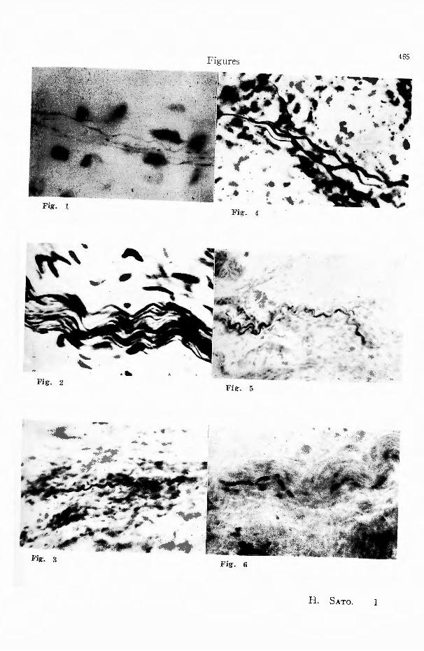
Fig. 1 "Nervose Terminalreticulum" in the me-
dulla of human ovary. x 1000 (BrnLsceowsKv-si;TO’宮 stain)
Fig. 2 SETO’S sensory nerve, which ran be dis-
tinguished clearly from the autonomic nerves,
in the nerve bundle at the hilum of human
ovary. x 500 (BrnLSCHOWSKY-SErO’s stain)
Fig. 3 SETO’s sensory nerve in the medulla of
human ovaηr. X 300 (BrnLSCROWdKY-Si>TO’s
stain)
Fig. 4 SETO・5sensory nerve near its end, in the
cortex of human ovary. x 300 (BCELSCHOWSKY-
SETO’s stain) Fig. 5 Myelinated五hersin the medulla of hu-
man ovary. x 200 (EHRLICH’s stain)
Fig. 6 Degeneration of a myelinated fiber in the medulla of a dog's right ovary extirpated
7 days after rhizotomy of Th. lO・・・L. 2 on the right side. x 400 (EHRL!Ca's stain)
Fig. 7 SETO’S sensory nerves, which can be distinguished clearly from the autonomic nerves,
in the nerve bundle at the hilum of normal
adult .dog’s ovary. x 800 (BIELSCHOWSKY-Suz-
uu's stain)
Fig. 8 Autonomic nerve五bersin the cortex in
contact with the medulla of a normal adult
dog’s ovary. x 500 (BIEL耳CHOWSKv-Sロz口KI’sstain)
Fig. 9 "Nervose Terminalreticulum”in the me-
dulla of a normal adult dog’s ovary. x 1500
(BIELSCROIVSKY -SuzuKr's stain)
Fig. 10 $ETO’s sensory nerve near its end in
the cortex in contact with the medulla of a normal adult dog’s ovary. x 500 (BIELSCH-
owsKv-SuzuK1's stain) Fig. 11 Nerve bundle containing many myelinated
fib町sat the hilum of a normal adult dog’s
ovary. x 300 (ErrnLICH’s stain)
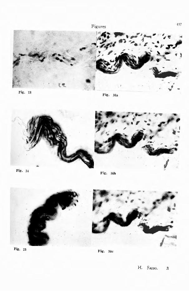
Fig. 12 Nerve bundle showing degeneration of
most of the myelinated fibers, at the hilum of
a dog’s right ovary extirpated 5 days after
rhizotomy of Th. 10・・L.2 on the right side.
X 400 (EHRLICH’s stain〕Fig. 13 Degeneration of myelinated fibers in the
medulla of a dog’s right ovary extirpated 5
days after rhizotomy of Th. 10 ・・・ L. 2 on the
right side. x 400 (EHRLICH’s stain)
Fig・. 14 Degeneration of a few myelinated fibers
in the r.erve bundle at the hilum of a dog’s left ovary extirpat吋 5days after rhizotomy
of Th. 9…Th. 12 on both sides. x 300 (Ea~- L!CR’s stain)
Fig・. 15 Degeneration of a mye!inated fiber in
the nerve bundle at the hilum of a dog’s right ovary extirpated 5 days after seヶtionof the
ventral and dorsal roots of S. 1・・・Co.5 on the right side. x 300 (EHRL!CR’s stain)
Fig. 16a, 16b and 16c Degeneration of SETo's
sensory n町vesin the nerve bundle at the hi-
!um of a dog’s right ovary extirpat吋 5days
after rhizotomy of Th. 10 ・・・ L. 2 on the right side. Photographs were taken in three layers.
x 300 (BIELSCHOIVSKY-SuzuKI's stain)
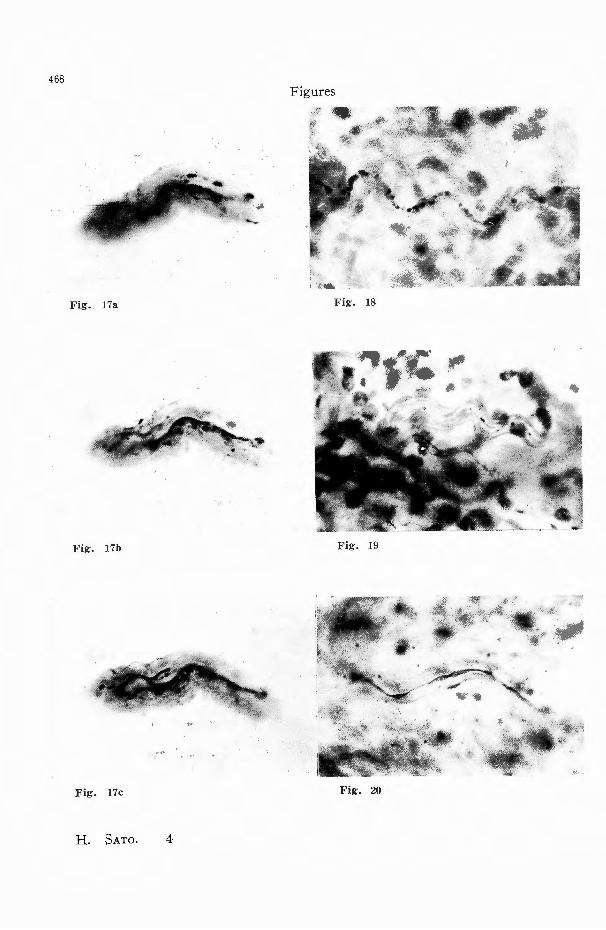
Fig-. 17a, 17b and 17c Degeneration of all the
myelinated fibers in the nerve bundle at the
hilLim of a dog’s right ovary extirpated 5 days
after rhizotomy of Th. 13・・・L. 4 on both sides. Photographs were taken in three layers. x 300
(EHRL!CR’S stain) Fig・. 18 Degeneration of a myelinated五her in
the medulla of a dog’s right ovary extirpated
5 days after section of the ventral and dorsal
roots of S. l ・・Co.5 on the right side. x 400
(EHRLICH’s stain)
Fig-. 19 Degeneration of SETO’s sensory nerve in
the vessel-plexus at the hilum of a dog's right ovary extirpated 5 days after section of
the ventral and dorsal roots of S. I・・Co.5 on
the right side. x 500 (BIELSCROWSKv-Sozo”
Ki's stain)
Fig・. 20 Deformation of SETO’s sensory nerve due to degeneration, in the medulla of a dog’s
right ovary extirpated 5 days after section of
the ventral and dorsal roots of S. I・・Co.5 on
the right side. x 650 (BraLSCHOWSKY-SI>TO’s
stain)
REFERENCES
1) Asai, Sh. : The Law of Dt1al Sensory Inner-vation of Viscerae (Naizo Chikaku no '.¥fijyu
Shinkei Shihai Hosoku, in Japanese). Kyoto
Igaku Zasshi, 23, 518, 559, 1926. 2) Bain, ¥V.
464 日本外科宝函第24巻第5号
A., Irving, J. T. and :¥[c Swiney, B. A. : The
Afferent Fibers from the Abdomen in the Splan-
chnic Nerves. Jour. Physiol., 84, 323, 1935. 3)
Cannon, B. : A MethoゴofStimulating Autono-
mic Nerves in the Unanesthetized Cat with Ob-
servation on the :¥Iotor and Sensory E任ects.
Am. Jour. Physiol., 105, 366, 1933. 4〕Clara,
M. : Die Anatomie der Sensibilit忌tunter beson-
derer Beriicksichtigung der vegetativen Leitungs-
bahnen. Acta Neuro-Veg., 7Bd, Heft 1-.1, 1953.
5) Dahl : ¥Iiillers Lebensnerven S., 681, 1931
cited by Kure, T. and Okinaka, Sh. : Jiritsu
Shinkeikei, 1949. 6) Edgeworth, F. H. : On a
Large-Fibered Sensory Supply of the Thoracic
and Abdominal Viscera. Jour. Physiol., 13, 260,
1892. 7) Fulton, J. F.: Physiology of the I¥er-
vous System. 1949. 8) Goecke, M. : Die Endaus-
breitung des vegetativen l¥en・engewebes im men-
schlichen Ovarium und ihre Bedeutung fiir die
Funktion des Ovariums. Arch. f. Gynakol., 166
Bd, 187, 1938. 9) Hiramatsu, S. : Supplements
to the Knowledge on the Sensory Innervation
of the Stomach (I Chikaku Dendoro ni kansuru
Chiken Hoi, in Japanese). Tokyo Igakukai Za-
sshi, 49, 1033, 1935. 10) Inoue, H. : A Histo司
logical Study of Sensory Xぞrvesin the Biliary
Tract. Arch. f. jap. Chir., 24, 257, 1955. 11)
Ishikawa, N. : Questions on the Visceral Sym-
pathetic Sensory Innervation. (Naizo Kokan-
Shinkei no Chikaku ¥Iondai ni kans口ru Gigi,
in Japanese). J. jap. Surg. Soc., 22, 137, 1921.
12) Kawakami, K. : Studies on Visceral Sensi-
tivity (Naiz5 no Chikaku ni tsuite, in japanese).
Kyoto Teikoku-Daigaku Igakubu Seirigaku-
Kyoshitsu Ronbunshii 2. 13) Kimura, Ch. :
Surgery of the Autonomic Nervous System (Ji-
ritsu-Shinkei no Geka, in Japanese). J. Jap.
Surg. Soc., 52, 450, 1951.…: The Problem of
Abdominal Pain. Arch. f. jap. Chir., 22, 59,
1953 ..・・ : Physiology of Abdominal Pain (Fu-
kutsii no Seiri, in Japanese). Rinsh百 noShinpo,
7, 1953 ..・・ : Development of the Dual Afferent
Innervation Theory of the Viscera (Naiz5・Chi-
kaku Nijyii-Shihai Gakusetsu no Shinten, in
Japanese). :¥ihon Rinsh5, 11, 85, 1953.…: Vis ceral Sensory Nerves and Autonomic Nerves (
Naizo Chikaku-Shinkeikei to Jiritsu-Shinkeil刊しin Japanese). Saishin Igaku, 9, 5, 34, 1954.・・・ :
Peripheral Structure ofγisceral Sensory X e1 ,-es
(l¥aiz5 Chikaku Shinkei no '¥fassh5z5, in Jap-
anese l. Rinsh5 Geka, 9, 255, 1954. 14) Kno-
che, H. : Untersuchung iiber die nervりseVer-
sorgung der Eizellen. Acta Neuro-Veg., 10, 4,
502, 1953. 15) Kuoo,人: Studies on s~ns itivi
ty of the Thoracic and Abdominal Viscera (
Kyofukukii-J:¥aizu no Chikaku ni tsuite, in Jap-
anese). Sl:inl.;:eisaku Zasshi, 24, 118, 1924. 16)
Kuntz,入.: The Autonomic Nervous System.
1947. 17) Kure, T. and Okinaka, Sh. : The
Autonomic Nervous System (Jiritsu Shinkeikei,
in Japanese). 1949. 18) Langley, J. ;-{. and
Anderson, H. K. : The Innervation of the Pel・
vie and Adjoining Viscera. Part VI Histological
and Physiological Observations upon the Efferts
of Section of the Sacral Nerves. Jour. Physiol.,
19, 37'.2, 1895. 19) Langley, J. N. : Observations
on the 1¥Iedullated Fibers of the Sympathetiこ
System and chiefly on those of the Grey Rami
Communicantes. Jour. Physiol., 20, 55, 1896.・
The Autonomic Nervous System. Brain, 26, 1.
1903. 20) Mitchell, G. A. G. : The Innervation
of the Ovary, Uterine Tube, Testis and Epidi-
dymis. Jour. Anat., 72, 508, 1938. 21) Nitta, Y.
: The Pelvic l¥e1 ,・e (Kotsuban-旬、inkeini twite,
in Japanese). Tokyo Igakukai Zasshi, 43, 610,
1929. 22) t¥euman, K. 0. : The A妊erent Fibers
of ti;巴 AbdominalVagus in the Rabbit and Cat.
Jour. Physiol., 49, 3-l, 1915. 23〕Otsu,A. : A
Histological Study of Sensory Ner<e Endings in
the c¥limentary Canal of Human Beings and
Dogs. Acta Sch. :Mel. Uni1'. Kyoto Jap., 31, 103,
1953. 24〕Ranson,S. vV. and Eillingsley, P.
R. : An Experimental Analysis of the Sympa-
thetic Trunk and Greater Splanchnic l'\en•e in the Cat. Jour. Comp. Neural., 29, 441, 1918.・・ .
The Thoracic Trunks Sympathicus, Rami Com-
munirantes and Splanchnic !¥'en'es in the Cat.
Ibid., 29, 405, 1918. 25) Ranson, S. W., Foley,
]. 0. and Alpert, C. D. : Observation of the
Structure of the ¥Tagus l¥erve. Am. Jour. Anat.,
53, 289, 1933. 26) Ranson, S. ¥V. : The Auto-
nomic !\'町、 ousSystem. 1946. 27) Sakaguchi, J.
: Histological Observations on the Nerve-Distri-
bution of Human Ovaries 〔ト;ingenRans5 no
Shinkei Btmpu ni taisuru Soshikigakuteki Kan-
satsu, in Japanese). Manshu Igaku Zasshi, 30,
795, 1939. 28) Seto, H. : A Discussion on Vis-
ceral Sensitivity from the Histological Stand-
point (Soshi kigaku・jyo kara mita Naizo no
Chikaku, in Japanese). Igaku no Shinpo, 5, 225,
1949. ・ ・ : A l¥fo,Jified Bielschowsky’s ・silver Im-
pregnation. Tohoku Jour. E:xp. ¥led., 54, I, 1951.
29) St凸hr,Jr. P.: I.e:1rbuch der Histologie und
der mikroskopischeλnatornie des l¥Iensrhen. Sp-
ringer-Verlag, 1951. : l¥likroskopische Ana-
tomie des vegetativen Nervensystems. 1951. 30)
怜、、,•'
Fig. 2
~- ろ
Fig. 3
,、,
-‘
‘“' .』
'i
‘4首
‘,.
·~ ,,, .. :、月
Fig・. 4
N
,...
4’ . 蜘タ~)
H. SATO.
465
-
,
1
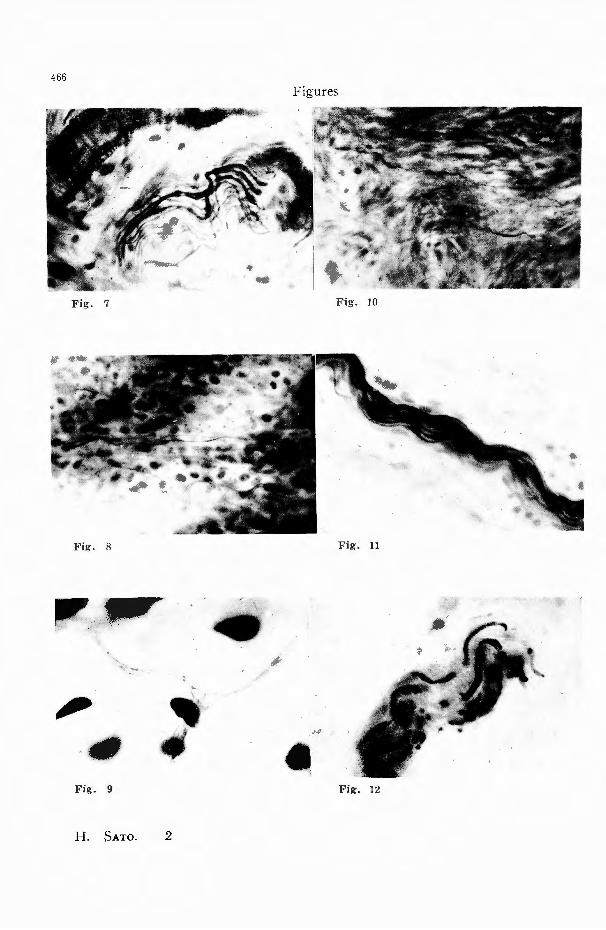
Fig. 7
Fig. 8
JI',,
, Fig. 9
、、一、『ー
H. SATO.
-. .fi 2
少
4・
..
....
Fig. 12
Fig・. 13
\
Fig. 14
Fig. 15
Figures
、,..;j川崎蝿,P シef’@ ム’r.. :
、除
467
可’F-~ 、 , 、皆 晦
Fig・. 16a
Fig・. 16b
Fig. 16c
H. SATO. 3
468
・合
Fig・. 17a
”・
Fig・. J7b
Fig. 17c
H. SATO. 4
Figures
Fig. 18
Fig. J9
Fig. 20
重均匂ゼ
、考
THE AFFERENT INNERVATION OF THE mr ARY OF TITE DOG 469
Suzuki, K. : Note of Technique to make Tisssue-
Preparations (Soshiki Hyohon Seisaku Gijyutsu
Noto, in Japanese). (IV). Noshinkei Ryoiki, 5,
184, 1952. 31) Tanaka, N. : A Histological
Study of the Dual Afferent Innervation of the
Esophagus of the Dog. Arch. £. jap. Chir., 22,
439, 1953. 32) Weddell, G. and Sinclair, D. C.
: The Anatomy of Pain Sensibility. Acta Neuro-
和文抄録
Veg., 7, 1-1, 135, 1953. 33) White, J. C. and
Smithwick, R. H. : The Autonomic :¥ervuus
System. 1946. 34) Yagita, i¥f.:人 Histological
Study of Sensory Nerves in the Lung and the
Visceral Pleura. Arch. f. jap. Chir., 23, 569, 1954.
35) Yoshiike, Y. : Physiologic Studies on the
Visceral ~ensation of the Uro界 nital Organs.
Arch. f. jap. Chir., 23, 433, 195-L
犬卵巣の求心性神経支配に関する組織学的研究
京都大学医学部外科学教室第2講座(指導青柳安誠教皮)
佐藤博正
BIELSCHOWSKY氏神経軸索鍍銀染色法の瀬戸 (2)犬卵巣の求心性有髄神経の大部分は同側の脊髄
氏変法及び鈴木氏変法並びに EHRLICH氏神経髄翰 神I怪後根を通り Th.13-L.2を中心とする胸膜髄に入
染色法を用いて犬卵巣の求心性神経について検討し, る.
更に犬の神経斡切断による卵巣内の仰経の二次的変性 (3)犬卵巣♂)求心性有髄神経のうちの一小部分に同
を追求することにより卵巣の求心性ic1J経文配経路を検 側の仙骨神経を通り仙慌に入るものが存在するがこれ
討して次の如き結論を得た. は;('uil隻i生支j切に比して極;〉て劣必であ之J・
(1)犬卵巣に於いても有償神経及ひ知覚神経(瀬 (4)犬卵巣の迷走'i~D経性求心性三二月日立証明し得な
戸)が存在する. い.