Embed Size (px)
Citation preview
HLA alloreactivity by human viral specific memory T-cellsD'Orsogna, L.J.A.
CitationD'Orsogna, L. J. A. (2010, December 8). HLA alloreactivity by human viralspecific memory T-cells. Retrieved from https://hdl.handle.net/1887/16223 Version: Corrected Publisher’s Version
License:Licence agreement concerning inclusion of doctoralthesis in the Institutional Repository of the Universityof Leiden
Downloaded from: https://hdl.handle.net/1887/16223 Note: To cite this publication please use the final published version (ifapplicable).

Chapter 4
Tissue specificity of cross-reactive allogeneic responses by EBV EBNA3A specific memory T-cellsLloyd J.A. D’Orsogna, Dave L. Roelen, Ellen M.W. van der Meer-Prins, Pieter van der Pol, Marry E.I. Franke-van Dijk, Michael Eikmans, Jacqy Anholts, Jamie Rossjohn, James McCluskey, Arend Mulder, Cees van Kooten, Ilias I.N. Doxiadis, Frans H.J. Claas.
Transplantation 2010 (In press)
Chapter 4
78
ABSTRACT
Background: The cross-reactivity of Epstein-Barr virus (EBV EBNA3A) specific CD8 T-cells against allogeneic HLA-B*44:02 has been shown to be dependent on presentation of self-peptide EEYLQAFTY by the target antigen. Here we report that allogeneic HLA-B*44:02+ proximal tubular epithelial cells (PTECs) and human umbilical vein endothelial cells (HU-VECs) are poor targets for EBV EBNA3A specific T cells. Methods: The EEY peptide was exogenously loaded onto HLA-B*44:02 and HLA-B*44:03 ex-pressing PTECs and HUVECs. EEY peptide loaded, and unloaded, PTECs and HUVECs were then incubated with serial dilutions of our EBNA3A T-cell clone, in a cytotoxicity assay. Results: While HLA-B*44:02 expressing PTECs were specifically lysed in proportion to the effector/target ratio by the EBNA3A T-cell clone, without peptide loading, lysis was greatly increased by exogenous EEY peptide loading (15% vs. 75%; p<0.0001). HLA-B*44:02 ex-pressing HUVECs were only lysed when loaded with exogenous EEY peptide (0% vs. 64%; p<0.0001). Lack of HLA expression and lack of ABCD3 gene expression were excluded as a cause for these results. PTECs and HUVECs were specifically targeted by another alloreac-tive T-cell clone without exogenous peptide loading, suggesting that the lack of recognition of HLA-B*44:02+ epithelial and endothelial cells by the EBV EBNA3A T-cell clone was due to lack of EEYLQAFTY peptide presentation. Conclusions: Tissue specific (peptide dependent) alloreactivity may have important implica-tions for transplantation monitoring and rejection.
Tissue specificity
79
4
INTRODUCTION
Viral infection is associated with solid organ transplant rejection and is a potent barrier to transplantation tolerance (1-10). It has recently been shown that allo-HLA crossreactivity from viral specific memory T-cells is far more common than predicted; using EBV trans-formed B-cells (EBV LCLs) and HLA-transfected target cells (11). This allo-HLA crossre-activity from viral specific memory T-cells has been shown to be dependent on endogenous self-peptide presentation by the donor cell (11-13), and therefore alloreactivity could be tissue cell type specific if the recognized peptide is differentially expressed by target tissues.
Early work suggested that the explanation for the presence of alloreactive memory T-cells in non-sensitized individuals could be crossreactivity from viral specific memory T-cells against allo-HLA molecules (14-15). Burrows and colleagues demonstrated the dual specificity of EBV EBNA3A specific T-cell clones for the immunodominant EBV peptide FLRGRAYGL presented on HLA-B*08:01 and the alloantigen HLA-B*44:02, to which the individual had never been exposed (15). The clinical relevance of this finding is reinforced by the fact that EBV EBNA3A specific CD8 T-cells are capable of specifically lysing HLA-B*44:02+ target cells in cytotoxicity assays (15-16), and that HLA-B44 is an immunogenic mismatch in HLA-B8 kidney recipients (17).
This EBV EBNA3A T-cell allo-HLA-B*44:02 crossreactivity is dependent on presentation of the EEYLQAFTY self-peptide derived from the ABCD3 gene, via molecular mimicry (12). Despite extensive polymorphism between HLA-B*08:01 and HLA-B*44:02, and the dispa-rate sequences of their bound viral and self-peptides respectively, the HLA-B8/FLR restricted TCR engages these different peptide/HLA complexes identically.
In our laboratory, various cell lines have been generated for in-vitro testing in kidney trans-plantation research. Proximal tubular epithelial cells (PTECs) are derived from proximal tu-bule cells taken from kidney transplant biopsy specimens (18-20), and are a useful model to examine alloreactivity from graft infiltrating lymphocytes (21-22). Human umbilical vein endothelial cells (HUVECs) are derived from healthy human post-partum umbilical tissue (23-25), but are also useful as a model of kidney vascular endothelial cell transplantation (26). Here we investigate tissue specificity of the crossreactive alloresponse by EBV EBNA3A specific memory T-cells, using PTECs and HUVECs as a model system for human kidney transplantation.
We report that in contrast to other allogeneic HLA-B*44:02+ cell lines, allogeneic HLA-B*44:02+ PTECs are poor targets for EBV EBNA3A specific CD8 T-cells, and HLA-B*44:02 HUVECs are not targeted at all. We hypothesized that this differential lysis of HLA-B*44:02 expressing PTEC and HUVEC cell lines could be explained by differential tissue presentation of the EEYLQAFTY self-peptide. Our work with the EBV EBNA3A T-cell clone, presented here, confirms that (peptide-dependent) tissue specificity of allo-HLA responses from viral specific memory T-cells may indeed be relevant in the kidney transplantation setting. Essen-tially, alloreactivity can be tissue specific.

Chapter 4
80
RESULTS
HLA-B*44:02 expressing epithelial and endothelial cell lines are poor targets for EBV EBNA3A specific CD8 memory T-cellsWe and others have previously shown that EBV EBNA3A specific T-cell clones exert cytolytic activity against allogeneic HLA-B*44:02+ EBV LCLs, PHA Blasts and HLA-B*44:02 trans-fected K562 cells (SALs) (11, 15-16). We therefore performed cytolytic assays using HLA-B*44:02+ PTEC and HUVEC targets to determine if allo-HLA crossreactivity from viral spe-cific memory T-cells, as determined by hematological target cells types, corresponds with solid organ alloreactivity (Figure 1). In contrast to other allogeneic HLA-B*44:02+ cell lines, allogeneic HLA-B*44:02+ PTECs are poor targets for EBV EBNA3A specific CD8 T cells (spe-cific lysis 12%). HLA-B*44:02+ expressing HUVECs are not targeted by an EBV EBNA3A specific T-cell clone (specific lysis 0%).
Figure 1. HLA-B*44:02 expressing PTECs and HUVECs are poor targets for EBV EBNA3A specific T-cells. In contrast to allogeneic HLA-B*44:02+ PHA Blasts, allogeneic HLA-B*44:02+ PTECs are poor targets for EBV EBNA3A specific CD8 T cells in a four hour cytotoxicity assay (specific lysis 12%). HLA-B*44:02+ expressing HUVECs are not targeted by an EBV EBNA3A T-cell clone (specific lysis 0%). Experiments performed in triplicate, mean values shown with SD. E:T ratio 30:1, targets=5000.
Tissue specificity
81
4
The lack of recognition of epithelial and endothelial cell lines is not due to lack of HLA-B*44:02 expressionTo exclude lack of HLA expression as the cause for these results we performed cytotoxicity assays before and after IFNg stimulation of the PTEC and HUVEC targets. PTECs demon-strated higher baseline HLA-B*44:02 expression as compared to HUVECs (data not shown). IFNg pre-stimulation was associated with increased HLA-B*44:02 surface expression in both PTECs and HUVECs (data not shown). Regardless, the increased HLA-B*44:02 expression following IFNg stimulation did not result in increased lysis of the PTEC or HUVEC target cells (data not shown).
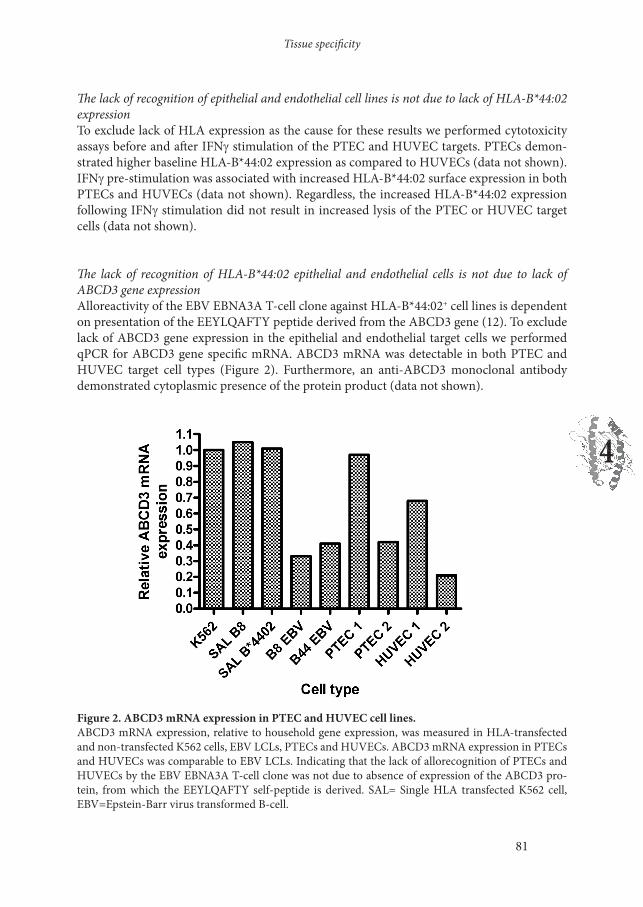
The lack of recognition of HLA-B*44:02 epithelial and endothelial cells is not due to lack of ABCD3 gene expressionAlloreactivity of the EBV EBNA3A T-cell clone against HLA-B*44:02+ cell lines is dependent on presentation of the EEYLQAFTY peptide derived from the ABCD3 gene (12). To exclude lack of ABCD3 gene expression in the epithelial and endothelial target cells we performed qPCR for ABCD3 gene specific mRNA. ABCD3 mRNA was detectable in both PTEC and HUVEC target cell types (Figure 2). Furthermore, an anti-ABCD3 monoclonal antibody demonstrated cytoplasmic presence of the protein product (data not shown).
Figure 2. ABCD3 mRNA expression in PTEC and HUVEC cell lines. ABCD3 mRNA expression, relative to household gene expression, was measured in HLA-transfected and non-transfected K562 cells, EBV LCLs, PTECs and HUVECs. ABCD3 mRNA expression in PTECs and HUVECs was comparable to EBV LCLs. Indicating that the lack of allorecognition of PTECs and HUVECs by the EBV EBNA3A T-cell clone was not due to absence of expression of the ABCD3 pro-tein, from which the EEYLQAFTY self-peptide is derived. SAL= Single HLA transfected K562 cell, EBV=Epstein-Barr virus transformed B-cell.
Chapter 4
82
The lack of recognition of HLA-B*44:02 expressing epithelial and endothelial cells is likely due to quantitative lack of EEYLQAFTY peptide presentation on the cell surfaceTo confirm that the lack of allorecognition of the HLA-B*44:02+ epithelial and endothelial cell lines was due to lack of peptide presentation we performed cytolytic assays using EEY-LQAFTY peptide loaded and unloaded PTEC and HUVEC targets. HLA-B*44:02 express-ing PTECs were poorly targeted by an EBV EBNA3A T-cell clone without peptide loading (specific lysis 15%; P=0.004 vs. HLA-B*44:03 PTEC), and then only at high effector/target ratio (Figure 3a). The specific lysis of HLA-B*44:02 expressing PTECs was greatly increased by exogenous EEY peptide loading (15% vs. 75%; P<0.0001) (Figure 3a). HLA-B*44:02 ex-pressing HUVECs were only targeted by an EBV EBNA3A specific clone when loaded with exogenous EEY peptide (0% vs. 64%; P<0.0001) (Figure 3b). Furthermore, stimulation of the EBV EBNA3A specific T-cell clone with HLA-B*44:02+ HUVECs did not elicit cytokine production in a 24 hour luminex assay (data not shown). The EBV EBNA3A specific T-cell clone was able to target EEY peptide loaded HLA-B*44:02 PTECs and HUVECs even without IFNg pre-stimulation, suggesting that the baseline HLA-B*44:02 expression in these cell lines is sufficient for CTL targeting if sufficient allopeptide is presented. HLA-B*44:03 PTECs and HUVECs were not targeted irrespective of peptide loading, as predicted (12). Thus organ (kidney) specificity of the allo-HLA crossreactivity from the EBV EBNA3A specific T-cell is dependent on endogenous self-peptide (EEY) processing and presentation.
Figure 3. The lack of recognition of HLA-B*44:02+ PTECs and HUVECs is likely due to lack of EEYLQAFTY peptide presentation on the cell surface. (a) HLA-B*44:02+ PTECs were poorly targeted by the EBV EBNA3A specific T-cell clone (specific lysis 15%; **P=0.004 vs. HLA-B*44:03 PTEC). The specific lysis of HLA-B*44:02+ PTECs was greatly increased by exogenous EEY peptide loading (specific lysis 15% vs. 75%; ***P<0.0001). (b) HLA-B*44:02+ HUVECs were only targeted by an EBV EBNA3A specific T-cell clone when loaded with exogenous EEY peptide (specific lysis 0% vs. 64%; ***P<0.0001). The EBV EBNA3A T-cell clone did not recognize HLA-B*44:03+ PTECs or HUVECs irrespective of peptide loading. Experiments shown here were performed without IFNg pre-stimulation further dem-onstrating that the baseline HLA-B*44:02 expression is sufficient to elicit cytotoxicity if the EEY peptide is present. Experiments performed in triplicate, mean values shown with SD. Targets=5000.

Tissue specificity
83
4
Cognate antigen recognition and allorecognition increase in proportion to the concentration of exogenously added viral or allo-peptideTo determine the concentration of specific peptide required to elicit cytolytic effector function by the EBV EBNA3A specific T-cells, FLR or EEY peptide were loaded onto HLA-B*08:01+ or HLA-B*44:02+ target cells respectively, in a peptide dose-response experiment (Figure 4). Cognate viral antigen recognition and allorecognition increase in proportion to the concen-tration of exogenously added cognate or allo-peptide (Figure 4). Equivalent concentrations of the FLR cognate peptide on HLA-B*08:01+ target cells and EEY allopeptide on HLA-B*44:02+ target cells was required to elicit cytolytic effector function by the EBV EBNA3A specific T-cells. For both cognate and allo-peptides 50% of the maximum specific lysis occurred be-tween 10mg/ml and 50mg/ml of exogenously added peptide.
Figure 4. Cognate antigen recognition and allorecognition increase in proportion to the concentra-tion of exogenously added viral or allo-peptide. To determine the concentration of specific peptide required to elicit cytolytic effector function by the EBV EBNA3A specific T-cells, FLR or EEY peptide was loaded onto HLA-B*08:01+ or HLA-B*44:02+ target cells respectively, in a peptide dose-response experiment. Cytolytic effector function of the EBV EBNA3A specific T-cells increases in proportion to exogenously added peptide concentration for both the cognate viral peptide and the allopeptide. Equivalent concentrations of the FLR cognate peptide on HLA-B*08:01+ target cells and EEY allopep-tide on HLA-B*44:02+ target cells is required to elicit cytolytic effector function by the EBV EBNA3A specific T-cells. Assays performed with a HLA-B*08:01+ and two different HLA-B*44:02+ PTEC and HUVEC target cells. Experiments performed in triplicate, mean values shown with SD. Effector:target ratio 20:1, targets=5000.

Chapter 4
84
Epithelial and endothelial cells are not resistant to lysis by CTL clonesFinally, to exclude the possibility that the EEYLQAFTY peptide is presented on the target HLA molecule but the cells are not targeted due to lytic resistance or CTL suppression we performed cytolytic assays using the EBV EBNA3A clone and a HLA-A2 alloreactive T-cell clone (JS132) in parallel. The JS132 clone specifically lysed HLA-A2+ B*44:02+ heterozygote PTECs and HUVECs, without any exogenous peptide addition (Figure 5). The EBV EBNA3A CD8 T-cell clone was unable to efficiently target the identical epithelial or endothelial cell lines without exogenous addition of EEYLQAFTY peptide (Figure 5). Thus, the epithelial and endothelial cells can be suitable targets for CTL clones without addition of exogenous peptide.
Figure 5.

Tissue specificity
85
4
Figure 5. PTECs and HUVECs are suitable targets for CTL mediated killing without exogenous peptide addition. The JS132 HLA-A2 alloreactive T-cell clone specifically lysed HLA-A*02+ B*44:02+ heterozygous PTECs and HUVECs irrespective of exogenous peptide loading (specific lysis >85%). The EBV EBNA3A CD8 T-cell clone was unable to efficiently target the identical epithelial or endothelial cell lines without exogenous addition of EEYLQAFTY peptide. Furthermore, the EBV EBNA3A clone lysed both HLA-B*08:01+ and HLA-B*44:02+ epithelial and endothelial cell lines when loaded with FLR pep-tide or EEY peptide respectively, but not RAK (HLA-B*08:01 control) nor EEK (HLA-B*44:02 control) peptides; confirming that the viral specificity and alloreactivity are peptide dependent and mediated by the same T-cell. Thus, it is highly likely that the lack of recognition of HLA-B*44:02+ epithelial and endothelial cells is due to a quantitative lack of EEY peptide presentation. Experiments performed in triplicate, mean values shown with SD. Effector:target ratio 30:1, targets=5000.
DISCUSSION
In this report we demonstrate that allo-HLA crossreactivity by viral specific memory T-cells can be tissue cell-type specific because of differential tissue specific self-peptide presentation. We have confirmed that not only is the HLA-B*44:02 alloreactivity from the EBV EBNA3A specific T-cell clone self-peptide dependent but that normal allogeneic kidney cells may not be targeted unless sufficient EEY self-peptide is processed and presented. Alloreactivity is mediated by cytotoxicity, when the peptide is presented, indicating the potential clinical rel-evance of cross-reactive alloresponses against cell types present in kidney transplant tissue.
Our results do not suggest that allo-HLA crossreactivity from the EBV EBNA3A T-cell is irrelevant to kidney transplantation. The EBV EBNA3A specific T-cell does have cytolytic activity against HLA-B*44:02+ kidney epithelial cells in a 4 hour assay. Memory T-cells persist and therefore could perform effector functions over a prolonged period, or at times when immunosuppression is tapered (2). Furthermore T-cells mediate effector functions through a variety of mechanisms, including cytokine production, not just cytotoxicity (27). The EBV EBNA3A specific immune response is a public TCR response present in all HLA-B8+ B44- in-dividuals (28) and HLA-B44 mismatching has been identified as high risk in HLA-B8 kidney recipients (17).
However results presented here suggest it is unlikely that EBV EBNA3A specific T-cells ex-hibit effector functions against HLA-B*44:02+ endothelial cells present in solid organ tissue. Conversely, a viral specific T-cell that targets a kidney cell specific peptide presented on an allogeneic HLA molecule may not recognize PBMCs or spleen cells from the same allogeneic donor.
In light of our findings it is worth considering some of the possible mechanisms by which organ specific alloreactivity could occur. Quantitative differences in HLA expression could explain organ specific alloreactivity, but has been excluded in the present study. Differences in co-stimulation and accessory molecule expression are also feasible but there is little evidence for this as memory CD8 T-cells have reduced requirements for co-stimulation and do not require CD4 T-cell help (29-30). Furthermore, the EBV EBNA3A clone used here is clearly
Chapter 4
86
capable of targeting HLA-B*44:02 transfected K562 cells which have absent co-stimulatory molecules (16).
Tissue specific expression of a protein that is the source of the self-peptide recognized on the allo-HLA molecule would be extremely likely to result in organ specific alloreactivity. For ex-ample, a peptide derived from a renal specific ion transporter will only be presented on renal tubular cells. Furthermore, alloreactivity might only be induced when the gene expression is up regulated.
Results presented here are of particular interest because we have demonstrated expression of the ABCD3 protein product in the target epithelial and endothelial cells. The HLA-B*44:02+ PTECs were targeted albeit to a lower level of lysis (15%), therefore there must be naturally some EEY peptide presented on the cell surface but not enough to trigger a high percentage of specific lysis. The HLA-B*44:02+ HUVECs are not targeted and therefore it is likely that insufficient EEY peptide is presented on the surface.
Differences in antigen processing and presentation could account for tissue specific allore-activity, even if similar levels of the ABCD3 gene product are expressed within the epithelial and endothelial cells. For example, EBV LCLs, PHA Blasts and K562 cells constitutively ex-press the immunoproteosome which may generate novel antigenic peptides. Furthermore, the study of Macdonald defines the EEYLQAFTY peptide as an antigenic target of the EBV EBNA3A T-cell presented via allogeneic HLA-B*44:02 (12), however this study does not ex-clude the possibility that several different peptides presented on HLA-B*44:02 are capable of activating the EBV EBNA3A specific T-cell. Theoretically, these additional peptides may not be presented by epithelial or endothelial cell types.
Alternatively, differences in expression of a protein that contains a peptide capable of compet-ing with an antigenic peptide for the peptide-binding groove of the allogeneic molecule could also cause organ specific alloreactivity, as also suggested by others (31). A tissue specific com-peting peptide may reduce the amount of the target self-peptide/allo-HLA complex available for recognition by the alloreactive CTL.
HLA-B*44:02 is a highly tapasin-dependent HLA molecule (32-33) and therefore limited tapasin expression in PTECs and/or HUVECs could decrease EEY peptide presentation in these cell lines. However tapasin mRNA is strongly induced in endothelial cells following IFNg treatment (34), and IFNg treatment did not increase the targeting of HUVECs in our assays despite inducing elevated HLA-B44 expression. The HLA-B*44:05 molecule is also a target of the EBV EBNA3A T-cell (12) and can load peptides independently of tapasin, unfor-tunately no HLA-B*44:05 expressing PTECs or HUVECs are available.
Our assays using the JS132 clone exclude the possibility that the EEY peptide is presented but that epithelial and/or endothelial cells are resistant to lysis or are tolerogenic. The HLA-A2 alloreactive JS132 clone was generated by stimulating PBMCs with HLA-A2 mismatched irradiated EBV LCLs in-vitro. The JS132 allo-A2 reactivity is likely peptide dependent and therefore we conclude that the antigenic peptide recognized in the context of HLA-A2 is con-

Tissue specificity
87
4
stitutively presented by the epithelial and endothelial cell lines.
The ultimate proof that our results are attributable to lower/absent EEYLQAFTY peptide presentation could be provided by peptide elution studies. However elution of peptides from HLA-B*44:02+ PTECs and HUVECs is not feasible due to the large number of cells required for peptide elution and mass spectrometry analysis. Nonetheless, we favour the conclusion that the differential allorecognition of HLA-B*44:02+ PTECs and HUVECs by the EBV EB-NA3A specific T-cell clone is the result of differential quantitative presentation of the EEY-LQAFTY self-peptide by the target cells.
The finding of organ specific allorecognition is extensively described in mice (31, 35-38). For example, priming of mice with normal allogeneic spleen cells generated peptide-dependent Kb-specific alloreactive CTL clones that exhibited cell-type specific allorecognition (31). Hu-man tissue specific alloreactivity has been suggested by studies using graft-infiltrating lym-phocytes obtained from renal allografts undergoing rejection (21, 39-43). Graft infiltrating lymphocytes were shown to exhibit T cell functional activity against PTEC grown from the corresponding biopsy, but not donor derived splenocytes nor PTEC from biopsies obtained from other patients. For example, van der Woude and colleagues found that thirteen out of forty (33%) of graft infiltrating cell lines reacted in a donor-specific fashion to PTEC but not to donor splenocytes (41).
Results presented here may have important clinical implications for renal transplantation monitoring, rejection and tolerance. Monitoring of alloreactive T-cells may allow individu-alization of immunosuppression (44), but such assays routinely use donor PBMCS or spleen cells as stimulator. Allo-HLA crossreactivity by viral specific memory T-cells as defined against hematological target cell types will not correspond with solid organ alloreactivity un-less the targeted self-peptide is ubiquitously and equally presented. If alloreactive CTL rec-ognize allo-HLA presenting a specific peptide then it is possible that competitive peptides could be designed to inhibit allorecognition, as has also been suggested by others (31, 45). We have confirmed that the absence of a single tissue specific self-peptide is enough to abrogate alloreactivity. Also, long term immunosuppressive free graft survival is the ultimate aim of much transplantation research, but our work suggests induction of tolerance by using pre-transplant blood transfusion may not delete organ specific CTLs.
Finally, we acknowledge that this study uses umbilical vein endothelial cells as a model for kidney vascular endothelial cell transplantation, however, gene expression and/or functional differences have not been reported between kidney and umbilical endothelial cells. Others have also found that donor-derived gonadal vein endothelial cells can be specifically targeted by graft infiltrating alloreactive T-cells (39).
In conclusion, we show that the EBV EBNA3A T-cell exhibits tissue cell type specific alloreac-tivity because of quantitative differences in presentation of the recognized self-peptide. Tissue specific allorecognition may have important clinical consequences, especially for monitoring, rejection and tolerance induction of solid organ grafts. Future work should determine if tissue specific allorecognition is a common characteristic of human alloreactive CTL.

Chapter 4
88
MATERIALS AND METHODS
Generation of EBV EBNA3A viral specific CD8 memory T-cell cloneThe generation and allo-HLA-B*44:02 crossreactivity of the EBV EBNA3A CD8 memory T-cell clone used here has been previously described (16). Briefly, EBV EBNA3A specific CD8 T-cell clones (HLA-B8/FLRGRAYGL restricted) were derived from a healthy donor with HLA typing HLA-A*01:01,02:01; B*08:01,-; DRB1*03:01,-; using single cell sorting based on viral peptide/tetramer complex staining. Clonality of the T-cell clone was confirmed using RT-PCR to determine TCR AV and BV usage (16).
Generation of JS132 cloneThe generation and the allo-HLA-A2 alloreactivity of the JS132 CD8 T-cell clone have been previously described (46-47). Briefly, PBMCs from healthy donor JS (HLA-A3,3; B7,7; DR2,2; DQ1,1) were stimulated with irradiated EBV transformed B-cell line JY (HLA-A2,2; B7,7; DR4,6). Following several rounds of stimulation and enrichment the HLA-A2 alloreactive population was cloned by limiting dilution at 0.5 cell/well.
Generation and culture of PTECs and HUVECsGeneration of PTECs (18-19) and HUVECs (23-24) has been previously described. PTECs were cultured from cortical tissue of human kidneys not suitable for transplantation because of anatomical reasons or from pretransplant biopsies, and HUVECs from umbilical vein of human umbilical cord. Morphologic appearance and immunofluorescence staining con-firmed specific outgrowth of PTECs and HUVECs.
HLA typing and FACS staining for HLA expression of epithelial and endothelial cellsMolecular typing for class I and class II was performed in the tissue typing laboratory Lei-den University Medical Centre, the Netherlands. The relative amount of HLA surface ex-pression was determined using human monoclonal antibodies specific for the HLA molecule expressed. Epithelial and endothelial cell lines were treated with trypsin, harvested and then washed two times. Cells were incubated with the human HLA specific monoclonal antibody for 30 minutes and then washed twice. Cells were then labeled with a rabbit-anti-human-FITC secondary detection antibody for a further 30 minutes and then washed three times. HLA expression was determined before and after IFNg treatment, 500 units/ml for 24 hours.
ABCD3 gene expression in PTEC and HUVEC cell linesFor detection of ABCD3 mRNA expression cells were harvested and preserved in RNAlater solution (Qiagen, Chatsworth, CA, USA). RNA was extracted using the RNeasy mini kit (Qia-gen) following the manufacturers instructions. RNA was treated with DNase (Qiagen) on the spin columns. RNA quantity was assessed with a spectrophotometer (Nanodrop technolo-gies, Wilmington, DE, USA) and all samples showed A260/A280 ratios between 1.9 and 2.1. Quantitative PCR was performed as per standard protocols. The forward and reverse primer
Tissue specificity
89
4
sequences used in the quantitative PCR for ABCD3 mRNA were CCTGGTGCTGGAGA-AATCAT and CCCCAGATCGAACTTCAAAA respectively, giving an amplicon of 118 bp. The PCR was performed using an iCycler MyiQ (Bio-Rad). The PCR program was finalized with a melting curve analysis. The signal of the stably expressed household genes b-actin and GAPDH served as normalization factors.
Cytotoxicity AssaysThe EBV EBNA-3A specific T-cell clone and/or the JS132 CD8 T-cell clone were evaluated for cytotoxicity by incubating 5000 PTEC or HUVEC target cells with serial dilutions of the T-cell clone(s) for 4 hours in 51Cr release assays. HLA-B*44:02+ target cells were loaded with either the EEYLQAFTY allopeptide or EEKLIVVLF control peptide, or no peptide. HLA-B*08:01+ target cells were loaded with either FLRGRAYGL cognate peptide or RAKFKQLL control peptide, or no peptide. In the peptide dose-response assays HLA-B*08:01+ target cells or HLA-B*44:02+ target cells were incubated with different concentrations of the FL-RGRAYGL cognate peptide or the EEYLQAFTY allopeptide respectively, for one hour and then washed twice. The peptide-dose response assays were performed with an effector:target ratio of 20:1 only. Target cells were incubated with chromium for 60 minutes. Supernatants were harvested for gamma counting: percent specific lysis= (experimental release-spontaneous release)/(Max release-spontaneous release) x 100%. Results are expressed as the mean of tripli-cate samples, with standard deviation.
StatisticsValues for specific lysis are presented as the mean of triplicate wells, with standard deviation. Comparative analyses are non-parametric (unpaired) t-tests, p<0.05 is considered to be sig-nificant. Statistics are derived using Graph Pad Prism 4 for Windows (Version 4.02, 2004).

Chapter 4
90
REFERENCES
1. Adams A, Williams M, Jones T et al. Heterologous immunity provides a potent barrier to transplantation tolerance. J Clin Invest 2003; 111: 1887-18952. Brook M, Wood K, Jones N. The impact of memory T-cells on rejection and the induction of tolerance. Transplantation 2006; 82: 1-93. Selin L, Brehm M. Frontiers in Nephrology: Heterologous Immunity, T cell cross reactivity and alloreactivity. J Am Soc Nephrol 2007; 18: 2268-22774. Welsh R, Selin L. No one is naïve: the significance of heterologous T-cell immunity. Nat Rev Immunol 2002; 2: 417-4265. Burrows S, Khanna R, Silins S, Moss D. The influence of antiviral T-cell responses on the alloreactive repertoire. Immunology Today 1999; 20: 203-2076. Yang H, Welsh R. Induction of alloreactive cytotoxic T cells by acute virus infection of mice. J Immunol 1986; 136: 1186-11937. Wang T, Chen L, Ahmed E et al. Prevention of allograft tolerance by bacterial infection with Listeria Monocytogenes. J Immunol 2008; 180: 5991-98. Welsh R, Markees T, Woda B et al. Virus-induced abrogation of transplantation tolerance induced by donor-specific transfusion and anti-CD154 antibody. J Virol 74 2000; 74: 2210-89. Webb S, Sprent J. T-cells with multiple specificities. Int Rev Immunol 1986; 1: 15110. Wang T, Ahmed E, Chen L et al. Infection with the intracellular bacterium, Listeria monocytogenes, overrides established tolerance in a Mouse cardiac allograft model. Am J Transplant 2010; 10: 1524-3311. Amir A, D’Orsogna L, Roelen D et al. Allo-HLA reactivity of viral specific memory T-cells is common. Blood 2010; 115: 3146-5712. Macdonald W, Chen Z, Gras S et al. T cell recognition via molecular mimicry. Immunity 2009; 31: 897-90813. Archbold J, Macdonald W, Miles J et al. Alloreactivity between disparate cognate and allogeneic pMHC-I complexes is the result of highly focused, peptide-dependent structural mimicry. Journal of Biological Chemistry 2006; 281: 34324-3214. Gaston J, Rickinson A, Epstein M. Crossreactivity of self-HLA-Restricted Epstein-Barr virus- specific cytotoxic T lymphocytes for allo-HLA determinants. J Exp Med 1983; 158: 1804-182115. Burrows S, Khanna R, Burrows J, Moss D. An alloresponse in humans is dominated by cytotoxic T-lymphocytes (CTL) cross-reactive with a single Epstein-Barr virus CTL epitope: Implications for graft-vs-host disease. J Exp Med 1994; 179: 1155-116116. D’Orsogna L, Amir A, Zoet Y et al. New tools to monitor the impact of viral infection on the alloreactive T-cell repertoire. Tissue antigens 2009; 74: 290-717. Maruya E, Takemoto S, Terasaki P. HLA matching: identification of permissible HLA mismatches. Clin Transpl 1993; 9: 511-52018. Woltman A, De Haij S, Boonstra J, Gobin S, Daha M, van Kooten C. Interleukin-17 and CD40-Ligand synergistically enhance cytokine and chemokine production by renal epithelial cells. J Am Soc Nephrol 2000; 11: 2044-5519. van Kooten C, Gerritsma J, Paape M, van Es L, Banchereau J, Daha M. Possible role for the CD40-CD40L in the regulation of interstitial infiltration in the kidney. Kidney International 1997; 51: 711-72120. Stobbe I, van der Meer-Prins E, Smits J, Doxiadis I, Claas F. In vitro reactivity of allo-specific cytotoxic T lymphocytes does not explain the taboo phenomenon. Transplant Immunology 1999; 7: 215-22021. Miltenburg A, Meijer-Paape M, Daha M et al. Donor-specific lysis of human proximal tubular epithelial cells by renal allograft-infiltrating lymphocytes. Transplantation 1989; 48: 296-302
Tissue specificity
91
4
22. van der Woude F, Daha M, Miltenburg A et al. Renal allograft-infiltrated lymphocytes and proximal tubular cells: further analysis of donor-specific lysis. Hum Immunol 1990; 28: 186-9223. Maciag T, Hoover G, Stemerman M, Weinstein R. Serial propagation of human endothelial cells in vitro. J Cell Biol 1981; 91: 420-624. Gordon P, Sussman I, Hatcher V. Long-term culture of human endothelial cells. In vitro 1983; 19: 661-7125. Lewis LJ, Hoak JC, Maca RD, Fry GL. Replication of human endothelial cells in culture. Science 1973; 181: 45326. Jin Y, Jindra P, Gong K, Lepin E, Reed E. Anti-HLA class I antibodies activate endothelial cells and promote chronic rejection. Transplantation 2005; 79; S19-2127. Seder R, Darrah P, Roederer M. T-cell quality in memory and protection: implications for vaccine design. Nat Rev Immunol 2008; 8: 247-5828. Venturi V, Price D, Douek D, Davenport M. The molecular basis for public T-cell responses? Nat Rev Immunol 2008; 8: 231-829. Byrne J, Butler J, Cooper M. Differential activation requirements for virgin and memory T cells. J Immunol 1988; 141: 3249-325730. Hamann D, Baars P, Rep M et al. Phenotypic and Functional Separation of memory and effector human CD8+ T cells. J Exp Med 1997; 186: 1407-141831. Heath W, Sherman L. Cell-type-specific recognition of allogeneic cells by alloreactive cytotoxic T cells: a consequence of peptide-dependent allorecognition. Eur J Immunol 1991; 21: 153-15932. Peh C, Burrows S, Barnden M et al. HLA-B27-restricted antigen presentation in the absence of tapasin reveals polymorphism in mechanisms of HLA class I peptide loading. Immunity 1998; 8: 531-4233. Williams A, Peh C, Purcell A, McCluskey J, Elliott T. Optimization of the MHC class I peptide cargo is dependent on tapasin. Immunity 2002: 16; 509-2034. Johnson D, Mook-Kanamori B. Dependence of elevated human leukocyte antigen class I molecule expression on increased heavy chain, light chain (b2-Microglobulin), transporter associated with antigen processing, tapasin and peptide. J Biol Chem 2000: 275; 16643-935. Schild H, Rotzschke O, Kalbacher H, Rammensee H. Limit of T Cell Tolerance to Self- Proteins by Peptide Presentation. Science 1990; 247: 1587-8936. Minami M, Kawasaki H, Taira S, Nariuchi H. Alloantigen presentation by B-cells; Two types of alloreactive T cell hybridomas, B cell reactive and B cell-nonreactive. J Immunol 1985; 135: 111-637. Molina I, Huber B. The Expression of Tissue-Specific Self-Peptide is Required for Allorecognition. J Immunol 1990; 144: 2082-8838. Marrack P, Kappler J. T Cells can distinguish between allogeneic major histocompatibility complex products on different cell types. Nature 1988; 332: 840-4339. Deckers J, Daha M, van der Kooij S, van der Woude F. Epithelial and endothelial-cell specificity of renal graft infiltrating T cells. Clin Transplant 1998; 12: 285-9140. Yard B, Claas F, Paape M et al. Recognition of a tissue-specific polymorphism by graft infiltrating T-cell clones isolated from a renal allograft with acute rejection. Nephrol Dial Transplant 1994; 9: 805-1041. Van der Woude F, Deckers J, Mallat M et al. Tissue antigens in tubulointerstitial and vascular rejection. Kidney Int Suppl 1995; 52: S11-3.42. Boonstra J, Deckers J, Laterveer J et al. Pancreas and kidney allograft-infiltrating cells in simultaneous pancreas-kidney transplantation. Transplantation 1997; 63: 1470-543. Deckers J, Boonstra J, van der Kooij S, Daha M, van der Woude F. Tissue-specific characteristics of cytotoxic graft-infiltrating T cells during renal allograft rejection.

Chapter 4
92
Transplantation 1997; 64: 178-8144. Bestard O, Nickel P, Cuzado J et al. Circulating alloreactive T cells correlate with graft function in longstanding renal transplant recipients. J Am Soc Nephrol 2008; 19: 1419-2945. Burrows S, Khanna R, Moss D. Direct alloreactivity by human T lymphocytes can be inhibited by altered peptide ligand antagonism. Blood 1999; 93: 1020-446. Borst J, de Vries E, Spits H, de Vries J, Boylston A, Matthews E. Complexity of T-cell receptor recognition sites for defined alloantigens. J Immunol 1987; 139: 1952-5947. Spits H, Paliard X, Engelhard V, de Vries J. Cytotoxic activity and lymphokine production of T cell receptor (TCR)-alpha beta+ and TCR-gamma delta+ cytotoxic T lymphocyte (CTL) clones recognizing HLA-A2 and HLA-A2 mutants. Recognition of TCR-gamma delta+ CTL clones is affected by mutations at positions 152 and 156. J Immunol 1990; 144: 4156-4162


![Spinal muscular atrophy: from tissue specificity to therapeutic … · 2016-05-25 · example, SMND7 mice exhibit cardiac defects [42,43] and distal tissue necrosis [29,44–46] that](https://img.dokumen.tips/doc/110x75/5e70e4427a0ffd770f060305/spinal-muscular-atrophy-from-tissue-specificity-to-therapeutic-2016-05-25-example.jpg)