Embed Size (px)
Citation preview

Tissue Response to Injury
Visit connect.mcgraw-hill.com for further exercises to apply your knowledge:
• Clinical application scenarios covering physiological events that occur during healing, healing process in bone, and assessment of pain
• Click-and-drag questions covering tissue response to injury, infl ammatory response, and soft-tissue healing
• Multiple-choice questions covering healing process, management of acute injuries, techniques and mechanisms for assessing pain, and factors that impede healing
• Selection questions covering factors that impede healing and treatment of infl ammation
■ Connect Highlights
margination leukocytes diapedesis exudate neutrophils phagocytes vasoconstriction macrophages lymphocytes fi broblasts fi broplasia
collagen proteoglycans glycosaminoglycans microtears macrotears NSAIDs prolotherapy platelet-rich plasma (PRP) avascular necrosis trigger points nociceptors
■ Key Terms ■ Outline
The Healing Process 266
Soft-Tissue Healing 271
Bone Healing 275
Pain 277
Summary 282
■ Objectives When you fi nish this chapter you should be able to
• Contrast the three phases of the healing process.
• Classify the physiological events that must take place during each phase of healing.
• Identify those factors that may impede the healing process.
• Discuss treatment techniques for modifying soft tissue healing, including using anti-infl ammatory medications, therapeutic modalities, exercise rehabilitation, and platelet-rich plasma injections.
• Discuss the healing process relative to various soft-tissue structures, including cartilage, ligament, muscle, tendon, and nerve.
• Describe the healing process as it occurs in bone.
• Formulate a management plan for treating acute fractures.
• Defi ne pain and discuss the various types of pain.
• Understand the neurophysiology of pain.
• Differentiate among the three mechanisms of pain control.
• Examine the various techniques for assessing pain.
10
pre22649_ch10_265-284.indd 265pre22649_ch10_265-284.indd 265 07/11/12 11:48 PM07/11/12 11:48 PM
CONFIRMING PAGES

266 Part Three ■ Pathology of Sports Injury
THE HEALING PROCESS It is essential for the athletic trainer to possess an in-depth understanding of the healing process. The heal-ing process consists of three phases: the infl ammatory
response phase, the fi -broblastic repair phase, and the maturation-remodeling phase. The athletic trainer should recognize both the se-quence and the time frames for these phases of healing and realize that certain physiologi-cal events must occur during each of the phases. Anything that an athletic trainer does that interferes with this healing process will likely slow the re-
turn to full activity. The healing process must have an opportunity to accomplish what it is supposed to. At best, the goal of the athletic trainer should be to try to create an environment that is conducive to the heal-ing process. There is little that can be done to speed up the process physiologically, but there are many things that may be done during rehabilitation to im-pede healing. Although the phases of healing are often discussed as three separate entities, the healing process is a continuum. Phases of the healing process overlap one another and have no defi nitive begin-ning or end points (Figure 10–1).
Inflammatory Response Phase Once a tissue is injured, the process of healing be-gins immediately (Figure 10–2A). 17 , 44 The destruc-tion of tissue produces direct injury to the cells of the various soft tissues. 45 Cellular injury results in altered metabolism and the liberation of chemical
mediators that initiate the infl ammatory response (Figure 10–3). It is characterized symptomatically by redness ( rubor ), swelling ( tumor ), tenderness and pain ( dolor ), in-creased tempera-ture ( calor ), and loss of function ( functio laesa ). 38 This initial infl am-matory response is critical to the en-tire healing process. If this response does not accomplish what it is supposed to, or if it does not subside, normal healing cannot take place. 15
Chemical Mediators The events in the infl amma-tory response are initiated by a series of interac-tions involving several chemical mediators. 16 Some of these chemi-cal mediators are derived from the invading organ-ism, some are released by the damaged tissue, others are generated by several plasma enzyme systems, and still others are prod-ucts of various white blood cells participating in the infl ammatory response. Three chemical mediators, histamine, leukotrienes, and cytokines, are important in limiting the amount of exudate, and thus swell-ing, after injury. 3 Histamine, released from the in-jured mast cells, causes vasodilation and increased cell permeability, owing to a swelling of endothelial cells and then separation between the cells. Leu-kotrienes and prostaglandins are responsible for margination, in which leukocytes (neutrophils and macrophages) adhere along the cell walls (Fig-ure 10–2B). They also increase cell permeability lo-cally, thus affecting the passage of fl uid, proteins, and neutrophils through cellwalls via diapedesis to form exudate in the extra-vascular spaces. Therefore, vasodilation and active hy-peremia are important in exudate (plasma) formation and in supplying neutro-phils to the injured area. As swelling continues and the extravascular pressure increases, the vascular fl ow to and the lymphatic fl ow from the area are decreased. The amount of swelling that occurs is directly related to the extent of vessel dam-age. Cytokines—in particular,
10–1
Cli
nica
l App
lica
tion
Exe
rcis
e A volleyball player has sprained her ankle just 2 days prior to the beginning of the conference tournament. The athlete, her parents, and her coach are extremely concerned that she is going to miss the tournament and want to know if anything can be done to help her get well more quickly.
? What can the athletic trainer tell this patient about the healing process?
FIGURE 10–1 The three phases of the healing process fall along a continuum.
Injury Day 4 Week 6
Inflammatory Response PhaseFibroblastic Repair PhaseMaturation-Remodeling Phase
2–3 Years
Signs of infl ammation:
• Redness (rubor)• Swelling (tumor)• Tenderness (dolor)• Increased temperature (calor)• Loss of function (functio laesa)
Chemical mediators:
• Histamine• Leukotrienes• Cytokines
margination Neutrophils and macrophages line up along the cell wall.
leukocytes Phagocytic cells.
diapedesis Movement of white blood cells out of small arterial vessels.
exudate Accumulation of fl uid that penetrates through vessel walls into and joining extravascular space.
neutrophils A type of leukocyte.
pre22649_ch10_265-284.indd 266pre22649_ch10_265-284.indd 266 07/11/12 11:48 PM07/11/12 11:48 PM
CONFIRMING PAGES

www.mhhe.com/prentice15e Chapter Ten ■ Tissue Response to Injury 267
chemokines and interleukin, are the primary regula-tors of leukocyte traffi c and help attract phagocytes
to the site of infl ammation. 16 Responding to the pres-ence of chemokines, mac-rophages and leukocytes migrate to the site of infl am-mation within a few hours.
Vascular Reaction The vascular reaction is con-trolled by chemical mediators and involves vascu-lar spasm, the formation of a platelet plug, blood coagulation, and the growth of fi brous tissue. 22 The immediate vascular response to tissue damage is
vasoconstriction of the vascular walls in the ves-sels leading away from the site of injury that lasts for approximately 5 to 10 minutes. This vasoconstric-tion presses the opposing endothelial wall linings together to produce a local anemia that is rapidly re-placed by hyperemia of the area due to vasodilation. This increase in blood fl ow is transitory and gives way to slowing of the fl ow in the dilated vessels, thus enabling the leukocytes to slow down and ad-here to the vascular endothelium. Eventually, there is stagnation and stasis. 23 The initial effusion of blood and plasma lasts for 24 to 36 hours.
Epidermisof skin
Wound
Dermisof skin
Fibroblast
Scar tissue(fibrosis)
Regeneratedepithelium(epidermis)
Granulation tissueMacrophages
Fibroblast
Regrowth ofblood vessel
Blood clot
Macrophages
Fibroblast
Neutrophils
Scab
A B
C D
Blood clot
Leukocyte
FIGURE 10–2 Initial injury and inflammatory response phase of the healing process. (A) Cut blood vessels bleed into the wound. ( B) Blood clot forms, and leukocytes clean the wound. ( C) Blood vessels regrow, and granulation tissue forms in the fibroblastic repair phase of the healing process. ( D) Epithelium regenerates, and connective tissue fibrosis occurs in the maturation-remodeling phase of the healing process.
phagocytes Neutrophils, macrophages, and leukocytes that ingest microorganisms, other cells, and foreign particles.
vasoconstriction Decrease in diameter of a blood vessel.
pre22649_ch10_265-284.indd 267pre22649_ch10_265-284.indd 267 07/11/12 11:48 PM07/11/12 11:48 PM
CONFIRMING PAGES

268 Part Three ■ Pathology of Sports Injury
Function of Platelets Platelets do not normally adhere to the vascular wall. However, injury to a vessel disrupts the endothelium and exposes the collagen fi bers. Platelets adhere to the collagen fi -bers to create a sticky matrix on the vascular wall, to which additional platelets and leukocytes adhere, eventually forming a plug. These plugs obstruct local lymphatic fl uid drainage and thus localize the injury response. 15
Formation of a Clot The initial event that pre-cipitates clot formation is the conversion of fi brin-ogen to fi brin. This transformation occurs because of a cascading effect beginning with the release of a protein molecule called thromboplastin from the damaged cell. Thromboplastin causes prothrombin
to be changed into thrombin which in turn causes the con-version of fi -brinogen into a very sticky fi brin clot that shuts off blood supply to the injured area. 52 Clot for-
mation begins around 12 hours after injury and is completed within 48 hours.
As a result of a combination of these factors, the injured area becomes walled off during the infl am-matory stage of healing. The leukocytes phago-cytize most of the foreign debris toward the end of the infl ammatory phase, setting the stage for the fi broblastic phase. This initial infl ammatory
response lasts for approximately 2 to 4 days after initial injury.
Chronic Infl ammation A distinction must be made between the acute infl ammatory response as previ-ously described and chronic infl ammation. 47 Chronic infl ammation occurs when the acute infl amma-tory response does not respond suf-fi ciently to elimi-nate the injuring agent and re-store tissue to its normal physiological state. Thus, only low concentrations of the chemical mediators are present. The neutrophils that are normally pres-ent during acute infl ammation are replaced by mac-rophages, lymphocytes, fi broblasts, and plasma cells. 47 As this low-grade infl ammation persists, damage occurs to connective tissue, resulting in tissue ne-crosis and fi brosis, prolong-ing the healing and repair process. Chronic infl amma-tion involves the produc-tion of granulation tissue and fi brous connective tis-sue. These cells accumulate in a highly vascularized and innervated loose connec-tive tissue matrix in the area of injury. 47 The specifi c mechanisms that cause an insuffi cient acute infl ammatory response are un-known, but they appear to be related to situations that involve overuse or overload with cumulative microtrauma to a particular structure. 15 , 23 There is
Injury to cell
Chemical mediators liberated(histamine, leukotrienes, cytokines)
Platelets and leukocytes adhere to vascular wall
Phagocytosis
Clot formation
Vascular reaction(Vasoconstriction Vasodilation Exudate creates stasis)
FIGURE 10–3 Inflammatory response sequence.
Blood coagulation:
Thromboplastin↓
Prothrombin↓
Thrombin↓
Fibrinogen↓
Insoluble fi brin clot
macrophages Phagocytic cells of the immune system.
lymphocytes Cells that are the primary means of providing the body with immune capabilities.
fi broblasts Cells that produce collagen and elastin.
Chronic infl ammation occurs from repeated acute micro-traumas and overuse.
pre22649_ch10_265-284.indd 268pre22649_ch10_265-284.indd 268 07/11/12 11:48 PM07/11/12 11:48 PM
CONFIRMING PAGES

www.mhhe.com/prentice15e Chapter Ten ■ Tissue Response to Injury 269
no specifi c time frame in which the acute infl am-mation transitions to chronic infl ammation. It does appear that chronic infl ammation is resistant to both physical and pharmacological treatments. 18
Fibroblastic Repair Phase During the fi broblastic repair phase of healing, pro-liferative and regenerative activity leading to scar formation and repair of the injured tissue follows the vascular and exudative phenomena of infl am-mation (see Figure 10–2C). 22 The period of scar
formation, referred to as fi broplasia, begins within the fi rst few days after in-
jury and may last for as long as 4 to 6 weeks. Dur-ing this period, many of the signs and symptoms associated with the infl ammatory response subside. The patient may still indicate some tenderness to touch and will usually complain of pain when par-ticular movements stress the injured structure. As scar formation progresses, complaints of tenderness or pain gradually disappear. 44
During this phase, the growth of endothelial cap-illary buds into the wound is stimulated by a lack of oxygen, after which the wound is capable of healing aerobically. Along with increased oxygen delivery comes an increase in blood fl ow, which delivers nu-trients essential for tissue regeneration in the area. 3
The formation of a delicate connective tissue called granulation tissue occurs with the breakdown of the fi brin clot. Granulation tissue consists of
fi broblasts, collagen, and capillaries. It appears as a reddish, granular mass of
connective tissue that fi lls in the gaps during the healing process.
As the capil-laries continue to grow into the area, fi broblasts accumulate at the wound site, arranging them-selves parallel to the capillar-ies. Fibroblastic cells begin to
synthesize an extracellular matrix that contains protein fi bers of collagen and elas-tin, a ground substance that consists of nonfi brous pro-teins called proteoglycans, glycosaminoglycans, and fl uid. On about the sixth or
seventh day, fi broblasts also begin producing colla-gen fi bers that are deposited in a random fashion throughout the forming scar. There are at least 16 types of collagen, but 80 to 90 percent of the col-lagen in the body consists of Types I, II, and III. Type I collagen is found in skin, fasciae, tendon, bone, ligaments, cartilage, and interstitial tissues; Type II can be found in hyaline cartilage and ver-tebral disks; and Type III is found in skin, smooth muscle, nerves, and blood vessels. Type III collagen has less tensile strength than does Type I and tends to be found more in the fi broblastic repair phase. 37 As the collagen continues to proliferate, the tensile strength of the wound rapidly increases in propor-tion to the rate of collagen synthesis. As the tensile strength increases, the number of fi broblasts di-minishes to signal the beginning of the maturation phase.
This normal sequence of events in the repair phase leads to the formation of minimal scar tissue. Occasionally, a persistent infl ammatory response and continued release of infl ammatory products promotes extended fi broplasia and excessive fi bro-genesis that can lead to irreversible tissue damage. 12 Fibrosis can occur in synovial structures, as with adhesive capsulitis in the shoulder; in extraarticular tissues, such as tendons and ligaments; in bursae; or in muscle.
Maturation-Remodeling Phase The maturation-remodeling phase of healing is a long-term process (Figure 10–2D). This phase fea-tures a realignment or re-modeling of the collagen fi bers that make up scar tissue according to the tensile forces to which that scar is subjected. It involves a decrease in Type III collagen fi -bers and an increase in Type I fi bers. 12 Ongo-ing breakdown and syn-thesis of collagen occur with a steady increase in the tensile strength of the scar matrix as well as a decrease in capillar-ies in that scar. With increased stress and strain, the collagen fi bers realign in a position of maxi-mum effi ciency parallel to the lines of tension. The tissue gradually assumes normal appearance and function, although a scar is rarely as strong as the normal uninjured tissue. Usually, by the end of ap-proximately 3 weeks, a fi rm, strong, contracted, nonvascular scar exists. The maturation phase of healing may require several years to be complete.
fi broplasia Period of scar formation.
collagen A strong, fi brous protein found in connective tissue.
Granulation tissue:
• Fibroblasts• Collagen• Capillaries
Extracellular matrix:
• Collagen• Elastin• Ground substance• Proteoglycans• Glycosaminoglycans
proteoglycans Molecules made of protein and carbohydrate.
glycosaminoglycans Carbohydrates that partially compose proteoglycans.
A football player sustains a grade 2 medial collateral
ligament sprain in his left knee. The athlete
expresses concern with prolonged immobilization because he does not want
to lose strength.
? What methods can be used to prevent atrophy from occurring but still
allow healing to take place?
10–2
Cli
nica
l App
lica
tion
Exe
rcis
e
pre22649_ch10_265-284.indd 269pre22649_ch10_265-284.indd 269 07/11/12 11:48 PM07/11/12 11:48 PM
CONFIRMING PAGES

270 Part Three ■ Pathology of Sports Injury
The Role of Progressive Controlled Mobility dur-ing the Healing Process Wolff’s law states that bone and soft tissue will respond to the physical de-mands placed on them, causing them to remodel or realign along lines of tensile force. 37 Therefore, it is critical that injured structures be exposed to progres-sively increasing loads throughout the rehabilitative process. 25
Controlled mobilization is superior to immobili-zation for scar formation, revascularization, muscle regeneration, and reorientation of muscle fi bers and tensile properties in animal models. 29 However, a brief period of immobilization of the injured tis-sue during the infl ammatory response phase is rec-ommended and will likely facilitate the process of healing by controlling infl ammation, thus reduc-ing clinical symptoms. As healing progresses to the repair phase, controlled activity directed toward return to normal fl exibility and strength should be combined with protective support or bracing. 17 Generally, clinical signs and symptoms disappear at the end of this phase.
As the remodeling phase begins, aggressive active range of motion and strengthening exercises should
be incorporated to fa-cilitate tissue remodel-ing and realignment. 48 To a great extent, pain dictates the rate of progression. With initial injury, pain is intense and tends to decrease and eventu-ally subside altogether as healing progresses. Any exacerbation of pain, swelling, or other clini cal symptoms dur-ing or after a particular exercise or activity in-dicates that the load is
too great for the level of tissue repair or remodeling. The athletic trainer must be aware of the time re-quired for the healing process and realize that being overly aggressive can interfere with that process.
Factors That Impede Healing Extent of Injury The nature or amount of the in-fl ammatory response is determined by the extent of the tissue injury. Microtears of soft tissue involve
only minor damage and are most often associated with overuse. Macrotears in-volve signifi cantly greater destruction of soft tissue and
result in clinical symptoms and functional alterations. Macrotears are generally caused by acute trauma. 16
Edema The increased pressure caused by swelling retards the healing process, causes separation of tis-sues, inhibits neuromuscular control, produces re-fl exive neurological changes, and impedes nutrition in the injured part. Edema is best controlled and managed during the initial fi rst-aid management period, as described previously. 32
Hemorrhage Bleeding occurs with even the small-est amount of damage to the capillaries. Bleeding produces the same negative effects on healing as does the accumulation of edema, and its presence produces additional tissue damage and thus exacer-bation of the injury. 52
Poor Vascular Supply Injuries to tissues with a poor vascular supply heal poorly and slowly. This re-sponse is likely related to a failure in the initial de-livery of phagocytic cells and fi broblasts necessary for scar formation. 52
Separation of Tissue Mechanical separation of tissue can signifi cantly affect the course of heal-ing. A wound that has smooth edges that are in good apposition will tend to heal by primary intention with minimal scarring. Conversely, a wound that has jagged, separated edges must heal by secondary intention, with granulation tissue fi lling the defect and excessive scarring. 9 , 52
Muscle Spasm Muscle spasm causes traction on the torn tissue, separates the two ends, and prevents ap-proximation. Local and generalized ischemia may result from spasm.
Atrophy Wasting away of muscle tissue begins im-mediately with injury. Strengthening and early mo-bilization of the injured structure retard atrophy. 9
Corticosteroids The use of corticosteroids in the treatment of infl ammation is controversial. Steroid use in the early stages of healing has been demon-strated to inhibit fi broplasia, capillary proliferation, collagen synthesis, and increases in tensile strength of the healing scar. Their use in the later stages of healing and with chronic infl ammation is debatable. 16
Keloids and Hypertrophic Scars Keloids occur when the rate of collagen production exceeds the rate of collagen breakdown during the maturation phase of healing. This process leads to hypertrophy
10–3
Cli
nica
l App
lica
tion
Exe
rcis
e A wrestler receives a sudden twist to his right shoulder, causing a grade 2 strain to the teres minor muscle.
? What hemodynamic changes occur in the fi rst hour of this acute injury?
microtears Overuse.
macrotears Acute trauma.
A patient sustained a grade 2 lateral ankle sprain 3 weeks ago. It was given proper
immediate and follow-up care.
? What repair has taken place during this
time?
10–4
Cli
nica
l App
lica
tion
Exe
rcis
e
pre22649_ch10_265-284.indd 270pre22649_ch10_265-284.indd 270 07/11/12 11:48 PM07/11/12 11:48 PM
CONFIRMING PAGES

www.mhhe.com/prentice15e Chapter Ten ■ Tissue Response to Injury 271
of scar tissue, particularly around the periphery of the wound.
Infection The presence of bacteria in the wound can delay healing and cause excessive granulation tissue, and frequently causes large, deformed scars. 16
Humidity, Climate, and Oxygen Tension Humid-ity signifi cantly infl uences the process of epitheliza-tion. Occlusive dressings stimulate the epithelium to migrate twice as fast without crust or scab forma-tion. The formation of a scab occurs with dehydra-tion of the wound and traps wound drainage, which promotes infection. Keeping the wound moist al-lows the necrotic debris to more easily go to the sur-face and be shed.
Oxygen tension relates to the neovascularization of the wound, which translates into optimal satu-ration and maximal tensile strength development. Circulation to the wound can be affected by isch-emia, venous stasis, hematomas, and vessel trauma.
Health, Age, and Nutrition The elastic qualities of the skin decrease with aging. Degenerative dis-
eases, such as diabetes and arteriosclerosis, also become a con-cern for older patients and may affect wound healing. 43 Nutrition is important for wound healing. In particular, vitamins C (collagen synthesis and immune system), K (clotting), and A (immune sys-tem); zinc for the enzyme systems; and amino acids play criti-cal roles in the healing
process. 28 Meeting the Dietary Recommended Intake (DRI) for vitamins is suffi cient for wound healing.
SOFT-TISSUE HEALING Cell Structure and Function All organisms, from the simplest to the most com-plex, are composed of cells (Figure 10–4). The properties of a specifi c soft tissue of the body are derived from the structure and function of the cells. Individual cells contain a nucleus surrounded by cytoplasm and are enclosed by a cell membrane that selectively allows substances to enter and leave the cell. The nucleus contains chromosomes , which con-sist of DNA and protein. The functional and struc-tural elements within the cell are called organelles and include mitochondria , ribosomes , endoplasmic retic-ulum , centrioles , Golgi apparatusus, and microtubules. 37
All the tissues of the body can be defi ned as soft tissue except for bone. The human body has four types of soft tissue: epithelial tissue, which consists of the skin and the lining of vessels and many or-gans; connective tissue, which consists of tendons, ligaments, cartilage, fat, and blood vessels; muscle, which can be skeletal (striated), cardiac, or smooth; and nervous tissue, which consists of the brain, spi-nal cord, and nerves. 37
Soft tissue can undergo adaptations as a result of healing and of the rehabilitative process following in-jury. 13 Soft-tissue adaptations include the following:
• Metaplasia—coversion of one kind of tissue into a form that is not normal for that tissue.
• Dysplasia—abnormal development of tissue • Hyperplasia—excessive proliferation of normal
cells in the normal tissue arrangement. • Atrophy—a decrease in the size of tissue due to cell
death and resorption or decreased cell proliferation. • Hypertrophy—an increase in the size of a tissue
without necessarily increasing the number of cells.
FIGURE 10–4 Structure of a cell.
Secretiongranule
Golgi complex
Nuclear membrane
Mitochondrion
LysosomeChromatinCell membrane
Microtubule
Roughendoplasmicreticulum
Cytoplasm
Ribosome
Lew
CentrioleNucleolus
Nucleus
Smoothendoplasmicreticulum
10–5
Cli
nica
l App
lica
tion
Exe
rcis
e A patient complains of a swollen ankle that never became completely resolved since he sustained a sprain 9 months ago.
? What is the reason for this chronic swelling?
pre22649_ch10_265-284.indd 271pre22649_ch10_265-284.indd 271 07/11/12 11:48 PM07/11/12 11:48 PM
CONFIRMING PAGES

272 Part Three ■ Pathology of Sports Injury
Cartilage Healing Cartilage has a relatively limited healing capacity. 30 When chondrocytes are destroyed and the matrix is disrupted, the course of healing is variable, depend-ing on whether damage is to cartilage alone or also to subchondral bone. 46 Injuries to articular cartilage alone fail to elicit clot formation or a cellular re-sponse. For the most part, the chondrocytes adja-cent to the injury are the only cells that show any signs of proliferation and synthesis of matrix. Thus, the defect fails to heal, although the extent of the damage tends to remain the same. 19
If subchondral bone is also affected, infl amma-tory cells enter the damaged area and formulate granulation tissue. In this case, the healing process proceeds normally, with differentiation of granu-lation tissue cells into chondrocytes occurring in about 2 weeks. 19 By approximately 2 months, nor-mal collagen has been formed. 8
Ligament Healing The healing process in the sprained ligament follows a course of repair similar to that of other vascular tissues. 41 Immediately after injury and for approxi-mately 72 hours, there is a loss of blood from dam-aged vessels and an attraction of infl ammatory cells into the injured area. If a ligament is sprained outside of a joint capsule (extraarticular ligament), bleeding occurs in a subcutaneous space. If an in-traarticular ligament is injured, bleeding occurs in-side the joint capsule until either clotting occurs or the pressure becomes so great that bleeding ceases. 10
During the next 6 weeks, vascular proliferation with new capillary growth begins to occur, along with fi broblastic activity, resulting in the forma-tion of a fi brin clot, 27 It is essential that the torn ends of the ligament be reconnected by bridging of this clot. A synthesis of collagen and a ground substance of proteoglycan in an intracellular ma-trix contributes to the proliferation of the scar that bridges the torn ends of the ligament. Initially, this scar is soft and viscous, but eventually it becomes more elastic. Collagen fi bers are arranged in a ran-dom woven pattern with little organization. Gradu-ally, there is a decrease in fi broblastic activity, a decrease in vascularity, and a maximum increase in the collagen density of the scar. 41 Failure to pro-duce enough scar and failure to reconnect the liga-ment to the appropriate location on a bone are the two reasons ligaments are likely to fail.
Over the next several months, the scar con-tinues to mature, with the realignment of colla-gen occurring in response to progressive stresses and strains. 27 The maturation of the scar may re-quire as long as 12 months to complete. 3 The exact length of time required for maturation depends on
mechanical factors, such as apposition of torn ends and length of immobilization. 51
Factors Affecting Ligament Healing Surgically repaired extraarticular ligaments have healed with decreased scar formation and are generally stronger than unrepaired ligaments initially, although this strength advantage may not be maintained as time progresses. 10 Nonrepaired ligaments heal by fi brous scarring, effectively lengthening the ligament and producing some degree of joint instability. With in-traarticular ligament tears, the presence of synovial fl uid dilutes the hematoma, thus preventing the for-mation of a fi brin clot and spontaneous healing. 51
Several studies have shown that actively ex-ercised ligaments are stronger than those that are immobilized. Ligaments that are immobilized for several weeks after injury tend to decrease in ten-sile strength and exhibit weakening of the insertion of the ligament to bone. 41 Thus, it is important to minimize periods of immobilization and progres-sively stress the injured ligaments while exercising caution relative to biomechanical considerations for specifi c ligaments. 21
It is not likely that the inherent stability of the joint provided by the ligament before injury will be regained. Thus, to restore stability to the joint, the structures that surround that joint, primarily mus-cles and their tendons, must be strengthened. The increased muscle tension provided by strength train-ing can improve the stability of the injured joint. 25
Muscle Healing Injuries to muscle tissue involve processes of heal-ing and repair similar to those of other tissues. Ini-tially, there will be hemorrhage and edema followed almost immediately by phagocytosis to clear debris. Within a few days, there is a proliferation of ground substance, and fi broblasts begin producing a gel-type matrix that surrounds the connective tissue, leading to fi brosis and scarring. At the same time, myoblastic cells (satellite cells) form in the area of injury, which eventually leads to the regeneration of new myofi -brils. Thus, the regeneration of both connective tis-sue and muscle tissue has begun. 25
Collagen fi bers undergo maturation and orient themselves along lines of tensile force according to Wolff’s law. Active contraction of the muscle is crit-ical in regaining normal tensile strength. 25 , 50
Regardless of the severity of the strain, the time required for rehabilitation is fairly lengthy. In many instances, rehabilitation time for a muscle strain is longer than for a ligament sprain. These incapacitat-ing muscle strains occur most often in the large, force-producing hamstring and quadriceps muscles of the lower extremity. The treatment of hamstring strains requires a healing period of at least 6 to 8 weeks and
pre22649_ch10_265-284.indd 272pre22649_ch10_265-284.indd 272 07/11/12 11:48 PM07/11/12 11:48 PM
CONFIRMING PAGES

www.mhhe.com/prentice15e Chapter Ten ■ Tissue Response to Injury 273
a considerable amount of patience. Trying to return to activity too soon often causes reinjury to the area of the musculotendinous unit that has been strained, and the healing process must begin again.
Tendon Healing Unlike most soft-tissue healing, tendon injuries pose a problem. 4 , 17 The injured tendon requires dense fi brous union of the separated ends and both extensibility and fl exibility at the site of attach-ment. 33 Thus, an abundance of collagen is required to achieve good tensile strength. Unfortunately, collagen synthesis can become excessive, resulting in fi brosis, in which adhesions form in surround-ing tissues and interfere with the gliding that is es-sential for smooth motion. 36 Fortunately, over a period of time the scar tissue of the surrounding tissues becomes elongated in its structure because of a breakdown in the cross-links between fi brin units and thus allows the necessary gliding motion. A tendon injury that occurs where the tendon is surrounded by a synovial sheath can be potentially devastating. 49
A typical time frame for tendon healing would be that during the second week the healing tendon adheres to the surrounding tissue to form a single mass. 29 During the third week, the tendon separates to varying degrees from the surrounding tissues. However, the tensile strength is not suffi cient to permit a strong pull on the tendon for at least 4 to
5 weeks, the danger being that a strong contraction can pull the tendon ends apart. 24 , 35 , 42
Nerve Healing Specialized tissue, such as nerve cells, cannot re-generate once the nerve cell dies. In an injured peripheral nerve, however, the nerve fi ber can re-generate signifi cantly if the injury does not affect the cell body. 37 The proximity of the axonal injury to the cell body can signifi cantly affect the time re-quired for healing. The closer an injury is to the cell body, the more diffi cult the regenerative process. In the case of a severed nerve, surgical intervention can markedly enhance regeneration.
For regeneration to occur, an optimal environ-ment for healing must exist. 5 When a nerve is cut, several degenerative changes occur that interfere with the neural pathways (Figure 10–5). Within the fi rst 3 to 5 days, the portion of the axon dis-tal to the cut begins to degenerate and breaks into irregular segments. There is also a concomitant in-crease in metabolism and protein production by the nerve cell body to facilitate the regenerative process. The neuron in the cell body contains the genetic material and produces the chemicals neces-sary to maintain the axon. These substances cannot be transmitted to the distal part of the axon, and eventually there will be complete degeneration. 37
In addition, the myelin portion of the Schwann cells around the degenerating axon also degenerates,
FIGURE 10–5 Neuron regeneration. (A) If a neuron is severed through a myelinated axon, the proxi-mal portion may survive, but (B) the distal portion will degener-ate through phagocytosis. ( C & D) The myelin layer provides a pathway for regeneration of the axon. (E) Innervation is restored.
Motor neuroncell body
AxonSite of injury
Neurolemmocytes
Distal portion of nervefiber degenerates
Skeletalmusclefiber
Proximal end of injured nerve fiberregenerating into tube of sheath cells
Neurolemmocytesproliferate
Former connectionreestablished
Axonalgrowth
A
B
C
D
E
pre22649_ch10_265-284.indd 273pre22649_ch10_265-284.indd 273 07/11/12 11:48 PM07/11/12 11:48 PM
CONFIRMING PAGES

274 Part Three ■ Pathology of Sports Injury
and the myelin is phagocytized. The Schwann cells divide, forming a column of cells in place of the axon. If the cut ends of the axon contact this col-umn of Schwann cells, the chances are good that an axon will eventually reinnervate distal struc-tures. If the proximal end of the axon does not make contact with the column of Schwann cells, reinnervation will not occur. 37
The axon proximal to the cut has minimal de-generation initially and then begins the regenera-tive process with growth from the proximal axon. Bulbous enlargements and several axon sprouts form at the end of the proximal axon. Within about two weeks, these sprouts grow across the scar that has developed in the area of the cut and enter the column of Schwann cells. Only one of these sprouts will form the new axon, while the others will degenerate. Once the axon grows through the Schwann cell columns, remaining Schwann cells proliferate along the length of the degenerating fi ber and the neurolemmocytes form new myelin around the growing axon, which will eventually reinnervate distal structures. 20
Regeneration is slow, at a rate of only 3 to 4 millimeters per day. Axon regeneration can be obstructed by scar formation due to excessive fi -broplasia. Damaged nerves within the central ner-vous system regenerate very poorly compared with nerves in the peripheral nervous system. Cen-tral nervous system axons lack connective tissue sheaths, and the myelin-producing Schwann cells fail to proliferate. 7
Modifying Soft-Tissue Healing The healing process is unique in each patient. In ad-dition, different tissues vary in their ability to regen-erate. For example, cartilage regenerates to some degree from the perichondrium, striated muscle is limited in its regeneration, and peripheral nerve fi -bers can regenerate only if their damaged ends are opposed. Usually, connective tissue will readily re-generate, but, as is true of all tissue, this possibility is dependent on the availability of nutrients.
Age and general nutrition can play a role in healing. Older patients may be more delayed in healing than are younger patients. In a patient with a poor nutritional status, injuries may heal more slowly than normal. Patients with certain organic
disorders may heal slowly. For example, blood conditions such as anemia and diabetes often in-hibit the healing
process because of markedly impaired collagen de-position. Many of the current treatment approaches
are designed to enhance the healing process. Cur-rent treatments are anti-infl ammatory medications, therapeutic modalities, exercise rehabilitation, and prolotherapy.
Anti-infl ammatory Medications It is a common practice for a physician to routinely prescribe non-steroidal anti-infl ammatory drugs (NSAIDs) for patients who have sustained an injury. 18 These med-ications are certainly effec-tive in minimizing the pain and swelling associated with infl ammation and may enhance a return to full ac-tivity. However, there are some concerns that the use of NSAIDs acutely following injury may actually interfere with infl ammation, thus delaying the heal-ing process. The use of NSAIDs is further discussed in Chapter 17.
Therapeutic Modalities Both cold and heat are used for different conditions. In general, heat facili-tates an acute infl ammatory response and cold slows the infl ammatory response. 32
A number of electrical modalities are used for the treatment of infl ammation stemming from sports injuries. These procedures involve penetrating heat devices, such as shortwave and ultrasound therapy, and electrical stimulation, including transcutaneous electrical nerve stimulation (TENS) and electrical muscle stimulation (EMS) 32 (see Chapter 15).
Exercise Rehabilitation A major aim of soft-tissue rehabilitation through exercise is pain-free move-ment, full-strength power, and full extensibility of associated muscles. The ligamentous tissue, if related to the injury, should become pain free and have full tensile strength and full range of motion. The dy-namic joint stabilizers should regain full strength and power. 18
Immobilization of a part after injury or surgery is not always good for all injuries. When a part is im-mobilized over an extended period of time, adverse biochemical changes occur in collagenous tissue. Early mobilization used in exercise rehabilitation that is highly controlled may enhance the healing process 19 (see Chapter 16).
Prolotherapy Prolotherapy is a technique that involves injection of an irritant, nonpharmacologi-cal solution (e.g., dextrose, phenol, glycerine, lidocaine) into soft tissue for the pur-pose of increasing the in-fl ammatory response, thus enhancing the healing pro-cess. 39 It is also referred to in the literature as proliferation therapy, prolifera-tive injection therapy, and regenerative injection
Methods to modify soft-tissue healing:
• Anti-infl ammatory medications• Therapeutic modalities• Exercise rehabilitation
NSAIDs Nonsteroidal anti-infl ammatory drugs.
Prolotherapy Injecting an irritant solution into a tendon or ligament to facilitate healing.
pre22649_ch10_265-284.indd 274pre22649_ch10_265-284.indd 274 07/11/12 11:48 PM07/11/12 11:48 PM
CONFIRMING PAGES

www.mhhe.com/prentice15e Chapter Ten ■ Tissue Response to Injury 275
therapy. Prolotherapy has been used primarily to facilitate strengthening of weakened connective tis-sue (i.e., tendons and ligaments) and to reduce pain although it has also been used in treating a variety of other musculoskeletal conditions. 39 Injections are typically repeated every 3 to 6 weeks until no lon-ger necessary. There is currently limited evidence in the literature to support the use of prolotherapy, although a number of randomized clinical trials are looking at this technique. Consequently, third-party payers are reluctant to reimburse for this therapeu-tic treatment.
Platelet-Rich Plasma (PRP) Injections Platelet-rich plasma (PRP) injections are a type of prolotherapy that uses the patient’s own platelets to promote the
natural healing of a variety of musculoskeletal conditions such as tendinosis, tendini-tis, ligament sprains, muscle strains, injuries to fi brocarti-lage, osteoarthritis, and wound healing. 6 , 31 To prepare a PRP
injection, a small amount of the patient’s own blood is drawn into a vial. The blood is then spun in a cen-trifuge to separate the blood into its various com-ponents: plasma, platelets and white blood cells, and red blood cells. The red blood cells are drained away, and the concentrated platelets, white cells, and some plasma are centrifuged again to sepa-rate the platelet-rich plasma from the platelet-poor plasma; then an anticoagulant is added to prevent early platelet clotting. The PRP is then injected into and around the injured tissues. 34 The concentrated platelets release bioactive proteins that include growth factors and signaling proteins that stimulate wound healing and tissue repair. Growth factors are peptides secreted by many different tissues (includ-ing platelets) that activate intracellular pathways
responsible for growth, differentiation, and devel-opment of cells. Specifi c growth factors are respon-sible for healing in musculoskeletal tissues. 6 The concentrated platelets can increase the growth fac-tors by as much as eightfold. 31 The signaling pro-teins attract stem cells that multiply and function to repair and rebuild damaged tissue. Following an injection, there is usually an increase in pain for 5 to 10 days. The number of injections that are neces-sary depends upon the type of condition, severity, and the age of the patient. 34 Because this is a rela-tively new technique for treating musculoskeletal injuries, there are comparatively few randomized controlled trial studies that offer strong evidence supporting its effectiveness. Also, it is currently an expensive treatment, costing about $1,000 per injection.
BONE HEALING Healing of injured bone tissue is similar to soft- tissue healing in that all phases of the healing pro-cess may be identifi ed, although bone regeneration capabilities are somewhat limited. However, the functional elements of healing differ signifi cantly from those of soft tissue. The tensile strength of the scar is the single most critical factor in soft-tissue healing, whereas bone has to contend with a number of additional forces, including torsion, bending, and compression. 26 Trauma to bone may vary from contusions of the periosteum to closed, nondisplaced fractures to severely displaced open fractures that also involve signifi cant soft-tissue damage. When a fracture occurs, blood vessels in the bone and the periosteum are damaged, result-ing in bleeding and subsequent clot formation (Figure 10–6). Hemorrhaging from the marrow is contained by the periosteum and the surrounding soft tissue in the region of the fracture. In about
Platelet-rich plasma (PRP) Using blood plasma that has been enriched with platelets to stimulate healing of bone and soft tissue.
FIGURE 10–6 The healing of a fracture. (A) Blood vessels are broken at the fracture line; the blood clots and forms a fracture hematoma. (B ) Blood vessels grow into the fracture and a fibrocartilage soft callus forms. (C ) The fibrocartilage becomes ossified and forms a bony callus made of spongy bone. (D ) Osteoclasts remove excess tissue from the bony callus and the bone eventually resembles its original appearance.
Marrowcavity
Fibrocartilage
Soft callus
Hard callus
Spongy bone
New bloodvessels
Hematoma
Compactbone
A B C D
pre22649_ch10_265-284.indd 275pre22649_ch10_265-284.indd 275 07/11/12 11:48 PM07/11/12 11:48 PM
CONFIRMING PAGES

276 Part Three ■ Pathology of Sports Injury
one week, fi broblasts have begun laying down a fi -brous collagen network. The fi brin strands within the clot serve as the framework for proliferating vessels. Chondroblast cells begin producing fi brocar-tilage, creating a callus between the broken bones. At fi rst, the callus is soft and fi rm because it is com-posed primarily of collagenous fi brin. The callus becomes fi rm and more rubbery as cartilage begins to predominate. Bone-producing cells called osteo-blasts begin to proliferate and enter the callus, form-ing cancellous bone trabeculae, which eventually replace the cartilage. Finally, the callus crystallizes into bone, at which point remodeling of the bone begins. The callus can be divided into two portions, the external callus located around the periosteum on the outside of the fracture and the internal callus found between the bone fragments. The size of the callus is proportional both to the damage and to the amount of irritation to the fracture site during the healing process. Also during this time, osteoclasts begin to appear in the area to resorb bone fragments and clean up debris. 26 , 37
The remodeling process is similar to the growth process of bone in that the fi brous cartilage is grad-ually replaced by fi brous bone and then by more structurally effi cient lamellar bone. Remodeling in-volves an ongoing process during which osteoblasts lay down new bone and osteoclasts remove and break down bone according to the forces placed on the healing bone. 26 Wolff’s law maintains that a bone will adapt to mechanical stresses and strains by changing size, shape, and structure. Therefore, once the cast is removed, the bone must be sub-jected to normal stresses and strains, so that tensile strength may be regained before the healing pro-cess is complete. 2
The time required for bone healing is variable and based on a number of factors, such as the se-verity of the fracture, site of the fracture, exten-siveness of the trauma, and age of the patient. 2 Normal periods of immobilization range from as short as three weeks for the small bones in the hands and feet to as long as eight weeks for the long bones of the upper and lower extremities. In some instances—for example, the four small toes— immobilization may not be required for heal-ing. The healing process is certainly not complete when the splint or cast is removed. Osteoblastic and osteoclastic activity may continue for 2 to 3 years after severe fractures.
Management of Acute Fractures In the treat-ment of acute fractures, the bones commonly must be immobilized completely until X-ray studies re-veal that the hard callus has been formed. It is up to the physician to know the various types of frac-tures and the best form of immobilization for each
fracture. 26 Fractures can keep a patient from ac-tivity for several weeks or months, depending on the nature, extent, and site of the fracture. Dur-ing this period, certain conditions can seriously interfere with the healing process: 2 , 26 , 37
• If there is a poor blood supply to the fractured area and one of the parts of the fractured bone is not properly supplied by the blood, that part will die and union or healing of the fracture will not take place. This condition is known as avascular necrosis and often oc-curs in the head of the femur, the navicular bone in the wrist, the talus in the ankle, and iso-lated bone fragments. The condition is relatively rare among vital, healthy, young patients except in the navicular bone of the wrist.
• Poor immobilization of the fracture site, resulting from poor casting by the physician and permit-ting motion between the bone parts, may not only prevent proper union but also, in the event that union does transpire, cause deformity to develop.
• Infection can materially interfere with the normal healing process, particularly in the case of a compound fracture, which offers an ideal situation for the development of a severe streptococcal or staphylococcal infection. The increased use of antibiotics has considerably reduced the prevalence of these infections coincidental with or immediately after a frac-ture. The closed fracture is not immune to contamination because infections within the body or poor blood supply can render it suscep-tible. If the fracture site becomes and remains infected, the infection can interfere with the proper union of the bone.
• Soft tissues that become positioned between the severed ends of the bone—such as muscle, con-nective tissue, or other soft tissue immediately adjacent to the fracture—can prevent proper bone union, often necessitating surgical intervention.
A fi eld hockey player falls and sustains an
acute fracture of the left humerus.
? What are the healing events typical of this acute bone fracture?
10–6
Cli
nica
l App
lica
tion
Exe
rcis
e
Conditions that interfere with fracture healing:
• Poor blood supply• Poor immobilization• Infection• Soft tissues between severed ends
of bone.
avascular necrosis A portion of the bone degenerates due to a poor blood supply.
pre22649_ch10_265-284.indd 276pre22649_ch10_265-284.indd 276 07/11/12 11:48 PM07/11/12 11:48 PM
CONFIRMING PAGES
www.mhhe.com/prentice15e Chapter Ten ■ Tissue Response to Injury 277
Healing of Stress Fractures As discussed in Chapter 9, stress fractures may be created by cyclic forces that adversely load a bone at a susceptible site. Fractures may be the result of axial compression or tension created by the pull of muscles. Stress on ligamentous and bony tissue can be either positive and increase relative strength or negative and lead to tissue weakness. Bone pro-duces an electrical potential in response to the stress of tension and compression. As a bone bends, tension is created on its convex side along with a positive electrical charge; conversely, on the con-
cave or compressional side, a negative elec-trical charge is cre-ated. Torsional forces produce tension cir-cumferentially. Con-stant tension caused by axial compression or stress by muscular activity can result in an increase in bone resorption and, subse-quently, a microfrac-ture. In other words, if the osteoclastic ac-tivity is greater than
the osteoblastic activity, the bone becomes increas-ingly susceptible to stress fractures. 14
Like the healing of acute fractures, the heal-ing of stress fractures involves restoring a balance of osteoclastic and osteoblastic activity. Achieving this balance requires recognition of the situation as early as possible. Stress fractures that go unhealed will eventually develop into complete cortical frac-tures that may, over a period of time, become dis-placed. A decrease in activity and the elimination of other factors in training that cause stress will allow the bone to remodel and to develop the ability to withstand stress. 14
PAIN Pain can be defi ned as “an unpleasant sensory and emotional experience associated with actual or po-tential tissue damage, or described in terms of such damage.” 1 Pain is a subjective sensation with more than one dimension and an abundance of descrip-tors of its qualities and characteristics. Pain is com-posed of a variety of human discomforts, rather than being a single entity. The perception of pain can be subjectively modifi ed by past experiences and expectations. Much of what is done to treat pa-tients’ pain is to change their perceptions of pain. Certainly, reducing pain is an essential part of treat-ment. The athletic trainer’s goal is to control acute
pain by encouraging the body to heal through ex-ercise designed to progressively increase functional capacity and to return the patient to full activity as swiftly and safely as possible.
Types of Pain Pain can be described according to a number of cat-egories, such as pain sources, acute versus chronic pain, and referred pain. 12
Pain Sources Pain sources are cutaneous, deep so-matic, visceral, and psychogenic. 11 Cutaneous pain is usually sharp, bright, and burning and can have a fast or slow onset. Deep somatic pain stems from structures such as ten-dons, muscles, joints, periosteum, and blood vessels. Visceral pain originates from internal organs. Visceral pain is diffused at fi rst and later may be localized, as in appendicitis. In psycho-genic pain, the individual feels pain but the cause is emotional rather than physical. 12
Acute versus Chronic Pain Acute pain lasts less than 6 months. Tissue damage occurs and serves as a warning to the patient. Chronic pain, on the other hand, has a duration longer than 6 months. The International Association for the Study of Pain de-scribes chronic pain as that which continues beyond the usual normal healing time. 8
Referred Pain Referred pain occurs away from the actual site of irritation. This pain has been called an error in perception. Each referred pain site must also be considered unique to each individual. Symptoms and signs vary according to the nerve fi bers affected. Response may be motor, sensory, or both. Four types of referred pain are myofascial, sclerotomic, myotomic, and dermatomic pain.
Myofascial Pain As discussed in Chapter 9, trig-ger points are small, hyperirritable areas within a muscle in which nerve im-pulses bombard the central nervous system and are ex-pressed as a referred pain. Acute and chronic musculoskeletal pain can be caused by myofascial trigger points. 12 Such pain sites have variously been described as fi brositis, myositis, myalgia, myofasciitis, and muscular strain.
An active trigger point is hyperirritable and causes an obvious complaint. Pain radiating from an active
A cross-country runner sustains a stress fracture of her left tibia. Her left leg is ¾ inch shorter than her right leg.
? ?What is a possible cause of this injury?
10–7
Cli
nica
l App
lica
tion
Exe
rcis
e
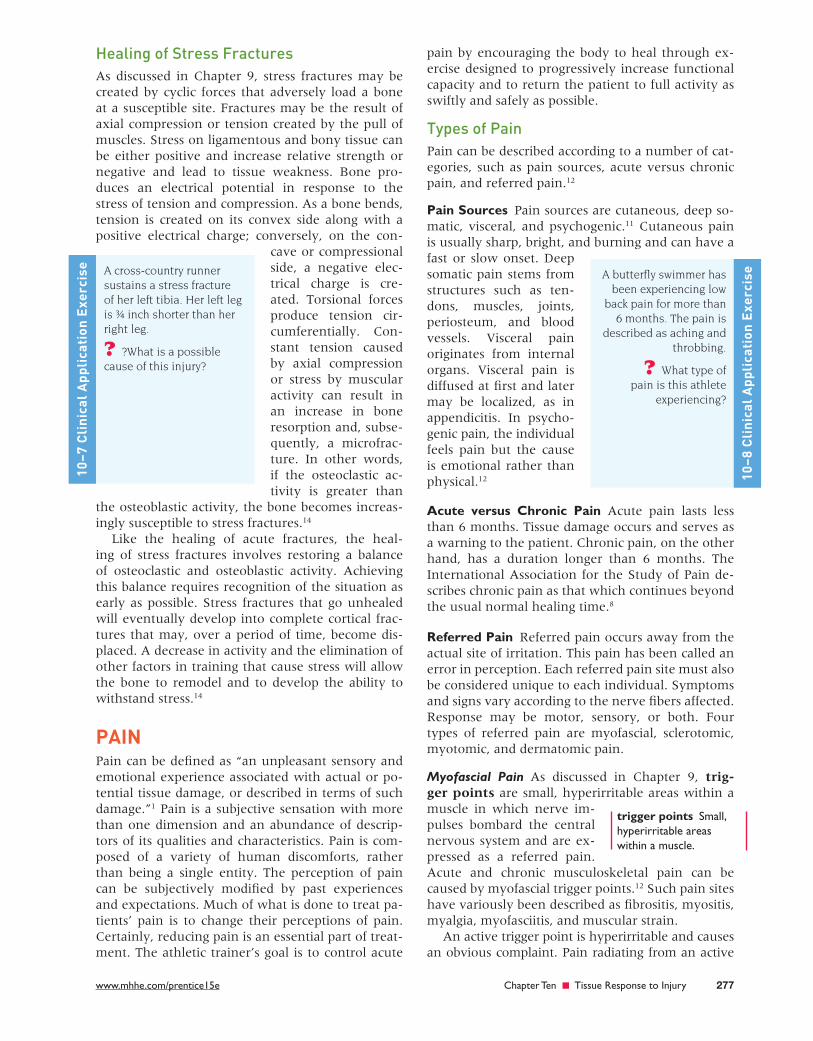
A butterfl y swimmer has been experiencing low
back pain for more than 6 months. The pain is
described as aching and throbbing.
? What type of pain is this athlete
experiencing?
10–8
Cli
nica
l App
lica
tion
Exe
rcis
e
trigger points Small, hyperirritable areas within a muscle.
pre22649_ch10_265-284.indd 277pre22649_ch10_265-284.indd 277 07/11/12 11:48 PM07/11/12 11:48 PM
CONFIRMING PAGES

278 Part Three ■ Pathology of Sports Injury
trigger point does not follow a usual area of distri-bution, such as sclerotomes, dermatomes, or periph-eral nerves. The trigger point pain area is called the reference zone, which can be close to the point of irritation or a considerable distance from it. 12
Sclerotomic, Myotomic, and Dermatomic Pain Deep pain may originate from sclerotomic, myotomic, or dermatomic nerve irritation or injury. 11 A sclero-tome is an area of bone or fascia that is supplied by a single nerve root. Myotomes are muscles supplied by a single nerve root. Dermatomes also are in an area of skin supplied by a single nerve root.
Sclerotomic pain is deep, aching, and poorly lo-calized pain. Sclerotomic pain impulses can be projected to regions in the brain such as the hypo-thalamus, limbic system, and reticular formation and can cause depression, anxiety, fear, or anger. Autonomic changes, such as changes in vasomotor tone, blood pressure, and sweating, may also occur.
Dermatomic pain, in contrast to sclerotomic pain, is sharp and well localized. Unlike sclerotomic pain, dermatomic pain projects mainly to the thala-mus and is relayed directly to the cortex, skipping autonomic and affective responses.
Nociceptors and Neural Transmission Pain receptors known as nociceptors, or free nerve endings, are sensitive to mechanical, ther-mal, and chemical energy. 11 They are commonly
found in skin, periosteum surrounding bone, teeth, meninges, and some organs.
Afferent nerve fi bers transmit impulses from the nociceptors toward the spinal cord, while efferent nerve fi bers, such as motor neurons, transmit im-pulses from the spinal cord toward the periphery. First-order, or primary, afferents transmit impulses from a nociceptor to the dorsal horn of the spinal cord. There are four types of fi rst-order neurons: Aa, Ab, Ad, and C. Aa and Ab fi bers are charac-terized as large-diameter afferents and Ad and C fi -bers as small-diameter afferents. Ad and C fi bers transmit sensations of pain and temperature. Ad neurons originate from nociceptors located in skin and transmit “fast pain,” while C neurons originate from both superfi cial tissue (skin) and deeper tissue (ligaments and muscle) and transmit “slow pain.” 12
Second-order afferent fi bers carry sensory mes-sages from the dorsal horn to the brain and are categorized as nociceptive specifi c. Second-order afferents receive input from Ab, Ad, and C fi bers. Second-order afferents serve relatively large, overlapping receptor fi elds. Nociceptive-specifi c second-order afferents respond exclusively to nox-ious stimulation and receive input only from Ad and C fi bers. All of these neurons synapse with
third-order neurons, which carry information via as-cending spinal tracts to various brain centers, where the input is integrated, interpreted, and acted upon. 12
Facilitators and Inhibitors of Synaptic Transmission For information to pass between neurons, a trans-mitter substance must be released from one neuron terminal, enter the synaptic cleft, and attach to a receptor site on the next neu-ron. This occurs primarily due to chemicals called neurotransmitters. However, it has been shown that several compounds that are not true neurotrans-mitters can facilitate or inhibit synaptic activity. These include serotonin, which is active in descend-ing pathways; norepinephrine, which inhibits pain transmission between fi rst- and second-order neu-rons; substance P, which is active in small-diameter primary afferent neurons; enkephalins, found in de-scending pathways; and b -endorphin, found in the central nervous system. 40
Mechanisms of Pain Control The neurophysiological mechanisms of pain con-trol have not been fully explained. To date, three models of pain control have been proposed: the gate control theory, descending pathway pain con-trol, and the release of b-endorphin. It is likely that some as-yet-unexplained combination of these three models is responsible for pain modulation. 1
Gate Control Theory Sensory information coming from cutaneous receptors in the skin enters the as-cending Ab afferents and is carried to the substan-tia gelatinosa in the dorsal horn of the spinal cord (Figure 10–7). Likewise, pain messages from the nociceptors are carried along the Ad and C afferent fi bers and enter the dorsal horn. Sensory informa-tion coming from Ab fi bers overrides or inhibits the “pain information” carried along Ad and C fi bers, thus inhibiting, or effectively “closing the gate” to, the transmission of pain information to second- order neurons. Consequently, pain information is not transmitted and never reaches sensory centers in the brain. The gate control theory of pain control occurs at the spinal cord level. 12
Descending Pathway Pain Control Stimulation of descending pathways in the spinal cord may also inhibit pain impulses carried along the Ad and C af-ferent fi bers (Figure 10–8). It is theorized that pre-vious experiences, emotional infl uences, sensory
nociceptors Pain receptors.
Neurotransmitters:
• Serotonin• Norepinephrine• Substance P• Enkephalins• b-endorphin
pre22649_ch10_265-284.indd 278pre22649_ch10_265-284.indd 278 07/11/12 11:48 PM07/11/12 11:48 PM
CONFIRMING PAGES

www.mhhe.com/prentice15e Chapter Ten ■ Tissue Response to Injury 279
perception, and other factors infl uence the trans-mission of pain messages and thus the perception of pain. The information coming from higher centers in the brain along efferent descending pathways in the spinal cord causes a release of two neurotransmitter- like substances, enkephalin and norepinephrine, into the dorsal horn, which together block or
inhibit the synaptic transmission of impulses from the Ad and C afferent fi bers to second-order affer-ent neurons. 12
Release of b-Endorphin It has been shown that noxious (painful) stimulation of nociceptors result-ing in the transmission of pain information along Ad and C afferents can stimulate the release of an opiate-like chemical called b - endorphin from the hypothalamus and anterior pituitary (Figure 10–9). b-endorphin is endogenous to the central nervous system and is known to have strong analgesic ef-fects. The exact mechanisms by which b-endorphin produces these potent analgesic effects are unclear. Acupuncture, acupressure, and point stimulation using electrical currents are all techniques that may stimulate the release of b-endorphin. 12
Pain Assessment Pain is a complex phenomenon that is diffi cult to evaluate and quantify because it is subjective. Thus, obtaining an accurate and standardized assessment of pain is problematic. A number of validated as-sessment tools are available that allow the athletic trainer to develop a pain profi le by identifying the type of pain a patient is experiencing, quantifying the intensity of pain, evaluating the effect of the pain experience on a patient’s level of functioning, and assessing the psychosocial impact of pain.
FIGURE 10–7 Gate control theory. Sensory information carried on Ab fibers “closes the gate” to pain information carried on Ad and C fibers in the substantia gelatinosa, preventing the transmission of pain to sensory centers in the cortex.
−−+
Dorsalhorn
Closingthe gate
mechanism
Substantiagelatinosa
First-orderneurons
Second-orderneurons
+ = Transmitted− = Inhibited
Ascendinglateralspinothalamictract
To sensorycortex
Posterior
Anterior
AδC
Aβ
FIGURE 10–8 Descending pathway pain control. Influ-ence from the thalamus stimulates the periaqueductal grey, the raphe nucleus, and the pons to inhibit the trans-mission of pain impulses through the ascending tracts.
Thalamus
Periaqueductalgrey
Raphe nucleus
Ascendinglateralspinothalamictract
Enkephalinreleased
Second-orderneuron
Substantiagelatinosa
Medulla
Pons
Midbrain
Dorsal lateralprojection
Second descendingprojection
Cerebrum
Aδ + C fibers
FIGURE 10–9 b-endorphin released from the hypotha-lamus and dynorphin released from the periaqueductal grey and the medulla.
Hypothalamus
Periaqueductalgrey
Lateralspinothalamicand spinoreticulartractsDynorphin
released
Dynorphinreleased
Enkephalinreleased
Medulla
Pons
Midbrain
Cerebrum
β-endorphinreleased
pre22649_ch10_265-284.indd 279pre22649_ch10_265-284.indd 279 07/11/12 11:48 PM07/11/12 11:48 PM
CONFIRMING PAGES

280 Part Three ■ Pathology of Sports Injury
Pain measurement tools include simple unidi-mensional scales or multidimensional questionnaires. Pain measurement should include both the time frame and the clinical context of the pain. With unidimensional scales, individuals with acute pain are usually asked to describe their pain “right now” and may be asked about the average inten-sity over a fi xed period to provide information on the course of the pain. Examples of commonly used unidimensional scales are verbal rating scales, the numeric rating scale, and the visual analog scale. Multidimensional pain assessment tools are more comprehensive pain assessments that require the determination of the quality of the pain and its ef-fect on mood and function. They are used mainly to quantify these aspects of pain, and they take lon-ger to administer than the unidimensional scales. The McGill Pain Questionnaire is an example of a multidimensional pain assessment tool.
Visual Analog Scales Visual analog scales are quick and simple tests that consist of a line, usually 2½ inches (10 cm) in length, the extremes of which are taken to represent the limits of the pain experience. The patient simply places a mark on that line based on the perceived level of pain. Scales can be completed daily or more often (Figure 10–10).
Pain Charts Pain charts are used to establish spatial properties of pain. They involve a two-dimensional graphic chart on which the patient assesses the loca-tion of pain and a number of subjective components. The patient colors the chart in areas that correspond to pain (Figure 10–11).
McGill Pain Questionnaire The McGill Pain Ques-tionnaire lists 78 words that describe pain, which are grouped into 20 sets and divided into four catego-ries representing dimensions of the pain experience. It may take up to 20 minutes to complete, and the questionnaire is administered every 2 to 4 weeks (Figure 10–12).
Activity Pain Indicators Profi le The Activity Pain Indicators Profi le is a 64-question self-report tool used to assess functional impairment associated with pain. It measures the frequency of certain behaviors, such as housework, recreation, and social activities, that produce pain.
Numeric Rating Scale The numeric rating scale is the most common acute pain profi le used in sports medicine. The patient is asked to verbally rate pain on a scale from 1 to 10, with 10 representing the worst pain he or she has experienced or can imag-ine. Usually, the scale is administered verbally before
No painrelief
No painat all
Complete painrelief
Worst painpossible
FIGURE 10–10 Visual analog scales.
FIGURE 10–11 Pain chart. Use the following instructions: “Please use all of the figures to show me exactly where all your pains are, and where they radi-ate to. Shade or draw with blue marker. Only the athlete is to fill out this sheet. Please be as precise and detailed as possible. Use yellow marker for numbness and tingling. Use red marker for burning or hot areas, and green marker for cramping. Please remember: blue 5 pain, yellow 5 numbness and tingling, red 5 burning or hot areas, green 5 cramping.” Used with permission from Melzack R: Pain measurement and assessment, New York, 1983, Raven Press.
Left Left Left
Left
Left
Left
Left
Left
Left
Right
RightRight
Right
Right
Right
Right
Right
pre22649_ch10_265-284.indd 280pre22649_ch10_265-284.indd 280 07/11/12 11:48 PM07/11/12 11:48 PM
CONFIRMING PAGES

www.mhhe.com/prentice15e Chapter Ten ■ Tissue Response to Injury 281
and after treatment. When treatments provide pain relief, questions are asked about the extent and du-ration of the relief.
Treating Pain An athletic trainer can approach pain management using a variety of treatment options, including therapeutic modalities and medications.
Therapeutic Modalities Many therapeutic mo-dalities can provide pain relief. 32 There is not one best therapeutic agent for pain control. The athletic trainer must select the therapeutic agent that is most appropriate for each athlete, based on the athletic trainer’s knowledge of the modalities and profes-sional judgment (see Chapter 15). In no situation should the athletic trainer apply a therapeutic agent
FIGURE 10–12 McGill Pain Questionnaire. The descriptors fall into four groups: sensory, 1 to 10; affective, 11 to 15; evaluative, 16; and miscellaneous, 17 to 20. The rank value for each descriptor is based on its position in the word set. The sum of the rank values is the pain rating index (PRI). The present pain intensity (PPI) is based on a scale of 0 to 5. Used with permission from Melzack R: Pain measurement and assessment, New York, 1983, Raven Press.
McGill Pain Questionnaire
Patient's Name
PRI S A E
FlickeringQuiveringPulsingThrobbingBeatingPounding
1
JumpingFlashingShooting
2
PrickingBoringDrillingStabbingLancinating
3
SharpCuttingLacerating
4
PinchingPressingGnawingCrampingCrushing
5
TuggingPullingWrenching
6
HotBurningScaldingSearing
7
TinglingItchySmartingStinging
8
DullSoreHurtingAchingHeavy
9
TenderTautRaspingSplitting
10
NaggingNauseatingAgonizingDreadfulTorturing
20
No painMildDiscomfortingDistressingHorribleExcruciating
012345
CoolColdFreezing
19
TightNumbDrawingSqueezingTearing
18
SpreadingRadiatingPenetratingPiercing
17
AnnoyingTroublesomeMiserableIntenseUnbearable
16
WretchedBlinding
15
PunishingGruellingCruelViciousKilling
14
FearfulFrightfulTerrifying
13
SickeningSuffocating
12
TiringExhausting
BriefMomentaryTransient
RhythmicPeriodicIntermittent
ContinuousSteadyConstant
11
M PRI (T) PPI
Date
(1–20)(17–20)(16)(11–15)(1–10)
Time am/pm
PPI
COMMENTS
E = External = InternalI
pre22649_ch10_265-284.indd 281pre22649_ch10_265-284.indd 281 07/11/12 11:48 PM07/11/12 11:48 PM
CONFIRMING PAGES

282 Part Three ■ Pathology of Sports Injury
without fi rst developing a clear rationale for the treatment. The therapeutic modalities used to control pain do little to promote tissue healing. They should be used to relieve acute pain following injury or sur-gery or to control pain and other symptoms, such as swelling, to promote progressive exercise. The ath-letic trainer should not lose sight of the effects of the modalities or the importance of progressive exercise in restoring the athlete’s functional ability.
The athletic trainer can make use of the gate control mechanism of pain control by using su-perfi cial heat or cold, electrical stimulating cur-rents, massage, and counterirritants to stimulate the large-diameter Aa and Ab efferent nerve fi bers. Noxious stimulation of acupuncture and trigger points using either electrical stimulating currents or deep acupressure massage techniques can mediate the release of b-endorphin. 12
Medications A physician may choose to prescribe oral or injectable medications in treating a patient. The most commonly used medications are clas-sifi ed as analgesics, anti-infl ammatory agents, or both. 10 The athletic trainer should become familiar with these drugs and note whether the patient is taking any medications (see Chapter 17). It is also important to work with the referring physician or a pharmacist to make sure that the patient takes the medications appropriately.
Psychological Aspect of Pain Pain, especially chronic pain, is a subjective psycho-logical phenomenon. 40 When painful injuries are treated, the total patient must be considered, not just the pain or condition. Even in the most well-adjusted person, pain creates emotional changes. Constant pain often causes self-centeredness and an increased sense of dependency. Chapter 11 dis-cusses in great detail the psychosocial aspects of dealing with injury and managing pain.
Patients vary in their pain thresholds (Fig-ure 10–13). Some can tolerate enormous pain, whereas others fi nd mild pain almost unbearable. Pain is perceived as being worse at night because persons are alone, more aware of themselves, and
devoid of external diversions. Personality differ-ences can also cause differences in pain toleration. For example, patients who are anxious, dependent, and immature have less tolerance for pain than those who are relaxed and emotionally in control.
A number of theo-ries about how pain is produced and perceived by the brain have been advanced. Only in the last few decades has sci-ence demonstrated that pain is both a psycho-logical and a physiologi-cal phenomenon and is therefore unique to each individual. 4,40 Sports ac-tivities demonstrate this fact clearly. Through con-ditioning, an athlete learns to endure the pain of rigorous activity and to block the sensations of a minor injury. It is perhaps most critical for the athletic trainer to recognize that all pain is very real to the patient.
FIGURE 10–13 Coping with pain in sports is as much psychological as it is physical.
A gymnast is receiving electrical stimulation for
chronic low back pain.
? What is the purpose of administering
electrical stimulation for the chronic pain?
10–9
Cli
nica
l App
lica
tion
Exe
rcis
e
SUMMARY • The three phases of the healing process are the
infl ammatory response phase, the fi broblastic repair phase, and the maturation-remodeling phase, which occur in sequence but overlap one another in a continuum.
• During the infl ammatory response phase, debris is phagocytized (cleaned up). The fi broblastic
repair phase involves the deposition of collagen fi bers to form a fi rm scar. During the maturation-remodeling phase, collagen is realigned along lines of tensile force and the tissue gradually as-sumes normal appearance and function.
• Factors that may impede the healing proc-ess include the extent of the injury, edema,
282 Part Three ■ Pathology of Sports Injury
pre22649_ch10_265-284.indd 282pre22649_ch10_265-284.indd 282 07/11/12 11:48 PM07/11/12 11:48 PM
CONFIRMING PAGES

www.mhhe.com/prentice15e Chapter Ten ■ Tissue Response to Injury 283
hemorrhage, poor vascular supply, separation of tissue, muscle spasm, atrophy, corticosteroids, keloids and hypertrophic scars, infection, climate and oxygen tension, health, age, and nutrition.
• Treatment techniques that can be used to modify soft-tissue healing include anti-infl ammatory med-ications, therapeutic modalities, exercise rehabilita-tion, and platelet-rich plasma injections.
• The healing of soft tissues, including cartilage, ligament, muscle, and nerve, follows a similar course. Unlike the other tissues, nerve has the capability of regenerating.
• Bone healing following fracture involves increased activity of osteoblastic cells and osteoclastic cells.
• Pain is a response to a noxious stimulus that is subjectively modifi ed by past experiences and expectations.
• Pain is classifi ed as either acute or chronic and can exhibit many different patterns.
• Three models of pain control are the gate control theory, descending pathway pain control, and the release of b-endorphin from higher centers of the brain.
• Pain perception may be infl uenced by a variety of cognitive processes mediated by the higher brain centers.
SOLUTIONS TO CLINICAL APPLICATION EXERCISES
American Academy of Pain Management: www.aapainmanage.org
American Pain Society: www.ampainsoc.org World Union of Wound Healing Societies:
www.wuwhs.org
10–1 Little can be done to speed up the healing process physi-ologically. This athlete must realize that certain physiologi-cal events must occur during each phase of the healing process. Any interference with this healing process during a rehabilitation program will likely slow return to full ac-tivity. The healing process must have an opportunity to accomplish what it is supposed to.
10–2 Immobilization during the infl ammatory process may be benefi cial; however, controlled mobilization helps the tis-sue decrease atrophy and enhance the healing process. Controlled mobilization allows the athlete to perform pro-gressive strengthening exercises in a timely manner.
10–3 Initially, a transitory vasoconstriction with the start of blood coagulation of the broken blood vessels occurs. Dila-tion of the vessels in the region of injury follows, along with the activation of chemical mediators via key cells.
10–4 A grade 2 lateral ankle sprain implies that the joint capsule and ligaments are partially torn. At 3 weeks, the injury has been cleaned of debris and is undergoing the process of
secondary healing. Granulation tissue fi lls the torn areas, and fi broblasts are beginning to form scar tissue.
10–5 In its acute phase, the injury was not allowed to heal properly. As a result, the injury became chronic, with a proliferation of scar tissue, lymphocytes, plasma cells, and macrophages.
10–6 Uncomplicated acute bone healing goes through fi ve stages: hematoma formation, cellular proliferation, callus formation, ossifi cation, and remodeling.
10–7 Because it is shorter, the left leg has the greater stress dur-ing running. This stress creates increased tension on the tibia’s concave side, causing an increase in osteoclastic activity.
10–8 The pain is considered to be chronic, deep somatic pain stemming from the low back muscles. It is conducted pri-marily by the C-type nerve fi bers.
10–9 The purpose is to stimulate the large, rapidly conducting nerve fi bers to inhibit the smaller and slower nerves that carry pain impulses.
WEB SITES
REVIEW QUESTIONS AND CLASS ACTIVITIES
1. What are the three phases of healing, and what are the ap-proximate time frames for each of these three phases?
2. What are the physiological events associated with the in-fl ammatory response phase of the healing process?
3. How can you differentiate between acute and chronic infl ammation?
4. How is collagen laid down in the area of injury during the fi broblastic repair phase of healing?
5. Explain Wolff’s law and the importance of controlled mobil-ity during the maturation-remodeling phase of healing.
6. What are some of the factors that can have a negative impact on the healing process?
7. Discuss treatment techniques for modifying soft-tissue heal-ing, including anti-infl ammatory medications, therapeutic modalities, exercise rehabilitation, and platelet-rich plasma injections.
8. Compare and contrast the course of healing in cartilage, liga-ments, muscle, and nerve.
9. What is a basic defi nition of pain? 10. What are the different types of pain? 11. What are the characteristics of the various sensory receptors? 12. How does the nervous system relay information about pain-
ful stimuli? 13. Describe how the gate control mechanism of pain modula-
tion may be used to modulate pain. 14. How does descending pathway pain control modulate pain? 15. What are the opiate-like substances, and how do they mod-
ulate pain? 16. What are the assessment scales available to help the athletic
trainer determine the extent of pain perception? 17. How can pain perception be modifi ed by cognitive factors?
pre22649_ch10_265-284.indd 283pre22649_ch10_265-284.indd 283 07/11/12 11:48 PM07/11/12 11:48 PM
CONFIRMING PAGES

284 Part Three ■ Pathology of Sports Injury
REFERENCES
1. Aronson PA: Pain theories: A review for application in athletic training and therapy, Athletic Therapy Today 7(4):8, 2002.
2. Bailon-Plaza A: Benefi cial effects of moder-ate, early loading and adverse effects of de-layed or excessive loading on bone healing, Journal of Biomechanics 36(8):1069, 2003.
3. Barrientos S: Growth factors and cytokines in wound healing, Wound Repair and Regen-eration , 16(5):585–602, 2008.
4. Battery L: Infl ammation in overuse ten-don injuries, Sports Medicine and Arthroscopy Reviews , 19(3):213–17, 2011.
5. Black KP: Perpheral afferent nerve regener-ation. In Lephart S, Fu F, editors: Propriocep-tion and neuromuscular control in joint stability , Champaign, IL, 2000, Human Kinetics.
6. Borrione P: Platelet-rich plasma in muscle healing, American Journal of Physical Medi-cine and Rehabilitation , 89(1):854, 2010.
7. Campbell W: Evaluation on management of peripheral nerve injury, Clinical Neuro-physiology , 119(9):1951–65, 2008.
8. Casey C: Transition from acute to chronic pain and disability: A model including cog-nitive, affective and trauma factors, Pain , 134(2):69–79, 2008.
9. Clark B: In vivo alterations in skeletal mus-cle form and function after disuse atrophy, Medicine and Science in Sport and Exercise , 2009.
10. Creighton A, Spang J, Dahners L: Basic science of ligament healing: Medial col-lateral ligament healing with and without treatment, Sports Med Arthroscopy Review 13(3):145, 2005.
11. Deleo J: Basic science of pain, Bone Joint Surg 88(s):58, 2006.
12. Denegar CR, Prentice W: Managing pain with therapeutic modalities. In Prentice WE, editor: Therapeutic modalities in sports medicine and athletic training, ed 6, San Francisco, 2010, McGraw-Hill.
13. Diegelmann R: Wound healing: An over-view of acute, fi brotic, and delayed heal-ing, Frontiers in Bioscience , (2):283–89, 2004.
14. Ferrry A: Stress fractures in athletes, Physi-cian and Sports Medicine , 38(2):109–18, 2010.
15. Gallin J, Snyderman R, Haynes B: Infl amma-tion: Basic principles and clinical correlates, Balti-more, 1999, Lippincott Williams and Wilkins.
16. Hildebrand KA, Behm C, Kydd A: The ba-sics of soft tissue healing and general fac-tors that infl uence such healing, Sports Med Arthroscopy Review 13(3):136, 2005.
17. Hill C: Rehabilitation of soft tissue and musculoskeletal injury. In O’Young B: Physical medicine and rehabilitation secrets , Philadelphia, 2007, Hanley-Belfus.
18. Hubbel S, Buschbacher R: Tissue injury and healing: Using medications, modalities, and exercise to maximize recovery. In Bush-bacher R, Branddom R, editors: Sports medi-cine and rehabilitation: A sport specifi c approach, Philadelphia, 2008, Elsevier Health Services.
19. Irrgang JJ, Pezzullo D: Rehabilitation fol-lowing surgical procedures to address artic-ular cartilage lesions in the knee, J Orthop Sports Phys Ther 28(4):232, 1998.
20. Isaacs J: Treatmnt of acute peripheral nerve injuries: Current concepts, Journal of Hand Surgery , 35(3):491–97, 2010.
21. Kocher MS: Ligament healing and augmen-tation. In Proceedings, National Athletic Trainers’ Association, 10th annual meeting and clinical sym-posia, Champaign, IL, 1999, Human Kinetics.
22. Ley K: Physiology of infl ammation, Bethesda, MD, 2001, American Physiological Society.
23. Li J: Pathophysiology of acute wound heal-ing, Clinics in Dermatology , 25(1):9–18, 1997.
24. Maffulli N: Basic science of tendons, Sports Med Arthroscopy Review 8(1):1, 2000.
25. Malone T, Garrett W, Zachewski J: Muscle: Deformation, injury and repair. In Zachaze-wski J, Magee D, Quillen W, editors: Ath-letic injuries and rehabilitation, Philadelphia, 1996, WB Saunders.
26. Marsell R: The biology of fracture healing, Injury , 42(6):551–55, 2011.
27. Molloy T: The roles of growth factors in tendon and ligament healing, Sports Med 33(5):381, 2003.
28. Murdoch S: Managing the infl ammatory response through nutritional supplements, Athletic Therapy Today 6(5):46, 2001.
29. Murrell GAC, Jang D, Lily E, Best T: The ef-fects of immobilization and exercise on ten-don healing, Journal of Science and Medicine in Sport 2(1 Supplement):40, 1999.
30. Musahl V: Cartilage injuries. In Ranawat A: Musculoskeletal examination of the hip and knee , Thorofare, NJ, 2011, Slack.
31. Paoloni J: Platelet-rich plasma treatment for ligament and tendon injuries, Clinical Jour-nal of Sports Medicine , 21(1):37–45, 2011.
32. Prentice WE: Therapeutic modalities in rehabil-itation , ed 4, New York, 2011, McGraw-Hill.
33. Rees J: Management of tendinopathy, Ameri-can Journal of Sports Medicine , 37(9):1855–67, 2009.
34. Redler L: Platelet-rich plasma therapy: An systematic literature review and evidence for clinical use, Physician and SportsMedicine 39(1):42–51, 2011.
35. Sandrey M: Acute and chronic tendon inju-ries: Factors affecting the healing response and treatment, J Sport Rehabil 12(1):70, 2003.
36. Sandrey MA: Effects of acute and chronic pathomechanics on the normal histology and biomechanics of tendons: A review, J Sport Rehabil 9(4):339, 2000.
37. Seeley R, Stephens T, Tate P: Seeley’s anatomy and physiology, St. Louis, 2010, McGraw-Hill.
38. Serhan C: Fundamentals of infl ammation , Cambridge, UK, 2010, Cambridge Univer-sity Press.
39. Shah R: Utilization of prolotherapy for fa-cilitation of ligament and tendon healing, Athletic Therapy Today , 15(6):25, 2010.
40. Shelemay K: Pain and its transformations: The interface of biology and culture , Boston, 2007, Harvard University Press.
41. Steiner D: Pathophysiology of ligament injuries. In Comfort P: Sports rehabilitation and injury prevention, New York, 2010, John Wiley & Sons.
42. Taylor M: Treating chronic tendon injuries, Athletic Therapy Today 5(6):50, 2000.
43. Thompson L: Skeletal muscle adaptations with age, inactivity, and therapeutic exer-cise, J Orthop Sports Phys Ther 32(2):2002.
44. Velnar T: The wound healing process: An overview of the cellular and molecular mechanisms, Journal of International Medical Research , 37(5):1528–42, 2009.
45. Volgas D: A manual of soft tissue manage-ment in orthopedic trauma , New York, 2011, Thieme.
46. Walker JM: Pathomechanics and classifi -cation of cartilage lesions, facilitation of repair, J Orthop Sports Phys Ther 28(4):216, 1998.
47. Ward P: Acute infl ammation and chronic infl ammation. In Serhan C: Fundamentals of infl ammation , Cambridge, UK, 2010, Cambridge University Press.
48. Warren G, Ingalls C, Lowe D: What mecha-nisms contribute to the strength loss that occurs during and in the recovery from skeletal muscle injury? J Orthop Sports Phys Ther 32(2):2002.
49. Weintraub W: Tendon and ligament healing: A new approach to sports and overuse injury, Brookline , MA, 2005, Paradigm.
50. Westerblad H: Skeletal muscle: En-ergy metabolism, fi ber types fatigue and adaptability, Experimental Cell Research , 316(18):3093–99, 2010.
51. Woo S, Moon D, Miura K: Basic science of ligament healing: anterior cruciate ligament graft biomechanics and knee kinematics, Sports Med Arthroscopy Review 13(3):161, 2005.
52. Young, A: The physiology of wound heal-ing, Surgery , 29(10):475–79, 2011.
ANNOTATED BIBLIOGRAPHY
Damjanov I, editor: Anderson’s pathology, ed 10, Philadelphia, 1996, Elsevier Science.
A major pathology text that discusses infl ammation and healing in depth.
Kloth L, McCulloch J: Wound healing: alternatives in management, Philadelphia, 2001, Davis.
An excellent discussion of factors infl uencing wound healing, evaluation, and methods of treatment.
Melzack R, Wall P: Handbook of pain management, Philadelphia, 2003, Elsevier.
A summary of current knowledge of pain states and their management for all health care professionals involved in the diagnosis and treatment of patients with a wide variety of acute and chronic pain problems.
Porth CM: Pathophysiology: Concepts of altered health states , ed 8, Philadelphia, 2010, Lippincott, Williams and Wilkins.
An in-depth text on the physiology of altered health; contains an excellent discussion on infl ammation, healing, and pain.
pre22649_ch10_265-284.indd 284pre22649_ch10_265-284.indd 284 07/11/12 11:48 PM07/11/12 11:48 PM
CONFIRMING PAGES