Embed Size (px)
Citation preview
TISSUE FREEZING METHODS FOR CRYOSTAT SECTIONING
Basic Tissue Freezing Methods Preparing Tissue for Freezing
Then a quick overview of MHPL Cryostat sectioning Techniques: Using The Brush;
Using The Anti-Roll Plate;
Speaker: Donna J. Emge, HT-ASCP, MHPL Manager
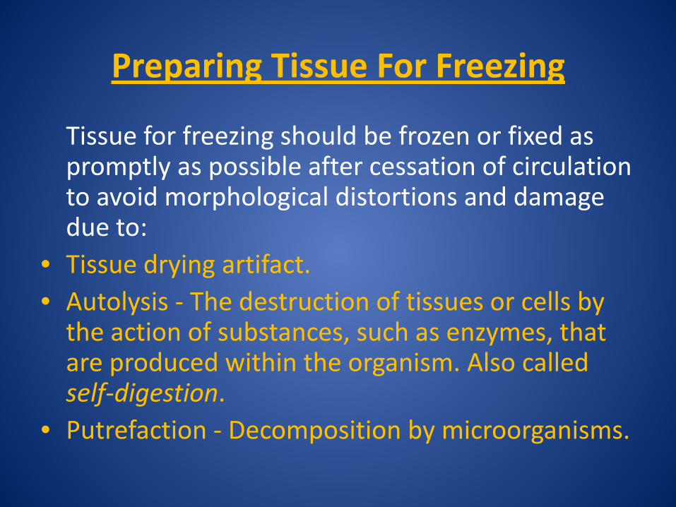
Preparing Tissue For Freezing
Tissue for freezing should be frozen or fixed as promptly as possible after cessation of circulation to avoid morphological distortions and damage due to:
• Tissue drying artifact. • Autolysis - The destruction of tissues or cells by
the action of substances, such as enzymes, that are produced within the organism. Also called self-digestion.
• Putrefaction - Decomposition by microorganisms.
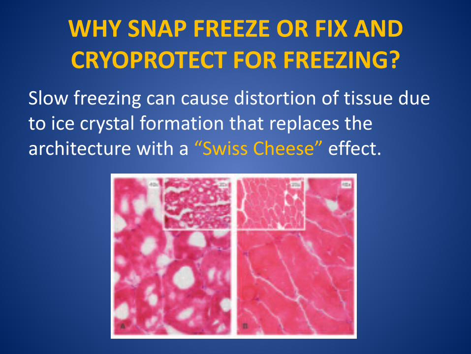
WHY SNAP FREEZE OR FIX AND CRYOPROTECT FOR FREEZING?
Slow freezing can cause distortion of tissue due to ice crystal formation that replaces the architecture with a “Swiss Cheese” effect.
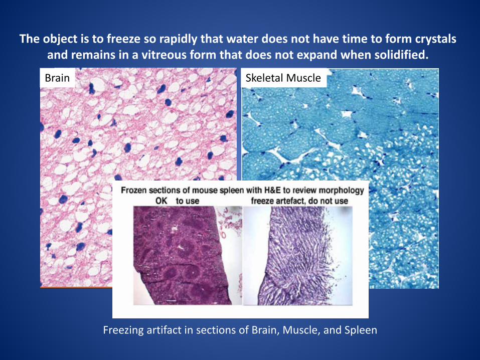
The object is to freeze so rapidly that water does not have time to form crystals and remains in a vitreous form that does not expand when solidified.
Freezing artifact in sections of Brain, Muscle, and Spleen
Brain Skeletal Muscle
SHORT ARTICLE ON THE SUBJECT OF WATER CRYSTAL FORMATION:
“FREEZING BIOLOGICAL SAMPLES”
Charles W. Scouten & Miles Cunningham http://www.myneurolab.com/global/Manuals/Tips%20an
d%20Techniques%20Freezing%20Artifact.pdf
METHODS OF TISSUE FREEZING 1. Fresh tissue freezing – Tissue is in OCT and flash
frozen fresh.
2. 4% PFA fixed, sucrose cryoprotected tissue freezing – Tissue is in OCT and may be frozen using dry ice or the flash frozen method.
3. Enzyme study tissue freezing – Often used for fresh muscle tissue. A fresh frozen method with no OCT matrix. Tissue protrudes from a Tragacanth or other support medium.
MHPL Protocols for these methods are included in your workshop folder.
WHY NOT JUST USE LIQUID NITROGEN?
• It Boils – this creates a vapor barrier that causes freezing in a slower, unpredictable pattern.
• Tissue and OCT often cracks – due to this unpredictable freezing pattern.
AFTER ALL: “Liquid nitrogen is one of the coldest liquids routinely available and it does not mix with tissue.”
Review the article: “FREEZING BIOLOGICAL SAMPLES”
Charles W. Scouten & Miles Cunningham http://www.myneurolab.com/global/Manuals/Tips%20and%20Techniques%20Freezing%
20Artifact.pdf
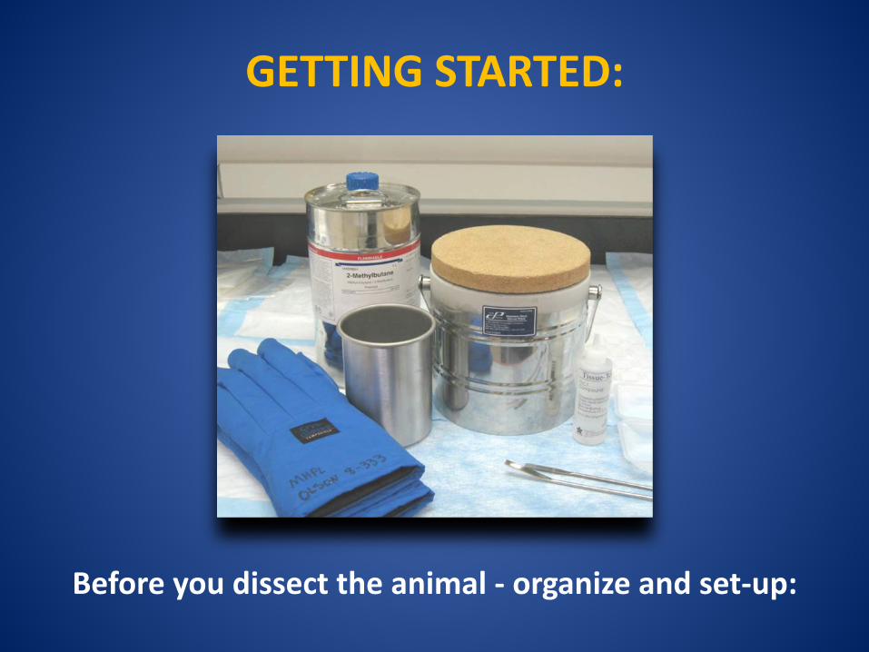
GETTING STARTED:
Before you dissect the animal - organize and set-up:
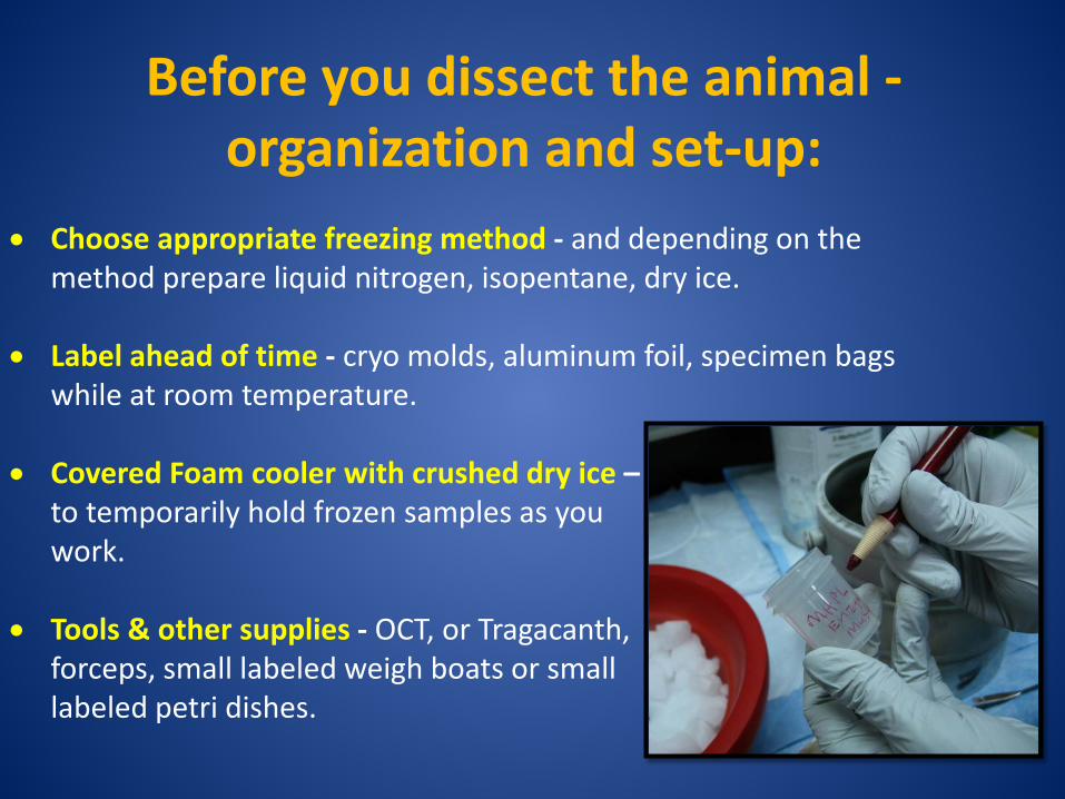
Before you dissect the animal - organization and set-up:
• Choose appropriate freezing method - and depending on the method prepare liquid nitrogen, isopentane, dry ice.
• Label ahead of time - cryo molds, aluminum foil, specimen bags while at room temperature.
• Covered Foam cooler with crushed dry ice – to temporarily hold frozen samples as you work.
• Tools & other supplies - OCT, or Tragacanth, forceps, small labeled weigh boats or small labeled petri dishes.
FRESH TISSUE FREEZING
Pros • Fastest of all methods. • Excellent for IHC, IF, ISH. No antigen retrieval
required since no cross-linking fixative. • Often easiest to section – depending upon tissue. Cons • Poorest morphology. • Prone to freezing artifact – must be snap frozen. • ISH integrity – extreme clean techniques required
or RNA will be rapidly and easily degraded.
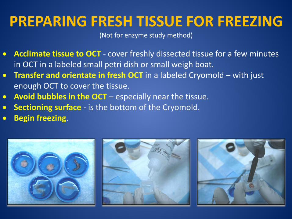
PREPARING FRESH TISSUE FOR FREEZING (Not for enzyme study method)
• Acclimate tissue to OCT - cover freshly dissected tissue for a few minutes
in OCT in a labeled small petri dish or small weigh boat. • Transfer and orientate in fresh OCT in a labeled Cryomold – with just
enough OCT to cover the tissue. • Avoid bubbles in the OCT – especially near the tissue. • Sectioning surface - is the bottom of the Cryomold. • Begin freezing.
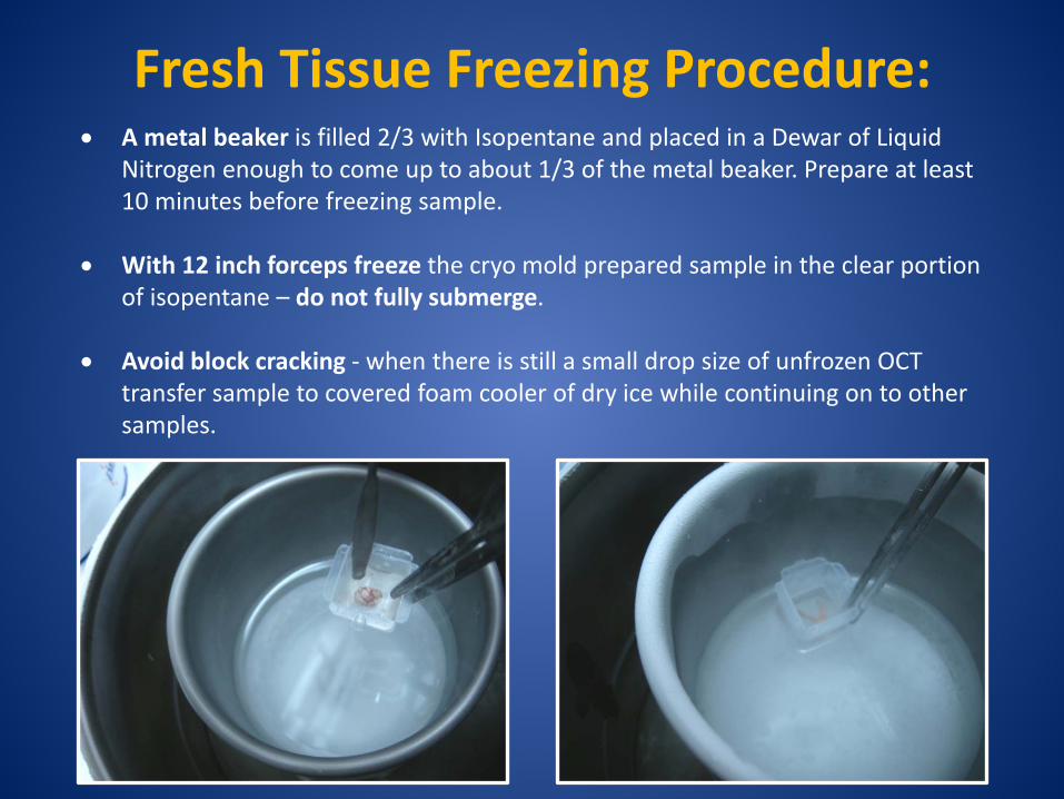
Fresh Tissue Freezing Procedure: • A metal beaker is filled 2/3 with Isopentane and placed in a Dewar of Liquid
Nitrogen enough to come up to about 1/3 of the metal beaker. Prepare at least 10 minutes before freezing sample.
• With 12 inch forceps freeze the cryo mold prepared sample in the clear portion of isopentane – do not fully submerge.
• Avoid block cracking - when there is still a small drop size of unfrozen OCT transfer sample to covered foam cooler of dry ice while continuing on to other samples.
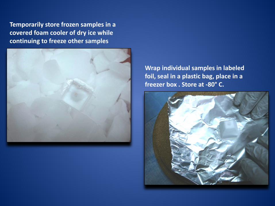
Temporarily store frozen samples in a covered foam cooler of dry ice while continuing to freeze other samples
Wrap individual samples in labeled foil, seal in a plastic bag, place in a freezer box . Store at -80° C.
4% PFA FIXED, SUCROSE CRYOPROTECTED TISSUE FREEZING
Pros • Excellent morphology compared to other methods. • May use a slower freeze in crushed powder dry ice
alone, slush of dry ice and 100% alcohol, or in a beaker of isopentane surrounded by dry ice - without incurring freezing artifact or block cracking.
• Any of the freezing methods discussed can be used. • Good for most IHC, IF and ISH. Cons • Time consuming • Most IHC will require antigen retrieval. • Although the fixative cross-linking is protective for ISH
techniques there is some RNA degradation.
PREPARING FIXED, SUCROSE CRYOPROTECTED TISSUE
• 4% PFA transcardial perfuse animal. • Drop fix in 4% PFA for a few hours to O/N. • 15% sucrose in 1XPBS until tissue sinks. • 30% sucrose in 1XPBS until tissue sinks.
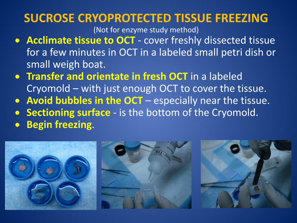
SUCROSE CRYOPROTECTED TISSUE FREEZING (Not for enzyme study method)
• Acclimate tissue to OCT - cover freshly dissected tissue for a few minutes in OCT in a labeled small petri dish or small weigh boat.
• Transfer and orientate in fresh OCT in a labeled Cryomold – with just enough OCT to cover the tissue.
• Avoid bubbles in the OCT – especially near the tissue. • Sectioning surface - is the bottom of the Cryomold. • Begin freezing.
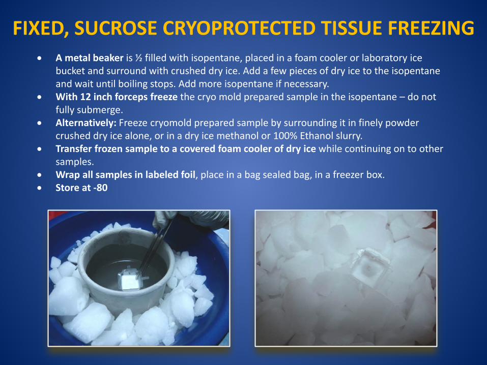
• A metal beaker is ½ filled with isopentane, placed in a foam cooler or laboratory ice bucket and surround with crushed dry ice. Add a few pieces of dry ice to the isopentane and wait until boiling stops. Add more isopentane if necessary.
• With 12 inch forceps freeze the cryo mold prepared sample in the isopentane – do not fully submerge.
• Alternatively: Freeze cryomold prepared sample by surrounding it in finely powder crushed dry ice alone, or in a dry ice methanol or 100% Ethanol slurry.
• Transfer frozen sample to a covered foam cooler of dry ice while continuing on to other samples.
• Wrap all samples in labeled foil, place in a bag sealed bag, in a freezer box. • Store at -80
FIXED, SUCROSE CRYOPROTECTED TISSUE FREEZING
ENZYME STUDY TISSUE FREEZING Pros
• Excellent for Enzyme histochemistry and Immunohistochemistry studies.
• Best method for muscle tissue. Cons • Advanced skill needed for sectioning – no supportive
OCT matrix. Anti-roll plate better than brush technique.
• Time and technique skill to prepare. • Extremely susceptible to any freeze thaw – leading to
loss of morphologic detail in muscle or brain tissue.
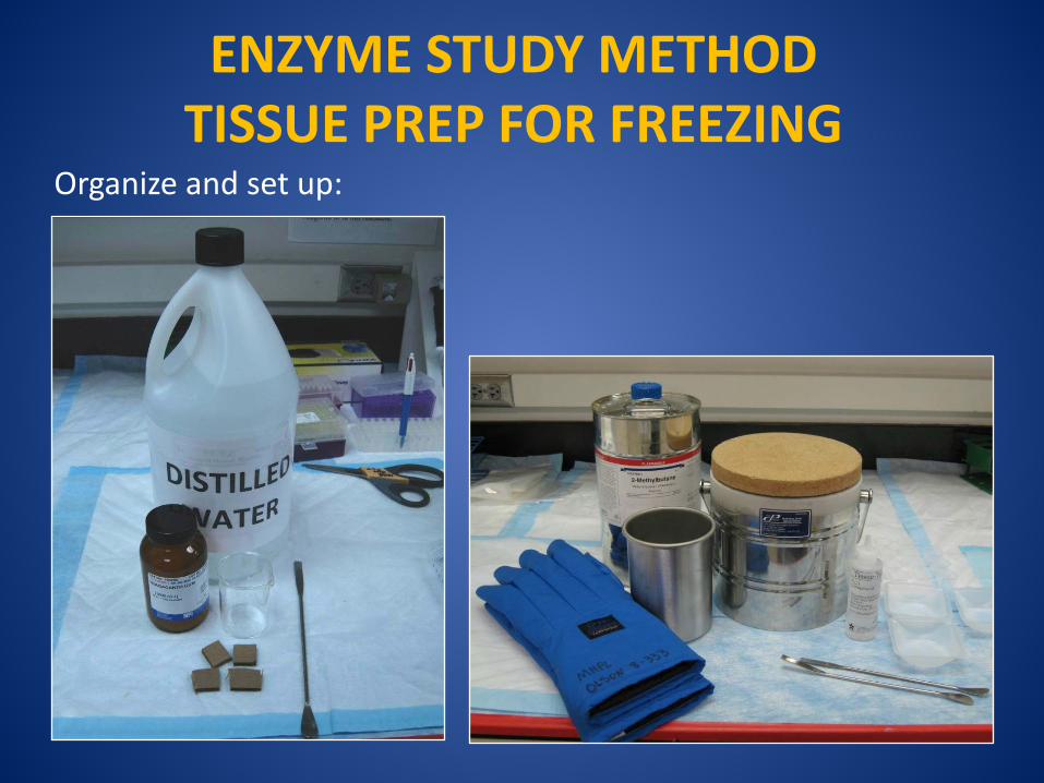
ENZYME STUDY METHOD TISSUE PREP FOR FREEZING
Organize and set up:
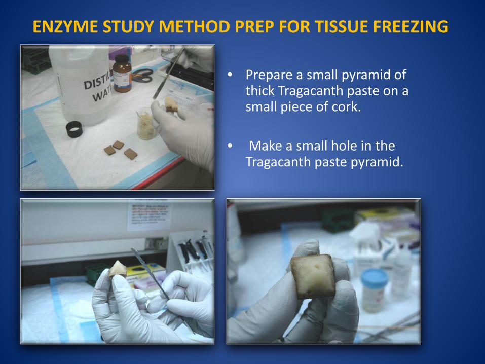
ENZYME STUDY METHOD PREP FOR TISSUE FREEZING
• Prepare a small pyramid of
thick Tragacanth paste on a small piece of cork.
• Make a small hole in the Tragacanth paste pyramid.
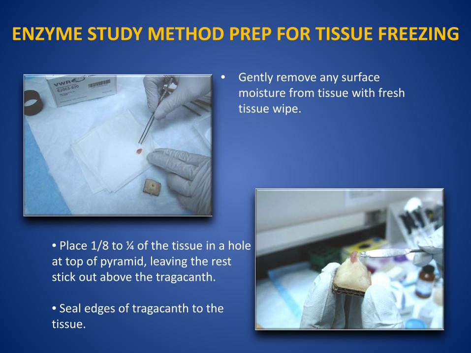
ENZYME STUDY METHOD PREP FOR TISSUE FREEZING
• Gently remove any surface moisture from tissue with fresh tissue wipe.
• Place 1/8 to ¼ of the tissue in a hole at top of pyramid, leaving the rest stick out above the tragacanth.
• Seal edges of tragacanth to the tissue.
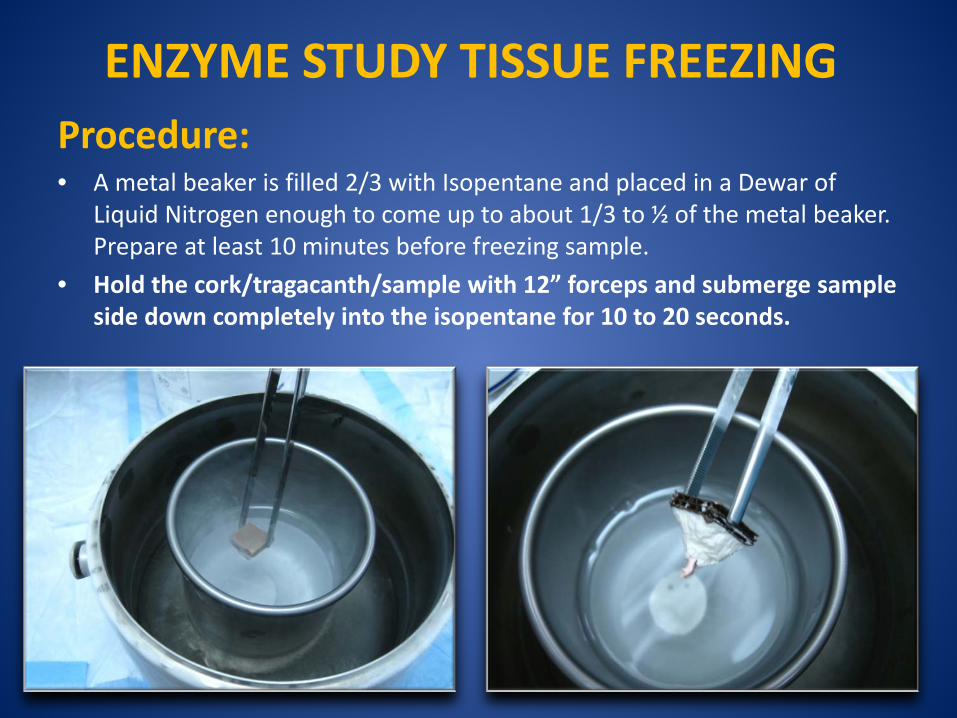
ENZYME STUDY TISSUE FREEZING Procedure:
• A metal beaker is filled 2/3 with Isopentane and placed in a Dewar of Liquid Nitrogen enough to come up to about 1/3 to ½ of the metal beaker. Prepare at least 10 minutes before freezing sample.
• Hold the cork/tragacanth/sample with 12” forceps and submerge sample side down completely into the isopentane for 10 to 20 seconds.
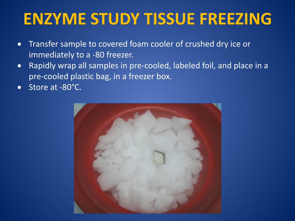
ENZYME STUDY TISSUE FREEZING • Transfer sample to covered foam cooler of crushed dry ice or
immediately to a -80 freezer. • Rapidly wrap all samples in pre-cooled, labeled foil, and place in a
pre-cooled plastic bag, in a freezer box. • Store at -80°C.
CRYOSECTIONING PREP
• Remove the frozen block from the -70°C freezer and allow it to equilibrate in the cryostat chamber temperature for approximately 30 minutes.
• The optimal temperature for cryostat sectioning
depends on the nature of the tissue and on whether the tissues have been freshly frozen or pre-fixed with subsequent cryoprotection.
• Note the reference chart for temperature setting guidelines for tissue types in your folder.
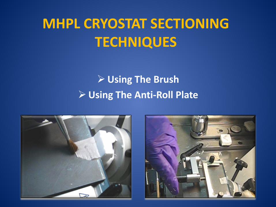
MHPL CRYOSTAT SECTIONING TECHNIQUES
Using The Brush
Using The Anti-Roll Plate
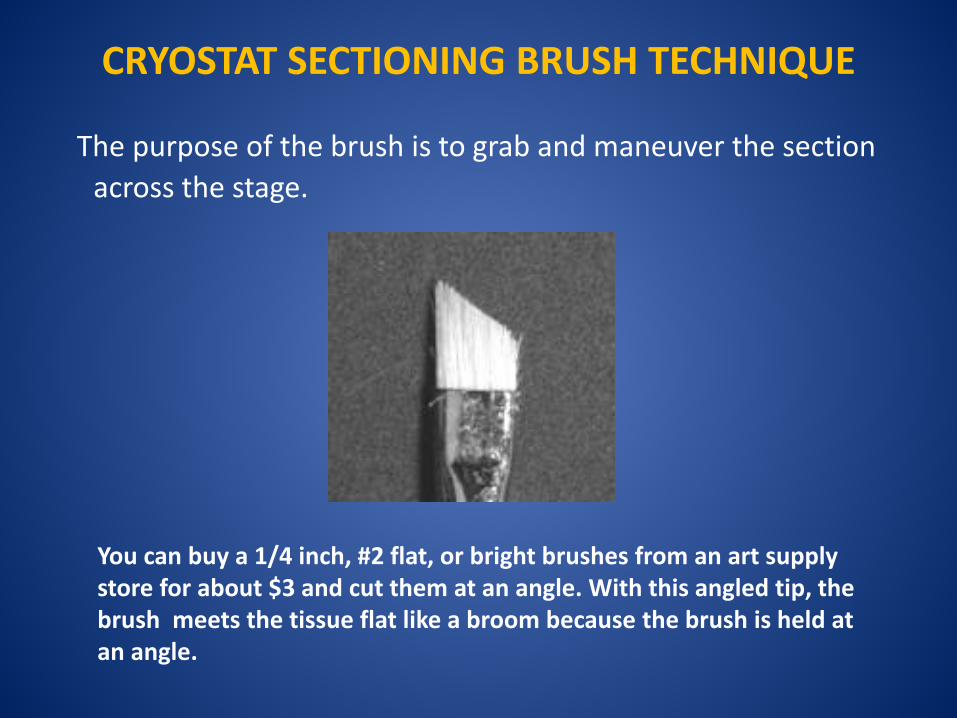
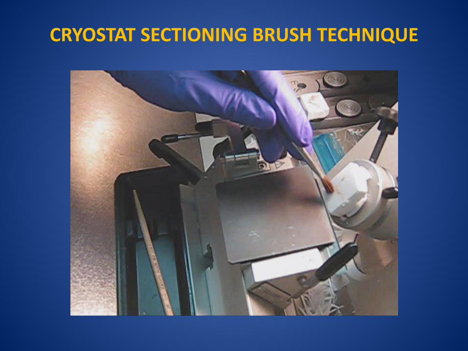
CRYOSTAT SECTIONING BRUSH TECHNIQUE
The purpose of the brush is to grab and maneuver the section across the stage.
You can buy a 1/4 inch, #2 flat, or bright brushes from an art supply store for about $3 and cut them at an angle. With this angled tip, the brush meets the tissue flat like a broom because the brush is held at an angle.
Stephen R Peters M.D. Pathology Innovations, LLC
http://www.pathologyinnovations.com/frozen_section_technique.htm
CRYOSTAT SECTIONING BRUSH TECHNIQUE
CRYOSTAT SECTIONING BRUSH TECHNIQUE
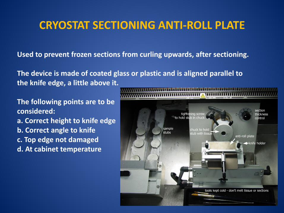
CRYOSTAT SECTIONING ANTI-ROLL PLATE Used to prevent frozen sections from curling upwards, after sectioning. The device is made of coated glass or plastic and is aligned parallel to the knife edge, a little above it. The following points are to be considered: a. Correct height to knife edge b. Correct angle to knife c. Top edge not damaged d. At cabinet temperature
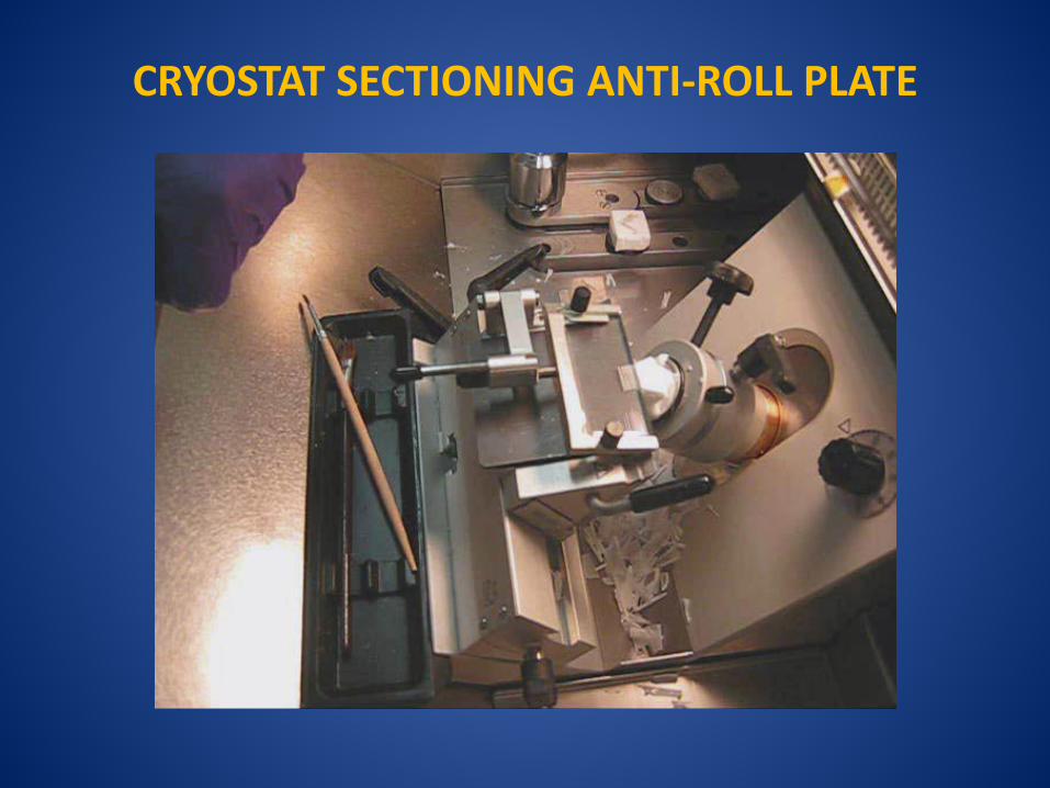
CRYOSTAT SECTIONING ANTI-ROLL PLATE
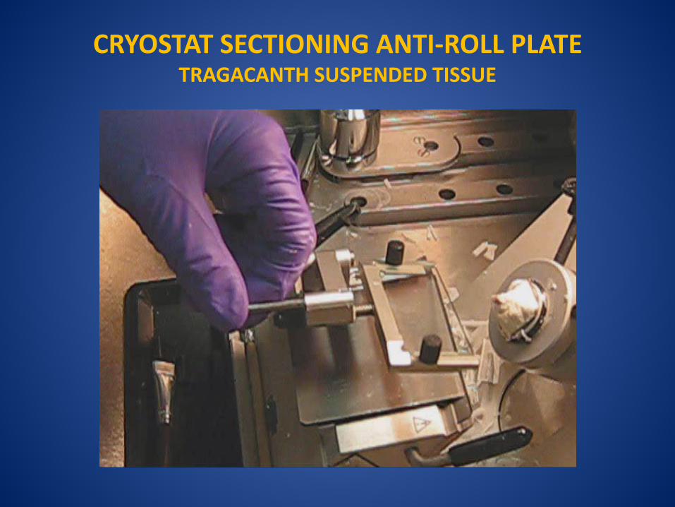
CRYOSTAT SECTIONING ANTI-ROLL PLATE TRAGACANTH SUSPENDED TISSUE
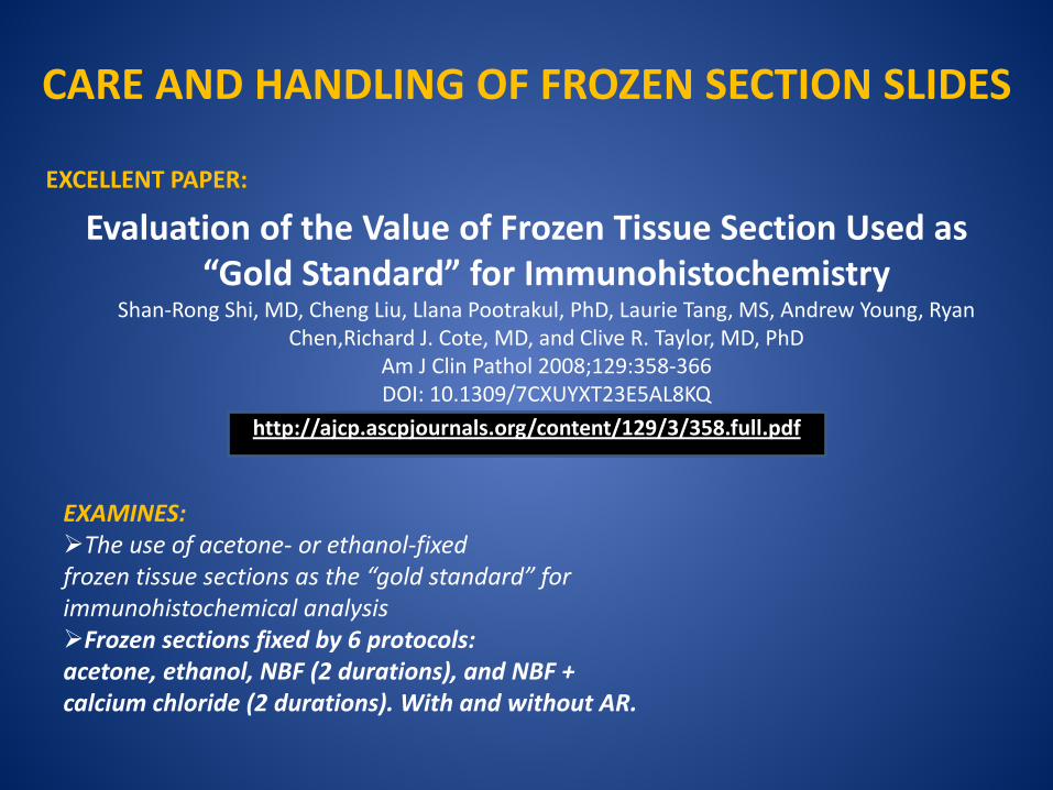
EXCELLENT PAPER:
Evaluation of the Value of Frozen Tissue Section Used as “Gold Standard” for Immunohistochemistry
Shan-Rong Shi, MD, Cheng Liu, Llana Pootrakul, PhD, Laurie Tang, MS, Andrew Young, Ryan Chen,Richard J. Cote, MD, and Clive R. Taylor, MD, PhD
Am J Clin Pathol 2008;129:358-366 DOI: 10.1309/7CXUYXT23E5AL8KQ
http://ajcp.ascpjournals.org/content/129/3/358.full.pdf
CARE AND HANDLING OF FROZEN SECTION SLIDES
EXAMINES: The use of acetone- or ethanol-fixed frozen tissue sections as the “gold standard” for immunohistochemical analysis Frozen sections fixed by 6 protocols: acetone, ethanol, NBF (2 durations), and NBF + calcium chloride (2 durations). With and without AR.
Acknowledgements
Warren Tourtellote, M.D., Ph.D, Director MHPL members: Lin Li, M.D., Assoc. Director Donna Emge, HT-ASCP, Manager Hong Chang, HT-ASCP Sheela Patel, HT-ASCP
Ephie Bakou, MBA Administrative Volunteer
Faculty Advisory Committee Anjen Chenn, MD, PhD Alexander Stegh, PhD Chyung-Ru Wang, PhD Richard M. Pope, MD Raymond Bergan, MD