Embed Size (px)
Citation preview

UNIVERSITÉ DE STRASBOURG
Ecole Doctorale Des Science De La Vie Et De La Santé (ED 414)
Inserm UMR 1121 Biomatériaux et Bioingénierie
THÈSE
Présentée par :
Rizwan ASLAM
Soutenue le : 15 Avril 2013
Pour obtenir le grade de : Docteur de l’université de Strasbourg
Discipline/ Spécialité : Aspects Moléculaire et Cellulaire de la Biologie
Les peptides antimicrobiens dérivés de la chromogranine A et Staphylococcus aureus:
de l’analyse de l’interaction hôte-pathogène au développement de revêtement de polymère antimicrobien
THÈSE dirigée par :
Mme METZ BOUTIGUE Marie Hélène Directrice de Recherche, Université de Strasbourg M. PREVOST Gilles MCU-PH, Université de Strasbourg
RAPPORTEURS : M. DRIDER Djamel Pr, Université de Lille-1 M. BERJEAUD Jean-Marc Pr, Université de Poitiers M. BECHINGER Burkhard Pr, Université de Strasbourg
AUTRES MEMBRES DU JURY : M. JACQUES Philippe Pr, Université de Lille-1


i
Acknowledgement I humbly bow my head before God, the most beneficent and merciful, whose blessing
flourished my thoughts and thrived my ambitions. I could never have done this
without the faith I have in You, the Almighty. I would like to express my deep and
sincere gratitude to my supervisor, Dr. Marie Hélène METZ-BOUTIGUE, for making it
possible for me to work in such a prestigious scientific environment. She has been
very supportive, encouraging and kind. I owe a great deal of appreciation for her
valuable advice, constructive criticism and extensive discussions around my work. It
has been a great honor to have her as a supervisor.
I feel immense pleasure to gratefully acknowledge and to express a deep sense of
gratitude to my co-supervisor Dr. Gilles PRÉVOST for his creative suggestions,
motivation and exemplary guidance throughout the course of my doctoral research
and also during thesis writing.
I wish to express my cordial appreciation to my examiners, Prof. Bukhard Bechinger,
Prof. Djamel DRIDER, Prof. Jean-Marc BERJEAUD, and Prof. Philippe JACQUES for
the acceptance to be a referee. I feel immense pleasure to gratefully acknowledge
and to express a deep sense of gratitude to Prof. Pierre SCHAAF, Prof. Jean-Claude
VOEGEL, Dr. Francis SCHNEIDER, Dr. Fouzia BOULMEDAIS, Dr. Alain VAN
DORSSELAER, Dr. Jean-Marc STRUB, for their enthusiasm, creative suggestions
and motivation throughout the course of my doctoral research.
Moreover, I offer my profound gratitude to Prof. Youssef HAIKEL for his moral and
financial support. Words are lacking to express my gratitude for Higher Education
Commission of Pakistan and especially Dr. Atta ur Rehman, for providing me with a
scholarship during my study in France. The opportunity of studying in France has
really broadened my horizon and widened my perspectives in life.
My sincere thanks are also extended to Dr. Jean Francois CHICH and Dr. Peiman
SHOOSHTARIZADEH for their help in learning various techniques and to evaluate
results more precisely and also offer them my zealous thanks. I also benefited by
outstanding support of Bernard GUEROLD, for providing various synthetic peptides
for my experiments. My heartfelt thanks are also extended to Dr. Philippe LAVALLE
and Dr. Roxane FABRE for their expertise in confocal microscopy and offer my
zealous thanks to Dr. Gwenaelle CADO and Lydie SEON for preparation of
ii
multicoated PEM films. I would like to offer my gratitude to Dr. Benoît-Joseph
LAVENTIE, Daniel KELLER, Mira TAWK and Raymonde GIRARDOT for support to
complete flow cytometry analysis and providing bacterial strains and PMNs.
I sincerely acknowledge my colleagues especially Dr. Atiandou M. Cynthia, Charlotte
BACH, Martine RIVET, Cosette BETSCHA, Sophie HELLE, Merium SAHRAOUI, Dr.
Céline MARBAN, Ouria TAHAR, and Qiongue Hu for their support during my projects
and for facilitating all experimental or administrative issues.
I own my sincere appreciations to Sultan ALI, and Ghulam HUSSAIN for their
support, guidance and affection. I would also like to extend huge, warm thanks to my
friends Asghar Shabbir, Azeem Sultan, Sarfraz Shafiq, Muhammad Nauman Zahid,
Tariq Mahmood, Adnan Khan Niazi and Azhar Ayyaz Pirzadoo for their love and
sincerity. I am eternally grateful to my beloved mother, my brothers and my sister for
their unconditional love, fidelity, endurance and encouragement. I would like to
thanks to a person whose ambidextrous nature made me more enthusiastic. There
are so many others whom I may have inadvertently left out and I sincerely thank all of
them for their help.
iii
Table of Contents Acknowledgements………………………………………………..………….. i
Table of contents………………………………………………………………..……….iii
Abbreviations………………………………………………………….………ix
List of Figures…………………………………………………………………….……..xii
List of Tables……………………………………………………………………………xiv
Part I: Introduction
1 Staphylococcus aureus ................................................................................. - 19 -
1.1 Virulence factors of the S. aureus ....................................................................... - 20 -
1.1.1 Surface proteins ............................................................................................... - 21 -
1.1.1.1 Cell wall and capsular factors ................................................................ - 22 -
1.1.1.2 Microbial Surface Components Recognizing Adhesive Matrix Molecules
(MSCRAMM) ......................................................................................................... - 22 -
1.1.1.2.1 Fibronectin binding proteins (A and B) ............................................. - 23 -
1.1.1.2.2 Protein A ............................................................................................ - 24 -
1.1.1.2.3 Collagen adhesion proteins ................................................................ - 24 -
1.1.1.2.4 Clumping factors (A and B) .............................................................. - 24 -
1.1.1.3 Secretable expanded repertoire adhesive molecules (SERAMs) ........... - 25 -
1.1.2 Secreted proteins ............................................................................................. - 26 -
1.1.2.1 Toxins .................................................................................................... - 26 -
1.1.2.1.1 Staphylococcal Enterotoxins ............................................................. - 26 -
1.1.2.1.2 Exfoliative toxins ............................................................................... - 28 -
1.1.2.1.3 Toxic shock syndrome toxins ............................................................ - 29 -
1.1.2.1.4 Staphylococcal Hemolysins ............................................................... - 31 -
1.1.2.1.4.1 Alpha hemolysin ......................................................................... - 31 -
1.1.2.1.4.2 Beta hemolysin ........................................................................... - 32 -
1.1.2.1.4.3 Gamma hemolysins .................................................................... - 32 -
1.1.2.1.4.4 Delta hemolysin .......................................................................... - 33 -
1.1.2.1.5 Panton valentine leucocidin ............................................................... - 33 -
1.1.2.1.6 Leukotoxin LukE/D ........................................................................... - 34 -
1.1.2.1.7 Other Bi-component cytolytic toxins ................................................ - 35 -

iv
1.1.2.2 Proteases of S. aureus ............................................................................ - 35 -
1.2 Host defences against S.aureus infection ............................................................ - 36 -
1.3 Immune evasion strategies of S. aureus .............................................................. - 39 -
1.3.1 Resistance to phagocytosis .............................................................................. - 40 -
1.3.2 Complement inactivation ................................................................................ - 40 -
1.3.3 Inhibition of neutrophil chemotaxis ................................................................ - 42 -
1.3.4 Leukocytes destruction by toxins .................................................................... - 43 -
1.3.5 Resistance to lysozyme ................................................................................... - 44 -
1.3.6 Survival of S. aureus in neutrophil phagosomes ............................................. - 44 -
1.3.7 Subversion of the humoral immune response ................................................. - 44 -
1.3.8 Resistance to killing by antimicrobial peptides ............................................... - 45 -
1.4 Resistance to chemotherapeutic agents ................................................................... - 45 -
2 Immune system ........................................................................................... - 47 -
2.1 Introduction ............................................................................................................. - 47 -
2.1.1 Innate immune system ..................................................................................... - 48 -
2.1.1.1 Components of innate immune system .................................................. - 48 -
2.1.1.1.1 Physical and chemical barriers .......................................................... - 49 -
2.1.1.1.2 Pattern recognition receptors (PRRs) ................................................ - 50 -
2.1.1.1.3 Cells of innate immune system .......................................................... - 51 -
2.1.1.1.3.1 Mast cells .................................................................................... - 51 -
2.1.1.1.3.2 Natural killer cells ...................................................................... - 52 -
2.1.1.1.3.3 Granulocytes (basophils and eosinophils) .................................. - 53 -
2.1.1.1.3.4 Phagocytic cells .......................................................................... - 54 -
2.1.1.1.3.4.1 Macrophages ........................................................................ - 55 -
2.1.1.1.3.4.2 Dendritic cells ...................................................................... - 55 -
2.1.1.1.3.4.3 Polymorphonuclear neutrophils ........................................... - 55 -
2.1.1.1.4 Complement system .......................................................................... - 57 -
2.1.1.1.5 Cytokines ........................................................................................... - 59 -
2.1.1.1.6 Antimicrobial peptides ...................................................................... - 61 -
2.1.2 Interrelation between immune, nervous and endocrine system. ..................... - 61 -
3 Antimicrobial peptides ................................................................................ - 64 -
3.1 Introduction ............................................................................................................. - 64 -
3.2 Structure of antimicrobial peptides ......................................................................... - 66 -
3.2.1 Chemical properties of the peptides ................................................................ - 66 -
3.2.2 Primary structure of AMPs ............................................................................. - 67 -
v
3.2.3 Secondary structure of AMPs ......................................................................... - 68 -
3.2.4 Posttranslational modifications ....................................................................... - 68 -
3.3 Classification of antimicrobial peptides .................................................................. - 69 -
3.3.1 Cationic AMPs rich in specific amino acids ................................................... - 70 -
3.3.2 Cationic AMPs lacking cysteine ..................................................................... - 70 -
3.3.3 Anionic AMPs ................................................................................................. - 70 -
3.3.4 Cationic/anionic AMPs having disulfide bond ............................................... - 71 -
3.3.5 Cationic/anionic peptides derived from the large proteins .............................. - 71 -
3.4 Mechanism of action of antimicrobial peptides ...................................................... - 72 -
3.4.1 Interaction with membranes ............................................................................ - 72 -
3.4.1.1 Carpet like mechanism ........................................................................... - 72 -
3.4.1.2 Barrel stave model ................................................................................. - 73 -
3.4.1.3 Toroidal model ....................................................................................... - 73 -
3.4.2 Interaction with intracellular components ....................................................... - 74 -
3.5 Biological activities of antimicrobial peptides ........................................................ - 75 -
3.5.1 Direct antimicrobial activity ............................................................................ - 75 -
3.5.2 Immune modulation by AMPs ........................................................................ - 76 -
3.5.2.1 Recruitment of innate immune cells ...................................................... - 77 -
3.5.2.2 Endotoxin binding .................................................................................. - 77 -
3.5.2.3 AMPs induce cellular proliferation and differentiation ......................... - 77 -
3.5.2.4 Adaptive immune cells activation .......................................................... - 78 -
3.6 Antimicrobial peptide avoidance mechanisms adopted by microorganisms .......... - 78 -
4 Granins ...................................................................................................... - 80 -
4.1 Adrenal glands ......................................................................................................... - 80 -
4.2 Chromaffin cells ...................................................................................................... - 80 -
4.3 Granin family .......................................................................................................... - 81 -
4.3.1 Biological activities of the granin derived peptides ........................................ - 82 -
4.3.2 Chromogranin A .............................................................................................. - 84 -
4.3.2.1 Biological activities of the CgA derived peptides ................................. - 85 -
4.3.2.1.1 Catestatin ........................................................................................... - 88 -
4.3.2.1.2 Cateslytin ........................................................................................... - 89 -
4.3.3 Chromogranin B .............................................................................................. - 90 -
5 AMPS conjugated polyelectrolyte films ........................................................ - 93 -
5.1 Introduction to polyelectrolyte complexes .............................................................. - 93 -
vi
5.2 Application of polyelectrolyte complexes in the biomedical field ......................... - 93 -
5.3 Polyelectrolyte multilayered films .......................................................................... - 93 -
5.3.1 History of multilayered films .......................................................................... - 94 -
5.3.2 Methods to develop polyelectrolyte multilayer films ..................................... - 95 -
5.3.2.1 The soaking method (dipping method) .................................................. - 96 -
5.3.2.2 The centrifugation method (Spin coating method) ................................ - 96 -
5.3.2.3 Spray coating method ............................................................................ - 97 -
5.4 Application of polyelectrolyte multilayered films in the field of biomaterials ....... - 98 -
5.4.1 Biomaterials .................................................................................................... - 98 -
5.4.2 Cell adhesion to the PEM films....................................................................... - 99 -
5.4.2.1 Adhesion to films based on synthetic polyelectrolytes .......................... - 99 -
5.4.2.2 Adhesion of the films based on biopolymers (polysaccharides and
polypeptides) ......................................................................................................... - 100 -
5.4.3 Insertion of active molecules in a film .......................................................... - 100 -
5.4.3.1 Adsorption and absorption of molecules ............................................. - 100 -
5.4.3.2 Insertion of active molecules by coupling of the molecules ................ - 101 -
5.4.4 Antibacterial PEM films ................................................................................ - 101 -
5.4.4.1 Passive antibacterial PEM films .......................................................... - 101 -
5.4.4.2 Active antibacterial PEM films ............................................................ - 102 -
Part II: Materials and methods
1 Preparation and purification of biological materials ...................................... - 103 -
1.1 Preparation of Leucocidin E-D ............................................................................. - 103 -
1.2 Gel electrophoresis and coomassie blue staining of the gel .................................. - 103 -
1.3 RP-HPLC of Leucocidins E-D .............................................................................. - 104 -
1.4 Isolation of polymorphonuclear neutrophils ......................................................... - 104 -
1.5 Preparation of secretions after PMNs stimulation by Leucocidin E-D ................. - 105 -
1.5.1 Purification of PMNs secretions by RP-HPLC ............................................. - 105 -
1.5.2 Desalting of the PMNs secretions ................................................................. - 105 -
1.6 Synthesis of peptides ............................................................................................. - 106 -
1.6.1 Purification of synthetic peptides by RP-HPLC ........................................... - 106 -
1.7 Isolation and characterization of staphylococcal strains ....................................... - 106 -
2 Preparation and characterization of material for polymer development.......... - 108 -
2.1 Preparation of modified synthetic antimicrobial peptide (CTL-Cys) for insertion in
biomaterials ................................................................................................................... - 108 -
2.1.1 Synthesis of rhodaminated peptides .............................................................. - 109 -

vii
2.2 Preparation of modified HA. ................................................................................. - 109 -
2.2.1 Preparation of HA-CTL-Cys ......................................................................... - 109 -
2.2.2 Preparation of HAFITC
.................................................................................... - 111 -
2.2.3 Preparation of HAFITC
-CTL ........................................................................... - 112 -
2.3 Gel filtration chromatography ............................................................................... - 112 -
2.4 PEM films buildup ................................................................................................ - 113 -
2.4.1 Polysaccharide solutions ............................................................................... - 113 -
2.4.2 Film buildup .................................................................................................. - 113 -
2.5 Physico-chemical characterization of the polymer films ...................................... - 114 -
2.5.1 Quartz Crystal Microbalance ........................................................................ - 114 -
2.5.2 Atomic Force Microscopy (AFM) ................................................................ - 114 -
2.5.3 Confocal Laser Scanning Microscopy (CLSM) ............................................ - 114 -
3 Methods of proteomic analysis ..................................................................... - 115 -
3.1 Sequence characterization by Edman sequencing ................................................. - 115 -
3.2 Western blot analysis of the PMNs secretion ........................................................ - 115 -
3.3 Matrix assisted laser desorption ionization time-of-flight Mass spectrometry (Maldi-
TOF) .............................................................................................................................. - 116 -
3.4 Nano LC-MS/MS Mass spectrometry analysis ..................................................... - 117 -
4 Methods to characterize biological activities ................................................. - 118 -
4.1 Flow-cytometry measurements: ............................................................................ - 118 -
4.2 Analysis of antimicrobial activity ......................................................................... - 119 -
4.2.1 Antibacterial test ........................................................................................... - 119 -
4.2.1.1 Kinetics of bacterial growth inhibition ................................................ - 120 -
4.2.2 Antifungal activity ......................................................................................... - 120 -
4.2.2.1 Kinetics of fungal growth inhibition .................................................... - 121 -
4.3 Confocal laser scanning microscopy(CLSM) ...................................................... - 121 -
4.4 Degradation analysis of synthetic peptides by S. aureus strains ........................... - 122 -
4.4.1 Purification of bacterial supernatants by RP-HPLC ..................................... - 122 -
4.5 PSG degradation by the V8 protease ..................................................................... - 122 -
4.6 Human gingival fibroblasts viability assays .......................................................... - 123 -
Part III: Results and discussion
Manuscript 1………………………………………………………………...….- 124-
viii
Activation of neutrophils by the two-component leukotoxin LukE/D from
Staphylococcus aureus: a proteomic analysis of the secretions
Manuscript 2……………………………………………………………………- 156-
Cateslytin a chromogranin A derived peptide is active against Staphylococcus
aureus and resistant to degradation by its proteases
Manuscript 3…………………………………………………………………….-176-
Staphylococcus aureus Glu-C protease subverts innate immunity by degradation of
Chromogranins and production of new antifungal peptides
Manuscript 4
Self-defensive biomaterial coating against bacteria and yeasts: polysaccharide
multilayer film with embedded antimicrobial peptide
Conclusion and Perspectives……………………………………………………….-191-
References………………………………………………………………………………-193-
Annexes
Chromogranin A-derived peptides are involved in innate immunity
The Natural Antimicrobial Chromogranins/Secretogranins- Derived Peptides –
Production, Lytic Activity and Processing by Bacterial Proteases
Proteomic analysis of activated PMNs secretion, by Staphylococcus aureus
leukotoxins LukE/D
Summary

ix
Abbreviations A. fumigatus: Aspergillus fumigatus
AFM: Atomic force microscopy measurements
ATCC: American Type Culture Collection
bCAT: Bovine catestatin
C. albicans: Candida albicans
C. glabrata: Candida glabrata
C. tropicalis: Candida tropicalis
CBS: Centraalbureau voor Schimmelcultures
CFU: Colony forming unit
CgA: Chromogranin A
CgB: Chromogranin B
CgC: Chromogranin C
Cgs: Chromogranins
CHI/HA: Chitosan/Hyaluronic acid
CHI: Chitosan
CHR: Chromofungin
CLSM: Confocal laser scanning microscopy
CTL: Cateslytin
DDT: Dichloro-Diphenyl-Trichloroethane
DMEM: Dulbecco's Modified Eagle Medium
E. coli: Escherichia coli
ECL:
ECM: Extra Cellular Matrix
EDC: N-(3-Dimethylaminopropyl)-N′-ethylcarbodiimide hydrochloride
EDTA: Ethylene-Diamine-Tetra-Acetic Acid
EGTA: Ethylene glycol-bis-(L-aminoethyl ether) N, N, Nˊ, Nˊ-tetraacetic acid
ETs: Exfoliative toxins (A, B)
FITC: Fluorescein Isothiocyanate
Fmoc: Fluorenylmethoxycarbonyl
FnBPA: Fibrinectin binding protein A
x
FnBPB: Fibrinectin binding protein B
FPLC: Fast performance liquid chromatography
HA: Hyaluronic acid
hCAT: Human catestatin
HCl: Hydrochloric acid
HEPES: (4-(2-hydroxyethyl)-1-piperazineethanesulfonic acid
HGFs: Human gingival fibroblasts
Hlg: Gamma hemolysin (A, B, C)
IgG: Immunoglobulin G
IgM: Immunoglobulin M
KCl: Potassium chloride
LbL: Layer-by-Layer
MALDI-TOF: Matrix-assisted laser desorption/ionization-Time Of Flight
MH: Mueller Hinton
MRSA: Methicillin-resistant S. aureus
MSCRAMMs: Microbial surface components recognizing adhesive matrix molecules
MSSA: Methicillin-susceptible S. aureus
N. crassa: Neurospora crassa
NaCl: Sodium chloride
NaOH: Sodium hydrooxide
NH4HCO3: Ammonium bi-carbonate
NMR: Nuclear magnetic resonance
OD: Optical density
PAH/PAA: poly-allylamine hydrochloride/poly-acrylic acid
PAH/PAAm: Poly-alanine hydrochloride/polyacrylamide
PAH: Poly-allylamine hydrochloride
PDB: Potato Dextrose Broth
PDMS: Polydimethylsiloxane
PEC: Polyelectrolyte complex
PEI: Polyethyleneimine
PEM: Polymer electrolyte membrane
PEMs: Polyelectrolyte multilayers
PLL / PGA-g-PEG: Poly-L-lysine/Poly-glutamic acid -poly-ethylene glycol
PLL/PGA: Poly-L-lysine/Poly-glutamic acid)
xi
PMNs: Polymorphonuclear neutrophils
PSS: Poly-sodium styrene sulfonate
PTH-aas: Phenylthiothiohydantoine-amino acids
PVDF: Polyvinylidene Fluoride
PVL: Panton Valentine leucocidin
QCM: Quartz crystal microbalance
RP-HPLC: Reverse phase-high performance liquid chromatography
S.aureus: Staphylococcus aureus
SDS: Sodium dodecyle sulphate
SDS-PAGE: Sodium dodecyle sulphate polyacrylamide gel electrophoresis
SERAM: Secretable expanded repertoire adhesive molecule
SEs: Staphylococcal enterotoxins
SP sepharose: Sulfopropyl sepharose
Sulfo-NHS: 3-sulfo-N-hydroxysuccinimide ester
T. mentagrophytes: Trichophyton mentagrophytes
TFA: Trifluoroacetic acid
TRIS: 2-Amino-2-hydroxymethyl-propane-1, 3-diol
TSSTs: Toxic shock syndrome toxins (0, I)
YCP: Yeast extract-casamino acids-pyruvate

xii
List of Figures Figure 1: Structure of S. aureus ......................................................................................... - 21 -
Figure 2: Ribbon presentation of exfoliative toxin A (ETA) sharing structural similarities
with glutamylendopeptidase (V8). ................................................................... - 29 -
Figure 3: Crystal structure representation of the binding site of TSST-1 with hVβ chain of T-
cell receptor (D-10 varient and wild type) and SpeC (Streptococcus pyogenic
exotoxin C). ...................................................................................................... - 30 -
Figure 4: Three-dimensional presentation of alpha toxin. ................................................. - 31 -
Figure 5: Immune activities against Staphylococcal infection following tissue invasion. - 38 -
Figure 6: Mechanisms employed by S. aureus to resist immune response. ....................... - 39 -
Figure 7: Schematic model of complement activation pathways. ...................................... - 41 -
Figure 8: Schematic representation of different methods employed by S. aureus to inhibit
chemotaxis. ....................................................................................................... - 42 -
Figure 9: Presentation of different cell lineage involved in immunity. ............................. - 49 -
Figure 10: Different componets of the innate and adaptive immune system. .................... - 51 -
Figure 11: Role of mast cells in tissue injury. .................................................................... - 52 -
Figure 12: Modulation of immune cells by eosinophils. .................................................... - 54 -
Figure 13: Human polymorphonuclear neutrophil. ............................................................ - 56 -
Figure 14: Complement activation pathways. .................................................................... - 58 -
Figure 15: Immune, nervous and endocrine system interaction. ....................................... - 62 -
Figure 16: Structural classes of antimicrobial peptides (AMPs). ...................................... - 69 -
Figure 17: Different types of interaction between AMPs and microbial membrane. ........ - 74 -
Figure 18: Multiple functions of AMPs in host defense .................................................... - 76 -
Figure 19: Different mechanism adopted by the pathogens to avoid AMPs. .................... - 79 -
Figure 20: Presentation of mature bovine CgA. ................................................................. - 85 -
xiii
Figure 21: Presentation of mature human CgA. ................................................................. - 86 -
Figure 22: Presentation of mature bovine CgB. ................................................................. - 91 -
Figure 23: Schematic construction of multilayer polyelectrolyte adsorptions by successive
polycations and polyanions. ............................................................................. - 95 -
Figure 24: Representation of the soaking/dipping method. ............................................... - 96 -
Figure 25: Representation of PEMs film construction by spin coating method. ............... - 97 -
Figure 26: Representation of the spraying method. ........................................................... - 98 -
Figure 27: Cateslytin (CGA 344-358) + cysteine at C-terminal ...................................... - 108 -
Figure 28: Coupling reaction of HA to maleimide and followed by conjugation of CTL
(CGA344-358) to HA. .................................................................................... - 110 -
Figure 29: Preparation of HAFITC
..................................................................................... - 111 -
Figure 30: Preparation of HAFITC
-CTL ............................................................................ - 112 -

xiv
List of Tables
Table I: MSCRAMMs produced by Staphylococcus aureus. ............................................ - 23 -
Table II: SERAMs produced by Staphylococcus aureus. .................................................. - 26 -
Table III: The superantigens of S. aureus. ......................................................................... - 27 -
Table IV: The bicomponent toxins produced by S. aureus. ............................................... - 34 -
Table V: Characteristics of few important cytokines implicated in immune system, with their
principle source and targets.. ............................................................................ - 60 -
Table VI: AMPs timeline: representative AMPs selected by antimicrobial peptide database
(APD). .............................................................................................................. - 65 -
Table VII: Different types of antimicrobial peptides based on their structural characteristics
with their characteristics and few prominent examples of the domain. ........... - 71 -
Table VIII: Different members (human origin) of the granin family. ............................... - 82 -
Table IX: Different granin derived peptides and their associated functions. ..................... - 83 -
Table X: Antimicrobial peptides derived from CgA. ......................................................... - 87 -
Table XI: Sequence presentation of Catestatin and cateslytin. .......................................... - 90 -
Table XII: Antimicrobial peptides derived from CgA. ...................................................... - 92 -


Résume
- 1 -
Résume
L’étude que j’ai menée dans le cadre de ma thèse de l’Université de Strasbourg, a
porté principalement sur l’analyse de l’interaction hôte-pathogène et le développement de
polymères de recouvrement antimicrobiens par conjugaison de peptides antimicrobiens sur les
matériaux.
Dans une première partie, nous avons d’une part évalué l’interaction de la leukotoxine
LukE/D de Staphylococcus aureus avec les neutrophiles polynucléaires (PMNs) et d’autre
part nous avons examiné le rôle des protéases de S. aureus sur la dégradation des peptides
antimicrobiens dérivés principalement de la chromogranine A (CgA). Dans la seconde partie
de ma thèse, nous avons préparé un revêtement de polymère antimicrobien biodégradable par
conjugaison de la cateslytine (CTL), peptide correspondant à la séquence de la CgA bovine
344-358, à l’acide hyaluronique dans un modèle de multi-couches acide
hyaluronique/chitosan.
Leukotoxine LukE/D et PMNs
Staphylococcus aureus (S. aureus), est une bactérie présente dans 20-30% de la
population et responsable de nombreuses infections nosocomiales. Elle est la cause de
nombreuses maladies allant d’infections cutanées bénignes à des pathologies mettant en
danger la vie du malade, telles que le sepsis, la pneumonie, l’endocardite, la méningite et
l’ostéomyélite. En géneral, les infections à S. aureus démarrent par une étape d’adhésion de la
bactérie au tissu de l’hôte et se pousuivent par l’étape de colonisation. La colonisation est
associée aux protéines de la famille “Microbial Surface Components Recognizing Adhesive
Matrix Molecules” (MSCRAMMs) qui sont importantes pour l’attachement de la bactérie à la
fibronectine. De plus, S. aureus produit un grand nombre de facteurs de virulence incluant des
toxines, telles que les entérotoxines (SEs), la toxine-1 du syndrome de choc toxique (TSST-
1), les toxines exfoliatives A et B (ETA et ETB), la leukotoxine de Panton-Valentine (PVL) et
la leukotoxine LukE/D. Cette dernière est composée de deux sous-unités: LukE et LukD qui
se lient à la membrane des leucocytes en induisant la formation de pores membranaires à
Ca2+ qui provoquent ensuite la lyse cellulaire. L’expression de LukE/D est associée à

Résume
- 2 -
plusieurs pathologies. Ainsi, elle a été isolée avec les deux épidermolysines A et B dans 78%
des cas d’impetigo. Par ailleurs, LukE/D a été isolée, associée aux enterotoxines chez 93.6%
de patients avec des diarrhées post-antibiotiques. Cependant, actuellement aucune étude n’a
examiné le rôle de LukE/D sur les interactions hôte-pathogènes.
Les neutrophiles sont des constituants importants de la première ligne de défense
contre les pathogènes, parce qu’ils participent à l’élimination des pathogènes et contribuent à
l’activation de l’immunité adaptative. Des résultats précédents ont indiqué que chez
l’Homme, les Cgs (CgA et CgB) sont présentes dans les granules de sécretion des
neutrophiles et sont surexprimées dans le plasma de patients avec le syndrome de réponse
inflammatoire systémique (SIRS) ou un sepsis. Plusieurs peptides endogènes dérivés des CgA
et CgB sont décrits comme présentant d’intéressantes propriétés antimicrobiennes. Par
ailleurs, en plus de leurs effets directs contre les pathogènes, ils activent les neutrophiles, en
induisant un influx de calcium extra-cellulaire, via une liaison à la calmoduline
cytoplasmique, puis une sécrétion par exocytose des cytokines et des molécules de l’immunité
innée, comme les peptides antimicrobiens (PAMs).
La compréhension des mécanismes de régulation de la réponse des neutrophiles à une
agression par des pathogènes peut être obtenue par l’analyse protéomique des sécretions des
neutrophiles activés. En utilisant les techniques d’analyse protéomique, nous avons identifié
les protéines sécrétées par les neutrophiles activés par LukE/D puis, discuté les effets de cette
leukotoxine sur la regulation dynamique des neutrophiles. Nous avons montré que LukE/D est
capable de stimuler les PMNs (Manuscrit 1: Figure 2), comme ce qui est décrit pour le LPS
bactérien par une rapide augmentation du calcium intracellulaire (Manuscrit 1: Figure 2). Le
facteur C3 du complément (CD35) est détecté en tant que marqueur de l’activation des PMNs.
Il est retrouvé après traitement par la PVL, ce qui indique une activation des neutrophiles.
Dans ce travail, nous avons démontré qu’en plus de la toxicité induite, Luk E/D peut aussi
stimuler les PMNs. L’identification dans les sécretions de PMNs de proteines provenant des
différents types de granules, confirme le rôle de LukE/D dans la dégranulation de ces
différents compartiments.
Ainsi, les PMNs actives produisent le materiel contenu dans les granules azurophiles
et spécifiques très enrichies en PAMs. L’analyse protéomique de ces sécrétions a été réalisée
en collaboration avec le laboratoire de spectrométrie de masse bio-organique de Strasbourg
(CNRS 7178, Dr A. Van Dorsselaer). La comparaison avec les banques de données
Résume
- 3 -
protéiques, qui a été obtenue par utilisation du Mascot Ion Score, a permis d’identifier les
nombreux facteurs et de les classer suivant leurs fonctions physiologiques par le “gene
ontology term mapper” (Manuscrit 1: Figure 4). Nous avons ainsi répertorié un grand nombre
de protéines impliquées dans les mécanismes d’activation de l’immunité adaptative (34%), les
mécanismes inflammatoires (8%), les protéines anti-oxydantes (3%) et les enzymes (13%)
etc... (Manuscrit 1: Figure 4). En complément de cette analyse, par Western blot et à l’aide
d’anticorps spécifiques de CgA et CgB, nous avons identifié plusieurs fragments dérivés
montrant leur expression lors de la réponse immunitaire à une infection à Straphyloccoques.
Parmi tous les fragments identifiés, en utilisant les techniques de tests antimicrobiens, nous
avons caractérisé plusieurs peptides actifs contre S. aureus, Micrococcus luteus, Candida
albicans, Candida tropicalis, Neurospora crassa et Aspergillus fumigatus.
L’ensemble de ces résultats souligne l’importance de la stimulation des PMNs par
LukE/D en relation avec les défenses immunitaires (innées et adaptatives).
Les protéases de S. aureus et les peptides
antimicrobiens de la Chromogranine A
Dans la deuxième partie, nous avons analysé l’activité de plusieurs peptides
antimicrobiens dérivés de la CgA contre plusieurs souches de S. aureus. Les Cgs sont les
protéines majoritaires des granules de sécrétion des cellules chromaffines (Cgs) et sont
exprimées dans les cellules nerveuses, endocrines et immunitaires. Elles sont naturellement
maturées pour produire de nombreux peptides aux propriétés biologiques très variées, mais
intervenant toutes pour produire le retour à l’homéostasie après la réponse au stress.
Pendant les dix dernières années, notre groupe a caractérisé de nouveaux peptides
antimicrobiens dérives des CgA et CgB. Les séquences de ces peptides sont très conservées
au cours de l’évolution, ce qui suggère l’importance de leur rôle dans l’immunité innée. Dans
les situations de sress et de sepsis les peptides dérivés des Cgs sont produits par les cellules
chromaffines, entérochromaffines et les neutrophiles. En plus de leurs effets antimicrobiens
directs, quelques peptides tels que la sécrétoneurine (un fragment dérivé de la CgC)
établissent un lien entre les systèmes endocrine et immunitaire.
Résume
- 4 -
La CgA contient deux domaines antimicrobiens, la vasostatine I (1-76/78) et la
prochromacine (79-431). Le domaine actif de la vasostatine-1 correspond à la chromofungine
(CgA47-66), qui est à la fois antibactérienne et antifongique, à une concentration de l’ordre
du micromolaire. La prochromacine inclut la séquence de la catestatine (CgA344-364) et du
fragment raccourci cateslytin (CgA344-358) qui est à la fois antibactérien et antifongique à
une concentration de l’ordre du micromolaire. En 2009, il a été montré par notre laboratoire
que la chromofungine (CHR, CgA47-66) et la catestatine (CAT, CgA344-364), activent les
PMNs en induisant un influx de calcium extra-cellulaire.
S. aureus est le pathogène à Gram-positif le plus fréquemment isolé dans les cas de
sepsis, souvent mis en cause dans les problèmes liés à la formation de caillots et à la
destruction du tissu cardiaque. S. aureus a développé plusieurs mécanismes pour éviter la
réponse immunitaire. En résistant à l’action des PAMs, en évitant le recrutement des
phagocytes, en interférant avec le complément, en évitant les pièges des neutrophiles liés à
leur lyse et en resistant aux conséquences de l’oxydation et aux liaisons non spécifiques des
imunoglobulines. Les mécanismes mis en place par S. aureus pour déjouer les PAMs
comprennent la dégradation protéolytique par les métallo-protéases, les protéases à sérine et à
cystéine. L’expression des enzymes protéolytiques est controlée directement par les
régulateurs des facteurs de virulence tels que agr, sar et indirectement par ailleurs, SarA est
aussi un régulateur du facteur de résistance à la méthicilline (fmtA). Précédemment, il a été
indiqué que la métallo-protéase aureolysine peut cliver la cathélicidine LL-37 pour l’inactiver
et contribuer ainsi à l’échappement du système immunitaire.
Notre étude se focalise sur les effets antimicrobiens des catestatine humaine et bovine
(bCgA344-364 et hCgA352-372) et du fragment cateslytine bovine (CTL, bCgA344-358)
contre S. aureus qui est connu pour coloniser la peau et l’épithélium. Parce que la CTL se
révèle beaucoup plus active que la CAT contre la croissance de S. aureus, nous avons analysé
la dégradation potentielle de ces deux peptides par les protéases de cette bactérie en utilisant
les techniques d’HPLC, séquençage et spectrométrie de masse. Ainsi, nous avons décrit (1)
une relation entre la séquence des deux peptides et leur sensibilité aux protéases bactériennes
et (2) la possibilité de les utiliser comme agents antimicrobiens en combinaison avec des
antibiotiques conventionnels.
En comparaison avec les autres peptides testés, CTL se révèle être le peptide le plus
actif contre les différentes souches testées ((ATCC49775, ATCC25923, S1, and S2). Les
Résume
- 5 -
souches S1 et S2 sont isolées à partir de patients de l’hôpital: S1 est une souche MRSA
(résistante à la méthicilline) et S2 est une souche MSSA (sensible à la méthicilline). Les
activités de ces deux peptides sont comparées avec celle de LL-37 (fragment C-terminal de
hCAP-18) (Manuscrit 2: Tableau 1). L’activité de CTL est comparable à celle de LL-37
contre S1 et S2, mais significativement différente contre ATCC49775 et ATCC25923. Les
valeurs des concentrations minimales d’inhibition de bCAT et hCAT sont 2-4 fois plus
élevées que pour le court fragment bCTL (Manuscrit 2: Tableau 1).
Afin de préciser l’activité antimicrobienne de CTL, nous avons déterminé la cinétique
de l’activité antibactérienne contre la souche ATCC 25923. CTL agit très rapidement et à une
concentration de 2 x CMI, CTL tue toutes les bactéries en 40 minutes et avec la CMI, le
même résultat est obtenu en 60 minutes et les bactéries ne repoussent plus pendant 24h
(Manuscrit 2: Figure 2AB). Parce que CTL est le peptide le plus actif, nous posons
l’hypothèse que ce fragment est plus résistant aux protéases produites par S. aureus.
Pour démontrer la stabilité de CTL par comparaison avec celles des CAT humaine et
bovine nous avons incubé les trois peptides avec les surnageants des quatre souches
différentes de S. aureus. Les facteurs de S. aureus agr et sar régulent l’expression des
protéases, qui sont souvent modulées par le régulateur SarA pendant la formation du biofilm.
Ces protéases sont principalement exprimées dans la dernière phase de croissance et sécrétées
dans le milieu extra-cellulaire qui facilite la dispersion bactérienne. Les peptides synthétiques
ont été incubés par les surnageants de S. aureus et séparés par RP-HPLC. Les profils
chromatographiques obtenus pour les deux CAT et CTL sont comparés avec le profil du
milieu MHB et ceux des surnageants bactériens. Les pics chromatographiques correspondant
à la dégradation bactérienne ont été analysés par séquençage automatique d’Edman et en
spectrométrie de masse par la technique MALDI-TOF. Les résultats obtenus montrent que
contrairement aux CAT bovine et humaine, CTL n’est pas dégradé.
L’HPLC de bCAT indique deux pics majeurs élués à 38.7 et 40 min (Manuscrit 2:
Figure 2A), correspondant au peptide complet, d’après les résultats de séquençage et MALDI-
TOF (2426 Da) (Manuscrit 2: Figure 3). La présence de ces deux formes peut s’expliquer à la
conformation induite par le résidu de proline en position 360 et des états d’isomérisation
(cis/trans). Dans le milieu MHB seul bCAT n’est pas dégradé, tandis qu’en présence des
surnageants des cultures des souches S49775, S25923 et S1, bCAT est mature pour produire
les fragments élués à 38.3 et 38.6 min, correspondant aux isoformes de bCgA349-364 (1782
Résume
- 6 -
Da) (Manuscrit 2: Figure 3), ce qui montre le rôle de la bactérie dans la dégradation du
peptide.
En présence du surnageant de S2, bCAT est fortement dégradé pour produire des
fragments élués à 28.0, 36.6 et 37.5 min (Manuscrit 2: Figure 2A). le séquençage et l’analyse
par MALDI-TOF indiquent que ces fragments correspondent à bCgA350-356 (826 Da) et les
deux isoformes de bCgA357-364 (887 Da) (Manuscrit 2: Figure 3).
De plus, le profil HPLC de hCAT montre un pic majoritaire qui correspond au peptide
complet d’après le séquençage et l’analyse en MALDI-TOF (2327 Da) (Manuscrit 2: Figure
3). En presence du milieu MHB, hCAT n’est pas clivée (Manuscrit 2: Figure 2C). Cependant,
après incubation avec les surnageants des souches S49775, S25923 et S1, hCAT est
partiellement dégradé pour générer un fragment élué à 36 min (Manuscrit 2: Figure 2C). Le
séquençage et la spectrométrie de masse MALDI-TOF indiquent que ce fragment correspond
à hCgA357-372 (1780 Da) (Manuscrit 2: Figure 3). En présence du surnageant S2, hCAT est
complètement dégradé pour générer des fragments élués à 28.5 et 33 min (Manuscrit 2: Figure
2C). Le séquençage et l’analyse MALDI-TOF indiquent que ces fragments correspondent à
hCgA358-364 (840 Da) et hCgA365-372 (871 Da) (Manuscrit 2: Figure 3).
De plus, le court peptide bCTL (bCgA344-358), est élué comme un peptide unique et
n’est pas dégradé par les souches de S. aureus testées (Manuscrit 2: Figure 2E).
Nous avons alors testé l’effet d’inhibiteurs de protéases sur la dégradation des peptides
par les surnageants bactériens. Les trois peptides testés bCAT, hCAT et bCTL ne sont plus
dégradés en présence des surnageants bactériens (Manuscrit 2: Figure 3B/D/E), démontrant
ainsi que la dégradation des CAT est due aux protéases du S. aureus.
Il est intéressant de noter que les points de clivage des CAT par les surnageants des
souches S49775, S25923 et S1 sont identiques. Ils correspondent aux liaisons L348-S349 et
L356-S357 respectivement pour bCAT et hCAT (Manuscrit 2: Figure 3). Avec la souche S2,
ils correspondent aux liaisons S349-F350+G356-F357 et S357-F358+G364-F365,
respectivement pour bCAT et hCAT (Manuscrit 2: Figure 3). Il est important de noter que le
court peptide bCTL (bCgA344-358) (Manuscrit 2: Figure 3) n’est pas clivé par les protéases
de S. aureus ce qui démontre que sa structure primaire lui permet de résister au mélange
protéolytique produit par S. aureus.
Résume
- 7 -
A cause du développement de la résistance de S. aureus aux antibiotiques
conventionnels, l’intérêt pour les peptides antimicrobiens est croissant. Par ailleurs, certains
peptides antimicrobiens sont toxiques pour les cellules de l’hôte, ce qui n’est pas le cas pour
les peptides antimicrobiens dérivés des chromogranines. Jusqu’à présent, aucun résultat
concernant des activités antibactériennes contre S. aureus a été rapporté pour les peptides
dérivés des chromogranines. CAT est décrit comme étant actif contre plusieurs souches
bactériennes au niveau du micromolaire et CTL est bien caractérisé pour ses activités
antibactériennes contre Micrococcus luteus, des levures et des souches de champignons. De
plus il n’est pas lytique contre les érythrocytes. Au cours de ma thèse, pour la première fois,
nous avons caractérisé l’activité de CTL contre plusieurs souches de S. aureus avec une CMI
de 37-45 µg/mL (Manuscrit 2: Tableau 1).
Au cours d’une infection, in vivo il s’établit une communication dynamique entre les
cellules de l’hôte et les bactéries. De nombreuses souches bactériennes expriment une grande
variété de protéases qui dégradent de nombreuses proteines jouant un rôle dans l’immunité
innée. L’absence de dégradation par S. aureus pour le peptide bCTL par comparaison avec
bCAT et hCAT peut expliquer la différence d’activité (d’un facteur 3-4) entre les peptides
testés (Manuscrit 2: Tableau 1). En effet, vis-à-vis des quatre souches testées, bCAT et hCAT
sont degradés, tandis que bCTL résiste à la dégradation protéolytique.
Dans une autre série d’expériences, en utilisant la protéase Glu-C de S. aureus et le
mélange complexe des proteines sécrétées par les cellules chromaffines stimulées, nous avons
identifié plusieurs sites de clivage identifiés après traitement (Manuscrit 3). Les protéines
chromogranine A (CgA) et chromogranine B (CgB) possèdent très peu de régions
antimicrobiennes qui ne soient pas dégradées par la protéase Glu-C. Les nouveaux fragments
générés sont purifiés par RP-HPLC et analysés pour leurs propriétés antimicrobiennes. Les
activités antibactériennes sont perdues, mais de manière intéressante les peptides présentent
des activités antifongiques contre Neurospora crassa. Parmi les fractions d’HPLC actives
nous avons identifié deux peptides de la CGA correspondant à CgA (47-60 et 418-426)
(Manuscrit 3: Figure 2) et trois peptides de la CgB (279-291, 450-464 et 470-486)
(Manuscrit 3: Figure 3). Les activités antimicrobiennes de ces fragments sont validées par
l’utilisation des peptides synthétiques correspondants avec une CMI de 2µM à 80µM contre
différents Candida et des souches de champignons filamenteux (Manuscrit 3: Tableau 1).
Résume
- 8 -
La biosynthèse de CTL résulte de l’action de la prohormone convertase PC1/2,
présente dans la matrice des granules chromaffines. La prohormone thiol protease (PTP) est
aussi primordiales pour la formation de CAT (bCgA344-364) par clivage de D-R et L-R. De
plus, dans les vésicules de sécrétion des cellules chromaffines la cathepsine L (CTSL) produit
CTL par un clivage supplémentaire R-G de la CAT. La CAT est capable d’activer les
neutrophiles en induisant un influx de calcium extra-cellulaire via la calmoduline. Dans mon
travail de thèse, j’ai montré la résistance de CTL en présence de S. aureus, ce qui renforce son
rôle dans l’immunité innée.
Les séquences de CAT et CTL sont très conservées au cours de l’évolution (Manuscrit
2: Figure 4). Le taux d’arginine est important puisqu’il module l’interaction avec les charges
négatives de la membrane du microorganisme. Ainsi, il a été démontré que les résidus
d’arginine ont une forte tendance à interagir avec les lipides comme il a été suggéré pour
d’autres peptides tels la protéine Tat, activateur transcriptionnel de HIV-1. Pour hCAT,
bCAT, et bCTL les taux d’arginine sont, respectivement de 15%, 23%, and 33%. Le fort
pourcentage d’arginine pour bCTL est en faveur d’une forte interaction avec les
phospholipides de la bicouche. La relation structure-activité de CTL avec la membrane
bactérienne est aussi démontrée par des expériences récentes concernant le peptide FLE-CTL
dans lequel le peptide CTL a été rallongé à l’extrémité N-terminale de FLE. FLE-CTL (FLE-
RSMRLSFRARGYGFR). En utilisant une combinaison de l’HPLC et des tests
antimicrobiens, nous avons montré que ce peptide synthétique est inactif contre S. aureus à
400 µg/mL et interagit fortement avec la membrane bactérienne. Au contraire le peptide
raccourci auquel il manque l’extrémité C-terminale (YGFR): FLE-RSMRLSFRARG, possède
une activité antibactérienne avec une CMI de 200 µg/mL. Ce dernier résultat montre que CTL
correspondant à une séquence avec les critères requis pour obtenir une activité
antibactérienne.
Concernant la structure secondaire de CTL, une structure en feuillets β aggrégés a été
décrite en utilisant des expériences de dichroïsme circulaire et d’ATR (Attenuated total
reflectance). Au contraire , hCAT et bCAT forment de courtes structures en hélice (résidu 7 à
résidu 11) en présence de fortes concentrations de DPC, ce qui a été confirmé par des
expériences de dichroïsme circulaire et de RMN. CTL adopte majoritairement un caractère en
feuillets β en présence de membranes chargées négativement, tandis qu’il est principalement
non structuré dans l’eau. Les structures en feuillets β donnent une plus grande stabilité que les
structures en hélices décrites pour CAT. De plus, l’analyse (HR-MAS) 1H NMR de CTL
Résume
- 9 -
indique que l’arginine et les résidus hydrophobes sont localisés à proximité induisant ainsi
une pénétration profonde des résidus chargés dans la membrane. Ainsi, les interactions
électrostatiques entre les résidus d’arginine chargés positivement et les lipides chargés
négativement seraient responsables de la liaison de CTL à la bicouche et les résidus
arômatiques stabiliseraient l’interaction lipide-peptide.
Dans notre groupe, nous avons récemment évalué l’effet synergique de trois peptides
derivés de la CgA (CAT, CTL et CTL amidée) avec des antibiotiques conventionnels. Nous
avons montré que ce co-traitement induit une diminution de la concentration en antibiotiques
et potentialise leurs activités. Les tests antimicrobiens sont réalisés à des concentrations
inférieures à la CMI et des tests contrôles sont réalisés avec le peptide et l’antibiotique à des
doses similaires. Pour toutes ces expériences, nous avons évalué la « Fractional Inhibitory
Concentration » (FIC) des peptides dérivés de la CgA en combinaison avec la Minocycline et
le Voriconazole. FIC correspond à un effet synergique, s’il est ≤0.5, à un effet additif, s’il est
>0.5 et <2, et à un effet antagoniste s’il est >2. Dans le cas de la combinaison de la CTL
amidée et de la Minocycline, nous obtenons un FIC de 0,37 contre S. aureus, et pour la
combinaison de CTL et du Voriconazole, nous obtenons un FIC de 0,25, et 0,5
respectivement, contre Candida albicans et Candida tropicalis. D’après ces résultats, on peut
imaginer un mécanisme dans lequel les peptides pourraient favoriser la déstabilisation de la
membrane bactérienne permettant ainsi, une meilleure pénétration des antibiotiques pour
atteindre leurs sites d’action. Ces résultats obtenus in vitro pourraient se produirent in vivo
pendant les états d’inflammation systémique.
En effet, au cours du sepsis on observe de nombreux changements dans les activités
des protéases et de leurs inhibiteurs. Ces régulations sont fortement reliées avec la gravité du
sepsis. Dans des situations de stress oxydatif, se produisant au cours du sepsis, CAT et CTL
seront produites plus difficilement à partir de la CgA complète. L’oxydation produit des
modifications structurales de la CgA tels que l’oxydation des résidus de méthionine, des
résidus arômatiques, des glycanes, des phosphorylations et aussi l’aggrégation de la protéine
complète qui pourraient empêcher la dégradation de la protéine pour produire les peptides
bénéfiques.
La structure des peptides est très importante pour l’expression des activités
antimicrobiennes. Il a été montré qu’une courte séquence hydrophobe terminale peut
augmenter l’activité. Dans notre étude nous avons ajouté un résidu de cystéine séparément
Résume
- 10 -
aux deux extrémités de CTL et nous avons montré une augmentation de l’activité lorsque le
résidu C est positionné à l’extrémité C-terminale et une diminution de l’activité lorsqu’il est
positionné à l’extrémité N-terminale. Le peptide CTL-C a alors été utilisé pour developper
des revêtements de polymères via le depôt alternatif de polyélectrolytes conjugués à CTL-C.
L’activité de CTL n’est pas altérée lorsqu’il est inséré dans le biomatériau. De plus il n’est
pas toxique pour les cellules de l’hôte (fibroblastes gingivaux).
Finalement pour la première fois, nous présentons l’importance de l’interaction des
protéases bactériennes sur la dégradation des peptides antimicrobiens et montrons le caractère
très résistant de la CTL qui représente un peptide intéressant pour des études futures.
Revêtements multicouches de
polyélectrolytes antimicrobiens
Dans cette dernière partie, nous avons décidé d’utiliser CTL pour préparer des
matériaux antimicrobiens qui recouvriront des intruments médicaux et chirurgicaux.
Les implants sont très utilisés en chirurgie, non seulement pour remplacer des tissus
altérés ou perdus, mais aussi dans les services de soins intensifs pour l’administration de gaz
ou fluides par cathéters ou tube trachéal. Ces implants constituent une porte d’entrée pour
l’invasion microbienne. La prévention de la colonisation microbienne des implants est une
importante question médicale et financière, puisque les infections nosocomiales représentent
une des plus sérieuses complications après la chirurgie et les soins intensifs. En effet chaque
année en Europe, 5% des patients admis à l’hôpital souffrent d’infections acquises lors de leur
séjour à l’hôpital et conduisant à 10% de mortalité.
S. aureus, une bactérie à Gram-positif est responsable des infections acquises lors
d’hospitalisations et plus particulièrement dans les cas de patients immuno-déprimés. C’est
une des bactéries les plus virulentes conduisant à de forts pourcentages d’infections
systémiques dues aux implants et au décès. Une étude récente a caractérisé du point de vue
génétique les souches de S. aureus responsables des infections dues à la pose de cathéters et
démontré que 82% de ces souches sont résistantes à la méthicilline et contiennent plusieurs
gènes impliqués dans la formation du biofilm et la dispersion bactérienne.

Résume
- 11 -
C. albicans, la levure pathogène pour l’homme la plus répandue, a la capacité de
former des biofilms qui sont une source d’infections locales et systémiques. De plus, les
biofilms colonisés par C. albicans permettent la formation de microcolonies de S. aureus à
leur surface et d’augmenter la résistance de S. aureus aux antibiotiques. Quand elles sont
associées aux infections bactériennes, les infections par champignons augmentent la gravité
de l’état du patient. La résistance récente de C. albicans aux thérapies antifongiques et de S.
aureus aux antibiotiques soulève le besoin de revêtements multifonctionnels assurant à la fois
la protection contre les bactéries et les champignons.
Plusieurs approches basées sur l’immobilisation et la libération de substances
bactéricides, utilisant des assemblages de monocouches ou des structures de polymères ont été
explorées et développées. Les films de multicouches de polyélectrolytes (PEM), basés sur le
dépôt alterné de polycations et polyanions sur la surface solide, représentent une méthode
simple et efficace pour fonctionnaliser les surfaces de manière contrôlée. Les premiers films
antibactériens ont été obtenus par insertion de nanoparticules d’argent. Ensuite, des
bactéricides hydrophobes et des liposomes contenant de l’argent sont insérés dans des films
PEM pour obtenir des revêtements bactéricides efficaces.
Les films PEM à base de chitosan sont antibactériens contre Escherichia coli et
Enterococcus faecalis. Les antibiotiques tels que la gentamicine ont été insérés dans des films
PEM pour être ensuite libérés. Malgrés une augmentation du pouvoir bactéricide des films,
l’utilisation d’antibiotiques et de particules d’argent présente des limites dues à leur toxicité
ou à leur rôle dans l’émergence de pathogènes multi-résistants.
Les peptides antimicrobiens naturels (PAMs), sécrétés par de nombreux organismes
vivants contre les pathogènes, suscitent l’intérêt par leur large spectre d’activité
antimicrobienne et leur faible cytotoxicité. Ils causent principalement une destruction de la
membrane plasmique des agents pathogènes et les empêchent de développer des mécanismes
de résistance. Les PAMs chargés positivement sont utilisés comme partie intégrante des
structures de PEM pour obtenir l’effet antibactérien par contact ou après libération.
Guyomard et al. 2008 ont réussi à intégrer dans des films PEM un complexe de PAMs
hydrophobes. Ces films sont antibactériens contre les bactéries à Gram-positif. Des exemples
de films PEM intégrant des PAMs antifongiques sont aussi rapportés. Il est urgent de
developper des revêtements portant à la fois les activités antibactériennes et antifongiques.
Résume
- 12 -
Jusqu’à présent peu de revêtement possèdent les deux propriétés. Ils sont principalement
basés sur les revêtements d’argent ou les groupements ammonium quaternaires et cationiques,
les polymères synthétiques et les silanes.
A notre connaissance, aucun revêtement de base sur les PAMs possèdent les deux
propriétés. Dans le but de créer un tel matériau, nous avons utilisé le peptide cateslytine
(CTL), un peptide dérivé de la chromogranine A (CGA), protéine sécrétée pendant les
infections avec de nombreux peptides dérivés par les cellules nerveuses, endocrines et
immunitaires dans le cadre de l’immunité innée.
CTL, un PAM correspondant à CGA344-358, agit à l’échelle du micromolaire avec un
large spectre d’activités antimicrobiennes contre les bactéries à Gram-positif, les
champignons filamenteux et les levures, il est sans activité cytotoxiques contre les cellules de
l’hôte. De plus, il est très stable vis-à-vis des protéases bactériennes. Nous avons utilisé des
films d’acide hyaluronique, comme polyanions fonctionnalisés par le peptide CTL-C dans
lequel un résidu Cystéine a été rajouté à l’extrémité C-terminale du peptide CTL, et de
chitosan comme polycations (HA-CTL-C/CHI) qui sont disposés sur une surface plane afin
d’être efficace contre les bactéries et les champignons. L’acide hyaluronique et le chitosan
sont biodégrédables par hydrolyse enzymatique, respectivement par la hyaluronidase et la
chitosanase. Ces deux molécules sont déjà largement utilisées pour des applications
biomédicales à cause de leurs propriétés intrinsèques. La capacité de S. aureus, M. luteus et
les Candida de dégrader HA en produisant la hyaluronidase, permet la libération de CTL-C
par les films PEM, exclusivement en présence des pathogènes. La liberation des composés
antimicrobiens (PAMs ou antibiotiques conventionnels) est habituellement obtenue par
diffusion passive des films à pH physiologique ou par dégradation des films induite par
variation du pH.
Pavlukhina et al. 2010 ont publié la libération des agents antimicrobiens en fonction de
la variation de pH liée à la pousse microbienne. Cependant, la libération des agents
antimicrobiens par variation du pH reste limitée étant donné que le pH devient acide au site de
l’infection quand la survie du patient est déjà engagée. A notre connaissance, nous avons
développé ici le premier revêtement d’auto-défense par dégradation du film induite par le
pathogène lui-même. La masse des polysaccharides absorbée est déterminée par Surface
Plasmon Resonance (SPR). La construction et la topographie des films ont été caractérisés par
microscopie à force atomique (AFM). Les activités antibactériennes et antifongiques des
Résume
- 13 -
solutions de HA-CTL-C et des films de HA-CTL-C/CHI ont été testées contre deux souches
de bactéries Gram-positif (S. aureus et M. luteus) et une souche de levure C. albicans, en
utilisant le test liquide par micro-dilution.
La microscopie confocale par laser (CLSM) permet de suivre par fluorescence la
pénétration de HAFITC-CTL-C (en solution ou intégré dans le film PEM) dans la membrane
de C. albicans. Par la suite, la cytotoxicité des films de HA-CTL-C/CHI a été testée sur des
fibroblastes gingivaux humains (HGFs).
Le peptide CTL-C a été couplé de façon covalente à HA par deux étapes successives
comprenant le greffage d’une fonction maléimide sur HA et le couplage d’un thiol maléimide
pour greffer CTL-C sur le HA modifié. Ces réactions sont décrites dans le document
complémentaire de la thèse (manuscrit 4). Après dialyse, séchage puis congélation, un rapport
de 5% de couplage a été determiné par RMN du proton, correspondant au greffage de 5
molécules de CTL-C pour 100 dimères de HA (Manuscrit 4: Figure 1A).
Les activités antimicrobiennes de CTL et CTL-C sont testées en solution contre deux
souches bactériennes M. luteus et S. aureus (ATCC 25923) ainsi que la levure C. albicans. La
concentration minimale d’inhibition (CMI) de CTL, CTL-C et HA-CTL-C a été determinée
après analyse des tests antibactériens et antifongiques par la technique de microdilution (Voir
la partie complémentaire de la thèse). Ainsi, le peptide CTL-C est antimicrobien à une
concentration de l’ordre du micromolaire (< 100 µM) et présente une plus forte activité
antibactérienne contre M. luteus par rapport au peptide CTL seul (sans C).
La comparaison avec le peptide CTL-C non greffé, la CMI de HA-CTL-C augmente
de 35 à 45 µM pour S. aureus et de 20 à 25 µM pour C. albicans (Manuscrit 4: Tableau 1).
Dans le cas des essais avec M. luteus, la CMI de HA-CTL-C (5 µM) est cinq fois plus élevée
que celle de CTL-C (1 µM).
Dans le cas de M. luteus, la CMI de HA-CTL-C (5 µM) est cinq fois plus élevée que
celle de CTL-C (1 µM). Bien qu’il y ait une diminution de l’efficacité, le peptide CTL-C lié
de façon covalente au polymère HA est encore antimicrobien à une concentration de l’ordre
du micromolaire. La construction du film HA-CTL-C/CHI est suivie par SPR. Une
augmentation linéaire de la masse absorbée est observée à chaque depôt de couche de
polysaccharide indiquant que la construction se déroule normalement (Manuscrit 4: Figure
1B).
Résume
- 14 -
Nous avons recherché la topographie des films de HA-CTL-C/CHI avec différents
niveaux de depôts par ZFM en état sec (Manuscrit 4: Figure S1). Il est difficile d’obtenir une
bonne qualité des images d’AFM dans l’état sec, à cause de la viscoélasticité des films. Les
images AFM permettent de mesurer l’épaisseur des films. Avec 5 bicouches, la surface est
presque entièrement couverte avec un film de 5 nm d’épaisseur et une rugosité de 1,6 nm. Le
film atteint une épaisseur de 52 nm et une rugosité de 16.5 nm avec 30 bicouches (Manuscrit
4: Figure S2).
Après caractérisation de la construction du film HA-CTL-C/CHI, les activités
antibactériennes et antifongiques des films fonctionnalisés ont été évaluées contre M. luteus et
S. aureus.
Après la caractérisation de la construction couche par couche de HA-CTL-C/CHI, les
activités antibactériennes et antifongiques du film fonctionnalisé ont été évaluées contre deux
souches de bactéries M. luteus et S. aureus ainsi que la levure C. albicans (Manuscrit 4:
Figure 2A-C). L’influence du nombre de couches fonctionnalisées est analysée par la
construction de différents fims avec des nombres croissants de couches et pour lesquels on
évalue les activités antimicrobiennes. Ainsi, des films PEI-(HA/CHI)15-n-(HA-CTL-C/CHI)n
avec n= 0, 5, 10, 15 et d’autres avec PEI-(HA-CTL-C/CHI)30 ont été élaborés. Les
pathogènes ont été incubés 24h avec les films de HA-CTL-C/CHI à 37°C pour les souches
bactériennes et à 30°C pour C. albicans (Manuscrit 4: Figure 2C). Pour chaque pathogène, la
croissance microbienne est mesurée à différents temps (1 h, 4 h, 6 h et 24 h) par détermination
de la densité optique à 620 nm de la suspension bactérienne en présence du film (Manuscrit 4:
Figure 2).
Les résultats ont été normalisés et exprimés comme des pourcentages de croissance
détectés à une Densité optique de 620 nm. Pour chaque pathogène testé, nous observons
qu’en augmentant le nombre de couches de HA-CTL-C/CHI, la croissance microbienne
diminue. Une diminution importante de la croissance microbienne est obtenue pour au moins
5 bicouches pour M. luteus et C. albicans et 15 couches pour S. aureus (Manuscrit 4: Figure
2). Après 6h d’incubation, au moins 70% d’inhibition est obtenue pour 15 couches pour tous
les pathogènes testés (Manuscrit 4: Figure 2).
Après 24h d’incubation la croissance de M. luteus, C. albicans et S. aureus est
complètement inhibée avec 5, 15 et 30 couches de HA-CTL-C/CHI. Ces résultats sont en

Résume
- 15 -
accord avec les CMI de HA-CTL-C en solution. Afin de vérifier si les films peuvent être
réutilisés plusieurs fois comme revêtement antimicrobiens, la croissance de 3 pathogènes a été
controlée après incubation pendant 24h de films de PEI-(HA-CTL-C/CHI)15 pour M. luteus
et C. albicans, ainsi que des films PEI-(HA-CTL-C/CHI)30 pour S. aureus (Manuscrit 4:
Figure 2D). Dans ce but, une suspension de pathogène a été mise en contact du film
fonctionnalisé. Après une incubation de 24h, le surnageant a été retiré et recouvert d’une
suspension fraîche. Après chaque retrait, la DO 620 nm du surnageant a été mesurée pour
déterminer la croissance du pathogène après 24h d’incubation. Quand les suspensions de M.
luteus et C. albicans sont en contact avec le film de (HA-CTL-C/CHI)15 et réutilisé chaque
24h, une inhibition complète a été observée , respectivement pour 2 et 3 cycles (Manuscrit 4:
Figure 2D). Une diminution significative de l’efficacité du film (HA-CTL-C/CHI)15 est
détectable après le renouvellement de la suspension du pathogène. La croissance de S. aureus
est inhibée de 40% par la seconde utilisation du film (HA-CTL-C/CHI)30 et il devient
complètement inefficace pour la troisième utilisation (Manuscrit 4: Figure 2D). En parallèle
nous avons incubé les surnageant retirés avec des suspensions fraîches de pathogènes pendant
24h et la DO 620 nm a aussi été mesurée (Manuscrit 4: Figure S3).
Nous n’avons pas observé d’inhibition, ce qui indique l’absence (ou une très faible
quantité) de CTL-C relargué en solution.
Pour clarifier le mécanisme d’inhibition de HA-CTL-C, nous avons synthétisé HA-
FITC-CTL-C et HA-FITC pour analyser leurs interactions avec C. albicans lorsqu’ils sont en
solution ou intégrés dans des films multicouches (Manuscrit 4: Figure 4).
Une étude a montré par microscopie confocale que les peptides pénètrent dans les
membranes plasmiques et s’accumulent dans les levures. Après 45 minutes d’incubation à
30°C avec HA-FITC ou HA-FITC-CTL-C en solution (Manuscrit 4: Figure 4A-B), C.
albicans est observé et HA-FITC-CTL-C a été détectable dans le cytoplasme sans provoquer
de lyse cellulaire. Au contraire, HA-FITC est clairement détecté autour des cellules de levure,
s’accumulant sur les membranes et conduisant à une structure en nid d’abeille. Ceci suggère
que CTL-C peut traverser la membrane plasmique, même quand il est couplé à HA et
s’accumuler à l’intérieur du cytoplasme. Les films ont été traités à la paraformaldéhyde (PFA)
et cette procédure n’induit pas de changement dans le cas des films de HA-FITC/CHI, au
contraire des films de HA-FITC-CTL-C/CHI qui apparaissent hétérogènes (Manuscrit 4:
Figure 4C-D).
Résume
- 16 -
C. albicans a été incubé pendant 45 min à 30°C au contact de film de PEI-(HA-
FITC-CTL-C/CHI)15 et observé ensuite par microscopie confocale (Manuscrit 4: Figure 4).
Parmi les hétérogénéités dues au traitement par PFA, une forte fluorescence verte est observée
principalement à l’intérieur de la levure. Dans le cas de films de HA-FITC/CHI, une faible
fluorescence est localisée à l’intérieur des levures et seulement quelques cellules paraissent
fortement fluorescentes. Même inséré dans le film de PEM, CTL-C permet la pénétration de
HA-FITC-CTL-C à l’intérieur des levures, justifiant ainsi l’activité antimicrobienne des films.
S. aureus and C. albicans sont connus pour sécréter de la hyaluronidase, une classe
d’enzymes capables d’hydrolyser HA. Le découpage d’un des partenaires du film peut
conduire à sa destruction comme cela a été observé par Etienne et al 2005.
Nous avons alors examiné par microscopie confocal des films de PEI-(HA-
FITC/CHI)15 avant et après 24h de contact avec les pathogènes (Manuscrit 4: Figure 3). Les
trois pathogènes induisent la dégradation des films HA/CHI avec cependant quelques
différences dans la morphologie résultante des films. Après 24h d’incubation avec S. aureus,
les films de HAFITC/CHI films sont presque tous dégradés (Manuscrit 4: Figure 3A). Le film
apparaît peu homogène avec des taches fluorescentes après incubation avec M. luteus. et C.
albicans (Manuscrit 4: Figure 3B-C) induisant la formation de structures en nid d’abeilles
dans le film, par dégradation de HA. La dégradation de HA peut conduire à la libération de
CTL-C dans le surnageant et à la promotion de l’interaction entre les peptides CTL-C et les
pathogènes.
Pour vérifier cette hypothèse, nous avons construit des films résistants à la
hyaluronidase, CTL-C étant greffé sur la poly(allylamine hydrochloride) pour des matériaux
de (PAA/PAH-CTL-C). Après 24h d’incubation les films de (PAA/PAH-CTL-C)15 ne
montrent pas d’inhibition contre C. albicans. Ceci renforce le fait que l’activité
antimicrobienne des films de HA-CTL-C/CHI est due à la dégradation par la hyaluronidase.
Ainsi, les pathogènes initient leur propre mort lorsqu’ils sont au contact du film HA-CTL-
C/CHI. De manière intéressante, même si le film est dégradé avec le temps en présence du
pathogène, il peut être utilisé au moins 2 à 3 fois sans perdre son activité contre M. luteus et
C. albicans.
Résume
- 17 -
Il est ensuite important de s’assurer que le film n’est pas toxique pour les cellules de
l’hôte. Les fibroblastes sont les premières cellules qui viennent se déposer sur la surface de
l’implant pendant le processus de cicatrisation.
La viabilité des fibroblastes gingivaux humains (HGFs) cultivés sur des films PEI-
(HA-CTL-C/CHI)15 , comparés avec des films PEI-(HA/CHI)15 et le substrat en verre, a été
évaluée au travers de l’activité mitochondriale, suivi par un test Alamar blue™. Après une
journée déjà, l’activité métabolique mesurée sur un film d’HA-CTL-C/CHI est
comparativement inférieure au film d’ HA/CHI ainsi qu’au substrat non coaté. La bonne
compatibilité du film de HA/CHI pour HGFs, pour le récepteur CD44 , a été rapporté lors de
notre travail précédent. Après sept jours en culture, le nombre d’HGFs viables sur le film
HA-CTL-C/CHI représente 25% des HGFs viables déposées sur le film HA/CHI (Manuscrit
4: Figure 5A). La fonctionnalisation de HA par le peptide CTL-C induit une faible adhésion
des HGFs qui semblent ralentir la prolifération des cellules. Sachant que HA-CTL-C en
solution à 100 µM n’est pas cytotoxique, un tel comportement suggère que les multicouches
ne sont pas cytotoxiques.
Pour faire la différence entre la cytotoxicité et une faible propriété d’adhésion initiale
d’HA-CTL-C/CHI, nous avons effectué deux expériences.. Premièrement après 24 h de
contact, nous avons récupéré la suspension de cellules du surnageant du film HA-CTL-
C/CHI et nous les avons passées sur une nouvelle plaque de culture. Après 24 h de culture,
plusieurs cellules facilement attachées se propagent comme des cellules fraîches.
Deuxièmement, nous avons analysé la biocompatibilité des films avec un test complémentaire
d’étallement des cellules avec un réarrangement du cytosquelette quand les cellules sont
ensemencées sur un substrat en verre à moitié coaté par (HA-CTL-C/CHI)15. Après 24 h, les
HGFs adhèrent sur le substrat en verre (Manuscrit 4: Figure 5BI), mais à un degré moindre
sur un film (HA-CTL-C/CHI)15 (Manuscrit 4: Figure 5BII). Après 24 h de culture, à
confluence, on observe une couche de cellules fibroblastiques typiques et des fibres
polymérisées d’F-actine, sur le substrat en verre. Sur le film d’HA-CTL-C/CHI, apparaissent
des cellules moins allongées ayant une distribution d’actine à leur périphérie. Ceci nous
indique que le film d’ (HA-CTL-C/CHI)15 n’est pas cytotoxique pour le HGFs mais, semble
être plutôt anti-adhérent. L’échec de l’implantation peut être du à des fonctions des
fibroblastes en relation avec la formation des tissus fibreux. Ces résultats qui diminuent
l’adhésion des fibroblastes sur les substrats fonctionnalisés avec les films d’HA-CTL-C/CHI .
Ces résultats nous incitent à développer ces matériaux pour des implants.

Résume
- 18 -
En conclusion de cette dernière partie de notre étude, nous avons choisi une nouvelle
surface recouverte avec des films de multicouches de polysaccharides contenant de l’HA
fonctionnalisé avec 5% de CTL-C, un peptide possédant les deux propriétés , antibactérienne
et antifongique. Les propriétés antimicrobiennes de CTL-C sont préservées quand elles sont
greffées sur HA en solution ou quand elles sont enfouies dans des films PEM. Après 24 h
d’incubation, les films d’HA-CTL-C/CHI inhibent le développement de S. aureus et C.
albicans, qui sont des agents pathogènes communs et virulents rencontrés dans les maladies.
La présence du peptide CTL-C sur HA permet la pénétration du polysaccharide
modifié à l’intérieur de C. albicans après 45 min de contact. La sécrétion de hyaluronidase
pour tous les agents pathogènes testés, semble être responsable de la libération d’ HA-CTL-C
du film et de son activité. Le film garde son activité durant trois cycles d’utilisation avec des
suspension fraîches de C. albicans. Par ailleurs, l’adhésion limitée de fibroblastes, sans
cytotoxicité, sur les films d’ HA-CTL-C/CHI met en évidence une application clinique pour
prévenir les infections sur des cathéters ou des tubes trachéaux, où les encapsulations de tissu
fibreux sont indésirables.

Part : I
Introduction

Staphylococcus aureus

Introduction
- 19 -
1 Staphylococcus aureus
Staphylococcus aureus belongs to genus Staphylococcus and family Micrococcaceae.
This genus is composed of gram positive, non-sporulated and aerobic bacteria. Most of the
members of the genus are opportunistic pathogens, but few are pathogenic. S. aureus was first
isolated by Scottish surgeon Alexander Ogston in 1880, from the pus (Ogston, 1881). Later,
he observed microscopically and named as “clustered micrococci” (Staphylococci) in 1882
(Ogston, 1882). S. aureus is one of the most commonly isolated human pathogen. S. aureus is
distinguished from other staphylococcal species because it produces gold pigmentation of
colonies, coagulase positive as well mannitol fermenter and gives positive results for the
deoxyribonuclease test (Wilkinson, 1997). It is commensal, mostly found in many biological
fluids including skin, throat, nares and many more. Mostly, it is found extracellular, but can
be observed intra cellular in some cases (Sendi and Proctor, 2009). S. aureus colonizes at
multiple sites in human beings but anterior nares are the most common site of colonization
(about 30% of general population) (Rich, 2005; Wertheim et al., 2005), extra-nares that
typically harbor S. aureus colonization include skin, perineum, pharynx (Wertheim et al.,
2005). Other sites, harboring less colonization as compared to skin or nares may include the
oral cavity, gastrointestinal tract, vagina, axillae etc. (Wertheim et al., 2005; Williams, 1963).
The transmission is mostly through contact of infected skin between two persons,
ruptured skin can provide favorable environment for progressive colonization of pathogen.
Innate immune system controls the colonization of skin pathogens, but horizontal
transmission between two persons can disturb the equilibrium between innate immune control
of host and skin pathogens (Dethlefsen et al., 2007). Once the favorable conditions are
furnished such as rupture of skin barrier or inadequate immune strength, Staphylococcus can
rapidly colonize resulting in an invasive growth deep into skin. Followed by the bacteremia
and once Staphylococci is in blood it can penetrate into vital organs of body, resulting in
mortal pathogenesis such as endocarditis, toxic shock, pneumonia and septicemia (Lina et al.,
1997).
S. aureus is responsible for infections, very diverse in nature: from minor skin infections
(pimples, impetigo, cellulitis, folliculitis, carbuncles, scalded skin syndrome, and skin
abscesses) to life threatening diseases such as sepsis, bacteremia, pneumonia, osteomyelitis,
toxic shock syndrome, endocarditis, and meningitis, (Freeman-cook and Freeman-cook, 2006
). This diverse variability of infections indicates that the pathogen is well adapted to the
Introduction
- 20 -
different environmental conditions. Moreover, S. aureus has developed resistance against
various families of antibiotics that is progressing day by day, often due to non rational use of
antibiotics during the last two decades (Campion et al., 2004; Denis et al., 2003; Hiramatsu,
2001; Hiramatsu et al., 2001; Zhang et al., 2008). This day by day increasing resistance is
provoking hindrance in effective treatment of infections.
Meanwhile, S. aureus is capable of forming biofilm, which helps to protect it, against the
adverse environmental conditions like action of detergents and even can interfer process of
sterilization. Then, it becomes difficult to eliminate pathogen by some conventional
sterilization process used in the hospitals, which results in about 30% more nosocomial
infections caused by MRSA each decade (Klevens et al., 2006). Finally, the increasing
frequency and the gravity of infections caused by S. aureus is becoming an adverse public
health problem.
S. aureus growing pathogenicity is due to production of a large number of virulence
factors, which are expressed at the site of infection (Foster, 2004; Holden et al., 2004; Novick,
2003). These virulence factors are responsible for infection severity; these include surface
proteins, exo-proteins, toxins, and cell adherence proteins. All these virulence factors not only
help to combat against the immune system but also to settlement and nutrition (Novick,
2003). Most of the adherence proteins are synthesized by Staphylococcus during the initial
growth phase while exoproteins are synthesized later during the exponential phase (Dunman
et al., 2001; Vandenesch et al., 1991). This model of different stages of virulence factors
production is well explained in-vivo, where first attempt is to well stick at the site of infection
which is helped by the production of various adhesion proteins during initial phase of growth.
After well settlement next step is to get nutrient, to colonize and to combat against the
gathering immune cells, which is favored by the production of exo-toxins. These help to
invade deep into tissue and later disperse through blood to other organs and to colonize at new
sites of infection.
1.1 Virulence factors of the S. aureus
S. aureus is capable of producing a large number of virulence factors, can be broadly
divided into two categories: surface proteins and the secretory proteins.
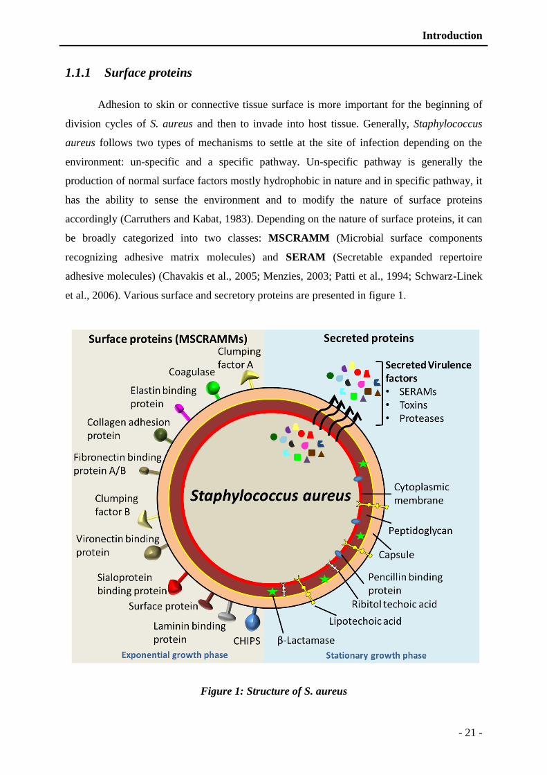
Introduction
- 21 -
1.1.1 Surface proteins
Adhesion to skin or connective tissue surface is more important for the beginning of
division cycles of S. aureus and then to invade into host tissue. Generally, Staphylococcus
aureus follows two types of mechanisms to settle at the site of infection depending on the
environment: un-specific and a specific pathway. Un-specific pathway is generally the
production of normal surface factors mostly hydrophobic in nature and in specific pathway, it
has the ability to sense the environment and to modify the nature of surface proteins
accordingly (Carruthers and Kabat, 1983). Depending on the nature of surface proteins, it can
be broadly categorized into two classes: MSCRAMM (Microbial surface components
recognizing adhesive matrix molecules) and SERAM (Secretable expanded repertoire
adhesive molecules) (Chavakis et al., 2005; Menzies, 2003; Patti et al., 1994; Schwarz-Linek
et al., 2006). Various surface and secretory proteins are presented in figure 1.
Figure 1: Structure of S. aureus
Introduction
- 22 -
Left panel presents the different surface and secreted proteins. The synthesis of these proteins is controlled by
regulatory genes locus (agr) and is dependent on growth phase. Lower right Panels presents the cross sections of
the cell envelope.
1.1.1.1 Cell wall and capsular factors
The staphylococcal cell wall is composed of peptidoglycan which consists of
alternating polysaccharide subunits of N-acetylmuramic acid and N-acetylglucosamine
(Figure 1). Peptidoglycan may have endotoxin-like activity, which can stimulate release of
cytokines by neutrophils and macrophages, leading to activation of complement system and
aggregation of platelets (Matsui and Nishikawa, 2012; Wang et al., 2000). Staphylococcal
strains results in variation in disseminated intravascular coagulation, which is due to
differences in the peptidoglycan structure (Kessler et al., 1991). Most of Staphylococci
produce microcapsules. Capsule is composed of polysaccharides, 11 different serotypes based
on the capsular antigens have been recognized out of which serotypes 5 and 8 are mainly
identified in human infections. Most of methicillin-resistant S. aureus (MRSA) isolates are
type 5 (Lee, 1996).
1.1.1.2 Microbial Surface Components Recognizing Adhesive Matrix
Molecules (MSCRAMM)
MSCRAMM can be classified as molecule of interest that helps to localize pathogen
and help to recognize a macromolecular ligand in host, which can be found in the ECM (Extra
Cellular Matrix). These macromolecular ligands are exclusively found in the ECM such as
collagen, laminin, fibronectin, fibrinogen and vitronectin. These ligands are recognized as
MSCRAMM ligands. Third important thing is the MSCRAMM’s affinity towards these ECM
ligands. Some molecules that recognize carbohydrate determinants exclusively present in
many macromolecules do not fall under the category of MSCRAMM such as lectin type
adhesion molecules. MSCRAMMs adhesion proteins are attached to the microbial membrane
by covalent interaction (Foster and Hook, 1998).
Usually, a single MSCRAMM can bind to several ECM ligands and inversely
pathogen can produce several MSCRAMMs to bind a single ECM ligand. For example as S.
aureus is capable of several fibrinogen binding proteins at a time (Boden and Flock, 1989;

Introduction
- 23 -
McDevitt et al., 1994). Several MSCRAMMs produced by S. aureus are listed in the table
below (Table I).
Microbial MSCRAMMs may be virulence factors that mediate host tissue
colonization, which in many cases is a prerequisite for infection development. Staphylococcus
aureus and many other microorganisms have developed several adhesion proteins (Table I,
Figure 1). These factors can be operated in combination or independently of each other. This
scenario appears to be particularly important when microbes invade a host tissue. Some of the
more important MSCRAMMs are discussed below briefly (Table I).
Table I: MSCRAMMs produced by Staphylococcus aureus.
MSCRAMM ECM ligand Gene MW Reference
FnBPA Fibronectin A fnbA 110 kDa (Wann et al., 2000)
FnBPB Fibronectin B fnbB 101 kDa (Howden et al., 2010)
Protein A Vh3 region of IgM spa 55 kDa (Sjodahl, 1977)
Collagen adhesion protein Collagen cna 133 kDa (Allon et al., 2012)
Vironectin binding protein Vironectin 60 kDa (Liang et al., 1995)
Laminin binding protein Laminin eno 47 kDa (Carneiro et al., 2004)
Surface protein Plasmin pls 174 kDa (Hilden et al., 1996)
Thrombospondin (Rennemeier et al., 2007)
Elastin binding protein Elastin ebpS 53 kDa (Downer et al., 2002)
Sialoprotein binding protein Bone sialoprotein bbp 124 kDa (Yacoub et al., 1994)
Clumping factor A Fibrinogen clfa 97 kDa (McDevitt et al., 1994)
Clumping factor B Fibrinogen clfb 97 kDa (Ni Eidhin et al., 1998)
1.1.1.2.1 Fibronectin binding proteins (A and B)
Most of S. aureus strains specifically bind to the fibronectin. Fibronectin is a 440 kDa
glycoprotein serves as an ECM ligand for the bacterial MSCRAMMs, found in the
extracellular matrix and body fluids. The interaction between fibronectin binding protein
(FnBP) and its ligands is highly specific. A signal peptide at the N-terminal of FnBP is
Introduction
- 24 -
followed by a unique sequence that is interrupted by the 35 amino acids long unit and another
domain of 38 amino acids is repeated, which is rich in proline residues. The 38 amino acids
domain contains a motif “LPXTGX” is found in most of surface proteins produced by gram
positive bacteria; it represents the cell wall domain. The cell wall domain is followed by the
membrane spanning domain rich in hydrophobic residues (Fischetti et al., 1990). C-terminal
extremities mostly contain positively charged residues which is probably the cytoplasmic
domain.
1.1.1.2.2 Protein A
Protein A is 55 kDa surface protein of S. aureus, present in more than 95% S. aureus
strains and represents about 7% of total proteins (Patel et al., 1992). It has multiple functions
either serves as MSCRAMM or in immune modulation of the host. Its anti-opsonic function is
very potent, which help to modulate macrophages based immune clearance. It binds to the Fc
region of the immunoglobulin G (King and Wilkinson, 1981). Due to this binding ability; it
inhibits the specific detection of antigen and blocks macrophage phagocytosis (Foster, 2005).
Moreover, it specifically binds to the Vh3 region of the IgM molecules that is located adjacent
to the antigen binding domain. The cells bearing Vh3 region of IgM are readily exposed to B
lymphocytes. Finally, stimulated to proliferate and undergo apoptosis (Goodyear and
Silverman, 2004). Protein A can also activate the complement system and can stimulate the
secretion of histamine by the basophils (Gomez et al., 2004; Nguyen et al., 2000).
1.1.1.2.3 Collagen adhesion proteins
Collagen based proteins are the major ECM constituents, which represent the major target
for most of gram positive pathogens. These proteins follow the pattern of FnBPs, containing a
signal peptide and followed by the 187 amino acids long domain which is repeated three
times. C-terminal contains a motif of 64 amino acids rich in lysine and proline residues, and
hydrophobic motif of representing the membrane domain. Collagen binding MSCRAMM
ligands on S. aureus is both necessary and sufficient for adhesion of pathogens to cartilage.
Only cells expressing the MSCRAMMs can adhere to cartilage (Speziale et al., 1986).
1.1.1.2.4 Clumping factors (A and B)
Clumping factors are also called fibrinogen binding proteins due to its specificity to bind
fibrinogen. Fibrinogen is found in high concentration in blood plasma, and plays an important
Introduction
- 25 -
role in the formation of blood clot structures. Clumping factors are composed of a stretch of
15 amino acids in the carboxy terminal of the protein that helps to anchor fibrinogen
monomers (Strong et al., 1982). The fibrinogen-binding proteins contain the typical feature of
gram-positive surface proteins such as the LPXTGX motif.
1.1.1.3 Secretable expanded repertoire adhesive molecules (SERAMs)
These adhesion proteins are identified, having the property to adhere to various
proteins present in the extra-cellular matrix for example fibrinogen, collagen, prothrombin
(Chavakis et al., 2005). Two hypotheses are established either this family of proteins is
secreted directly into the extracellular matrix or they form bonds with membrane proteins
(Chavakis et al., 2005). Although, SERAMs and MSCRAMMs are structurally unrelated but
functionally these are same. In addition, these do not form covalent interaction with the
bacterial membrane as premeditated in MSCRAMMs. Two important functions are
designated with SERAMs, to bind to ECMs present in the host and to bind to a large array of
host proteins, which results in modulation of host response against infection. A list of few
well studied SERAMs and their ECM ligands are presented in Table II.
Most important and most studied SERAM is coagulase which is somehow
characteristic of Staphylococcus spp and was used for the detection of Staphylococcus
infections, despite now MALDI-TOF assited bacterial identification gives a direct
identification of these bacteria. Coagulase interacts with thrombin and form a
staphylothrombin complex, this complex has the capacity to polymerize with fibrin and
finally inhibit agglutination (Palma et al., 1998). Moreover, these proteins can act as
immunoregulators and can provoke vascular pathies (Chavakis et al., 2002). For example
protein Eap (Extracellular adherence protein) (Hussain et al., 2001; Hussain et al., 2002) and
Efb (Extracellular fibrinogen binding protein) (Palma et al., 1998) can modulateimmune
responsese by facilitating internalization in phagocytes and demolish inflammatory response
(Haggar et al., 2003; Hansen et al., 2006). Moreover, Efb can effectively block complement
system, as these are homologous to C3 convertases of the complement system (Jongerius et
al., 2007). As Eap blocks inflammatory process, thereby it can affect pro-angiogenic activity
of inflammatory cells and so indirectly modulate neovascularization. Moreover, Eap interacts
with proteins of wound repair that form a provisional adhesive fibrillar network thereby
regulating diverse functions of tissue and vascular cells that contribute to repair angiogenesis
(Plow et al., 2000; Springer, 1994). Another secreted protein Emp (Extracellular matrix
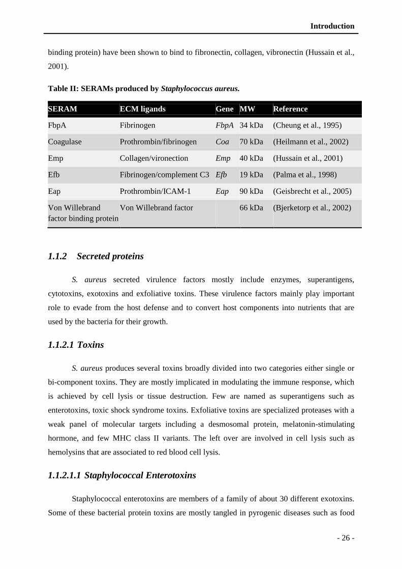
Introduction
- 26 -
binding protein) have been shown to bind to fibronectin, collagen, vibronectin (Hussain et al.,
2001).
Table II: SERAMs produced by Staphylococcus aureus.
SERAM ECM ligands Gene MW Reference
FbpA Fibrinogen FbpA 34 kDa (Cheung et al., 1995)
Coagulase Prothrombin/fibrinogen Coa 70 kDa (Heilmann et al., 2002)
Emp Collagen/vironection Emp 40 kDa (Hussain et al., 2001)
Efb Fibrinogen/complement C3 Efb 19 kDa (Palma et al., 1998)
Eap Prothrombin/ICAM-1 Eap 90 kDa (Geisbrecht et al., 2005)
Von Willebrand
factor binding protein
Von Willebrand factor
66 kDa
(Bjerketorp et al., 2002)
1.1.2 Secreted proteins
S. aureus secreted virulence factors mostly include enzymes, superantigens,
cytotoxins, exotoxins and exfoliative toxins. These virulence factors mainly play important
role to evade from the host defense and to convert host components into nutrients that are
used by the bacteria for their growth.
1.1.2.1 Toxins
S. aureus produces several toxins broadly divided into two categories either single or
bi-component toxins. They are mostly implicated in modulating the immune response, which
is achieved by cell lysis or tissue destruction. Few are named as superantigens such as
enterotoxins, toxic shock syndrome toxins. Exfoliative toxins are specialized proteases with a
weak panel of molecular targets including a desmosomal protein, melatonin-stimulating
hormone, and few MHC class II variants. The left over are involved in cell lysis such as
hemolysins that are associated to red blood cell lysis.
1.1.2.1.1 Staphylococcal Enterotoxins
Staphylococcal enterotoxins are members of a family of about 30 different exotoxins.
Some of these bacterial protein toxins are mostly tangled in pyrogenic diseases such as food
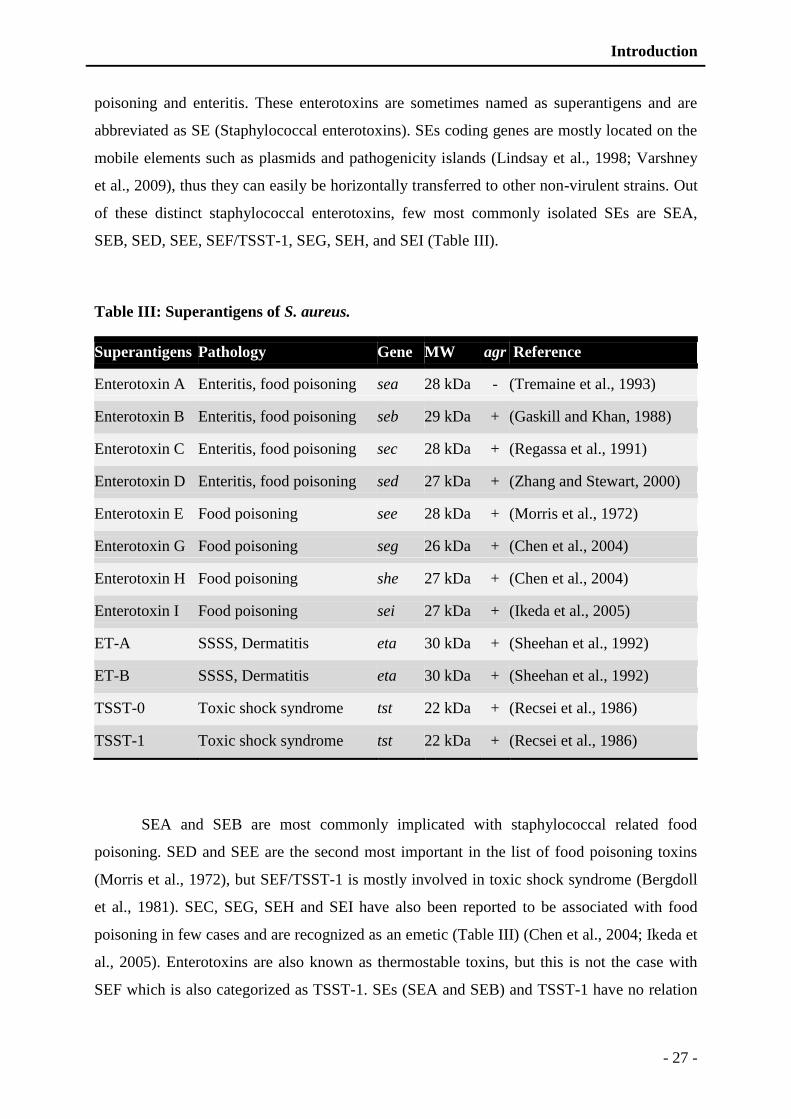
Introduction
- 27 -
poisoning and enteritis. These enterotoxins are sometimes named as superantigens and are
abbreviated as SE (Staphylococcal enterotoxins). SEs coding genes are mostly located on the
mobile elements such as plasmids and pathogenicity islands (Lindsay et al., 1998; Varshney
et al., 2009), thus they can easily be horizontally transferred to other non-virulent strains. Out
of these distinct staphylococcal enterotoxins, few most commonly isolated SEs are SEA,
SEB, SED, SEE, SEF/TSST-1, SEG, SEH, and SEI (Table III).
Table III: Superantigens of S. aureus.
Superantigens Pathology Gene MW agr Reference
Enterotoxin A Enteritis, food poisoning sea 28 kDa - (Tremaine et al., 1993)
Enterotoxin B Enteritis, food poisoning seb 29 kDa + (Gaskill and Khan, 1988)
Enterotoxin C Enteritis, food poisoning sec 28 kDa + (Regassa et al., 1991)
Enterotoxin D Enteritis, food poisoning sed 27 kDa + (Zhang and Stewart, 2000)
Enterotoxin E Food poisoning see 28 kDa + (Morris et al., 1972)
Enterotoxin G Food poisoning seg 26 kDa + (Chen et al., 2004)
Enterotoxin H Food poisoning she 27 kDa + (Chen et al., 2004)
Enterotoxin I Food poisoning sei 27 kDa + (Ikeda et al., 2005)
ET-A SSSS, Dermatitis eta 30 kDa + (Sheehan et al., 1992)
ET-B SSSS, Dermatitis eta 30 kDa + (Sheehan et al., 1992)
TSST-0 Toxic shock syndrome tst 22 kDa + (Recsei et al., 1986)
TSST-1 Toxic shock syndrome tst 22 kDa + (Recsei et al., 1986)
SEA and SEB are most commonly implicated with staphylococcal related food
poisoning. SED and SEE are the second most important in the list of food poisoning toxins
(Morris et al., 1972), but SEF/TSST-1 is mostly involved in toxic shock syndrome (Bergdoll
et al., 1981). SEC, SEG, SEH and SEI have also been reported to be associated with food
poisoning in few cases and are recognized as an emetic (Table III) (Chen et al., 2004; Ikeda et
al., 2005). Enterotoxins are also known as thermostable toxins, but this is not the case with
SEF which is also categorized as TSST-1. SEs (SEA and SEB) and TSST-1 have no relation
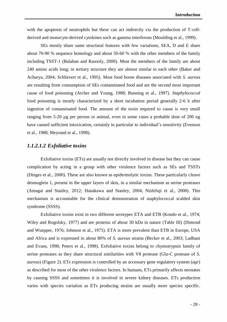
Introduction
- 28 -
with the apoptosis of neutrophils but these can act indirectly via the production of T-cell-
derived and monocyte-derived cytokines such as gamma interferons (Moulding et al., 1999).
SEs mostly share same structural features with few variations, SEA, D and E share
about 70-90 % sequence homology and about 50-60 % with the other members of the family
including TSST-1 (Balaban and Rasooly, 2000). Most the members of the family are about
240 amino acids long; in tertiary structure they are almost similar to each other (Baker and
Acharya, 2004; Schlievert et al., 1995). Most food borne diseases associated with S. aureus
are resulting from consumption of SEs contaminated food and are the second most important
cause of food poisoning (Archer and Young, 1988; Bunning et al., 1997). Staphylococcal
food poisoning is mostly characterized by a short incubation period generally 2-6 h after
ingestion of contaminated food. The amount of the toxin required to cause is very small
ranging from 5-20 µg per person or animal, even in some cases a probable dose of 200 ng
have caused sufficient intoxication, certainly in particular to individual’s sensitivity (Evenson
et al., 1988; Meyrand et al., 1998).
1.1.2.1.2 Exfoliative toxins
Exfoliative toxins (ETs) are usually not directly involved in disease but they can cause
complication by acting in a group with other virulence factors such as SEs and TSSTs
(Dinges et al., 2000). These are also known as epidermolytic toxins. These particularly cleave
desmoglein 1, present in the upper layers of skin, in a similar mechanism as serine proteases
(Amagai and Stanley, 2012; Hanakawa and Stanley, 2004; Nishifuji et al., 2008). This
mechanism is accountable for the clinical demonstration of staphylococcal scalded skin
syndrome (SSSS).
Exfoliative toxins exist in two different serotypes ETA and ETB (Kondo et al., 1974;
Wiley and Rogolsky, 1977) and are proteins of about 30 kDa in nature (Table III) (Dimond
and Wuepper, 1976; Johnson et al., 1975). ETA is more prevalent than ETB in Europe, USA
and Africa and is expressed in about 80% of S. aureus strains (Becker et al., 2003; Ladhani
and Evans, 1998; Peters et al., 1998). Exfoliative toxins belong to chymotrypsin family of
serine proteases as they share structural similarities with V8 protease (Glu-C protease of S.
aureus) (Figure 2). ETs expression is controlled by an accessary gene regulatory system (agr)
as described for most of the other virulence factors. In humans, ETs primarily affects neonates
by causing SSSS and sometimes it is involved in severe kidney diseases. ETs production
varies with species variation as ETs producing strains are usually more species specific.
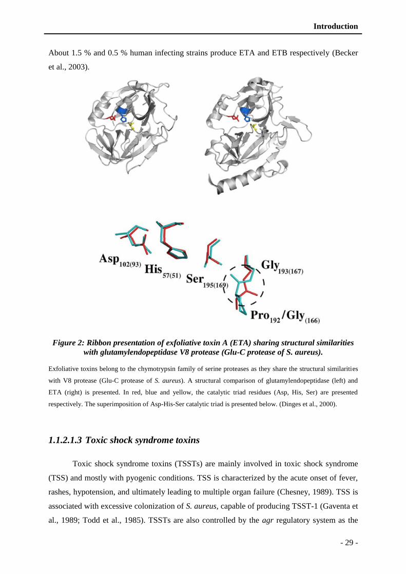
Introduction
- 29 -
About 1.5 % and 0.5 % human infecting strains produce ETA and ETB respectively (Becker
et al., 2003).
Figure 2: Ribbon presentation of exfoliative toxin A (ETA) sharing structural similarities
with glutamylendopeptidase V8 protease (Glu-C protease of S. aureus).
Exfoliative toxins belong to the chymotrypsin family of serine proteases as they share the structural similarities
with V8 protease (Glu-C protease of S. aureus). A structural comparison of glutamylendopeptidase (left) and
ETA (right) is presented. In red, blue and yellow, the catalytic triad residues (Asp, His, Ser) are presented
respectively. The superimposition of Asp-His-Ser catalytic triad is presented below. (Dinges et al., 2000).
1.1.2.1.3 Toxic shock syndrome toxins
Toxic shock syndrome toxins (TSSTs) are mainly involved in toxic shock syndrome
(TSS) and mostly with pyogenic conditions. TSS is characterized by the acute onset of fever,
rashes, hypotension, and ultimately leading to multiple organ failure (Chesney, 1989). TSS is
associated with excessive colonization of S. aureus, capable of producing TSST-1 (Gaventa et
al., 1989; Todd et al., 1985). TSSTs are also controlled by the agr regulatory system as the
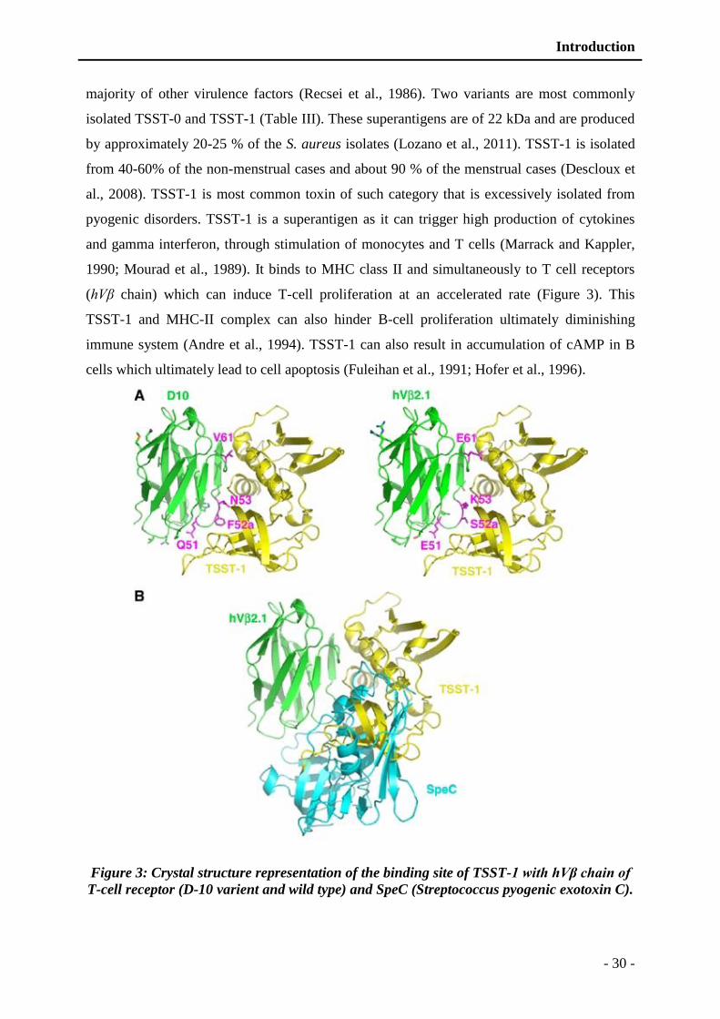
Introduction
- 30 -
majority of other virulence factors (Recsei et al., 1986). Two variants are most commonly
isolated TSST-0 and TSST-1 (Table III). These superantigens are of 22 kDa and are produced
by approximately 20-25 % of the S. aureus isolates (Lozano et al., 2011). TSST-1 is isolated
from 40-60% of the non-menstrual cases and about 90 % of the menstrual cases (Descloux et
al., 2008). TSST-1 is most common toxin of such category that is excessively isolated from
pyogenic disorders. TSST-1 is a superantigen as it can trigger high production of cytokines
and gamma interferon, through stimulation of monocytes and T cells (Marrack and Kappler,
1990; Mourad et al., 1989). It binds to MHC class II and simultaneously to T cell receptors
(hVβ chain) which can induce T-cell proliferation at an accelerated rate (Figure 3). This
TSST-1 and MHC-II complex can also hinder B-cell proliferation ultimately diminishing
immune system (Andre et al., 1994). TSST-1 can also result in accumulation of cAMP in B
cells which ultimately lead to cell apoptosis (Fuleihan et al., 1991; Hofer et al., 1996).
Figure 3: Crystal structure representation of the binding site of TSST-1 with hVβ chain of
T-cell receptor (D-10 varient and wild type) and SpeC (Streptococcus pyogenic exotoxin C).
Introduction
- 31 -
(A) Crystal structure of the TSST-1 complex with D-10 variant (left) and of the wild-type hV (right). TSST-1
is in yellow and hV is in green. (B) Superposition of the TSST-1–hV 2.1 and SpeC–hV complexes. The
SpeC is in cyan. (Moza et al., 2007).
1.1.2.1.4 Staphylococcal Hemolysins
Hemolytic toxins are entitled due to their capacity to specifically cause hemolysis. These
hemolytic toxins are subdivided into four types: alpha (α) hemolysin (Hla), beta (β)
hemolysin (Hlb), gamma (γ) hemolysin (Hlg), and delta (δ) hemolysin (Hld) based on their
structural characteristics. Hemolysins are mostly secreted by the general secretory pathway
(sec) (Peng et al., 1988; Sibbald et al., 2006).
1.1.2.1.4.1 Alpha hemolysin
Alpha toxin is encoded by hla gene and is important for S. aureus sepsis, arthritis,
pneumonia, sepsis, corneal infection and brain abscess (Bubeck Wardenburg et al., 2007;
Callegan et al., 1994; Kielian et al., 2001; Nilsson et al., 1999). Hla is a 33kDa pore forming
toxin and is secreted by most of S. aureus isolates (Bhakdi and Tranum-Jensen, 1991). Alpha
toxin is secreted as monomer which represents cap domain and a β-strand. Monomers
combine to form a heptamer where β-strands join to for stem which is inserted into the
membrane (Figure 4).
Figure 4: Three-dimensional presentation of alpha toxin.
View of alpha hemolysin a heptamer drawn in a ribbon representation which is approximately parallel to the
membrane plane. Each protomer is presented in a different color. Different domains cap, rim, and stem are
Introduction
- 32 -
labeled (left). View of the heptamer (right), the axis from the top of the Cap domain to bottom. (Gouaux et al.,
1997)
This toxin is active against a vast variety of mammalian cells with a remarkable activity
against rabbit erythrocytes (Bhakdi and Tranum-Jensen, 1991; Husmann et al., 2009). Moreover, it is
capable of triggering cytokines and chemokines release such as IL-1a, IL-1b, IL-6, IL-8, TNF-α and
MIP-2 from neutrophils (Bartlett et al., 2008; Bhakdi et al., 1989; Onogawa, 2002).
1.1.2.1.4.2 Beta hemolysin
Beta toxins are also known as sphingomyelinase, it is encoded by hlb gene. Beta
hemolysin is a 35 kDa protein toxin that is hemolytic in nature (Huseby et al., 2007). In
contrast to alpha hemolysin it is more active on sheep RBCs rather than rabbit erythrocytes
(Dinges et al., 2000). Sometimes, β-toxin is depicted as “hot-cold” toxin due to their ability to
become more active at temperature below 10°C (Dinges et al., 2000). The β-toxin has been
isolated from cases of corneal and lung infections and is often associated with pneumonia due
to its inhibitory effect on the ciliary movement of nasal epithelial cells (Kim et al., 2000;
O'Callaghan et al., 1997).
1.1.2.1.4.3 Gamma hemolysins
Gamma hemolysins are the most important of hemolysin family members produced by S.
aureus. They are encoded by hlg locus, in contrast to the other hemolysins, there are three
open reading frames designated as hlgA, hlgB and hlgC (Cooney et al., 1988). Each of the
encoded proteins is preceded by a putative signal translation sequence and mature forms are
nominated as HlgA, HlgB and HlgC each of 31.9, 34.1 and 32.5 kDa respectively (Table IV)
(Cooney et al., 1988). The sequence comparison demonstrates 70 % residual identity between
HlgA and HlgC proteins, where each has approximately 30 % sequence similarity with HlgB
(Cooney et al., 1993). Additionally, alpha toxins and HlgB have 27 % sequence identity there
is almost no sequence identity between HlgA/C and alpha toxins (Cooney et al., 1993).
These toxins can act in combination with other γ-toxins components and sometimes
even with the leucocidins. So, these are demarcated as bi-component toxins. HlgB and
HlgA/C are nominated as class F and class S components respectively, due to their
remarkable similarity with class F and class S components of PVL (98.5 and 99.1 %
respectively) (Cooney et al., 1993). Biological activity usually results from the combination
of one class S component with one class F component (Colin et al., 1994). Likewise, each
toxin component in the gamma hemolysin assembly could in fact yield two biologically active
Introduction
- 33 -
toxins. These are hemolytic (HlgA-HlgB) in nature, but complexity of their toxicity is more
preeminent due to their ability of cross combine with other bi-component family members
(Dalla Serra et al., 2005). Gamma hemolysins have leukotoxic effect in addition to hemolytic
activity. HlgA-HlgB pair is more active on leukocytes in contrast to the HlgC - HlgB pair
which is more active on erythrocytes (Colin et al., 1994; Prevost et al., 1995c). Recently,
toxicity of gamma hemolysins has also been demonstrated towards neurons. In neuron cell
cultures these can mediate rise in free intracellular Ca2+ via acidic stores and activation of
store operated channels (Jover et al., 2012).
1.1.2.1.4.4 Delta hemolysin
Delta hemolysin is encoded by hld gene and the product is a 26 amino acid peptide
capable of causing erythrolysis (Dinges et al., 2000; Peng et al., 1988; Wiseman, 1975). This
toxin is more prevalent as compared to other hemolysin members, produced by about 97% of
S. aureus isolates. These are also active for variety of mammalian erythrocytes, in addition
can bind to sub-cellular structures that are mostly membrane bound (Bhakoo et al., 1982;
Freer and Birkbeck, 1982). Delta toxin does not possess a cleavable signal sequence as
present in alpha and beta toxins (Dinges et al., 2000).
1.1.2.1.5 Panton valentine leucocidin
Panton-Valentine leucocidin (PVL) is one of the most important bi-component toxins
that are composed of one class S (LukS-PV) and one class F (LukF-PV) component (Finck-
Barbancon et al., 1991; Panton and Valentine, 1932; Ward and Turner, 1980; Woodin, 1960).
LukS-PV is 33 kDa and LukF-PV is 34 kDa, these toxins act synergistically on mammalian
polymorphonuclear cells, monocytes, and macrophages (Prevost et al., 1995c). PVL has been
described as a marker of virulence, due to its strong association with necrotizing cutaneous
infections (Couppie et al., 1994; Prevost et al., 1995a; Prevost et al., 1995b). PVL is active
against a variety of cells with varying activity but primarily target PMNs. They display
different activities in different species such as these are highly dermonecrotic for rabbit skin
as compared to other mammalians (Couppie et al., 1994). As previously described for gamma
hemolysins (Dalla Serra et al., 2005), these can combine with other components too,
including Luk-E, Luk-D, γhlg (A, B, C), Luk-S, LukF, Luk-M and Luk-F' (Fromageau et al.,
2010). However, the heterologous pairs are not as much active as parent pairs (Prevost et al.,
1995c). PVL, at sublytic amounts have been demonstrated to stimulate PMNs, and lead to the
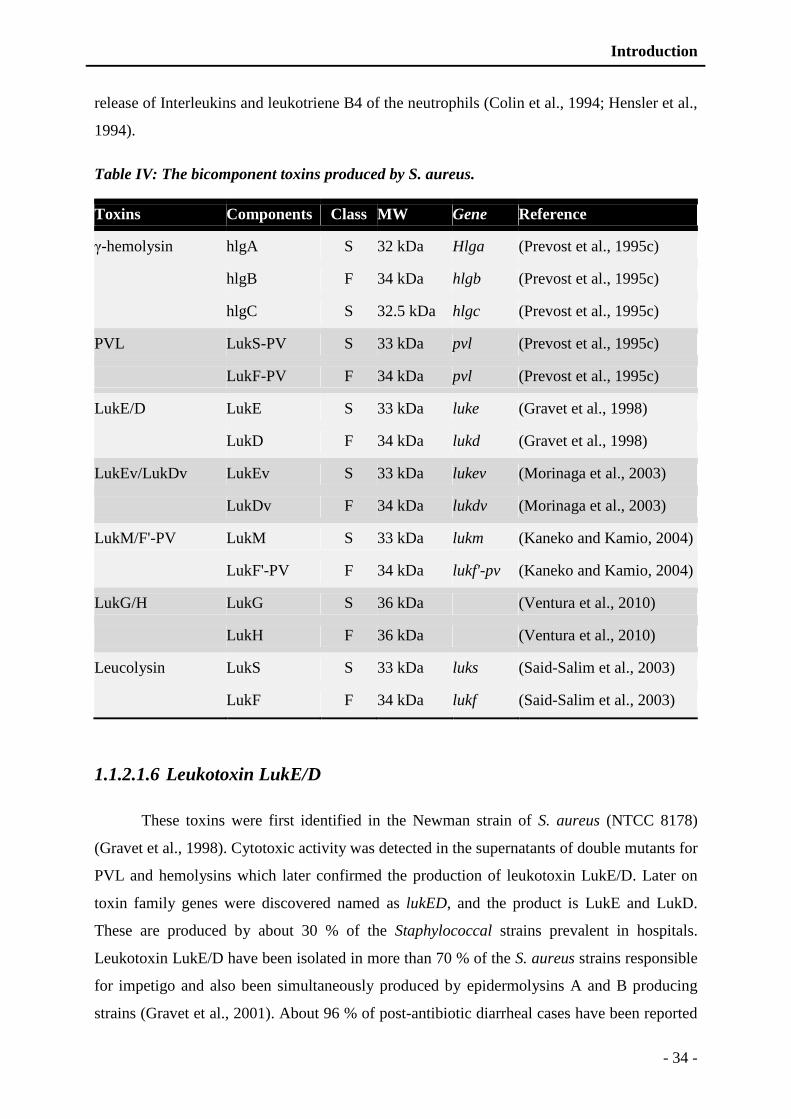
Introduction
- 34 -
release of Interleukins and leukotriene B4 of the neutrophils (Colin et al., 1994; Hensler et al.,
1994).
Table IV: The bicomponent toxins produced by S. aureus.
Toxins Components Class MW Gene Reference
γ-hemolysin hlgA S 32 kDa Hlga (Prevost et al., 1995c)
hlgB F 34 kDa hlgb (Prevost et al., 1995c)
hlgC S 32.5 kDa hlgc (Prevost et al., 1995c)
PVL LukS-PV S 33 kDa pvl (Prevost et al., 1995c)
LukF-PV F 34 kDa pvl (Prevost et al., 1995c)
LukE/D LukE S 33 kDa luke (Gravet et al., 1998)
LukD F 34 kDa lukd (Gravet et al., 1998)
LukEv/LukDv LukEv S 33 kDa lukev (Morinaga et al., 2003)
LukDv F 34 kDa lukdv (Morinaga et al., 2003)
LukM/F'-PV LukM S 33 kDa lukm (Kaneko and Kamio, 2004)
LukF'-PV F 34 kDa lukf'-pv (Kaneko and Kamio, 2004)
LukG/H LukG S 36 kDa (Ventura et al., 2010)
LukH F 36 kDa (Ventura et al., 2010)
Leucolysin LukS S 33 kDa luks (Said-Salim et al., 2003)
LukF F 34 kDa lukf (Said-Salim et al., 2003)
1.1.2.1.6 Leukotoxin LukE/D
These toxins were first identified in the Newman strain of S. aureus (NTCC 8178)
(Gravet et al., 1998). Cytotoxic activity was detected in the supernatants of double mutants for
PVL and hemolysins which later confirmed the production of leukotoxin LukE/D. Later on
toxin family genes were discovered named as lukED, and the product is LukE and LukD.
These are produced by about 30 % of the Staphylococcal strains prevalent in hospitals.
Leukotoxin LukE/D have been isolated in more than 70 % of the S. aureus strains responsible
for impetigo and also been simultaneously produced by epidermolysins A and B producing
strains (Gravet et al., 2001). About 96 % of post-antibiotic diarrheal cases have been reported
Introduction
- 35 -
to have leukotoxin LukE/D in association with enterotoxins (more than 80 %) concurrently
(Gravet et al., 2001). These present similar activity as PVL for rabbit skin model but are non-
hemolytics and week leukocytic in nature. Additionally, these have been demonstrated to
target T lymphocytes. Recently leucocidin ED have been described to contribute to systemic
infection by targeting neutrophils in murine and rabbit cells (Alonzo et al., 2012). Two
variants of leukotoxin LukE/D (LukEv and LukDv) have also been characterized with 91 and
94% sequence similarities with LukE and LukD respectively (Morinaga et al., 2003).
Interestingly, these variants have few regions of similarity with PVL components that is might
responsible for a little enhanced leukocytic week hemolytic activity against rabbit PMNs and
erythrocytes (Morinaga et al., 2003). Neither LukED nor LukEDv have been reported to have
K/R-R/K-X-T/S motif, which is known to be phosphorylated by protein kinase and is
essential for leukocytolytic activity but genome sequencing of few strains (N315, Mu50) have
revealed presence of K/R-R/K-X-T/S motif, on pathogenicity islands in chromosome of these
strains. (Kuroda et al., 2001). There is a conflict of studies about the prevalence of lukED
genes in different strains but regardless of this, LukE/D are important for virulence of S.
aureus as the deletion of LukE and LukD gene can result in attenuation of virulence and
decrease in lethality in murine blood stream infection model (Alonzo et al., 2012). LukE/D
also mediates specific toxicity towards T-lymphocytes by specifically binding to CCR5.
Binding with the CCR5 recepter is meadiated by LukE compoenent but not the LukD of the
leukotoxin (Alonzo et al., 2013).
1.1.2.1.7 Other Bi-component cytolytic toxins
Few other cytolytic toxins have also been characterized in different strains of S. aureus
including Luk-S/Luk-F, Luk-M/Luk-F' and LukG/LukH, targeting different mammalian
species. Luk-S (33 kDa) and Luk-F (34 kDa) pair is cytolytic in nature, mostly targeting
human and rabbit PMNs and specifically rabbit erythrocytes but have no effect on human
erythrocytes (Kaneko and Kamio, 2004). Luk-M and Luk-F' is mostly involved in mastitis
cases with a very astonishing specificity towards bovine leukocytes (Fromageau et al., 2010).
1.1.2.2 Proteases of S. aureus
S. aureus expresses several kinds of proteases. SspA serine protease (V8 protease) plays
an important role in the degradation of surface proteins of the bacterium (Karlsson et al.,
2001; McGavin et al., 1997). So, it is responsible for degradation of protein A and most

Introduction
- 36 -
fibrinogen binding proteins which are expressed at the end of exponential growth phase.
Aureolysin is another major protease, which has a dual function: it acts similarly to SspA on
the degradation of surface proteins and also it catalyzes the activation of many proteases
cleaving precursors at specific sites (McAleese et al., 2001; Rice et al., 2001). There is
interdependence in activation of different proteases. For example, SspA aureolysin lead to
maturation of AspA and mature SspA turn SSPB cysteine protease (Rice et al., 2001).
Proteases of S. aureus are also important to reshape bacterial membrane by modification.
They are also actively involved in fight against the immune system. Some are especially
capable of degrading immunoglobulin heavy chain and several protease inhibitors (Prokesova
et al., 1995).Additionally, they protect bacterial cell from antimicrobial peptides such as
aureolysin (Sieprawska-Lupa et al., 2004). Finally, proteases provide a source of energy since
bacteria degrade all macromolecules present in extracellular medium after apoptosis of host
cells (Shaw et al., 2004). Recently, several new serine protease encoded on the same operon
were identified such as proteases sPL A-F (Reed et al., 2001). The function of these proteins
is still unknown but crystallographic structure of sPLA shows strong similarities with SspA
(Stec-Niemczyk et al., 2009). sPLA-F proteases are probably involved in virulence of the
bacterium; especially expression of SPLC has already been confirmed in patients with
endocarditis (Rieneck et al., 1997).
1.2 Host defences against S.aureus infection
S. aureus infection usually starts with a breach in outer physical barrier of body,
including skin and different mucous surfaces. The organism is confronted by the host immune
system, comprising both innate and acquired responses. S. aureus infection of skin triggers a
strong inflammatory response, which involves the migration of first line defence cells
(neutrophils and macrophages) to site of infection. A brief exhibition of events occurring
during the initial development of infection and triggered inflammatory responses are
presented in figure 5 (Foster, 2005). These inflammatory cells will attempt to engulf and
dispose of entering pathogens with the help of available antibodies and through complement
activation. This is where the first vital internal conflict between S. aureus and host defense
occurs. Complement system is a family of proteins that have many roles in innate and
acquired immunity, comprising killing of foreign cells and regulation of other effectors of
immune response (Moore, 2004). During S. aureus infection the role of complement is to
recruit effector molecules that are essential for tagging the cells and target them for

Introduction
- 37 -
demolition by immune effector cells such as neutrophils. The process of complement fixation
takes place by three different pathways as follows: alternative, lectin and classical pathway
(Figure 7). Alternative and lectin pathways are carried out by components of innate immunity,
whereas the classical pathway requires interaction of antigen bounded with antibodies on
target cells (Moore, 2004). One of key purposes of complement fixation is opsonization
which promotes phagocytosis of infected cells by macrophages and neutrophils.
During the complement activation several small peptides are released such as (C3a and
C5a) which results in chemo-attraction of Phagocytes to site of infection, also by formylated
peptides released by developing bacteria. These small molecules have specific receptor on
the phagocytic cell membrane that enhances phagocytosis efficiency. Similarly neutrophils
also have specific receptor that recognize Fc region of the immunoglobulin (Figure 5). By the
help of complement system, invading microorganisms and their products are cleared by
immune cells and transported to lymphnodes, where the resting B-lymphocytes are activated.
These activated cells differentiate between host and pathogen components and secret specific
antibodies that help to neutralize circulating toxins and promote efficient killing of remaining
infectious agents (Foster, 2005).
Regardless of high immune response, S. aureus survive such dreadful conditions. Most
of time antibodies to S. aureus antigens are present in humans and there is rapid rise in titre
following infection (Dryla et al., 2005; Etz et al., 2002). However, these antibodies and
immunological memory appear to be inadequate to prevent S. aureus subsequent infections.
Moreover, several mechanisms have been evolved by S. aureus to disrupt immune defense.
Previously it was regarded as a non-invasive pathogen, but recent studies have suggested that
S. aureus can invade different type of host cells by a mechanism which involves formation of
fibronectin bridge between host α5β1 integrin molecules and bacterial fibronectin binding
proteins (Schwarz-Linek et al., 2004; Schwarz-Linek et al., 2003). After internalization into
host cells, it can survive in a semi-dormant form denoted as small colony variants (von Eiff et
al., 2000). In addition to this dormancy, various mechanisms are hired by S. aureus that let
them to evade innate and acquired immunity.
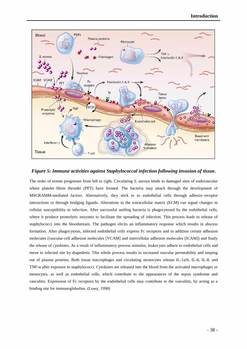
Introduction
- 38 -
Figure 5: Immune activities against Staphylococcal infection following invasion of tissue.
The order of events progresses from left to right. Circulating S. aureus binds to damaged sites of endovascular
where platelet–fibrin thrombi (PFT) have formed. The bacteria may attach through the development of
MSCRAMM-mediated factors. Alternatively, they stick to to endothelial cells through adhesin–receptor
interactions or through bridging ligands. Alterations in the extracellular matrix (ECM) can signal changes in
cellular susceptibility to infection. After successful settling bacteria is phagocytosed by the endothelial cells,
where it produce proteolytic enzymes to facilitate the spreading of infection. This process leads to release of
staphylococci into the bloodstream. The pathogen elicits an inflammatory response which results in abscess
formation. After phagocytosis, infected endothelial cells express Fc receptors and in addition certain adhesion
molecules (vascular-cell adhesion molecules [VCAM] and intercellular adhesion molecules [ICAM]) and finaly
the release of cytokines. As a result of inflammatory process stimulus, leukocytes adhere to endothelial cells and
move to infected site by diapedesis. This whole process results in increased vascular permeability and seeping
out of plasma proteins. Both tissue macrophages and circulating monocytes release IL-1a/b, IL-6, IL-8, and
TNF-α after exposure to staphylococci. Cytokines are released into the blood from the activated macrophages or
monocytes, as well as endothelial cells, which contribute to the appearances of the sepsis syndrome and
vasculitis. Expression of Fc receptors by the endothelial cells may contribute to the vasculitis, by acting as a
binding site for immunoglobulins. (Lowy, 1998)
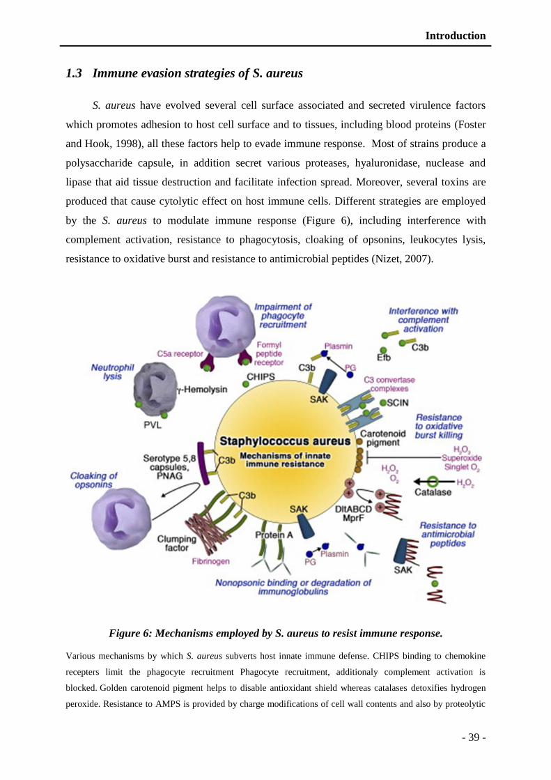
Introduction
- 39 -
1.3 Immune evasion strategies of S. aureus
S. aureus have evolved several cell surface associated and secreted virulence factors
which promotes adhesion to host cell surface and to tissues, including blood proteins (Foster
and Hook, 1998), all these factors help to evade immune response. Most of strains produce a
polysaccharide capsule, in addition secret various proteases, hyaluronidase, nuclease and
lipase that aid tissue destruction and facilitate infection spread. Moreover, several toxins are
produced that cause cytolytic effect on host immune cells. Different strategies are employed
by the S. aureus to modulate immune response (Figure 6), including interference with
complement activation, resistance to phagocytosis, cloaking of opsonins, leukocytes lysis,
resistance to oxidative burst and resistance to antimicrobial peptides (Nizet, 2007).
Figure 6: Mechanisms employed by S. aureus to resist immune response.
Various mechanisms by which S. aureus subverts host innate immune defense. CHIPS binding to chemokine
recepters limit the phagocyte recruitment Phagocyte recruitment, additionaly complement activation is
blocked. Golden carotenoid pigment helps to disable antioxidant shield whereas catalases detoxifies hydrogen
peroxide. Resistance to AMPS is provided by charge modifications of cell wall contents and also by proteolytic

Introduction
- 40 -
degradation by the proteases. Protein A binds to Fc domains of immunoglobulins in a nonspecific manner,
whereas clumping factors and surface polysaccharide capsule and proteins and poly-N-acetylglucosamine
(PNAG) act to cloak surface bound opsonins from phagocyte recognition. The pore-forming toxins of S. aureus
target leukocyte membranes and ultimately cause cell lysis. The staphylokinase (SAK) activates the zymogen to
active protease plasmin, which degrades immunoglobulin Fc domain and complement opsonin C3b. (Nizet,
2007)
1.3.1 Resistance to phagocytosis
S. aureus has the capability to resist phagocytosis by production of antiopsonin
proteins, in addition polysaccharide capsule is produced which can hinder complement
activation, either by classical pathway or alternative pathway. As the phagocytosis require
bounded complement and antibodies which is prevented by capsule formation, regardless of
high titer of antibodies against different cell surface components of S. aureus. Well known
surface-protein of S. aureus, protein A is cell wall associated protein with four or five
domains, which interact with the Fc region of IgG (Uhlen et al., 1984). Due to binding of IgG
with protein A, bacterial cell surface is covered with immunoglobulins in an improper
orientation that is unrecognizable by neutrophils Fc receptors.
Another protein produced by S. aureus is clumping factor A which is very dominant
fibrinogen binding protein (Bischoff et al., 2004; O'Brien et al., 2002). Clf-A is predominantly
produced during the exponential phase of growth and during stationary phase of growth
unlike other surface proteins, it is 10 fold increased (O'Brien et al., 2002). When cells are
densely packed in vivo, the γ-chain of Clf-A can bind to two different fibrinogen molecules
(McDevitt et al., 1997). In this way, bacterial cell is covered with fibrinogens which inhibit
deposition of opsonins.
Most of the S. aureus strains produce capsule especially MRSA, that is composed of
various serotypes such as 5, 8 and 336 are more common (Roghmann et al., 2005). In vitro,
phagocytosis assays revealed that presence of polysaccharide capsules reduced the uptake of
cells by neutrophils which indicate the anti-opsonic property of capsule.
1.3.2 Complement inactivation
For the successful activation of complement, attachment of C3 convertases to bacterial
surface is pre-requisite. C3 convertases (C4bC2a and C3bBb in classical/lecithin and
alternative pathways respectively) lead to conversion of C3 protein to C3a (classical and
lecithin pathways) and C3b (alternative pathway) (Figure 7). S. aureus secretes protein named
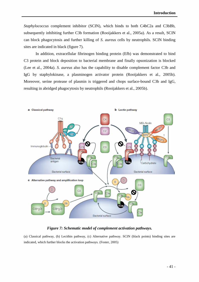
Introduction
- 41 -
Staphylococcus complement inhibitor (SCIN), which binds to both C4bC2a and C3bBb,
subsequently inhibiting further C3b formation (Rooijakkers et al., 2005a). As a result, SCIN
can block phagocytosis and further killing of S. aureus cells by neutrophils. SCIN binding
sites are indicated in black (figure 7).
In addition, extracellular fibrinogen binding protein (Efb) was demonstrated to bind
C3 protein and block deposition to bacterial membrane and finally opsonization is blocked
(Lee et al., 2004a). S. aureus also has the capability to disable complement factor C3b and
IgG by staphylokinase, a plasminogen activator protein (Rooijakkers et al., 2005b).
Moreover, serine protease of plasmin is triggered and chops surface-bound C3b and IgG,
resulting in abridged phagocytosis by neutrophils (Rooijakkers et al., 2005b).
Figure 7: Schematic model of complement activation pathways.
(a) Classical pathway, (b) Lecithin pathway, (c) Alternative pathway. SCIN (black points) binding sites are
indicated, which further blocks the activation pathways. (Foster, 2005)

Introduction
- 42 -
1.3.3 Inhibition of neutrophil chemotaxis
Immediately, with the start of bacterial infection several chemo attractant are liberated,
which are recognized by neutrophils. These chemo-attractants include C3a, C5a fragments of
complement activation and several formylated petides (FPs) secreted by bacteria during
growth. These factors results in strong chemotactic response, and are recognized by the
transmembrane G-protein coupled receptors of neutrophils (Murdoch and Finn, 2000). With
the recognition of these chemo attractants intracellular signaling cascade is activated, which
result in migration of neutrophils from blood stream to site of infection. Approximately 60%
of S. aureus strains produce CHIPS (chemotaxis inhibitory proteins) that can bind to C5a
receptor and formyl peptide receptor (Figure 8) and finally block the recognition process (de
Haas et al., 2004).
Figure 8: Schematic representation of methods employed by S. aureus to inhibit
chemotaxis.
S. aureus produces CHIPS which occupy the C5a recepters and formyl receptor on the neutrophils cell surface.
Additionally, Eap blocks neutrophil adhesion by occupying ICAM-1.

Introduction
- 43 -
In addition to CHIPS, S. aureus also secretes extracellular adhesion protein (Eap) also
called major histocompatibility class II analogue protein (MAP), which has a ligand on the
endothelial cells surface called intercellular adhesion molecule-1 (ICAM-1) (Figure 8).
Interaction between these two blocks the binding of LFA-1 (lymphocyte function associated
antigen-1) of neutrophil surface (Chavakis et al., 2002). With blockade of LFA-1 neutrophil
adhesion, extravasation and diapedesis is compromised, ultimately resulting in chemotaxis
inhibition.
1.3.4 Leukocytes destruction by toxins
The expression of cytolytic toxins that lyse leukocytes is cardinal feature of S. aureus.
Neutrophil lysis contributes to development of abscesses and diminishes their attempt to
engulf bacteria. Most of the cytolytic toxins from S. aureus form beta-barrel pore in
membrane, ultimately causing leakage and consequently cell death. Alpha-toxin is model of
this class of virulence factors which is produced as monomers and these monomers join to
form a heptamer in cell membrane. The β-strand from each monomer assemble into a β-barrel
pore (Montoya and Gouaux, 2003).
The bi-component leukotoxins are secreted in two separate subunits (class S and class
F component), those can assemble to form either hexamer or heptamer which have strong
affinity for leukocytes. As previously described in the virulence factor section, S. aureus
produces mainly four different type of bicomponent toxins including γ-toxins, PVL,
leucocidin ED and Leucocidin M-F (PV like). The γ-hemolysin is more prevalent, it is
produced by approximately more than 90% of the strains while PVL genes are found only in
1-2% strains (Peacock et al., 2002; Prevost et al., 1995c). PVL is more associated with the
resistant strains (CA-MRSA), these community acquired methicillin resistant S. aureus strains
has been widely isolated from cases of necrotizing pneumonia, and skin infections (Gillet et
al., 2002; Said-Salim et al., 2003). These CA-MRSA strains have been demonstrated to carry
staphylococcal cassette chromosome (type IV or V -SCCmec), which encode resistance to
meticillin and other β-lactam antibiotics (Lina et al., 1999). Additionally, encoded by pvl
genes located in a lysogenic bacteriophage (Narita et al., 2001).
Introduction
- 44 -
1.3.5 Resistance to lysozyme
Lysozyme (muramidase) is produced by a vast array of host cell and is present almost
all body fluids. Muramidase expression is enhanced by stimulated phagocytic cells.
Muramidase cause cleavage of glycosidic link between N-acetylglucosamine and N-acetyl
muramic acid, present in the bacterial cell wall (Bera et al., 2005). S. aureus resistance to
lysozyme is attributed to a membrane-bound structure (O-acetyltransferase) which modifies
C6 hydroxyl group of muramic acid (Bera et al., 2005). Ultimately, this leads to protection of
glycosidic linkage from action of lysozyme.
1.3.6 Survival of S. aureus in neutrophil phagosomes
Neutrophils are most important in number and the first line of defense against
pathogens especially S. aureus, as patient of granulomatous disease suffer recurrent
infections. Recurrent staphylococcal infections are due to defective genes encoding of
NADPH phagocyte oxidase subunits. Defective encoding for NADPH compromise
production of superoxide radicals during respiratory burst (Heyworth et al., 2003). S. aureus
has developed several mechanisms that resist the killing by phagocytes, including resistance
to antimicrobial substances released and interference with endosome fusion (Gresham et al.,
2000). Moreover, bacterium avoids the toxic effects of oxygen free radicals that are formed
during respiratory burst. Carotenoid pigments are produced by S. aureus which give yellow
coloration to colonies, helps to scavenge oxygen free radicals (Liu et al., 2005). In addition,
S. aureus produces two superoxide dismutase enzymes that help to remove free oxygen
radicals (Karavolos et al., 2003). Several other measure are employed to counter toxic effect
of phagocyte environment including manganese homeostasis (Horsburgh et al., 2002),
methionine sulphoxide reductases production (Singh and Moskovitz, 2003), high catalase
production and several toxins are upregulated such as γ-hemolysin (Voyich et al., 2005).
1.3.7 Subversion of the humoral immune response
S. aureus heavily produce fibronectin binding protein (A and B) as surface adhesion
proteins, which binds to platelets and finally lead to their activation. With the activation of
platelets initiat thrombus starts to grow. S. aureus reside inside the thrombus where it is
protected from neutrophils (Moreillon and Que, 2004). Moreover, fibronectin binding
Introduction
- 45 -
proteins bind on the other end with immunoglobulins, which cover thrombus surface and
finally pathogen saves itself from humural immune response.
1.3.8 Resistance to killing by antimicrobial peptides
Antimicrobial peptides are integral part of innate immune defense, mostly these are
are attracted to wards bacteria due to negative charge, where they cause destabilization and
rupture of bacterium. S. aureus execute few natural modifications to lipoteochoic acid, wall
teichoic acid (WTA), and to membrane phospholipids. Dlt proteins expression result in d-
alanine substitutions of lipoteichoic acid and ribitol teichoic acid that can partially neutralize
the negative charge and moderate attraction of cationic force (Peschel et al., 1999). Similarly,
MprF protein adds an L-lysine residue to phosphatidylglycerol (Staubitz et al., 2004). In both
cases, cationic AMPs are repelled from the cytoplasmic membrane, in addition positively
charges AMPs are also resisted such as lactoferrin and phospholipase A2. Mutants defective
in MprF and Dlt are rapidly killed by cationic antimicrobial proteins and neutrophils in vitro
and have reduced virulence in several animal infection models (Collins et al., 2002; Kristian
et al., 2003).
In addition to neutralization of the charge, S. aureus also secretes several proteins that
neutralize cationic peptides such as staphylokinase, a prothrombin activator. Staphylokinase
degrade IgG and C3 factor of complement system, dissolve fibrin clot and moreover has high
affinity for defensing peptides (Jin et al., 2004). Also, the extracellular proteases produce by
S. aureus degrade antimicrobial peptides such as metalloprotease aureolysin cleaves and
inactivates cathelicidin LL-37 and contributes considerably to resistance to antimicrobial
peptide in vitro (Sieprawska-Lupa et al., 2004).
1.4 Resistance to chemotherapeutic agents
Chemotherapy is used against many bacterial infectious diseases including gram
positive and gram negative pathogens. Chemotherapeutic agents such as aminoglycosides, β-
lactams, and quinolones are used against S. aureus. The emergence of resistance to antibiotics
makes it a huge problem to treat S. aureus infections. Initially, S. aureus became resistant to
β-lactams antibiotics by acquiring the penicillinase (β-lactamase) gene by horizontal transfer
(Chambers, 2001; Kirby, 1944; Thornsberry, 1988). To counter resistance to β-lactam
antibiotics modified β-lactams resistant to β-lactamase, such as oxacillin and methicillin, were
developed. Subsequently, through extensive use of these modified β-lactam antibiotics,

Introduction
- 46 -
MRSA strains have emerged (Maple et al., 1989; Martin, 1994). Penicillin-binding-protein
(PBP)-2 (2A) is responsible for this resistance against the modified β-lactams (Berger-Bachi,
2002; de Lencastre et al., 1994). S. aureus generally possesses four PBPs. In susceptible
strains, they are inactivated in the presence of β-lactams, results in inhibition of cell wall
biosynthesis and ultimately leading to cell death. MRSA can survive in presence of β-lactams
due to an extra PBP2'. The difference between MRSA and methicillinsusceptible S. aureus
(MSSA) is due to the presence or absence of the mec region including the mecA gene coding
for PBP2'. The mec region is composed of mobile genetic elements that are called SCCmec
cassettes. To date, five types of SCCmecs have been identified (Hiramatsu et al., 2001;
Hiramatsu et al., 2002; Ma et al., 2002). These cassettes have a variable size of about 20–70
kbp and contain mecA, ccrA (recombinase A), ccrB (recombinase B), and other orf s of
unknown function (Robinson and Enright, 2004).
The glycopeptides vancomycin and teicoplanin are powerful agents with significant
activity in MRSA infections. Vancomycin binds to peptidoglycan precursor and then inhibits
the incorporation of newly synthesized precursors in the cell wall peptidoglycan, thus causing
inhibition of cell wall synthesis. Recently, vancomycin-intermediate susceptible S. aureus
(VISA), or glycopeptide-intermediate susceptible S. aureus (GISA), emerged (Bobin-Dubreux
et al., 2001; Cosgrove et al., 2004; Hiramatsu, 2001; Sieradzki et al., 1999), followed by a
vancomycin-resistant S. aureus (VRSA) (Goldrick, 2002; Whitener et al., 2004). The
mechanism of vancomycin resistance in the VRSA is due to expression of vanA gene, which
modifies the structure of peptidoglycan, causing a loss of affinity of vancomycin for the
modified peptidoglycan precursor (Clark et al., 2005).
Antibiotic resistance in MRSA has evolved to multiple drug resistances against
chemotherapies. This implies that MRSA strains acquire extra factors at high frequency
compared with the MSSA strains. This pattern has continued among the antibotics such as
linezolid, which was introduced in 2000. Only a year later a resistance strain is reported
named as linezolid-resistant MRSA (Quiles-Melero et al., 2012; Tsiodras et al., 2001).
Similary fate was achieved daptomycin which was introduced in 2003 and MRSA was able to
develop resistance against daptomycin within two years (Mangili et al., 2005; Marty et al.,
2006). Therefore, it will become difficult in near future to cure MRSA infections because S.
aureus adapts to new chemotherapeutics and new approaches to therapy will be required.

Immune system


Introduction
- 47 -
2 Immune system
2.1 Introduction
The immune system evolved as a set of very well organized event that protect the host
from the invading pathogens and cancer cells. Immune system helps to discriminate between
self and non-self components. It acts as a mechanism of defense against the pathogens, such
as bacteria, viruses, fungi, parasites and cancerous cells. Immune system usually functions in
two steps first is the pathogen recognition and then response. Recognition is remarkably
highly specific in nature, which carried out by various components of immune system. In
recognition, subtle chemical differences are recognized by innate immune components, which
distinguish invading agents from the host components. Later, effector cells come into play
and clear the hazardous agents from the host body (Kindt et al., 2006).
On basis of time of play of the actors of immunity, it is broadly categorized in innate
and adaptive immune systems. When the pathogen gets in, the components which first
encounter are categorized as innate immune components; those build the first line of defense
(Abbas and Lichtman, 2005). Innate immune components present the pathogen to adaptive
immunity, which differentiate minor details of the pathogen and memorize the details for
future. This form of immunity is not very rapid; it adapts itself according to situation, so
named as adaptive immunity.
Adaptive immunity is able to recognize a large number of non-microbial and microbial
substances. It possesses a great capacity to discriminate very finely different molecules and
microbes, even in the case of high similarity. The adaptive immunity has further two main
branches, the humoral and cellular immunity (Kindt et al., 2006). Humoral immunity is
usually active against bacteria and viruses that invade into body fluids. Its major tools of
action are immunoglobulins also known as antibodies (Burmester et al., 2003; Kindt et al.,
2006). The cellular immunity targets infected cells by the action of T-lymphocytes and
natural killer cells. Cellular immunity is very specific and potent in action, as it is able to clear
infection reservoirs (Biron, 2010). With recent discoveries of various aspects of immune
system, different categories of immune systems seem to be intermingled, these work in
coordination, interdependent on each other. In addition to clear pathogens, immunity also
maintains a homeostasis, by effectively clearing dead or dying cells from the body.
Introduction
- 48 -
2.1.1 Innate immune system
Innate immune system is the first line of defense against the infections. It is composed
of the components that recognize the pathogen well before the adaptive immunity comes into
play. Innate immunity is one of the oldest defense mechanism that is evolved in multicellular
organisms including insects and plants. While the adaptive system evolved in vertebrates as a
more specific immune system (Danilova, 2006).
Innate immunity is very much important in defense mechanisms, as dysfunction of
innate system remarkably increases the suceptibity to infection regardless of the persistence of
adaptive immune system. A classic example is chronic granulomatous disease (CGD), which
is characterized by the gene mutation encoding NADPH oxidase in neutrophils (Heyworth et
al., 2003). Patients of CGD are more prone to recurrent infections due to inability of
neutrophils to successfully lyse pathogens (Marodi and Notarangelo, 2007). Moreover, innate
system provide base for the development of adaptive system, components of innate system
such as phagocytes and complement system are used by the adaptive immune system.
Additionally, innate immune system presents infection to adaptive system to trigger an
effective immune response.
With the invasion of the infectious agents, innate immune response is triggered. First
step is recognition of pathogens and declare it as self or non-self that is carried out by the
pattern recognition receptors (PRRs) (Biron, 2010). After declaration of agent as non-self,
cellular components comes into action and try to nullify the infection, meanwhile these
develop chemical components of the immune system including complement system,
cytokines and antimicrobial peptides (Biron, 2010).
2.1.1.1 Components of innate immune system
Innate immune system composed of following factors: physical/chemical barriers,
pattern recognition receptors, cells of innate immune system, complement system, cytokines,
and antimicrobial peptides.
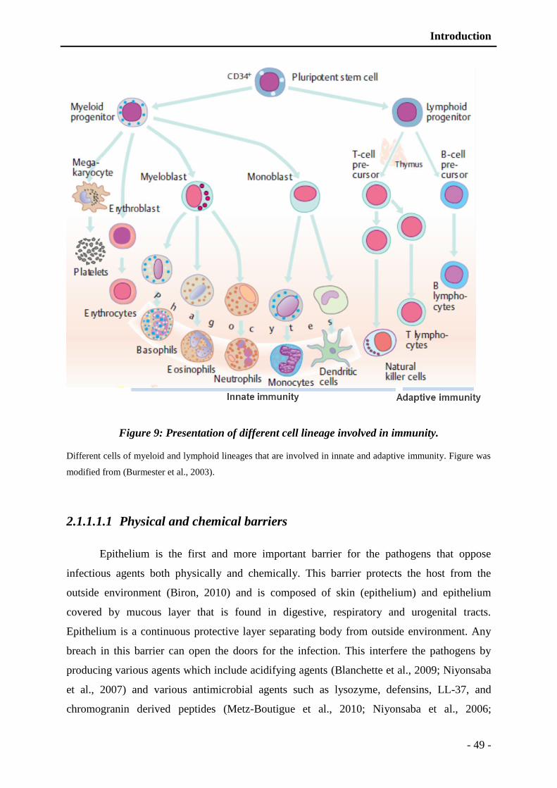
Introduction
- 49 -
Figure 9: Presentation of different cell lineage involved in immunity.
Different cells of myeloid and lymphoid lineages that are involved in innate and adaptive immunity. Figure was
modified from (Burmester et al., 2003).
2.1.1.1.1 Physical and chemical barriers
Epithelium is the first and more important barrier for the pathogens that oppose
infectious agents both physically and chemically. This barrier protects the host from the
outside environment (Biron, 2010) and is composed of skin (epithelium) and epithelium
covered by mucous layer that is found in digestive, respiratory and urogenital tracts.
Epithelium is a continuous protective layer separating body from outside environment. Any
breach in this barrier can open the doors for the infection. This interfere the pathogens by
producing various agents which include acidifying agents (Blanchette et al., 2009; Niyonsaba
et al., 2007) and various antimicrobial agents such as lysozyme, defensins, LL-37, and
chromogranin derived peptides (Metz-Boutigue et al., 2010; Niyonsaba et al., 2006;
Introduction
- 50 -
Niyonsaba and Ogawa, 2005; Schroeder et al., 2011). Moreover, the chemical barrier provides
an acidic pH at which it’s too harsh for pathogens to survive (Niyonsaba et al., 2007). Inner
tracts epithium carries various recognition receptors such as TLRs and PARs, that sense the
infection and transmits signal (Biron, 2010; Kato et al., 2007). Later certain mediators are
released, such as cytokines and chemokines in addition to AMPs. These chemokines and
cytokines helps to attract and activate phagocytic cells (Maury et al., 2003).
2.1.1.1.2 Pattern recognition receptors (PRRs)
PRRs are the proteins expressed by various immune cells that help to recognize the
pathogens with a remarkable high specificity. PRRs identify the pathogens by bind to much
conserved molecules of pathogens called pathogen associated molecular patterns (PAMPs).
Various PRRs are found in the host cells but few of them got remarkable importance
including toll like receptors (TLRs), C-type lectin receptors (CLR), nucleotide binding
oligomerization domain (NOD), lectins, collectin and ficolin. PRRs are broadly categorized
into three groups:
Endocytic receptors: they are expressed on the surface of phagocytes and are often
involved in the ingestion and transport of microorganisms to the lysosomes. This is an
example of scavenger receptors (Dommett et al., 2006).
Secreted receptors: these bind to pathogen cell wall and mark the microbes for
degradation by the system complement or phagocytosis such as mannose binding lectin
(MBL) (Fraser et al., 1998).
Signaling receptors: they are involved in the activation immune cells when they come in
direct contact with the microorganism. These are mostly membrane bound (TLRs) and
cytoplasmic (NOD like receptors) (Delneste et al., 2007; Franchi et al., 2006).
TLRs are very important in the pattern recognition; these are found on various cell surfaces,
mostly, which are in direct contact with the external environment, such as lung epithelial
cells, skin cells etc. These were first discovered in drosophila (Hoffmann et al., 1996). TLRs
are transmembrane receptors with a rich leucine extracellular domain and a cysteine rich
transmembrane domain. Till today 11 different TLRs have been identified, few are located in
the plasma membrane (TLR-1, -2, -4, -5, and -6) and few are in the endosomes (TLR-3, -7, -8
and -9) (Kabelitz and Medzhitov, 2007). Upon recognition of the no-self factors these TLRs
rapidly transmit signal to produce proinflammatory cytokines and interferons (Takeda and
Akira, 2004, 2005).
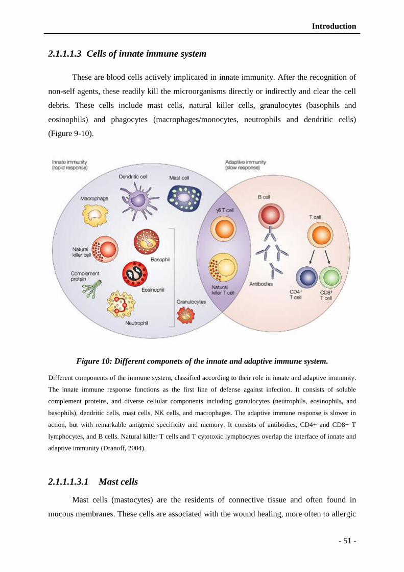
Introduction
- 51 -
2.1.1.1.3 Cells of innate immune system
These are blood cells actively implicated in innate immunity. After the recognition of
non-self agents, these readily kill the microorganisms directly or indirectly and clear the cell
debris. These cells include mast cells, natural killer cells, granulocytes (basophils and
eosinophils) and phagocytes (macrophages/monocytes, neutrophils and dendritic cells)
(Figure 9-10).
Figure 10: Different componets of the innate and adaptive immune system.
Different components of the immune system, classified according to their role in innate and adaptive immunity.
The innate immune response functions as the first line of defense against infection. It consists of soluble
complement proteins, and diverse cellular components including granulocytes (neutrophils, eosinophils, and
basophils), dendritic cells, mast cells, NK cells, and macrophages. The adaptive immune response is slower in
action, but with remarkable antigenic specificity and memory. It consists of antibodies, CD4+ and CD8+ T
lymphocytes, and B cells. Natural killer T cells and T cytotoxic lymphocytes overlap the interface of innate and
adaptive immunity (Dranoff, 2004).
2.1.1.1.3.1 Mast cells
Mast cells (mastocytes) are the residents of connective tissue and often found in
mucous membranes. These cells are associated with the wound healing, more often to allergic
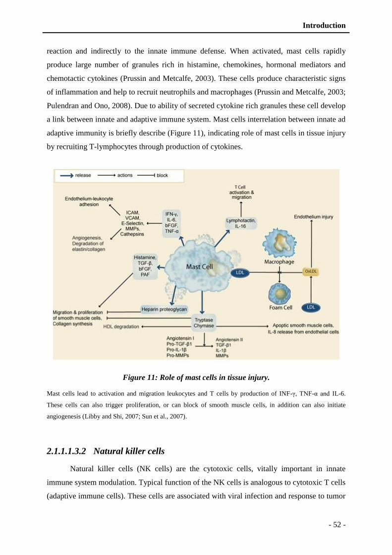
Introduction
- 52 -
reaction and indirectly to the innate immune defense. When activated, mast cells rapidly
produce large number of granules rich in histamine, chemokines, hormonal mediators and
chemotactic cytokines (Prussin and Metcalfe, 2003). These cells produce characteristic signs
of inflammation and help to recruit neutrophils and macrophages (Prussin and Metcalfe, 2003;
Pulendran and Ono, 2008). Due to ability of secreted cytokine rich granules these cell develop
a link between innate and adaptive immune system. Mast cells interrelation between innate ad
adaptive immunity is briefly describe (Figure 11), indicating role of mast cells in tissue injury
by recruiting T-lymphocytes through production of cytokines.
Figure 11: Role of mast cells in tissue injury.
Mast cells lead to activation and migration leukocytes and T cells by production of INF-γ, TNF-α and IL-6.
These cells can also trigger proliferation, or can block of smooth muscle cells, in addition can also initiate
angiogenesis (Libby and Shi, 2007; Sun et al., 2007).
2.1.1.1.3.2 Natural killer cells
Natural killer cells (NK cells) are the cytotoxic cells, vitally important in innate
immune system modulation. Typical function of the NK cells is analogous to cytotoxic T cells
(adaptive immune cells). These cells are associated with viral infection and response to tumor
Introduction
- 53 -
formation. NK cells are unique in their function as recognize stressed cells even in the
absence of MHC-1 (major histocompatibility complex-1) (Vivier et al., 2011). Unlike other
innate immune cells, NK cells belong to the lymphoid lineage rather than myeloid lineage.
NK cells can differentiate into natural killer T cells. Moreover, these have the ability to adjust
promptly according to the environment, and to formulate antigen-specific immunological
memory (Arina et al., 2007; O'Leary et al., 2006). Various functions are attributed to NK cells
including cytolytic granule mediated apoptosis, antibody dependent cell mediated cytotoxicity
(Smyth et al., 2002), cytokine induced activation by secreting TNF-α and INF-γ (Lodoen and
Lanier, 2005), tumor surveillance (Smyth et al., 2002), and memory maintenance and recall
(Lodoen and Lanier, 2005).
2.1.1.1.3.3 Granulocytes (basophils and eosinophils)
Basophils and eosinophils are the granulocytes of the innate immunity; basophils are
least common blood cells (0.01-0.3%) (Grattan et al., 2003) with characteristic staining by
basic dyes while eosinophils are harbored by acidic dyes. These cells release large amount of
granules rich in chemokines, histamine that modulate different immune cells functions
(Figure 12). Additionally highly toxic proteins and free radicals are also released to kill the
bacteria and parasites. In addition to kill pathogens, these cells are also responsible for tissue
damage that is liable for allergic reactions.
Secretions of basophils are rich in histamine, cytokines, leukotriene C4 (Karasuyama et
al., 2011) and they often bear characteristic receptors that help to bind immunoglobulin (IgE)
(Mukai et al., 2005; Mukai et al., 2009; Schroeder, 2009). While eosinophils secrete cationic
proteins (eosinophil cationic protein), in addition reactive oxygen radicals (superoxide,
peroxide etc.) are also produced that are associated with direct killing of microbes (Dyer et
al., 2010; Saito et al., 2004; Trulson et al., 2007). Eosinophils present various Toll like
receptors including TLR1, TLR4, TLR7, TLR9 and TLR10 (Nagase et al., 2003).
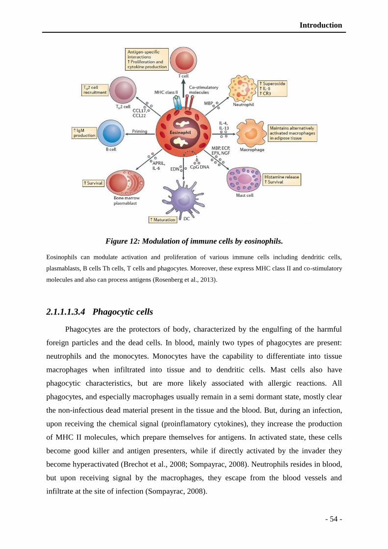
Introduction
- 54 -
Figure 12: Modulation of immune cells by eosinophils.
Eosinophils can modulate activation and proliferation of various immune cells including dendritic cells,
plasmablasts, B cells Th cells, T cells and phagocytes. Moreover, these express MHC class II and co-stimulatory
molecules and also can process antigens (Rosenberg et al., 2013).
2.1.1.1.3.4 Phagocytic cells
Phagocytes are the protectors of body, characterized by the engulfing of the harmful
foreign particles and the dead cells. In blood, mainly two types of phagocytes are present:
neutrophils and the monocytes. Monocytes have the capability to differentiate into tissue
macrophages when infiltrated into tissue and to dendritic cells. Mast cells also have
phagocytic characteristics, but are more likely associated with allergic reactions. All
phagocytes, and especially macrophages usually remain in a semi dormant state, mostly clear
the non-infectious dead material present in the tissue and the blood. But, during an infection,
upon receiving the chemical signal (proinflamatory cytokines), they increase the production
of MHC II molecules, which prepare themselves for antigens. In activated state, these cells
become good killer and antigen presenters, while if directly activated by the invader they
become hyperactivated (Brechot et al., 2008; Sompayrac, 2008). Neutrophils resides in blood,
but upon receiving signal by the macrophages, they escape from the blood vessels and
infiltrate at the site of infection (Sompayrac, 2008).
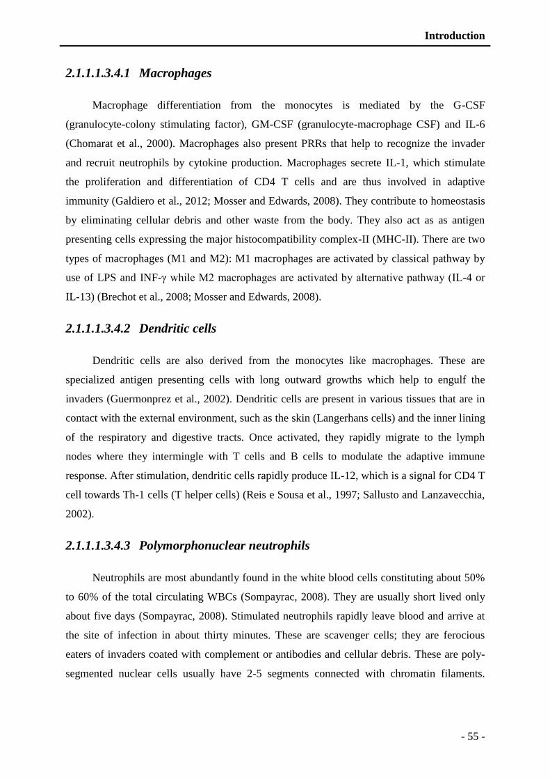
Introduction
- 55 -
2.1.1.1.3.4.1 Macrophages
Macrophage differentiation from the monocytes is mediated by the G-CSF
(granulocyte-colony stimulating factor), GM-CSF (granulocyte-macrophage CSF) and IL-6
(Chomarat et al., 2000). Macrophages also present PRRs that help to recognize the invader
and recruit neutrophils by cytokine production. Macrophages secrete IL-1, which stimulate
the proliferation and differentiation of CD4 T cells and are thus involved in adaptive
immunity (Galdiero et al., 2012; Mosser and Edwards, 2008). They contribute to homeostasis
by eliminating cellular debris and other waste from the body. They also act as as antigen
presenting cells expressing the major histocompatibility complex-II (MHC-II). There are two
types of macrophages (M1 and M2): M1 macrophages are activated by classical pathway by
use of LPS and INF-γ while M2 macrophages are activated by alternative pathway (IL-4 or
IL-13) (Brechot et al., 2008; Mosser and Edwards, 2008).
2.1.1.1.3.4.2 Dendritic cells
Dendritic cells are also derived from the monocytes like macrophages. These are
specialized antigen presenting cells with long outward growths which help to engulf the
invaders (Guermonprez et al., 2002). Dendritic cells are present in various tissues that are in
contact with the external environment, such as the skin (Langerhans cells) and the inner lining
of the respiratory and digestive tracts. Once activated, they rapidly migrate to the lymph
nodes where they intermingle with T cells and B cells to modulate the adaptive immune
response. After stimulation, dendritic cells rapidly produce IL-12, which is a signal for CD4 T
cell towards Th-1 cells (T helper cells) (Reis e Sousa et al., 1997; Sallusto and Lanzavecchia,
2002).
2.1.1.1.3.4.3 Polymorphonuclear neutrophils
Neutrophils are most abundantly found in the white blood cells constituting about 50%
to 60% of the total circulating WBCs (Sompayrac, 2008). They are usually short lived only
about five days (Sompayrac, 2008). Stimulated neutrophils rapidly leave blood and arrive at
the site of infection in about thirty minutes. These are scavenger cells; they are ferocious
eaters of invaders coated with complement or antibodies and cellular debris. These are poly-
segmented nuclear cells usually have 2-5 segments connected with chromatin filaments.
Introduction
- 56 -
Generally PMNs do not leave the bone marrow until maturity, but in case of severe infection
neutrophil precursors (myelocytes and promyelocytes) are released (Linderkamp et al., 1998).
Neutrophils are highly enriched in granules containing bactericidal proteins, reactive
oxygen compounds and protein destroying factors (Soehnlein et al., 2008b). In addition, they
can secrete products that are helpful to stimulate monocytes and macrophages (Soehnlein et
al., 2008b).
By the process of chemotaxis neutrophils migrate towards the infected or inflamed site
which is under the influence of various chemical factors. Cell surface bears several receptors
including cytokine receptors, complement receptors, receptors for chemokines, receptors to
detect and adhere to endothelium, and Fc receptors for opsonin that allow neutrophils to
detect the chemo-attractants such as interferon gamma (INF-γ), interleukin-8 (IL-8),
complement factor C5a, and leukotriene B4 (Segal, 2005).
Neutrophils mainly produce three types of granules: azurophilic/primary granules,
specific/secondary granules, gelatinase/tertiary granules (Borregaard and Cowland, 1997;
Borregaard et al., 1993) (Figure 13). In addition to these granules PMNs also possesses
several secretory vesicles (Figure 13) (Borregaard and Cowland, 1997; Borregaard et al.,
1993; Lominadze et al., 2005).
Figure 13: Human polymorphonuclear neutrophil.
Human polymorphonuclear neutrophil (PMNs), different granules with major characteristic proteins are
demonstrated.
Introduction
- 57 -
Secretory vesicles are produced by endocytosis having membranes rich in receptors,
adhesion proteins and signaling proteins (Borregaard and Cowland, 1997). While the other
three granules contain various host defense proteins (HDPs) and enzymes (Borregaard and
Cowland, 1997; Lominadze et al., 2005). Secretions from the primary granules stimulate the
phagocytosis of IgG-coated pathogens (Soehnlein et al., 2008a). Different granules are
mobilized in a specific manner called graded exocytosis. Gelatinase granules are most easily
mobilized followed by secondary and primary granules (Sengelov et al., 1995). These
granules are highly enriched in antimicrobial peptides such as defensins, cathepsins,
chromogranins, lactoferrins and lysozymes (Borregaard and Cowland, 1997; Faurschou and
Borregaard, 2003; Lominadze et al., 2005).
In addition to direct killing by engulfing and degranulating, neutrophils are capable of
constructing NETs (neutrophil extracellular traps) which is a hallmark of neutrophil function
(Brinkmann et al., 2004). NETs comprise a web composed of chromatin fibers and serine
proteases that trap and kill microbes. NETs provide localized high concentration of various
antimicrobial components secreted by degranulation that facilitates adhesion and killing of
microbes. NETs also serve to localize and prevent further spread of infection by providing a
physical barrier (Clark et al., 2007). In addition NETs also exhibit pro-thrombotic effect
(Borissoff and ten Cate, 2011; Fuchs et al., 2010).
2.1.1.1.4 Complement system
The complement system aids the antibodies and phagocytic cells to clear pathogens
from an organism. It is part of the innate immune system; however it can be recruited by the
adaptive immune system (Abbas and Lichtman, 2005). Components of the complement
system include large number of small proteins, generally synthesized by the liver, and
circulating in blood as inactive precursors. It is triggered by several factors, when stimulated
proteases in the system cleave specific proteins to initiate an amplifying cascade of further
cleavages. The end-result of this cleaving cascade is activation of the cell killing mechanism
by formation of membrane attack complex (Sims and Wiedmer, 2000). Complement system is
composed of more than 25 different proteins and their fragments. Several functions are
attributed to complement system including opsonization, chemotaxis, cell lysis, and clumping
of antigen bearing agents that are as a result of enhancement of phagocytosis, attracting
phagocytic cells and rupturing membrane of foreign invaders respectively (Goldman and
Prabhakar, 1996; Sims and Wiedmer, 2000).
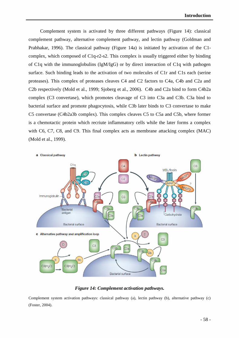
Introduction
- 58 -
Complement system is activated by three different pathways (Figure 14): classical
complement pathway, alternative complement pathway, and lectin pathway (Goldman and
Prabhakar, 1996). The classical pathway (Figure 14a) is initiated by activation of the C1-
complex, which composed of C1q-r2-s2. This complex is usually triggered either by binding
of C1q with the immunoglobulins (IgM/IgG) or by direct interaction of C1q with pathogen
surface. Such binding leads to the activation of two molecules of C1r and C1s each (serine
proteases). This complex of proteases cleaves C4 and C2 factors to C4a, C4b and C2a and
C2b respectively (Mold et al., 1999; Sjoberg et al., 2006). C4b and C2a bind to form C4b2a
complex (C3 convertase), which promotes cleavage of C3 into C3a and C3b. C3a bind to
bacterial surface and promote phagocytosis, while C3b later binds to C3 convertase to make
C5 convertase (C4b2a3b complex). This complex cleaves C5 to C5a and C5b, where former
is a chemotactic protein which recriute inflammatory cells while the later forms a complex
with C6, C7, C8, and C9. This final complex acts as membrane attacking complex (MAC)
(Mold et al., 1999).
Figure 14: Complement activation pathways.
Complement system activation pathways: classical pathway (a), lectin pathway (b), alternative pathway (c)
(Foster, 2004).
Introduction
- 59 -
The lectin pathway (Figure 14b) is very much homologous to the classical pathway, that
is triggered by mannose-binding lectin (MBL), opsonin, and ficolins, instead of C1q. Binding
of MBL to mannose residues on the pathogen surface, stimulates the MBL-associated serine
proteases (MASP-1, and MASP-2) (Degn et al., 2009; Wallis et al., 2010). These serine
proteases can split C4 and C2 into C4a, C4b and C2a, C2b respectively (Sjoberg et al., 2006).
Remaining pathway is same as described for the classical complement activation pathway
(Mold et al., 1999).
The alternative pathway (Figure 14c) does not require binded antibodies and is
continuously activated due to spontaneous C3 hydrolysis. The surface-bound C3b generated
by cleavage of C3, binds to factor B to form C3bB (Conrad et al., 1978). Another factor D
promotes the cleavage of B into Ba and Bb. C3b remain attached to Bb and this complex
C3bBb (C3 convertase) complex is stabilized by binding of factor P. The complex C3bBbP
can further intiate cleavage of C3 and this newly-bound C3b recruits more B, D and P factors
and greatly amplifies the complement activation (McRae et al., 2005; Pangburn et al., 1977).
Complement regulatory proteins such as CD35, CD46, CD55 and CD59 limit the alternative
pathway activation. While pathogens do not have such complement regulatory proteins. Thus,
the alternative complement pathway is able to distinguish self from non-self. Once the
alternative C3 convertase enzyme is developed, it can bind to other C3b factor and form a
complex C3bBbC3bP that act as C5 convertase.
2.1.1.1.5 Cytokines
Cytokines are soluble proteins that are modulators of immune response and
inflammatory reaction. These develop a link between innate and adaptive immune cells.
These are secreted by various immune cells, such as activated macrophages and neutrophils
by presence of microbes. They are also produced by T lymphocytes, supplementing adaptive
immunity (Lacy and Stow, 2011). An illustration of few cytokines with their role in innate
immunity is given below (Table V).
Cytokines have been classed as interleukins (IL) chemokines, and lymphokines, based
on their presumed function, cell of secretion, or target of action. Function based classification
is more common now a days in research and clinical point of view which includes pro-
inflammatory and anti-inflammatory cytokines (Abbas and Lichtman, 2005). Prior stimulates
immune cells to secret more while the later works in inverse. This group of mediators
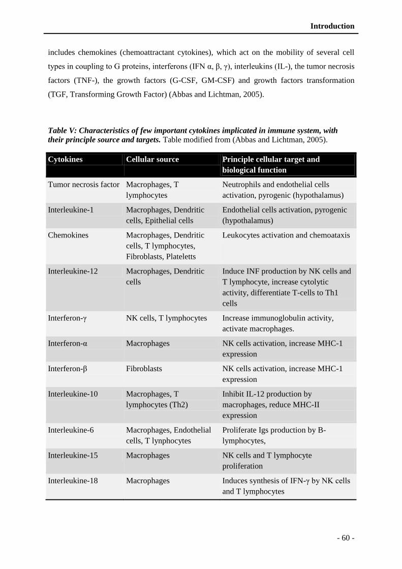
Introduction
- 60 -
includes chemokines (chemoattractant cytokines), which act on the mobility of several cell
types in coupling to G proteins, interferons (IFN α, β, γ), interleukins (IL-), the tumor necrosis
factors (TNF-), the growth factors (G-CSF, GM-CSF) and growth factors transformation
(TGF, Transforming Growth Factor) (Abbas and Lichtman, 2005).
Table V: Characteristics of few important cytokines implicated in immune system, with
their principle source and targets. Table modified from (Abbas and Lichtman, 2005).
Cytokines Cellular source Principle cellular target and
biological function
Tumor necrosis factor Macrophages, T
lymphocytes
Neutrophils and endothelial cells
activation, pyrogenic (hypothalamus)
Interleukine-1 Macrophages, Dendritic
cells, Epithelial cells
Endothelial cells activation, pyrogenic
(hypothalamus)
Chemokines Macrophages, Dendritic
cells, T lymphocytes,
Fibroblasts, Plateletts
Leukocytes activation and chemoataxis
Interleukine-12 Macrophages, Dendritic
cells
Induce INF production by NK cells and
T lymphocyte, increase cytolytic
activity, differentiate T-cells to Th1
cells
Interferon-γ NK cells, T lymphocytes Increase immunoglobulin activity,
activate macrophages.
Interferon-α Macrophages NK cells activation, increase MHC-1
expression
Interferon-β Fibroblasts NK cells activation, increase MHC-1
expression
Interleukine-10 Macrophages, T
lymphocytes (Th2)
Inhibit IL-12 production by
macrophages, reduce MHC-II
expression
Interleukine-6 Macrophages, Endothelial
cells, T lynphocytes
Proliferate Igs production by B-
lymphocytes,
Interleukine-15 Macrophages NK cells and T lymphocyte
proliferation
Interleukine-18 Macrophages Induces synthesis of IFN-γ by NK cells
and T lymphocytes
Introduction
- 61 -
2.1.1.1.6 Antimicrobial peptides
Antimicrobial peptides are the unique component of the innate immune response, that
are highly conserved through evolutionary process (Ganz, 2003). AMPs are also called host
defense peptides (Auvynet and Rosenstein, 2009). These are remarkably appreciable in their
selection of target cells. These peptides are highly effective broad-spectrum antibiotics, which
demonstrate their potential use as novel therapeutic agents. Antimicrobial peptides have been
demonstrated to kill bacteria (including resistant strains), mycobacteria, enveloped viruses,
fungi and even transformed or cancerous cells (Auvynet and Rosenstein, 2009). In addition,
they also have the ability to enhance immunity by functioning as immunomodulators
(Auvynet and Rosenstein, 2009). A detailed description of structure, mode of action and
different classes is provided in the relevant chapter.
2.1.2 Interrelation between immune, nervous and endocrine system.
The hypothalamic-pituitary-adrenal (HPA) axis helps to maintain homeostasis and
participate to reproduction, growth and differentiation. Endocrine informations are controlled
from the central nervous system. Adrenal cortex and adrenal medulla is predominantly
composed of chromaffin cells. These secretory cells contain numerous granules rich in
adrenaline, nor-adrenaline, serotonin, dopamine and simultaneously various proteins having
diverse biological function such as chromogranins (Helle, 2004). Later, the discovery to TLRs
on the adrenal paved the way to further establish the immune-nervous-endocrine triade
(Sternberg, 2006). Stressful situations lead to secretion of AMPs from the endocrine and
immune cells. These actions are well coordinated and regulated by the nervous system via the
endocrine system. The relationship between nervous system and immune system via the
endocrine system are not unidirectional but bidirectional. Several interrelations of neuro-
endocrine immune system are demonstrated in figure 15. Brain controls immune system via
endocrine system and directly by growth hormones and prolactin. Furthermore, several
feedbacks involving of cytokines and autonomic system are also present. Several studies
indicate the interrelation of nervous and immune system; for example an electric stimulus
triggers the release of antibodies by lymphocytes (Liburdy, 1992). Conversely, interlukine-2
lead to changes in bioelectric pulse (Bindoni et al., 1988).
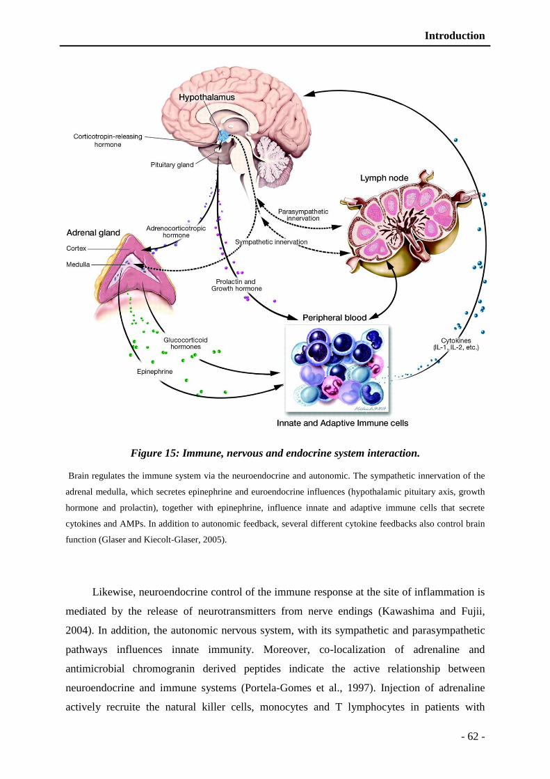
Introduction
- 62 -
Figure 15: Immune, nervous and endocrine system interaction.
Brain regulates the immune system via the neuroendocrine and autonomic. The sympathetic innervation of the
adrenal medulla, which secretes epinephrine and euroendocrine influences (hypothalamic pituitary axis, growth
hormone and prolactin), together with epinephrine, influence innate and adaptive immune cells that secrete
cytokines and AMPs. In addition to autonomic feedback, several different cytokine feedbacks also control brain
function (Glaser and Kiecolt-Glaser, 2005).
Likewise, neuroendocrine control of the immune response at the site of inflammation is
mediated by the release of neurotransmitters from nerve endings (Kawashima and Fujii,
2004). In addition, the autonomic nervous system, with its sympathetic and parasympathetic
pathways influences innate immunity. Moreover, co-localization of adrenaline and
antimicrobial chromogranin derived peptides indicate the active relationship between
neuroendocrine and immune systems (Portela-Gomes et al., 1997). Injection of adrenaline
actively recruite the natural killer cells, monocytes and T lymphocytes in patients with
Introduction
- 63 -
rheumatoid arthritis (Kittner et al., 2002). Similarly, norepinephrine plays a role in the central
neural regulation of the immune system (Besedovsky and del Rey, 1996; Elenkov et al., 2000;
Sanders and Straub, 2002). Likewise, acetylcholine binds macrophages and endothelial cells
through the α7 nicotinic receptor, reducing their activation and secretion of pro-inflammatory
cytokines (Saeed et al., 2005). Other hormones such as CRH (corticotrophin releasing
hormone), the substance P, CGRP (calcitonin gene-regulated peptides) are vigorously
involved in the defense by controlling the pro-inflammatory activity of the innate immune
cells. For example, stress may stimulate CRH production via cytokines secretion such as
TNF-α, IL-1 or IL-6 secretion and finally increases the adrenocorticotropic hormone (ACTH)
and cortisol. Conversely, cortisol inhibits pro-inflammatory cytokine secretion by
macrophages. Substance P favors the release of vasoactive mediators, such as serotonin and
histamine by mast cells (Guhl et al., 2005). Similarly, the β-endorphin and enkephalin inhibit
the production of ACTH. In addition, sex hormones and glucocorticoids are also well
implicated in immunity (Silva et al., 2010). Furthermore, stress and anxiety lead to weakening
of the immune system due to release of adrenaline and prostaglandins that decrease NK cells
activity (Inbar et al., 2011).
Similar to control of immune system by the endocrine system, components of immune
system also regulates it in vice versa. Such as, AMPs modulate immune cell secretions. For
example, the peptides derived from chromogranins such as catestatine has a chemotactic
effect and thus contributes to mobilization of polymorphonuclear the site of inflammation
(Shooshtarizadeh et al., 2010), further detail about the chromogranin derived peptides is
provided in the relevant chapter. Similarly, catestatin and chromofungin trigger neutrophil
secretion via SOC channels (Channels Store Operated) by inducing intracellular calcium
influx (Zhang et al., 2009b).

Antimicrobial peptides

Introduction
- 64 -
3 Antimicrobial peptides
3.1 Introduction
Antimicrobial peptides (AMPs) are small molecules that are integral part of innate
immune defense (Yount et al., 2006; Zasloff, 1992), found in all living organisms including
bacteria, archaea, protists, fungi, plants, animals and humans. These are evolutionary
conserved components, existing through the history of life on earth (Yount et al., 2006).
AMPs are generally 12-50 amino acids long peptides, although several large proteins are also
discovered having antimicrobial properties such as lysozymes, lactoferrin and chromogranin
(Strub et al., 1996a). AMPs are usually positively charged which can vary from +2 to +9
depending on the number of positively charged amino acids (arginine and lysine) (Hancock
and Chapple, 1999). In addition to this positive charge a large hydrophobic portion is also
present (>50%) (Hancock and Chapple, 1999; Yeaman and Yount, 2003; Yount et al., 2006).
Few AMPs are negatively charged having large number of anionic residues such as aspartic
acid or glutamic acid (Hancock and Chapple, 1999; Yeaman and Yount, 2003). Most of the
AMPs are non-structured in solutions but they immediately attain their specific configuration
in membranes or hydrophobic solutions (Hancock and Rozek, 2002). AMPs display a broad
spectrum of activity against viruses, fungi, bacteria, and parasites (Hancock and Sahl, 2006;
Izadpanah and Gallo, 2005). In addition to antimicrobial activities, AMPs also display various
immuno-modulatory activities, such as anticancer activities and immune boosting properties
(chemotactic peptides) (Izadpanah and Gallo, 2005).
AMPs could not get the research focus until late twenth century, which was further
accelerated in the last two decades. The first AMP, lysozyme was described by the Alexander
Flemming in 1922 (Fleming and Allison, 1922), he isolated lysozymes from the secretions
produced against infectious agents. Few early reports are also present about AMPs described
in 1902-1921, but a well-established report about the existence of AMPs was published in
1928 by Ridley (Ridley, 1928). Lysozymes were later used in medication as bacteriolytic
agents in 1964 (Corone, 1964), which initiated the research to discover new AMPs and their
use as potential antimicrobial agents. Later in the year 1930, the franco-american
microbiologist, Rene Dubos discovered gramicidin D (Dubos and Cattaneo, 1939). This AMP
was isolated from Bacillus brevis and is composed of 15 amino acids. It increases cationic
permeability of plasma membrane by pore forming.
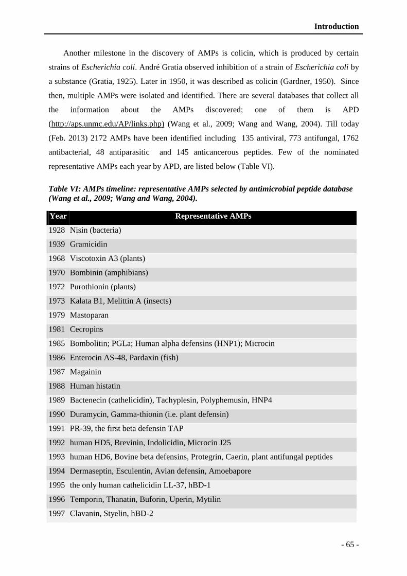
Introduction
- 65 -
Another milestone in the discovery of AMPs is colicin, which is produced by certain
strains of Escherichia coli. André Gratia observed inhibition of a strain of Escherichia coli by
a substance (Gratia, 1925). Later in 1950, it was described as colicin (Gardner, 1950). Since
then, multiple AMPs were isolated and identified. There are several databases that collect all
the information about the AMPs discovered; one of them is APD
(http://aps.unmc.edu/AP/links.php) (Wang et al., 2009; Wang and Wang, 2004). Till today
(Feb. 2013) 2172 AMPs have been identified including 135 antiviral, 773 antifungal, 1762
antibacterial, 48 antiparasitic and 145 anticancerous peptides. Few of the nominated
representative AMPs each year by APD, are listed below (Table VI).
Table VI: AMPs timeline: representative AMPs selected by antimicrobial peptide database
(Wang et al., 2009; Wang and Wang, 2004).
Year Representative AMPs
1928 Nisin (bacteria)
1939 Gramicidin
1968 Viscotoxin A3 (plants)
1970 Bombinin (amphibians)
1972 Purothionin (plants)
1973 Kalata B1, Melittin A (insects)
1979 Mastoparan
1981 Cecropins
1985 Bombolitin; PGLa; Human alpha defensins (HNP1); Microcin
1986 Enterocin AS-48, Pardaxin (fish)
1987 Magainin
1988 Human histatin
1989 Bactenecin (cathelicidin), Tachyplesin, Polyphemusin, HNP4
1990 Duramycin, Gamma-thionin (i.e. plant defensin)
1991 PR-39, the first beta defensin TAP
1992 human HD5, Brevinin, Indolicidin, Microcin J25
1993 human HD6, Bovine beta defensins, Protegrin, Caerin, plant antifungal peptides
1994 Dermaseptin, Esculentin, Avian defensin, Amoebapore
1995 the only human cathelicidin LL-37, hBD-1
1996 Temporin, Thanatin, Buforin, Uperin, Mytilin
1997 Clavanin, Styelin, hBD-2
Introduction
- 66 -
1998 Lactoferrin, Ranaturin, Maculatin, Lycotoxin, Granulysin
1999 Circulin, RTD-1 (Theta-defensin, circular), Cyclotides (Kalata B1, circular)
2000 Aurein, rCRAMP, Palustrin
2001 hBD-3, hBD-4, Ponericin, Dermcidin, Piscidin
2002 human RNase 7; Maximin, Hepcidin, Cupiennin, Retrocyclin, Halocidin
2003 human angiogenins, human chemokine, Alo-1, Circularin A, PhD1, Halocin C8
2004 Arenicin, Brazzein, Ascaphin, Caenacin (Gly-rich)
2005 human psoriasin (S100A7), Plectasin, Phylloseptin
2006 human RNase 8; Latarcin, Cycloviolacin, Lividin
2007 Dybowskin, Pleurain, Hyposin, Odorranain, AvBD, Longicin
2008 human RNase 3; BTD, Vibi
2009 cn-AMP, Kassinatuerin, Lasioglossin, Lichennicidin, Myxinidin
2010 Caenopore-5, Lucifensin, Temporin-SHf, Centrocin, Beta-amyloid peptide, Thuricin
2011 LCI, Glycocin F (class 4 bacteriocin is found), frog cathelicidin-AL, PAM1
2012 Geobacillin I (7 thioether rings), uncyclotide ChaC7 (i.e open chained), Microcin S
(103AA), Slerocin, fungal Eurocin, fungal Micasin
3.2 Structure of antimicrobial peptides
AMPs structural classification and characteristics are typically based on the amino acid
sequences. It has been demonstrated that alteration in the sequence and modification of the
charged amino acids vastly alter activities of peptides. In addition to sequence and nature of
amino acids, secondary and tertiary structures are equally important in physiological
characteristics. Generally four characteristics play important role in AMPs activities: chemical
properties, primary structure, secondary structure and posttranslational modifications
(Izadpanah and Gallo, 2005; Jenssen et al., 2006; Strub et al., 1997).
3.2.1 Chemical properties of the peptides
AMPs are widely isolated from various species including animals, plants and microbes;
such diverse nature gives AMPs a vast variety of physico-chemical characteristics. These
characteristics include polarity and non-polarity. Generally, AMPs possesses a unique balance
of hydrophobic and hydrophilic residues that gives amphiphilic nature to peptides important
for their activity (Helle et al., 2007; Izadpanah and Gallo, 2005; Yount et al., 2006).
Introduction
- 67 -
Polarity of AMPs: Polarity is the first important characteristic of AMPs: there are two
types of AMPs: cationic and anionic AMPs. Cationic antimicrobial peptides are more
common in nature than anionic peptides. Generally, charge varies from +2 to +9 for cationic
AMPs, but can be up to +12 in some cases. Positive charge of the AMPs is important for
interaction with negatively charged microbial membrane. Bacterial surface usually contains
high concentration of LPS (lipopolysaccharides) and techoic acid which gives negative charge
to surface in Gram-negative and Gram-positive bacteria respectively (Madigan and Martinko,
2005). In case of fungi, phosphomannans is important to provide negative charge to surface
(Salzman et al., 2004). In addition to surface proteins, microbial membrane is also rich in
phospholipids, such as phosphatidylcholines which facilitate AMPs insertion deep into
membrane (Yount et al., 2006).
While anionic AMPs are rich in glutamic acid and aspartic acid residues that denotes
negative charge. These are more abundantly found in respiratory system of mammals.
Anionic peptides generally require zinc co-factors to present antibacterial activities (Fales-
Williams et al., 2002). Studies on anionic AMPs dermicidin and enkelytin indicate that
negative charge and amphiphilic characteristics are important for their antimicrobial activities
(Goumon et al., 1998; Kieffer et al., 1998). In addition to electrostatic interaction between
peptide and membrane, polarity also provides a unique orientation to peptide that facilitates
penetration to bacterial membrane (Dennison et al., 2006; Lins and Brasseur, 2008).
Amphiphilic nature of AMPs: A unique balance of hydrophilic and lipophilic residues
provides amphiphilic character to AMPs. Most of the AMPs are amphipathic or they became
amphipathic, when came across anisotropic medium (Yount et al., 2006). It has been
demonstrated that alteration in this balance can change the properties of AMPs. By increasing
amphiphilic character of synthetic peptides their effectiveness can be increased (Jin et al.,
2005). Generally, AMPs contain about 50% hydrophobic residues that facilitate insertion and
rests are hydrophilic (Helle et al., 2007; Jin et al., 2005; Yount et al., 2006). This dual nature
provides the capability to solubilize both in hydrophilic and lipophilic solvents. However,
excessive increase in the hydrophobicity can rapidly decrease their activity but can increase
toxicity to mammalian cells (Zelezetsky et al., 2005).
3.2.2 Primary structure of AMPs
Antimicrobial activity is determined by the amino acids composition of AMPs. Lysine,
arginine, and to a lesser extent, histidine, are likely to provide a positive charge to peptide
Introduction
- 68 -
(cationic), while aspartic acid and glutamic acid are responsible for negative charge.
Additionally, tryptophan, tyrosine, phenylalanine, leucine, isoleucine and methionine are
responsible for hydrophobicity of AMPs. Alteration in amino acids sequence can profoundly
change their antimicrobial activity, such as two tryptophan residues are essential for the
activity of lactoferrin (Strom et al., 2002). Similarly, addition of few amino acids can increase
the activity of AMPs by facilitating insertion into bacterial membrane such as tryptophan end
tags (Pasupuleti et al., 2009a; Pasupuleti et al., 2009b; Schmidtchen et al., 2009).
3.2.3 Secondary structure of AMPs
Secondary structures can widely alter activity of AMPs. General characteristics found in
AMPs are α-helices, β-sheets, and extended coils (Figure 16). However, AMPs mostly
possesses several mixed structures, which provide an extended spectrum of activity. In
general, secondary structures are observed only when the peptide interacts with membranes
(Jenssen et al., 2006; Yount et al., 2006). For example indolicidin is mostly unstructured in
aqueous solutions but it adopts a boat like conformation in the presence of lipid membranes
(Hsu and Yip, 2007). A cathelicidin derived peptide tritrpticin is unstructured but in organic
solvents such as dodecylphosphocholine micelles it adopts turn-turn amphipathic structure
(Schibli et al., 2006).
3.2.4 Posttranslational modifications
In addition to structural and physico-chemical characteristics, posttranslational
modifications also play important role in activity spectrum of AMPs. These modifications
include phosphorylation, glycosylation, and ubiquitination. As, in case of enkelytine (AMP
derived from proenkephalin) phosphorylation is essential for antimicrobial activity of the
peptide (Goumon et al., 2000). Similarly, phosphorylations and glycosylations are important
for chromacin (chromogranin A derived peptide) (Strub et al., 1996a).
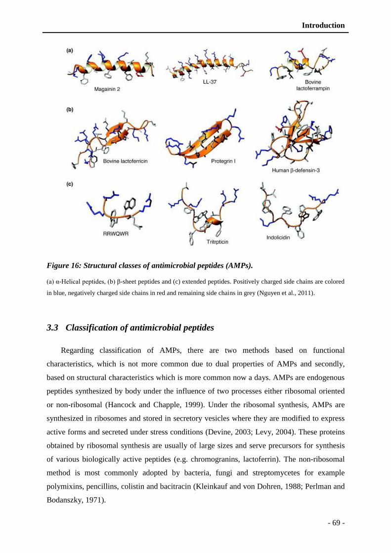
Introduction
- 69 -
Figure 16: Structural classes of antimicrobial peptides (AMPs).
(a) α-Helical peptides, (b) β-sheet peptides and (c) extended peptides. Positively charged side chains are colored
in blue, negatively charged side chains in red and remaining side chains in grey (Nguyen et al., 2011).
3.3 Classification of antimicrobial peptides
Regarding classification of AMPs, there are two methods based on functional
characteristics, which is not more common due to dual properties of AMPs and secondly,
based on structural characteristics which is more common now a days. AMPs are endogenous
peptides synthesized by body under the influence of two processes either ribosomal oriented
or non-ribosomal (Hancock and Chapple, 1999). Under the ribosomal synthesis, AMPs are
synthesized in ribosomes and stored in secretory vesicles where they are modified to express
active forms and secreted under stress conditions (Devine, 2003; Levy, 2004). These proteins
obtained by ribosomal synthesis are usually of large sizes and serve precursors for synthesis
of various biologically active peptides (e.g. chromogranins, lactoferrin). The non-ribosomal
method is most commonly adopted by bacteria, fungi and streptomycetes for example
polymixins, pencillins, colistin and bacitracin (Kleinkauf and von Dohren, 1988; Perlman and
Bodanszky, 1971).

Introduction
- 70 -
AMPs are posttranslationaly modified in secretory vesicles, which helps to protect
peptides from degradation. These modifications include amidation, phosphorylation,
glycosylation (Bulet et al., 1999; Goumon et al., 1998; Strub et al., 1997) that are helpful in
ultimate structural modifications in their active form. Based on active form generation and
structural characteristics AMPs are divided into following categories (Table VII). Generally,
based on the physic-chemical characteristics AMPs are divided into five classes.
3.3.1 Cationic AMPs rich in specific amino acids
These types of AMPs are very vast in number having a single or multiple amino acids
high in number. These amino acids include proline (abaecin), arginine (bactericin),
phenylalanine (prophenin), glycine (hymenotaecin), histadin (histatin) and tryptophan
(indolicidin). These can be linear in structure or can adopt different conformations. Generally
these have less than 40 residues, out of which 40-60% are hydrophobic residues (Tossi et al.,
2000). This category of AMPs include a large number of peptides rich in the amino acids such
as proline (abacin), trptophan (indolicidin), glycine (hymenotaecin), histidin (histatin),
arginine (bactenicin), phenylalanine (prophenin), or few other amino acids (Baumann et al.,
2010; Brogden, 2005; Bulet et al., 1999; Kichler et al., 2006; Scocchi et al., 2011; Yu et al.,
2010).
3.3.2 Cationic AMPs lacking cysteine
Cationic AMPs lacking cysteine are not very common in nature. These lack specifically
cysteine amino acid, which limit their ability to form disulfide bond. Moreover, these strictly
follow the conformation of α helices. These also possess 40-60% hydrophobic residues as
previously described. Examples include cercopins, LL37, and magainin (Boman, 1991; Zaiou
et al., 2003; Zasloff, 1987).
3.3.3 Anionic AMPs
These peptides are rich in aspartic acid or glutamic acid, which provide negative charge
to peptides. These peptides are more commonly found in the respiratory system of the
mammals, where they act as surfactants. Few examples include scygonadine, Maximin H5,
and enkelytine (Goumon et al., 1998; Lai et al., 2002; Wang et al., 2007). The mode of action
of these peptides is not clear few studies indicate their role as surfactants while others suggest
that they act through a structure orientation by oblique helical shape (Dennison et al., 2006).

Introduction
- 71 -
Table VII: Different types of antimicrobial peptides based on their structural
characteristics with their characteristics and few prominent examples of the domain.
Cationic AMPs rich in specific amino acids
Rich in proline, arginine, phenylalanine,
glycine, tryptophan
abaecin, apidaecins, prophenin, indolicidin,
drosocine, pyrrhocoricine, bactenicines,
PR39, prophenine, hymenotaecine, histatin,
coleoptericine, holotricine
Cationic AMPs lacking cysteine
Lack in cysteine
Linear α-helix in structure
Andropin, moricin, ceratotoxin cecropins,
melittin, bombinin, magainin, dermaseptin,
esculentins, brevinin-1, buforin II, CAP18,
LL37, BMAP, SMAP
Anionic AMPs
Rich in glutamic and aspartic acid Maximin H5, scygonadine, dermcidin,
enkelytin
Cationic/anionic AMPs having disulfide bond
Having cysteine and 1-3 disulfide bonds brevinins, protegrin, tachyplesins, defensins,
drosomycin, SPAG11, cryptidine
Cationic/anionic peptides derived from the large proteins
Shorter fragments derived from the large
proteins
Lactoferricin, casocidin, lysozyme, catestatin
antimicrobial domains of lactabumine,
hemoglobin, ovalbumine
3.3.4 Cationic/anionic AMPs having disulfide bond
AMPs rich in cysteine residues are usually cyclic peptides with antiparallel β sheets that
are stabilized by disulfide bridges. These cationic peptides may contain between 2 and 8
cysteine residues that can bind to form disulfide bonds and adopt a specific structures such as
"hairpin". The main representative of this class of peptides is defensin (Dimarcq et al., 1998;
Dimarcq et al., 1990; Lehrer and Ganz, 1996).
3.3.5 Cationic/anionic peptides derived from the large proteins
AMPs generated from large proteins are obtained by proteolysis carried out either by host
proteases or bacterial proteases. Few examples of the AMPs belong to this category includes:
peptides derived from granins (Briolat et al., 2005; Lugardon et al., 2001; Strub et al., 1995;
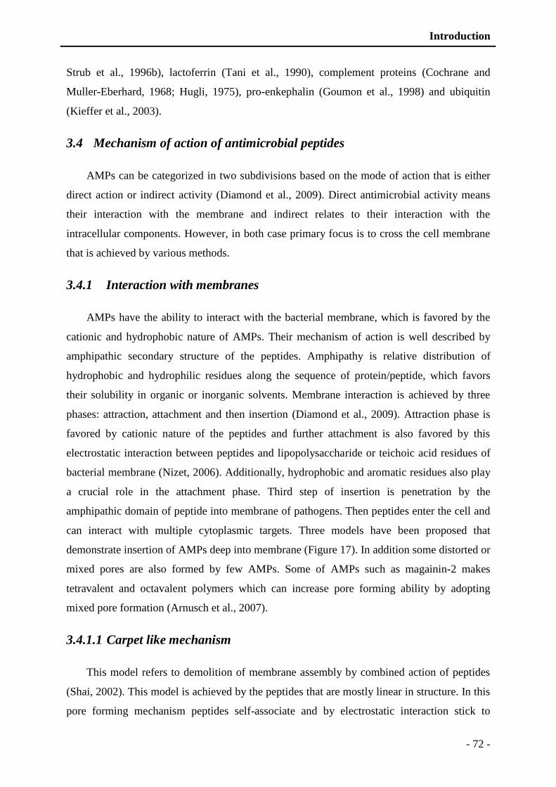
Introduction
- 72 -
Strub et al., 1996b), lactoferrin (Tani et al., 1990), complement proteins (Cochrane and
Muller-Eberhard, 1968; Hugli, 1975), pro-enkephalin (Goumon et al., 1998) and ubiquitin
(Kieffer et al., 2003).
3.4 Mechanism of action of antimicrobial peptides
AMPs can be categorized in two subdivisions based on the mode of action that is either
direct action or indirect activity (Diamond et al., 2009). Direct antimicrobial activity means
their interaction with the membrane and indirect relates to their interaction with the
intracellular components. However, in both case primary focus is to cross the cell membrane
that is achieved by various methods.
3.4.1 Interaction with membranes
AMPs have the ability to interact with the bacterial membrane, which is favored by the
cationic and hydrophobic nature of AMPs. Their mechanism of action is well described by
amphipathic secondary structure of the peptides. Amphipathy is relative distribution of
hydrophobic and hydrophilic residues along the sequence of protein/peptide, which favors
their solubility in organic or inorganic solvents. Membrane interaction is achieved by three
phases: attraction, attachment and then insertion (Diamond et al., 2009). Attraction phase is
favored by cationic nature of the peptides and further attachment is also favored by this
electrostatic interaction between peptides and lipopolysaccharide or teichoic acid residues of
bacterial membrane (Nizet, 2006). Additionally, hydrophobic and aromatic residues also play
a crucial role in the attachment phase. Third step of insertion is penetration by the
amphipathic domain of peptide into membrane of pathogens. Then peptides enter the cell and
can interact with multiple cytoplasmic targets. Three models have been proposed that
demonstrate insertion of AMPs deep into membrane (Figure 17). In addition some distorted or
mixed pores are also formed by few AMPs. Some of AMPs such as magainin-2 makes
tetravalent and octavalent polymers which can increase pore forming ability by adopting
mixed pore formation (Arnusch et al., 2007).
3.4.1.1 Carpet like mechanism
This model refers to demolition of membrane assembly by combined action of peptides
(Shai, 2002). This model is achieved by the peptides that are mostly linear in structure. In this
pore forming mechanism peptides self-associate and by electrostatic interaction stick to

Introduction
- 73 -
bacterial membrane just like beta barrel antiparallel sheets. Peptide accumulation is
specifically onto the acidic phospholipid-rich regions of lipid bilayers. This carpet structure
continues to grow until a certain threshold level is reached which lead to permeation of
membrane by destabilizing (Figure 17). AMPs rapidly permeate into the membrane after
destabilization by mounting the bilayer positive potential (Li et al., 2006).
3.4.1.2 Barrel stave model
In this model, AMPs form clusters in hydrophobic regions of the membranes and creates
pore by formation of transmembrane bundles (Yang et al., 2001). Initial steps are same as
attraction and attachment which in followed by insertion of peptides perpendicular to the
membrane axis. Peptides bind to the membrane and spread their hydrophilic heads towards
phospholipids, and then they enter their pin hydrophobic heads by pushing the phospholipids.
Peptides assemble themselves to carbon chains phospholipids leaving them to free their
hydrophilic domains that form pores (Bechinger, 1999; Nguyen et al., 2011; Yang et al.,
2001) (Figure 17). One typical example following this mode of action is alamethicin which
was isolated from fungus Trichoderma viride (Bechinger, 1999; North et al., 1995).
3.4.1.3 Toroidal model
In this model, AMPs builds toroidal pores in lipid bilayers. The lipid polar head and helix
bundles that orient vertically to the membrane to achieve pore construction. AMPs aggregate
and induce the lipid monolayers to bend continuously through the pores so that both the
inserted peptides and the lipid head groups line towards water core (Brogden, 2005). Toroidal
pores act as channels for ions or metabolites, and eventually preventing bacteria from
maintaining the homeostasis (Boland and Separovic, 2006) (Figure 17). This model of action
is adopted mostly by α-helical peptides such as magainin (Matsuzaki et al., 1996), melittin
(Lee et al., 2004b), LL-37 (Brogden, 2005; Henzler-Wildman et al., 2004), protegrin-1
(Yamaguchi et al., 2002), 2002).
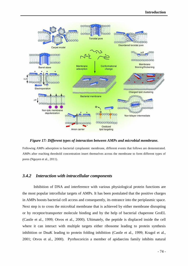
Introduction
- 74 -
Figure 17: Different types of interaction between AMPs and microbial membrane.
Following AMPs adsorption to bacterial cytoplasmic membrane, different events that follows are demonstrated.
AMPs after reaching threshold concentration insert themselves across the membrane to form different types of
pores (Nguyen et al., 2011).
3.4.2 Interaction with intracellular components
Inhibition of DNA and interference with various physiological protein functions are
the most popular intrcellular targets of AMPs. It has been postulated that the positive charges
in AMPs boosts bacterial cell access and consequently, its entrance into the periplasmic space.
Next step is to cross the microbial membrane that is achieved by either membrane disrupting
or by receptor/transporter molecule binding and by the help of bacterial chaperone GroEL
(Castle et al., 1999; Otvos et al., 2000). Ultimately, the peptide is displaced inside the cell
where it can interact with multiple targets either ribosome leading to protein synthesis
inhibition or DnaK leading to protein folding inhibition (Castle et al., 1999; Kragol et al.,
2001; Otvos et al., 2000). Pyrrhocoricin a member of apidaecins family inhibits natural

Introduction
- 75 -
ATPase activity in E. coli (Kragol et al., 2001). In contrast, PR-39 kills bacteria by interfering
with DNA and ultimately blocking protein synthesis (Boman et al., 1993). Interestingly, PR-
39 can hamper NADPH-dependent redox reactions by production of syndecans (Shi et al.,
1996). In addition to ribosomal and DNA interference, AMPs have various other target sites
including enzymatic activity and cell wall synthesis inhibition (Brogden et al., 2003; Jenssen
et al., 2006).
3.5 Biological activities of antimicrobial peptides
AMPs are widely associated with host defense either by exerting direct antimicrobial
activities or by modulating immune cell’s function (Hancock and Sahl, 2006). Their spectrum
of activity is so much diverse that they are often termed as host defense peptides (HDPs)
rather than AMPs (Yeung et al., 2011; Yu et al., 2007).
3.5.1 Direct antimicrobial activity
AMPs can directly target and destroy microbes and infected cells. This direct action
involves destabilization of microbial membrane as previously described in section 3.4 of this
chapter. Based on this type of interaction these can be either antimicrobial or anticancerous.
Antimicrobial nature of these peptides is attributed to negatively charged membranes of
bacteria, fungi, viruses and parasites. Similar to this mode of action, AMPs can differentiate
between healthy normal cells and cancer cells, due to selective presentation of negative
charge on the surface. This selective presentation of negative charge on the surface is due to
demonstration of some anionic molecules on the surface such as phosphatidylserine
(Dobrzynska et al., 2004; Utsugi et al., 1991). Sometimes this recognition is potentiated by
selective demonstration of O-glycosylated saccharide components (Burdick et al., 1997; Yoon
et al., 1997). In addition to selective presentation, cancer cells grow larger than the normal
cells with remarkably distorted orientation that renders them more prone to AMPs (Chan et
al., 1998; Hoskin and Ramamoorthy, 2008; Schweizer, 2009).
AMPs are also effective against the DNA or RNA viruses by interacting to heparin
sulfate that is required for viral entry into cells, for example lactoferricin and rabbit alpha
defensin NP-1 (Jenssen et al., 2006; Sinha et al., 2003). Some peptides, such as NK-18 can
simultaneously destabilize microbial membrane and interact with microbial genome,
ultimately potentiating antimicrobial activity (Yan et al., 2013). AMPs can also interact with
multiple targets to block various biological processes and ultimately inhibiting microbial
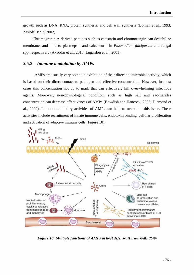
Introduction
- 76 -
growth such as DNA, RNA, protein synthesis, and cell wall synthesis (Boman et al., 1993;
Zasloff, 1992, 2002).
Chromogranin A derived peptides such as catestatin and chromofungin can destabilize
membrane, and bind to plasmepsin and calceneurin in Plasmodium falciparum and fungal
spp. respectively (Akaddar et al., 2010; Lugardon et al., 2001).
3.5.2 Immune modulation by AMPs
AMPs are usually very potent in exhibition of their direct antimicrobial activity, which
is based on their direct contact to pathogen and effective concentration. However, in most
cases this concentration not up to mark that can effectively kill overwhelming infectious
agents. Moreover, non-physiological condition, such as high salt and saccharides
concentration can decrease effectiveness of AMPs (Bowdish and Hancock, 2005; Diamond et
al., 2009). Immunomodulatory activities of AMPs can help to overcome this issue. These
activities include recruitment of innate immune cells, endotoxin binding, cellular proliferation
and activation of adaptive immune cells (Figure 18).
Figure 18: Multiple functions of AMPs in host defense. (Lai and Gallo, 2009)
Introduction
- 77 -
3.5.2.1 Recruitment of innate immune cells
AMPs are able to trigger selective production of pro-inflammatory and anti-
inflammatory cytokines and ultimately leukocyte chemotaxis. For example, LL-37 attracts
neutrophils, monocytes, T cells, and mast cells; by production of G-protein coupled receptors
and N-formyl peptide like receptors that induce selective degranulation of phagocytes (De et
al., 2000; Mookherjee and Hancock, 2007). LL-37 can also act synergistically with IL-1β, to
enhance production of proinflammatory cytokines such as IL-6, IL-10, MCP-1, and MCP-3
(Yu et al., 2007). Catestatin, a chromogranin-A derived peptide has been demonstrated to
attract neutrophils in vitro (Shooshtarizadeh et al., 2010). Defensins can also interact with
MIP-3a receptor present on the inflammatory cells and regulate inflammatory response
(Mookherjee and Hancock, 2007). Another AMP, IDR1002 (innate defense regulator) can
modulate production of IL-1β by the inflammatory cells (Turner-Brannen et al., 2011).
3.5.2.2 Endotoxin binding
During infection a large amount of endotoxins are produced, which can initiate
inflammatory response. Inflammation is beneficial as well as life threatening when exceeded
by a certain limit (Mookherjee and Hancock, 2007). AMPs such as LL-37, indolicidin and
cecropins, can modulate inflammatory response by binding to endotoxins and protect from
endotoxemia (Bowdish and Hancock, 2005; Mookherjee et al., 2006). Furthermore, AMPs
can inhibit expression of proinflammatory genes induced by endotoxin, such as LL-37 which
can block TNF-alpha production mediated by endotoxins (Bowdish and Hancock, 2005;
Mookherjee et al., 2006).
3.5.2.3 AMPs induce cellular proliferation and differentiation
AMPs can also modulate proliferation and differentiation of phagocytic cells, such as
neutrophils by inhibiting apoptosis, activating mast cells, wound healing and promoting
angiogenesis (Chen et al., 2006; Koczulla et al., 2003; Nagaoka et al., 2006). AMPs, such as
LL-37, can also improve expression of CDs by innate immune cells and assist in antigen
presentation (Mookherjee and Hancock, 2007; Tokumaru et al., 2005). Cathelicidin and
neutrophil β-defensins can activate MAPK-p38 (Mitogen-Activated Protein Kinase) and
ERK-1/2 (Extracellular signal-Regulated Kinase) pathways in mast cells, keratinocytes and
monocytes (Bowdish et al., 2004; Chen et al., 2006; Niyonsaba et al., 2006). Furthermore,

Introduction
- 78 -
AMPs can modulate transcription by regulation of STAT-3 (signal transducer and activator of
transcription) (Tokumaru et al., 2005).
3.5.2.4 Adaptive immune cells activation
Besides the innate immune response modulation, AMPs can activate adaptive immune
cells (Mookherjee and Hancock, 2007). Different AMPs such as human neutrophil defensins,
β-defensins, cathelicidin (PR-39 and LL-37) can express CDs receptor and ultimately T and B
lymphocyte activation (Chertov et al., 1996; Huang et al., 1997). Furthermore, LL-37 can also
modulate dendritic cells differentiation and dendritic cells induced T-cells polarization
(Davidson et al., 2004). Defensins can also increase proliferation of T cell induced by IFN-g,
IL-10 and IL-6 (Lillard et al., 1999a; Lillard et al., 1999b).
3.6 Antimicrobial peptide avoidance mechanisms adopted by microorganisms
Microorganisms have adopted several mechanisms to disable antimicrobial host
defense. Most of them are extra-cellular in origin including production of capsules,
production of components that can bind and trap AMPs, degradation of AMPs, neutralization
of charge, modification of cell surface proteins, change in membrane fluidity and expulsion of
AMPs (Figure 19) (Gruenheid and Le Moual, 2012; Koprivnjak and Peschel, 2011).
Few resistant pathogens such as S. aureus, produces several proteins such as
staphylokinase and SIC (Streptococcal inhibitor of complement) that can bind and trap the
AMPs including α-defensin, CAMP (cathelicidin antimicrobial peptide) (Jin et al., 2004).
Neutralization of charge is carried out either production of capsules or various
modifications of surface proteins including D-alanylation of teichoic acid. Most of bacteria
produce cationic exopolymer polysaccharide intercellular adhesin (PIA) and anionic poly-c-
glutamic acid (PGA), which can modulate surface charge and ultimately resist in expulsion of
both cationic and anionic AMPs (Kocianova et al., 2005; Vuong et al., 2004a; Vuong et al.,
2004b). Due to protection polysaccharide capsules, both cationic and anionic AMPs are
expelled from bacterial surface by both electrostatic and mechanical interaction.

Introduction
- 79 -
Figure 19: Different mechanism adopted by the pathogens to avoid antimicrobial peptides.
The most common mechanisms of resistance triggered by pathogens to decrease their susceptibility to AMPs are
described (Teixeira et al., 2012).
In addition, bacteria such as Salmonella typhimurium, which can sense cationic AMPs
and can alters Lipid A by acylation, carry out few modifications. This type of modification
can ultimately reduce the membrane fluidity and permeability (Guina et al., 2000; Guo et al.,
1997; Miller et al., 1990).
Furthermore, AMPs can be degraded by the bacterial proteases, such aureolysin and
Glu-C in case of S. aureus. AMPs degradation ultimately renders them inactive or can
decrease the effectiveness. For example, as previously demonstrated LL-37 is degraded by
aureolysin, a metallo-protease produced by S. aureus (Sieprawska-Lupa et al., 2004).

granins

Introduction
- 80 -
4 granins
Granin is a family of acidic proteins that are ubiquitous in various cells and tissues of
body. Most common sites include chromaffin cells, endocrine glands, nervous cells, and
immune cells (Eiden, 1987; Huttner et al., 1991a; Huttner et al., 1991b). These proteins are
acid soluble and mostly heat stable in nature, have diverse biological functions. Members of
granin family consist of single-polypeptide chains of about 180-700 residues, bearing a signal
peptide at amino terminal. Granins are widely distributed in secretory granules of chromaffin
cells (adrenal medulla), which is most abundant reservoir of chromogranins/secretogranins.
Before to present different members of granin family and their physiological functions, let’s
have a look on the brief introduction of the adrenal glands and chromaffin cells.
4.1 Adrenal glands
Adrenal glands are one of the most important paired glands present in mammals. These
are triangular in shape and are present on the upper edge of both kidneys. They are located at
12th thoracic vertebra and weigh about 4-10 g. Adrenal glands are composed of two parts:
cortex and medulla.
Adrenal cortex is peripheral portion of adrenal gland that represents about 90% of the
adrenal gland. Adrenal cortex usually mediates stress response via production of
glucocorticoids (cortisol), mineralocorticoids (aldosterone) and androgens. Adrenal medulla is
inner part of adrenal gland that is surrounded by adrenal cortex in parametric fashion. Adrenal
medulla is originated from the embryonic neural crest and is innervated by the splanchnic
nerve. It is reservoir of large number of secretory cells such as chromaffin cells. Chromaffin
cell secretions are highly enriched in catecholamines (noradrenaline and adrenaline),
dopamine, proteolytic enzymes (prohormone convertases like) (Seidah and Chretien, 1999),
morphine and certain proteins like proenkephalin-1, chromogranins and many derived
peptides (Goumon et al., 1996; Metz-Boutigue et al., 1998; Taupenot et al., 2003).
4.2 Chromaffin cells
Chromaffin cells are the most important secretory cells of adrenal medulla that are
densely packed with secretory granules. These cells have high affinity towards chromium
salts, which gives dark coloration to them that is why named as chromaffin cells (Ogata and
Ogata, 1917). Preganglionic sympathetic nerves innervate chromaffin cells. These are rich in
Introduction
- 81 -
secretory granules and provide a research model for analysis of molecular mechanisms of
exocytosis (Aunis, 1998).
Secretory granules of chromaffin cells are storage vesicles of catecholamines (Blaschko
and Welch, 1953; Hillarp, 1958; Hillarp and Hokfelt, 1953) and many other secretory
proteins. Proteomic analysis of the secretory vesicles revealed different stress related
components, including catecholamines, granins, cathelicidins (Wegrzyn et al., 2007).
Chromaffin secretory granules highly enriched in different chromogranin/secretogranin
family members. Recent proteomic analysis revealed eight out of nine members of granin
family (Wegrzyn et al., 2007), which constitutes a major portion of chromaffin secretory
granules. These secretory granules contain a vast variety of prohormone precursors such as
chromogranins, cathepsin L, neurotransmitters and various other proteins categorized as
enzymes/proteases (tyrosine-3-hydroxylase, prohormone convertase 1/2, and cystatin C),
signal transduction proteins (RAS/Rho proteins and annexins), redox factors (cytochrome C)
(Wegrzyn et al., 2007). The proteins present in the secretory granules usually act paracrine or
autocrine upon secretion. Granule secretion is achieved by exocytosis or through a mechanism
of Kiss and Run (Aunis, 1998; Henkel et al., 2001).
4.3 Granin family
Soluble proteins of secretory granules of chromaffin cells were found in 1958 (Hillarp,
1958). First member of granin family, CgA (chromogranin A) was identified in 1965 that is
co-secreted with catecholamine (Banks and Helle, 1965; Blaschko et al., 1967). Later, other
proteins have also been discovered and are integrated into granin family. Up-till now, nine
members have been discovered (Bartolomucci et al., 2011; Chakraborty et al., 2006; Helle,
2004) that are divided into two groups: chromogranins (Cgs) and secretogranins (Sgs). The
Cgs are acidic soluble proteins having multiple di- and tri-basic sites that are potential
proteolytic cleavage sites, which can generate several biologicaly active short fragments
(Metz-Boutigue et al., 1993). CgA, CgB/SgI and SgII constitute about 40%, 10%, and 2%
respectively, of total protein constituents of bovine chromaffin cell secretions (Fischer-
Colbrie and Schober, 1987; Wegrzyn et al., 2007). Relative proportion of different Cgs vary
with stress condition and also with species: CgA is more dominant in bovine, sheep and
horses but CgB is more in human and rat (Fischer-Colbrie and Schober, 1987). Different
members of granin family of human origin, are presented in Table VIII with their number of
residues, molecular weight and pI index.
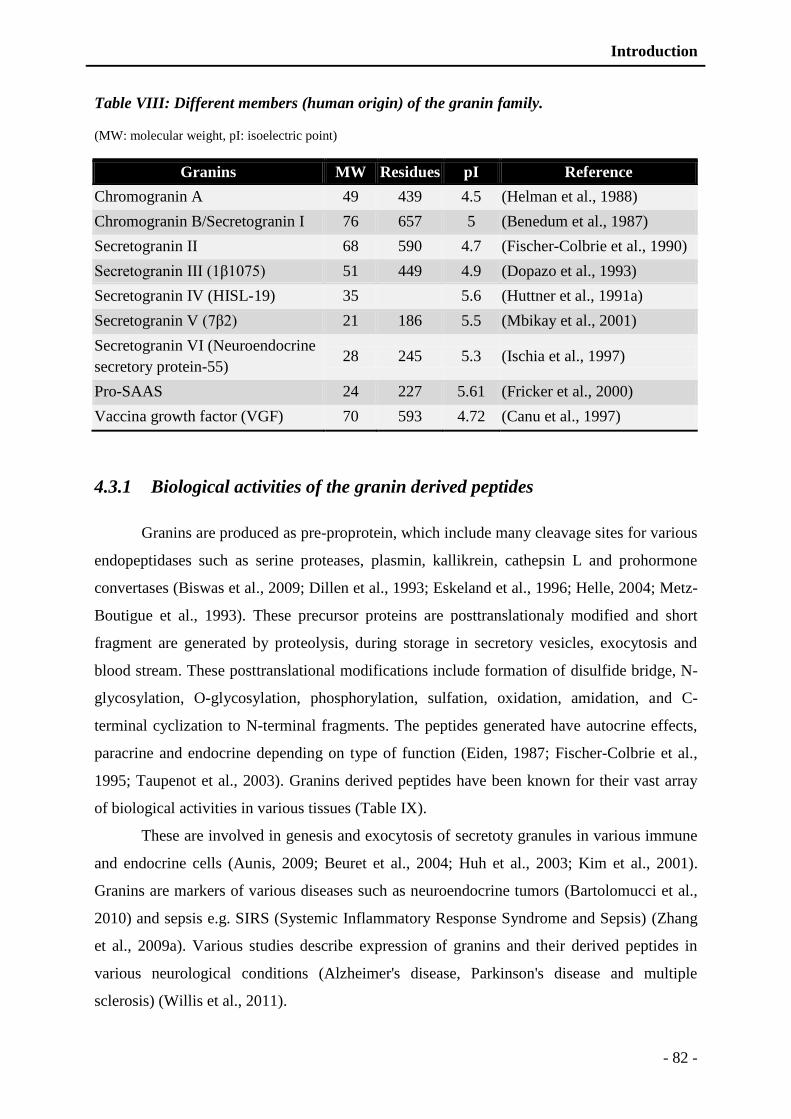
Introduction
- 82 -
Table VIII: Different members (human origin) of the granin family.
(MW: molecular weight, pI: isoelectric point)
Granins MW Residues pI Reference
Chromogranin A 49 439 4.5 (Helman et al., 1988)
Chromogranin B/Secretogranin I 76 657 5 (Benedum et al., 1987)
Secretogranin II 68 590 4.7 (Fischer-Colbrie et al., 1990)
Secretogranin III (1β1075) 51 449 4.9 (Dopazo et al., 1993)
Secretogranin IV (HISL-19) 35 5.6 (Huttner et al., 1991a)
Secretogranin V (7β2) 21 186 5.5 (Mbikay et al., 2001)
Secretogranin VI (Neuroendocrine
secretory protein-55) 28 245 5.3 (Ischia et al., 1997)
Pro-SAAS 24 227 5.61 (Fricker et al., 2000)
Vaccina growth factor (VGF) 70 593 4.72 (Canu et al., 1997)
4.3.1 Biological activities of the granin derived peptides
Granins are produced as pre-proprotein, which include many cleavage sites for various
endopeptidases such as serine proteases, plasmin, kallikrein, cathepsin L and prohormone
convertases (Biswas et al., 2009; Dillen et al., 1993; Eskeland et al., 1996; Helle, 2004; Metz-
Boutigue et al., 1993). These precursor proteins are posttranslationaly modified and short
fragment are generated by proteolysis, during storage in secretory vesicles, exocytosis and
blood stream. These posttranslational modifications include formation of disulfide bridge, N-
glycosylation, O-glycosylation, phosphorylation, sulfation, oxidation, amidation, and C-
terminal cyclization to N-terminal fragments. The peptides generated have autocrine effects,
paracrine and endocrine depending on type of function (Eiden, 1987; Fischer-Colbrie et al.,
1995; Taupenot et al., 2003). Granins derived peptides have been known for their vast array
of biological activities in various tissues (Table IX).
These are involved in genesis and exocytosis of secretoty granules in various immune
and endocrine cells (Aunis, 2009; Beuret et al., 2004; Huh et al., 2003; Kim et al., 2001).
Granins are markers of various diseases such as neuroendocrine tumors (Bartolomucci et al.,
2010) and sepsis e.g. SIRS (Systemic Inflammatory Response Syndrome and Sepsis) (Zhang
et al., 2009a). Various studies describe expression of granins and their derived peptides in
various neurological conditions (Alzheimer's disease, Parkinson's disease and multiple
sclerosis) (Willis et al., 2011).
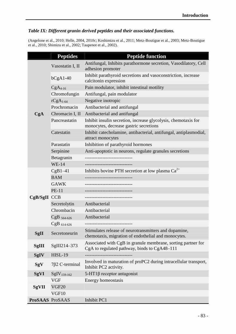
Introduction
- 83 -
Table IX: Different granin derived peptides and their associated functions.
(Angelone et al., 2010; Helle, 2004, 2010c; Koshimizu et al., 2011; Metz-Boutigue et al., 2003; Metz-Boutigue
et al., 2010; Shimizu et al., 2002; Taupenot et al., 2002).
Peptides Peptide function
CgA
Vasostatin I, II Antifungal, Inhibits parathormone secretion, Vasodilatory, Cell
adhesion promoter
bCgA1-40 Inhibit parathyroid secretions and vasoconstriction, increase
calcitonin expression
CgA4-16 Pain modulator, inhibit intestinal motility
Chromofungin Antifungal, pain modulator
rCgA1-64 Negative inotropic
Prochromacin Antibacterial and antifungal
Chromacin I, II Antibacterial and antifungal
Pancreastatin
Inhibit insulin secretion, increase glycolysis, chemotaxis for
monocytes, decrease gastric secretions
Catestatin
Inhibit catecholamine, antibacterial, antifungal, antiplasmodial,
attract monocytes
Parastatin Inhibition of parathyroid hormones
Serpinine Anti-apoptotic in neurons, regulate granules secretions
Betagranin ------------------------------
WE-14 ------------------------------
CgB/SgII
CgB1–41 Inhibits bovine PTH secretion at low plasma Ca2+
BAM ------------------------------
GAWK ------------------------------
PE-11 ------------------------------
CCB ------------------------------
Secretolytin Antibacterial
Chrombacin Antibacterial
CgB 564-626 Antibacterial
CgB 614-626 ------------------------------
SgII Secretoneurin Stimulates release of neurotransmitters and dopamine,
chemotaxis, migration of endothelial and monocytes.
SgIII SgIII214–373 Associated with CgB in granule membrane, sorting partner for
CgA to regulated pathway, binds to CgA48–111
SgIV HISL-19 ------------------------------
SgV 7β2 C-terminal Involved in maturation of proPC2 during intracellular transport,
Inhibit PC2 activity.
SgVI SgIV159-162 5-HT1β receptor antagonist
SgVII
VGF Energy homeostasis
VGF20
VGF10
ProSAAS ProSAAS Inhibit PC1
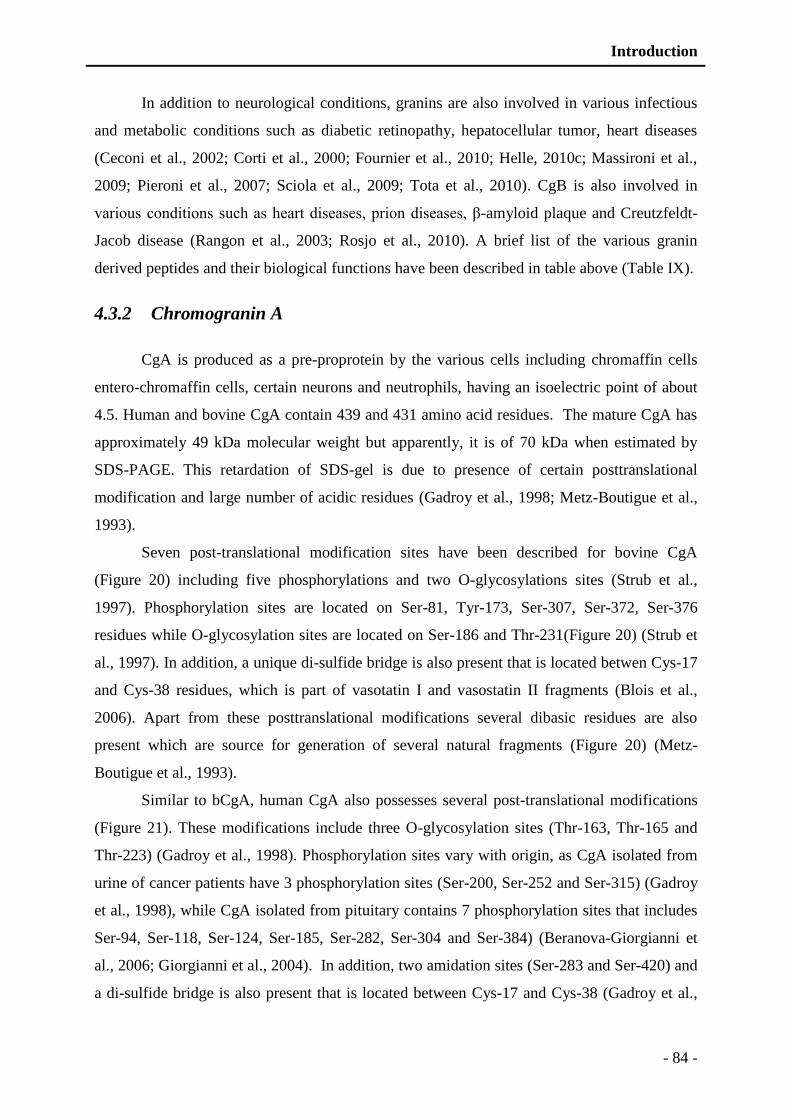
Introduction
- 84 -
In addition to neurological conditions, granins are also involved in various infectious
and metabolic conditions such as diabetic retinopathy, hepatocellular tumor, heart diseases
(Ceconi et al., 2002; Corti et al., 2000; Fournier et al., 2010; Helle, 2010c; Massironi et al.,
2009; Pieroni et al., 2007; Sciola et al., 2009; Tota et al., 2010). CgB is also involved in
various conditions such as heart diseases, prion diseases, β-amyloid plaque and Creutzfeldt-
Jacob disease (Rangon et al., 2003; Rosjo et al., 2010). A brief list of the various granin
derived peptides and their biological functions have been described in table above (Table IX).
4.3.2 Chromogranin A
CgA is produced as a pre-proprotein by the various cells including chromaffin cells
entero-chromaffin cells, certain neurons and neutrophils, having an isoelectric point of about
4.5. Human and bovine CgA contain 439 and 431 amino acid residues. The mature CgA has
approximately 49 kDa molecular weight but apparently, it is of 70 kDa when estimated by
SDS-PAGE. This retardation of SDS-gel is due to presence of certain posttranslational
modification and large number of acidic residues (Gadroy et al., 1998; Metz-Boutigue et al.,
1993).
Seven post-translational modification sites have been described for bovine CgA
(Figure 20) including five phosphorylations and two O-glycosylations sites (Strub et al.,
1997). Phosphorylation sites are located on Ser-81, Tyr-173, Ser-307, Ser-372, Ser-376
residues while O-glycosylation sites are located on Ser-186 and Thr-231(Figure 20) (Strub et
al., 1997). In addition, a unique di-sulfide bridge is also present that is located betwen Cys-17
and Cys-38 residues, which is part of vasotatin I and vasostatin II fragments (Blois et al.,
2006). Apart from these posttranslational modifications several dibasic residues are also
present which are source for generation of several natural fragments (Figure 20) (Metz-
Boutigue et al., 1993).
Similar to bCgA, human CgA also possesses several post-translational modifications
(Figure 21). These modifications include three O-glycosylation sites (Thr-163, Thr-165 and
Thr-223) (Gadroy et al., 1998). Phosphorylation sites vary with origin, as CgA isolated from
urine of cancer patients have 3 phosphorylation sites (Ser-200, Ser-252 and Ser-315) (Gadroy
et al., 1998), while CgA isolated from pituitary contains 7 phosphorylation sites that includes
Ser-94, Ser-118, Ser-124, Ser-185, Ser-282, Ser-304 and Ser-384) (Beranova-Giorgianni et
al., 2006; Giorgianni et al., 2004). In addition, two amidation sites (Ser-283 and Ser-420) and
a di-sulfide bridge is also present that is located between Cys-17 and Cys-38 (Gadroy et al.,
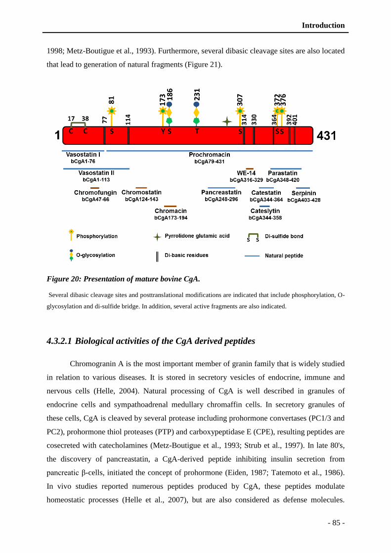
Introduction
- 85 -
1998; Metz-Boutigue et al., 1993). Furthermore, several dibasic cleavage sites are also located
that lead to generation of natural fragments (Figure 21).
Figure 20: Presentation of mature bovine CgA.
Several dibasic cleavage sites and posttranslational modifications are indicated that include phosphorylation, O-
glycosylation and di-sulfide bridge. In addition, several active fragments are also indicated.
4.3.2.1 Biological activities of the CgA derived peptides
Chromogranin A is the most important member of granin family that is widely studied
in relation to various diseases. It is stored in secretory vesicles of endocrine, immune and
nervous cells (Helle, 2004). Natural processing of CgA is well described in granules of
endocrine cells and sympathoadrenal medullary chromaffin cells. In secretory granules of
these cells, CgA is cleaved by several protease including prohormone convertases (PC1/3 and
PC2), prohormone thiol proteases (PTP) and carboxypeptidase E (CPE), resulting peptides are
cosecreted with catecholamines (Metz-Boutigue et al., 1993; Strub et al., 1997). In late 80's,
the discovery of pancreastatin, a CgA-derived peptide inhibiting insulin secretion from
pancreatic β-cells, initiated the concept of prohormone (Eiden, 1987; Tatemoto et al., 1986).
In vivo studies reported numerous peptides produced by CgA, these peptides modulate
homeostatic processes (Helle et al., 2007), but are also considered as defense molecules.
Introduction
- 86 -
During last two decades, several new antimicrobial CgA-derived peptides have been
characterized that participate actively against the combat with infectious agents (Helle et al.,
2007; Lugardon et al., 2000; Metz-Boutigue et al., 1998; Strub et al., 1996a; Strub et al.,
1996b) (Figure 21: Table IX).
Figure 21: Presentation of mature human CgA.
Several dibasic cleavage sites and posttranslational modifications are indicated that include phosphorylation, O-
glycosylation and di-sulfide bridge. In addition, several active fragments are also indicated.
Sequences of CgA-derived peptides are highly conserved during evolution.
Interestingly, main cleavage site in bovine CgA (bCgA) at position 78-79 and subsequently
removal of the two basic residues K77 and K78 by carboxypeptidase H (Metz-Boutigue et al.,
1993), produces two major fragments: vasostatin-I (VS-I; bCgA1-76) (Lugardon et al., 2000)
and prochromacin (ProChrom; bCgA79-431) (Strub et al., 1996a). In addition, another N-
terminal fragment called vasostatin II (VS-II; bCgA1-113) is also generated that possesses
similar activities as VS-1(Helle et al., 1993). Synthetic vasostatin and its shorter fragment

Introduction
- 87 -
bCgA16-40 can actively inhibit PTH release from bovine gland (Angeletti et al., 1996).
Moreover, these present antimicrobial activities against filamentous fungi, yeasts and Gram-
positive bacteria in micromolar range from 0.1-2 µM (Lugardon et al., 2000) (Table X).
Table X: Antimicrobial peptides derived from CgA.
Peptides Bacteria (G+) Bacteria (G-) Fungi Yeast Parasites Reference
Vasostatine I
(bCGA1−76) X X X
(Lugardon et al., 2000)
Chromofungin
(bCGA47-66) X
(Lugardon et al., 2001)
Prochromacin
(bCGA79−431) X X
(Strub et al., 1996a)
Chromacin
(bCGA173−186) X
(Strub et al., 1996a)
Catestatin
(bCGA344−364) X X X X X
(Briolat et al., 2005)
Furthermore, it has been previously demonstrated that the N-terminal bCgA1-40
fragment can interact with a model of phospholipid membranes (Blois et al., 2006; Maget-
Dana et al., 2002; Shooshtarizadeh et al., 2010). Another important peptide derived from
vasostatin region of CgA is chromofungin (CHR; CgA47-66) that is not naturally occurring
but generated by bacterial proteases such as Glu-C from S. aureus. CHR belongs to CgA
domain that is well conserved through evolution. CHR presents antifungal activity at 2-15 µM
concentration against yeasts (Candida albicans, Candida tropicalis, and Candida
neoformans) and filamentous fungi (Fusarium culmorum, Fusarium oxysporum, Aspergillus
fumigatus, Neurospora crassa, Alternaria brassicola, and Nectria haematococca) (Lugardon
et al., 2001).
Similarly, prochromacin also generates several fragments including chromacin
(Chrom; bCgA173-194), catestatin (CAT; bCgA344-364), cateslytin (CTL; bCgA344-358),
pancreastatin (bCgA248-296), parastatin (bCgA348-420), chromastatin (bCgA124-143), and
WE-14 (bCgA216-229) (Helle, 2004, 2010a, b, c; Shooshtarizadeh et al., 2010; Strub et al.,
1996b). These C-terminal derived peptides also display several biological activities; few of
them are summarized in Table IX. Most important multifunctional C-terminal derived
peptides are cateslytin and catestatin that are described in detail below.
Introduction
- 88 -
4.3.2.1.1 Catestatin
Catestatin (CAT) is short CgA derived fragment, which is produced by cleavage of
prochromacin region of CgA (bovine and human CAT CgA344-364 and CgA352-372
respectively) (Table XI). CAT was first identified as non-competitive nicotinic cholinergic
antagonist in chromaffin cells in 1997 (Mahata et al., 1997). Later studies revealed that CAT
is produced in chromaffin granules by action of prohormone thiol protease (PTP), which was
demonstrated to act on D343-R344 and L364-R365 (Lee et al., 2003). This cleavage leads to
liberation of CAT, a further cleavage is also been demonstrated on the motif R-G of CAT that
liberate cateslytin (CTL), the short active form of CAT (Biswas et al., 2009). Three human
variants were identified: Gly364Ser, Pro370Leu and Arg374Gln. These varients have same
properties as wild type CAT but their activity varies and their order of activity to inhibit
nicotinic process is as follows Pro370Leu > wild type > Gly364Ser > Arg374Gln (Mahata et
al., 2004).
CAT represents various biological activities: it can regulate blood pressure, is
sympatho-inhibitor and attenuates sympathetic barosensitivity, and serves as an endogenous
sympathetic nervous system regulator (Gaede and Pilowsky, 2012; Rao et al., 2007; Schillaci
et al., 2011). Plasma concentration of CAT in hypertension patients is increased (Meng et al.,
2011), which underlines its role as vasodilator (Angelone et al., 2011; Kennedy et al., 1998;
Liao et al., 2011). Few contradictory results are also published, which indicate reduced
plasma expression in hypertension patient than healthy persons (O'Connor et al., 2002).
Despites all, CAT serves as heart protector by inhibiting adrenergic release, regulating blood
pressure and as vasodilator (Bassino et al., 2011; Liao et al., 2011). It also serves as marker
for various diseases such as heart failure, myocardial infarction, and carcinoid tumors of
intestine (Conlon, 2010; Ji et al., 2012; Meng et al., 2012; Prommegger et al., 2003). CAT is
also involved in mobilization of fat from adipose tissue through regulation of adrenergic and
leptin signaling (Bandyopadhyay et al., 2012). Furthermore, CAT also serves as angiogenic
cytokine by promoting fibroblast growth via a basic fibroblast growth factor dependent
mechanism (Theurl et al., 2010).
In addition, CAT is very potent AMPs against bacteria, fungi and yeasts (Akaddar et
al., 2010; Briolat et al., 2005; Metz-Boutigue et al., 2010; Shooshtarizadeh et al., 2010). CAT
can effectively inhibit various fungal, parasite and bacterial growth in micromolar range. It
interacts with plasmepsin and ultimately inhibits growth of plasmodium species (Akaddar et
al., 2010). Antimicrobial properties of CAT are due to asymmetric structure adopted in

Introduction
- 89 -
hydrophobic solvents. It is mostly unstructured but readily adopts β-strand-loop-β-strand
conformation when it is in contact with lipid membrane. A short loop is formed between
residue 7 and 11 of CAT. Three arginine residues adopt a conformation of positive side chain
that facilitates its interaction with membrane (Sugawara et al., 2010; Tsigelny et al., 1998).
This model is well explained for catecholamine release inhibition and is assumed to interact
similarly against pathogens.
In addition to direct antimicrobial activities, it also acts as immune modulator peptide.
Catestatin acts as chemoattractant for the monocytes (Egger et al., 2008), and as angiogenic
cytokine for fibroblast (Theurl et al., 2010). These studies suggest its activity in immune
modulation by acting as inflammatory cytokine. Furthermore, it can trigger production of IL-8
by keratinocytes via activation of mitogen-activated protein kinases (Aung et al., 2011a) and
stimulates mast cells degranulation to release histamine that further initiates inflammatory
cascade (Aung et al., 2011b; Kruger et al., 2003). Catestatin induce influx of calcium in
neutrophils by calmodulin-regulated calcium independent phospholipase A2 (Zhang et al.,
2009b). It also contributes in wound healing by increasing migration and proliferation of
keratinocytes at wound site (Hoq et al., 2011).
4.3.2.1.2 Cateslytin
Cateslytin (bCTL; bCgA 344-358) is the short active form of CAT (Table XI).
Endogenous production of CAT is previously described, which is further processed by an
additional cleavage R358-G359 of catestatin by cysteine protease cathepsin L (CTSL) in
chromaffin secretory vesicles (Lee et al., 2003). Cateslytin interacts with fungal membrane,
by adopting an aggregated antiparallel beta-sheet structure at membrane interfaces (Jean-
Francois et al., 2009). This mechanism lead to separation of fluid and rigid membrane
structures on ergosterol (fungal membrane) containing models and ultimately destabilization
of membrane (Jean-Francois et al., 2009). In vitro, studies by using zwitterionic
biomembranes, CTL was demonstrated to form beta-sheets and generates pores of 1 nm
diameter (Jean-Francois et al., 2008b). By using NMR studies CTL was demonstrated to be
converted into antiparallel beta-sheets that aggregate at negatively charged surface of bacterial
membranes (Jean-Francois et al., 2008a; Jean-Francois et al., 2007). Arginine residues are
known to be important in binding to negatively charged lipids. Following the interaction of
CTL to membrane, several rigid and thicker membrane domains were observed (Jean-
Francois et al., 2008a).
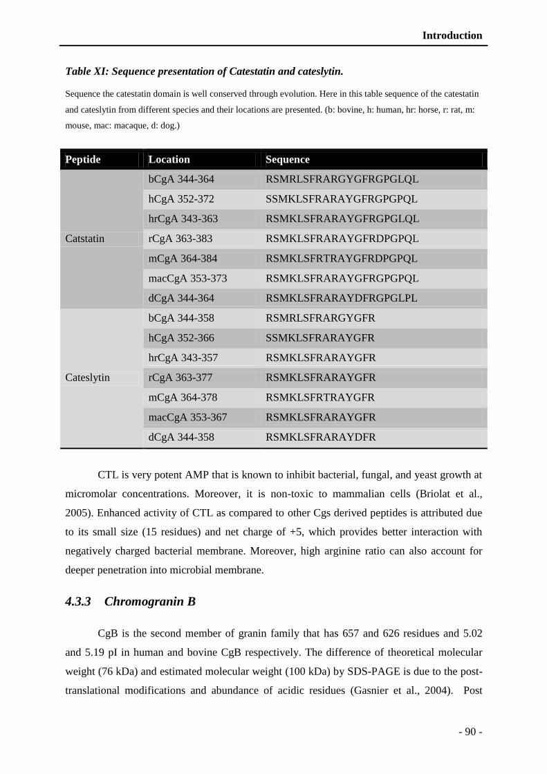
Introduction
- 90 -
Table XI: Sequence presentation of Catestatin and cateslytin.
Sequence the catestatin domain is well conserved through evolution. Here in this table sequence of the catestatin
and cateslytin from different species and their locations are presented. (b: bovine, h: human, hr: horse, r: rat, m:
mouse, mac: macaque, d: dog.)
Peptide Location Sequence
Catstatin
bCgA 344-364 RSMRLSFRARGYGFRGPGLQL
hCgA 352-372 SSMKLSFRARAYGFRGPGPQL
hrCgA 343-363 RSMKLSFRARAYGFRGPGLQL
rCgA 363-383 RSMKLSFRARAYGFRDPGPQL
mCgA 364-384 RSMKLSFRTRAYGFRDPGPQL
macCgA 353-373 RSMKLSFRARAYGFRGPGPQL
dCgA 344-364 RSMKLSFRARAYDFRGPGLPL
Cateslytin
bCgA 344-358 RSMRLSFRARGYGFR
hCgA 352-366 SSMKLSFRARAYGFR
hrCgA 343-357 RSMKLSFRARAYGFR
rCgA 363-377 RSMKLSFRARAYGFR
mCgA 364-378 RSMKLSFRTRAYGFR
macCgA 353-367 RSMKLSFRARAYGFR
dCgA 344-358 RSMKLSFRARAYDFR
CTL is very potent AMP that is known to inhibit bacterial, fungal, and yeast growth at
micromolar concentrations. Moreover, it is non-toxic to mammalian cells (Briolat et al.,
2005). Enhanced activity of CTL as compared to other Cgs derived peptides is attributed due
to its small size (15 residues) and net charge of +5, which provides better interaction with
negatively charged bacterial membrane. Moreover, high arginine ratio can also account for
deeper penetration into microbial membrane.
4.3.3 Chromogranin B
CgB is the second member of granin family that has 657 and 626 residues and 5.02
and 5.19 pI in human and bovine CgB respectively. The difference of theoretical molecular
weight (76 kDa) and estimated molecular weight (100 kDa) by SDS-PAGE is due to the post-
translational modifications and abundance of acidic residues (Gasnier et al., 2004). Post

Introduction
- 91 -
translational modifications include two O-glycosylations (T93 and T170), one N-
glycosylation (N168), two sulfatations (Y138 and Y295), three phosphorylations (S564, S573
and S578) and two pyrolidone glutamic acid residues (Q456 and Q547) (Figure 22) (Gasnier
et al., 2004). In addition, a disulfide bridge is also present at the N-terminal of CgB that is
essential for CgB interaction with membrane and also mediates membrane binding in the
TGN (Trans-Golgi-Network) (Glombik et al., 1999).
Figure 22: Presentation of mature bovine CgB.
Several dibasic cleavage sites and posttranslational modifications are indicated that include
phosphorylation, O-glycosylation, N-glycosylation, sulfatation and disulfide bridge. In
addition, several active fragments are also indicated.
CgB was first characterized from rat PC12 cells as a tyrosine-sulphated protein and
later named chromogranin B when it was detected as a conspicuous component of bovine
adrenal medulla (Eiden et al., 1987; Falkensammer et al., 1985; Lee and Huttner, 1983).
Several CgB derived peptides have been identified in various tissues, including extracts of
human pituitary glands, pheochromocytomas of human, brain extracts, splanchnic nerve in

Introduction
- 92 -
pigs, and cephalo-rachidian fluid samples (Benjannet et al., 1985; Conlon et al., 1992;
Depreitere et al., 2002; Depreitere et al., 2004; Marksteiner et al., 1999; Stark et al., 2001).
Likewise, CgA derived peptides, CgB and its derived peptides also denote various biological
activities, such as N-terminal peptide CgB1-41 that inhibits PTH secretion in bovine (Helle,
2004). Similarly, N-terminal domain (CgB1-49) has been implicated in interaction with
membrane vesicles (Yoo and Kang, 1997).
C-terminal peptide (bCgB614-626) was identified in 1995 in secretions of stimulated
chromaffin cells (Strub et al., 1996b). This C-terminal peptide is well known for its
antibacterial activities, inhibiting growth of various bacteria at micro molar range (Strub et al.,
1996b). Another C-terminal peptide CgB564-626, also presents antimicrobial activities (Strub
et al., 1995), this peptide is homologous to N-terminal domain of cecropin, a well-known
insect AMP. CgB derived AMPs are listed in the Table XII.
Table XII: Antimicrobial peptides derived from CgA.
Peptides Bacteria (G+) Bacteria (G-) Fungi Yeast Reference
Secretolytin
(bCgB614−626)
X X (Strub et al., 1996b)
CCB
(bCgB564-626)
X (Metz-Boutigue et al., 1998)
Several other CgB derived peptides have been isolated but still their biological
activities are not very clear such as GAWK, BAM-1745, SR-17, HQ-34 and PE-11
(Benjannet et al., 1985; Flanagan et al., 1990; Laslop et al., 1998; Wang et al., 2001). CgB
have also been detected in vagus nerves of human fetus, where may be playing important role
in fetal developmental organization (Bitsche et al., 2006).

AMP conjugated polyelectrolyte films

Introduction
- 93 -
5 AMPS conjugated polyelectrolyte films
5.1 Introduction to polyelectrolyte complexes
Polyelectrolytes have emerged as tools to study the complexity of the polymers and to
develop a multilayer polyelectrolyte base. In this chapter: the evolution and the use of charged
polymers to complex formation and development of multilayer by dipping or spraying
methods will be discussed briefly. Finally, we focus on the comparison polyelectrolyte
complex films and their potential applications in the field of biomaterials. Polyelectrolytes are
polymers whose repeating unit is electrostatically charged. They may be synthetic, such as
poly-allylamine hydrochloride (PAH) or poly-sodium styrene sulfonate (PSS) or natural
polysaccharides such as DNA. The polyelectrolyte complex (PEC) is formed from the mixture
of solutions of polyanion and polycation. They can be in the form of soluble complexes
(Kabanov, 1994; Tsuchida and Abe, 1982), of liquid coacervates (Burke et al., 2007) or solid
precipitates (Michaels and Miekka, 1961).
5.2 Application of polyelectrolyte complexes in the biomedical field
PECs are widely used in the field of biomedical, these present very diverse applications.
These are widely used in various studies for example, transfection of DNA for which
nanoparticles of chitosan are used (Duceppe and Tabrizian, 2009; Erbacher et al., 1998). The
PECs are also used for the encapsulation such as targeted or non-targeted release of insulin for
the oral treatment of diabetes, for asthma treatment, for targeted anticancer therapies (Damgé,
1991; He et al., 2009; Jain and Jain, 2008; Oyarzun-Ampuero et al., 2009; Yin et al., 2009)
(Jain and Jain 2008; Choi et al., 2009; He et al. 2009).
5.3 Polyelectrolyte multilayered films
The material surface is the preferred interaction site between it and its environment. To
control these interactions, it is necessary to control the properties of this interface. To control
cell-material interaction is one of the most crucial aspects in the field of biomaterials. This is
made possible by the deposition of thin films with controlled surface properties. The
polyelectrolyte multilayers, thanks to their ease of implementation as well as the wide range
of available compounds, constitute an appropriate method of functionalization of biomaterials
by surface modification.
Introduction
- 94 -
5.3.1 History of multilayered films
Two types of coating materials exist: inorganic and organic materials. Organic
material films were initially started to construct in the early twentieth century by Langmuir
and Blodgett, commonly known as "Langmuir-Blodgett films" (LB). These films are
constructed by applying a monolayer of surfactants or amphiphilic molecules that are
transferred from the air/water interface of a solution on the vessel substrate by dipping
(Blodgett, 1934; Blodgett and Langmuir, 1937). The adsorption of the film is based on the
hydrophilic / hydrophobic type of interactions with the substrate. A similar second immersion
is carried out for the adsorption of a second layer. This method can be repeated several times
varying species each deposition step. The thickness of the films ranges from a few angstroms
to several nanometers. This method has many disadvantages such as instability of films, a
limitation related to the shape of the substrate and the presence of parasitic reactions between
the uncontrolled and the solvent molecules in the film.
Another method of preparation of multilayer films was proposed in 1966 by Iler (Iler,
1966). It is based on electrostatic interactions between the anionic and cationic colloidal
particles for the construction of multilayer films. Later in 1980, Fromherz is the first to
mention the application of this concept to the adsorption of proteins and oppositely charged
polyelectrolytes (Fromherz, 1980; Gölander et al., 1982). Each deposit must be accompanied
by a change of surface charge, by overcompensation of charge (Gölander et al., 1982).
Later in 1980s, other self-assembly methods have been developed to form organic
films based on the systems of coordination chemistry of metal/phosphonate and covalent
chemistry (Guang et al., 1992; Netzer and Sagiv, 1983). The first consists in the self-assembly
of molecules, composed of a head and a tail phosphonate hydrophobic carbon, by fixing heads
phosphonate on a metal substrate by coordination chemistry and hydrophobic interaction.
These processes, however, are limited to a number of chemical systems and do not provide
reproducibly homogeneous films.
In the early 1990s, with the adaptation of work to Iler polyelectrolytes, Decher have
really exploited the capabilities of the method of layer-by-layer (LbL) assembly (Decher,
1997; Decher and Hong, 1991; Helm et al., 1992). This technique is based on the adsorption
of polycations and polyanions alternatively, each deposition step being followed by a rinsing
step with a buffer solution or water. Films made by this method are called PEMs
(polyelectrolyte multilayers).

Introduction
- 95 -
5.3.2 Methods to develop polyelectrolyte multilayer films
This method was well established in the 1990s, the layer by layer deposition is shown
schematically in Figure 23. The successive deposition of polyelectrolytes are made on a
substrate, usually negatively charged in contact with a solution of polycation followed by a
rinsing step and then a solution of polyanion also followed by a rinsing step for a given time
(5 to 20 min). Putting in contact with the polyelectrolyte solution allows the adsorption of the
compounds in solution (Advincula et al., 1996; Hoogeveen et al., 1996; Ramsden et al.,
1995), the surplus or unbound compounds are removed by rinsing step (Caruso et al., 1998;
Lvov et al., 1999). At the end of the adsorption step and rinsing excessive polycations, the
surface is positively charged: there is overcompensation of charge (Caruso et al., 1998;
Ladam et al., 1999). This overcompensation load has been demonstrated by zeta potential
measurements (Hoogeveen et al., 1996; Ladam et al., 1999). It is the driving force behind the
construction of polyelectrolyte multilayer films.
Figure 23: Schematic construction of multilayer polyelectrolyte adsorptions by successive
polycations and polyanions. (Decher, 1997; Decher and Hong, 1991)
Newly positively charged surface, can be then exposed to a solution of polyanions to
be electrostatically adsorbed. Further rinsing allows the elimination of the surplus of
polyanions. The surface is negatively charged. A pair of layers is formed finally. Consecutive
cycles, alternating adsorption of polycations and polyanions, thus allow the growth stages of
the multilayer film.
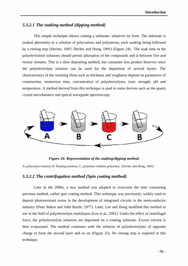
Introduction
- 96 -
5.3.2.1 The soaking method (dipping method)
This simple technique allows coating a substrate, whatever its form. The substrate is
soaked alternately in a solution of polycations and polyanions, each soaking being followed
by a rinsing step (Decher, 1997; Decher and Hong, 1991) (Figure 24). The soak time in the
polyelectrolyte solutions should permit adsorption of the compounds and is between five and
twenty minutes. This is a slow depositing method, but consumes less product however since
the polyelectrolyte solution can be used for the deposition of several layers. The
characteristics of the resulting films such as thickness and roughness depend on parameters of
construction: immersion time, concentration of polyelectrolytes, ionic strength, pH and
temperature. A method derived from this technique is used in some devices such as the quartz
crystal microbalance and optical waveguide spectroscopy.
Figure 24: Representation of the soaking/dipping method.
A: polycation solution, B: flushing solution, C: polyanion solution polyanion. (Decher and Hong, 1991)
5.3.2.2 The centrifugation method (Spin coating method)
Later in the 2000s, a new method was adopted to overcome the time consuming
previous method, called spin coating method. This technique was previously, widely used to
deposit photoresistant resins in the development of integrated circuits in the semiconductor
industry (Peter Bakos and John Rasile, 1977). Later, Lee and Hong modified this method to
use in the field of polyelectrolyte multilayers (Lee et al., 2001). Under the effect of centrifugal
force, the polyelectrolyte solutions are deposited on a rotating substrate. Excess solvent is
then evaporated. The method continues with the solution of polyelectrolytes of opposite
charge to form the second layer and so on (Figure 25). No rinsing step is required in this
technique.

Introduction
- 97 -
With a spin coating, the time required for the construction of a polyelectrolyte bilayer
is more than about 80 seconds. The thickness and surface roughness of the films depends on
the rotational speed, the viscosity of the solution and the volatility of the solvent in addition to
the parameters already mentioned in the case of dipping. This method allows to obtain films
with better mechanical strength than the films obtained by dipping (Jiang et al., 2004).
Figure 25: Representation of PEMs film construction by spin coating method.
5.3.2.3 Spray coating method
Another coating method was developed to build PEM films, known as spraying or
assisted spray-coating method. The multilayer construction is done by spraying solutions of
polyelectrolytes on a vertical or slightly inclined substrate, allowing the drainage of excess
solution (Schlenoff et al., 2000). A step of spraying polyelectrolyte and then a rinsing step
followed (Figure 26). When spraying, forming a thin film of liquid on the substrate is
observed. The main disadvantage of this method is the high consumption solution. In fact, the
majority of spray solutions is lost by drainage and is not reusable thereafter. Roughness and
thickness of the films obtained are comparable to those of the films obtained by dipping. The
flow of solutions sprayed, the spraying distance, solution concentrations, ionic strength, pH
and temperature are the parameters that influence construction of the film. Sometimes rinsing
step is deliberately removed to increase the thickness and roughness of the films (Izquierdo et
al., 2005). Now a day, this technique is widely used for the surface treatment of contact lenses
to make them hydrophilic.
Another modification of this technique is also used that uses simultaneous deposition
of polyelectrolytes rather than alternatively spraying (Porcel et al., 2005).
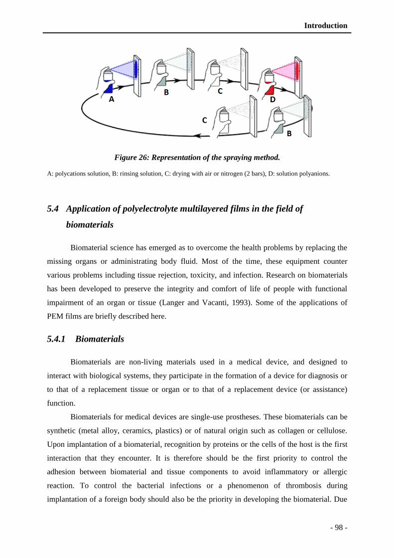
Introduction
- 98 -
Figure 26: Representation of the spraying method.
A: polycations solution, B: rinsing solution, C: drying with air or nitrogen (2 bars), D: solution polyanions.
5.4 Application of polyelectrolyte multilayered films in the field of
biomaterials
Biomaterial science has emerged as to overcome the health problems by replacing the
missing organs or administrating body fluid. Most of the time, these equipment counter
various problems including tissue rejection, toxicity, and infection. Research on biomaterials
has been developed to preserve the integrity and comfort of life of people with functional
impairment of an organ or tissue (Langer and Vacanti, 1993). Some of the applications of
PEM films are briefly described here.
5.4.1 Biomaterials
Biomaterials are non-living materials used in a medical device, and designed to
interact with biological systems, they participate in the formation of a device for diagnosis or
to that of a replacement tissue or organ or to that of a replacement device (or assistance)
function.
Biomaterials for medical devices are single-use prostheses. These biomaterials can be
synthetic (metal alloy, ceramics, plastics) or of natural origin such as collagen or cellulose.
Upon implantation of a biomaterial, recognition by proteins or the cells of the host is the first
interaction that they encounter. It is therefore should be the first priority to control the
adhesion between biomaterial and tissue components to avoid inflammatory or allergic
reaction. To control the bacterial infections or a phenomenon of thrombosis during
implantation of a foreign body should also be the priority in developing the biomaterial. Due
Introduction
- 99 -
to these secondary infections, most of the time removal of implant becomes unavoidable. The
new challenge in this area is the development of materials with mechanical and structural
properties are adapted to their future application but also interact actively with the
surrounding living environment. The control response of the material is sought to enable
minimize reactions of the body to this foreign element and modulate the activation of immune
cells (Ratner, 1996; Ratner and Bryant, 2004). These materials are called "bioactive" interact
with cells and induce a specific response of the surrounding tissue.
To limit the discharge phenomena, biomaterials must be biocompatible.
Biocompatibility is defined as the ability of the material to be accepted by the body. Recently,
research has turned to the development of biodegradable compounds when their presence is
required for a given time. Synthetic absorbable suture threads such as Vicryl® or Dexon®,
bioresorbable stents, biodegradable membranes for wound healing, sponges to regenerate the
bone tissue are some of classical examples that are extensively used (Freed et al., 1993;
Ishaug et al., 1997; Jürgens et al., 2006; Zilberman and Eberhart, 2006).
5.4.2 Cell adhesion to the PEM films
Multilayer films can be used as coatings for biomaterials to control of the response of
the medium surrounding the implanted biomaterial. In this perspective, understanding and
studying the behavior of cells is extremely important. Indeed, depending on the desired
application, the biomaterial must be attached or not to the cells. When cell migration should
be encouraged to regenerate damaged tissue, the material is preferably non-stick. Conversely,
to enable the integration of an implant in vivo following the removal of an organ, it may
promote adhesion of healthy cells while disfavoring the tumor cells that may still be present
in the surrounding tissue.
5.4.2.1 Adhesion to films based on synthetic polyelectrolytes
A means of controlling cell adhesion on film PAH/PAA (poly-allylamine
hydrochloride/poly-acrylic acid), is pH change during the construction of the film.
Mendelsohn studied the adhesion of mouse fibroblasts (NR6WT) on these two polyelectrolyte
films, in function of pH to control cellular growth (Mendelsohn et al., 2003). Construction of
the film at pH 6.5 allows good adhesion of fibroblasts, whereas the same film constructed at
pH 2 is non-adherent. This change in pH control the hydration of the molecules, ultimately
growth of cells via internal swelling capacity in relation to pH. At low pH molecules are
Introduction
- 100 -
usually less hydrated and develop thin layer ultimately allowing cells to grow. Moreover, the
amount of proteins adsorbed on the films is independent of the pH of construction.
PEM films (PAH / PSS) constructed on gels of polydimethylsiloxane (PDMS) by the
microfluidics network technique, are non-adherent for the murine neural cells and becomes
adherent with the passage of time. PEMs constructed by this method are also selective for
growth pattern, as more elongated cell growth was observed (Reyes et al., 2004). Later, some
in-vivo studies showed improved endothelial cell growth when implanted in arteries and
human umbilical cord (Kerdjoudj et al., 2007).
5.4.2.2 Adhesion of the films based on biopolymers (polysaccharides and
polypeptides)
Cell adhesion can also be modulated by the use of natural polymer based films instead
of using synthetic polymers, such as CHI/HA (Chitosan/Hyaluronic acid) films. These films
can regulate growth of many cells from different origin e.g. platelets and leukocytes (Thierry
et al., 2003), chondrosacroma (Richert et al., 2004a; Richert et al., 2004b), MC3T3
preosteoblasts (Hillberg et al., 2009) or stem cells derived from bone marrow (Liu et al.,
2010). Low cellular adhesion and proliferation on the films CHI/HA is mainly due to the
mechanical properties of these films, which are not rigid enough. Another modification of
these films is to crosslink, which can render these films more stiffer and resistant to enzymes
such as hyaluronidases (Richert et al., 2004a).
5.4.3 Insertion of active molecules in a film
To make bioactive biomaterials to induce an appropriate response in vivo, it is
necessary to add or graft bioactive molecules such as proteins, peptides or drugs.
5.4.3.1 Adsorption and absorption of molecules
The multilayer films are made by adsorption of polyanions and polycations. This
property of the films can make it possible to incorporate a wide variety of charged molecules
(particles, inorganic molecules, proteins). One method is to create reservoir films that may
contain active molecules by use of adsorption. Berg in 2006, made a porous film by treatment
at different pH after alternative building of PAH/PAA and PAH/PSS and include the
ketoprofen and cytochalasin D (Berg et al., 2006). In this technique, exponential growth of the
crosslinked films may serve as a reservoir for active molecules (Crouzier et al., 2010). The

Introduction
- 101 -
amount of active molecules is modulated by varying the number of layers of the film and the
release is affected by the modification of the medium or degradation of the film. A similar
technique was adopted to construct a multilayered film made up of PLL/PGA (Poly-L-
lysine/Poly-glutamic acid), with the insertion of Protein A from S. aureus (Jessel et al., 2003).
Protein A has various biological activities, including antitumor, antifungal and antiparasitic.
This film is gradually degraded when it is in contact with the cells, which enables them to
liberate protein A.
5.4.3.2 Insertion of active molecules by coupling of the molecules
Another method of inserting active molecules to the film is the coupling with one of
the components of the film. The α-MSH (α-melanocyte-stimulating hormone), anti-
inflammatory peptide, has been successfully integrated into multilayer films of PLL/PGA
while remaining active (Chluba et al., 2001). The α-MSH was grafted onto the PLL. Later, in
2005 this peptide is covalently coupled by thiol functionalization with the PGA to create
prosthetic trachea (Fioretti et al., 2010; Schultz et al., 2005). The anti-inflammatory activity of
α-MSH could be demonstrated in-vivo by detection of IL-10 in rats implanted with
functionalized trachea.
5.4.4 Antibacterial PEM films
5.4.4.1 Passive antibacterial PEM films
Most of the pathogenic bacteria can form biofilms. Silverstein and Donatucci describe
different stages of biofilm formation for various biomaterials such as catheters, heart valves
(Silverstein and Donatucci, 2003). As previously described in the first chapter about S.
aureus, infection usually starts with the adhesion of bacteria to host surface that is potentiated
by use of MSCRAMs. Various studies have enabled the creation of multi-resistant bacterial
adhesion films. Ultimately, reduced adhesion of the pathogen can indirectly reduce infection.
PEMs films based on PLL/PGA can be potentiated by applying of last layer which is grafted
with poly-ethylene glycol (PLL / PGA-g-PEG) (Boulmedais et al., 2004). PEG is indeed a
hydrophilic molecule and its hydration prevents bacterial adhesion. Reisch et al. also
proposed another approach to make non-adherent surfaces to inhibit fungal adhesion. The
surface covering by deposition of polyelectrolyte multilayer films modified with
oligoethylene glycol chains or phosphorylcholine (Reisch et al., 2010; Reisch et al., 2008).

Introduction
- 102 -
Chitosan is extensively used for the development of PEM films. It is a hydrophilic
polymer obtained industrially by N-deacetylation of chitin, also has antimicrobial properties
against a wide range of fungi, algae and some bacteria. In addition, it can regulate growth of
some viruses (Rabea et al., 2003).
5.4.4.2 Active antibacterial PEM films
Films can be made by inserting molecules having antibacterial properties. Initially,
antibacterial films were developed by the insertion of silver nanoparticles in multilayers of
PAH/PAAm (Poly-alanine hydrochloride/polyacrylamide) (Lee et al., 2005). Antibacterial
activity can be controlled by the deposition of varying number of layers on the planner
surface. The zone of inhibition increases as the film thickness increases, which can be
described by a simple diffusion model. It was also demonstrated that the release of silver ions,
responsible for antibacterial, can be controlled by an oxidation mechanism on the surface of
nanoparticles. The deposition of these layers on magnetic microspheres allows release of the
antimicrobial agent to specific sites. Similar experiments were carried out in 2008, by using
the antibacterial encapsulated silver ions in liposomes inserts that are inserted in the films
based on PLL/HA (Malcher et al., 2008). The bactericidal action was observed against E. coli,
which is due to the diffusion of silver ions through the film under the effect of temperature.
Similarly, it is also possible to incorporate antibiotics or AMPs in multilayers such as
gentamycin and gramicidin, which was coupled to HA of the film (Chuang et al., 2008;
Guyomard et al., 2008; Guyomard et al., 2006). These bactericidal compounds inserted films
can effectively kill gram-positive and gram-negative bacteria and these are non-toxic to host
cells (murine MC3T3 osteoblasts). The integrated amount of peptide is modulated by
changing the number of deposited layers. The antibacterial activity is due to two phenomena:
the contact between the bacteria and the film on the one hand and the release of the peptide in
the surrounding medium. It is therefore possible to integrate many molecules in multilayer
films and release them to give them different properties.
Part: II Materials and Methods

Materials and Methods
- 103 -
1 Preparation and purification of biological
materials
1.1 Preparation of Leucocidin E-D
Staphylococcal leucocidin E-D were purified from S. aureus Newman strain (NTCC
8178) according to previous studies (Finck-Barbancon et al., 1991; Gravet et al., 1998;
Morinaga et al., 2003). Briefly, one colony was grown in 5mL of YCP medium (yeast extract-
Casamino Acids-pyruvate) composed of 3% w/v yeast extract (Oxoid), 2% w/v bacto-
casaminoacids (Difco), 2 % w/v sodium pyruvate (Merck), 0.25 % w/v Na2HPO4, 0.042 %
w/v KH2PO4, pH 7.0 at 37°C for 8h with vigorous rotary shaking. Proteins were precipitated
from the supernatant by the help of 80 % w/v ammonium sulfate and resuspended and
subjected to a first chromatography on a SP sepharose fast flow column (Pharmacia, Uppsala,
Sweden) (Finck-Barbancon et al., 1991). The further purification was achieved by subjecting
the eluted fractions separately to a two steps chromatography including MonoS-cation-
exchange FPLC (Fast performance liquid chromatography, Pharmacia, Uppsala, Sweden) and
later to an Alkyl-Superose hydrophobic FPLC (Finck-Barbancon et al., 1993; Staali et al.,
1998). Purified compounds were controlled by SDS-PAGE (12 %) on PHAST System
(Pharmacia) and then solubilized in 50 mM Na-phosphate buffer (pH 7.0) supplemented with
150 mM NaCl, (Gravet et al., 1998; Prevost et al., 1995c) at OD of 1.0 at 280 nm. Leucocidin
E-D were stored at -80° C until further use. LukE and LukD were further analysed by the
Edman sequencing and gel-electrophoresis (12 %) to analyze purity and integrity. After
Edman degradative analysis, for sequences identification a query was made in the database
SWISS-prot (http://www.expasy.org/tools/Blast/). The protein characteristic parameters are
given by the (http://www.uniport.org/). Uniport accession numbers are as follows: LukD
A6QI08 and LukE A6QI09.
1.2 Gel electrophoresis and coomassie blue staining of the gel
o Preparation of gel
Western blot analysis was carried out by the use of the SDS-PAGE (sodium dodecyle
sulphate polyacrylamide gel electrophoresis) by the protocol previously described (Laemmli,
1970). For electrophoresis 4-12 % polyacrylamide gel was prepared which composed of two
gel portion separating gel and stacking or concentrating gel. First the 15 % gel was

Materials and Methods
- 104 -
polymerized between two glass plates fixed in jacket and followed by polymerization of 4 %
polyacrylamide gel polymerization after insertion of the comb (10 wells; 1 mm)
o Sample preparation for electrophoresis
Leucocidin E-D were prepared for electrophoresis separately, by adding Laemmli buffer
(4x) (Laemmli, 1970) (SDS 3 %, EDTA 1 mM, Tris HCl 10 mM, DDT 20 mM, glycerol 40
%, bromophenol blue 0.025 % at pH 6.8). Samples were denatured by heating at 95° C for 6
min.
o Migration of the samples
Samples after denaturing were poured to wells and migration was achieved at 45 V for 30
min and followed by 110 V till the migration was completed till the lower end of the gel.
Migration buffer composed of glycine 192 mM, SDS 0.1 %, Tris HCl 0.025 M, pH 8.2.
Mixture of known molecular markers was used to determine the mass of the proteins.
o Coomassie blue staining
After the migration of the proteins onto 12 % polyacrylamide gel, gel was stained by the
coomassie blue solution (40 % methanol, 10 % acetic acid, 50 % water; one gram coomassie
blue) for 1 h with shaking. After staining, destaining was done with decoloration solution (7.5
% methanol, 10 % acetic acid and 82.5 % water).
1.3 RP-HPLC of Leucocidins E-D
Leucocidins E-D were further analyzed for the purity by using RP-HPLC (Reverse
phase-high performance liquid chromatography). DIONEX-Ultimate 3000 Dual Gradient
System (Dionex, Sunnyvale USA) was used to achieve purification which comprises of dual
solvent (A and B) system which was used at a flow rate of 700 µL/min. Solvent A comprises
of 0.1 % (v/v) Trifluoroacetic acid and solvent B comprises of 0.09 % (v/v) Trifluoroacetic
acid in 70 % (v/v) acetonitrile. Gradient used for analysis is presented on the relevant
chromatograms. Absorbance was monitored at 214 nm and whole process of purification was
carried out at room temperature.
1.4 Isolation of polymorphonuclear neutrophils
Human PMNs were prepared to about 98 % homogeneity as previously described,
(Finck-Barbancon et al., 1993) from Buffy coat of healthy donors of either sex, obtained from
the Centre de Transfusion Sanguine de Strasbourg (France). Briefly, twelve mL of J-Prep
solution (TechGen) added added to 30 mL of buffy coats from healthy donors and centrifuged

Materials and Methods
- 105 -
at 200 x g for 20 min after centrifugation lower dark band was collected without disturbing
the sediment and washed with EGTA buffer (140 mM NaCl, 5 mM KCl, 10 mM glucose, 0.1
mM ethylene glycol-bis-(L-aminoethyl ether) N,N,Nˊ,Nˊ-tetraacetic acid, 10 mM HEPES, 3
mM Tris-base, pH 7.3). PMNs were further processed by dextran sedimentation (Colin et al.,
1994) for 30 min decantation and then followed by centrifugation at 1600 x g for 6 min.
Remaining impurities of red blood cells were lysed by the hypotonic shock of distilled water
(18 mL) for 45s and immediately suspended in 2 mL hypertonic NaCl (9 % w/v) solution to
maintain a concentration of 0.9 % (w/v) (Barrio et al., 2006). This step of RBCs lysis can be
repeated if necessary but for shorter time. Later two washings were performed by EGTA
buffer and suspended at a concentration of 5 × 10⁶
cells/mL in the same buffer.
1.5 Preparation of secretions after PMNs stimulation by Leucocidin E-D
PMNs were suspended in EGTA buffer solution (140 mM NaCl, 5 mM KCl, 10 mM
Hepes, 0.1 mM EGTA and 10 mM glucose pH 7.3) at 108 cells per mL. A total of 10 mL
(108) cell suspension was used. Two components of leucocidins of S. aureus Luk-D and Luk-
E were used each at 10 nM. Before stimulating the cells CaCl2 was used at a concentration
approximate to the concentration of normal biological solutions (1.1 mM) (Moussa et al.,
2006). For proper stimulation cell suspensions were kept at 37° C for 25 min. Centrifuged
(800 × g) for 10 min and supernatants were recovered and stored at -80° C.
1.5.1 Purification of PMNs secretions by RP-HPLC
PMNs secretion were purified by using RP-HPLC to separate in different fractions for
identification by Lc-MS/MS. DIONEX-Ultimate 3000 Dual Gradient System (Dionex,
Sunnyvale USA) was used with a Nucleosil RP300-5C18 column (Macherey-Nagel, Hoerdt,
France) (0.4 x 25 cm: 5 µm particle size, 300-Å porosity). Dual solvent (A and B) system was
used at a flow rate of 700 µL/min. Absorbance was monitored at 214 nm at room temperature.
Gradient with reference to time is indicated on the chromatograms and fractions were
collected. Fractions were collected according to one minute time interval.
1.5.2 Desalting of the PMNs secretions
Desalting method was used to eliminate the salts of the PMNs secretion medium.
PMNs secretions were dispatched to tube each containing 1 mL, were concentrated by speed
vacuum (concentrator 5301, Hamburg, Germany) to approximately one third of the volume
Materials and Methods
- 106 -
and 3-4 tubes were combined to one tube and desalting was performed with Sep-PAK C18
cartridge (Waters, Ireland). Protocol used is as follows: first cartridge was activated with 10
mL of absolute methanol, followed by 10 mL of acetonitrile (pure). After activation, cartridge
was calibrated with 10 mL of 0.1 % TFA (trifluoroacetic acid). After those PMNs secretions
were passed through the cartridge, followed by 10 mL of 0.1 % TFA to remove the salts.
Proteins were eluted from the cartridge by 5-7 ml of acetonitrile. Eluted proteins were
lyophilized to remove acetonitrile.
1.6 Synthesis of peptides
All the synthetic peptides were prepared on an Applied Biosystems 433A peptide
synthesizer (Foster City, USA). Amino acids and the all the reagents used were purchased
from Novabiochem (Novabiochem, Darmastadt, Germany). Stepwise solid-phase approach
with 9-fluorenylmethoxycarbonyl (Fmoc) chemistry was used for the synthesis of the peptides
(Merrifield, 1965). Then, the synthetic peptides were purified by a Dionex HPLC system
(Ultimate 3000; Sunnyvale, CA USA) on a Macherey Nagel Nucleosil RP 300-5C18 column
(10 × 250 mm; particle size 5µm and pore size 100 nm). Synthetic peptides were analysed by
mass spectrometry (MALDI-TOF) and automated Edman sequencing on an Applied
Sequencing System Procise (Applied Biosystems, Foster City, USA). MALDI-TOF mass
measurements were carried out on an Ultraflex™ TOF/TOF (BrukerDaltonics, USA) to
perform a rapid control of synthetic peptides (Metz-Boutigue et al., 1998; Sizova et al., 2007).
1.6.1 Purification of synthetic peptides by RP-HPLC
Peptides after synthesis were analyzed and purified by using RP-HPLC, DIONEX-
Ultimate 3000 Dual Gradient System (Dionex, Sunnyvale USA) was used with a C18 Vydac
Grace 218TP510 column (Grace Davidson, USA) (1 x 25 cm: particle size was 5 µm and pore
size of 300 A). Dual solvent (A and B) system was used at a flow rate of 1500 µL/min.
Absorbance was monitored at 214 nm at room temperature.
1.7 Isolation and characterization of staphylococcal strains
S. aureus strains used to demonstrate peptide degradation S. aureus ATCC-25923,
ATCC-49775, S1 and S2 were provided by the Institute of Bacteriology, Strasbourg, France.
S1 was isolated from the blood of an 83 y. o. patient and S2 was isolated from the sputum of a
12 days old neonate. After isolation and identification, S. aureus strains were assessed for
Materials and Methods
- 107 -
their susceptibility to various antibiotics, using the agar disc diffusion method. S1 was found
resistant to Amoxicillin, Oxacillin, Amikacin, Tobramycin, Fluoroquinolones, Erythromycin
and Clindamycin, but was susceptible to Gentamicin, Synercid, Co-trimoxazole, Rifampicin,
Fusidic acid, Vancomycin, Teicoplanin and Linezolid. These tests indicate that S1 is a MRSA
(Methicillin resistant S. aureus) strain. In contrast, S2 was MSSA (Methicillin susceptible S.
aureus) as is sensitive to all the antibiotics tested.
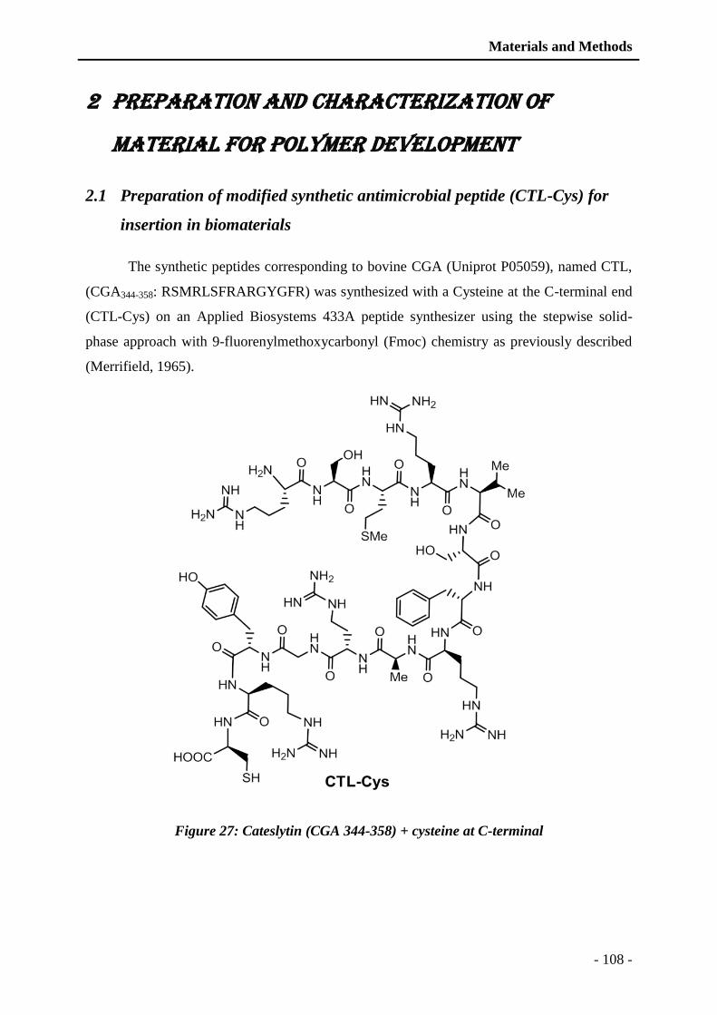
Materials and Methods
- 108 -
2 Preparation and characterization of
material for polymer development
2.1 Preparation of modified synthetic antimicrobial peptide (CTL-Cys) for
insertion in biomaterials
The synthetic peptides corresponding to bovine CGA (Uniprot P05059), named CTL,
(CGA344-358: RSMRLSFRARGYGFR) was synthesized with a Cysteine at the C-terminal end
(CTL-Cys) on an Applied Biosystems 433A peptide synthesizer using the stepwise solid-
phase approach with 9-fluorenylmethoxycarbonyl (Fmoc) chemistry as previously described
(Merrifield, 1965).
Figure 27: Cateslytin (CGA 344-358) + cysteine at C-terminal
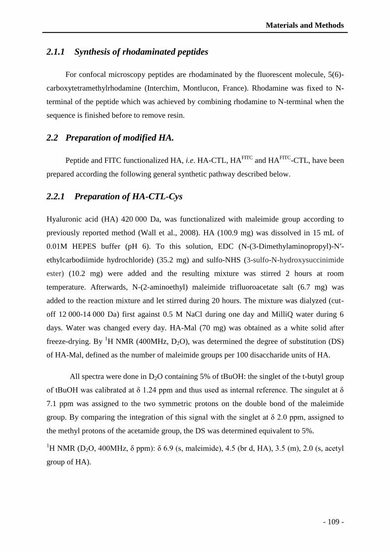
Materials and Methods
- 109 -
2.1.1 Synthesis of rhodaminated peptides
For confocal microscopy peptides are rhodaminated by the fluorescent molecule, 5(6)-
carboxytetramethylrhodamine (Interchim, Montlucon, France). Rhodamine was fixed to N-
terminal of the peptide which was achieved by combining rhodamine to N-terminal when the
sequence is finished before to remove resin.
2.2 Preparation of modified HA.
Peptide and FITC functionalized HA, i.e. HA-CTL, HAFITC
and HAFITC
-CTL, have been
prepared according the following general synthetic pathway described below.
2.2.1 Preparation of HA-CTL-Cys
Hyaluronic acid (HA) 420 000 Da, was functionalized with maleimide group according to
previously reported method (Wall et al., 2008). HA (100.9 mg) was dissolved in 15 mL of
0.01M HEPES buffer (pH 6). To this solution, EDC (N-(3-Dimethylaminopropyl)-N′-
ethylcarbodiimide hydrochloride) (35.2 mg) and sulfo-NHS (3-sulfo-N-hydroxysuccinimide
ester) (10.2 mg) were added and the resulting mixture was stirred 2 hours at room
temperature. Afterwards, N-(2-aminoethyl) maleimide trifluoroacetate salt (6.7 mg) was
added to the reaction mixture and let stirred during 20 hours. The mixture was dialyzed (cut-
off 12 000-14 000 Da) first against 0.5 M NaCl during one day and MilliQ water during 6
days. Water was changed every day. HA-Mal (70 mg) was obtained as a white solid after
freeze-drying. By 1H NMR (400MHz, D2O), was determined the degree of substitution (DS)
of HA-Mal, defined as the number of maleimide groups per 100 disaccharide units of HA.
All spectra were done in D2O containing 5% of tBuOH: the singlet of the t-butyl group
of tBuOH was calibrated at δ 1.24 ppm and thus used as internal reference. The singulet at δ
7.1 ppm was assigned to the two symmetric protons on the double bond of the maleimide
group. By comparing the integration of this signal with the singlet at δ 2.0 ppm, assigned to
the methyl protons of the acetamide group, the DS was determined equivalent to 5%.
1H NMR (D2O, 400MHz, δ ppm): δ 6.9 (s, maleimide), 4.5 (br d, HA), 3.5 (m), 2.0 (s, acetyl
group of HA).
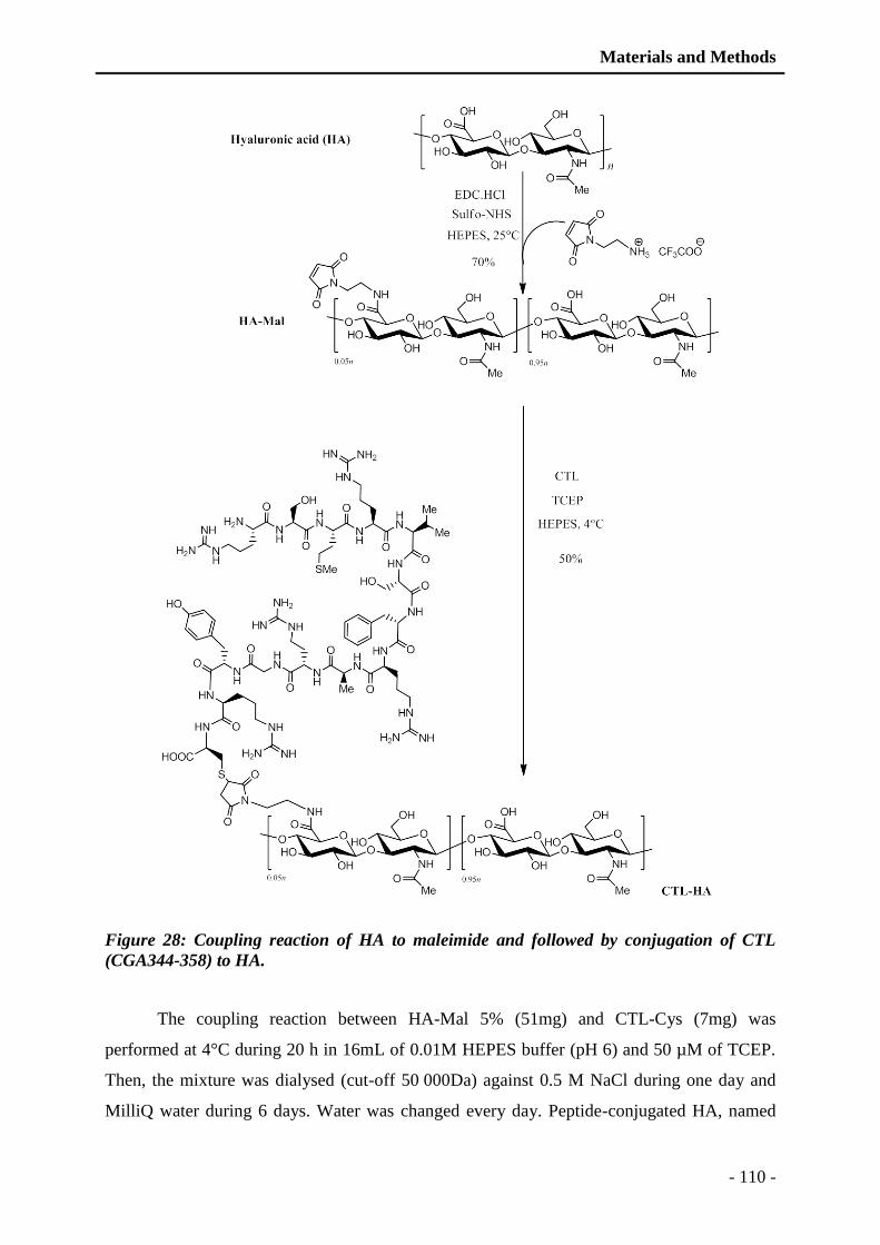
Materials and Methods
- 110 -
Figure 28: Coupling reaction of HA to maleimide and followed by conjugation of CTL
(CGA344-358) to HA.
The coupling reaction between HA-Mal 5% (51mg) and CTL-Cys (7mg) was
performed at 4°C during 20 h in 16mL of 0.01M HEPES buffer (pH 6) and 50 µM of TCEP.
Then, the mixture was dialysed (cut-off 50 000Da) against 0.5 M NaCl during one day and
MilliQ water during 6 days. Water was changed every day. Peptide-conjugated HA, named
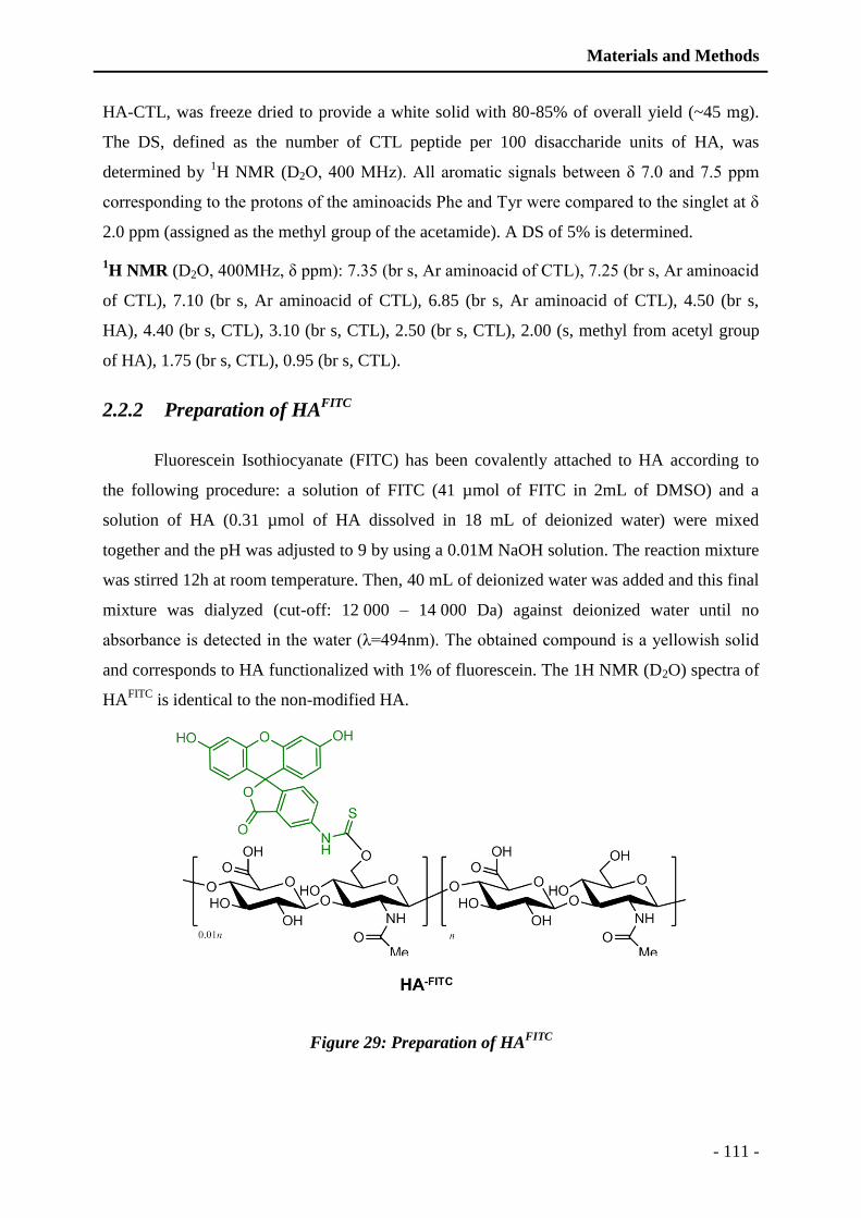
Materials and Methods
- 111 -
HA-CTL, was freeze dried to provide a white solid with 80-85% of overall yield (~45 mg).
The DS, defined as the number of CTL peptide per 100 disaccharide units of HA, was
determined by 1H NMR (D2O, 400 MHz). All aromatic signals between δ 7.0 and 7.5 ppm
corresponding to the protons of the aminoacids Phe and Tyr were compared to the singlet at δ
2.0 ppm (assigned as the methyl group of the acetamide). A DS of 5% is determined.
1H NMR (D2O, 400MHz, δ ppm): 7.35 (br s, Ar aminoacid of CTL), 7.25 (br s, Ar aminoacid
of CTL), 7.10 (br s, Ar aminoacid of CTL), 6.85 (br s, Ar aminoacid of CTL), 4.50 (br s,
HA), 4.40 (br s, CTL), 3.10 (br s, CTL), 2.50 (br s, CTL), 2.00 (s, methyl from acetyl group
of HA), 1.75 (br s, CTL), 0.95 (br s, CTL).
2.2.2 Preparation of HAFITC
Fluorescein Isothiocyanate (FITC) has been covalently attached to HA according to
the following procedure: a solution of FITC (41 µmol of FITC in 2mL of DMSO) and a
solution of HA (0.31 µmol of HA dissolved in 18 mL of deionized water) were mixed
together and the pH was adjusted to 9 by using a 0.01M NaOH solution. The reaction mixture
was stirred 12h at room temperature. Then, 40 mL of deionized water was added and this final
mixture was dialyzed (cut-off: 12 000 – 14 000 Da) against deionized water until no
absorbance is detected in the water (λ=494nm). The obtained compound is a yellowish solid
and corresponds to HA functionalized with 1% of fluorescein. The 1H NMR (D2O) spectra of
HAFITC
is identical to the non-modified HA.
Figure 29: Preparation of HAFITC

Materials and Methods
- 112 -
2.2.3 Preparation of HAFITC
-CTL
HAFITC
-CTL polymer has been prepared from HA-Mal. This polymer was labeled with
1% FITC has described above to provide HAFITC
-Mal. Then, CTL-Cys peptide reacted with
free maleimide group to lead to HAFITC
-CTL as a yellow solid. The procedure used to get
HAFITC
-CTL was identical to the one described above to prepare HA-CTL. 1H NMR (D2O)
spectra of HAFITC
-CTL was identical to the spectra of HA-CTL.
Figure 30: Preparation of HAFITC
-CTL
2.3 Gel filtration chromatography
Gel filtration chromatography was used to perform a rapid control of the HA and HA-CTL
prepared. For chromatography HPLC DIONEX-Ultimate 3000 (Dionex, Sunnyvale USA)
system was used, instead of using dual gradient only one solvent system was used which
comprises of 100% H2O. A constant flow rate of 1 mL was used and absorption was
monitored at 214 nm. Gel filtration column used was TSK-GEL G3000PWXL (TOSOH
Materials and Methods
- 113 -
Bioscience; Zettachring, Stuttgart, Germany) (0.75 x 30 cm: 6 µm particle size). All the
elutions were performed at room temperature.
2.4 PEM films buildup
2.4.1 Polysaccharide solutions
Chitosane (CHI, PROTASAN Ultrapure Chitosan CL213, 260 000 g/mol, DA 83 %)
was purchased by Novamatrix (Sandvika, Norway). Dried Sodium Hyaluronate (HA, 420 000
g/mol) was purchased by Lifecore (Chaska, USA). Poly (ethylene imine) (PEI, 60 000 g/mol,
50% in water), sodium chloride, sodium dodecyl sulfate (SDS), HCl and NaOH were
purchased by Sigma-Aldrich (Quentin-Fallavier, France). All products were used without
further purification. Both solutions were prepared by dissolution of 0.3 mg/mL in 150 mM
NaCl aqueous solution prepared using ultrapure Milli-Q® water having a resistivity of 18.2
MΩ.cm and were adjusted at pH 4 with NaOH or HCl solutions. PEI solution was prepared at
0.5 mg/mL in 150 mM NaCl solution and used without adjusting the pH.
2.4.2 Film buildup
Before PEM buildup, glass coverslips with a 14 mm in diameter (VWR, Strasbourg,
France) were cleaned in a SDS solution at 10-2
M, ultrasonicated during 5 min and then
submitted to a hot 0.1 M HCl solution during 10 min and finally rinsed with ultrapure Milli-
Q® water. A precursor PEI layer was adsorbed with an adsorption time of 15 min followed by
a rinsing step of 5 min. CHI/HA films were deposited by using an automated spraying device
described in a previous work (Cado et al., 2012). CHI/HA-CTL and CHI/HAFITC
-CTL films
have been prepared in 24 wells plates on glass coverslips. 300 µl of polycation solution has
been deposited for 5 min on glass coverslips followed by three rinsing steps (1 mL) with ultra
Milli Q water. Then, 300 µl of polyanionic solution has been added for 5 min followed by
three rinsing steps (1 mL) with ultra Milli Q water. To test the antimicrobial activity of PEM
films depending of the number of HA-CTL layers, PEI-[HA/CHI]15-n[HA-CTL/CHI]n with n
equal to 0, 5, 10 and 15 and PEI-[HA-CTL/CHI]30 films were built.
Materials and Methods
- 114 -
2.5 Physico-chemical characterization of the polymer films
2.5.1 Quartz Crystal Microbalance
The construction of multilayer films was monitored in situ by quartz crystal
microbalance (QCM) (Q-Sense E4, Goteborg, Sweden). The measurement methodology has
been addressed in details elsewhere (Boulmedais et al., 2004). Briefly, after cleaning of the
crystal with a 2% (v/v) Hellmanex solution during half an hour, rinse with Milli-Q water,
further immersion in a 0.1M HCl solution and intensive rinse with Milli-Q water, 600 μL of
NaCl solution were injected into the measurement cell. After stabilization of the signals, 600
μL of the polycation solution containing NaCl were injected, left in the cell for 5 min, and
rinsed with the NaCl solution and left again for 5 min. The frequency shifts were recorded
during the whole process. The same procedure was used for the deposition of the polyanion.
A negative value of Δfn (or of the normalized shift Δfn/n) reveals a decrease of the resonance
frequency during the deposition process and an increase of the mass adsorbed.
2.5.2 Atomic Force Microscopy (AFM)
Atomic force microscopy measurements (AFM) were realized with a Veeco
Multimode Nanoscope IIIA (Digital Instrument) in the wet state and in PeakForce TappingR
mode (Bruker) using cantilevers with a spring constant of 3 N/m.
2.5.3 Confocal Laser Scanning Microscopy (CLSM)
Confocal laser scanning microscopy (CLSM) observations were carried out with a Zeiss
LSM-510 microscope using a 63x/1.4 oil immersion objective and a 0.43-mm z-section
interval. FITC fluorescence was detected upon excitation at 488 nm, through a cut-off
dichroic mirror and an emission band-pass filter of 505–530 nm (green).
Materials and Methods
- 115 -
3 Methods of proteomic analysis
3.1 Sequence characterization by Edman sequencing
Sequencing was used to perform a rapid control of the amino acid sequence of the
leucotoxins purified, synthetic peptides and for the identification. Edman sequencing is
recurrent process of detaching a single amino acid starting from the N-terminal of the proteins
and comparing a satandard of amino acids. Degradation analysis is based on the reaction of
Edman (Edman, 1949).
The N-terminal sequencing was carried out by the method of automatic Edman
sequencing with a bi-sequencer cartridge (Procise 473A, Applied Biosystems, Foster city,
USA). All the products for the peptide sequencing were purchased from Applied Biosystem
(Applied Biosystem, Warrington, UK). The sensitivity of detection is in Picomoles (5-
10pmol: 0.01-0.02mL). Samples were deposited on a fiber filter by repeating cycles of sample
addition (10µl volume) and drying by argon gas. Fiber filters were previously treated with
10µL of polybrene, (BioBrène plus, Applied Biosystems) was used. Amino acids are
identified as Phenylthiothiohydantoine-amino acids (PTH-aas) by HPLC on reverse phase
column coated with C18 (PTH-C18, 2.1 x 200µm, Applied Biosystems, Warrington, UK) at a
temperature of 55°C. Solvents used for elution consists of dual solvent system comprising:
solvent A (Tetrahydrofurane 3.5% and Premix 2% in water) and solvent B (Acetonitrile 88%
and Propanol 12%). Detection is performed by measuring the absorbance at 269 nm and the
identification of PTH-ass is done by comparison with the chromatogram of a standard mixture
of different PTH-aas.
For sequences identification a query was made in the database SWISS-prot
(http://www.expasy.org/tools/Blast/). The protein characteristic parameters are given by the
(http://www.uniport.org/)
3.2 Western blot analysis of the PMNs secretion
o Gel electrophoresis
Western blot analysis was carried out by the use of the SDS-PAGE (sodium dodecyle
sulphate polyacrylamide gel electrophoresis) by the protocol previously described (Laemmli,
1970). For electrophoresis 4-15% polyacrylamide gel was prepared. Desalted PMNs secretion
were used, which were prepared by adding Laemmli buffer (4x) (Laemmli, 1970) (SDS 3%,
Materials and Methods
- 116 -
EDTA 1mM, Tris HCl 10mM, DDT 20mM, glycerol 40%, bromophenol blue 0.025% at pH
6.8). Samples were denatured by heating at 95°C for 6 min. Migration was achieved at 45V
for 30 min and followed by 110 V till the migration was completed till the lower end of the
gel. Migration buffer composed of glycine 192 mM, SDS 0.1%, Tris HCl 0.025M, pH 8.2.
Mixture of known molecular markers was used to determine the mass of the proteins
identified and in case of PMNs secretions soluble protein rich granules of the chromaffin cells
were used as positive control which is rich in chromogranins.
o Electro-transfer onto PVDF membrane
Transfer was achieved by subjecting to 75 V for 50 min. PVDF membrane (Amersham
Biosciences, Little Chalfon, UK) with a pore size of 0.22 µm was used for transfer. Transfer
buffer composed of 20% of the migration buffer, 10% methanol and 70% water.
o Immuno detection
For immunodetection Millipor Snap i.d protein detection system was used using 0.5 %
BSA (bovine serum albumin) (Euromedex, Muldosheim, France) in PBS as blocking agent.
Proteins were immunodetected by the antibodies specific to the proteins, two type of
antibodies were used monoclonal anti-CGA (anti-CgA47–68) and polyclonal anti-CgB (anti-
CGB547-560) for CGA and CGB respectively. ECL conjugated antibodies were used as
secondary antibodies: anti-mouse IgG and anti-rabbit IgG ((GE Healthcare, Little Chaflond,
UK) for anti-CgA and anti-CgB respectively. Finally, detection was achieved by the use of
SuperSignal (SuperSignal West Femto Maximum Sensitivity Substrate, Thermoscientific,
Rochford, USA) and visualized by the use of ChemiDOC (BioRad).
3.3 Matrix assisted laser desorption ionization time-of-flight Mass
spectrometry (Maldi-TOF)
Mass determination was carried out on a Brucker BIFLEX™ matrix-assisted laser
desorption time-of-flight mass spectrometer (MALDI-TOF) equipped with the high resolution
optics (SCOUT™) with X-Y multi-sample probe, a grid less reflector and with the HIMAS™
linear-detector. With a maximum accelerating potential of 30 kV, the system can be operated
either in linear or the reflective mode. Ionization was carried out with a 148 337-nm beam
from a nitrogen laser with a repetition rate of 3 Hz. The output signal from the detector was
digitized at a sampling rate of 250 MHz in linear mode and 500 MHz in reflector mode using
a 1-GHz digital oscilloscope (Lecroy model). For the data processing and the instrumental
control, software supplied by Bruker using a Sun sparcworkstation was accomplished (Pons et
Materials and Methods
- 117 -
al., 2002). These studies were realized using the matrix α-cyano-4-hydroxycinnamic acid,
obtained from Sigma, and prepared as a saturated solution in acetone. A total of 1-2 Nl of the
sample matrix aliquot solution was deposited on the probe and air dried by ambient air. A thin
layer was obtained of matrix crystal after fast spreading and evaporation (Kussmann et al.,
1997). A micro-molar analyte solution was applied to the matrix and allowed to dry under
moderate vacuum. The whole preparation was washed by 1 μl of a 0.5% trifluoroacetic acid
aqueous solution. This cleaning procedure often leads to an increase in sensitivity and mass
accuracy by removing the remaining alkali cations
3.4 Nano LC-MS/MS Mass spectrometry analysis
Secretions were digested by the modified porcine trypsin (promega, Madison, WI, USA)
in 25 mM NH4HCO3 at 37°C for 14 hrs. The generated peptides were analyzed directly by
NanoLC-MS/MS on an Agilent 1100 series HPLC-Chip/MS system (Agilent Technologies,
Palo Alto, USA). The voltage applied to the capillary cap was optimized to -1850V. For
tandem MS experiments, the system was operated with automatic switching between MS and
MS/MS modes. The three most abundant peptides, performing doubly charged ions, were
selected on each MS spectrum for further fragmentation. The MS/MS scanning was
performed in the ultra-scan resolution mode at a scan rate of 26.000m/z per second. A total of
six scans were averaged to obtain a MS/MS spectrum. The complete system was fully
controlled by ChemStation (Agilent Technologies) and EsquireControl (BrukerDaltonics)
software. Mass data collected during nanoLC-MS/MS analysis were processed before
interpretation using a local MascotTM
(Matrix Science, London, UK) server. The searches
were performed against the NCBI database (Delalande et al., 2005).

Materials and Methods
- 118 -
4 Methods to characterize biological
activities
4.1 Flow-cytometry measurements:
For the flow cytometry PMNs were suspended at a concentration of 5x 10⁵ cells per
mLin the EGTA buffer (pH 7.3 adjusted with Tris HCl) containing EGTA 0.1 mM, Hepes
10mM, NaCL 140mM, KCl 5mM, Glucose 10 mM. Flow cytometry measurements were
carried out using a FacSort flow cytometer (Becton-Dickinson, Le pont de Claix, France)
equipped with a 15mW argon laser tuned to 488 nm (Meunier et al., 1995).
Activity of leucotoxins (E-D) on the human PMNs was evaluated using flow-
cytometry of cells previously incubated with Ethidium Bromide (25nM) (Moussa et al., 2006)
for 10 min before addition of LukE/LukD. Pore formation by the toxins was revealed by the
penetration of ethidium through the pores. Ethidium fluorescence was measured using Cell
QuestProTM
software (Becton, Dickinson and company, Franklin Lakes-USA) by using (FL3:
λEm = 650nm) (Meunier et al., 1995). Each component constituting the leucotoxins was
generally added at concentrations as follows: 2nM, 3nM, 6nM, 10nM, 20nM, 40nM and
60nM. First image was taken at the time of addition of toxins then after 5 min fluorescence
was recorded till 30 min of time. Fluorescence is increased when the molecule entered the
cells by the pores formed through the plasma membrane and combined with nucleic acids. As
positive control, the fluorescence corresponding to the dead cells was considered with a value
of 100. Fluorescence without toxins was subtracted from the data and the results are presented
as percentile by take positive control as 100%. Kinetics of the intracellular calcium were
measured by using flow-cytometry of the cells previously loaded with 5 µM of Fluo-3
(Molecular Probes, New Brunswick, USA) during 1 h at 37°C and the washed and
resuspended in EGTA buffer supplemented with 1.1mM Ca2+ (Moussa et al., 2006). Each
component constituting the leucotoxins was generally added at concentrations as follows:
2nM, 3nM, 6nM, 10nM, 20nM, 40nM and 60nM. Fluo-3 measurement were carried out from
the fluorescence light 1(FL1: λEm = 530 nm) using cell Quest ProTM software (Becton-
Dickinson, Le Pont de Claix, France)(Meunier et al., 1995) for every 30 seconds during 10
minutes. Data provided is the average of at-least three individual experiments.
Materials and Methods
- 119 -
4.2 Analysis of antimicrobial activity
Antimicrobial activities of the peptides and secretion of the PMNs were analyzed. For
the demonstration of activity two types of assays were carried out: antibacterial activities and
the antifungal activities. Bacterial strains used are as follows: Micrococcus luteus (A270),
Staphylococcus aureus (ATCC25923, ATCC49775, S1, S2) and E. coli (ATCC 25922).
Fungal and yeast strains used to demonstrate antifungal activities were: Neurospora crassa
(CBS 327–54), Aspergilus fumigatus, Trichophyton mentagrophytes, Candida albicans,
Candida glabrata and Candida tropicalis.
4.2.1 Antibacterial test
Bacterial strains were precultured aerobically at 37°C in a Mueller-95 Hinton-Broth
medium (Difco Laboratories, Detroit, MI), pH 7.3. For which two isolated S. aureus strains
were plated on the agar plates and cultivated for 24 h at 37°C. After incubation, one colony
per isolate was transferred to 10 mL of the Mueller-Hinton-Broth medium and incubated at
37°C for about 18-22 h to the stationary growth phase.Bacteria were suspended in the
Mueller-Hinton Broth medium, and antibacterial activity was tested by measuring the
inhibition of bacterial growth. 10 μL final volumes of synthetic peptides or the polymer (HA-
CTL-C) and 20µL in case of PMNs secretion fractions purified by RP-HPLC were incubated
in microtitration plates (Falcon, Becton Dickinson, USA) with 90 μl of a mid-logarithmic
phase culture of bacteria, with a starting absorbance of 0.001 at 620 nm. To test the
antibacterial and properties of the multilayer films, PEI-(HA/CHI)15-n-(HA-CTL/CHI)n with
n = 0,3, 5, 10, 15 and PEI-(HA-CTL/CHI)30 films were prepared. 400 μL of a mid-
logarithmic-phase culture of bacteria or yeast with OD620 of 0.001 were placed in 24-well
plate containing multilayer films. In the initial inoculums with absorbance of 0.001, bacteria
were quantified by agar plate spreading method which was 5x10⁵ Colony forming unit
(CFU)/mL (NCCLS, 2003). Tetracycline (10 μg/mL) and Cefotaxime (0.1 μg/mL) were used
as positive control. Microbial growth was assessed by the increase of absorbance after 16 h
incubation at 37°C (Bulet et al., 1993; Wu and Hancock, 1999). The A620 nm value of
control cultures growing in the absence of peptide and antibiotics was taken as 100% growth
and when the A620 nm is zero with the antibiotics (Tetracycline and Cefotaxime) was taken
as 100% inhibition. Absence of bacterial growth was verified by agar plate spreading. Each
assay was verified three times at least.
Materials and Methods
- 120 -
4.2.1.1 Kinetics of bacterial growth inhibition
For the bacterial growth inhibition kinetics in respect to time, the initial inoculum was
prepared at concentration of 5x10⁵ CFU/mL (NCCLS, 2003) (OD620nm = 0.001) as
described above. Quantification was performed by converting OD to bacterial count by
devising a conversion factor (Lee et al., 2009). Briefly, OD of fresh bacteria culture was
measured at 620 nm and was plated to MH agar for 24 h at 37°C. Colony count was
performed and plotted versus the OD620 and the gradient of the linear line was taken as
conversion factor. Later on intermittent controls were performed by colony counting to verify
the accuracy of conversion factor. Quantification was performed at different time intervals
(0h, 1h, 2h, 3h, 4h, 5h, 6h, 7h, 8h, and 24h). For each tested film, 100 μL of the supernatant
was taken to measure the OD620 of the supernatant by a microplate reader. The withdrawn
volume was compensated with the appropriated fresh bacteria or yeast solution of the same
measured OD620. Intermittent controls were performed by inoculating the culture medium on
MH agar plates by spreading method and counting colonies for verification at different steps.
Several controls were used: fresh medium without inoculation of pathogens was used to
ensure sterility, mixture of Tetracycline (10 μg/mL) and Cefotaxime (0.1 μg/mL) were used
as positive control (90 μL of culture and 10 μL of antibiotics) and fresh inoculated culture
medium without any addition was taken as negative control. Each assay was performed in
triplicate and the experiments were repeated minimum three times.
4.2.2 Antifungal activity
To analyze the antifungal activities of the synthetic peptides, PMNs secretion or the
polymer either in solution or the multilayered film, two types of fungi were used: Moulds
(filamentous fungi) and yeasts. Filamentous fungi used were Neurospora crassa (CBS 327–
54) and Aspergilus fumigatus, and three candida strains were used C. albicans C. glabrata, C.
tropicalis.
o Filamentous fungi:
For the filamentous fungi, spores were suspended at a concentration 104 spores/mL, in a
growth medium containing Potato Dextrose Broth (PDB) (Difco, Becton Dickson
Microbiology system) in half-strength, supplemented with Tetracycline (10 μg/mL) and
Cefotaxime (0.1 μg/mL) to suppress bacterial growth (Broekaert et al., 1990). Synthetic
peptides and the polymers in solution form (10 µL) and aliquots of RP-HPLC fractions (20
μL) of PMNs secretions were incubated in micro-titration plates (Falcon, Becton Dickinson,
Materials and Methods
- 121 -
USA) with 90 and 80μL respectively, of fungal spores already diluted. The suspension was
incubated at 30 C° for 24-72 h without agitation (Broekaert et al., 1990; Wu and Hancock,
1999). Growth of fungi was monitored microscopically after 24 h in case of Neurospora and
Aspergilus spp. and 72 h in case of Trichophyton spp. Inoculated media was used as negative
control and was taken as 100% fungal growth and voriconazole (1µg/mL) was used as
positive control which was taken as 100% growth inhibition.
o Yeasts:
For the yeast spp. method used was the same as previously described for the antibacterial
assay with few exceptions. Broth medium used was sabouraud medium (BioMerieux S.A.,
Marcy l’Etoile, France) and the incubation temperature was modified to 30°C instead of 37°
as in case of bacterial strains with agitation. Inoculated media (OD620nm = 0.001) was used as
negative control and was taken as 100% fungal growth and voriconazole (1µg/mL) was used
as positive control which was taken as 100% growth inhibition.
4.2.2.1 Kinetics of fungal growth inhibition
For the quantification of fungal growth with respect to time Candida albicans was used.
Protocol used was the same as previously described for the antibacterial assay with few
exceptions. Broth medium used was sabouraud medium (BioMerieux S.A., Marcy l’Etoile,
France) and the incubation temperature was modified to 30°C instead of 37° as in case of
bacterial strains with agitation. For the colony counting sabouraud agar (BioMerieux S.A.,
Marcy l’Etoile, France) was used.
4.3 Confocal laser scanning microscopy(CLSM)
To follow the interaction of fluorescently labeled HAFITC
-CTL in solution and HAFITC
-
CTL/CHI films with C. albicans, CLSM was used a protocol previously described with few
modifications(Lugardon et al., 2001). Briefly for experiments done with HAFITC
-CTL in
solution, Poly-L-Lysine coated microscopic slides were covered with fresh medium
containing C. albicans (OD620 = 0.001) in SB culture medium and incubated during 24 h at
30°C without agitation. The medium was then removed and replaced by 50 μM (in peptide) of
HAFITC
-CTL. After an incubation of 45 min, the slides were washed with fresh culture
medium and subsequently treated for 30 min with 4% paraformaldehyde in phosphate buffer
saline (PBS) at pH 7.3.
Materials and Methods
- 122 -
After several rinsing with PBS, slides were covered with Moviol 4-88 (Aldrich,
Steinheim, Germany). For experiments done with HAFITC
-CTL/CHI, fresh C. albicans
medium, previously incubated at OD620 =0.001 in SB culture medium during 24 h at 30°C
without agitation, was put in contact with PEI-(HAFITC
-CTL/CHI)15 films during 45 min at
30°C without agitation. HAFITC
in solution and HAFITC
/CHI films were used as control. C.
albicans were subjected to optical serial sectioning (0.2-0.3 μm) to produce image in the x-y
plane. Each optical section was scanned several times to obtain an average image. Images
were recorded digitally in a 768 x 576 pixel format.
4.4 Degradation analysis of synthetic peptides by S. aureus strains
S. aureus strains (ATCC25923, ATCC49775, S1 and S2) were precultured in MH broth
medium at 37°C for 18-20h. Preculture was diluted to OD620nm 0.001, which contains 5x10⁵
CFU/mL. Synthetic peptides were incubated with the diluted preculture at ½ MICs as
previously determined. Suspension of bacteria and peptides were incubated at 37°C with
agitation for 18-20 h. Triplicate wells were treated for each concentration of peptide. Media
was sampled at different time intervals (0h, 2h, 4h, 8h, and 20h). Samplings were centrifuged
at 10000 x g for 15 min and the supernatants were filtered using Millex®-GV 0.22 μM
(Millipore, Carrigtwohill, Ireland) to eliminate bacteria. The supernatants were stored -20°C
until further use. In order to check sterility, 1 mL of each supernatant was incubated at 37°C
for 72 h. Absence of a colony was interpreted as lack of viable bacteria. In parallel, several
controls were performed: i) without peptides for bacterial growth control and ii) without
bacteria, with peptides in the MHB medium.
4.4.1 Purification of bacterial supernatants by RP-HPLC
Bacterial supernatants were analyzed by using RP-HPLC to separate in different
fractions for comparison. RP-HPLC system used was the same as described for PMNs
secretions except the change of gradient which is indicated on the relevant chromatograms.
Fractions were collected according to one minute time interval.
4.5 PSG degradation by the V8 protease
Secreted proteins from stimulated chromaffin cells were isolated from bovine adrenal
medulla as previously described (Metz-Boutigue et al., 1998). The soluble material (1 mg)
was incubated for 18 h at 37°C with S. aureus V8 protease Glu-C (E.C. 3.4.21.19;
Materials and Methods
- 123 -
Boehringer, Mannheim, Germany) at a protein-to enzyme weight ratio of 50:1 in 200 μL of 50
mM Tris-HCl, pH 8.3. The reaction was stopped by addition of 0.1% (v/v) trifluoroacetic acid
(TFA) and concentrated by evaporation, but not to dryness, using a speed-vac centrifuge.
4.6 Human gingival fibroblasts viability assays
The biocompatibility of films was tested using Human gingival fibroblasts (HGFs).
HGFs were extracted from human gingival connective tissue of healthy individuals according
to a protocol approved by the ethics committee for patient protection of CPP Strasbourg
Hospitals. Cells were grown in DMEM containing, 1g/L of glucose and supplemented with
10% fetal bovine serum and 100 IU/mL penicillin-streptomycin (all from GibcoR). Cells were
cultured at 37°C in 5% CO2 in 75 cm2 flasks. HGF were used between the 6th and the 9th
passage. Before cell seeding, glass coverslips (O = 14 mm) coated with different architectures
were irradiated by UV for 15 min. Then HGFs were seeded at 3x10⁴ cells per cm
2 and
cultivated at 37°C under a 5% CO2 humidified atmosphere for different culture times (Day 1,
Day 2 and Day 7).
Cell viability was assessed by Alamar BlueTM assay (Biosource International). This
assay is based on the reduction of the blue, non-fluorescent resazurin dye to the pink and
fluorescent resorufin dye by living cells (Page et al., 1993). The overall conversion rate is
proportional to the metabolic activity of living cells (Voytik-Harbin et al., 1998). Viability
was assessed for different times 1, 2 and 7 days.
After rinsing, cells with PBS, 10% reagent in complete medium were incubated for 2 h.
After incubation optical density (OD) at 570 nm and 630 nm were determined with a
microplate reader. The percentage of reduction of Alamar BlueTM was calculated according
to the procedure provided by the manufacturer. The experiments were performed in triplicate.


Part: III Results and Discussion

Manuscript: I Proteomic analysis of activated neutrophils by LukE/D


Results and Discussion
- 124 -
Activation of neutrophils by the two-component
leukotoxin LukE/D from Staphylococcus aureus:
proteomic analysis of the secretions
(Under revision in Journal of proteome research)
Rizwan Aslam ‡, Benoît-Joseph Laventie
≠†, Céline Marban
‡, Gilles Prévost
≠, Daniel Keller
≠, Jean-Marc Strub
§, Alain van Dorsselaer
§, Youssef Haikel
‼, Corinne Taddei
‼ and Marie-
Hélène Metz-Boutigue ‡*
‡ Inserm UMR 1121, Université de Strasbourg, Strasbourg, F-67000, France
≠ EA 7290 Virulence bactérienne précoce, Fédération de Médecine Translationnelle de
Strasbourg, Université de Strasbourg - CHRU Strasbourg, Institut de Bactériologie, F-67000
Strasbourg, France
§ CNRS UMR 7178, Université de Strasbourg, Strasbourg, F-67000, France
‼ Faculté d’Odontologie, Université de Strasbourg, Strasbourg, F-67000, France
KEYWORDS: Staphylococcus aureus, Leukotoxin LukE/D, Neutrophils, Chromogranin A/B,
Proteomic Analysis, Antimicrobial Peptides, Innate Immunity.
Results and Discussion
- 125 -
ABSTRACT
Staphylococcus aureus is responsible for severe bacterial infections in hospitals and
healthcare facilities. It produces single and bi-component toxins (leukotoxins and hemolysins)
that hinder innate immune function. Leukotoxin subunits bind to leukocyte cell membrane
thus inducing transmembrane pores and subsequently, cell lysis. Leukotoxin LukE/D is a
member of the bi-component toxin family, but to date, no study concerning its involvement in
host-pathogen interactions has been reported. In the present study, we performed the
proteomic analysis of the secretions recovered after activation of human neutrophils by
leukotoxin LukE/D. The neutrophil secretions were purified by RP-HPLC and different
fractions were analyzed by Edman sequencing, LC-MS/MS, immunoblotted for
chromogranin-derived peptides and further analyzed for antimicrobial properties. Proteomic
analysis revealed that neutrophil secretions constitute a large number of proteins related with
immune boosting mechanisms, proteolytic degradation, inflammatory process and antioxidant
reactions.

Results and Discussion
- 126 -
INTRODUCTION
Staphylococcus aureus (S. aureus), a commensal bacterial strain present in about 20–30%
of the general population, is responsible for numerous nosocomial infections.1 It causes a
wide range of illnesses from minor skin infections to some of life-threatening diseases, such
as sepsis, osteomyelitis, pneumonia, endocarditis, and meningitis.2 Mostly, S. aureus infection
starts with bacterial adhesion to host tissues and continues with colonization.3, 4
Colonization
is associated with adhesion proteins that belong to the “Microbial Surface Components
Recognizing Adhesive Matrix Molecules” (MSCRAMMs) family. These proteins are
important for the attachment of bacteria to fibronectin5, plasma clot, etc.
6 In addition, S.
aureus produces several virulence factors, including toxins7 such as staphylococcal
enterotoxins (SEs), toxic shock syndrome toxin-1 (TSST-1), exfoliative toxins A and B (ETA
and ETB), α-, β-, γ- and δ-hemolysins, the bicomponent Panton-Valentine Leucocidin (PVL)8
and the LukE/D9 leukotoxin. LukE/D consists of two subunits: LukE and LukD. The toxin
subunits bind to leukocyte cell membranes, inducing Ca2+
activated trans-membrane pores and
subsequent cell lysis.9-11
Expression of LukE/D is associated with several common diseases.
As an example, it has been isolated with both epidermolysin A and B from 78% of
retrospective and prospective cases of impetigo.10
In addition, LukE/D has been isolated from
93.6% of patients with S. aureus associated post-antibiotic diarrhea, in combination with the
enterotoxins (80.9% of isolates).12
However, no study regarding the involvement of LukE/D
on the host-pathogen interactions have been reported so far.
Neutrophils (PMNs) are part of the first line of defense against pathogens, as they are
involved in infection clearing and contribute to the development of adaptive immunity.
Previous data indicate that in humans, chromogranins (CgA and CgB) are present in secretory
granules of PMNs13, 14
and are overexpressed in plasma of the patients with systemic
inflammatory response syndrome (SIRS) or sepsis.15
Several endogenous CgA- and CgB-
Results and Discussion
- 127 -
derived fragments have been described as antimicrobial peptides (AMPs).16
In addition to
their direct antimicrobial effect against pathogens, they activate PMNs,17
inducing a
calmodulin-mediated influx of calcium and therefore the secretion of innate immune
components such as AMPs and cytokines. 18
A comprehensive understanding of the
regulation of neutrophils responses could be obtained by proteomic analysis of the secretions
from activated neutrophils.19, 20
Here, by using proteomics and computational analysis, we
identified the secreted proteins from LukE/D-activated neutrophils in order to better define
the effects of the dynamic regulatory network of neutrophils.
Results and Discussion
- 128 -
MATERIALS AND METHODS
PREPARATION OF LEUKOTOXIN LUKE/D
LukE and LukD were purified from S. aureus Newman strain (NTCC 8178) according to
previous studies.9, 21, 22
Briefly, one colony was grown in 5 mL of YCP medium composed of
3% (w/v) yeast extract (Oxoid), 2% (w/v) Bacto-Casamino acids (Difco), 2% (w/v) sodium
pyruvate (Merck), 0.25% (w/v) Na2HPO4, 0.042% (w/v) KH2PO4, pH 7.0 at 37°C for 18 h
with vigorous shaking. Proteins were precipitated from the supernatant with the help of 80%
(w/v) ammonium sulfate, resuspended and subjected to a first chromatography on a 50 mL
XK26 SP Sepharose fast flow column onto an AktaPurifyer-10® (GE-Healthcare, Uppsala,
Sweden).21
The further purification was achieved by subjecting the eluted fractions separately
to a two-step chromatography including a 8 mL Mono S® cation exchange FPLC column
(GE-Healthcare, Uppsala, Sweden) followed by a 8 mL ReSource-Iso®
hydrophobic FPLC
column (GE-Healthcare, Uppsala, Sweden) conditioned into a HR-10®
column (GE-
Healthcare, Uppsala, Sweden).21, 22
Purified compounds were controlled by SDS-PAGE
(12%) on PHAST System (GE-Healthcare, Uppsala, Sweden) and solubilized in 50 mM Na-
phosphate buffer (pH 7.0) supplemented with 150 mM NaCl8, 9
at A280 nm of 1.0. LukE and
LukD were stored at -80°C until use. Purity and integrity of LukE and LukD were analyzed
by Edman sequencing and MALDI-TOF.
ISOLATION OF POLYMORPHONUCLEAR NEUTROPHILS
Human PMNs were prepared with a purity of 98% as previously described23, 24
from Buffy
Coat from healthy sex non-discriminated donors, obtained from the Centre de Transfusion
Sanguine de Strasbourg (France). Briefly, 12 mL of lympho-Prep solution (EuroBio,
Courtaboeuf Cedex B, France) was added to 30 mL of Buffy Coat from healthy donors and
centrifuged at 200 g for 20 min. After centrifugation the lower dark band was collected
without disturbing the sediment and washed with neutrophil buffer (EGTA buffer) (140 mM
Results and Discussion
- 129 -
NaCl, 5 mM KCl, 10 mM glucose, 0.1 mM EGTA (ethylene glycol-bis-(2-aminoethylether)
N,N,N`,N`-tetraacetic acid), 10 mM HEPES, 3 mM Tris-base, pH 7.3). PMNs were further
processed by dextran sedimentation25
for 30 min and then centrifugated at 1600 g for 6 min.
The remaining impurities of red blood cells were lysed by hypotonic shock in distilled water
(18 mL) for 45 s and immediately suspended in a 2 mL hypertonic NaCl (9% w/v) solution to
a concentration of 0.9% (w/v).26
Finally, cells were washed with mM EGTA buffer and
suspended at a concentration of 5 × 10⁶ cells/mL in the same buffer.
FLOW CYTOMETRY MEASUREMENTS
For the flow cytometry analysis, PMNs were cultured at a concentration of 5 x 10⁵ cells/mL
in EGTA buffer. Flow cytometry measurements were carried out using a FacSort flow
cytometer (Becton-Dickinson, Le Pont de Claix, France) equipped with a 1 mW argon laser
tuned to 488 nm.27
Activity of LukE/D on PMNs was evaluated using flow cytometry of cells
previously incubated with ethidium bromide (25 nM)28
for 10 min prior to addition of LukE
and LukD. Ethidium fluorescence was measured using the Cell QuestProTM software
(Becton, Dickinson and company, Franklin Lakes, USA) by using (FL3: λEm = 650 nm).27
Cells were supplemented with different concentrations of each leukotoxin component: 2, 3, 6,
10, 20, 40 and 60 nM. The first image was taken at the time of addition of the toxins.
Subsequently, fluorescence was recorded from 5 to 30 min post-treatment. As a control, the
fluorescence measuring for dead cells was considered as 100. Fluorescence measurements in
the absence of toxin were subtracted from the data and the results are presented as percentiles.
Intracellular calcium kinetics were measured by using flow cytometry with cells previously
loaded with 5 μM of Fluo-3 (Molecular Probes, New Brunswick, USA) for 1 h at 37°C,
washed and resuspended in 0.1 mM EGTA buffer supplemented with 1.1mM Ca2+
.28
Fluo-3
measurement were carried out every 30 s during 10 min from the fluorescence light 1 (FL1:
Results and Discussion
- 130 -
λEm = 530 nm) using the Cell Quest ProTM
software (Becton-Dickinson, Le Pont de Claix,
France)27
. Data represent an average of at least three independent experiments.
PREPARATION OF SECRETIONS AFTER PMNs STIMULATION BY
LEUKOTOXIN LUKE/D
PMNs were cultured in EGTA buffer solution at 107 cells/mL. The two components of
leukotoxin (LukE and LukD) were used each at 10 nM. Cells were stimulated with 1.1 mM
CaCl2 for 25 min at 37°C and centrifuged at 800 g for 10 min.28
The supernatants were
recovered and stored at -80°C.
RP-HPLC OF NEUTROPHILS SECRETIONS
Total protein concentration was measured using a Bradford Assay,29
and 1 mg of total
proteins was purified by RP-HPLC using a DIONEX Dual Gradient System (Dionex,
Sunnyvale USA) and a Nucleosil RP300-5C18 column (5 µm particle size, 300-Å porosity;
Macherey-Nagel, Hoerdt, France) at 20°C. Absorbance was monitored at 214 nm, at room
temperature (25° C). The solvent system consists of 0.1% (v/v) trifluoroacetic acid in water
(solvent A) and 0.1% (v/v) trifluoroacetic acid in 70% (v/v) acetonitrile-water (solvent B)
with a flow rate at 700 µL/min. A gradient with elution time reference is indicated on the
chromatograms. The fractions were collected every 1 min interval and concentrated by speed-
vac and stored at -20°C.
WESTERN BLOT ANALYSIS
Secretion samples were loaded on a 15% SDS-PAGE and transferred to PVDF membrane
(HybondTM
-P, Amersham Bioscience, Little Chalfont, Bucks, UK). During migration a
voltage of 50 V was applied for the first 30 min and then 110 V till end and 75 V during
transfer for 70 min. For immunodetection Millipore Snap i.d.® 2.0 protein detection system
(Millipore, Billerica, MA, USA) was used, using ChemiBlockerTM
as blocking agent. The
antibodies used are as follows: mouse monoclonal anti-CgA (anti-CgA53–57),13, 30
rabbit
Results and Discussion
- 131 -
polyclonal anti-CgB (anti-CgB547-560)31
. Immunolabelled bands were visualized by
autoradiography after addition of ECL reagent (Amersham Bio-science, Buckinghamshire,
UK).
ANTIBACTERIAL ASSAY
Micrococcus luteus (A270) and S. aureus (ATCC 25923) strains were used to assess the
antibacterial properties of the secreted material. Bacterial strains were precultured aerobically
at 37°C in a Mueller Hinton Broth (MHB) medium (Merck KGaA, Darmstadt, Germany), pH
7.3. For each isolate, one colony was transferred to 10 mL of MHB medium and incubated at
37°C for 18-20 h. Bacteria were resuspended in MHB medium, and antibacterial activity was
assessed by measuring the inhibition of bacterial growth. 10 to 20 μL of PMNs secretion
fractions purified by RP-HPLC were incubated with 90 μL of a mid-logarithmic phase culture
of bacteria, with a starting absorbance of 0.001 at 620 nm. In the initial inoculums, bacteria
were quantified to 5 x 10⁵ colony forming unit (CFU)/mL by agar plate spreading method.
32
Tetracycline (10 μg/mL) and Cefotaxime (0.1 μg/mL) were used as a positive control.
Microbial growth was assessed by the increase of absorbance after 20h incubation at 37°C.33,
34 The A620nm values of control cultures growing in the absence of secretions and antibiotics
were considered as 100% growth. Moreover, the A620nm value with the antibiotics
(Tetracycline and Cefotaxime) was considered as 100% inhibition (absence of bacterial
growth was confirmed by agar plate spreading). Each assay was performed at least three
times.
ANTIFUNGAL ACTIVITY
Neurospora crassa, Aspergillus fumigatus, Candida albicans, and Candida tropicalis
strains were tested to evaluate the antifungal properties for the secreted material.
For the filamentous fungi, spores were resuspended at a concentration of 104 spores/mL, in
Potato Dextrose Broth (PDB) medium (Difco, Becton Dickson Microbiology system, Sparks,

Results and Discussion
- 132 -
MD, USA) in half-strength, supplemented with Tetracycline (10 μg/mL) and Cefotaxime (0.1
μg/mL) to suppress bacterial growth.35
Aliquots of PMN secretions purified by RP-HPLC (20
μL) were incubated with 80 μL of fungal spores. The suspension was incubated at 30° C for
24 h without agitation.34, 35
Fungal growth was monitored microscopically after 24 h. Media
alone was used as negative control and corresponds to 100% fungal growth and voriconazole
(1 μg/mL) was used as a positive control and corresponds to 100% growth inhibition.
Concerning yeast, the protocol described above to test the antibacterial properties of the
secretions was used, with two exceptions: the culture medium was Sabouraud (BioMerieux
S.A., Marcy l’Etoile, France) and the incubation temperature was modified to 30°C with
agitation. Media alone was used as negative control and corresponds to 100% fungal growth
and voriconazole (1 μg/mL) was used as a positive control and corresponds to 100% growth
inhibition.
PROTEOMIC CHARACTERIZATION BY EDMAN SEQUENCING
The N-terminal sequencing was carried out by automatic Edman sequencing method, with a
bi-sequencer cartridge (Procise 473A, Applied Biosystems, Foster City, USA).13
For
sequences identification a query was made in the SWISS-prot database
http://www.expasy.org/tools/Blast/. The protein characteristic parameters were given by
http://www.uniport.org/
TRYPTIC DIGESTION
After RP-HPLC, collected fractions were concentrated using a SpeedVac. Collected fractions
were submitted to a reduction with 10 µl of 10 mM DTT in 25 mM NH4HCO3 (1 h at 57°C)
and alkylation with 10 µl of 55 mM iodoacetamide in 25 mM NH4HCO3 at room temperature.
For tryptic digestion, the trypsin (12.5 ng/μL; Promega V5111, Madison, WI, USA) was
suspended in 2 ml of 25 mM NH4HCO3.The enzymatic digestion was performed at 37°C

Results and Discussion
- 133 -
during 4 h at an estimated ratio enzyme/substrate 1/50. Before nano LC-MS/MS analysis, 1 µl
of H2O/TFA 0.1% was added and fractions were concentrated using a SpeedVac.
MASS SPECTROMETRY ANALYSIS
NanoLC-MS/MS was performed using a nanoACQUITY ultra performance liquid
chromatography (UPLC®) system (Waters, Milford, MA, USA) coupled to a maXis 4G Q-
TOF mass spectrometer (BrukerDaltonics, Bremen, Germany). The system was fully
controlled by HyStar 3.2 (BrukerDaltonics). The UPLC system was equipped with a
Symmetry C18 precolumn (20 х 0.18 mm, 5 µm particle size, Waters, Milford, USA) and an
ACQUITY UPLC® BEH130 C18 separation column (75 µm × 200 mm, 1.7 µm particle size,
Waters). The solvent system consisted of 0.1% formic acid in water (solvent A) and 0.1%
formic acid in acetonitrile (solvent B). 3 µL of each sample was injected. Peptides were
trapped during 1 min at 15 µL/min with 99% A and 1% B. Elution was performed at 60 °C at
a flow rate of 450 nL/min, using a linear gradient of 6-35% B over 28 minutes. The mass
spectrometer was operating in positive mode, with the following settings: source temperature
was set to 160°C while dry gas flow was at 5 liter per minute. The nanoelectrospray voltage
was optimized to −5000 V. External mass calibration of the TOF was achieved before each
set of analyses using Tuning Mix (Agilent Technologies, Paolo Alto, USA ) in the mass range
of 322-2722 m/z. Mass correction was achieved by recalibration of acquired spectra to the
applied lock masses (methylstearate ([M+H]+ 299.2945 m/z) and hexakis(2,2,3,3,-
tetrafluoropropoxy)phosphazine ([M+H]+ 922.0098 m/z)). For tandem MS experiments, the
system was operated with automatic switching between MS and MS/MS modes in the range
of 100-2500 m/z (MS acquisition time of 0.4 s, MS/MS acquisition time between 0.05 s
(intensity > 250,000) and 1.25 s (intensity <5000)). The 6 most abundant ions (absolute
intensity threshold of 1500), were selected from each MS spectrum for further isolation and
CID fragmentation using nitrogen as collision gas. Ions were excluded after acquisition of one

Results and Discussion
- 134 -
MS/MS spectrum and the exclusion was released after 0.2 minutes. Peak lists in mascot
generic format (.mgf) were generated using Data Analysis (version 4.0; Bruker Daltonics,
Bremen, Germany).
MS/MS DATA INTERPRETATION
The peak list has been searched against a NCBInr-derived combined target-decoy database
(created 2010-03-23, containing 219120 target sequences plus the same number of reversed
decoy sequences) using Mascot (version 2.4.1, Matrix science, London, England). During the
database search, up to one missed cleavage by trypsin and two variable modifications
(oxidation of Methionine (+16 Da), carbamidomethylation of Cysteine (+57 Da), were
considered. The search window was set to 15 ppm for precursor ions and 0.05 Da for
fragment ions. Mascot result files (.dat) were imported into the Scaffold 3 software (version
3.00.03; Proteome Software Inc., Portland, OR, USA) used to validate MS/MS based peptide
and protein identifications. Mascot identifications required at least ion minus identity scores
greater than 0 and ion scores greater than 40. Protein identification was accepted if the protein
contained at least 1 identified peptide. The false discovery rate (FDR) was calculated to be
<1% based on the number of decoy hits.

Results and Discussion
- 135 -
RESULTS:
PURIFICATION OF LUKE/D
LukE/D were isolated from the Staphylococcus aureus Newman strain (NTCC 8178) as
described in the Materials and Methods section. Once isolated, they were analyzed by RP-
HPLC to determine their purity (Figure 1). HPLC peaks were collected and analyzed by SDS-
PAGE after Coomassie Blue staining and later by Edman sequencing (Figure 1).
Experimental masses obtained by SDS-PAGE and MALDI-TOF were in accordance with the
theoretical molecular values reported in www.uniprot.org, (LukE: UniProt; A6QI09, LukD:
UniProt; A6QI08). For each protein, the N-terminal sequence determined by Edman
sequencing confirms the identification of LukE/D compared to UniProt sequences.
Figure 1: Reversed-phase HPLC of LukE and LukD and sequence analysis.
LukE and LukD (10 µg) were separately loaded on a reverse phase Nucleosil RP300-5C18 column. Elution was
performed at a flow rate of 700 µL/min with a linear gradient as indicated by dotted line on the right-hand scale.
Fractions with one min time interval were collected for mass spectrometry. LukE and LukD were further loaded
on a 12% SDS-PAGE and stained by Coomassie Blue.
LUKE/D STIMULATE PMNs
To determine the effects of LukE/D on PMNs activation, we carried out flow cytometry.
Two parameters were evaluated: firstly, the kinetics of the ethidium bromide entry into the
cells (Figure 2A) and secondly the kinetics of Ca2+
influx into the cells (Figure 2B). LukE and
LukD were used at a concentration ranging from 2 nM to 60 nM (Figure 2A, 2B). Ethidium
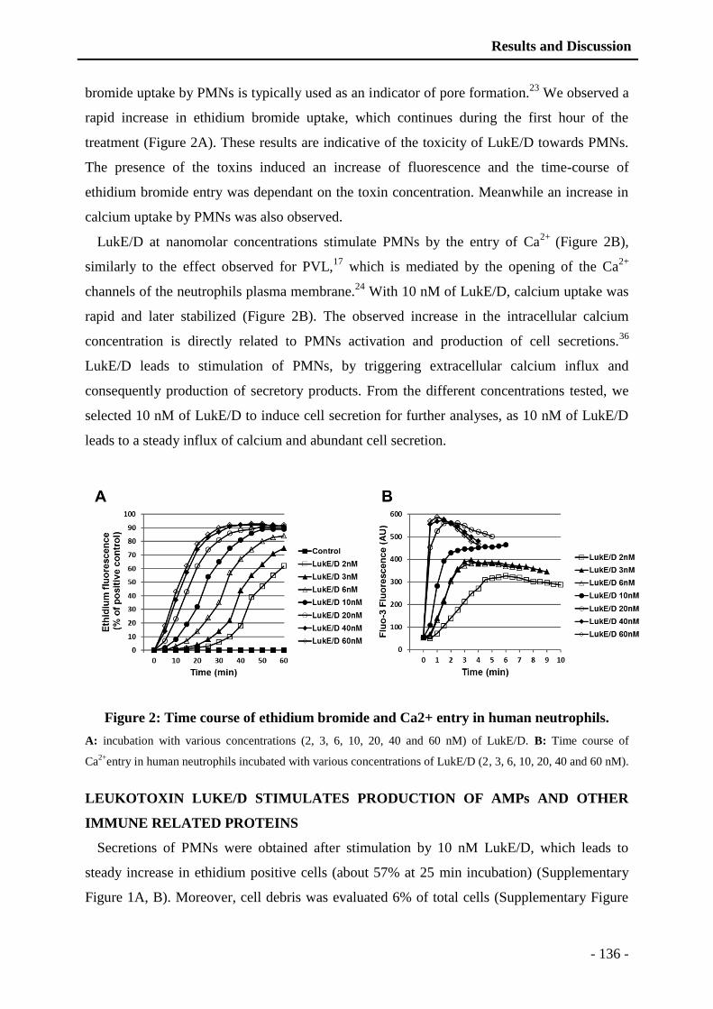
Results and Discussion
- 136 -
bromide uptake by PMNs is typically used as an indicator of pore formation.23
We observed a
rapid increase in ethidium bromide uptake, which continues during the first hour of the
treatment (Figure 2A). These results are indicative of the toxicity of LukE/D towards PMNs.
The presence of the toxins induced an increase of fluorescence and the time-course of
ethidium bromide entry was dependant on the toxin concentration. Meanwhile an increase in
calcium uptake by PMNs was also observed.
LukE/D at nanomolar concentrations stimulate PMNs by the entry of Ca2+
(Figure 2B),
similarly to the effect observed for PVL,17
which is mediated by the opening of the Ca2+
channels of the neutrophils plasma membrane.24
With 10 nM of LukE/D, calcium uptake was
rapid and later stabilized (Figure 2B). The observed increase in the intracellular calcium
concentration is directly related to PMNs activation and production of cell secretions.36
LukE/D leads to stimulation of PMNs, by triggering extracellular calcium influx and
consequently production of secretory products. From the different concentrations tested, we
selected 10 nM of LukE/D to induce cell secretion for further analyses, as 10 nM of LukE/D
leads to a steady influx of calcium and abundant cell secretion.
Figure 2: Time course of ethidium bromide and Ca2+ entry in human neutrophils.
A: incubation with various concentrations (2, 3, 6, 10, 20, 40 and 60 nM) of LukE/D. B: Time course of
Ca2+
entry in human neutrophils incubated with various concentrations of LukE/D (2, 3, 6, 10, 20, 40 and 60 nM).
LEUKOTOXIN LUKE/D STIMULATES PRODUCTION OF AMPs AND OTHER
IMMUNE RELATED PROTEINS
Secretions of PMNs were obtained after stimulation by 10 nM LukE/D, which leads to
steady increase in ethidium positive cells (about 57% at 25 min incubation) (Supplementary
Figure 1A, B). Moreover, cell debris was evaluated 6% of total cells (Supplementary Figure

Results and Discussion
- 137 -
1A, B). PMNs secretions were purified by RP-HPLC as described in the Materials and
Methods section (Figure 3). One mg of PMN secretions was loaded on the HPLC column.
Absorbance was monitored at 214 nm and elution was performed at a flow rate of 700 µL/min
with a linear gradient, as indicated by the dotted line. Fractions were collected with 1 min
time interval and marked according to the collection time. Collected fractions were
concentrated using a speed-vac. Compared to the control of non-stimulated PMNs (PBS)
(Figure 3: Chromatogram 1), LukE/D stimulated neutrophils (Figure 3: Chromatogram 2) and
induced a rapid secretion of a large number of immune related factors. The 223 secreted
proteins identified by mass spectrometry are listed according to the RP-HPLC fraction
numbers (Supplementary data 1), together with their number of unique peptides, sequence
coverage, Mascot scores and the peptides used to identify them (Supplementary data 2).
Figure 3: Reverse-phase HPLC of proteic material from LukE/D-induced PMNs
secretions compared to the control.
(1) After stimulation with 10 nM of LukE/D and (2) as a control, after treatment with PBS.
Using gene ontology analysis software (GoTermMapper), all these factors were classified
into different functional categories on the basis of their molecular functions, according to the
Gene ontology database analysis (Figure 4). Based on the theme of innate immunity, we
decided to classify the proteins into the following functional categories: (1) AMPs and other

Results and Discussion
- 138 -
immune related proteins, (2) binding proteins, (3) inflammatory proteins, (4) enzymatic
proteins, (5) cell growth and differentiation proteins, (6) transport proteins, (7) cytoskeleton
proteins, (8) antioxidant proteins, (9) proteins involved in proteolysis and (10) proteins related
to protein synthesis (Figure 4).
The pie chart representing the identified proteins revealed a major secretion of AMPs and
other immune related proteins (27% of total secreted products) (Figure 4; Table 1). HPLC
fraction numbers containing the identified proteins were listed together with their NCBI
accession numbers and their identification probability. In addition, the type of granule
containing the proteins is also listed next to their position. Other abundant secreted proteins
belong to the binding proteins (11%), inflammatory proteins (8%), enzymatic proteins (13%),
cell growth and differentiation proteins (12%), transport proteins (11%), cytoskeleton proteins
(10%), antioxidant proteins (2%), proteins involved in proteolysis (4%) and proteins related to
protein synthesis (2%) (Figure 4). All these secreted factors well illustrate acute activation of
the neutrophil cell machinery.
Figure 4: Pie chart representing the protein distribution according to their molecular
function (GoTermMapper).
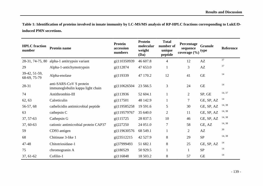
Results and Discussion
- 139 -
Table 1: Identification of proteins involved in innate immunity by LC-MS/MS analysis of RP-HPLC fractions corresponding to LukE/D-
induced PMN secretions.
HPLC fraction
number Protein name
Protein
accession
numbers
Protein
molecular
weight
(Da)
Total
number of
unique
peptide
Percentage
sequence
coverage (%)
Granule
type Reference
28-31, 74-75, 80 alpha-1 antitrypsin variant gi|110350939 46 607.8 4 12 AZ 37
29 Alpha-1-antichymotrypsin gi|112874 47 653.0 1 3 AZ 37
39-42, 51-59,
68-69, 75-79 Alpha-enolase gi|119339 47 170.2 12 41 GE
14
28-31 anti-SARS-CoV S protein
immunoglobulin kappa light chain gi|110626504 23 566.5 3 24 GE
14
74 Antithrombin-III gi|113936 52 604.1 1 2 SP, GE 14, 37
62, 63 Calreticulin gi|117501 48 142.9 1 7 GE, SP, AZ 14
56-57, 68 cathelicidin antimicrobial peptide gi|119585258 19 591.6 5 30 GE, SP, AZ 14, 38
63 cathepsin C gi|119579767 35 640.0 2 11 GE, SP, AZ 14, 38
37, 57-63 Cathepsin G gi|115725 28 837.5 10 46 GE, SP, AZ 14, 38
37, 60-63 cationic antimicrobial protein CAP37 gi|227250 24 051.0 7 58 GE, AZ 14, 38
59 CD93 antigen gi|119630576 68 549.1 1 2 AZ 39
68 Chitinase 3-like 1 gi|23512215 42 527.9 8 29 SP 14, 38
47-48 Chitotriosidase-1 gi|37999493 51 682.1 8 25 GE, SP, AZ 14
75 chromogranin A gi|180529 50 929.5 1 1 SP 14
37, 61-62 Cofilin-1 gi|116848 18 503.2 8 57 GE 14

Results and Discussion
- 140 -
61 complement factor properdin gi|119579721 49 664.4 1 3 GE, SP, SV 14, 38
59, 76 dermcidin isoform 2 gi|148271063 12 414.5 1 9 GE, SP 14
53-59 Eosinophil cationic protein gi|147744558 18 385.3 5 37 GE, SP, AZ 14
78-80 Neuroleukin gi|17380385 63 148.5 16 43 GE 14
28, 31, 34 hCG2029987 gi|119602340 43 900.2 7 28 GE 14
44 histocompatibility (minor) HA-1 gi|119589958 122 920.8 1 1 GE, SP 14, 38
28 Ig alpha-1 chain C region gi|113584 37 653.8 3 20 GE, SP, AZ 14
28 Ig gamma 2 H chain gi|243169 38 721.8 3 14 GE 14
56-60 IgG receptor IIIB gi|11344591 26 189.0 3 15 GE, SP 14
57-61 IgG receptor precursor gi|183037 26 299.0 3 14 GE, SP 14
28, 31 immunoglobulin heavy chain constant
region gi|10799664 35 919.3 4 19 GE, SP, AZ
14
37-38, 57-70,
72-79 lactoferrin gi|34412 78 338.5 42 69 GE, SP, AZ
14, 38
61-63 lactoferrin precursor gi|12083188 78 382.5 42 69 GE, SP, AZ 14, 38
77-79 Leukotriene A-4 hydrolase gi|126353 69 287.1 15 36 SP 14
38-40, 42 Lymphocyte-specific protein 1; WP34 gi|462553 37 191.1 1 8 GE, SP 14
37-38, 60-63 lysozyme gi|1335210 14 700.5 6 59 GE, SP, AZ 14, 38
44, 47, 70-72 matrix metallopeptidase 8 (neutrophil
collagenase) gi|119587431 53 426.3 10 31 GE, SP
14
69-79 myeloperoxidase gi|119614877 88 763.7 17 28 GE, SP, AZ 14
40, 47, 50, 70-
73, 79 Neutrophil collagenase gi|116862 53 414.2 10 31 SP
14
54-55 Neutrophil defensin 4 gi|399352 10 504.1 1 18 SP, GE 14, 38
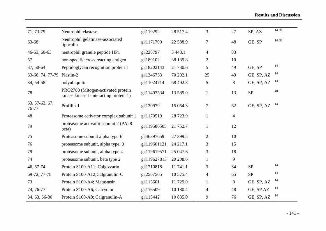
Results and Discussion
- 141 -
71, 73-79 Neutrophil elastase gi|119292 28 517.4 3 27 SP, AZ 14, 38
63-68 Neutrophil gelatinase-associated
lipocalin gi|1171700 22 588.9 7 48 GE, SP
14, 38
46-53, 60-63 neutrophil granule peptide HP1 gi|228797 3 448.1 4 83
57 non-specific cross reacting antigen gi|189102 38 139.8 2 10
37, 60-64 Peptidoglycan recognition protein 1 gi|18202143 21 730.6 5 49 GE, SP 14
63-66, 74, 77-79 Plastin-2 gi|1346733 70 292.1 25 49 GE, SP, AZ 14
34, 54-58 polyubiquitin gi|11024714 68 492.8 5 8 GE, SP, AZ 14
78 PRO2783 (Mitogen-activated protein
kinase kinase 1-interacting protein 1) gi|11493534 13 589.0 1 13 SP
40
53, 57-63, 67,
76-77 Profilin-1 gi|130979 15 054.3 7 62 GE, SP, AZ
14
48 Proteasome activator complex subunit 1 gi|1170519 28 723.9 1 4
79 proteasome activator subunit 2 (PA28
beta) gi|119586505 21 752.7 1 12
75 Proteasome subunit alpha type-6 gi|46397659 27 399.5 2 10
76 proteasome subunit, alpha type, 3 gi|119601121 24 217.1 3 15
79 proteasome subunit, alpha type 4 gi|119619571 25 047.6 3 18
74 proteasome subunit, beta type 2 gi|119627813 20 208.6 1 9
46, 67-74 Protein S100-A11; Calgizzarin gi|1710818 11 741.1 3 34 SP 14
69-72, 77-78 Protein S100-A12;Calgranulin-C gi|2507565 10 575.4 4 65 SP 14
73 Protein S100-A4; Metastasin gi|115601 11 729.0 1 8 GE, SP, AZ 14
74, 76-77 Protein S100-A6; Calcyclin gi|116509 10 180.4 4 48 GE, SP AZ 14
34, 63, 66-80 Protein S100-A8; Calgranulin-A gi|115442 10 835.0 9 76 GE, SP, AZ 14
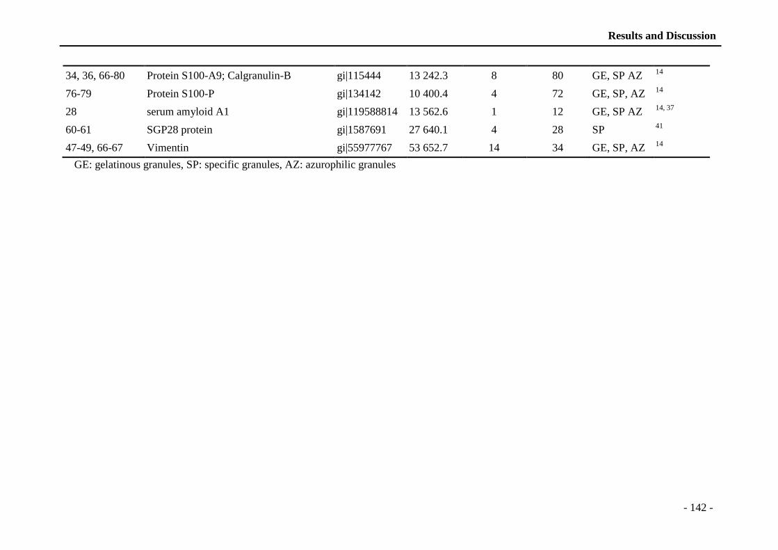
Results and Discussion
- 142 -
34, 36, 66-80 Protein S100-A9; Calgranulin-B gi|115444 13 242.3 8 80 GE, SP AZ 14
76-79 Protein S100-P gi|134142 10 400.4 4 72 GE, SP, AZ 14
28 serum amyloid A1 gi|119588814 13 562.6 1 12 GE, SP AZ 14, 37
60-61 SGP28 protein gi|1587691 27 640.1 4 28 SP 41
47-49, 66-67 Vimentin gi|55977767 53 652.7 14 34 GE, SP, AZ 14
GE: gelatinous granules, SP: specific granules, AZ: azurophilic granules
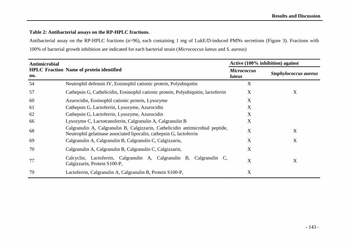
Results and Discussion
- 143 -
Table 2: Antibacterial assays on the RP-HPLC fractions.
Antibacterial assay on the RP-HPLC fractions (n=96), each containing 1 mg of LukE/D-induced PMNs secretions (Figure 3). Fractions with
100% of bacterial growth inhibition are indicated for each bacterial strain (Micrococcus luteus and S. aureus)
Antimicrobial
HPLC Fraction
no.
Name of protein identified
Active (100% inhibition) against
Micrococcus
luteus Staphylococcus aureus
54 Neutrophil defensin IV, Eosinophil cationic protein, Polyubiquitin X
57 Cathepsin G, Cathelicidin, Eosinophil cationic protein, Polyubiquitin, lactoferrin X X
60 Azurocidin, Eosinophil cationic protein, Lysozyme X
61 Cathepsin G, Lactoferrin, Lysozyme, Azurocidin X
62 Cathepsin G, Lactoferrin, Lysozyme, Azurocidin X
66 Lysozyme C, Lactotransferrin, Calgranulin A, Calgranulin B X
68 Calgranulin A, Calgranulin B, Calgizzarin, Cathelicidin antimicrobial peptide,
Neutrophil gelatinase associated lipocalin, cathepsin G, lactoferrin X X
69 Calgranulin A, Calgranulin B, Calgranulin C, Calgizzarin, X X
70 Calgranulin A, Calgranulin B, Calgranulin C, Calgizzarin, X
77 Calcyclin, Lactoferrin, Calgranulin A, Calgranulin B, Calgranulin C,
Calgizzarin, Protein S100-P, X X
79 Lactoferrin, Calgranulin A, Calgranulin B, Protein S100-P, X
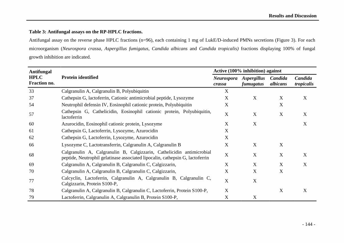
Results and Discussion
- 144 -
Table 3: Antifungal assays on the RP-HPLC fractions.
Antifungal assay on the reverse phase HPLC fractions (n=96), each containing 1 mg of LukE/D-induced PMNs secretions (Figure 3). For each
microorganism (Neurospora crassa, Aspergillus fumigatus, Candida albicans and Candida tropicalis) fractions displaying 100% of fungal
growth inhibition are indicated.
Antifungal
HPLC
Fraction no.
Protein identified
Active (100% inhibition) against
Neurospora
crassa
Aspergillus
fumugatus
Candida
albicans
Candida
tropicalis
33 Calgranulin A, Calgranulin B, Polyubiquitin X
37 Cathepsin G, lactoferrin, Cationic antimicrobial peptide, Lysozyme X X X X
54 Neutrophil defensin IV, Eosinophil cationic protein, Polyubiquitin X X
57 Cathepsin G, Cathelicidin, Eosinophil cationic protein, Polyubiquitin,
lactoferrin X X X X
60 Azurocidin, Eosinophil cationic protein, Lysozyme X X X
61 Cathepsin G, Lactoferrin, Lysozyme, Azurocidin X
62 Cathepsin G, Lactoferrin, Lysozyme, Azurocidin X
66 Lysozyme C, Lactotransferrin, Calgranulin A, Calgranulin B X X X
68 Calgranulin A, Calgranulin B, Calgizzarin, Cathelicidin antimicrobial
peptide, Neutrophil gelatinase associated lipocalin, cathepsin G, lactoferrin X X X X
69 Calgranulin A, Calgranulin B, Calgranulin C, Calgizzarin, X X X X
70 Calgranulin A, Calgranulin B, Calgranulin C, Calgizzarin, X X X
77 Calcyclin, Lactoferrin, Calgranulin A, Calgranulin B, Calgranulin C,
Calgizzarin, Protein S100-P, X X
78 Calgranulin A, Calgranulin B, Calgranulin C, Lactoferrin, Protein S100-P, X X X
79 Lactoferrin, Calgranulin A, Calgranulin B, Protein S100-P, X X
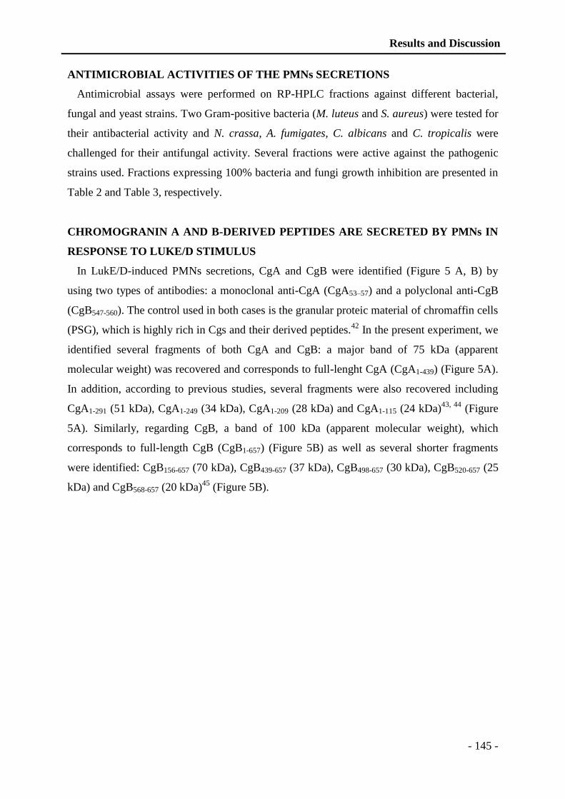
Results and Discussion
- 145 -
ANTIMICROBIAL ACTIVITIES OF THE PMNs SECRETIONS
Antimicrobial assays were performed on RP-HPLC fractions against different bacterial,
fungal and yeast strains. Two Gram-positive bacteria (M. luteus and S. aureus) were tested for
their antibacterial activity and N. crassa, A. fumigates, C. albicans and C. tropicalis were
challenged for their antifungal activity. Several fractions were active against the pathogenic
strains used. Fractions expressing 100% bacteria and fungi growth inhibition are presented in
Table 2 and Table 3, respectively.
CHROMOGRANIN A AND B-DERIVED PEPTIDES ARE SECRETED BY PMNs IN
RESPONSE TO LUKE/D STIMULUS
In LukE/D-induced PMNs secretions, CgA and CgB were identified (Figure 5 A, B) by
using two types of antibodies: a monoclonal anti-CgA (CgA53–57) and a polyclonal anti-CgB
(CgB547-560). The control used in both cases is the granular proteic material of chromaffin cells
(PSG), which is highly rich in Cgs and their derived peptides.42
In the present experiment, we
identified several fragments of both CgA and CgB: a major band of 75 kDa (apparent
molecular weight) was recovered and corresponds to full-lenght CgA (CgA1-439) (Figure 5A).
In addition, according to previous studies, several fragments were also recovered including
CgA1-291 (51 kDa), CgA1-249 (34 kDa), CgA1-209 (28 kDa) and CgA1-115 (24 kDa)43, 44
(Figure
5A). Similarly, regarding CgB, a band of 100 kDa (apparent molecular weight), which
corresponds to full-length CgB (CgB1-657) (Figure 5B) as well as several shorter fragments
were identified: CgB156-657 (70 kDa), CgB439-657 (37 kDa), CgB498-657 (30 kDa), CgB520-657 (25
kDa) and CgB568-657 (20 kDa)45
(Figure 5B).
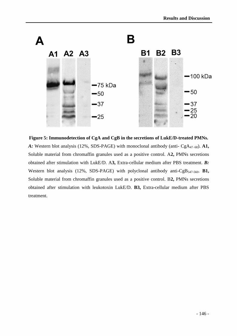
Results and Discussion
- 146 -
Figure 5: Immunodetection of CgA and CgB in the secretions of LukE/D-treated PMNs.
A: Western blot analysis (12%, SDS-PAGE) with monoclonal antibody (anti- CgA47–68). A1,
Soluble material from chromaffin granules used as a positive control. A2, PMNs secretions
obtained after stimulation with LukE/D. A3, Extra-cellular medium after PBS treatment. B:
Western blot analysis (12%, SDS-PAGE) with polyclonal antibody anti-CgB547-560. B1,
Soluble material from chromaffin granules used as a positive control. B2, PMNs secretions
obtained after stimulation with leukotoxin LukE/D. B3, Extra-cellular medium after PBS
treatment.
Results and Discussion
- 147 -
DISCUSSION
LukE/D, a pore-forming leukotoxin produced by S. aureus strains infecting human, is
specific against host defense cells and T-lymphocytes.46-48
Several recent studies have
underlined the crucial role of LukE/D in S. aureus virulence. LukE/D gene expression was
significantly higher in community-associated methicillin-resistant S. aureus (CA-MRSA)
compared with methicillin sensitive S. aureus (MSSA), suggesting that they contribute to
bacterial virulence.49
Analysis of the most prevalent genes associated with MRSA strains with
type III SCCmec points out LukE/D in 72.9% of the isolates.50
Furthermore, it was reported
that in addition to PVL, a high prevalence of LukE/D was recorded in HIV (+) furuncles
patients.51
The role of LukE/D to trigger inflammation by acting synergistically with PVL to
amplify IL-1b release in human macrophages has been demonstrated.52
A biological
mechanism explaining the toxic activity of LukE/D was proposed in a recent study.48
Indeed,
the authors demonstrate that LukE/D is specifically cytotoxic on human T-cell line expressing
CCR5. Moreover, LukE, but not LukD binds to CCR5 in a time-dependent manner.48
In the present study, we characterized the proteins secreted by PMNs upon S. aureus
LukE/D activation. PMNs are potent actors of innate immunity and serve as an important
interface between innate and adaptive immunity by producing reactive oxygen species,
antimicrobial peptides, chemokines and cytokines.53, 54
The 223 proteins identified in the neutrophil secretions after proteomic analysis were
classified according to their molecular function by using go.princeton.edu/cgi-
bh/GoTermMapper (Figure 4). Major proteins correspond to AMPs and immune related
proteins (27%). PMNs display a potent microbicidal activity including oxygen-dependent and
oxygen-independent strategies to destroy invading pathogens.54
The production of ROS
derived from NADPH oxidase is critical for host defense mechanisms against invading
microorganisms.55
In relation with oxidative mechanisms, the proteomic analysis indicates
that 2% of antioxidant proteins and oxygen-independent microbicidal agents include AMPs38
such as defensins, cathelicidins, azurocidin, cathepsins, lactoferrin, chromogranin A and
lysozyme (Table 1). Enzymatic proteins, such as cathepsin, lysozyme, pyruvate kinase,
neutrophil collagenase, eosinophil peroxidase and matrix metallo peptidase correspond to
13% and are important for proteolysis of AMPs and to manage the neutrophil turnover.
Proteins for cell growth and development, such as epidermal growth factor (EGF), elongation
factors, olfactomedin 4 (OLFM4) and neuroleukin represent 12% of the total released proteins
and they participate to the rapid turnover of PMNs (Figure 4). All these proteins are crucial
Results and Discussion
- 148 -
for the dynamics of host-pathogen interactions with the multistep progression into mature
PMNs.56
Binding proteins correspond to 11% of the total proteins and include DNA binding
proteins (histones, high mobility group protein B2), calcium binding proteins (calmodulin and
its isoforms), adenylate cyclase and fatty acid binding proteins. The proteomic analysis also
includes proteins involved in transport activity (hemoglobin, lactoferrin, and neutrophil
gelatinase associated lipocalin), which play an important role in boosting innate defense at the
site of infection. PMNs are the most efficient vertebrate motile defense cells. Their directional
movement is governed by the binding of chemoattractants and chemokines to G-proteins
coupled receptors. These motility responses start with the polymerization of F-actin and the
rearrangement of cytoskeleton and cell crawling. The stimulation of neutrophils by LukE/D
induces the release of cytoskeleton proteins (10%) such as actin, actinin, gelsolin, profilin and
vimentin. The proteins involved in the modulation of inflammation represent 8% of the total
proteins and play a key role in the early pro-inflammatory signals, as they release
proinflammatory molecules to promote efficient pathogen recognition and removal.54
Numerous secreted proteins are pro-inflammatory molecules such as cathepsin,
chitotriosidase, cofilin, complement C3, cysteine rich secretory protein, eosinophil cationic
protein, eosinophil peroxidase, neutrophil elastase, defensin-4, proteasome proteins, S100
A12, calgranulin, vimentin. In contrast, others such as alpha-1 antitrypsin, serpin, anti-
thrombin III, neuroleukin, lactoferrin, lysozyme, MMP8, peptidoglycan recognition protein 1
and polyubiquitin are anti-inflammatory. Collectively, these observations suggest that
LukE/D can enhance rather than hinder the host innate immune response to S. aureus
infection. Nevertheless, the ability of LukE/D to enhance neutrophils bactericidal activity in
vivo needs further investigation. For the first time Brinkmann et al. reported that activated
PMNs release their DNA that is laden with antimicrobial molecules and form PMNs
extracellular traps (NETs).57
Later, in 2007 with a very detailed series of imaging experiments
it was demonstrated that the formation of NETs in response to S. aureus is the last step in a
process of active PMN death.58
It was also reported that over the period of 3h, the PMNs
underwent important morphological changes. The granular membranes disappeared and the
nuclear, cytoplasmic and granular components were mixed together, allowing the extrusion of
the NETs and therefore the antimicrobial role of PMNs even after cell death. PVL was
described to be the major NET inducer, after inhibition of NET production by treatment with
antiserum against PVL.59
Our proteomic analysis of the PMNs secretions induced by LukE/D
shows the presence of 6.25% cytoskeleton proteins (Figure 4) also suggesting the
involvement of severe morphological changes.
Results and Discussion
- 149 -
Calcium is a universal secondary messenger involved in many cellular signal transduction
pathways, regulating crucial functions such as secretion, cell motility, proliferation and cell
death. Increase in intracellular Ca2+
derives mainly from two sources: internal stores releasing
Ca2+
into the cytosol and Ca2+
channels in the plasma membrane that open to allow external
Ca2+
to flow into the cell. In PMNs, calcium signaling has been reported to be involved in
oxidase activation, cell degranulation and priming response to a wide range of
proinflammatory molecules.60
In the present study, we demonstrated that calcium binding
proteins represent ~11% of the PMNs secreted proteins highlighting the importance of
calcium in the stimulation of PMNs by LukE/D. For instance, vesicular amine transport
(VAT-I) plays a crucial role in calcium regulated cell activation.61
Our previous studies have established the presence of CgA in neutrophils and its role as an
immune activator.17
Immunodetection of CgA (Figure 5) showed that LukE/D induces the
predominant release of full-length CgA (75 kDa) but also of minor fragments with apparent
molecular masses of 50, 35, 30 and 25 kDa that might correspond to fragments 1-298, 1-210,
1-182 and 1-115. Moreover, immunodetection of CgB (Figure 5) shows the release of 2 major
fragments corresponding to full-length CgB (100 kDa) and the 143-657 fragment (70 kDa). In
addition, several minor fragments 259-657 (50kDa), 306-657 (37 kDa), 555-657 (20 kDa) and
564-657 (10 kDa) were identified.
CONCLUSION
We previously described that an increase in intracellular free Ca2+
can induce secretion in
PMNs.62
These secretions can result from cell activation by the two LukE and LukD
components of the LukE/D toxin of S. aureus. A similar effect was observed previously by
using PVL of S. aureus.23
Complement C3 factor (CD35) was recovered upon PVL treatment,
which indicates neutrophil activation.63
Here, we demonstrated that in addition to toxicity,
LukE/D can also stimulate PMNs. Identification of trademark proteins from different PMNs
granules underlines the ability of this leukotoxin to degranulate all type of compartments.
PMNs secrete various proteins belonging to different functional categories, such as immune
related proteins, antioxidant proteins, proteases and inflammatory components. Beyond these
functional categories, AMPs and immune activation proteins represent a significant
proportion of the secretions in this specific inflammatory stimulus. The release of all these
factors is crucial for innate immune functions. As an example, we recovered Cgs-derived
Results and Discussion
- 150 -
fragments, which are active participants of the immune system. These findings underlie the
importance to further study the role of leukotoxin LukE/D as immune activating components.
ASSOCIATED CONTENTS
Extended information of all the proteins/peptides identified by LC-MS/MS in PMNs
secretions induced by S. aureus leukotoxin LukE/D, are listed in the supporting information.
Data are presented according to RP-HPLC fraction numbers, which correspond to collection
time (Supplementary data 1). Identified proteins are provided with their NCBI database
accession numbers, molecular weight, percent sequence coverage, and corresponding peptide
sequences (Supplementary data 1). Proteomic analysis was supplemented with Mascot Ion
score, Mascot identity score and ion score-identity score (Supplementary data 2).
Proteins/peptides identified by only one unique peptide and with Ion score less than 40 are
presented with MS/MS spectra (Supplementary data 3).This information is available free of
charge via the Internet at http://pubs.acs.org
* CORRESPONDING AUTHOR
Marie-Hélène METZ-BOUTIGUE,
Inserm UMR1121, Bâtiment Leriche, Faculté de Chirurgie Dentaire, Hôpital civil, Porte de
l’Hôpital, F-67000, Strasbourg, France.
E-mail: [email protected]; Tel. (+33)3 68 85 54 71; Fax (+33)3 68 85 54 72.
PRESENT ADDRESS
† Division of Molecular Microbiology, Biozentrum, University of Basel, CH-4056 Basel,
Switzerland.
AUTHOR CONTRIBUTIONS
The manuscript was written through contributions of all authors. Rizwan Aslam has
performed the experiments and prepared the manuscript and figures. Benoît-Josef Laventie
Results and Discussion
- 151 -
and Daniel Keller have purified and characterized LukE/D. Gilles Prévost supervised the
study and with Céline Marban participated in preparation of the manuscript. Jean-Marc Strub
and Alain van Dorsselaer brought their expertise in proteomic studies; Youssef Haikel and
Corinne Taddei participated to interesting discussions and Marie-Hélène Metz-Boutigue
supervised the experiments and the preparation of the manuscript. All authors have given
approval to the final version of the manuscript.
NOTES
The authors declare no competing financial interest.
ACKNOWLEDGMENT
Authors acknowledge the funding provided by the Inserm, the University of Strasbourg and
the Faculty of Odontology of Strasbourg. RA was the recipient of the financial support from
the Higher Education Commission of Pakistan and Faculty of Odontology of Strasbourg. We
are indebted to Angelo Corti from the San Raffaele research institute of Milan (Italy) for his
generous gift of specific CgA antibodies and the Institute of Bacteriology of Strasbourg for
the pathogenic strains. We thank Bernard Senger for manuscript improvement.
REFERENCES
1. Heyman, D. control of communicable diseases manual. 18 ed.; American Public
Health Association, Washington DC: 2004.
2. DeLeo, F. R.; Otto, M.; Kreiswirth, B. N.; Chambers, H. F. Community-associated
meticillin-resistant Staphylococcus aureus. Lancet. 2010, 375, (9725), 1557-68.
3. Foster, T. J.; Hook, M. Surface protein adhesins of Staphylococcus aureus. Trends
Microbiol. 1998, 6, (12), 484-8.
4. Gordon, R. J.; Lowy, F. D. Pathogenesis of methicillin-resistant Staphylococcus
aureus infection. Clin Infect Dis. 2008, 46 Suppl 5, S350-9.
5. Switalski, L. M.; Patti, J. M.; Butcher, W.; Gristina, A. G.; Speziale, P.; Hook, M. A
collagen receptor on Staphylococcus aureus strains isolated from patients with septic
arthritis mediates adhesion to cartilage. Mol Microbiol. 1993, 7, (1), 99-107.
6. Hauck, C. R.; Ohlsen, K. Sticky connections: extracellular matrix protein recognition
and integrin-mediated cellular invasion by Staphylococcus aureus. Curr Opin
Microbiol. 2006, 9, (1), 5-11.
7. Sauer, P.; Sila, J.; Stosova, T.; Vecerova, R.; Hejnar, P.; Vagnerova, I.; Kolar, M.;
Raclavsky, V.; Petrzelova, J.; Loveckova, Y.; Koukalova, D. Prevalence of genes
encoding extracellular virulence factors among meticillin-resistant Staphylococcus

Results and Discussion
- 152 -
aureus isolates from the University Hospital, Olomouc, Czech Republic. J Med
Microbiol. 2008, 57, (Pt 4), 403-10.
8. Prévost, G.; Cribier, B.; Couppie, P.; Petiau, P.; Supersac, G.; Finck-Barbançon, V.;
Monteil, H.; Piémont, Y. Panton-Valentine leucocidin and gamma-hemolysin from
Staphylococcus aureus ATCC 49775 are encoded by distinct genetic loci and have
different biological activities. Infect Immun. 1995, 63, (10), 4121-9.
9. Gravet, A.; Colin, D. A.; Keller, D.; Girardot, R.; Monteil, H.; Prévost, G.
Characterization of a novel structural member, LukE-LukD, of the bi-component
staphylococcal leucotoxins family. FEBS Lett. 1998, 436, (2), 202-8.
10. Gravet, A.; Couppie, P.; Meunier, O.; Clyti, E.; Moreau, B.; Pradinaud, R.; Monteil,
H.; Prévost, G. Staphylococcus aureus isolated in cases of impetigo produces both
epidermolysin A or B and LukE-LukD in 78% of 131 retrospective and prospective
cases. J Clin Microbiol. 2001, 39, (12), 4349-56.
11. Joubert, O.; Viero, G.; Keller, D.; Martinez, E.; Colin, D. A.; Monteil, H.; Mourey, L.;
Dalla Serra, M.; Prévost, G. Engineered covalent leucotoxin heterodimers form
functional pores: insights into S-F interactions. Biochem J. 2006, 396, (2), 381-9.
12. Gravet, A.; Rondeau, M.; Harf-Monteil, C.; Grunenberger, F.; Monteil, H.; Scheftel, J.
M.; Prévost, G. Predominant Staphylococcus aureus isolated from antibiotic-
associated diarrhea is clinically relevant and produces enterotoxin A and the
bicomponent toxin LukE-lukD. J Clin Microbiol. 1999, 37, (12), 4012-9.
13. Lugardon, K.; Raffner, R.; Goumon, Y.; Corti, A.; Delmas, A.; Bulet, P.; Aunis, D.;
Metz-Boutigue, M. H. Antibacterial and antifungal activities of vasostatin-1, the N-
terminal fragment of chromogranin A. J Biol Chem. 2000, 275, (15), 10745-53.
14. Lominadze, G.; Powell, D. W.; Luerman, G. C.; Link, A. J.; Ward, R. A.; McLeish, K.
R. Proteomic analysis of human neutrophil granules. Mol Cell Proteomics. 2005, 4,
(10), 1503-21.
15. Zhang, D.; Lavaux, T.; Voegeli, A. C.; Lavigne, T.; Castelain, V.; Meyer, N.; Sapin,
R.; Aunis, D.; Metz-Boutigue, M. H.; Schneider, F. Prognostic value of chromogranin
A at admission in critically ill patients: a cohort study in a medical intensive care unit.
Clin Chem. 2008, 54, (9), 1497-503.
16. Metz-Boutigue, M. H.; Kieffer, A. E.; Goumon, Y.; Aunis, D. Innate immunity:
involvement of new neuropeptides. Trends Microbiol. 2003, 11, (12), 585-92.
17. Zhang, D.; Shooshtarizadeh, P.; Laventie, B. J.; Colin, D. A.; Chich, J. F.; Vidic, J.; de
Barry, J.; Chasserot-Golaz, S.; Delalande, F.; Van Dorsselaer, A.; Schneider, F.; Helle,
K.; Aunis, D.; Prévost, G.; Metz-Boutigue, M. H. Two chromogranin a-derived
peptides induce calcium entry in human neutrophils by calmodulin-regulated calcium
independent phospholipase A2. PLoS One. 2009, 4, (2), e4501.
18. Ma, X.; Chang, W.; Zhang, C.; Zhou, X.; Yu, F. Staphylococcal panton-valentine
leukocidin induces pro-inflammatory cytokine production and nuclear factor-kappa B
activation in neutrophils. PLoS One. 2012, 7, (4), e34970.
19. Borregaard, N.; Lollike, K.; Kjeldsen, L.; Sengelov, H.; Bastholm, L.; Nielsen, M. H.;
Bainton, D. F. Human neutrophil granules and secretory vesicles. Eur J Haematol.
1993, 51, (4), 187-98.
20. Luerman, G. C.; Uriarte, S. M.; Rane, M. J.; McLeish, K. R. Application of
proteomics to neutrophil biology. J Proteomics. 2009, 73, (3), 552-61.
21. Finck-Barbançon, V.; Prévost, G.; Piémont, Y. Improved purification of leukocidin
from Staphylococcus aureus and toxin distribution among hospital strains. Res
Microbiol. 1991, 142, (1), 75-85.

Results and Discussion
- 153 -
22. Morinaga, N.; Kaihou, Y.; Noda, M. Purification, cloning and characterization of
variant LukE-LukD with strong leukocidal activity of staphylococcal bi-component
leukotoxin family. Microbiol Immunol. 2003, 47, (1), 81-90.
23. Finck-Barbançon, V.; Duportail, G.; Meunier, O.; Colin, D. A. Pore formation by a
two-component leukocidin from Staphylococcus aureus within the membrane of
human polymorphonuclear leukocytes. Biochim Biophys Acta. 1993, 1182, (3), 275-
82.
24. Staali, L.; Monteil, H.; Colin, D. A. The staphylococcal pore-forming leukotoxins
open Ca2+ channels in the membrane of human polymorphonuclear neutrophils. J
Membr Biol. 1998, 162, (3), 209-16.
25. Colin, D. A.; Mazurier, I.; Sire, S.; Finck-Barbancon, V. Interaction of the two
components of leukocidin from Staphylococcus aureus with human
polymorphonuclear leukocyte membranes: sequential binding and subsequent
activation. Infect Immun. 1994, 62, (8), 3184-8.
26. Barrio, M. B.; Rainard, P.; Prévost, G. LukM/LukF'-PV is the most active
Staphylococcus aureus leukotoxin on bovine neutrophils. Microbes Infect. 2006, 8,
(8), 2068-74.
27. Meunier, O.; Falkenrodt, A.; Monteil, H.; Colin, D. A. Application of flow cytometry
in toxinology: pathophysiology of human polymorphonuclear leukocytes damaged by
a pore-forming toxin from Staphylococcus aureus. Cytometry. 1995, 21, (3), 241-7.
28. Moussa, L. B.; Werner, S.; Coraiola, M.; Colin, D. A.; Keller, D.; Sanni, A.; Serra, M.
D.; Monteil, H.; Prévost, G. Site-directed mutagenesis to assess the binding capacity
of class s protein of Staphylococcus aureus leucotoxins to the surface of
polymorphonuclear cells. J Biomed Biotechnol. 2006, 2006, (2), 80101.
29. Bradford, M. M. A rapid and sensitive method for the quantitation of microgram
quantities of protein utilizing the principle of protein-dye binding. Anal Biochem.
1976, 72, 248-54.
30. Corti, A.; Sanchez, L. P.; Gasparri, A.; Curnis, F.; Longhi, R.; Brandazza, A.;
Siccardi, A. G.; Sidoli, A. Production and structure characterisation of recombinant
chromogranin A N-terminal fragments (vasostatins) -- evidence of dimer-monomer
equilibria. Eur J Biochem. 1997, 248, (3), 692-9.
31. Gasnier, C.; Lugardon, K.; Ruh, O.; Strub, J. M.; Aunis, D.; Metz-Boutigue, M. H.
Characterization and location of post-translational modifications on chromogranin B
from bovine adrenal medullary chromaffin granules. Proteomics. 2004, 4, (6), 1789-
801.
32. NCCLS, Methods for Dilution Antimicrobial Susceptibility Tests for Bacteria That
Grow Aerobically: National Committee for Clinical Laboratory Standards. In
Approved Standard M07-A8. Eight ed.; NCCLS, Wayne, PA, USA.: 2003.
33. Bulet, P.; Dimarcq, J. L.; Hetru, C.; Lagueux, M.; Charlet, M.; Hegy, G.; Van
Dorsselaer, A.; Hoffmann, J. A. A novel inducible antibacterial peptide of Drosophila
carries an O-glycosylated substitution. J Biol Chem. 1993, 268, (20), 14893-7.
34. Wu, M.; Hancock, R. E. Interaction of the cyclic antimicrobial cationic peptide
bactenecin with the outer and cytoplasmic membrane. J Biol Chem. 1999, 274, (1), 29-
35.
35. Broekaert, W. F.; Terras, F. R. G.; Cammue, B. P. A.; Vanderleyden, J. An automated
quantitative assay for fungal growth inhibition. FEMS Microbiology Letters. 1990, 69,
(1-2), 55-59.
36. Granfeldt, D.; Harbecke, O.; Bjorstad, A.; Karlsson, A.; Dahlgren, C. Neutrophil
secretion induced by an intracellular Ca2+
rise and followed by whole-cell patch-clamp

Results and Discussion
- 154 -
recordings occurs without any selective mobilization of different granule populations.
J Biomed Biotechnol. 2006, 2006, (2), 97803.
37. Rosales, J. L.; Ernst, J. D. Calcium-dependent neutrophil secretion: characterization
and regulation by annexins. J Immunol. 1997, 159, (12), 6195-202.
38. Faurschou, M.; Borregaard, N. Neutrophil granules and secretory vesicles in
inflammation. Microbes and Infection. 2003, 5, (14), 1317-1327.
39. Burlak, C.; Whitney, A. R.; Mead, D. J.; Hackstadt, T.; Deleo, F. R. Maturation of
human neutrophil phagosomes includes incorporation of molecular chaperones and
endoplasmic reticulum quality control machinery. Mol Cell Proteomics. 2006, 5, (4),
620-34.
40. Mocsai, A.; Banfi, B.; Kapus, A.; Farkas, G.; Geiszt, M.; Buday, L.; Farago, A.;
Ligeti, E. Differential effects of tyrosine kinase inhibitors and an inhibitor of the
mitogen-activated protein kinase cascade on degranulation and superoxide production
of human neutrophil granulocytes. Biochem Pharmacol. 1997, 54, (7), 781-9.
41. Kjeldsen, L.; Cowland, J. B.; Johnsen, A. H.; Borregaard, N. SGP28, a novel matrix
glycoprotein in specific granules of human neutrophils with similarity to a human
testis-specific gene product and a rodent sperm-coating glycoprotein. FEBS Lett. 1996,
380, (3), 246-50.
42. Smith, A. D.; Winkler, H. Purification and properties of an acidic protein from
chromaffin granules of bovine adrenal medulla. Biochem J. 1967, 103, (2), 483-92.
43. Metz-Boutigue, M. H.; Garcia-Sablone, P.; Hogue-Angeletti, R.; Aunis, D.
Intracellular and extracellular processing of chromogranin A. Determination of
cleavage sites. Eur J Biochem. 1993, 217, (1), 247-57.
44. Gadroy, P.; Stridsberg, M.; Capon, C.; Michalski, J. C.; Strub, J. M.; Van Dorsselaer,
A.; Aunis, D.; Metz-Boutigue, M. H. Phosphorylation and O-glycosylation sites of
human chromogranin A (CGA79-439) from urine of patients with carcinoid tumors. J
Biol Chem. 1998, 273, (51), 34087-97.
45. Fournier, I.; Gaucher, D.; Chich, J. F.; Bach, C.; Shooshtarizadeh, P.; Picaud, S.;
Bourcier, T.; Speeg-Schatz, C.; Strub, J. M.; Van Dorsselaer, A.; Corti, A.; Aunis, D.;
Metz-Boutigue, M. H. Processing of chromogranins/secretogranin in patients with
diabetic retinopathy. Regul Pept. 2011, 167, (1), 118-24.
46. Foster, T. J. The Staphylococcus aureus "superbug". J Clin Invest. 2004, 114, (12),
1693-6.
47. Foster, T. J. Immune evasion by staphylococci. Nat Rev Microbiol. 2005, 3, (12), 948-
58.
48. Alonzo, F., 3rd; Kozhaya, L.; Rawlings, S. A.; Reyes-Robles, T.; DuMont, A. L.;
Myszka, D. G.; Landau, N. R.; Unutmaz, D.; Torres, V. J. CCR5 is a receptor for
Staphylococcus aureus leukotoxin ED. Nature. 2013, 493, (7430), 51-5.
49. Shukla, S. K.; Karow, M. E.; Brady, J. M.; Stemper, M. E.; Kislow, J.; Moore, N.;
Wroblewski, K.; Chyou, P. H.; Warshauer, D. M.; Reed, K. D.; Lynfield, R.; Schwan,
W. R. Virulence genes and genotypic associations in nasal carriage, community-
associated methicillin-susceptible and methicillin-resistant USA400 Staphylococcus
aureus isolates. J Clin Microbiol. 2010, 48, (10), 3582-92.
50. Emaneini, M.; Jabalameli, L.; Iman-Eini, H.; Aligholi, M.; Ghasemi, A.; Nakhjavani,
F. A.; Taherikalani, M.; Khoramian, B.; Asadollahi, P.; Jabalameli, F. Multiple-locus
variable number of tandem repeats fingerprinting (MLVF) and virulence factor
analysis of methicillin resistant Staphylococcus aureus SCCmec type III. Pol J
Microbiol. 2011, 60, (4), 303-7.

Results and Discussion
- 155 -
51. Baba-Moussa, L.; Sina, H.; Scheftel, J. M.; Moreau, B.; Sainte-Marie, D.; Kotchoni,
S. O.; Prévost, G.; Couppie, P. Staphylococcal Panton-Valentine leucocidin as a major
virulence factor associated to furuncles. PLoS One. 2011, 6, (10), e25716.
52. Perret, M.; Badiou, C.; Lina, G.; Burbaud, S.; Benito, Y.; Bes, M.; Cottin, V.; Couzon,
F.; Juruj, C.; Dauwalder, O.; Goutagny, N.; Diep, B. A.; Vandenesch, F.; Henry, T.
Cross-talk between Staphylococcus aureus leukocidins-intoxicated macrophages and
lung epithelial cells triggers chemokine secretion in an inflammasome-dependent
manner. Cell Microbiol. 2012, 14, (7), 1019-36.
53. Amulic, B.; Cazalet, C.; Hayes, G. L.; Metzler, K. D.; Zychlinsky, A. Neutrophil
function: from mechanisms to disease. Annu Rev Immunol. 2012, 30, 459-89.
54. Rigby, K. M.; DeLeo, F. R. Neutrophils in innate host defense against Staphylococcus
aureus infections. Semin Immunopathol. 2012, 34, (2), 237-59.
55. DeCoursey, T. E.; Cherny, V. V.; Zhou, W.; Thomas, L. L. Simultaneous activation of
NADPH oxidase-related proton and electron currents in human neutrophils. Proc Natl
Acad Sci U S A. 2000, 97, (12), 6885-9.
56. Glasser, L.; Fiederlein, R. L. Functional differentiation of normal human neutrophils.
Blood. 1987, 69, (3), 937-44.
57. Brinkmann, V.; Reichard, U.; Goosmann, C.; Fauler, B.; Uhlemann, Y.; Weiss, D. S.;
Weinrauch, Y.; Zychlinsky, A. Neutrophil extracellular traps kill bacteria. Science.
2004, 303, (5663), 1532-5.
58. Fuchs, T. A.; Abed, U.; Goosmann, C.; Hurwitz, R.; Schulze, I.; Wahn, V.;
Weinrauch, Y.; Brinkmann, V.; Zychlinsky, A. Novel cell death program leads to
neutrophil extracellular traps. J Cell Biol. 2007, 176, (2), 231-41.
59. Pilsczek, F. H.; Salina, D.; Poon, K. K.; Fahey, C.; Yipp, B. G.; Sibley, C. D.;
Robbins, S. M.; Green, F. H.; Surette, M. G.; Sugai, M.; Bowden, M. G.; Hussain, M.;
Zhang, K.; Kubes, P. A novel mechanism of rapid nuclear neutrophil extracellular trap
formation in response to Staphylococcus aureus. J Immunol. 2010, 185, (12), 7413-25.
60. Parekh, A. B.; Putney, J. W., Jr. Store-operated calcium channels. Physiol Rev. 2005,
85, (2), 757-810.
61. Koch, J.; Foekens, J.; Timmermans, M.; Fink, W.; Wirzbach, A.; Kramer, M. D.;
Schaefer, B. M. Human VAT-1: a calcium-regulated activation marker of human
epithelial cells. Arch Dermatol Res. 2003, 295, (5), 203-10.
62. Cockcroft, S.; Bennett, J. P.; Gomperts, B. D. The dependence on Ca2+
of
phosphatidylinositol breakdown and enzyme secretion in rabbit neutrophils stimulated
by formylmethionyl-leucylphenylalanine or ionomycin. Biochem J. 1981, 200, (3),
501-8.
63. Mann, B. S.; Chung, K. F. Blood neutrophil activation markers in severe asthma: lack
of inhibition by prednisolone therapy. Respir Res. 2006, 7, 59.
Manuscript: II
Chromogranin A derived Peptide
and S. aureus interaction

Results and Discussion
- 156 -
DOI: 10.1371/journal.pone.0068993
Cateslytin, a chromogranin A derived peptide is
active against Staphylococcus aureus and resistant
to degradation by its proteases
(Accepted in PloSOne)
Rizwan Aslam1, Céline Marban
1, Christian Corazzol
2, François Jehl
2, François Delalande
3,
Alain Van Dorsselaer3, Gilles Prévost
2, Youssef Haïkel
1,4, Corinne Taddei
4, Francis
Schneider5, Marie-Hélène Metz-Boutigue
1#
1 Inserm UMR-1121, Université de Strasbourg, Strasbourg, France
2 EA-7290, Virulence bactérienne précoce, Fédération de Médecine Translationnelle de
Strasbourg, Institut de Bactériologie, Université de Strasbourg - CHRU Strasbourg,
Strasbourg, France
3 CNRS UMR-7178, Institut Pluridisciplinaire Hubert Curien, Université de Strasbourg,
Strasbourg, France
4 Faculté de chirurgie dentaire, Université de Strasbourg, Strasbourg, France
5 Service de Réanimation Médicale, Hôpital de Hautepierre, Université de Strasbourg,
Strasbourg, France
#, Corresponding author: Marie-Hélène Metz-Boutigue, Inserm UMR 1121, Bâtiment
Leriche, Faculté d’Odontologie, Hôpital Civil, Porte de l’Hôpital, Strasbourg, F-67000,
France.
E-mail : [email protected]; Tel. +33 3 68 85 54 71; Fax +33 3 68 85 54 72.
Key words: Chromogranins, Staphylococcus aureus, antimicrobial peptides, proteases.
Results and Discussion
- 157 -
ABSTRACT
Innate immunity involving antimicrobial peptides represents an integrated and highly
effective system of molecular and cellular mechanisms that protects host against infections.
One of the most frequent hospital-acquired pathogens, Staphylococcus aureus, capable of
producing proteolytic enzymes, which can degrade the host defence agents and tissue
components. Numerous antimicrobial peptides derived from chromogranins, are secreted by
nervous, endocrine and immune cells during stress conditions. These kill microorganisms by
their lytic effect at micromolar range, using a pore-forming mechanism against Gram-positive
bacteria, filamentous fungi and yeasts. In this study, we tested antimicrobial activity of
chromogranin A-derived peptides (catestatin and cateslytin) against S. aureus and analysed S.
aureus-mediated proteolysis of these peptides using HPLC, sequencing and MALDI-TOF
mass spectrometry. Interestingly, this study is the first to demonstrate that cateslytin, the
active domain of catestatin, is active against S. aureus and is interestingly resistant to
degradation by S. aureus proteases.
Results and Discussion
- 158 -
INTRODUCTION
Chromogranins (Cgs) constitute the predominant family of proteins enclosed in secretory
vesicles of chromaffin cells.[1] They are naturally processed to produce numerous peptides
with various biological activities.[2-4] During the past decade, our group characterized
several new antimicrobial peptides (AMPs) derived from chromogranin A (CgA) [5-8] and
chromogranin B (CgB).[3,9] These peptides are released by stimulated chromaffin cells of
adrenal medulla and also by activated polymorphonuclear neutrophils (PMNs).[5,7]
Sequences of these peptides are highly conserved during evolution, suggesting that they are
well-integrated in innate immune system.[10] Among these AMPs: chromofungin (CHR,
CgA47-66) and catestatin (CAT, CgA344-364), derived from bovine CgA, activate PMNs and
induce a calcium influx into immune cells.[11]
Staphylococcus aureus is the most frequently isolated pathogen in Gram-positive sepsis, often
involved in blood clotting disorders and destruction of endocardial tissue.[12] S. aureus has
developed several mechanisms to avoid immune response including resistance to AMPs,[13]
impairment of phagocyte recruitment,[14] escape from neutrophil extracellular traps,[15]
interference with complement,[16] neutrophil lysis, resistance to oxidative burst[17] and non-
specific binding and degradation of immunoglobulins.[18] The AMPs evasion mechanisms
deployed by S. aureus include proteolytic degradation by extracellular proteases of three
major catalytic classes, namely metallo-, serine- and papain-like cysteine proteases.[19] The
expression of proteolytic enzymes is controlled directly by global regulators of virulence
factors such as agr, sar [20,21] and indirectly by RsbU that controls Sigma(B) activity.[22]
Moreover, SarA is also a regulator of methicillin resistance factor (fmtA).[23] It has been
previously reported that S. aureus metallo-protease aureolysin can cleave and inactivate
human cathelicidin LL-37, thereby contributing to bacterial escape from the innate immune
system.[13]
As staphylococci easily colonize skin and epithelia, regardless of the expression of
antimicrobial Cgs-derived peptides,[24] we aimed to investigate the antimicrobial effects of
CAT and its shorter fragment cateslytin (CTL, CgA344-358) against S. aureus. Taking into
account the difference in the activity of these peptides, using HPLC and proteomic analysis
(sequencing and MALDI-TOF mass spectrometry), we examined the degradation of these
CgA-derived peptides by staphylococcal proteases released into bacterial supernatants. Here,
we report the relationship between peptide sequences and their sensitivity to bacterial
proteases, as well as the possibility to use them as new antimicrobial agents in combination
with antibiotics.
Results and Discussion
- 159 -
MATERIALS AND METHODS
Preparation and analysis of synthetic antimicrobial peptides
Synthetic peptides were prepared on an Applied Biosystems 433A peptide synthesizer (Foster
City, USA), using the stepwise solid-phase approach with 9-fluorenylmethoxycarbonyl
(Fmoc) chemistry. Sequences of the synthetic peptides are as follows: bCAT (bCgA344-364:
RSMRLSFRARGYGFRGPGLQL), hCAT (hCgA352-372: SSMKLSFRARGYGFRGPGPQL),
bCTL (bCgA344-358: RSMRLSFRARGYGFR) and LL-37 (hCAP134-170
LLGDFFRKSKEKIGKEFKRIVQRIKDFLRNLVPRTES). Then, the synthetic peptides were
purified by a Dionex HPLC system (Ultimate 3000; Sunnyvale, CA USA) on a Macherey
Nagel Nucleosil RP 300-5C18 column (10 × 250 mm; particle size 5 µm and pore size 100
nm). Synthetic peptides were analysed by an automated Edman sequencing on an Applied
Sequencing System Procise (Applied Biosystems, Foster City, USA).[9] Finally, MALDI-
TOF mass measurements were carried out on an Ultraflex™ TOF/TOF (BrukerDaltonics,
USA), as previously described.[25]
Isolation and characterization of Staphylococcus aureus strains
Different S. aureus strains were used to demonstrate the peptide antimicrobial activity and
subsequently, the peptide degradation: strains ATCC 25923, ATCC49775, S1 and S2, were
provided by the Institute of Bacteriology, Strasbourg, France. S1 was isolated from the blood
of an 83 y. o. patient and S2 was isolated from the sputum of a 12 days old neonate. After
isolation and identification, S. aureus strains were assessed for their susceptibility to various
antibiotics, using the agar disc diffusion method.[26] S1 was found Methicillin resistant
(MRSA) and is also resistant to Amoxicillin, Oxacillin, Amikacin, Tobramycin,
Fluoroquinolones, Erythromycin and Clindamycin. However, it was susceptible to
Gentamicin, Synercid, Co-trimoxazole, Rifampicin, Fusidic acid, Vancomycin, Teicoplanin
and Linezolid. In contrast, S2 was found to be Methicillin susceptible (MSSA) and is
sensitive to all the antibiotics tested.
Antibacterial activity against S. aureus
Different S. aureus strains described above were first pre-cultured aerobically at 37°C for 20
h in a Mueller-Hinton-Broth (MHB) medium, pH 7.3 (Difco Laboratories, Detroit, MI).
Bacteria were suspended at absorbance of 0.001 at 620nm in the MHB medium. Antibacterial
activity was tested for 24 h incubation at 37°C with shaking by measuring the inhibition of
bacterial growth. Ten µl final volumes (10-200 µg/mL) of synthetic peptides (LL-37, bCAT,
hCAT and bCTL) were incubated in microtitration plates with 90 µl of a mid-logarithmic
phase culture of bacteria, with a starting absorbance of 0.001 at 620 nm. In the initial inoculi,
Results and Discussion
- 160 -
bacteria were quantified by the agar plate spreading method which was 5 x 10⁵ colony
forming units (CFU)/mL.[27] Tetracycline (10 mg/L) and Cefotaxime (0.1 mg/L) were used
as positive controls. Microbial growth was assessed by the increase of absorbance after 24 h
incubation at 37°C.[28,29] The A620 nm value of control cultures growing in the absence of
peptide and antibiotics was defined as 100% growth. A620 nm zero with the antibiotics
(Tetracycline and Cefotaxime) was taken as 100% inhibition. Absence of bacterial growth
was verified by agar plate spreading. Each assay was performed in triplicates.
Killing kinetics
Bacterial strains were first grown in MHB medium as described above. Bacteria killing
kinetic activity was measured according to the previously described method, after making few
modifications.[30] Initial inoculum was prepared at a concentration of 5 x 10⁵ CFU/mL with
absorbance of 0.001 at 620 nm, which was calculated by agar plate spreading method.[27]
Bacteria were incubated with different concentrations of peptides (MIC and 2 x MIC),
determined by MIC assay via microdilution method.[27] Viable bacterial count was then
assessed at different time intervals up to 24 h. Briefly, aliquots were taken at different time
intervals and were diluted in phosphate buffer saline (pH 7.4). After appropriate shaking 100
µL of the dilutions were plated on MH agar plates. Plates were incubated at 37°C and colony
count was performed to determine CFU/mL after 24 h.
Preparation of S. aureus supernatants
S. aureus strains were precultured and plated on the agar plates and cultivated for 24 h at
37°C. After incubation, one colony per isolate was transferred to 10 mL of the MHB medium
and incubated at 37°C for about 30 h to late stationary growth phase. After incubation,
cultures were centrifuged at 10,000 x g for 15 min and supernatants were filtered using a 0.22
µM Millex®-GV (Millipore, Carrigtwohill, Ireland) to eliminate bacteria. The supernatants
were stored at -20°C until further use. In order to check sterility, 1 mL of each supernatant
was incubated at 37°C for 48 h. Absence of a colony was interpreted as lack of viable
bacteria.
Degradation analysis of the synthetic antimicrobial peptides by S. aureus supernatants
Synthetic peptides catestatin (bovine and human) and cateslytin (bovine) were incubated at
37°C for 24 h, with the different strain supernatants, at a concentration corresponding to the
MIC values. Triplicate wells were treated for each concentration of peptide. In parallel,
several controls were performed: with and without peptides in MHB medium.
Results and Discussion
- 161 -
In order to prevent degradation, the above experiment was repeated. For which, protease
inhibitor cocktail (complete protease inhibitors except metalloproteases) (Roche Diagnostic
GmbH, Mannheim, Germany) supplemented with 100 µM of 1-10 phenanthroline
(metalloprotease inhibitor) (Sigma Aldrich GmbH, Steinheim, Germany) was added to
bacterial supernatants before the addition of synthetic peptides. The whole suspension was
incubated at 37°C for 24 h prior to being analyzed by RP-HPLC.
RP-HPLC purification of CgA-derived peptides after incubation with S. aureus
supernatants
The synthetic peptides treated with bacterial supernatants and different controls including
culture medium, bacterial supernatants, and synthetic peptides were separated using a Dionex
HPLC system (Ultimate 3000; Sunnyvale, CA USA) on a Nucleosil reverse-phase 300-5C18-
column (4×250 mm; particle size, 5 µm; porosity, 300 Å) (Macherey Nagel Hoerdt, France).
Absorbance was monitored at 214 nm, and the solvent system consisted of 0.1% (v/v) TFA in
water (solvent A) and 0.09% (v/v) TFA in 70% (v/v) acetonitrile-water (solvent B). Elution
was performed at a flow rate of 700 µL/min, with the gradient as indicated on
chromatograms. Fractions were collected with one min time interval. They were subsequently
concentrated by evaporation by using a speed-vac, however not allowing them to dry
completely.
Automatic Edman sequencing of CgA-derived peptides
The N-terminal sequence of purified peptides was determined by automatic Edman
degradation analysis on a Procise microsequencer (Applied Biosystems, Courtaboeuf,
France). Samples purified by HPLC were loaded on polybrene-treated glass-fiber filters.
Phenylthiohydantoin-amino acids (Pth-Xaa) were identified by chromatography on a C18
column (PTH C-18, 2.1 mm × 200 mm).[2]
Proteomic analysis
Mass determination was carried out on a Brucker BIFLEX™ Matrix-Assisted Laser
Desorption Ionization - Time-Of-Flight mass spectrometer (MALDI-TOF) (equipped with the
high resolution optics (SCOUT™) with X-Y multi-sample probe, a gridless reflector and with
the HIMAS™ linear detector). With a maximum accelerating potential of 30 kV, the system
can be operated either in linear or the reflective mode. Ionization was carried out with a 337-
nm beam from a nitrogen laser with a repetition rate of 3 Hz. The output signal from the
detector was digitized at a sampling rate of 250 MHz in linear mode and 500 MHz in reflector
mode using a 1 GHz digital oscilloscope (Lecroy model). Brucker supplied software hosted in
Sun sparcworkstation was used for data processing and the instrumental control.[31] These
Results and Discussion
- 162 -
studies were realized using the matrix α-cyano-4-hydroxycinnamic acid, obtained from
Sigma, and prepared as a saturated solution in acetone. A total of 1-2 µl of the sample matrix
aliquot solution was deposited on the probe and dried by ambient air. A thin layer matrix
crystal was obtained after rapid spreading and evaporation.[32] A micromolar analyte solution
was applied to the matrix and allowed to dry under moderate vacuum. The whole preparation
was washed by 1 μl of trifluoroacetic acid (0.5%) aqueous solution. This cleaning procedure
helps remove remaining alkaline cations and often leads to an increase in sensitivity.
Statistical analysis
The MIC values (µg/mL) are reported as means of three independent experiments. To
determine significance, between different peptides (rows) and bacterial strains (columns), one
way ANOVA was used. Overall significance and correlation was evaluated by 4x4 factorial
ANOVA for independent samples. Significance was accepted at p ≤ 0.05.
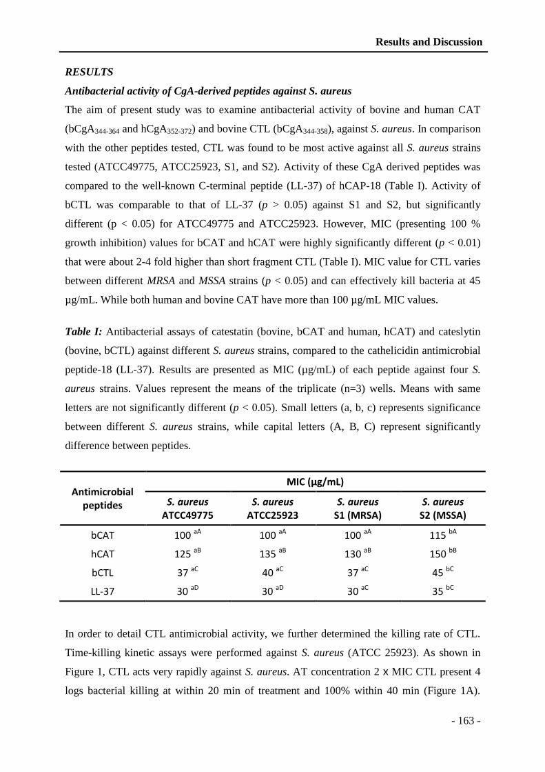
Results and Discussion
- 163 -
RESULTS
Antibacterial activity of CgA-derived peptides against S. aureus
The aim of present study was to examine antibacterial activity of bovine and human CAT
(bCgA344-364 and hCgA352-372) and bovine CTL (bCgA344-358), against S. aureus. In comparison
with the other peptides tested, CTL was found to be most active against all S. aureus strains
tested (ATCC49775, ATCC25923, S1, and S2). Activity of these CgA derived peptides was
compared to the well-known C-terminal peptide (LL-37) of hCAP-18 (Table I). Activity of
bCTL was comparable to that of LL-37 (p > 0.05) against S1 and S2, but significantly
different (p < 0.05) for ATCC49775 and ATCC25923. However, MIC (presenting 100 %
growth inhibition) values for bCAT and hCAT were highly significantly different (p < 0.01)
that were about 2-4 fold higher than short fragment CTL (Table I). MIC value for CTL varies
between different MRSA and MSSA strains (p < 0.05) and can effectively kill bacteria at 45
µg/mL. While both human and bovine CAT have more than 100 µg/mL MIC values.
Table I: Antibacterial assays of catestatin (bovine, bCAT and human, hCAT) and cateslytin
(bovine, bCTL) against different S. aureus strains, compared to the cathelicidin antimicrobial
peptide-18 (LL-37). Results are presented as MIC (µg/mL) of each peptide against four S.
aureus strains. Values represent the means of the triplicate (n=3) wells. Means with same
letters are not significantly different (p < 0.05). Small letters (a, b, c) represents significance
between different S. aureus strains, while capital letters (A, B, C) represent significantly
difference between peptides.
Antimicrobial peptides
MIC (µg/mL)
S. aureus ATCC49775
S. aureus ATCC25923
S. aureus S1 (MRSA)
S. aureus S2 (MSSA)
bCAT 100 aA 100 aA 100 aA 115 bA
hCAT 125 aB 135 aB 130 aB 150 bB
bCTL 37 aC 40 aC 37 aC 45 bC
LL-37 30 aD 30 aD 30 aC 35 bC
In order to detail CTL antimicrobial activity, we further determined the killing rate of CTL.
Time-killing kinetic assays were performed against S. aureus (ATCC 25923). As shown in
Figure 1, CTL acts very rapidly against S. aureus. AT concentration 2 x MIC CTL present 4
logs bacterial killing at within 20 min of treatment and 100% within 40 min (Figure 1A).
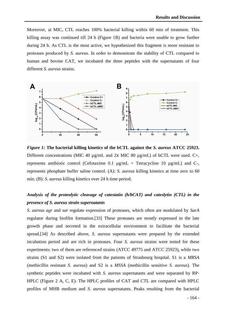
Results and Discussion
- 164 -
Moreover, at MIC, CTL reaches 100% bacterial killing within 60 min of treatment. This
killing assay was continued till 24 h (Figure 1B) and bacteria were unable to grow further
during 24 h. As CTL is the most active, we hypothesized this fragment is more resistant to
proteases produced by S. aureus. In order to demonstrate the stability of CTL compared to
human and bovine CAT, we incubated the three peptides with the supernatants of four
different S. aureus strains.
Figure 1: The bacterial killing kinetics of the bCTL against the S. aureus ATCC 25923.
Different concentrations (MIC 40 µg/mL and 2x MIC 80 µg/mL) of bCTL were used. C+,
represents antibiotic control (Cefotaxime 0.1 µg/mL + Tetracycline 10 µg/mL) and C-,
represents phosphate buffer saline control. (A): S. aureus killing kinetics at time zero to 60
min. (B): S. aureus killing kinetics over 24 h time period.
Analysis of the proteolytic cleavage of catestatin (h/bCAT) and cateslytin (CTL) in the
presence of S. aureus strain supernatants
S. aureus agr and sar regulate expression of proteases, which often are modulated by SarA
regulator during biofilm formation.[33] These proteases are mostly expressed in the late
growth phase and secreted in the extracellular environment to facilitate the bacterial
spread.[34] As described above, S. aureus supernatants were prepared by the extended
incubation period and are rich in proteases. Four S. aureus strains were tested for these
experiments: two of them are referenced strains (ATCC 49775 and ATCC 25923), while two
strains (S1 and S2) were isolated from the patients of Strasbourg hospital. S1 is a MRSA
(methicillin resistant S. aureus) and S2 is a MSSA (methicillin sensitive S. aureus). The
synthetic peptides were incubated with S. aureus supernatants and were separated by RP-
HPLC (Figure 2 A, C, E). The HPLC profiles of CAT and CTL are compared with HPLC
profiles of MHB medium and S. aureus supernatants. Peaks resulting from the bacterial

Results and Discussion
- 165 -
degradation were analyzed by automatic Edman sequencing and MALDI-TOF. In contrast to
bovine and human CAT (Figure 2A, 2C), CTL is not degraded (Figure 2E).
Figure 2: HPLC chromatograms of bCAT, hCAT and CTL alone or with different
bacterial strain supernatants, with or without protease inhibitors. (A): Alignment of the
HPLC chromatograms corresponding to: (1) bCAT, (2) bCAT+MHB, (3) bCAT+S49775, (4)
bCAT+S25923 (5) bCAT+S1 (6) bCAT+S2. (B): Alignment of the HPLC chromatograms
corresponding to: (1) bCAT, (2) bCAT+Pi+S49775, (3) bCAT+Pi+S25923 (4) bCAT+Pi+S1
(5) bCAT+Pi+S2. (C): Alignment of the HPLC chromatograms corresponding to: (1) hCAT,
Results and Discussion
- 166 -
(2) hCAT+MHB, (3) hCAT+S49775, (4) hCAT+S25923 (5) hCAT+S1 (6) hCAT+S2. (D):
Alignment of the HPLC chromatograms corresponding to: (1) hCAT, (2) hCAT+Pi+S49775,
(3) hCAT+Pi+S25923 (4) hCAT+Pi+S1 (5) hCAT+Pi+S2. (E): Alignment of the HPLC
chromatograms corresponding to: (1) bCTL, (2) bCTL+MHB, (3) bCTL+S49775, (4)
bCTL+S25923 (5) bCTL+S1 (6) bCTL+S2. (F): Alignment of the HPLC chromatograms
corresponding to: (1) bCTL, (2) bCTL+Pi+S49775, (3) bCTL+Pi+S25923 (4) bCTL+Pi+S1
(5) bCTL+Pi+S2.
HPLC of bCAT (Figure 2A) indicates 2 major peaks, eluted at 38.7 and 40 min,
corresponding to full-length peptide according to sequencing and MALDI-TOF mass
spectrometry (2426 Da) (Figure 3). Presence of these 2 isoforms may be related to beta-turn
conformation induced by proline residue in position 360 and its isomeric state (cis/trans).[35]
In MHB medium, bCAT was not processed. Whereas, in presence of culture supernatants
(S49775, S25923 and S1), bCAT was processed to generate fragments eluted at 38.3 and 38.6
min (Figure 2A). Sequencing and MALDI-TOF analysis indicate that these 2 fragments
correspond to the isoforms of bCgA349-364 (1782 Da) (Figure 3). In presence of culture
supernatant of S2, bCAT was largely cleaved to generate fragments eluted at 28.0, 36.6 and
37.5 min (Figure 2A). Sequencing and MALDI-TOF analysis indicate that these fragments
correspond to bCgA350-356 (826 Da) and the 2 isoforms of bCgA357-364 (887 Da) (Figure 3).
Moreover, the HPLC profile of hCAT (Figure 2C) showed one major peak at 38 min that
corresponds to the full-length peptide according to sequencing and MALDI-TOF mass
spectrometry (2327 Da) (Figure 3). In presence of MHB medium, hCAT was not cleaved.
However, after incubation with S49775, S25923 and S1, hCAT was partially processed to
generate a fragment eluted at 36 min. Sequencing and MALDI-TOF analysis indicated that
this fragment corresponds to hCgA357-372 (1780 Da) (Figure 3). In presence of supernatant S2,
hCAT was completely cleaved to generate fragments eluted at 28.5 and 33 min (Figure 2C).
Sequencing and MALDI-TOF analysis indicated that these fragments correspond to hCgA358-
364 (840 Da) and hCgA365-372 (871 Da) (Figure 3).
Finally, the shortest peptide bCTL (bCTL, CgA344-358), is eluted as a single peak (Figure 2E)
and is not degraded by any of the staphylococcal strains tested.
Interestingly, incubation of synthetic peptides with bacterial supernatants supplemented with
protease inhibitors, prevents the peptide degradation (Figure 2B, D, F). HPLC profiles of
bCAT (Figure 2B: chromatogram 2-5) indicate that it is not cleaved by any of the four strain

Results and Discussion
- 167 -
supernatants tested in presence of protease inhibitors. Similar results were obtained for hCAT
(Figure 2D: chromatogram 2-5). In addition, CTL remains unaffected by S. aureus
supernatants with or without protease inhibitors (Figure 2E, F).
Figure 3: The proteolytic cleavage sites induced after treatment of bCAT, hCAT and
CTL with different S. aureus strains (ATCC25923, ATCC49775, S1 and S2). The
experimental molecular masses obtained after MALDI-TOF analysis, are indicated.
The proteolytic cleavage sites of bCAT and hCAT obtained after incubation with S49775,
S25923 and S1 were identical (Figure 3). They corresponded to the peptide bonds L348-S349
and L356-S357 for bCAT and hCAT, respectively. With S2, they correspond to the peptide
bonds S349-F350+G356-F357 and S357-F358+G364-F365 for bCAT and hCAT, respectively. It is
important to point out that the short peptide bCTL (bCgA344-358) derived from CAT was not
cleaved by S. aureus proteases (Figure 2E, F) demonstrating that CTL resists to the
proteolytic cocktail produced by S. aureus.
Results and Discussion
- 168 -
DISCUSSION
Due to continuous resistance development by S. aureus, the interest for natural AMPs is
increasing because some of them display potent antibacterial activity. However, secondary
reactions such as hemolytic activity,[36] limit their use as systemic anti-infective drugs. Until
recently, no such reaction was reported for the antimicrobial Cgs-derived peptides in the
literature. Several CgA-derived peptides were previously characterized to display
antimicrobial properties,[9] however, data concerning killing of S. aureus is not reported.
CAT was previously reported, to be active against different bacterial strains at micromolar
range.[24] CTL is well characterized for antimicrobial activity against Micrococcus luteus,
few yeasts and fungal strains. Moreover, it is not hemolytic for human erythrocytes.[5]. In the
present study, for the first time, we brought to light the antibacterial activity of CTL against
different strains of S. aureus with a MIC value of 37-45 µg/mL (Table I).
In vivo, a cross talk is established via dynamics of the interactions between host and bacteria.
Many bacterial strains express a variety of proteases, ranging from non-specific and powerful
enzymes that degrade many proteins involved in innate immunity, to proteases that are
extremely specific in their mode of action.[37] Here, we questioned whether S. aureus
proteases can cleave the three CgA-derived peptides studied. This proteolytic degradation
could explain the 3-4 fold difference observed in antibacterial activity. To address this issue,
we hypothesized that the human and bovine CAT peptides are degraded by the staphylococcal
proteases and that CTL resists to the degradation and maintains its activity. Therefore, we
tested the effect of four staphylococcal supernatants and demonstrated that bovine and human
CAT are similarly degraded to generate inactive fragments, while CTL is not degraded. For
bovine and human CAT, the cleavage sites induced by the supernatants are different. For S2
these correspond to the cleavage of S-F/G-F. Whereas, for ATCC49775, ATCC25923, and
S1, it corresponds to cleavage of L-S (Figure 3). Upon cleavage by S. aureus proteases, the
CAT peptides are no longer active against S. aureus. Previous data showed that bovine
CgA344-351 and CgA348-358 display an antimicrobial activity against Micrococcus luteus at 20
µM, suggesting that L348/356 is important for its antibacterial activity.[5] Furthermore, in the
protease inhibition assay, we demonstrated that degradation of human and bovine CAT can be
prevented by the use of protease inhibitors. CTL maintains its position either with or without
protease inhibitors indicating that proteases have no effect on the CTL.
The biosynthesis of CTL results by the action of prohormone convertases PC1/2 present in the
granular matrix of chromaffin cells.[2,38] The prohormone thiol protease (PTP) was also
reported to be essential for CAT synthesis (bCgA344-364) by cleaving D-R and L-R.[38] In
Results and Discussion
- 169 -
addition, in chromaffin secretory vesicles, the cysteine protease cathepsin L (CTSL)[39]
generates CTL by the additional cleavage R-G of CAT. CAT fragment is known to activate
the neutrophils by inducing extra-cellular calcium influx in human neutrophils via
calmodulin-regulated calcium independent phospholipase A2.[11] Here, we demonstrate that
CTL is resistant to the degradation by staphylococcal proteases, which strengthens the
involvement of this CgA-derived domain in innate immunity.[10,40]
The primary structures of CAT and CTL are highly conserved during evolution (Figure 4).
Arginine ratio of these peptide sequences is important, since it modulates the interaction with
negatively charges of the microorganism’s membranes. In addition, it was previously shown
that arginine residues have a high tendency to interact with the lipids as suggested for other
peptides, such as HIV-1 transcriptional activator Tat protein.[41,42] For hCAT, bCAT, and
bCTL the arginine ratios are 15%, 23%, and 33% respectively. High arginine ratio of bCTL
supports a strong interaction with the negatively charged lipid bilayer as compared to the both
CAT. Structure activity relationship of CTL with bacterial membrane is also demonstrated by
recent experiments concerning the CTL derived peptide with a hydrophobic N-terminal end
FLE-CTL (FLE-RSMRLSFRARGYGFR) (5R). With the help of HPLC and antimicrobial
assays, we have shown that this synthetic peptide is inactive against S. aureus at 400 µg/mL
and interacts strongly with bacterial membrane. In contrast, the shorter peptide that is lacking
the C-terminal end (YGFR) of CTL: FLE-RSMRLSFRARG, displays antibacterial activities
at 200 µg/mL (data not shown). Thus, CTL sequence possesses the cationic amphipathic
features to exhibit potent antimicrobial activities.
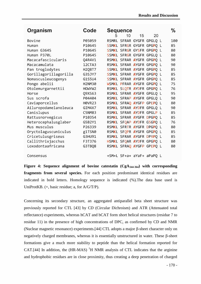
Results and Discussion
- 170 -
Figure 4: Sequence alignment of bovine catestatin (CgA344-364) with corresponding
fragments from several species. For each position predominant identical residues are
indicated in bold letters. Homology sequence is indicated (%).The data base used is
UniProtKB. (+, basic residue; a, for A/G/T/P).
Concerning its secondary structure, an aggregated antiparallel beta sheet structure was
previously reported for CTL [43] by CD (Circular Dichroism) and ATR (Attenuated total
reflectance) experiments, whereas hCAT and bCAT form short helical structures (residue 7 to
residue 11) in the presence of high concentrations of DPC, as confirmed by CD and NMR
(Nuclear magnetic resonance) experiments.[44] CTL adopts a major β-sheet character only on
negatively charged membranes, whereas it is essentially unstructured in water. These β-sheet
formations give a much more stability to peptide than the helical formation reported for
CAT.[44] In addition, the (HR-MAS) 1H NMR analysis of CTL indicates that the arginine
and hydrophobic residues are in close proximity, thus creating a deep penetration of charged
Results and Discussion
- 171 -
residues into the membrane.[42] Therefore, the electrostatic interaction between positively
charged arginine residues and negatively charged lipids appears to be responsible for binding
of CTL to the lipid bilayer and the aromatic residues stabilize the lipid-peptide interaction.
In our group, we recently evaluated the synergistic effect of three CgA-derived peptides
(CAT, CTL and amidated CTL) with antibiotics and demonstrated that this co-treatment
induce a reduction of the antibiotics concentration used and can potentiate their activities.
Antimicrobial assays were carried out by combining the AMPs at concentration below the
MIC. The comparison was made with antibiotic or peptide separately at the same doses. For
all these experiments, we evaluated the Fractional Inhibitory Concentration (FIC) of CgA-
derived peptides combined with Minocyclin or Voriconazole. FIC corresponds to a
synergistic effect in the range of ≤0.5, an additive effect when it is between >0.5 and <2, and
an antagonistic effect when it is >2.[45] For the combination of amidated CTL and
Minocyclin, we obtained a FIC of 0.37 against S. aureus, and for CTL and Voriconazole, we
obtained a FIC of 0.25, and 0.5 against Candida albicans and Candida tropicalis
respectively.[10] Regarding these data, one could imagine a mechanism in which, the peptides
could favor destabilization of bacterial membrane, thus allowing the antibiotics to rapidly
penetrate inside bacterial cells to reaching their site of action. These in vitro results are likely
to occur in vivo, especially during systemic inflammation. Indeed, sepsis triggers numerous
changes in proteases and inhibitors activity.[46] These regulations have been strongly related
with sepsis severity.[47] Under such influences, CAT and CTL could even be difficult to
release, owing to structural modifications of CgA,[48] due to sepsis-related oxidative stress.
These modifications include oxidation of methionine, aromatic residues, glycanes,
phosphorylation and also aggregation of the complete protein that might prevent the
processing of CgA to produce active antimicrobial peptides. The structure-function
relationship of AMPs is also very important to unravel the regular pattern of antibacterial
activity. It has been previously demonstrated, that selective end tagging or short hydrophobic
stretches can be, added to increase AMPs activity.[49,50] Recently, we studied the properties
of a modified CTL by inserting cysteine at C-terminal. This modified CTL was used to
develop antimicrobial polymer coatings via alternative deposition of polyelectrolytes
conjugated to CTL-C.[51] Activity of CTL is not altered, even with this modification or when
inserted into PEM (Polyelectrolyte multilayer) coatings. Moreover, it is non-toxic for human
gingival fibroblast.[51]
To conclude, for the first time we present the involvement of staphylococcal proteases on the
cleavage of antimicrobial CgA-derived peptides. As the CTL domain is resistant to

Results and Discussion
- 172 -
staphylococcal proteases, it constitutes a promising natural antibacterial peptide for further
studies.
ACKNOWLEDGMENTS
We are grateful for the generous gift of bacterial strains from the bacteriology Institute of
Civil Hospital of Strasbourg. Authors thank Cosette Betcha and Ouria Tahar for their
excellent technical assistance. We also acknowledge Azhar Ayyaz Pirzado and Adnan Khan
Niazi for their help in writing.
Transparency Declarations
None to declare.
REFERENCES
1. Helle KB (2010) Regulatory peptides from chromogranin A and secretogranin II: putative
modulators of cells and tissues involved in inflammatory conditions. Regul Pept 165:
45-51.
2. Metz-Boutigue MH, Garcia-Sablone P, Hogue-Angeletti R, Aunis D (1993) Intracellular
and extracellular processing of chromogranin A. Determination of cleavage sites. Eur
J Biochem 217: 247-257.
3. Strub JM, Garcia-Sablone P, Lonning K, Taupenot L, Hubert P, et al. (1995) Processing of
chromogranin B in bovine adrenal medulla. Identification of secretolytin, the
endogenous C-terminal fragment of residues 614-626 with antibacterial activity. Eur J
Biochem 229: 356-368.
4. Montero-Hadjadje M, Vaingankar S, Elias S, Tostivint H, Mahata SK, et al. (2008)
Chromogranins A and B and secretogranin II: evolutionary and functional aspects.
Acta Physiol (Oxf) 192: 309-324.
5. Briolat J, Wu SD, Mahata SK, Gonthier B, Bagnard D, et al. (2005) New antimicrobial
activity for the catecholamine release-inhibitory peptide from chromogranin A. Cell
Mol Life Sci 62: 377-385.
6. Lugardon K, Chasserot-Golaz S, Kieffer AE, Maget-Dana R, Nullans G, et al. (2001)
Structural and biological characterization of chromofungin, the antifungal
chromogranin A-(47-66)-derived peptide. J Biol Chem 276: 35875-35882.
7. Lugardon K, Raffner R, Goumon Y, Corti A, Delmas A, et al. (2000) Antibacterial and
antifungal activities of vasostatin-1, the N-terminal fragment of chromogranin A. J
Biol Chem 275: 10745-10753.
8. Strub JM, Goumon Y, Lugardon K, Capon C, Lopez M, et al. (1996) Antibacterial activity
of glycosylated and phosphorylated chromogranin A-derived peptide 173-194 from
bovine adrenal medullary chromaffin granules. J Biol Chem 271: 28533-28540.
9. Metz-Boutigue MH, Goumon Y, Lugardon K, Strub JM, Aunis D (1998) Antibacterial
peptides are present in chromaffin cell secretory granules. Cell Mol Neurobiol 18:
249-266.
10. Aslam R, Atindehou M, Lavaux T, Haikel Y, Schneider F, et al. (2012) Chromogranin A-
derived peptides are involved in innate immunity. Curr Med Chem 19: 4115-4123.
11. Zhang D, Shooshtarizadeh P, Laventie BJ, Colin DA, Chich JF, et al. (2009) Two
chromogranin a-derived peptides induce calcium entry in human neutrophils by
calmodulin-regulated calcium independent phospholipase A2. PLoS One 4: e4501.

Results and Discussion
- 173 -
12. Ohbayashi T, Irie A, Murakami Y, Nowak M, Potempa J, et al. (2011) Degradation of
fibrinogen and collagen by staphopains, cysteine proteases released from
Staphylococcus aureus. Microbiology 157: 786-792.
13. Sieprawska-Lupa M, Mydel P, Krawczyk K, Wojcik K, Puklo M, et al. (2004)
Degradation of human antimicrobial peptide LL-37 by Staphylococcus aureus-derived
proteinases. Antimicrob Agents Chemother 48: 4673-4679.
14. Postma B, Poppelier MJ, van Galen JC, Prossnitz ER, van Strijp JA, et al. (2004)
Chemotaxis inhibitory protein of Staphylococcus aureus binds specifically to the C5a
and formylated peptide receptor. J Immunol 172: 6994-7001.
15. Sumby P, Barbian KD, Gardner DJ, Whitney AR, Welty DM, et al. (2005) Extracellular
deoxyribonuclease made by group A Streptococcus assists pathogenesis by enhancing
evasion of the innate immune response. Proc Natl Acad Sci U S A 102: 1679-1684.
16. Rooijakkers SH, Ruyken M, Roos A, Daha MR, Presanis JS, et al. (2005) Immune
evasion by a staphylococcal complement inhibitor that acts on C3 convertases. Nat
Immunol 6: 920-927.
17. Karavolos MH, Horsburgh MJ, Ingham E, Foster SJ (2003) Role and regulation of the
superoxide dismutases of Staphylococcus aureus. Microbiology 149: 2749-2758.
18. Prokesova L, Potuznikova B, Potempa J, Zikan J, Radl J, et al. (1995) Cleavage of human
immunoglobulins by proteinase from Staphylococcus aureus. Adv Exp Med Biol
371A: 613-616.
19. Dubin G (2002) Extracellular proteases of Staphylococcus spp. Biol Chem 383: 1075-
1086.
20. Abdelnour A, Arvidson S, Bremell T, Ryden C, Tarkowski A (1993) The accessory gene
regulator (agr) controls Staphylococcus aureus virulence in a murine arthritis model.
Infect Immun 61: 3879-3885.
21. Chien Y, Manna AC, Projan SJ, Cheung AL (1999) SarA, a global regulator of virulence
determinants in Staphylococcus aureus, binds to a conserved motif essential for sar-
dependent gene regulation. J Biol Chem 274: 37169-37176.
22. Palma M, Cheung AL (2001) sigma(B) activity in Staphylococcus aureus is controlled by
RsbU and an additional factor(s) during bacterial growth. Infect Immun 69: 7858-
7865.
23. Zhao Y, Verma V, Belcheva A, Singh A, Fridman M, et al. (2012) Staphylococcus aureus
methicillin-resistance factor fmtA is regulated by the global regulator SarA. PLoS One
7: e43998.
24. Radek KA, Lopez-Garcia B, Hupe M, Niesman IR, Elias PM, et al. (2008) The
neuroendocrine peptide catestatin is a cutaneous antimicrobial and induced in the skin
after injury. J Invest Dermatol 128: 1525-1534.
25. Sizova D, Charbaut E, Delalande F, Poirier F, High AA, et al. (2007) Proteomic analysis
of brain tissue from an Alzheimer's disease mouse model by two-dimensional
difference gel electrophoresis. Neurobiol Aging 28: 357-370.
26. Davis MF, Peterson AE, Julian KG, Greene WH, Price LB, et al. (2013) Household Risk
Factors for Colonization with Multidrug-Resistant Staphylococcus aureus Isolates.
PLoS One 8: e54733.
27. National Committee for Clinical Laboratory Standards (2003) Methods for Dilution
Antimicrobial Susceptibility Tests for Bacteria That Grow Aerobically—Eighth
Edition: Approved Standard M07-A8.: NCCLS, Wayne, PA, USA.
28. Bulet P, Dimarcq JL, Hetru C, Lagueux M, Charlet M, et al. (1993) A novel inducible
antibacterial peptide of Drosophila carries an O-glycosylated substitution. J Biol
Chem 268: 14893-14897.
Results and Discussion
- 174 -
29. Wu M, Hancock RE (1999) Interaction of the cyclic antimicrobial cationic peptide
bactenecin with the outer and cytoplasmic membrane. J Biol Chem 274: 29-35.
30. Pag U, Oedenkoven M, Papo N, Oren Z, Shai Y, et al. (2004) In vitro activity and mode
of action of diastereomeric antimicrobial peptides against bacterial clinical isolates. J
Antimicrob Chemother 53: 230-239.
31. Pons AM, Zorn N, Vignon D, Delalande F, Van Dorsselaer A, et al. (2002) Microcin
E492 is an unmodified peptide related in structure to colicin V. Antimicrob Agents
Chemother 46: 229-230.
32. Kussmann M, Lassing U, Sturmer CA, Przybylski M, Roepstorff P (1997) Matrix-assisted
laser desorption/ionization mass spectrometric peptide mapping of the neural cell
adhesion protein neurolin purified by sodium dodecyl sulfate polyacrylamide gel
electrophoresis or acidic precipitation. J Mass Spectrom 32: 483-493.
33. Mrak LN, Zielinska AK, Beenken KE, Mrak IN, Atwood DN, et al. (2012) saeRS and
sarA act synergistically to repress protease production and promote biofilm formation
in Staphylococcus aureus. PLoS One 7: e38453.
34. Dunman PM, Murphy E, Haney S, Palacios D, Tucker-Kellogg G, et al. (2001)
Transcription profiling-based identification of Staphylococcus aureus genes regulated
by the agr and/or sarA loci. J Bacteriol 183: 7341-7353.
35. Vanhoof G, Goossens F, De Meester I, Hendriks D, Scharpe S (1995) Proline motifs in
peptides and their biological processing. Faseb J 9: 736-744.
36. Conlon JM, Mechkarska M, Prajeep M, Sonnevend A, Coquet L, et al. (2012) Host-
defense peptides in skin secretions of the tetraploid frog Silurana epitropicalis with
potent activity against methicillin-resistant Staphylococcus aureus (MRSA). Peptides
37: 113-119.
37. Potempa J, Pike RN (2009) Corruption of innate immunity by bacterial proteases. J Innate
Immun 1: 70-87.
38. Lee JC, Taylor CV, Gaucher SP, Toneff T, Taupenot L, et al. (2003) Primary sequence
characterization of catestatin intermediates and peptides defines proteolytic cleavage
sites utilized for converting chromogranin a into active catestatin secreted from
neuroendocrine chromaffin cells. Biochemistry 42: 6938-6946.
39. Biswas N, Rodriguez-Flores JL, Courel M, Gayen JR, Vaingankar SM, et al. (2009)
Cathepsin L colocalizes with chromogranin a in chromaffin vesicles to generate active
peptides. Endocrinology 150: 3547-3557.
40. Sternberg EM (2006) Neural regulation of innate immunity: a coordinated nonspecific
host response to pathogens. Nat Rev Immunol 6: 318-328.
41. Henriques ST, Melo MN, Castanho MA (2006) Cell-penetrating peptides and
antimicrobial peptides: how different are they? Biochem J 399: 1-7.
42. Ziegler A (2008) Thermodynamic studies and binding mechanisms of cell-penetrating
peptides with lipids and glycosaminoglycans. Adv Drug Deliv Rev 60: 580-597.
43. Jean-Francois F, Castano S, Desbat B, Odaert B, Roux M, et al. (2008) Aggregation of
cateslytin beta-sheets on negatively charged lipids promotes rigid membrane domains.
A new mode of action for antimicrobial peptides? Biochemistry 47: 6394-6402.
44. Sugawara M, Resende JM, Moraes CM, Marquette A, Chich JF, et al. (2010) Membrane
structure and interactions of human catestatin by multidimensional solution and solid-
state NMR spectroscopy. Faseb J 24: 1737-1746.
45. Serra P, Brandimarte C, Martino P, Carlone S, Giunchi G (1977) Synergistic treatment of
enterococcal endocarditis: in vitro and in vivo studies. Arch Intern Med 137: 1562-
1567.
Results and Discussion
- 175 -
46. Kwiecinski J, Josefsson E, Mitchell J, Higgins J, Magnusson M, et al. (2010) Activation
of plasminogen by staphylokinase reduces the severity of Staphylococcus aureus
systemic infection. J Infect Dis 202: 1041-1049.
47. Michel A, Agerer F, Hauck CR, Herrmann M, Ullrich J, et al. (2006) Global regulatory
impact of ClpP protease of Staphylococcus aureus on regulons involved in virulence,
oxidative stress response, autolysis, and DNA repair. J Bacteriol 188: 5783-5796.
48. Strub JM, Sorokine O, Van Dorsselaer A, Aunis D, Metz-Boutigue MH (1997)
Phosphorylation and O-glycosylation sites of bovine chromogranin A from adrenal
medullary chromaffin granules and their relationship with biological activities. J Biol
Chem 272: 11928-11936.
49. Pasupuleti M, Schmidtchen A, Chalupka A, Ringstad L, Malmsten M (2009) End-tagging
of ultra-short antimicrobial peptides by W/F stretches to facilitate bacterial killing.
PLoS One 4: e5285.
50. Malmsten M, Kasetty G, Pasupuleti M, Alenfall J, Schmidtchen A (2011) Highly selective
end-tagged antimicrobial peptides derived from PRELP. PLoS One 6: e16400.
51. Cado G, Aslam R, Séon L, Garnier T, Fabre R, et al. (2013) Self-defensive biomaterial
coating against bacteria and yeasts: polysaccharide multilayer film with embedded
antimicrobial peptide. Adv functional materials [DOI: 10.1002/adfm.201300416].

Manuscript: III
Chromogranins and
Glu-C Protease

Manuscript: IV
Biodegradeable antimicrobial
polyelectrolyte multilayer coating


www.afm-journal.de
FULL P
APER
1
www.MaterialsViews.com
wileyonlinelibrary.com© 2013 WILEY-VCH Verlag GmbH & Co. KGaA, Weinheim
G. Cado , R. Aslam , L. Séon , T. Garnier , R. Fabre , A. Parat , A. Chassepot , J.-C. Voegel , B. Senger , F. Schneider , Y. Frère , L. Jierry , P. Schaaf , H. Kerdjoudj , M.-H. Metz-Boutigue , and F. Boulmedais *
1. Introduction
Implantable medical devices are widely used in surgery not only to replace altered or lost tissues but also in critical care for fl uid or gas administration using cath-eters or tracheal tube, respectively. These devices constitute an open gate for patho-gens invasion. [ 1 ] Prevention of pathogen colonization of medical implants consti-tutes a major medical and fi nancial issue since nosocomial infection represents one of the most serious complications after surgery or critical care. Indeed each year in Europe, 5% of patients admitted to hospitals suffer from hospital-acquired infections leading to a mortality of 10%. [ 2 ] Staphylococcus aureus , a Gram-positive bacterium, is responsible for hospital-acquired infections especially in immu-nocompromized patients. It is one of the most virulent bacteria leading to high rates of device-related systemic infections and mortality. [ 3 ] A recent study has geneti-cally characterized the strains of S. aureus
Self-Defensive Biomaterial Coating Against Bacteria and Yeasts: Polysaccharide Multilayer Film with Embedded Antimicrobial Peptide
Prevention of pathogen colonization of medical implants is a major medical and fi nancial issue since infection by microorganisms constitutes one of the most serious complications after surgery or critical care. Immobilization of antimicrobial molecules on biomaterials surfaces is an effi cient approach to prevent biofi lm formation. Herein, the fi rst self-defensive coating against both bacteria and yeasts is reported where the release of the antimicrobial peptide is triggered by enzymatic degradation of the fi lm due to the patho-gens themselves. Biocompatible and biodegradable polysaccharide multilayer fi lms based on functionalized hyaluronic acid by cateslytin (CTL), an endog-enous host-defensive antimicrobial peptide, and chitosan (HA-CTL-C/CHI) were deposited on a planar surface with the aim of designing both antibacte-rial and antifungal coating. After 24 h of incubation, HA-CTL-C/CHI fi lms fully inhibit the development of Gram-positive Staphylococcus aureus bacteria and Candida albicans yeasts, which are common and virulent pathogens agents encountered in care-associated diseases. Hyaluronidase, secreted by the pathogens, leads to the fi lm degradation and the antimicrobial action of the peptide. Furthermore, the limited fi broblasts adhesion, without cytotoxicity, on HA-CTL-C/CHI fi lms highlights a medically relevant application to prevent infections on catheters or tracheal tubes where fi brous tissue encapsulation is undesirable.
DOI: 10.1002/adfm.201300416
Dr. G. Cado, L. Séon, Dr. T. Garnier, Dr. A. Parat, Dr. Y. Frère, Dr. L. Jierry, Prof. P. Schaaf, Dr. F. BoulmedaisCentre National de la Recherche Scientifi queInstitut Charles Sadron UPR 22, 23 rue du Loess,67034 Strasbourg Cedex 2, France E-mail: [email protected] R. Aslam, Dr. R. Fabre, A. Chassepot, Dr. J.-C. Voegel, Dr. B. Senger, Prof. P. Schaaf, Dr M.-H. Metz-BoutigueInstitut National de la Santé et de la Recherche Médicale INSERM UMR 1121, 11 rue Humann, 67085 Strasbourg Cedex, FranceUniversité de StrasbourgFaculté de Chirurgie Dentaire 1 place de l’Hôpital, 67000 Strasbourg, France Prof. F. SchneiderService de Réanimation MédicaleHôpital de HautepierreHôpitaux Universitaires de Strasbourg1 avenue Molière, 67098 Strasbourg Cedex, France
Dr. H. KerdjoudjEA 4691, Université de Reims Champagne ArdenneSFR-CapSanté, 1 avenue du Maréchal Juin51100 Reims, France Dr. L. Jierry, Prof. P. SchaafUniversité de Strasbourg Ecole Européenne de ChimiePolymères et Matériaux 25 rue Becquerel, 67087 Strasbourg Cedex 2, France Dr. L. Jierry, Prof. P. Schaaf, Dr. F. BoulmedaisInternational Center for Frontier Research in Chemistry8 allée Gaspard Monge, 67083 Strasbourg, France Prof. P. SchaafInstitut Universitaire de France103 boulevard Saint-Michel, 75005 Paris, France
Adv. Funct. Mater. 2013, DOI: 10.1002/adfm.201300416
FULL
PAPER
2
www.afm-journal.dewww.MaterialsViews.com
wileyonlinelibrary.com © 2013 WILEY-VCH Verlag GmbH & Co. KGaA, Weinheim
responsible of catheter-related infections and demonstrated that 82% of these strains are methicillin resistant and contain numerous genes involved in biofi lm formation and bacte-rial dispersion. [ 4 ] Candida albicans , the most common human yeast pathogen, possesses the ability to form biofi lms that are sources of local and systemic infection. Moreover C. albicans biofi lms allow the formation of S. aureus microcolonies on their surface and even enhanced S. aureus resistance to antibiotics. [ 5 ] When associated with bacterial infections, fungal proliferation induces an increased frequency or severity of diseases. [ 6–8 ] The recent resistance of C. albicans to antifungal therapies [ 9–10 ] and of S. aureus to antibiotics points out the need of multifunctional coatings that prevent infections of both yeast and bacteria.
Various approaches based on immobilization or release of bactericidal substances, using self-assembled monolayers or grafting of polymers have been explored and extensively reviewed. [ 11,12 ] Polyelectrolyte multilayer (PEM) fi lms, based on an alternated deposition of polycations and polyanions onto a solid surface, emerged as a simple and effi cient approach to functionalize surfaces in a controlled way. [ 13,14 ] The fi rst antibacterial PEM fi lms were designed by insertion of silver nanoparticles. [ 15–20 ] Later on, hydrophobic bactericide using dendritic block copolymer [ 21 ] and liposomes containing silver ion [ 22 ] were inserted in PEM fi lms to obtain effi cient bactericidal coatings. Chitosan based PEM fi lms were dem-onstrated to be antibacterial against Escherichia coli and Ente-rococcus faecalis. [ 16 , 23,24 ] Antibiotics, like gentamicin, have been inserted in hydrolysable [ 25 ] or crosslinked PEM fi lms [ 26 ] to be later released. Despite an improvement of the antibacterial activity of the fi lms, the use of antibiotics or silver particles have serious drawbacks because of their limited effi ciency, their toxicity or their role in the emergence of multi-resistant pathogens. [ 27,28 ]
Natural antimicrobial peptides (AMPs), secreted by numerous living organisms against pathogens, gain increased attention due to their broad spectrum of antimicrobial activity and their low cytotoxicity. [ 12 ] They predominantly cause disruption of the membrane integrity of pathogen agents and thus unlikely ini-tiate the development of resistance. [ 12 ] Positively charged AMPs were already used as a part of the PEM architecture to obtain antibacterial effect by contact [ 29 ] or by release. [ 26 , 30 ] Guyomard et al. succeeded in embedding in PEM fi lms a complex of poor water soluble AMPs and amphiphilic polyelectrolytes. They obtained antibacterial activity against Gram-positive bacteria. [ 31 ] Active PEM fi lms with embedded antifungal peptides were also reported. [ 32,33 ] Yet, it would be of high interest to design coatings bearing both anti-bacterial and antifungal properties. Up to now, only few systems share both properties. They are mainly based on silver coating [ 34 ] or quaternary ammonium cationic molecules as surfactant, [ 35 ] synthetic polymer, [ 36 ] or silane. [ 37 ]
To our knowledge, no coating based on AMP peptides possessing both properties has been reported so far. To achieve this goal, we used bovine cateslytin (CTL), a Chrom-ogranin A (CGA) derived peptide, an endog-enous protein, secreted with its numerous
natural derived peptides by nervous, endocrine and immune cells during infection [ 38 ] acting in the innate immunity system. [ 39 ] CTL, an AMP corresponding to CGA344-358, acts in the micromolar range with a wide spectrum of antimicrobial activities against Gram-positive bacteria, yeasts and fi lamen-tous fungi, without cytotoxic effect on mammalian cells. [ 40,41 ] Moreover, it is very stable against bacterial proteases. [ 42 ] We used polysaccharide multilayer fi lms based on CTL-C-function-alized hyaluronic acid as polyanion and chitosan as polycation, (HA-CTL-C/CHI), that were deposited on a planar surface with the aim of designing a self-defensive coating against both bac-teria and yeasts ( Scheme 1 ). A cysteine residue (C) was added at the C-terminal end of the CTL sequence to allow its grafting to HA. HA and CHI are biodegradable by enzymatic hydrol-ysis with hyaluronidase [ 43 ] and chitosanase [ 44 ] respectively. Both polysaccharides are already widely used in biomedical appli-cations due to their interesting intrinsic properties. [ 45,46 ] The ability of Staphylococcus, [ 47 ] Candida species [ 48 ] and M. luteus to degrade HA, by producing hyaluronidase, allows the CTL–C to be released from PEM fi lms only in the presence of the patho-gens. Release of antibacterial compounds (AMPs or classical antibiotics) are usually obtained by passive diffusion from the fi lms at physiological pH [ 31,32 ] or by pH-induced degradation of the fi lms. [ 25 , 30 ] Pavlukhina et al. reported the release of anti-microbial agents using pH variations associated with growth of bacteria as an internal trigger to release. [ 26 ] This coating can thus be named as self-defensive as it is related to a local change of the environment of the coating due to the pathogens themselves. This was the fi rst system developed based on this idea. We developed herein a new self-defensive coating where the release of the antimicrobial peptide is triggered by enzy-matic degradation of the fi lm due to the pathogens themselves. Polysaccharides adsorbed mass was determined by Surface Plasmon Resonance (SPR). The buildup and the topography of the fi lms were characterized by Atomic Force Microscopy (AFM). Antibacterial and antifungal activities of HA-CTL-C in solution and HA-CTL-C/CHI fi lms were tested against two strains Gram-positive bacteria, i.e., S. aureus and M. luteus and one strain of yeast strain C. albicans, respectively, by using microdilution assays. [ 49 ] Confocal laser scanning microscopy (CLSM) allowed following the penetration of the fl uorescently labeled HA FITC -CTL-C, diluted in solution or embedded in a PEM fi lm, into the cell membrane of C. albicans . Finally, the cytotoxicity of HA-CTL-C/CHI fi lms was tested through human gingival fi broblasts (HGFs) viability.
Scheme 1 . Schematic representation of CHI/HA multilayers functionalized by an antimicrobial peptide (AMP) and its activity towards bacteria and yeasts based on the degradation of the fi lm.
Adv. Funct. Mater. 2013, DOI: 10.1002/adfm.201300416

FULL P
APER
3
www.afm-journal.dewww.MaterialsViews.com
wileyonlinelibrary.com© 2013 WILEY-VCH Verlag GmbH & Co. KGaA, Weinheim
2. Results and Discussion
2.1. Physical-Chemical Characterization of HA-CTL-C/CHI Films
CTL-C peptide was covalently coupled to HA in two steps using successively the carbodiimide chemistry to graft maleimide functions on HA and the thiol-maleimide coupling reaction to graft CTL-C on the modifi ed HA. Synthetic procedures of CTL-C peptide and its conjugation to HA are described in Supporting Information (SI). After dialysis and freeze-drying, a coupling ratio of 5% was determined by 1 H-NMR, corresponding to the grafting of approximately 5 CTL-C peptides for 100 HA dimer units ( Figure 1 a). The antimicrobial and antifungal activities of CTL and modifi ed CTL were tested in solution against two bac-terial strains M. luteus and S. aureus (ATCC 25923) and a yeast C. albicans . Minimal inhibitory concentration (MIC) in peptide of CTL, CTL-C and HA-CTL-C were determined using bacterial and fungal assays described in SI. CTL-C remains antimicro-bial at micromolar concentration ( < 100 μ M) and displays an even better activity against M. luteus compared to the cysteine free CTL peptide. In comparison with the non-grafted CTL-C peptide, MIC value of HA-CTL-C increases from 35 to 45 μ M for S. aureus and from 20 to 25 μ M for C. albicans ( Table 1 ). In the case of M. luteus , the MIC of HA-CTL-C (5 μ M) is fi ve times higher than that of free CTL-C (1 μ M). In spite of this decrease in effi ciency, the CTL-C peptide covalently linked to
HA polymer can still be considered as antimicrobial, acting in the micromolar range. The buildup of HA-CTL-C/CHI fi lm was monitored by SPR. A linear increase of the mass adsorbed is observed at each deposited layer of polysaccharide indicating the buildup of the fi lm (Figure 1 b). We investigated the topog-raphy and the roughness of HA-CTL-C/CHI fi lms at different numbers of deposition steps by means of AFM in dry state (Supporting Information Figure S1). It was diffi cult to obtain good quality AFM images in the wet state due to the viscoelas-ticity of the fi lms as we found for HA/CHI fi lms in our pre-vious work. [ 50 ] AFM imaging allows measuring fi lm thickness after scratching. Supporting Information Table S1 summarizes the thicknesses and roughnesses measured by AFM. With 5 bilayers, the surface is already entirely covered with a 5-nm thick fi lm with a roughness of 1.6 nm. As the buildup process goes on, the fi lm thickness increases up to 52 nm at 30 bilayers. The fi lm grows linearly up to 30 bilayers (Supporting Informa-tion Figure S2) and the fi lm roughness increases up to 16.5 nm (for 30 pairs of layers).
2.2. Bacterial and Fungal Assays of HA-CTL-C/CHI Films
After characterization of HA-CTL-C/CHI multilayers buildup, the antibacterial and antifungal activities of the functionalized fi lms were evaluated against two strains of bacteria M. luteus and S. aureus and one of yeast strain C. albicans, respectively. The infl uence of the number of embedded functionalized layers was studied by monitoring pathogen growth for different fi lms with increasing number of HA-CTL-C/CHI bilayers. To this aim, PEI-(HA/CHI) 15-n -(HA-CTL-C/CHI) n fi lms, with n = 0, 5, 10 and 15, and PEI-(HA-CTL-C/CHI) 30 fi lms were built. Patho-gens were incubated for 24 h in contact with HA-CTL-C/CHI fi lms at 37 ° C for bacteria strains and at 30 ° C for C. albicans . For each pathogen, the microbial growth was measured at different times (1, 4, 6, and 24 h) by determination of the optical density at 620 nm (OD 620 ) of the bacterial suspension in contact with the fi lm. The data were normalized to OD 620 value obtained in similar conditions in the absence of fi lms and are expressed as a percentage of growth ( Figure 2 a–c). For each tested pathogen,
Figure 1 . a) Formula of CTL-C functionalized hyaluronic acid, HA-CTL-C, at 5% in grafting ratio. b) Evolution of the mass adsorbed, measured by SPR, (black disks) for the buildup of PEI-(HA-CTL-C/CHI) multilayer fi lm as a function of the number of deposited pairs of layers. The straight line serves to guide the eye.
Number of HA-CTL-C/CHI pairs of layers
0 1 2 3 4 5 6 7 8 9 10 11 12 13 14
Mas
s ad
sorb
ed (µ
g/cm
2 )
0
2
4
6
8a b
H2N NH
NHH2N
NH
OOH
HN
O
SMe
NH
O HN
O
HN
HN NH2
OHNHO O
NH
HN O
O
HN
HN
NH2
NH
NH
O
HN
HN
ONH
O
HO
O
NH
O NH HN
O
NHH2N
HN NH
NH2
O
NH
O OH
S
NO
O
NH
OHO
OO
OO
HO
OH
OH
NHO
OHO
OO
OO
HO
OH
OH
NHO
0.05n 0.95n
OH
Table 1. Minimal inhibitory concentration (MIC 100 ) of CTL, CTL-C and HA-CTL-C measured in solution leading to 100% of pathogens inhibi-tion. In the case of HA-CTL-C, the MIC values given correspond to the concentration in CTL-C.
Pathogens MIC 100 [ μ M in peptide]
CTL CTL-C HA-CTL-C
S. aureus 30 35 45
M. luteus 5 1 5
C. albicans 20 20 25
Adv. Funct. Mater. 2013, DOI: 10.1002/adfm.201300416
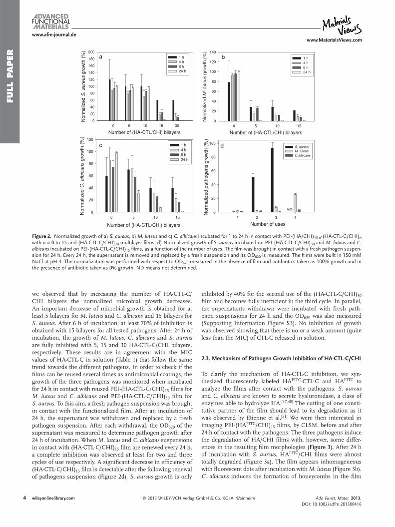
FULL
PAPER
4
www.afm-journal.dewww.MaterialsViews.com
wileyonlinelibrary.com © 2013 WILEY-VCH Verlag GmbH & Co. KGaA, Weinheim
we observed that by increasing the number of HA-CTL-C/CHI bilayers the normalized microbial growth decreases. An important decrease of microbial growth is obtained for at least 5 bilayers for M. luteus and C. albicans and 15 bilayers for S. aureus. After 6 h of incubation, at least 70% of inhibition is obtained with 15 bilayers for all tested pathogens. After 24 h of incubation, the growth of M. luteus , C. albicans and S. aureus are fully inhibited with 5, 15 and 30 HA-CTL-C/CHI bilayers, respectively. These results are in agreement with the MIC values of HA-CTL-C in solution (Table 1 ) that follow the same trend towards the different pathogens. In order to check if the fi lms can be reused several times as antimicrobial coatings, the growth of the three pathogens was monitored when incubated for 24 h in contact with reused PEI-(HA-CTL-C/CHI) 15 fi lms for M. luteus and C. albicans and PEI-(HA-CTL-C/CHI) 30 fi lm for S. aureus . To this aim, a fresh pathogen suspension was brought in contact with the functionalized fi lm. After an incubation of 24 h, the supernatant was withdrawn and replaced by a fresh pathogen suspension. After each withdrawal, the OD 620 of the supernatant was measured to determine pathogen growth after 24 h of incubation. When M. luteus and C. albicans suspensions in contact with (HA-CTL-C/CHI) 15 fi lm are renewed every 24 h, a complete inhibition was observed at least for two and three cycles of use respectively. A signifi cant decrease in effi ciency of (HA-CTL-C/CHI) 15 fi lm is detectable after the following renewal of pathogens suspension (Figure 2 d). S. aureus growth is only
inhibited by 40% for the second use of the (HA-CTL-C/CHI) 30 fi lm and becomes fully ineffi cient in the third cycle. In parallel, the supernatants withdrawn were incubated with fresh path-ogen suspensions for 24 h and the OD 620 was also measured (Supporting Information Figure S3). No inhibition of growth was observed showing that there is no or a weak amount (quite less than the MIC) of CTL-C released in solution.
2.3. Mechanism of Pathogen Growth Inhibition of HA-CTL-C/CHI
To clarify the mechanism of HA-CTL-C inhibition, we syn-thesized fl uorescently labeled HA FITC -CTL-C and HA FITC to analyze the fi lms after contact with the pathogens. S. aureus and C. albicans are known to secrete hyaluronidase, a class of enzymes able to hydrolyze HA. [ 47,48 ] The cutting of one consti-tutive partner of the fi lm should lead to its degradation as it was observed by Etienne et al. [ 51 ] We were then interested in imaging PEI-(HA FITC /CHI) 15 fi lms, by CLSM, before and after 24 h of contact with the pathogens. The three pathogens induce the degradation of HA/CHI fi lms with, however, some differ-ences in the resulting fi lm morphologies ( Figure 3 ). After 24 h of incubation with S. aureus , HA FITC /CHI fi lms were almost totally degraded (Figure 3 a). The fi lm appears inhomogeneous with fl uorescent dots after incubation with M. luteus (Figure 3 b). C. albicans induces the formation of honeycombs in the fi lm
Figure 2 . Normalized growth of a) S. aureus , b) M. luteus and c) C. albicans incubated for 1 to 24 h in contact with PEI-(HA/CHI) 15-n -(HA-CTL-C/CHI) n with n = 0 to 15 and (HA-CTL-C/CHI) 30 multilayer fi lms. d) Normalized growth of S. aureus incubated on PEI-(HA-CTL-C/CHI) 30 and M. luteus and C. albicans incubated on PEI-(HA-CTL-C/CHI) 15 fi lms, as a function of the number of uses. The fi lm was brought in contact with a fresh pathogen suspen-sion for 24 h. Every 24 h, the supernatant is removed and replaced by a fresh suspension and its OD 620 is measured. The fi lms were built in 150 mM NaCl at pH 4. The normalization was performed with respect to OD 620 measured in the absence of fi lm and antibiotics taken as 100% growth and in the presence of antibiotic taken as 0% growth. ND means not determined.
Number of (HA-CTL/CHI) bilayers 0 5 10 15 30
Nor
mal
ized
S. a
ureu
s gr
owth
(%
)
0
20
40
60
80
100
120
140
160
180
2001 h4 h 6 h 24 h
Number of (HA-CTL/CHI) bilayers 0 5 10 15
Nor
mal
ized
M. l
uteu
s gr
owth
(%
)
0
20
40
60
80
100
120
140
1 h4 h 6 h 24 h
Number of (HA-CTL/CHI) bilayers
0 5 10 15
Nor
mal
ized
C. a
lbic
ans
grow
th (
%)
0
20
40
60
80
100
1201 h4 h 6 h 24 h
Number of uses1 2 3 4
Nor
mal
ized
pat
hoge
ns g
row
th (
%)
0
20
40
60
80
100S. aureusM. luteusC.albicans
N.D.
a b
d c
Adv. Funct. Mater. 2013, DOI: 10.1002/adfm.201300416

FULL P
APER
5
www.afm-journal.dewww.MaterialsViews.com
wileyonlinelibrary.com© 2013 WILEY-VCH Verlag GmbH & Co. KGaA, Weinheim
Figure 4 . CLSM images of C. albicans, after 45 min of incubation a) in the presence of HA FITC -CTL-C in solution (50 μ M in peptide), b) in the presence of HA FITC in solution, c) in contact with PEI-(HA FITC -CTL-C/CHI) 15 , and d) in contact with PEI-(HA FITC /CHI) 15 multilayer fi lms. The scale bars represent 20 μ m
fi lms show no inhibition against C. albicans (data not shown). This emphasizes the fact that the antimicrobial activity of the HA-CTL-C/CHI fi lm is due to its degradation by the pathogens. This property renders the fi lm specifi cally active in the presence of hyaluro-nidase secreted by the pathogens. Pathogens thus initiate their own death when brought in contact with the HA-CTL-C/CHI fi lm. Even though the fi lm is degraded with time in the presence of pathogens, it can be reused at least two and three times without losing its activity against M. luteus and C. albicans .
Using fl uorescently labeled CTL-C, a pre-vious study showed that the peptides penetrate into cell mem-branes and accumulate inside yeasts. [ 40,41 ] The interactions of HA FITC -CTL-C and HA FITC with C. albicans was thus studied when solubilized in solution or embedded in multilayer fi lms. After 45 min of incubation at 30 ° C with HA FITC or HA FITC -CTL-C in solution, C. albicans were observed by CLSM. The fl uorescent HA FITC -CTL-C was detectable in cytoplasm without inducing cell lysis ( Figure 4 a). On the contrary, HA FITC is clearly
due to the degradation of HA (Figure 3 c). Degradation of HA should release CTL-C in the supernatant and promote the con-tact between CTL-C peptides and the pathogens. To check this hypothesis, we built hyaluronidase resistant fi lms functional-ized by CTL-C. CTL-C grafted on poly(allylamine hydrochloride) was thus synthesized and used to build poly(acrylic acid)/CTL-C functionalized poly(allylamine hydrochloride) (PAA/PAH-CTL-C) fi lm. After 24 h of incubation, (PAA/PAH-CTL-C) 15
Figure 3 . CLSM images of PEI-(HA FITC /CHI) 15 after 24 h of incubation with a) S. aureus , b) M. luteus and c) C. albicans . All the fi lms underwent a PFA treatment. The scale bars represent 20 μ m.
Adv. Funct. Mater. 2013, DOI: 10.1002/adfm.201300416
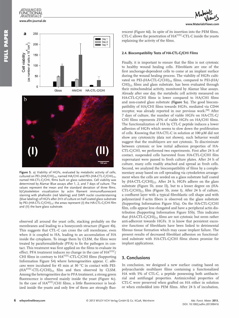
FULL
PAPER
6
www.afm-journal.dewww.MaterialsViews.com
wileyonlinelibrary.com © 2013 WILEY-VCH Verlag GmbH & Co. KGaA, Weinheim
observed all around the yeast cells, stacking probably on the membranes and leading to a honeycomb structure (Figure 4 b). This suggests that CTL–C can cross the cell membrane, even when it is coupled to HA, leading to an accumulation of HA inside the cytoplasm. To image them by CLSM, the fi lms were treated by paraformaldehyde (PFA) to fi x the pathogen in con-tact. This treatment was fi rst applied on the fi lms to evaluate its effect. PFA treatment induces no change in the case of HA FITC /CHI fi lms in contrary to HA FITC -CTL-C/CHI fi lms (Supporting Information Figure S4) where heterogeneities appear. C. albi-cans were incubated for 45 min at 30 ° C in contact with PEI-(HA FITC -CTL-C/CHI) 15 fi lm and then observed by CLSM. Among the heterogeneities due to PFA treatment, a strong green fl uorescence is observed mainly inside the yeast (Figure 4 c). In the case of HA FITC /CHI fi lms, a little fl uorescence is local-ized inside the yeasts and only few of them are strongly fl uo-
rescent (Figure 4 d). In spite of its insertion into the PEM fi lms, CTL-C allows the penetration of HA FITC -CTL-C inside the yeasts explaining the activity of the fi lms.
2.4. Biocompatibility Tests of HA-CTL-C/CHI Films
Finally, it is important to ensure that the fi lm is not cytotoxic to healthy wound healing cells. Fibroblasts are one of the fi rst anchorage-dependent cells to come at an implant surface during the wound healing process. The viability of HGFs culti-vated on PEI-(HA-CTL-C/CHI) 15 fi lms, compared to PEI-(HA/CHI) 15 fi lms and glass substrate, has been evaluated through their mitochondrial activity, monitored by Alamar blue assays. Already after one day, the metabolic cell activity measured on HA-CTL-C/CHI fi lms is lower compared to HA/CHI fi lms and non-coated glass substrate ( Figure 5 a). The good biocom-patibility of HA/CHI fi lms towards HGFs, mediated via CD44 receptor, was already reported in our previous work. [ 50 ] After 7 days of culture, the number of viable HGFs on HA-CTL-C/CHI fi lms represents 25% of viable HGFs on HA/CHI fi lms. The functionalization of HA by CTL-C peptide induces a lower adhesion of HGFs which seems to slow down the proliferation of cells. Knowing that HA-CTL-C in solution at 100 μ M did not show any cytotoxicity (data not shown), such behavior would suggest that the multilayers are not cytotoxic. To discriminate between cytotoxic or low initial adhesion properties of HA-CTL-C/CHI, we performed two experiments. First after 24 h of contact, suspended cells harvested from HA-CTL-C/CHI fi lm supernatant were passed to fresh culture plates. After 24 h of culture, many cells readily attached and spread as fresh cells. Second, we analyzed the biocompatibility of fi lms by a comple-mentary assay based on cell spreading via cytoskeleton arrange-ment when the cells are seeded on a glass substrate half coated by (HA-CTL-C/CHI) 15 . After 24 h, HGFs adhere on the glass substrate (Figure 5 b, zone II), but to a lesser degree on (HA-CTL-C/CHI) 15 fi lm (Figure 5 b, zone I). After 24 h of culture, a confl uent layer with a typical fi broblastic cell shape and with polymerized F-actin fi bers is observed on the glass substrate (Supporting Information Figure S5a). On the HA-CTL-C/CHI fi lm, cells appear less elongated and have a peripheral actin dis-tribution (Supporting Information Figure S5b). This indicates that (HA-CTL-C/CHI) 15 fi lms are not cytotoxic but seem rather anti-adherent towards HGFs. It is know that persistent exces-sive functions of fi broblasts have been linked to detrimental fi brous tissue formation which may cause implant failure. The present results of decreased fi broblast adhesion on functional-ized substrate with HA-CTL-C/CHI fi lms shows promise for implant applications.
3. Conclusions
In conclusion, we designed a new surface coating based on polysaccharide multilayer fi lms containing a functionalized HA with 5% of CTL-C, a peptide possessing both antibacte-rial and antifungal properties. Antimicrobial properties of CTL-C were preserved when grafted on HA either in solution or when embedded into PEM fi lms. After 24 h of incubation,
Figure 5 . a) Viability of HGFs, evaluated by metabolic activity of cells, cultured on PEI-(HA/CHI) 15 , named HA/CHI and PEI-(HA-CTL-C/CHI) 15 , named HA-CTL-C/CHI, fi lms built on glass substrates. Cell viability was determined by Alamar Blue assays after 1, 2, and 7 days of culture. The values represent the mean and the standard deviation of three fi lms. b)Cytoskeleton visualization by actin fi lament immunofl uorescent staining with phalloidin (red labeling) and DAPI nuclei counterstaining (blue labeling) of HGFs after 24 h of culture on half coated glass substrate by PEI-(HA-CTL-C/CHI) 15 : the areas represent (I) the HA-CTL-C/CHI fi lm and (II) the bare glass substrate.
Adv. Funct. Mater. 2013, DOI: 10.1002/adfm.201300416

FULL P
APER
7
www.afm-journal.dewww.MaterialsViews.com
wileyonlinelibrary.com© 2013 WILEY-VCH Verlag GmbH & Co. KGaA, Weinheim
home-made software based on Maxwell’s equations. The parameters involved are the refractive indices of the prism, the chromium and gold layers, the polymer fi lm ( n fi lm ) and the buffer solution ( n sol ), as well as the thickness of the chromium and gold layers and of the fi lm ( d fi lm ). From n fi lm , n sol and d fi lm , we derive the mass of fi lm per unit area:
m =
(nfilm − nsol)dfilm
dn/dc (1)
where d n /d c is the change in refractive index with concentration and is equal to 0.197 cm 3 /g for polyelectrolytes. [ 54 ] The SPR experiment was performed under a continuous fl ow rate of 100 μ L/min of 150 mM NaCl solution adjusted at pH 4. The time of deposition and of rinsing of polyelectrolytes was fi xed at 5 min.
Atomic Force Microscopy (AFM) : Atomic force microscopy (AFM) images were obtained in contact mode in dry state with the Nanoscope IV from Veeco (Santa Barbara, CA). The images were carried out with silicon nitride cantilevers, spring constant 0.03 N/m (model MSCTAUHW, Veeco, CA). Several scans were performed over a given surface area. These scans had to give reproducible images to ascertain that there is no sample damage induced by the tip. Defl ection and height mode images are scanned simultaneously at a fi xed scan rate (2 Hz) with a resolution of 512 × 512 pixels. Data evaluation was performed with the NanoScope software version 5.31r1 (Digital Instruments, Veeco). The buildup of the fi lm was made on glass substrate and the fi lm thickness was measured by using the “scratch” method. Profi lometric section analyses of a scratched fi lm allowed us to determine precisely the quality of the fi lm and its thickness over the scanned area. The scratches were achieved with a plastic cone tip and were always imaged perpendicular to the fast scan axis. The profi les correspond to a cross section along this axis. The mean thickness of the scratched fi lm was determined by measuring the thickness at least on three areas.
Confocal Laser Scanning Microscopy (CLSM) : Confocal laser scanning microscopy (CLSM) observations were carried out with a Zeiss LSM-510 microscope using a 40 × /1.31 oil immersion objective and a 0.43 μ m z-section interval. FITC fl uorescence was detected upon excitation at 488 nm, through a cut-off dichroic mirror and an emission bandpass fi lter of 505–530 nm (green).
Bacterial and Fungal Growth : To determine the antibacterial activity, microdilution assay was used on two bacterial strains M. luteus (A270) and S. aureus (ATCC25923). The antifungal activity was tested against one yeast strain C. albicans . Bacteria were precultured aerobically in Mueller-Hinton (MH) broth (Merck, Darmstadt, Germany) with agitation at 37 ° C for 18 h. C. albicans preculture was carried out on a Sabouraud broth (SB) medium (BioMérieux S.A., Marcy l’Etoile, France) with agitation at 30 ° C for 24 h. The antimicrobial activity was tested using a mid-logarithmic-phase culture of bacteria or yeast with an initial optical density at 620 nm (OD 620 ) of 0.001. For the tests performed in solution, aqueous solutions of CTL, CTL-C and HA-CTL-C were prepared in bacterial or yeast medium. Aliquots of CTL-C or HA-CTL-C (10 μ L) were incubated in 96-well microplates (Falcon, Becton Dickinson, USA) with 90 μ L of bacteria or yeast at fi nal concentrations of 100 μ M to 2 μ M of peptide. After 24 h of incubation at 37 ° C under gentle stirring, the OD 620 of the 96-well plate was measured by a microplate reader. [ 55–56 ] MIC 100 , the minimal inhibitory concentration, is the lowest concentration that is able to completely inhibit the growth of bacterial or fungi after 24 h of contact. To test the antibacterial and antifungal properties of the multilayer fi lms, PEI-(HA/CHI) 15-n -(HA-CTL-C/CHI) n with n = 0, 5, 10 and 15 and PEI-(HA-CTL-C/CHI) 30 fi lms were prepared. 400 μ L of a mid-logarithmic-phase culture of bacteria or yeast with OD 620 of 0.001 were placed in 24-well plate containing multilayer fi lms. For each tested fi lm, 100 μ L of the supernatant was taken to measure its OD 620 by a microplate reader. The withdrawn volume was compensated with the appropriated fresh bacteria or yeast solution of the same measured OD 620 . Intermittent controls were performed by inoculating the culture medium on MH (resp. SB) agar plates by spreading method and counting colonies for verifi cation at different steps. Several controls were used: a fresh medium without inoculation of pathogens was used
HA-CTL-C/CHI fi lms fully inhibit the development of S. aureus and C. albicans, which are common and virulent pathogens agents encountered in care-associated diseases. The presence of CTL-C peptides on HA allows the penetration of the modi-fi ed polysaccharide inside C. albicans after 45 min of contact. The secretion of hyaluronidase by all tested pathogens seems to be responsible for HA-CTL-C release from the fi lm and for its activity. The fi lm can keep its activity during 3 cycles of use against fresh incubated C. albicans suspension. Furthermore, the limited fi broblasts adhesion, without cytotoxicity, on HA-CTL-C/CHI fi lms highlights a medically relevant application to prevent infections on catheters or tracheal tubes where fi brous tissue encapsulation is undesirable.
4. Experimental Section Polysaccharide Solutions : Chitosan (CHI, PROTASAN Ultrapure
Chitosan CL213, 260 000 g/mol, DA 83%) was purchased by Novamatrix (Sandvika, Norway). Dried Sodium Hyaluronate (HA, 420 000 g/mol) was purchased by Lifecore (Chaska, USA). Poly(ethylene imine) (PEI, 60 000 g/mol, 50% in water), sodium chloride, sodium dodecyl sulfate (SDS), HCl and NaOH were purchased by Sigma-Aldrich (Quentin-Fallavier, France). All products were used without further purifi cation. 150 mM NaCl solution was prepared with Ultrapure Milli-Q water having a resistivity of 18.2 M Ω cm. CHI and HA solutions were prepared at 0.3 mg/mL in 150 mM NaCl adjusted at pH 4 with NaOH or HCl solutions. HA-CTL-C and HA FITC -CTL-C solutions were prepared at 0.2 mg/mL and 0.1 mg/mL, respectively. PEI solution was prepared at 0.5 mg/mL in 150 mM NaCl solution adjusted at pH 7.5 with NaOH or HCl solutions.
Film Buildup : Before PEM buildup, glass coverslips (Ø = 14 mm; VWR, Strasbourg, France) were cleaned in a SDS solution at 0.01 M, ultrasonicated for 5 min and then submitted to a hot 0.1 M HCl solution for 10 min and fi nally rinsed with ultrapure Milli-Q water. The precursor PEI layer and CHI/HA fi lms were deposited using an automated spraying device described in a previous work. [ 50 ] CHI/HA-CTL-C and CHI/HA FITC -CTL-C fi lms have been prepared in 24-well plates on PEI precoated glass substrates. 300 μ L of the polycation solution were deposited for 5 min on glass coverslips followed by a rinsing step (500 μ L) with ultrapure Milli-Q water. Then, 300 μ L of polyanion solution were deposited for 5 min followed by a rinsing step (500 μ L) with ultrapure Milli-Q water. To test the antimicrobial activity of PEM fi lms versus the number of HA-CTL-C layers, PEI-[HA/CHI] 15- n [HA-CTL-C/CHI] n with n equal to 0, 5, 10, and 15 and PEI-[HA-CTL-C/CHI] 30 fi lms were built.
Surface Plasmon Resonance : Surface plasmon resonance (SPR) [ 52,53 ] as some other detection techniques (optical waveguide lightmode spectroscopy, quartz crystal microbalance), provides a label-free, in situ method to monitor the buildup of e.g., a polymer fi lm on a solid surface. When a beam of light passes from a material of refractive index, n 1 , into a material with a lower refractive index, n 2 , some light is refl ected from the interface. When the angle of incidence of the light, θ , on the interface is greater than θ TIR = arcsin( n 2 / n 1 ), the light is completely refl ected (total internal refl ection). In the case of the SPR instrument used here (SPR Navi 200, Bionavis, Finland), the most refractive medium is a BK7-glass prism. Because the bottom surface of the prism is coated with a thin fi lm of a noble metal (gold), this refl ection is not total; some of the light is ‘lost’ into the metallic fi lm. There then exists a second angle, θ SPR > θ TIR , at which the intensity of the refl ected light goes through a minimum. In addition, an evanescent electrical fi eld travels for a short distance into the medium (e.g., fi lm) from the metallic fi lm. The probing distance reaches generally a few hundreds of nm depending on the refractive index of the probed medium and θ SPR is sensitive to the optical characteristics of the deposited fi lm. The refl ection curves, i.e., the variation of the intensity of the refl ected light with θ , corresponding to the successive polymer deposition steps have been analyzed with a
Adv. Funct. Mater. 2013, DOI: 10.1002/adfm.201300416
FULL
PAPER
8
www.afm-journal.dewww.MaterialsViews.com
wileyonlinelibrary.com © 2013 WILEY-VCH Verlag GmbH & Co. KGaA, Weinheim
of Alamar Blue was calculated according to the procedure provided by the manufacturer. The experiments were performed in triplicate. Actin fi lament staining with tetramethylrhodamine B isothiocyanate coupled phalloidin (Sigma-Aldrich) and DAPI (Invitrogen) nuclei counterstaining were performed as follows: cells were fi xed with 3.7% paraformaldehyde for 10 min at 4 ° C, permeabilized in 0.25% Triton X-100 in PBS for 10 min, and blocked in 1% BSA-PBS for 30 min. Thereafter, cells were incubated for 30 min at room temperature with 5 × 10 − 5 mg/mL phalloidin followed by nuclear counterstaining with DAPI (50 ng/mL) incubated for 2 min at room temperature. Washed slides were mounted on blades with DAKO fl uorescent mounting medium and fl uorescence distribution was examined by means of an inverse fl uorescence microscope (Axiovert, Zeiss).
Supporting Information Supporting Information is available from the Wiley Online Library or from the author.
Acknowledgements G.C., R.A., and L.S. contributed equally to this work. Aurélie Garnier is acknowledged for technical support. Karim Benmlih, Mathias Lefort and Joseph Hemmerlé are acknowledged for the automated sprayer development. Bernard Guerold is acknowledged for peptide synthesis. Géraldine Koenig is acknowledged for fruitful discussion. G.C. was supported by a “BDI” doctoral fellowship of the Centre National de la Recherche Scientifi que and the Région Alsace. L.S. was supported by the grant from Ministère de l’Enseignement Supérieur et de la Recherche (France) and R.A. by the grant from Higher Education Commission (Pakistan) and the Faculty of Dentistry of the University of Strasbourg. T.G. was supported by the icFRC foundation. We acknowledge the Pôle Matériaux et Nanosciences d’Alsace (PMNA) (“Jeunes Chercheurs du PMNA 2009” grant), the MICA Carnot (SELF-DECAMP) and the Faculty of Dentistry of the University of Strasbourg for fi nancial contribution. The contributions of the different authors are as follows: G.C. and L.S. performed the physico-chemical characterization of the fi lms, B.S. treated the SPR data, R.A. performed the antimicrobial assays, G.C., A.P. and T.G performed the synthesis of the polymers, R.F. performed the confocal images, A.C. and G.C. performed the biocompatibility tests, H.K. supervised the biocompatibility tests. L.J. supervised the synthesis of functionalized polymers, M-H.M.B. supervised the purifi cation of antimicrobial peptide and the antimicrobial assays, F.B. initiated the project, F.B. and P.S. supervised the work, Y.F., J-C.V., L.J., F.S., P.S., M-H.M.B. and F.B. participated actively in the discussions about the results, G.C., F.B., L.J., and P.S. wrote the paper.
Received: February 1, 2013 Revised: March 7, 2013
Published online:
to ensure sterility, a mixture of Tetracycline (10 μ g/mL) and Cefotaxime (0.1 μ g/mL) was used as positive control (90 μ L of culture and 10 μ L of antibiotics) and a fresh inoculated culture medium without any addition was taken as negative control. For the bacterial and yeast quantifi cation, a conversion factor was devised to convert OD into bacterial and yeast counting, respectively. Briefl y, OD of fresh bacteria (respectively yeast) culture was measured at 620 nm and was plated to MH (respectively. SB) agar for 24 h at 37 ° C (respectively 30 ° C). Colony count was performed and plotted versus the OD 620 and the slope of the linear curve was taken as conversion factor. Pathogen quantifi cation (colony forming unit (CFU) per mL) was performed at time zero and then at each hour for 6 h and then fi nally at 24 h. For quantifi cation, conversion factors were determined for each strain used separately. [ 57 ] Each assay was performed in triplicate and the experiments were repeated at least three times. The normalized growth of pathogens (%) was estimated by comparing the OD 620 values in the presence of multilayer fi lms and the positive and the negative controls. The OD 620 value of control cultures growing in the absence of fi lms and antibiotics was taken as 100% growth (negative control) and the OD 620 value of cultures growing in the presence of antibiotics (Tetracycline and Cefotaxime) was taken as 0% growth (positive control). The following equation defi nition was used:
Normalized pathogen growth(%)
=(OD 620 sample, − OD 620 positive, control)
(OD 620 negative control, − OD 620 positive, control )× 100
To follow the interaction of fl uorescently labeled HA FITC -CTL-C in solution and HA FITC -CTL-C/CHI fi lms with C. albicans, CLSM was used based on a protocol previously described with few modifi cations. [ 58 ] Briefl y, for experiments done with HA FITC -CTL-C in solution, poly-L-Lysine coated microscopic slides were covered with fresh medium containing C. albicans (OD 620 = 0.001) in SB culture medium and incubated for 24 h at 30 ° C without agitation. The medium was then removed and replaced by 50 μ M (in peptide) of HA FITC -CTL-C. After an incubation period of 45 min, the slides were washed with fresh culture medium and subsequently treated for 30 min with 4% paraformaldehyde in phosphate buffer saline (PBS) at pH 7.3. After several rinsing steps with PBS, slides were covered with Moviol 4-88 (Aldrich, Steinheim, Germany). For experiments performed with HA FITC -CTL-C/CHI, fresh C. albicans medium, previously incubated at OD 620 = 0.001 in SB culture medium for 24 h at 30 ° C without agitation, was put in contact with PEI-(HA FITC -CTL-C/CHI) 15 fi lms for 45 min at 30 ° C without agitation. HA FITC in solution and HA FITC /CHI fi lms were used as control. C. albicans were subjected to optical serial sectioning (0.2–0.3 μ m) to produce images in the x–y plane. Each optical section was scanned several times to obtain an average image. Images were recorded digitally in a 768 × 576 pixel format.
Human Gingival Fibroblasts Viability Assays : The biocompatibility of fi lms was tested using human gingival fi broblasts (HGFs). HGFs were extracted from human gingival connective tissue of healthy individuals according to a protocol approved by the ethics committee for patient protection of CPP Strasbourg Hospitals. Cells were grown in DMEM containing 1 g/L of glucose and supplemented with 10% fetal bovine serum and 100 IU/mL penicillin-streptomycin (all from Gibco). Cells were cultured at 37 ° C in 5% CO 2 in 75 cm 2 fl asks. HGFs were used between the 6th and the 9th passage. Before cell seeding, glass coverslips (Ø = 14 mm) coated with different architectures were irradiated by UV for 15 min. Then HGFs were seeded at 3 × 10 4 cells per cm 2 and cultivated at 37 ° C under a 5% CO 2 humidifi ed atmosphere for different culture times (Day 1, Day 2 and Day 7). Cell viability was assessed by Alamar Blue assay (Biosource International). This assay is based on the reduction of the blue, non-fl uorescent resazurin dye to the pink and fl uorescent resorufi n dye by living cells. [ 59 ] The overall conversion rate is proportional to the metabolic activity of living cells. [ 60 ] Viability was assessed for different times 1, 2, and 7 days. After rinsing with PBS, cells were incubated with 10% reagent in complete medium for 2 h. After incubation optical density (OD) at 570 nm and 630 nm were determined with a microplate reader. The percentage of reduction
[ 1 ] C. von Eiff , B. Jansen , W. Kohnen , K. Becker , Drugs 2005 , 65 , 179 . [ 2 ] J. P. Guggenbichler , O. Assadian , M. Boeswald , A. Kramer , GMS
Krankenhaushyg. Interdiszip. 2011 , 6 , 1 . [ 3 ] R. Katneni , S. S. Hedayati , Nat. Clin. Pract. Nephrol. 2007 , 3 , 256 . [ 4 ] G. Paniagua-Contreras , T. Sáinz-Espuñes , E. Monroy-Pérez ,
J. R. Rodríguez-Moctezuma , D. Arenas-Aranda , E. Negrete-Abascal , S. Vaca , Adv. Microbiol. 2012 , 2 , 476 .
[ 5 ] M. M. Harriott , M. C. Noverr , Antimicrob. Agents Chemother. 2009 , 53 , 3914 .
[ 6 ] M. E. Shirtliff , B. M. Peters , M. A. Jabra-Rizk , FEMS Microbiol. Lett. 2009 , 299 , 1 .
[ 7 ] E. Carlson , Infect. Immun. 1983 , 42 , 285 .
Adv. Funct. Mater. 2013, DOI: 10.1002/adfm.201300416

FULL P
APER
9
www.afm-journal.dewww.MaterialsViews.com
wileyonlinelibrary.com© 2013 WILEY-VCH Verlag GmbH & Co. KGaA, Weinheim
[ 36 ] F. X. Hu , K. G. Neoh , L. Cen , E. T. Kang , Biotechnol. Bioeng. 2005 , 89 , 474 .
[ 37 ] J. J. H. Oosterhof , K. J. D. A. Buijssen , H. J. Busscher , B. F. A. M. van der Laan , H. C. van der Mei , Appl. Environ. Microbiol. 2006 , 72 , 3673 .
[ 38 ] K. B. Helle , A. Corti , M.-H. Metz-Boutigue , B. Tota , Cell. Mol. Life Sci. 2007 , 64 , 2863 .
[ 39 ] D. Zhang , T. Lavaux , A. C. Voegeli , T. Lavigne , V. Castelain , N. Meyer , R. Sapin , D. Aunis , M.-H. Metz-Boutigue , F. Schneider , Clin. Chem. 2008 , 54 , 1497 .
[ 40 ] J. Briolat , S. D. Wu , S. K. Mahata , B. Gonthier , D. Bagnard , S. Chasserot-Golaz , K. B. Helle , D. Aunis , M.-H. Metz-Boutigue , Cell. Mol. Life Sci. 2005 , 62 , 377 .
[ 41 ] P. Shooshtarizadeh , D. Zhang , J. F. Chich , C. Gasnier , F. Schneider , Y. Haikel , D. Aunis , M.-H. Metz-Boutigue , Regul. Pept. 2010 , 165 , 102 .
[ 42 ] R. Aslam , M. Atindehou , T. Lavaux , Y. Haikel , F. Schneider , M.-H. Metz-Boutigue , Curr. Med. Chem. 2012 , 19 , 4115 .
[ 43 ] E. J. Menzel , C. Farr , Cancer Lett. 1998 , 131 , 3 . [ 44 ] T. Serizawa , M. Yamaguchi , M. Akashi , Macromolecules 2002 , 35 ,
8656 . [ 45 ] N. Volpi , J. Schiller , R. Stern , L. Soltes , Curr. Med. Chem. 2009 , 16 ,
1718 . [ 46 ] R. Riva , H. Ragelle , A. des Rieux , N. Duhem , C. Jerome , V. Preat ,
Adv. Polym. Sci. 2011 , 244 , 19 . [ 47 ] E. A. Larkin , R. J. Carman , T. Krakauer , B. G. Stiles , Curr. Med. Chem.
2009 , 16 , 4003 . [ 48 ] M. T. Shimizu , N. Q. Almeida , V. Fantinato , C. S. Unterkircher ,
Mycoses 1996 , 39 , 161 . [ 49 ] Methods for dilution antimicrobial susceptibility tests for bacteria that
grow aerobically; approved standards 8th version. CLSI document M07-A8 , Clinical and laboratory standards institute , Wayne, PA 2009 .
[ 50 ] G. Cado , H. Kerdjoudj , A. Chassepot , M. Lefort , K. Benmlih , J. Hemmerlé , J.-C. Voegel , L. Jierry , P. Schaaf , Y. Frère , F. Boulmedais , Langmuir 2012 , 28 , 8470 .
[ 51 ] O. Etienne , A. Schneider , C. Taddéi , L. Richert , P. Schaaf , J.-C. Voegel , C. Egles , C. Picart , Biomacromolecules 2005 , 6 , 726 .
[ 52 ] E. Kretschmann , Z. Phys. 1971 , 241 , 313 . [ 53 ] J. Homola , S. S. Yee , G. Gauglitz , Sens. Actuators, B 1999 , 54 , 3 . [ 54 ] X. Arys , A. M. Jonas , B. Laguitton , R. Legras , A. Laschewsky ,
E. Wischerhoff , Prog. Org. Coat. 1998 , 34 , 108 . [ 55 ] P. Bulet , J. L. Dimarcq , C. Hetru , M. Lagueux , M. Charlet , G. Hegy ,
A. Van Dorsselaer , J. A. Hoffmann , J. Biol. Chem. 1993 , 268 , 14893 . [ 56 ] M. H. Wu , R. E. W. Hancock , J. Biol. Chem. 1999 , 274 , 29 . [ 57 ] D. Y. Lee , C. M. Huang , T. Nakatsuji , D. Thiboutot , M. Monestier ,
R. L. Gallo , J. Invest. Dermatol. 2009 , 129 , S7 . [ 58 ] K. Lugardon , S. Chasserot-Golaz , A. E. Kieffer , G. Maget-Dana ,
G. Nullans , B. Kieffer , D. Aunis , M.-H. Metz-Boutigue , J. Biol. Chem. 2001 , 276 , 35875 .
[ 59 ] B. Page , M. Page , C. Noel , Int. J. Oncol. 1993 , 3 , 473 . [ 60 ] S. L. Voytik-Harbin , A. O. Brightman , B. Waisner , C. H. Lamar ,
S. F. Badylak , In Vitro Cell. Dev. Biol.: Anim. 1998 , 34 , 239 .
[ 8 ] S. A. Klotz , B. S. Chasin , B. Powell , N. K. Gaur , P. N. Lipke , Diagn. Microbiol. Infect. Dis. 2007 , 59 , 401 .
[ 9 ] G. Ramage , J. P. Martinez , J. L. Lopez-Ribot , Fems Yeast Res. 2006 , 6 , 979 .
[ 10 ] A. Lupetti , R. Danesi , M. Campa , M. Del Tacca , S. Kelly , Trends Mol. Med. 2002 , 8 , 76 .
[ 11 ] I. Banerjee , R. C. Pangule , R. S. Kane , Adv. Mater. 2011 , 23 , 690 . [ 12 ] K. Glinel , P. Thebault , V. Humblot , C. M. Pradier , T. Jouenne , Acta
Biomater. 2012 , 8 , 1670 . [ 13 ] G. Decher , Science 1997 , 277 , 1232 . [ 14 ] V. Gribova , R. Auzely-Velty , C. Picart , Chem. Mater. 2012 , 24 , 854 . [ 15 ] J. H. Dai , M. L. Bruening , Nano Lett. 2002 , 2 , 497 . [ 16 ] J. H. Fu , J. Ji , D. Z. Fan , J. C. Shen , J. Biomed. Mater. Res., Part A
2006 , 79A , 665 . [ 17 ] Z. Shi , K. G. Neoh , S. P. Zhong , L. Y. Yung , E. T. Kang , W. Wang , J.
Biomed. Mater. Res., Part A 2006 , 76 , 826 . [ 18 ] D. Lee , R. E. Cohen , M. F. Rubner , Langmuir 2005 , 21 , 9651 . [ 19 ] Z. Li , D. Lee , X. X. Sheng , R. E. Cohen , M. F. Rubner , Langmuir 2006 ,
22 , 9820 . [ 20 ] D. Lee , M. F. Rubner , R. E. Cohen , Chem. Mater. 2005 , 17 , 1099 . [ 21 ] P. M. Nguyen , N. S. Zacharia , E. Verploegen , P. T. Hammond ,
Chem. Mater. 2007 , 19 , 5524 . [ 22 ] M. Malcher , D. Volodkin , B. Heurtault , P. André , P. Schaaf ,
H. Möhwald , J.-C. Voegel , A. Sokolowski , V. Ball , F. Boulmedais , B. Frisch , Langmuir 2008 , 24 , 10209 .
[ 23 ] J. H. Fu , J. Ji , W. Y. Yuan , J. C. Shen , Biomaterials 2005 , 26 , 6684 . [ 24 ] S. Bratskaya , D. Marinin , F. Simon , A. Synytska , S. Zschoche ,
H. J. Busscher , D. Jager , H. C. van der Mei , Biomacromolecules 2007 , 8 , 2960 .
[ 25 ] H. F. Chuang , R. C. Smith , P. T. Hammond , Biomacromolecules 2008 , 9 , 1660 .
[ 26 ] S. Pavlukhina , Y. M. Lu , A. Patimetha , M. Libera , S. Sukhishvili , Biomacromolecules 2010 , 11 , 3448 .
[ 27 ] J. Verran , K. Whitehead , Int. J. Artif. Organs 2005 , 28 , 1138 . [ 28 ] K. Bruellhoff , J. Fiedler , M. Moller , J. Groll , R. E. Brenner , Int. J. Artif.
Organs 2010 , 33 , 646 . [ 29 ] O. Etienne , C. Picart , C. Taddéi , Y. Haikel , J. L. Dimarcq , P. Schaaf ,
J.-C. Voegel , J. A. Ogier , C. Egles , Antimicrob. Agents Chemother. 2004 , 48 , 3662 .
[ 30 ] A. Shukla , K. E. Fleming , H. F. Chuang , T. M. Chau , C. R. Loose , G. N. Stephanopoulos , P. T. Hammond , Biomaterials 2010 , 31 , 2348 .
[ 31 ] A. Guyomard , E. De , T. Jouenne , J. J. Malandain , G. Muller , K. Glinel , Adv. Funct. Mater 2008 , 18 , 758 .
[ 32 ] A. J. Karlsson , R. M. Flessner , S. H. Gellman , D. M. Lynn , S. P. Palecek , Biomacromolecules 2010 , 11 , 2321 .
[ 33 ] O. Etienne , C. Gasnier , C. Taddéi , J.-C. Voegel , D. Aunis , P. Schaaf , M.-H. Metz-Boutigue , A. L. Bolcato-Bellemin , C. Egles , Biomaterials 2005 , 26 , 6704 .
[ 34 ] R. Prucek , J. Tucek , M. Kilianova , A. Panacek , L. Kvitek , J. Filip , M. Kolar , K. Tomankova , R. Zboril , Biomaterials 2011 , 32 , 4704 .
[ 35 ] D. Botequim , J. Maia , M. M. F. Lino , L. M. F. Lopes , P. N. Simoes , L. M. Ilharco , L. Ferreira , Langmuir 2012 , 28 , 7646 .
Adv. Funct. Mater. 2013, DOI: 10.1002/adfm.201300416
Copyright WILEY-VCH Verlag GmbH & Co. KGaA, 69469 Weinheim, Germany, 2013.
Supporting Information for Adv. Funct. Mater., DOI: 10.1002/adfm.201300416 Self-Defensive Biomaterial Coating Against Bacteria and Yeasts: Polysaccharide Multilayer Film with Embedded Antimicrobial Peptide G. Cado, R. Aslam, L. Séon, T. Garnier, R. Fabre, A. Parat, A. Chassepot, J.-C. Voegel, B. Senger, F. Schneider, Y. Frère, L. Jierry, P. Schaaf, H. Kerdjoudj, M.-H. Metz-Boutigue, and F. Boulmedais*
S-1
Supporting Information
Self-defensive biomaterial coating against bacteria and yeasts: polysaccharide
multilayer film with embedded antimicrobial peptide
G. Cado, R. Aslam, L. Séon, T. Garnier, R. Fabre, A. Parat, A. Chassepot, J.-C. Voegel, B.
Senger, F. Schneider, Y. Frère, L. Jierry, P. Schaaf*, H. Kerdjoudj, M.-H. Metz-Boutigue
and F. Boulmedais*
Abbreviations
S-28
1. Preparation and analysis of antimicrobial peptides
S-28
2. Preparation of modified HA and PAH S-29 2.1. Preparation of HA-CTL-C S-29 2.2. Preparation of HAFITC S-30 2.3 Preparation of HAFITC-CTL-C 2.4 Preparation of PAH-CTL-C
S-31 S-32
3. References 4. Supplementary Figures
S-33
Figure S-1 Table S-1 Figure S-2 Figure S-3 Figure S-4 Figure S-5
S-34 S-35 S-35 S-36 S-37 S-38
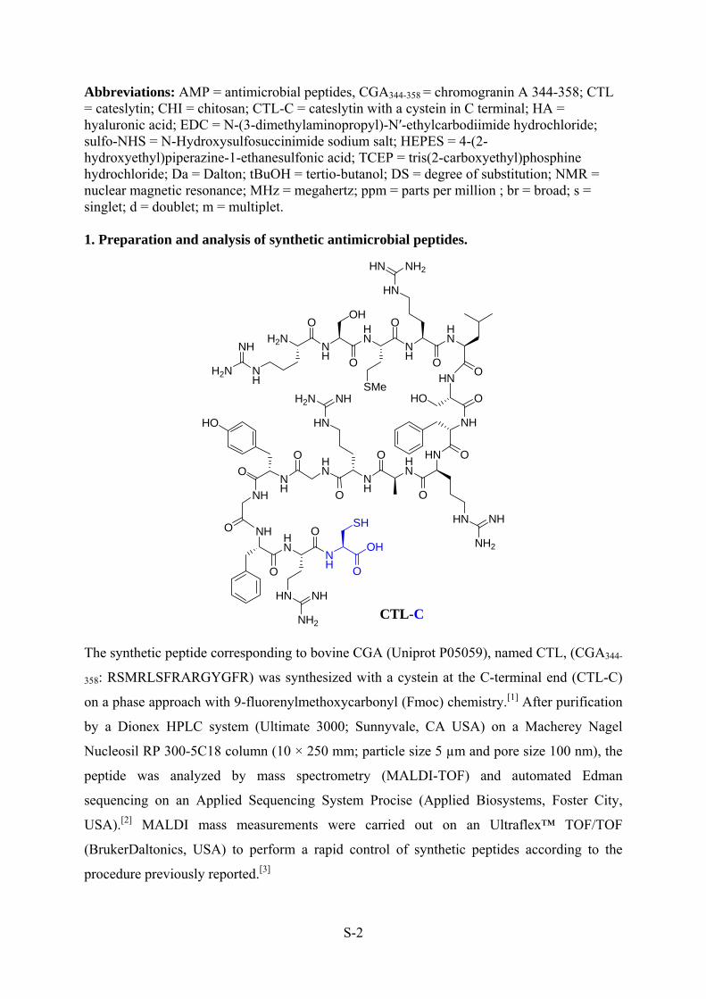
S-2
Abbreviations: AMP = antimicrobial peptides, CGA344-358 = chromogranin A 344-358; CTL = cateslytin; CHI = chitosan; CTL-C = cateslytin with a cystein in C terminal; HA = hyaluronic acid; EDC = N-(3-dimethylaminopropyl)-N′-ethylcarbodiimide hydrochloride; sulfo-NHS = N-Hydroxysulfosuccinimide sodium salt; HEPES = 4-(2-hydroxyethyl)piperazine-1-ethanesulfonic acid; TCEP = tris(2-carboxyethyl)phosphine hydrochloride; Da = Dalton; tBuOH = tertio-butanol; DS = degree of substitution; NMR = nuclear magnetic resonance; MHz = megahertz; ppm = parts per million ; br = broad; s = singlet; d = doublet; m = multiplet. 1. Preparation and analysis of synthetic antimicrobial peptides.
H2N NH
NHH2N
NH
OOH
HN
O
SMe
NH
OHN
O
HN
HN NH2
OHN
HO O
NH
HN O
O
HN
HN
NH2
NH
NH
O
HN
HN
O
NH
O
HO
O
NH
O NHHN
O
NHH2N
HN NH
NH2
O
NH
OH
O
SH
The synthetic peptide corresponding to bovine CGA (Uniprot P05059), named CTL, (CGA344-
358: RSMRLSFRARGYGFR) was synthesized with a cystein at the C-terminal end (CTL-C)
on a phase approach with 9-fluorenylmethoxycarbonyl (Fmoc) chemistry.[1] After purification
by a Dionex HPLC system (Ultimate 3000; Sunnyvale, CA USA) on a Macherey Nagel
Nucleosil RP 300-5C18 column (10 × 250 mm; particle size 5 µm and pore size 100 nm), the
peptide was analyzed by mass spectrometry (MALDI-TOF) and automated Edman
sequencing on an Applied Sequencing System Procise (Applied Biosystems, Foster City,
USA).[2] MALDI mass measurements were carried out on an Ultraflex™ TOF/TOF
(BrukerDaltonics, USA) to perform a rapid control of synthetic peptides according to the
procedure previously reported.[3]
CTL-C

S-3
2. Preparation of modified HA and PAH.
Peptide and FITC functionalized HA, i.e. HA-CTL-C, HAFITC and HAFITC-CTL-C, and
peptide modified PAH have been prepared according to the general synthetic pathway
described below.
2.1. Preparation of HA-CTL-C
Hyaluronic acid (HA) 420 000 Da, was functionalized with maleimide group according to
previously reported method.[4] HA (100.9 mg) was dissolved in 15 mL of 0.01M HEPES
buffer (pH 6). EDC (35.2 mg) and sulfo-NHS (10.2 mg) were added to this solution and the
resulting mixture was stirred 2 h at room temperature. Afterwards, N-(2-aminoethyl)
maleimide trifluoroacetate salt (6.7 mg) was added to the reaction mixture and let stirred for
20 h. The mixture was dialyzed (cut-off 12 000-14 000 Da) first against 0.5 M NaCl for one
day and MilliQ water for 6 days. Water was changed every day. HA-Mal (86 mg) was
obtained as a white solid after freeze-drying. The degree of substitution (DS) of HA-Mal,
defined as the number of maleimide groups per 100 disaccharide unit of HA, was determined
by 1H NMR (D2O, 400 MHz). All spectra were realized in D2O containing 5% of tBuOH: the
singlet of the t-butyl group of tBuOH was calibrated at δ 1.24 ppm and thus used as internal
reference. The singulet at δ 7.1 ppm was assigned to the two symmetric protons on the double
bond of the maleimide group. By comparing the integration of this signal with the singlet at δ
2.0 ppm, assigned to the methyl protons of the acetamide group, the DS was determined
equivalent to 5%.
1H NMR (D2O, 400MHz, δ ppm): δ 6.9 (s, maleimide), 4.5 (br d, HA), 3.5 (m), 2.0 (s, acetyl
group of HA).
The coupling reaction between HA-Mal 5% (80 mg) and CTL-C (10.1 mg) was
performed at 4°C for 20°h in 16mL of 0.01M HEPES buffer (pH 6) and 50 µM of TCEP.
Then, the mixture was dialyzed (cut-off 50 000Da) against 0.5 M NaCl for one day and
MilliQ water for 6 days. Water was changed every day. Peptide-conjugated HA, named HA-
CTL-C, was freeze-dried to provide a white solid with 80-85% of overall yield ( 79 mg).
The DS, defined as the number of CTL-C peptide per 100 disaccharide units of HA, was
determined by 1H NMR (D2O, 400 MHz). All aromatic signals between δ 7.0 and 7.5 ppm
corresponding to the protons of the aminoacids Phe and Tyr were compared to the singlet at δ
2.0 ppm (assigned as the methyl group of the acetamide). A DS of 5% is determined.

S-4
1H NMR (D2O, 400MHz, δ ppm): 7.35 (br s, Ar aminoacid of CTL-C), 7.25 (br s, Ar
aminoacid of CTL-C), 7.10 (br s, Ar aminoacid of CTL-C), 6.85 (br s, Ar aminoacid of CTL-
C), 4.50 (br s, HA), 4.40 (br s, CTL-C), 3.10 (br s, CTL-C), 2.50 (br s, CTL-C), 2.00 (s,
methyl from acetyl group of HA), 1.75 (br s, CTL-C), 0.95 (br s, CTL-C).
2.2. Preparation of HAFITC
Fluorescein Isothiocyanate (FITC) has been covalently attached to HA according to the
following procedure: a solution of FITC (41 µmol dissolved in 2 mL of DMSO) and a
solution of HA (0.31 µmol dissolved in 18 mL of deionized water) were mixed. The resulting
H2N NH
NHH2N
NH
OOH
HN
O
SMe
NH
OHN
O
HN
HN NH2
OHN
HO O
NH
HN O
O
HN
HN
NH2
NH
NH
O
HN
HN
O
NH
O
HO
O
NH
O NHHN
O
NHH2N
HN NH
NH2
O
NH
O OH
S
NO
O
NH
HA-CTL-C
O
HOO
OO
OHO
OH
OH
NHO
O
HOO
OO
OHO
OH
OH
NHO
0.05n 0.95n
OH
N
O
O
NH
O
HOO
OO
OHO
OH
OH
NHO
O
HOO
OO
OHO
OH
OH
NHO
0.05n 0.95n
OH
CTL-CTCEPHEPES, 4 °C
HA-Mal
OH
O
HOO
OO
OHO
OH
OH
NHO
n
Hyaluronic acid (HA)
EDC.HClSulfo-NHSHEPES, 25 °C70%
NNH3
O
OCF3COO
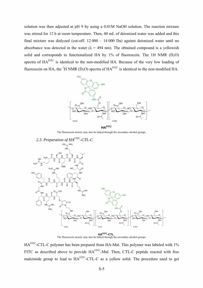
S-5
solution was then adjusted at pH 9 by using a 0.01M NaOH solution. The reaction mixture
was stirred for 12 h at room temperature. Then, 40 mL of deionized water was added and this
final mixture was dialyzed (cut-off: 12 000 – 14 000 Da) against deionized water until no
absorbance was detected in the water (λ = 494 nm). The obtained compound is a yellowish
solid and corresponds to functionalized HA by 1% of fluorescein. The 1H NMR (D2O)
spectra of HAFITC is identical to the non-modified HA. Because of the very low loading of
fluorescein on HA, the 1H NMR (D2O) spectra of HAFITC is identical to the non-modified HA.
The fluorescein moiety may also be linked through the secondary alcohol groups.
2.3. Preparation of HAFITC-CTL-C
The fluorescein moiety may also be linked through the secondary alcohol groups.
HAFITC-CTL-C polymer has been prepared from HA-Mal. This polymer was labeled with 1%
FITC as described above to provide HAFITC-Mal. Then, CTL-C peptide reacted with free
maleimide group to lead to HAFITC-CTL-C as a yellow solid. The procedure used to get
O
HOO
OO
OHO
OH
O
NHO
O
HOO
OO
OHO
OH
OH
NHO
0.01n 0.99n
OH
HAFITC
OH
SHN
O
O
HO
OH
O
O
HOO
OO
OHO
OH
O
NHO
0.01n
OH
SHN
O
O
HO
OH
O
H2N NH
NHH2N
NH
OOH
HN
O
SMe
NH
OHN
O
HN
HN NH2
OHN
HO O
NH
HN O
O
HN
HN
NH2
NH
NH
O
HN
HN
O
NH
O
HO
O
NH
O NHHN
O
NHH2N
HN NH
NH2
O
NH
O OH
S
N
O
O
NH
HAFITC-CTL
O
HOO
OO
OHO
OH
OH
NH
O
0.05n
O
HOO
OO
OHO
OH
OH
NH
O
0.94n
OH
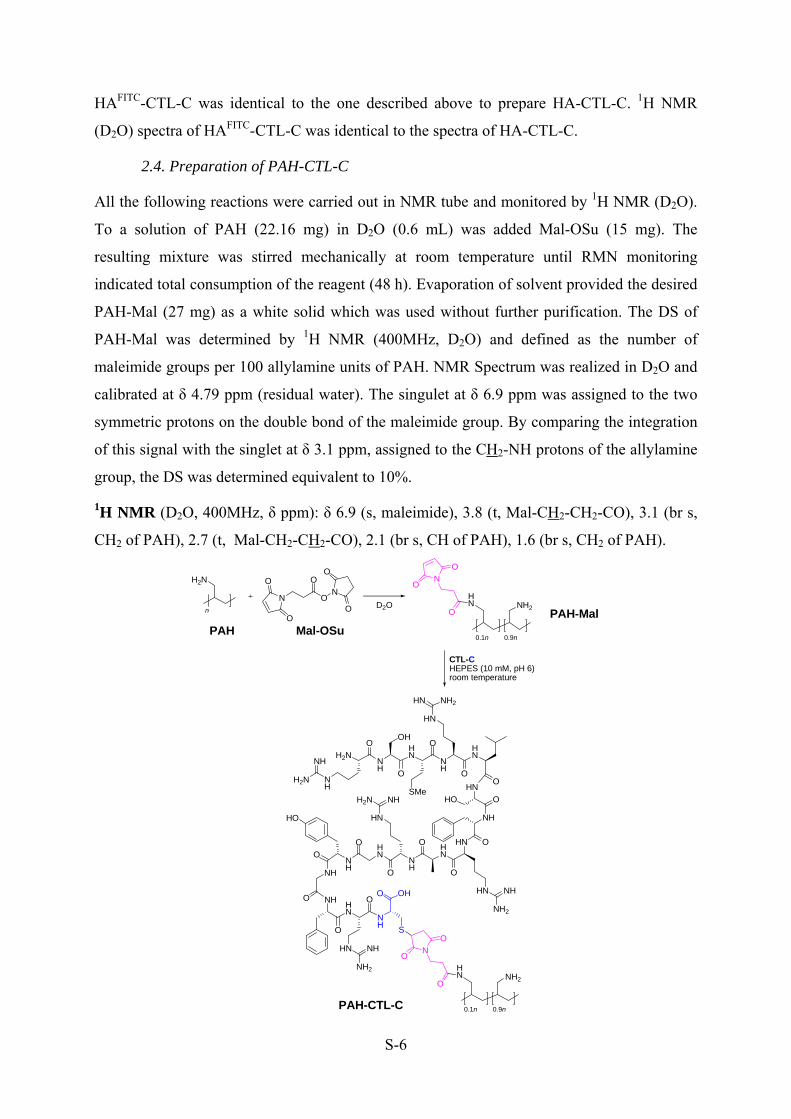
S-6
HAFITC-CTL-C was identical to the one described above to prepare HA-CTL-C. 1H NMR
(D2O) spectra of HAFITC-CTL-C was identical to the spectra of HA-CTL-C.
2.4. Preparation of PAH-CTL-C
All the following reactions were carried out in NMR tube and monitored by 1H NMR (D2O).
To a solution of PAH (22.16 mg) in D2O (0.6 mL) was added Mal-OSu (15 mg). The
resulting mixture was stirred mechanically at room temperature until RMN monitoring
indicated total consumption of the reagent (48 h). Evaporation of solvent provided the desired
PAH-Mal (27 mg) as a white solid which was used without further purification. The DS of
PAH-Mal was determined by 1H NMR (400MHz, D2O) and defined as the number of
maleimide groups per 100 allylamine units of PAH. NMR Spectrum was realized in D2O and
calibrated at δ 4.79 ppm (residual water). The singulet at δ 6.9 ppm was assigned to the two
symmetric protons on the double bond of the maleimide group. By comparing the integration
of this signal with the singlet at δ 3.1 ppm, assigned to the CH2-NH protons of the allylamine
group, the DS was determined equivalent to 10%.
1H NMR (D2O, 400MHz, δ ppm): δ 6.9 (s, maleimide), 3.8 (t, Mal-CH2-CH2-CO), 3.1 (br s,
CH2 of PAH), 2.7 (t, Mal-CH2-CH2-CO), 2.1 (br s, CH of PAH), 1.6 (br s, CH2 of PAH).
N
O
ON
O
O
O
O
H2N
nD2O
N
O
O
O
HN NH2
0.1n 0.9nPAH Mal-OSu
PAH-Mal
CTL-CHEPES (10 mM, pH 6)room temperature
H2N NH
NHH2N
NH
OOH
HN
O
SMe
NH
OHN
O
HN
HN NH2
OHN
HO O
NH
HN O
O
HN
HN
NH2
NH
NH
O
HN
HN
O
NH
O
HO
O
NH
O NHHN
O
NHH2N
HN NH
NH2
O
NH
O OH
S
N
O
O
O
HN NH2
0.1n 0.9nPAH-CTL-C

S-7
The coupling reaction between PAH-Mal 10% (1.5 mg) and CTL-C (15.6 mg) was performed
overnight at room temperature in 0.6 mL of 10 mM HEPES buffer solution in D2O (pH 6).
Then, the mixture was dialyzed (cut-off: 12 000 – 14 000 Da) against MilliQ water for 6 days
(water was changed every day). Peptide-conjugated PAH, named PAH-CTL-C, was freeze
dried to provide a white solid (17 mg). This quantitative yield and the 1H NMR described
below allowed us to define a 10% DS (number of CTL-C peptide per 100 allylamine units of
PAH).
1H NMR (D2O, 400MHz, δ ppm): 7.35 (br s, Ar aminoacid of CTL-C), 7.25 (br s, Ar
aminoacid of CTL-C), 7.13 (br s, Ar aminoacid of CTL-C), 6.86 (br s, Ar aminoacid of CTL-
C), 3.67-4.68 (m, CTL-C), 3.10 (br m, PAH and CTL-C), 2.50-2.68 (br m, CTL-C), 1.34-2.18
(br m, PAH and CTL-C), 0.96 (br s, CTL-C).
3. References
[1] R. B. Merrifield, Science 1965, 150, 178.
[2] M.-H. Metz-Boutigue, Y. Goumon, K. Lugardon, J. M. Strub, D. Aunis, Cell. Mol.
Neurobiol. 1998, 18, 249.
[3] D. Sizova, E. Charbaut, F. Delalande, F. Poirier, A. A. High, F. Parker, A. Van
Dorsselaer, M. Duchesne, A. Diu-Hercend, Neurobiol. Aging 2007, 28, 357.
[4] S. T. Wall, K. Saha, R. S. Ashton, K. R. Kam, D. V. Schaffer, K. E. Healy, Bioconjug.
Chem. 2008, 19, 806.

S-8
4. Supplementary Figures
a
z scale = 20 nm x (µm)
0 5 10 15 20
z (n
m)
0
5
10
15
b
z scale = 50 nm x (µm)
0 5 10 15 20z
(nm
)
0
5
10
15
20
25
c
z scale = 50 nm x (µm)
0 5 10 15 20
z (n
m)
0
10
20
30
40
50
d
z scale = 100 nm x (µm)
0 5 10 15 20
z (n
m)
0
20
40
60
80
100
Figure S-1: AFM images of non-scratched and 3D images and their profilometric sections of scratched PEI-
(HA-CTL-C/CHI)n films built at (a) 5, (b) 10, (c) 15 and (d) 30 bilayers obtained in height mode in dry state.

S-9
Table S-1. Thicknesses and roughnesses of PEI-(HA-CTL-C/CHI)n films built at different number of deposited
pair of layers, n. The errors on the thickness values measured by AFM were obtained from the measurement of
three different areas.
n dAFM (nm) RMS (nm)
5 5 ± 1 1.6 ± 0.1
10 17 ± 2 4.2 ± 0.8
15 28 ± 3 4.6 ± 0.9
30 52 ± 9 16.5 ± 2.8
Number of HA-CTL-C/CHI pairs of layers
0 5 10 15 20 25 30 35
Dry
thi
ckne
ss (
nm)
0
10
20
30
40
50
60
70
Figure S-2: Evolution of the thickness of (HA-CTL-C/CHI) films, measured by AFM in dry state after
scratching, at different number n of pairs of layers.
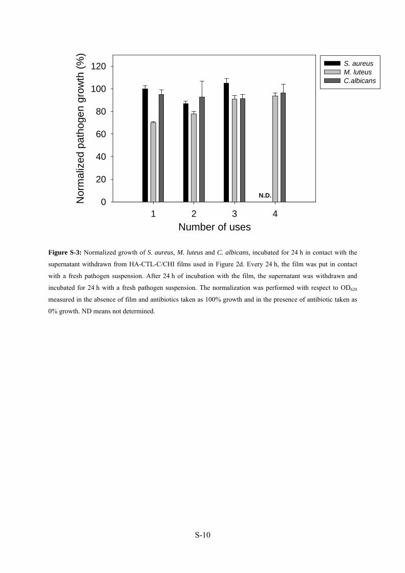
S-10
Number of uses1 2 3 4
Nor
mal
ized
pat
hoge
n gr
owth
(%
)
0
20
40
60
80
100
120 S. aureusM. luteusC.albicans
N.D.
Figure S-3: Normalized growth of S. aureus, M. luteus and C. albicans, incubated for 24 h in contact with the
supernatant withdrawn from HA-CTL-C/CHI films used in Figure 2d. Every 24 h, the film was put in contact
with a fresh pathogen suspension. After 24 h of incubation with the film, the supernatant was withdrawn and
incubated for 24 h with a fresh pathogen suspension. The normalization was performed with respect to OD620
measured in the absence of film and antibiotics taken as 100% growth and in the presence of antibiotic taken as
0% growth. ND means not determined.

S-11
Figure S-4: CLSM images of PEI-(HAFITC/CHI)15 (a) before and (b) after paraformaldehyde (PFA) treatment
and of PEI-(HAFITC-CTL-C/CHI)15 (c) before and (d) after PFA treatment. PFA treatment was performed to
allow the observation of polysaccharide multilayer films after contact with pathogens. The films are put into
contact for 30 min with 4% paraformaldehyde in phosphate buffer saline (PBS) at pH 7.3 and, after several
rinsing with PBS, were covered with Moviol 4-88 (Aldrich, Steinheim, Germany). The scale bars represent 20
µm.
a b
c d
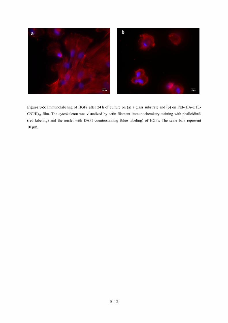
S-12
Figure S-5: Immunolabeling of HGFs after 24 h of culture on (a) a glass substrate and (b) on PEI-(HA-CTL-
C/CHI)15 film. The cytoskeleton was visualized by actin filament immunochemistry staining with phalloidin®
(red labeling) and the nuclei with DAPI counterstaining (blue labeling) of HGFs. The scale bars represent
10 µm.
a b

Conclusions

Conclusions
191
Conclusions My PhD thesis report data of basic and applied research with the development of new
antimicrobial materials. The basic studies correspond to the analysis of new cellular and
molecular connections in the dynamic of Host-Pathogens interactions. S. aureus is a
dangerous Gram-positive bacterium responsible of numerous severe diseases that release
more than 60 virulence factors. During my PhD I have studied S. aureus leukotoxins LukE/D
which are characterized to cause lysis of PMNs and other immune cells. By using a proteomic
approach we demonstrated that they induce the secretion of granular contents. Since, it has
been shown that LukE/D triggered a rise in intracellular free Ca2+ in PMNs which ultimately
incited cell secretion. These secretions are results of the cell activation generated by the two
LukE and LukD components, a similar effect was observed previously by using PVL of S.
aureus. Complement C3 factor (CD35) was recovered with neutrophils secretions, which
showing the cellular activation of neutrophils and the identification of trademark proteins of
different types of granules underlines the ability of leukotoxins LukE/D to degranulate all
vesicles. PMNs secrete numerous proteins (243) belonging to different functional categories,
such as immune related proteins, antioxidants proteins, proteases and inflammatory
components. Out of these functional categories AMPs and immune activation proteins present
major share in this specific inflammatory stimulus. Interestingly, we recovered Cgs-derived
fragments from CgA and CgB in the secretions of neutrophils, as active participants of the
immune system. Cgs play a bi-directional role as secretagogue (immunomodulators) and
antimicrobial agents. Because of these important properties with innate immunity, we decided
to analyze the antibacterial activity against S. aureus of several synthetic Cgs-derived
peptides included in the large pro-chromacin C-terminal fragment.
Chromogranins (Cgs) and their derived peptides are highly expressed during various
stress and infectious conditions including SIRS and severe sepsis. On the other hand, S.
aureus is well known for different immune evasion strategies, such as degradation of AMPs.
As demonstrated in manuscript II, CTL (CgA344-358) was found to be the only CgA derived
peptide that can efficiently effective against different S. aureus strains. Moreover, in
comparison to the other CgA derived AMPs, it can effectively resist degradation by proteases.
Furthermore, we investigated the processing of Cgs by the Glu-C protease of S. aureus and
observed that Glu-C can effectively process Cgs and render them inactive against different
bacterial spp. but interestingly, several new antifungal peptides are generated. These new
antifungal peptides are very active and can inhibit growth of different fungal and candida
Conclusions
192
strains at micromolar concentration. Moreover, it is important to point out that the CTL
containing domain of CgA is not processed by Glu-C protease. Additionally, CTL also
present promising ability of synergism in combination with other antibiotics. These can be
promising features for further studies and to use as potent antibacterial drugs in combination
with other antimicrobial agents.
Regarding the Cgs derived peptides production in response to S. aureus LukE/D, and
ultimate effectiveness of CTL to kill S. aureus coupled with its ability to resist bacterial
proteolytic degradation, underlines the necessity to use CTL as antimicrobial agent. Keeping
in view these features, we designed a new surface coating based on polysaccharide multilayer
films containing functionalized HA with 5% of CTL-C. Antimicrobial properties of CTL-C
were preserved when grafted on HA either in solution or when embedded into PEM films.
After 24 h of incubation, HA-CTL-C/CHI films fully inhibit the development of S. aureus and
C. albicans, which are common and virulent pathogens agents encountered in care-associated
diseases. The presence of CTL-C peptides on HA allows the penetration of the modified
polysaccharide inside C. albicans after 45 min of contact. The secretion of hyaluronidase by
all tested pathogens seems to be responsible for HA-CTL-C release from the film and for its
activity. The film can keep its activity during 3 cycles of use against fresh incubated C.
albicans suspension. Furthermore, the limited fibroblasts adhesion, without cytotoxicity, on
HA-CTL-C/CHI films highlights a medically relevant application to prevent infections on
catheters or tracheal tubes where fibrous tissue encapsulation is undesirable.

References

References
- 193 -
References Abbas, A.K., and Lichtman, A.H. (2005). General Properties of Immune Response. In
Cellular and Molecular Immunology (Saunders, Elsevier), pp. 3–15.
Advincula, R, Aust, E, Meyer, W, Knoll, and W (1996). In situ investigations of polymer self-
assembly solution adsorption by surface plasmon spectroscopy, Vol 12 (Washington,
DC, ETATS-UNIS, American Chemical Society).
Akaddar, A., Doderer-Lang, C., Marzahn, M.R., Delalande, F., Mousli, M., Helle, K., Van
Dorsselaer, A., Aunis, D., Dunn, B.M., Metz-Boutigue, M.H., et al. (2010). Catestatin,
an endogenous chromogranin A-derived peptide, inhibits in vitro growth of
Plasmodium falciparum. Cell Mol Life Sci 67, 1005-1015.
Allon, A.A., Ng, K.W., Hammoud, S., Russell, B.H., Jones, C.M., Rivera, J.J., Schwartz, J.,
Hook, M., and Maher, S.A. (2012). Augmenting the articular cartilage-implant
interface: Functionalizing with a collagen adhesion protein. J Biomed Mater Res A
100, 2168-2175.
Alonzo, F., 3rd, Benson, M.A., Chen, J., Novick, R.P., Shopsin, B., and Torres, V.J. (2012).
Staphylococcus aureus leucocidin ED contributes to systemic infection by targeting
neutrophils and promoting bacterial growth in vivo. Mol Microbiol 83, 423-435.
Alonzo, F., 3rd, Kozhaya, L., Rawlings, S.A., Reyes-Robles, T., DuMont, A.L., Myszka,
D.G., Landau, N.R., Unutmaz, D., and Torres, V.J. (2013). CCR5 is a receptor for
Staphylococcus aureus leukotoxin ED. Nature 493, 51-55.
Amagai, M., and Stanley, J.R. (2012). Desmoglein as a target in skin disease and beyond. J
Invest Dermatol 132, 776-784.
Andre, P., Cambier, J.C., Wade, T.K., Raetz, T., and Wade, W.F. (1994). Distinct structural
compartmentalization of the signal transducing functions of major histocompatibility
complex class II (Ia) molecules. J Exp Med 179, 763-768.
Angeletti, R.H., Mints, L., Aber, C., and Russell, J. (1996). Determination of residues in
chromogranin A-(16-40) required for inhibition of parathyroid hormone secretion.
Endocrinology 137, 2918-2922.
Angelone, T., Quintieri, A.M., Goumon, Y., Di Felice, V., Filice, E., Gattuso, A., Mazza, R.,
Corti, A., Tota, B., Metz-Boutigue, M.H., et al. (2010). Cytoskeleton mediates
negative inotropism and lusitropism of chromogranin A-derived peptides (human
vasostatin1-78 and rat CgA1-64) in the rat heart. Regul Pept 165, 78-85.
Angelone, T., Quintieri, A.M., Pasqua, T., Gentile, S., Tota, B., Mahata, S.K., and Cerra,
M.C. (2011). Phosphodiesterase type-2 and NO-dependent S-nitrosylation mediate the
cardioinhibition of the antihypertensive catestatin. Am J Physiol Heart Circ Physiol
302, H431-442.
Archer, D.L., and Young, F.E. (1988). Contemporary issues: diseases with a food vector. Clin
Microbiol Rev 1, 377-398.
Arina, A., Murillo, O., Dubrot, J., Azpilikueta, A., Alfaro, C., Perez-Gracia, J.L., Bendandi,
M., Palencia, B., Hervas-Stubbs, S., and Melero, I. (2007). Cellular liaisons of natural
killer lymphocytes in immunology and immunotherapy of cancer. Expert Opin Biol
Ther 7, 599-615.
Arnusch, C.J., Branderhorst, H., de Kruijff, B., Liskamp, R.M., Breukink, E., and Pieters, R.J.
(2007). Enhanced membrane pore formation by multimeric/oligomeric antimicrobial
peptides. Biochemistry 46, 13437-13442.
Aung, G., Niyonsaba, F., Ushio, H., Hoq, M.I., Ikeda, S., Ogawa, H., and Okumura, K.
(2011a). A neuroendocrine antimicrobial peptide, catestatin, stimulates interleukin-8
References
- 194 -
production from human keratinocytes via activation of mitogen-activated protein
kinases. J Dermatol Sci 61, 142-144.
Aung, G., Niyonsaba, F., Ushio, H., Kajiwara, N., Saito, H., Ikeda, S., Ogawa, H., and
Okumura, K. (2011b). Catestatin, a neuroendocrine antimicrobial peptide, induces
human mast cell migration, degranulation and production of cytokines and
chemokines. Immunology 132, 527-539.
Aunis, D. (1998). Exocytosis in chromaffin cells of the adrenal medulla. Int Rev Cytol 181,
213-320.
Aunis, D. (2009). GRANINS: thirty-five happy years in the granulosome world. Preface.
Regul Pept 165, 3-4.
Auvynet, C., and Rosenstein, Y. (2009). Multifunctional host defense peptides: antimicrobial
peptides, the small yet big players in innate and adaptive immunity. Febs J 276, 6497-
6508.
Baker, M.D., and Acharya, K.R. (2004). Superantigens: structure-function relationships. Int J
Med Microbiol 293, 529-537.
Balaban, N., and Rasooly, A. (2000). Staphylococcal enterotoxins. Int J Food Microbiol 61,
1-10.
Bandyopadhyay, G.K., Vu, C.U., Gentile, S., Lee, H., Biswas, N., Chi, N.W., O'Connor, D.T.,
and Mahata, S.K. (2012). Catestatin (chromogranin A(352-372)) and novel effects on
mobilization of fat from adipose tissue through regulation of adrenergic and leptin
signaling. J Biol Chem 287, 23141-23151.
Banks, P., and Helle, K. (1965). The release of protein from the stimulated adrenal medulla.
Biochem J 97, 40C-41C.
Barrio, M.B., Rainard, P., and Prevost, G. (2006). LukM/LukF'-PV is the most active
Staphylococcus aureus leukotoxin on bovine neutrophils. Microbes Infect 8, 2068-
2074.
Bartolomucci, A., Pasinetti, G.M., and Salton, S.R. (2010). Granins as disease-biomarkers:
translational potential for psychiatric and neurological disorders. Neuroscience 170,
289-297.
Bartolomucci, A., Possenti, R., Mahata, S.K., Fischer-Colbrie, R., Loh, Y.P., and Salton, S.R.
(2011). The extended granin family: structure, function, and biomedical implications.
Endocr Rev 32, 755-797.
Bassino, E., Fornero, S., Gallo, M.P., Ramella, R., Mahata, S.K., Tota, B., Levi, R., and
Alloatti, G. (2011). A novel catestatin-induced antiadrenergic mechanism triggered by
the endothelial PI3K-eNOS pathway in the myocardium. Cardiovasc Res 91, 617-624.
Baumann, T., Kampfer, U., Schurch, S., Schaller, J., Largiader, C., Nentwig, W., and Kuhn-
Nentwig, L. (2010). Ctenidins: antimicrobial glycine-rich peptides from the hemocytes
of the spider Cupiennius salei. Cell Mol Life Sci 67, 2787-2798.
Bechinger, B. (1999). The structure, dynamics and orientation of antimicrobial peptides in
membranes by multidimensional solid-state NMR spectroscopy. Biochim Biophys
Acta 1462, 157-183.
Becker, K., Friedrich, A.W., Lubritz, G., Weilert, M., Peters, G., and Von Eiff, C. (2003).
Prevalence of genes encoding pyrogenic toxin superantigens and exfoliative toxins
among strains of Staphylococcus aureus isolated from blood and nasal specimens. J
Clin Microbiol 41, 1434-1439.
Benedum, U.M., Lamouroux, A., Konecki, D.S., Rosa, P., Hille, A., Baeuerle, P.A., Frank,
R., Lottspeich, F., Mallet, J., and Huttner, W.B. (1987). The primary structure of
human secretogranin I (chromogranin B): comparison with chromogranin A reveals
homologous terminal domains and a large intervening variable region. Embo J 6,
1203-1211.
References
- 195 -
Benjannet, S., Leduc, R., Lazure, C., Seidah, N.G., Marcinkiewicz, M., and Chretien, M.
(1985). GAWK, a novel human pituitary polypeptide: isolation, immunocytochemical
localization and complete amino acid sequence. Biochem Biophys Res Commun 126,
602-609.
Bera, A., Herbert, S., Jakob, A., Vollmer, W., and Gotz, F. (2005). Why are pathogenic
staphylococci so lysozyme resistant? The peptidoglycan O-acetyltransferase OatA is
the major determinant for lysozyme resistance of Staphylococcus aureus. Mol
Microbiol 55, 778-787.
Beranova-Giorgianni, S., Zhao, Y., Desiderio, D.M., and Giorgianni, F. (2006).
Phosphoproteomic analysis of the human pituitary. Pituitary 9, 109-120.
Berg, M.C., Zhai, L., Cohen, R.E., and Rubner, M.F. (2006). Controlled drug release from
porous polyelectrolyte multilayers. Biomacromolecules 7, 357-364.
Bergdoll, M.S., Crass, B.A., Reiser, R.F., Robbins, R.N., and Davis, J.P. (1981). A new
staphylococcal enterotoxin, enterotoxin F, associated with toxic-shock-syndrome
Staphylococcus aureus isolates. Lancet 1, 1017-1021.
Berger-Bachi, B. (2002). Resistance mechanisms of gram-positive bacteria. Int J Med
Microbiol 292, 27-35.
Besedovsky, H.O., and del Rey, A. (1996). Immune-neuro-endocrine interactions: facts and
hypotheses. Endocr Rev 17, 64-102.
Beuret, N., Stettler, H., Renold, A., Rutishauser, J., and Spiess, M. (2004). Expression of
regulated secretory proteins is sufficient to generate granule-like structures in
constitutively secreting cells. J Biol Chem 279, 20242-20249.
Bhakdi, S., and Tranum-Jensen, J. (1991). Alpha-toxin of Staphylococcus aureus. Microbiol
Rev 55, 733-751.
Bhakoo, M., Birkbeck, T.H., and Freer, J.H. (1982). Interaction of Staphylococcus aureus
delta-lysin with phospholipid monolayers. Biochemistry 21, 6879-6883.
Bindoni, M., Perciavalle, V., Berretta, S., Belluardo, N., and Diamantstein, T. (1988).
Interleukin 2 modifies the bioelectric activity of some neurosecretory nuclei in the rat
hypothalamus. Brain Res 462, 10-14.
Biron, C.A. (2010). More things in heaven and earth: defining innate and adaptive immunity.
Nat Immunol 11, 1080-1082.
Bischoff, M., Dunman, P., Kormanec, J., Macapagal, D., Murphy, E., Mounts, W., Berger-
Bachi, B., and Projan, S. (2004). Microarray-based analysis of the Staphylococcus
aureus sigmaB regulon. J Bacteriol 186, 4085-4099.
Biswas, N., Rodriguez-Flores, J.L., Courel, M., Gayen, J.R., Vaingankar, S.M., Mahata, M.,
Torpey, J.W., Taupenot, L., O'Connor, D.T., and Mahata, S.K. (2009). Cathepsin L
colocalizes with chromogranin a in chromaffin vesicles to generate active peptides.
Endocrinology 150, 3547-3557.
Bitsche, M., Schrott-Fischer, A., Hinterhoelzl, J., Fischer-Colbrie, R., Sergi, C., Glueckert, R.,
Humpel, C., and Marksteiner, J. (2006). First localization and biochemical
identification of chromogranin B- and secretoneurin-like immunoreactivity in the fetal
human vagal/nucleus solitary complex. Regul Pept 134, 97-104.
Bjerketorp, J., Nilsson, M., Ljungh, A., Flock, J.I., Jacobsson, K., and Frykberg, L. (2002). A
novel von Willebrand factor binding protein expressed by Staphylococcus aureus.
Microbiology 148, 2037-2044.
Blanchette, C.D., Woo, Y.H., Thomas, C., Shen, N., Sulchek, T.A., and Hiddessen, A.L.
(2009). Decoupling internalization, acidification and phagosomal-
endosomal/lysosomal fusion during phagocytosis of InlA coated beads in epithelial
cells. PLoS One 4, e6056.
References
- 196 -
Blaschko, H., Comline, R.S., Schneider, F.H., Silver, M., and Smith, A.D. (1967). Secretion
of a chromaffin granule protein, chromogranin, from the adrenal gland after
splanchnic stimulation. Nature 215, 58-59.
Blaschko, H., and Welch, A.D. (1953). Localization of adrenaline in cytoplasmic particles of
the bovine adrenal medulla. Naunyn Schmiedebergs Arch Exp Pathol Pharmakol 219,
17-22.
Blodgett, K.B. (1934). Monomolecular films of fatty acids on glass. J Am Chem Soc 56, 495.
Blodgett, K.B., and Langmuir, I. (1937). Buildup films of barium stearate and their optical
properties. Phys Rev 51, 964-982.
Blois, A., Holmsen, H., Martino, G., Corti, A., Metz-Boutigue, M.H., and Helle, K.B. (2006).
Interactions of chromogranin A-derived vasostatins and monolayers of
phosphatidylserine, phosphatidylcholine and phosphatidylethanolamine. Regul Pept
134, 30-37.
Bobin-Dubreux, S., Reverdy, M.E., Nervi, C., Rougier, M., Bolmstrom, A., Vandenesch, F.,
and Etienne, J. (2001). Clinical isolate of vancomycin-heterointermediate
Staphylococcus aureus susceptible to methicillin and in vitro selection of a
vancomycin-resistant derivative. Antimicrob Agents Chemother 45, 349-352.
Boden, M.K., and Flock, J.I. (1989). Fibrinogen-binding protein/clumping factor from
Staphylococcus aureus. Infect Immun 57, 2358-2363.
Boland, M.P., and Separovic, F. (2006). Membrane interactions of antimicrobial peptides
from Australian tree frogs. Biochim Biophys Acta 1758, 1178-1183.
Boman, H.G. (1991). Antibacterial peptides: key components needed in immunity. Cell 65,
205-207.
Boman, H.G., Agerberth, B., and Boman, A. (1993). Mechanisms of action on Escherichia
coli of cecropin P1 and PR-39, two antibacterial peptides from pig intestine. Infect
Immun 61, 2978-2984.
Borissoff, J.I., and ten Cate, H. (2011). From neutrophil extracellular traps release to
thrombosis: an overshooting host-defense mechanism? J Thromb Haemost 9, 1791-
1794.
Borregaard, N., and Cowland, J.B. (1997). Granules of the human neutrophilic
polymorphonuclear leukocyte. Blood 89, 3503-3521.
Borregaard, N., Lollike, K., Kjeldsen, L., Sengelov, H., Bastholm, L., Nielsen, M.H., and
Bainton, D.F. (1993). Human neutrophil granules and secretory vesicles. Eur J
Haematol 51, 187-198.
Boulmedais, F., Frisch, B., Etienne, O., Lavalle, P., Picart, C., Ogier, J., Voegel, J.C., Schaaf,
P., and Egles, C. (2004). Polyelectrolyte multilayer films with pegylated polypeptides
as a new type of anti-microbial protection for biomaterials. Biomaterials 25, 2003-
2011.
Bowdish, D.M., Davidson, D.J., Speert, D.P., and Hancock, R.E. (2004). The human cationic
peptide LL-37 induces activation of the extracellular signal-regulated kinase and p38
kinase pathways in primary human monocytes. J Immunol 172, 3758-3765.
Bowdish, D.M., and Hancock, R.E. (2005). Anti-endotoxin properties of cationic host defence
peptides and proteins. J Endotoxin Res 11, 230-236.
Brechot, N., Gomez, E., Bignon, M., Khallou-Laschet, J., Dussiot, M., Cazes, A., Alanio-
Brechot, C., Durand, M., Philippe, J., Silvestre, J.S., et al. (2008). Modulation of
macrophage activation state protects tissue from necrosis during critical limb ischemia
in thrombospondin-1-deficient mice. PLoS One 3, e3950.
Brinkmann, V., Reichard, U., Goosmann, C., Fauler, B., Uhlemann, Y., Weiss, D.S.,
Weinrauch, Y., and Zychlinsky, A. (2004). Neutrophil extracellular traps kill bacteria.
Science 303, 1532-1535.
References
- 197 -
Briolat, J., Wu, S.D., Mahata, S.K., Gonthier, B., Bagnard, D., Chasserot-Golaz, S., Helle,
K.B., Aunis, D., and Metz-Boutigue, M.H. (2005). New antimicrobial activity for the
catecholamine release-inhibitory peptide from chromogranin A. Cell Mol Life Sci 62,
377-385.
Broekaert, W.F., Terras, F.R.G., Cammue, B.P.A., and Vanderleyden, J. (1990). An
automated quantitative assay for fungal growth inhibition. FEMS Microbiology
Letters 69, 55-59.
Brogden, K.A. (2005). Antimicrobial peptides: pore formers or metabolic inhibitors in
bacteria? Nat Rev Microbiol 3, 238-250.
Brogden, K.A., Ackermann, M., McCray, P.B., Jr., and Tack, B.F. (2003). Antimicrobial
peptides in animals and their role in host defences. Int J Antimicrob Agents 22, 465-
478.
Bubeck Wardenburg, J., Patel, R.J., and Schneewind, O. (2007). Surface proteins and
exotoxins are required for the pathogenesis of Staphylococcus aureus pneumonia.
Infect Immun 75, 1040-1044.
Bulet, P., Dimarcq, J.L., Hetru, C., Lagueux, M., Charlet, M., Hegy, G., Van Dorsselaer, A.,
and Hoffmann, J.A. (1993). A novel inducible antibacterial peptide of Drosophila
carries an O-glycosylated substitution. J Biol Chem 268, 14893-14897.
Bulet, P., Hetru, C., Dimarcq, J.L., and Hoffmann, D. (1999). Antimicrobial peptides in
insects; structure and function. Dev Comp Immunol 23, 329-344.
Bunning, V.K., Lindsay, J.A., and Archer, D.L. (1997). Chronic health effects of microbial
foodborne disease. World Health Stat Q 50, 51-56.
Burdick, M.D., Harris, A., Reid, C.J., Iwamura, T., and Hollingsworth, M.A. (1997).
Oligosaccharides expressed on MUC1 produced by pancreatic and colon tumor cell
lines. J Biol Chem 272, 24198-24202.
Burke, N.A.D., Jafar Mazumder, M.A., Hanna, M., and Stöver, H.D.H. (2007).
Polyelectrolyte complexation between poly(methacrylic acid, sodium salt) and
poly(diallyldimethylammonium chloride) or poly[2-(methacryloyloxyethyl)
trimethylammonium chloride] Journal of Polymer Science Part A : Polymer Chemistry
45, 4129–4143.
Burmester, G., Pezzutto, A., and Wirth, J. (2003). Color atlas of Immunology (Grammlich,
Pliezhausen, Germany).
Cado, G., Kerdjoudj, H., Chassepot, A., Lefort, M., Benmlih, K., Hemmerle, J., Voegel, J.C.,
Jierry, L., Schaaf, P., Frere, Y., et al. (2012). Polysaccharide films built by
simultaneous or alternate spray: a rapid way to engineer biomaterial surfaces.
Langmuir 28, 8470-8478.
Callegan, M.C., Engel, L.S., Hill, J.M., and O'Callaghan, R.J. (1994). Corneal virulence of
Staphylococcus aureus: roles of alpha-toxin and protein A in pathogenesis. Infect
Immun 62, 2478-2482.
Campion, J.J., McNamara, P.J., and Evans, M.E. (2004). Evolution of ciprofloxacin-resistant
Staphylococcus aureus in in vitro pharmacokinetic environments. Antimicrob Agents
Chemother 48, 4733-4744.
Canu, N., Possenti, R., Ricco, A.S., Rocchi, M., and Levi, A. (1997). Cloning, structural
organization analysis, and chromosomal assignment of the human gene for the
neurosecretory protein VGF. Genomics 45, 443-446.
Carneiro, C.R., Postol, E., Nomizo, R., Reis, L.F., and Brentani, R.R. (2004). Identification of
enolase as a laminin-binding protein on the surface of Staphylococcus aureus.
Microbes Infect 6, 604-608.
Carruthers, M.M., and Kabat, W.J. (1983). Mediation of staphylococcal adherence to mucosal
cells by lipoteichoic acid. Infect Immun 40, 444-446.
References
- 198 -
Caruso, F., Furlong, D.N., Ariga, K., Ichinose, I., and Kunitake, T. (1998). Characterization
of Polyelectrolyte−Protein Multilayer Films by Atomic Force Microscopy, Scanning
Electron Microscopy, and Fourier Transform Infrared Reflection−Absorption
Spectroscopy. Langmuir 14, 4559-4565.
Castle, M., Nazarian, A., Yi, S.S., and Tempst, P. (1999). Lethal effects of apidaecin on
Escherichia coli involve sequential molecular interactions with diverse targets. J Biol
Chem 274, 32555-32564.
Ceconi, C., Ferrari, R., Bachetti, T., Opasich, C., Volterrani, M., Colombo, B., Parrinello, G.,
and Corti, A. (2002). Chromogranin A in heart failure; a novel neurohumoral factor
and a predictor for mortality. Eur Heart J 23, 967-974.
Chakraborty, T.R., Tkalych, O., Nanno, D., Garcia, A.L., Devi, L.A., and Salton, S.R. (2006).
Quantification of VGF- and pro-SAAS-derived peptides in endocrine tissues and the
brain, and their regulation by diet and cold stress. Brain Res 1089, 21-32.
Chambers, H.F. (2001). The changing epidemiology of Staphylococcus aureus? Emerg Infect
Dis 7, 178-182.
Chan, S.C., Hui, L., and Chen, H.M. (1998). Enhancement of the cytolytic effect of anti-
bacterial cecropin by the microvilli of cancer cells. Anticancer Res 18, 4467-4474.
Chavakis, T., Hussain, M., Kanse, S.M., Peters, G., Bretzel, R.G., Flock, J.I., Herrmann, M.,
and Preissner, K.T. (2002). Staphylococcus aureus extracellular adherence protein
serves as anti-inflammatory factor by inhibiting the recruitment of host leukocytes.
Nat Med 8, 687-693.
Chavakis, T., Wiechmann, K., Preissner, K.T., and Herrmann, M. (2005). Staphylococcus
aureus interactions with the endothelium: the role of bacterial "secretable expanded
repertoire adhesive molecules" (SERAM) in disturbing host defense systems. Thromb
Haemost 94, 278-285.
Chen, T.R., Chiou, C.S., and Tsen, H.Y. (2004). Use of novel PCR primers specific to the
genes of staphylococcal enterotoxin G, H, I for the survey of Staphylococcus aureus
strains isolated from food-poisoning cases and food samples in Taiwan. Int J Food
Microbiol 92, 189-197.
Chen, X., Niyonsaba, F., Ushio, H., Nagaoka, I., Ikeda, S., Okumura, K., and Ogawa, H.
(2006). Human cathelicidin LL-37 increases vascular permeability in the skin via mast
cell activation, and phosphorylates MAP kinases p38 and ERK in mast cells. J
Dermatol Sci 43, 63-66.
Chertov, O., Michiel, D.F., Xu, L., Wang, J.M., Tani, K., Murphy, W.J., Longo, D.L., Taub,
D.D., and Oppenheim, J.J. (1996). Identification of defensin-1, defensin-2, and
CAP37/azurocidin as T-cell chemoattractant proteins released from interleukin-8-
stimulated neutrophils. J Biol Chem 271, 2935-2940.
Chesney, P.J. (1989). Clinical aspects and spectrum of illness of toxic shock syndrome:
overview. Rev Infect Dis 11 Suppl 1, S1-7.
Cheung, A.I., Projan, S.J., Edelstein, R.E., and Fischetti, V.A. (1995). Cloning, expression,
and nucleotide sequence of a Staphylococcus aureus gene (fbpA) encoding a
fibrinogen-binding protein. Infect Immun 63, 1914-1920.
Chluba, J., Voegel, J.C., Decher, G., Erbacher, P., Schaaf, P., and Ogier, J. (2001). Peptide
hormone covalently bound to polyelectrolytes and embedded into multilayer
architectures conserving full biological activity. Biomacromolecules 2, 800-805.
Chomarat, P., Banchereau, J., Davoust, J., and Palucka, A.K. (2000). IL-6 switches the
differentiation of monocytes from dendritic cells to macrophages. Nat Immunol 1,
510-514.
Chuang, H.F., Smith, R.C., and Hammond, P.T. (2008). Polyelectrolyte multilayers for
tunable release of antibiotics. Biomacromolecules 9, 1660-1668.
References
- 199 -
Clark, N.C., Weigel, L.M., Patel, J.B., and Tenover, F.C. (2005). Comparison of Tn1546-like
elements in vancomycin-resistant Staphylococcus aureus isolates from Michigan and
Pennsylvania. Antimicrob Agents Chemother 49, 470-472.
Clark, S.R., Ma, A.C., Tavener, S.A., McDonald, B., Goodarzi, Z., Kelly, M.M., Patel, K.D.,
Chakrabarti, S., McAvoy, E., Sinclair, G.D., et al. (2007). Platelet TLR4 activates
neutrophil extracellular traps to ensnare bacteria in septic blood. Nat Med 13, 463-469.
Cochrane, C.G., and Muller-Eberhard, H.J. (1968). The derivation of two distinct
anaphylatoxin activities from the third and fifth components of human complement. J
Exp Med 127, 371-386.
Colin, D.A., Mazurier, I., Sire, S., and Finck-Barbancon, V. (1994). Interaction of the two
components of leukocidin from Staphylococcus aureus with human
polymorphonuclear leukocyte membranes: sequential binding and subsequent
activation. Infect Immun 62, 3184-3188.
Collins, L.V., Kristian, S.A., Weidenmaier, C., Faigle, M., Van Kessel, K.P., Van Strijp, J.A.,
Gotz, F., Neumeister, B., and Peschel, A. (2002). Staphylococcus aureus strains
lacking D-alanine modifications of teichoic acids are highly susceptible to human
neutrophil killing and are virulence attenuated in mice. J Infect Dis 186, 214-219.
Conlon, J.M. (2010). Granin-derived peptides as diagnostic and prognostic markers for
endocrine tumors. Regul Pept 165, 5-11.
Conlon, J.M., Hamberger, B., and Grimelius, L. (1992). Isolation of peptides arising from the
specific posttranslational processing of chromogranin A and chromogranin B from
human pheochromocytoma tissue. Peptides 13, 639-644.
Conrad, D.H., Carlo, J.R., and Ruddy, S. (1978). Interaction of beta1H globulin with cell-
bound C3b: quantitative analysis of binding and influence of alternative pathway
components on binding. J Exp Med 147, 1792-1805.
Cooney, J., Kienle, Z., Foster, T.J., and O'Toole, P.W. (1993). The gamma-hemolysin locus
of Staphylococcus aureus comprises three linked genes, two of which are identical to
the genes for the F and S components of leukocidin. Infect Immun 61, 768-771.
Cooney, J., Mulvey, M., Arbuthnott, J.P., and Foster, T.J. (1988). Molecular cloning and
genetic analysis of the determinant for gamma-lysin, a two-component toxin of
Staphylococcus aureus. J Gen Microbiol 134, 2179-2188.
Corone, A. (1964). [Lysopain In Transtympanic Aerosols]. Ann Otolaryngol Chir Cervicofac
81, 367-368.
Corti, A., Ferrari, R., and Ceconi, C. (2000). Chromogranin A and tumor necrosis factor-alpha
(TNF) in chronic heart failure. Adv Exp Med Biol 482, 351-359.
Cosgrove, S.E., Carroll, K.C., and Perl, T.M. (2004). Staphylococcus aureus with reduced
susceptibility to vancomycin. Clin Infect Dis 39, 539-545.
Couppie, P., Cribier, B., and Prevost, G. (1994). Leukocidin from Staphylococcus aureus and
cutaneous infections: an epidemiologic study. Arch Dermatol 130, 1208-1209.
Crouzier, T., Szarpak, A., Boudou, T., Auzely-Velty, R., and Picart, C. (2010).
Polysaccharide-blend multilayers containing hyaluronan and heparin as a delivery
system for rhBMP-2. Small 6, 651-662.
Dalla Serra, M., Coraiola, M., Viero, G., Comai, M., Potrich, C., Ferreras, M., Baba-Moussa,
L., Colin, D.A., Menestrina, G., Bhakdi, S., et al. (2005). Staphylococcus aureus
bicomponent gamma-hemolysins, HlgA, HlgB, and HlgC, can form mixed pores
containing all components. J Chem Inf Model 45, 1539-1545.
Damgé, C. (1991). Oral insulin in Biotechnology of Insulin Therapy (London, UK., Oxford
press).
Danilova, N. (2006). The evolution of immune mechanisms. J Exp Zool B Mol Dev Evol 306,
496-520.
References
- 200 -
Davidson, D.J., Currie, A.J., Reid, G.S., Bowdish, D.M., MacDonald, K.L., Ma, R.C.,
Hancock, R.E., and Speert, D.P. (2004). The cationic antimicrobial peptide LL-37
modulates dendritic cell differentiation and dendritic cell-induced T cell polarization. J
Immunol 172, 1146-1156.
de Haas, C.J., Veldkamp, K.E., Peschel, A., Weerkamp, F., Van Wamel, W.J., Heezius, E.C.,
Poppelier, M.J., Van Kessel, K.P., and van Strijp, J.A. (2004). Chemotaxis inhibitory
protein of Staphylococcus aureus, a bacterial antiinflammatory agent. J Exp Med 199,
687-695.
de Lencastre, H., de Jonge, B.L., Matthews, P.R., and Tomasz, A. (1994). Molecular aspects
of methicillin resistance in Staphylococcus aureus. J Antimicrob Chemother 33, 7-24.
De, Y., Chen, Q., Schmidt, A.P., Anderson, G.M., Wang, J.M., Wooters, J., Oppenheim, J.J.,
and Chertov, O. (2000). LL-37, the neutrophil granule- and epithelial cell-derived
cathelicidin, utilizes formyl peptide receptor-like 1 (FPRL1) as a receptor to
chemoattract human peripheral blood neutrophils, monocytes, and T cells. J Exp Med
192, 1069-1074.
Decher, G. (1997). Fuzzy Nanoassemblies: Toward Layered Polymeric Multicomposites.
Science 277, 1232-1237.
Decher, G., and Hong, J.D. (1991). Build-up of ultrathin multilayer films by self-assembly
process : II Consecutive adsorption of ionic and cationic bipolar amphiphiles and
polyelectrolytes on charged surfaces. Ber Bunsenges Phys Chem 95: 1430-1434 95,
1430-1434.
Degn, S.E., Hansen, A.G., Steffensen, R., Jacobsen, C., Jensenius, J.C., and Thiel, S. (2009).
MAp44, a human protein associated with pattern recognition molecules of the
complement system and regulating the lectin pathway of complement activation. J
Immunol 183, 7371-7378.
Delalande, F., Carapito, C., Brizard, J.P., Brugidou, C., and Van Dorsselaer, A. (2005).
Multigenic families and proteomics: extended protein characterization as a tool for
paralog gene identification. Proteomics 5, 450-460.
Delneste, Y., Beauvillain, C., and Jeannin, P. (2007). [Innate immunity: structure and function
of TLRs]. Med Sci (Paris) 23, 67-73.
Denis, O., Deplano, A., De Ryck, R., Nonhoff, C., and Struelens, M.J. (2003). Emergence and
spread of gentamicin-susceptible strains of methicillin-resistant Staphylococcus aureus
in Belgian hospitals. Microb Drug Resist 9, 61-71.
Dennison, S.R., Howe, J., Morton, L.H., Brandenburg, K., Harris, F., and Phoenix, D.A.
(2006). Interactions of an anionic antimicrobial peptide with Staphylococcus aureus
membranes. Biochem Biophys Res Commun 347, 1006-1010.
Depreitere, J., Durinx, C., Wang, Z., Coen, E., Lambeir, A.M., Scharpe, S., De Potter, W.,
and Nouwen, E.J. (2002). Presence and release of SR-17 (chromogranin B(586-602))
in the porcine splenic nerve and its enzymatic degradation by CD26/dipeptidyl
peptidase IV. Regul Pept 106, 71-79.
Depreitere, J., Wang, Z., Liang, F., Coen, E., and Nouwen, E.J. (2004). Presence and release
of the chromogranin B-derived secretolytin-like peptide KR-11 from the porcine
spleen. Regul Pept 122, 99-107.
Descloux, E., Perpoint, T., Ferry, T., Lina, G., Bes, M., Vandenesch, F., Mohammedi, I., and
Etienne, J. (2008). One in five mortality in non-menstrual toxic shock syndrome
versus no mortality in menstrual cases in a balanced French series of 55 cases. Eur J
Clin Microbiol Infect Dis 27, 37-43.
Dethlefsen, L., McFall-Ngai, M., and Relman, D.A. (2007). An ecological and evolutionary
perspective on human-microbe mutualism and disease. Nature 449, 811-818.
References
- 201 -
Devine, D.A. (2003). Antimicrobial peptides in defence of the oral and respiratory tracts. Mol
Immunol 40, 431-443.
Diamond, G., Beckloff, N., Weinberg, A., and Kisich, K.O. (2009). The roles of antimicrobial
peptides in innate host defense. Curr Pharm Des 15, 2377-2392.
Dillen, L., Miserez, B., Claeys, M., Aunis, D., and De Potter, W. (1993). Posttranslational
processing of proenkephalins and chromogranins/secretogranins. Neurochem Int 22,
315-352.
Dimarcq, J.L., Bulet, P., Hetru, C., and Hoffmann, J. (1998). Cysteine-rich antimicrobial
peptides in invertebrates. Biopolymers 47, 465-477.
Dimarcq, J.L., Zachary, D., Hoffmann, J.A., Hoffmann, D., and Reichhart, J.M. (1990). Insect
immunity: expression of the two major inducible antibacterial peptides, defensin and
diptericin, in Phormia terranovae. Embo J 9, 2507-2515.
Dimond, R.L., and Wuepper, K.D. (1976). Purification and characterization of a
staphylococcal epidermolytic toxin. Infect Immun 13, 627-633.
Dinges, M.M., Orwin, P.M., and Schlievert, P.M. (2000). Exotoxins of Staphylococcus
aureus. Clin Microbiol Rev 13, 16-34, table of contents.
Dobrzynska, I., Szachowicz-Petelska, B., Skrzydlewska, E., and Figaszewski, Z.A. (2004).
Changes in electric charge and phospholipids composition in erythrocyte membrane of
ethanol--poisoned rats after administration of teas. Acta Pol Pharm 61, 483-487.
Dommett, R.M., Klein, N., and Turner, M.W. (2006). Mannose-binding lectin in innate
immunity: past, present and future. Tissue Antigens 68, 193-209.
Dopazo, A., Lovenberg, T.W., Danielson, P.E., Ottiger, H.P., and Sutcliffe, J.G. (1993).
Primary structure of mouse secretogranin III and its absence from deficient mice. J
Mol Neurosci 4, 225-233.
Downer, R., Roche, F., Park, P.W., Mecham, R.P., and Foster, T.J. (2002). The elastin-
binding protein of Staphylococcus aureus (EbpS) is expressed at the cell surface as an
integral membrane protein and not as a cell wall-associated protein. J Biol Chem 277,
243-250.
Dranoff, G. (2004). Cytokines in cancer pathogenesis and cancer therapy. Nat Rev Cancer 4,
11-22.
Dryla, A., Prustomersky, S., Gelbmann, D., Hanner, M., Bettinger, E., Kocsis, B., Kustos, T.,
Henics, T., Meinke, A., and Nagy, E. (2005). Comparison of antibody repertoires
against Staphylococcus aureus in healthy individuals and in acutely infected patients.
Clin Diagn Lab Immunol 12, 387-398.
Dubos, R.J., and Cattaneo, C. (1939). Studies On A Bactericidal Agent Extracted From A Soil
Bacillus : Iii. Preparation And Activity Of A Protein-Free Fraction. J Exp Med 70,
249-256.
Duceppe, N., and Tabrizian, M. (2009). Factors influencing the transfection efficiency of ultra
low molecular weight chitosan/hyaluronic acid nanoparticles. Biomaterials 30, 2625-
2631.
Dunman, P.M., Murphy, E., Haney, S., Palacios, D., Tucker-Kellogg, G., Wu, S., Brown,
E.L., Zagursky, R.J., Shlaes, D., and Projan, S.J. (2001). Transcription profiling-based
identification of Staphylococcus aureus genes regulated by the agr and/or sarA loci. J
Bacteriol 183, 7341-7353.
Dyer, K.D., Percopo, C.M., Xie, Z., Yang, Z., Kim, J.D., Davoine, F., Lacy, P., Druey, K.M.,
Moqbel, R., and Rosenberg, H.F. (2010). Mouse and human eosinophils degranulate in
response to platelet-activating factor (PAF) and lysoPAF via a PAF-receptor-
independent mechanism: evidence for a novel receptor. J Immunol 184, 6327-6334.
Edman, P. (1949). A method for the determination of amino acid sequence in peptides. Arch
Biochem 22, 475.
References
- 202 -
Egger, M., Beer, A.G., Theurl, M., Schgoer, W., Hotter, B., Tatarczyk, T., Vasiljevic, D.,
Frauscher, S., Marksteiner, J., Patsch, J.R., et al. (2008). Monocyte migration: a novel
effect and signaling pathways of catestatin. Eur J Pharmacol 598, 104-111.
Eiden, L.E. (1987). Is chromogranin a prohormone? Nature 325, 301.
Eiden, L.E., Huttner, W.B., Mallet, J., O'Connor, D.T., Winkler, H., and Zanini, A. (1987). A
nomenclature proposal for the chromogranin/secretogranin proteins. Neuroscience 21,
1019-1021.
Elenkov, I.J., Wilder, R.L., Chrousos, G.P., and Vizi, E.S. (2000). The sympathetic nerve--an
integrative interface between two supersystems: the brain and the immune system.
Pharmacol Rev 52, 595-638.
Erbacher, P., Zou, S., Bettinger, T., Steffan, A.M., and Remy, J.S. (1998). Chitosan-based
vector/DNA complexes for gene delivery: biophysical characteristics and transfection
ability. Pharm Res 15, 1332-1339.
Eskeland, N.L., Zhou, A., Dinh, T.Q., Wu, H., Parmer, R.J., Mains, R.E., and O'Connor, D.T.
(1996). Chromogranin A processing and secretion: specific role of endogenous and
exogenous prohormone convertases in the regulated secretory pathway. J Clin Invest
98, 148-156.
Etz, H., Minh, D.B., Henics, T., Dryla, A., Winkler, B., Triska, C., Boyd, A.P., Sollner, J.,
Schmidt, W., von Ahsen, U., et al. (2002). Identification of in vivo expressed vaccine
candidate antigens from Staphylococcus aureus. Proc Natl Acad Sci U S A 99, 6573-
6578.
Evenson, M.L., Hinds, M.W., Bernstein, R.S., and Bergdoll, M.S. (1988). Estimation of
human dose of staphylococcal enterotoxin A from a large outbreak of staphylococcal
food poisoning involving chocolate milk. Int J Food Microbiol 7, 311-316.
Fales-Williams, A.J., Brogden, K.A., Huffman, E., Gallup, J.M., and Ackermann, M.R.
(2002). Cellular distribution of anionic antimicrobial peptide in normal lung and
during acute pulmonary inflammation. Vet Pathol 39, 706-711.
Falkensammer, G., Fischer-Colbrie, R., and Winkler, H. (1985). Biogenesis of chromaffin
granules: incorporation of sulfate into chromogranin B and into a proteoglycan. J
Neurochem 45, 1475-1480.
Faurschou, M., and Borregaard, N. (2003). Neutrophil granules and secretory vesicles in
inflammation. Microbes and Infection 5, 1317-1327.
Finck-Barbancon, V., Duportail, G., Meunier, O., and Colin, D.A. (1993). Pore formation by
a two-component leukocidin from Staphylococcus aureus within the membrane of
human polymorphonuclear leukocytes. Biochim Biophys Acta 1182, 275-282.
Finck-Barbancon, V., Prevost, G., and Piemont, Y. (1991). Improved purification of
leukocidin from Staphylococcus aureus and toxin distribution among hospital strains.
Res Microbiol 142, 75-85.
Fioretti, F., Mendoza-Palomares, C., Helms, M., Al Alam, D., Richert, L., Arntz, Y.,
Rinckenbach, S., Garnier, F., Haikel, Y., Gangloff, S.C., et al. (2010). Nanostructured
assemblies for dental application. ACS Nano 4, 3277-3287.
Fischer-Colbrie, R., Gutierrez, J., Hsu, C.M., Iacangelo, A., and Eiden, L.E. (1990). Sequence
analysis, tissue distribution and regulation by cell depolarization, and second
messengers of bovine secretogranin II (chromogranin C) mRNA. J Biol Chem 265,
9208-9213.
Fischer-Colbrie, R., Laslop, A., and Kirchmair, R. (1995). Secretogranin II: molecular
properties, regulation of biosynthesis and processing to the neuropeptide
secretoneurin. Prog Neurobiol 46, 49-70.
References
- 203 -
Fischer-Colbrie, R., and Schober, M. (1987). Isolation and characterization of chromogranins
A, B, and C from bovine chromaffin granules and a rat pheochromocytoma. J
Neurochem 48, 262-270.
Fischetti, V.A., Pancholi, V., and Schneewind, O. (1990). Conservation of a hexapeptide
sequence in the anchor region of surface proteins from gram-positive cocci. Mol
Microbiol 4, 1603-1605.
Flanagan, T., Taylor, L., Poulter, L., Viveros, O.H., and Diliberto, E.J., Jr. (1990). A novel
1745-dalton pyroglutamyl peptide derived from chromogranin B is in the bovine
adrenomedullary chromaffin vesicle. Cell Mol Neurobiol 10, 507-523.
Fleming, A., and Allison, V.D. (1922). Observations on a bacteriolytic substance
("lysozyme") found in secretions and tissues. Br J Exp Pathol 3, 252–260.
Foster, T.J. (2004). The Staphylococcus aureus "superbug". J Clin Invest 114, 1693-1696.
Foster, T.J. (2005). Immune evasion by staphylococci. Nat Rev Microbiol 3, 948-958.
Foster, T.J., and Hook, M. (1998). Surface protein adhesins of Staphylococcus aureus. Trends
Microbiol 6, 484-488.
Fournier, I., Gaucher, D., Chich, J.F., Bach, C., Shooshtarizadeh, P., Picaud, S., Bourcier, T.,
Speeg-Schatz, C., Strub, J.M., Van Dorsselaer, A., et al. (2010). Processing of
chromogranins/secretogranin in patients with diabetic retinopathy. Regul Pept 167,
118-124.
Franchi, L., McDonald, C., Kanneganti, T.D., Amer, A., and Nunez, G. (2006). Nucleotide-
binding oligomerization domain-like receptors: intracellular pattern recognition
molecules for pathogen detection and host defense. J Immunol 177, 3507-3513.
Fraser, I.P., Koziel, H., and Ezekowitz, R.A. (1998). The serum mannose-binding protein and
the macrophage mannose receptor are pattern recognition molecules that link innate
and adaptive immunity. Semin Immunol 10, 363-372.
Freed, L.E., Marquis, J.C., Nohria, A., Emmanual, J., Mikos, A.G., and Langer, R. (1993).
Neocartilage formation in vitro and in vivo using cells cultured on synthetic
biodegradable polymers. J Biomed Mater Res 27, 11-23.
Freeman-cook, L., and Freeman-cook, K. (2006 ). Staphylococcus aureus infections (deadly
diseases and epidemics) (Chelsea House Publications, Philadelphia ).
Freer, J.H., and Birkbeck, T.H. (1982). Possible conformation of delta-lysin, a membrane-
damaging peptide of Staphylococcus aureus. J Theor Biol 94, 535-540.
Fricker, L.D., McKinzie, A.A., Sun, J., Curran, E., Qian, Y., Yan, L., Patterson, S.D.,
Courchesne, P.L., Richards, B., Levin, N., et al. (2000). Identification and
characterization of proSAAS, a granin-like neuroendocrine peptide precursor that
inhibits prohormone processing. J Neurosci 20, 639-648.
Fromageau, A., Gilbert, F.B., Prevost, G., and Rainard, P. (2010). Binding of the
Staphylococcus aureus leucotoxin LukM to its leucocyte targets. Microb Pathog 49,
354-362.
Fromherz, P. (1980). Assembling of Proteins at Lipid Monolayers (Berlin, Germany,
Springer).
Fuchs, T.A., Brill, A., Duerschmied, D., Schatzberg, D., Monestier, M., Myers, D.D., Jr.,
Wrobleski, S.K., Wakefield, T.W., Hartwig, J.H., and Wagner, D.D. (2010).
Extracellular DNA traps promote thrombosis. Proc Natl Acad Sci U S A 107, 15880-
15885.
Fuleihan, R., Mourad, W., Geha, R.S., and Chatila, T. (1991). Engagement of MHC-class II
molecules by staphylococcal exotoxins delivers a comitogenic signal to human B cells.
J Immunol 146, 1661-1666.
Gadroy, P., Stridsberg, M., Capon, C., Michalski, J.C., Strub, J.M., Van Dorsselaer, A.,
Aunis, D., and Metz-Boutigue, M.H. (1998). Phosphorylation and O-glycosylation

References
- 204 -
sites of human chromogranin A (CGA79-439) from urine of patients with carcinoid
tumors. J Biol Chem 273, 34087-34097.
Gaede, A.H., and Pilowsky, P.M. (2012). Catestatin, a chromogranin A-derived peptide, is
sympathoinhibitory and attenuates sympathetic barosensitivity and the chemoreflex in
rat CVLM. Am J Physiol Regul Integr Comp Physiol 302, R365-372.
Galdiero, M.R., Garlanda, C., Jaillon, S., Marone, G., and Mantovani, A. (2012). Tumor
associated macrophages and neutrophils in tumor progression. J Cell Physiol.
Ganz, T. (2003). The role of antimicrobial peptides in innate immunity. Integr Comp Biol 43,
300-304.
Gardner, J.F. (1950). Some antibiotics formed by bacterium coli. Br J Exp Pathol 31, 102-
111.
Gaskill, M.E., and Khan, S.A. (1988). Regulation of the enterotoxin B gene in
Staphylococcus aureus. J Biol Chem 263, 6276-6280.
Gasnier, C., Lugardon, K., Ruh, O., Strub, J.M., Aunis, D., and Metz-Boutigue, M.H. (2004).
Characterization and location of post-translational modifications on chromogranin B
from bovine adrenal medullary chromaffin granules. Proteomics 4, 1789-1801.
Gaventa, S., Reingold, A.L., Hightower, A.W., Broome, C.V., Schwartz, B., Hoppe, C.,
Harwell, J., Lefkowitz, L.K., Makintubee, S., Cundiff, D.R., et al. (1989). Active
surveillance for toxic shock syndrome in the United States, 1986. Rev Infect Dis 11
Suppl 1, S28-34.
Geisbrecht, B.V., Hamaoka, B.Y., Perman, B., Zemla, A., and Leahy, D.J. (2005). The crystal
structures of EAP domains from Staphylococcus aureus reveal an unexpected
homology to bacterial superantigens. J Biol Chem 280, 17243-17250.
Gillet, Y., Issartel, B., Vanhems, P., Fournet, J.C., Lina, G., Bes, M., Vandenesch, F.,
Piemont, Y., Brousse, N., Floret, D., et al. (2002). Association between
Staphylococcus aureus strains carrying gene for Panton-Valentine leukocidin and
highly lethal necrotising pneumonia in young immunocompetent patients. Lancet 359,
753-759.
Giorgianni, F., Beranova-Giorgianni, S., and Desiderio, D.M. (2004). Identification and
characterization of phosphorylated proteins in the human pituitary. Proteomics 4, 587-
598.
Glaser, R., and Kiecolt-Glaser, J.K. (2005). Stress-induced immune dysfunction: implications
for health. Nat Rev Immunol 5, 243-251.
Glombik, M.M., Kromer, A., Salm, T., Huttner, W.B., and Gerdes, H.H. (1999). The
disulfide-bonded loop of chromogranin B mediates membrane binding and directs
sorting from the trans-Golgi network to secretory granules. Embo J 18, 1059-1070.
Gölander, C.G., Arwin, H., Eriksson, J.C., Lundstrom, I., and Larsson, R. (1982). Heparin
surface film formation through adsorption of colloidal particles studied by
ellipsometry and scanning electron microscopy. Colloids and Surfaces 5, 1-16.
Goldman, A.S., and Prabhakar, B.S. (1996). The Complement System. In Baron's Medical
Microbiology, S. Baron, ed. (Univ of Texas Medical Branch.).
Goldrick, B. (2002). First reported case of VRSA in the United States. Am J Nurs 102, 17.
Gomez, M.I., Lee, A., Reddy, B., Muir, A., Soong, G., Pitt, A., Cheung, A., and Prince, A.
(2004). Staphylococcus aureus protein A induces airway epithelial inflammatory
responses by activating TNFR1. Nat Med 10, 842-848.
Goodyear, C.S., and Silverman, G.J. (2004). Staphylococcal toxin induced preferential and
prolonged in vivo deletion of innate-like B lymphocytes. Proc Natl Acad Sci U S A
101, 11392-11397.

References
- 205 -
Gouaux, E., Hobaugh, M., and Song, L. (1997). alpha-Hemolysin, gamma-hemolysin, and
leukocidin from Staphylococcus aureus: distant in sequence but similar in structure.
Protein Sci 6, 2631-2635.
Goumon, Y., Lugardon, K., Gadroy, P., Strub, J.M., Welters, I.D., Stefano, G.B., Aunis, D.,
and Metz-Boutigue, M.H. (2000). Processing of proenkephalin-A in bovine
chromaffin cells. Identification of natural derived fragments by N-terminal sequencing
and matrix-assisted laser desorption ionization-time of flight mass spectrometry. J Biol
Chem 275, 38355-38362.
Goumon, Y., Lugardon, K., Kieffer, B., Lefevre, J.F., Van Dorsselaer, A., Aunis, D., and
Metz-Boutigue, M.H. (1998). Characterization of antibacterial COOH-terminal
proenkephalin-A-derived peptides (PEAP) in infectious fluids. Importance of
enkelytin, the antibacterial PEAP209-237 secreted by stimulated chromaffin cells. J
Biol Chem 273, 29847-29856.
Goumon, Y., Strub, J.M., Moniatte, M., Nullans, G., Poteur, L., Hubert, P., Van Dorsselaer,
A., Aunis, D., and Metz-Boutigue, M.H. (1996). The C-terminal bisphosphorylated
proenkephalin-A-(209-237)-peptide from adrenal medullary chromaffin granules
possesses antibacterial activity. Eur J Biochem 235, 516-525.
Gratia, A. (1925). Sur un remarquable exemple d'antagonisme entre deux souches de
colibacille. Compt Rend Soc Biol 93, 1040-1042.
Grattan, C.E., Dawn, G., Gibbs, S., and Francis, D.M. (2003). Blood basophil numbers in
chronic ordinary urticaria and healthy controls: diurnal variation, influence of
loratadine and prednisolone and relationship to disease activity. Clin Exp Allergy 33,
337-341.
Gravet, A., Colin, D.A., Keller, D., Girardot, R., Monteil, H., and Prevost, G. (1998).
Characterization of a novel structural member, LukE-LukD, of the bi-component
staphylococcal leucotoxins family. FEBS Lett 436, 202-208.
Gravet, A., Couppie, P., Meunier, O., Clyti, E., Moreau, B., Pradinaud, R., Monteil, H., and
Prevost, G. (2001). Staphylococcus aureus isolated in cases of impetigo produces both
epidermolysin A or B and LukE-LukD in 78% of 131 retrospective and prospective
cases. J Clin Microbiol 39, 4349-4356.
Gresham, H.D., Lowrance, J.H., Caver, T.E., Wilson, B.S., Cheung, A.L., and Lindberg, F.P.
(2000). Survival of Staphylococcus aureus inside neutrophils contributes to infection. J
Immunol 164, 3713-3722.
Gruenheid, S., and Le Moual, H. (2012). Resistance to antimicrobial peptides in Gram-
negative bacteria. FEMS Microbiol Lett 330, 81-89.
Guang, C., Hong, H.G., and Mallouk, T.E. (1992). Layered Metal Phosphates and
Phosphonates from Crystals to Monolayers. Accounts of Chemical Research 25, 420-
427.
Guermonprez, P., Valladeau, J., Zitvogel, L., Thery, C., and Amigorena, S. (2002). Antigen
presentation and T cell stimulation by dendritic cells. Annu Rev Immunol 20, 621-
667.
Guhl, S., Lee, H.H., Babina, M., Henz, B.M., and Zuberbier, T. (2005). Evidence for a
restricted rather than generalized stimulatory response of skin-derived human mast
cells to substance P. J Neuroimmunol 163, 92-101.
Guina, T., Yi, E.C., Wang, H., Hackett, M., and Miller, S.I. (2000). A PhoP-regulated outer
membrane protease of Salmonella enterica serovar typhimurium promotes resistance
to alpha-helical antimicrobial peptides. J Bacteriol 182, 4077-4086.
Guo, L., Lim, K.B., Gunn, J.S., Bainbridge, B., Darveau, R.P., Hackett, M., and Miller, S.I.
(1997). Regulation of lipid A modifications by Salmonella typhimurium virulence
genes phoP-phoQ. Science 276, 250-253.
References
- 206 -
Guyomard, A., Dé, E., Jouenne, T., Malandain, J.-J., Muller, G., and Glinel, K. (2008).
Incorporation of a Hydrophobic Antibacterial Peptide into Amphiphilic
Polyelectrolyte Multilayers: A Bioinspired Approach to Prepare Biocidal Thin
Coatings. Advanced Functional Materials 18, 758-765.
Guyomard, A., Nysten, B., Muller, G., and Glinel, K. (2006). Loading and release of small
hydrophobic molecules in multilayer films based on amphiphilic polysaccharides.
Langmuir 22, 2281-2287.
Haggar, A., Hussain, M., Lonnies, H., Herrmann, M., Norrby-Teglund, A., and Flock, J.I.
(2003). Extracellular adherence protein from Staphylococcus aureus enhances
internalization into eukaryotic cells. Infect Immun 71, 2310-2317.
Hanakawa, Y., and Stanley, J.R. (2004). Mechanisms of blister formation by staphylococcal
toxins. J Biochem 136, 747-750.
Hancock, R.E., and Chapple, D.S. (1999). Peptide antibiotics. Antimicrob Agents Chemother
43, 1317-1323.
Hancock, R.E., and Rozek, A. (2002). Role of membranes in the activities of antimicrobial
cationic peptides. FEMS Microbiol Lett 206, 143-149.
Hancock, R.E., and Sahl, H.G. (2006). Antimicrobial and host-defense peptides as new anti-
infective therapeutic strategies. Nat Biotechnol 24, 1551-1557.
Hansen, U., Hussain, M., Villone, D., Herrmann, M., Robenek, H., Peters, G., Sinha, B., and
Bruckner, P. (2006). The anchorless adhesin Eap (extracellular adherence protein)
from Staphylococcus aureus selectively recognizes extracellular matrix aggregates but
binds promiscuously to monomeric matrix macromolecules. Matrix Biol 25, 252-260.
He, M., Zhao, Z., Yin, L., Tang, C., and Yin, C. (2009). Hyaluronic acid coated poly(butyl
cyanoacrylate) nanoparticles as anticancer drug carriers. Int J Pharm 373, 165-173.
Heilmann, C., Herrmann, M., Kehrel, B.E., and Peters, G. (2002). Platelet-binding domains in
2 fibrinogen-binding proteins of Staphylococcus aureus identified by phage display. J
Infect Dis 186, 32-39.
Helle, K.B. (2004). The granin family of uniquely acidic proteins of the diffuse
neuroendocrine system: comparative and functional aspects. Biol Rev Camb Philos
Soc 79, 769-794.
Helle, K.B. (2010a). Chromogranins A and B and secretogranin II as prohormones for
regulatory peptides from the diffuse neuroendocrine system. Results Probl Cell Differ
50, 21-44.
Helle, K.B. (2010b). Regulatory peptides from chromogranin A and secretogranin II. Cell
Mol Neurobiol 30, 1145-1146.
Helle, K.B. (2010c). Regulatory peptides from chromogranin A and secretogranin II: putative
modulators of cells and tissues involved in inflammatory conditions. Regul Pept 165,
45-51.
Helle, K.B., Corti, A., Metz-Boutigue, M.H., and Tota, B. (2007). The endocrine role for
chromogranin A: a prohormone for peptides with regulatory properties. Cell Mol Life
Sci 64, 2863-2886.
Helle, K.B., Marley, P.D., Angeletti, R.H., Aunis, D., Galindo, E., Small, D.H., and Livett,
B.G. (1993). Chromogranin A: secretion of processed products from the stimulated
retrogradely perfused bovine adrenal gland. J Neuroendocrinol 5, 413-420.
Helm, C., Lösche, M., Möhwald, H., Decher, G., and Schmitt, J. (1992). Fine-Tuning of
the film thickness of ultrathin multilayer films composed of consecutively alternating
layers of anionic and cationic polyelectrolytes. In Trends in Colloid and Interface
Science VI (Steinkopff), pp. 160-164.
References
- 207 -
Helman, L.J., Ahn, T.G., Levine, M.A., Allison, A., Cohen, P.S., Cooper, M.J., Cohn, D.V.,
and Israel, M.A. (1988). Molecular cloning and primary structure of human
chromogranin A (secretory protein I) cDNA. J Biol Chem 263, 11559-11563.
Henkel, A.W., Kang, G., and Kornhuber, J. (2001). A common molecular machinery for
exocytosis and the 'kiss-and-run' mechanism in chromaffin cells is controlled by
phosphorylation. J Cell Sci 114, 4613-4620.
Hensler, T., Konig, B., Prevost, G., Piemont, Y., Koller, M., and Konig, W. (1994).
Leukotriene B4 generation and DNA fragmentation induced by leukocidin from
Staphylococcus aureus: protective role of granulocyte-macrophage colony-stimulating
factor (GM-CSF) and G-CSF for human neutrophils. Infect Immun 62, 2529-2535.
Henzler-Wildman, K.A., Martinez, G.V., Brown, M.F., and Ramamoorthy, A. (2004).
Perturbation of the hydrophobic core of lipid bilayers by the human antimicrobial
peptide LL-37. Biochemistry 43, 8459-8469.
Heyworth, P.G., Cross, A.R., and Curnutte, J.T. (2003). Chronic granulomatous disease. Curr
Opin Immunol 15, 578-584.
Hilden, P., Savolainen, K., Tyynela, J., Vuento, M., and Kuusela, P. (1996). Purification and
characterisation of a plasmin-sensitive surface protein of Staphylococcus aureus. Eur J
Biochem 236, 904-910.
Hillarp, N.A. (1958). Isolation and some biochemical properties of the catechol amine
granules in the cow adrenal medulla. Acta Physiol Scand 43, 82-96.
Hillarp, N.A., and Hokfelt, B. (1953). Evidence of adrenaline and noradrenaline in separate
adrenal medullary cells. Acta Physiol Scand 30, 55-68.
Hillberg, A.L., Holmes, C.A., and Tabrizian, M. (2009). Effect of genipin cross-linking on the
cellular adhesion properties of layer-by-layer assembled polyelectrolyte films.
Biomaterials 30, 4463-4470.
Hiramatsu, K. (2001). Vancomycin-resistant Staphylococcus aureus: a new model of
antibiotic resistance. Lancet Infect Dis 1, 147-155.
Hiramatsu, K., Cui, L., Kuroda, M., and Ito, T. (2001). The emergence and evolution of
methicillin-resistant Staphylococcus aureus. Trends Microbiol 9, 486-493.
Hiramatsu, K., Katayama, Y., Yuzawa, H., and Ito, T. (2002). Molecular genetics of
methicillin-resistant Staphylococcus aureus. Int J Med Microbiol 292, 67-74.
Hofer, M.F., Newell, K., Duke, R.C., Schlievert, P.M., Freed, J.H., and Leung, D.Y. (1996).
Differential effects of staphylococcal toxic shock syndrome toxin-1 on B cell
apoptosis. Proc Natl Acad Sci U S A 93, 5425-5430.
Hoffmann, J.A., Reichhart, J.M., and Hetru, C. (1996). Innate immunity in higher insects.
Curr Opin Immunol 8, 8-13.
Holden, M.T., Feil, E.J., Lindsay, J.A., Peacock, S.J., Day, N.P., Enright, M.C., Foster, T.J.,
Moore, C.E., Hurst, L., Atkin, R., et al. (2004). Complete genomes of two clinical
Staphylococcus aureus strains: evidence for the rapid evolution of virulence and drug
resistance. Proc Natl Acad Sci U S A 101, 9786-9791.
Hoogeveen, N.G., Cohen Stuart, M.A., Fleer, G.J., and Böhmer, M.R. (1996). Formation
and Stability of Multilayers of Polyelectrolytes. Langmuir 12, 3675-3681.
Hoq, M.I., Niyonsaba, F., Ushio, H., Aung, G., Okumura, K., and Ogawa, H. (2011). Human
catestatin enhances migration and proliferation of normal human epidermal
keratinocytes. J Dermatol Sci 64, 108-118.
Horsburgh, M.J., Wharton, S.J., Cox, A.G., Ingham, E., Peacock, S., and Foster, S.J. (2002).
MntR modulates expression of the PerR regulon and superoxide resistance in
Staphylococcus aureus through control of manganese uptake. Mol Microbiol 44, 1269-
1286.
References
- 208 -
Hoskin, D.W., and Ramamoorthy, A. (2008). Studies on anticancer activities of antimicrobial
peptides. Biochim Biophys Acta 1778, 357-375.
Howden, B.P., Seemann, T., Harrison, P.F., McEvoy, C.R., Stanton, J.A., Rand, C.J., Mason,
C.W., Jensen, S.O., Firth, N., Davies, J.K., et al. (2010). Complete genome sequence
of Staphylococcus aureus strain JKD6008, an ST239 clone of methicillin-resistant
Staphylococcus aureus with intermediate-level vancomycin resistance. J Bacteriol 192,
5848-5849.
Hsu, J.C., and Yip, C.M. (2007). Molecular dynamics simulations of indolicidin association
with model lipid bilayers. Biophys J 92, L100-102.
Huang, H.J., Ross, C.R., and Blecha, F. (1997). Chemoattractant properties of PR-39, a
neutrophil antibacterial peptide. J Leukoc Biol 61, 624-629.
Hugli, T.E. (1975). Human anaphylatoxin (C3a) from the third component of complement.
Primary structure. J Biol Chem 250, 8293-8301.
Huh, Y.H., Jeon, S.H., and Yoo, S.H. (2003). Chromogranin B-induced secretory granule
biogenesis: comparison with the similar role of chromogranin A. J Biol Chem 278,
40581-40589.
Huseby, M., Shi, K., Brown, C.K., Digre, J., Mengistu, F., Seo, K.S., Bohach, G.A.,
Schlievert, P.M., Ohlendorf, D.H., and Earhart, C.A. (2007). Structure and biological
activities of beta toxin from Staphylococcus aureus. J Bacteriol 189, 8719-8726.
Hussain, M., Becker, K., von Eiff, C., Schrenzel, J., Peters, G., and Herrmann, M. (2001).
Identification and characterization of a novel 38.5-kilodalton cell surface protein of
Staphylococcus aureus with extended-spectrum binding activity for extracellular
matrix and plasma proteins. J Bacteriol 183, 6778-6786.
Hussain, M., Haggar, A., Heilmann, C., Peters, G., Flock, J.I., and Herrmann, M. (2002).
Insertional inactivation of Eap in Staphylococcus aureus strain Newman confers
reduced staphylococcal binding to fibroblasts. Infect Immun 70, 2933-2940.
Huttner, W.B., Gerdes, H.H., and Rosa, P. (1991a). Chromogranins/secretogranins –
widespread constituents of the secretory granule matrix in endocrine cells and neurons.
In Markers for neuronal and endocrine cells-Molecular and cell biology diagnostic
applications M. Gratzl, and K. Langley, eds. (Weinheim, Germany., VCH.
Verlagsgesellshaft mbH), pp. 94–131.
Huttner, W.B., Gerdes, H.H., and Rosa, P. (1991b). The granin (chromogranin/secretogranin)
family. Trends Biochem Sci 16, 27-30.
Ikeda, T., Tamate, N., Yamaguchi, K., and Makino, S. (2005). Mass outbreak of food
poisoning disease caused by small amounts of staphylococcal enterotoxins A and H.
Appl Environ Microbiol 71, 2793-2795.
Iler, R.K. (1966). Multilayers of colloidal particles. Journal of Colloid and Interface Science
21, 569-594.
Inbar, S., Neeman, E., Avraham, R., Benish, M., Rosenne, E., and Ben-Eliyahu, S. (2011). Do
stress responses promote leukemia progression? An animal study suggesting a role for
epinephrine and prostaglandin-E2 through reduced NK activity. PLoS One 6, e19246.
Ischia, R., Lovisetti-Scamihorn, P., Hogue-Angeletti, R., Wolkersdorfer, M., Winkler, H., and
Fischer-Colbrie, R. (1997). Molecular cloning and characterization of NESP55, a
novel chromogranin-like precursor of a peptide with 5-HT1B receptor antagonist
activity. J Biol Chem 272, 11657-11662.
Ishaug, S.L., Crane, G.M., Miller, M.J., Yasko, A.W., Yaszemski, M.J., and Mikos, A.G.
(1997). Bone formation by three-dimensional stromal osteoblast culture in
biodegradable polymer scaffolds. J Biomed Mater Res 36, 17-28.
Izadpanah, A., and Gallo, R.L. (2005). Antimicrobial peptides. J Am Acad Dermatol 52, 381-
390; quiz 391-382.

References
- 209 -
Izquierdo, A., Ono, S.S., Voegel, J.C., Schaaf, P., and Decher, G. (2005). Dipping versus
Spraying:  Exploring the Deposition Conditions for Speeding Up Layer-by-Layer
Assembly. Langmuir 21, 7558-7567.
Jain, A., and Jain, S.K. (2008). In vitro and cell uptake studies for targeting of ligand
anchored nanoparticles for colon tumors. Eur J Pharm Sci 35, 404-416.
Jean-Francois, F., Castano, S., Desbat, B., Odaert, B., Roux, M., Metz-Boutigue, M.H., and
Dufourc, E.J. (2008a). Aggregation of cateslytin beta-sheets on negatively charged
lipids promotes rigid membrane domains. A new mode of action for antimicrobial
peptides? Biochemistry 47, 6394-6402.
Jean-Francois, F., Desbat, B., and Dufourc, E.J. (2009). Selectivity of cateslytin for fungi: the
role of acidic lipid-ergosterol membrane fluidity in antimicrobial action. Faseb J 23,
3692-3701.
Jean-Francois, F., Elezgaray, J., Berson, P., Vacher, P., and Dufourc, E.J. (2008b). Pore
formation induced by an antimicrobial peptide: electrostatic effects. Biophys J 95,
5748-5756.
Jean-Francois, F., Khemtemourian, L., Odaert, B., Castano, S., Grelard, A., Manigand, C.,
Bathany, K., Metz-Boutigue, M.H., and Dufourc, E.J. (2007). Variability in secondary
structure of the antimicrobial peptide Cateslytin in powder, solution, DPC micelles
and at the air-water interface. Eur Biophys J 36, 1019-1027.
Jenssen, H., Hamill, P., and Hancock, R.E. (2006). Peptide antimicrobial agents. Clin
Microbiol Rev 19, 491-511.
Jessel, N., Atalar, F., Lavalle, P., Mutterer, J., Decher, G., Schaaf, P., Voegel, J.C., and Ogier,
J. (2003). Bioactive Coatings Based on a Polyelectrolyte Multilayer Architecture
Functionalized by Embedded Proteins. Advanced Materials 15, 692-695.
Ji, L., Pei, Z.Q., Ma, D.F., Zhang, J., Su, J.S., Gao, X.D., Xue, W.Z., Chen, X.P., and Wang,
W.S. (2012). [Prognostic value of circulating catestatin levels for in-hospital heart
failure in patients with acute myocardial infarction]. Zhonghua Xin Xue Guan Bing Za
Zhi 40, 914-919.
Jiang, C., Markutsya, S., and Tsukruk, V.V. (2004). Compliant, Robust, and Truly Nanoscale
Free-Standing Multilayer Films Fabricated Using Spin-Assisted Layer-by-Layer
Assembly. Advanced Materials 16, 157-161.
Jin, T., Bokarewa, M., Foster, T., Mitchell, J., Higgins, J., and Tarkowski, A. (2004).
Staphylococcus aureus resists human defensins by production of staphylokinase, a
novel bacterial evasion mechanism. J Immunol 172, 1169-1176.
Jin, Y., Hammer, J., Pate, M., Zhang, Y., Zhu, F., Zmuda, E., and Blazyk, J. (2005).
Antimicrobial activities and structures of two linear cationic peptide families with
various amphipathic beta-sheet and alpha-helical potentials. Antimicrob Agents
Chemother 49, 4957-4964.
Johnson, A.D., Metzger, J.F., and Spero, L. (1975). Production, purification, and chemical
characterization of Staphylococcus aureus exfoliative toxin. Infect Immun 12, 1206-
1210.
Jongerius, I., Kohl, J., Pandey, M.K., Ruyken, M., van Kessel, K.P., van Strijp, J.A., and
Rooijakkers, S.H. (2007). Staphylococcal complement evasion by various convertase-
blocking molecules. J Exp Med 204, 2461-2471.
Jover, E., Tawk, M.Y., Laventie, B.J., Poulain, B., and Prévost, G. (2012). Staphylococcal
leukotoxins trigger free intracellular Ca(2+) rise in neurones, signalling through acidic
stores and activation of store-operated channels. Cell Microbiol.
Jürgens, C., Schulz, A., Porté, T., Faschingbauer, M., and Seide, K. (2006). Biodegradable
Films in Trauma and Orthopedic Surgery. European Journal of Trauma 32, 160-171.
References
- 210 -
Kabanov, V. (1994). Physicochemical basis and the prospects of using soluble
interpolyelectrolyte complexes. Polym Sci 36, 183–197.
Kabelitz, D., and Medzhitov, R. (2007). Innate immunity--cross-talk with adaptive immunity
through pattern recognition receptors and cytokines. Curr Opin Immunol 19, 1-3.
Kaneko, J., and Kamio, Y. (2004). Bacterial two-component and hetero-heptameric pore-
forming cytolytic toxins: structures, pore-forming mechanism, and organization of the
genes. Biosci Biotechnol Biochem 68, 981-1003.
Karasuyama, H., Wada, T., Yoshikawa, S., and Obata, K. (2011). Emerging roles of basophils
in protective immunity against parasites. Trends Immunol 32, 125-130.
Karavolos, M.H., Horsburgh, M.J., Ingham, E., and Foster, S.J. (2003). Role and regulation of
the superoxide dismutases of Staphylococcus aureus. Microbiology 149, 2749-2758.
Karlsson, A., Saravia-Otten, P., Tegmark, K., Morfeldt, E., and Arvidson, S. (2001).
Decreased amounts of cell wall-associated protein A and fibronectin-binding proteins
in Staphylococcus aureus sarA mutants due to up-regulation of extracellular proteases.
Infect Immun 69, 4742-4748.
Kato, A., Favoreto, S., Jr., Avila, P.C., and Schleimer, R.P. (2007). TLR3- and Th2 cytokine-
dependent production of thymic stromal lymphopoietin in human airway epithelial
cells. J Immunol 179, 1080-1087.
Kawashima, K., and Fujii, T. (2004). Expression of non-neuronal acetylcholine in
lymphocytes and its contribution to the regulation of immune function. Front Biosci 9,
2063-2085.
Kennedy, B.P., Mahata, S.K., O'Connor, D.T., and Ziegler, M.G. (1998). Mechanism of
cardiovascular actions of the chromogranin A fragment catestatin in vivo. Peptides 19,
1241-1248.
Kerdjoudj, H., Boura, C., Moby, V., Montagne, K., Schaaf, P., Voegel, J.C., Stoltz, J.F., and
Menu, P. (2007). Re-endothelialization of Human Umbilical Arteries Treated with
Polyelectrolyte Multilayers: A Tool for Damaged Vessel Replacement. Advanced
Functional Materials 17, 2667-2673.
Kessler, C.M., Nussbaum, E., and Tuazon, C.U. (1991). Disseminated intravascular
coagulation associated with Staphylococcus aureus septicemia is mediated by
peptidoglycan-induced platelet aggregation. J Infect Dis 164, 101-107.
Kichler, A., Mason, A.J., and Bechinger, B. (2006). Cationic amphipathic histidine-rich
peptides for gene delivery. Biochim Biophys Acta 1758, 301-307.
Kieffer, A.E., Goumon, Y., Ruh, O., Chasserot-Golaz, S., Nullans, G., Gasnier, C., Aunis, D.,
and Metz-Boutigue, M.H. (2003). The N- and C-terminal fragments of ubiquitin are
important for the antimicrobial activities. Faseb J 17, 776-778.
Kieffer, B., Dillmann, B., Lefevre, J.F., Goumon, Y., Aunis, D., and Metz-Boutigue, M.H.
(1998). Solution conformation of the synthetic bovine proenkephalin-A209-237 by 1H
NMR spectroscopy. J Biol Chem 273, 33517-33523.
Kielian, T., Cheung, A., and Hickey, W.F. (2001). Diminished virulence of an alpha-toxin
mutant of Staphylococcus aureus in experimental brain abscesses. Infect Immun 69,
6902-6911.
Kim, C.S., Jeon, S.Y., Min, Y.G., Rhyoo, C., Kim, J.W., Yun, J.B., Park, S.W., and Kwon,
T.Y. (2000). Effects of beta-toxin of Staphylococcus aureus on ciliary activity of nasal
epithelial cells. Laryngoscope 110, 2085-2088.
Kim, T., Tao-Cheng, J.H., Eiden, L.E., and Loh, Y.P. (2001). Chromogranin A, an "on/off"
switch controlling dense-core secretory granule biogenesis. Cell 106, 499-509.
Kindt, T.J., Osborne, B.A., and Goldsby, R.A. (2006). Overview of immune system In Kuby
Immunology, T.J. Kindt, B.A. Osborne, and R.A. Goldsby, eds. (W. H. Freeman &
Company).
References
- 211 -
King, B.F., and Wilkinson, B.J. (1981). Binding of human immunoglobulin G to protein A in
encapsulated Staphylococcus aureus. Infect Immun 33, 666-672.
Kirby, W.M. (1944). Extraction of a Highly Potent Penicillin Inactivator from Penicillin
Resistant Staphylococci. Science 99, 452-453.
Kittner, J.M., Jacobs, R., Pawlak, C.R., Heijnen, C.J., Schedlowski, M., and Schmidt, R.E.
(2002). Adrenaline-induced immunological changes are altered in patients with
rheumatoid arthritis. Rheumatology (Oxford) 41, 1031-1039.
Kleinkauf, H., and von Dohren, H. (1988). Peptide antibiotics, beta-lactams, and related
compounds. Crit Rev Biotechnol 8, 1-32.
Klevens, R.M., Edwards, J.R., Tenover, F.C., McDonald, L.C., Horan, T., and Gaynes, R.
(2006). Changes in the epidemiology of methicillin-resistant Staphylococcus aureus in
intensive care units in US hospitals, 1992-2003. Clin Infect Dis 42, 389-391.
Kocianova, S., Vuong, C., Yao, Y., Voyich, J.M., Fischer, E.R., DeLeo, F.R., and Otto, M.
(2005). Key role of poly-gamma-DL-glutamic acid in immune evasion and virulence
of Staphylococcus epidermidis. J Clin Invest 115, 688-694.
Koczulla, R., von Degenfeld, G., Kupatt, C., Krotz, F., Zahler, S., Gloe, T., Issbrucker, K.,
Unterberger, P., Zaiou, M., Lebherz, C., et al. (2003). An angiogenic role for the
human peptide antibiotic LL-37/hCAP-18. J Clin Invest 111, 1665-1672.
Kondo, I., Sakurai, S., and Sarai, Y. (1974). New type of exfoliatin obtained from
staphylococcal strains, belonging to phage groups other than group II, isolated from
patients with impetigo and Ritter's disease. Infect Immun 10, 851-861.
Koprivnjak, T., and Peschel, A. (2011). Bacterial resistance mechanisms against host defense
peptides. Cell Mol Life Sci 68, 2243-2254.
Koshimizu, H., Cawley, N.X., Kim, T., Yergey, A.L., and Loh, Y.P. (2011). Serpinin: a novel
chromogranin A-derived, secreted peptide up-regulates protease nexin-1 expression
and granule biogenesis in endocrine cells. Mol Endocrinol 25, 732-744.
Kragol, G., Lovas, S., Varadi, G., Condie, B.A., Hoffmann, R., and Otvos, L., Jr. (2001). The
antibacterial peptide pyrrhocoricin inhibits the ATPase actions of DnaK and prevents
chaperone-assisted protein folding. Biochemistry 40, 3016-3026.
Kristian, S.A., Durr, M., Van Strijp, J.A., Neumeister, B., and Peschel, A. (2003). MprF-
mediated lysinylation of phospholipids in Staphylococcus aureus leads to protection
against oxygen-independent neutrophil killing. Infect Immun 71, 546-549.
Kruger, P.G., Mahata, S.K., and Helle, K.B. (2003). Catestatin (CgA344-364) stimulates rat
mast cell release of histamine in a manner comparable to mastoparan and other
cationic charged neuropeptides. Regul Pept 114, 29-35.
Kuroda, M., Ohta, T., Uchiyama, I., Baba, T., Yuzawa, H., Kobayashi, I., Cui, L., Oguchi, A.,
Aoki, K., Nagai, Y., et al. (2001). Whole genome sequencing of meticillin-resistant
Staphylococcus aureus. Lancet 357, 1225-1240.
Kussmann, M., Lassing, U., Sturmer, C.A., Przybylski, M., and Roepstorff, P. (1997). Matrix-
assisted laser desorption/ionization mass spectrometric peptide mapping of the neural
cell adhesion protein neurolin purified by sodium dodecyl sulfate polyacrylamide gel
electrophoresis or acidic precipitation. J Mass Spectrom 32, 483-493.
Kwiecinski, J., Josefsson, E., Mitchell, J., Higgins, J., Magnusson, M., Foster, T., Jin, T., and
Bokarewa, M. (2010). Activation of plasminogen by staphylokinase reduces the
severity of Staphylococcus aureus systemic infection. J Infect Dis 202, 1041-1049.
Lacy, P., and Stow, J.L. (2011). Cytokine release from innate immune cells: association with
diverse membrane trafficking pathways. Blood 118, 9-18.
Ladam, G., Schaad, P., Voegel, J.C., Schaaf, P., Decher, G., and Cuisinier, F. (1999). In Situ
Determination of the Structural Properties of Initially Deposited Polyelectrolyte
Multilayers. Langmuir 16, 1249-1255.
References
- 212 -
Ladhani, S., and Evans, R.W. (1998). Staphylococcal scalded skin syndrome. Arch Dis Child
78, 85-88.
Laemmli, U.K. (1970). Cleavage of structural proteins during the assembly of the head of
bacteriophage T4. Nature 227, 680-685.
Lai, R., Liu, H., Hui Lee, W., and Zhang, Y. (2002). An anionic antimicrobial peptide from
toad Bombina maxima. Biochem Biophys Res Commun 295, 796-799.
Lai, Y., and Gallo, R.L. (2009). AMPed up immunity: how antimicrobial peptides have
multiple roles in immune defense. Trends Immunol 30, 131-141.
Langer, R., and Vacanti, J.P. (1993). Tissue engineering. Science 260, 920-926.
Laslop, A., Weiss, C., Savaria, D., Eiter, C., Tooze, S.A., Seidah, N.G., and Winkler, H.
(1998). Proteolytic processing of chromogranin B and secretogranin II by prohormone
convertases. J Neurochem 70, 374-383.
Lee, D., Cohen, R.E., and Rubner, M.F. (2005). Antibacterial properties of Ag nanoparticle
loaded multilayers and formation of magnetically directed antibacterial microparticles.
Langmuir 21, 9651-9659.
Lee, D.Y., Huang, C.M., Nakatsuji, T., Thiboutot, D., Kang, S.A., Monestier, M., and Gallo,
R.L. (2009). Histone H4 is a major component of the antimicrobial action of human
sebocytes. J Invest Dermatol 129, 2489-2496.
Lee, J.C. (1996). The prospects for developing a vaccine against Staphylococcus aureus.
Trends Microbiol 4, 162-166.
Lee, J.C., Taylor, C.V., Gaucher, S.P., Toneff, T., Taupenot, L., Yasothornsrikul, S., Mahata,
S.K., Sei, C., Parmer, R.J., Neveu, J.M., et al. (2003). Primary sequence
characterization of catestatin intermediates and peptides defines proteolytic cleavage
sites utilized for converting chromogranin a into active catestatin secreted from
neuroendocrine chromaffin cells. Biochemistry 42, 6938-6946.
Lee, L.Y., Liang, X., Hook, M., and Brown, E.L. (2004a). Identification and characterization
of the C3 binding domain of the Staphylococcus aureus extracellular fibrinogen-
binding protein (Efb). J Biol Chem 279, 50710-50716.
Lee, M.T., Chen, F.Y., and Huang, H.W. (2004b). Energetics of pore formation induced by
membrane active peptides. Biochemistry 43, 3590-3599.
Lee, R.W., and Huttner, W.B. (1983). Tyrosine-O-sulfated proteins of PC12
pheochromocytoma cells and their sulfation by a tyrosylprotein sulfotransferase. J Biol
Chem 258, 11326-11334.
Lee, S.S., Hong, J.D., Kim, C.H., Kim, K., Koo, J.P., and Lee, K.-B. (2001). Layer-by-Layer
Deposited Multilayer Assemblies of Ionene-Type Polyelectrolytes Based on the Spin-
Coating Method. Macromolecules 3, 5358-5360.
Lehrer, R.I., and Ganz, T. (1996). Endogenous vertebrate antibiotics. Defensins, protegrins,
and other cysteine-rich antimicrobial peptides. Ann N Y Acad Sci 797, 228-239.
Levy, O. (2004). Antimicrobial proteins and peptides: anti-infective molecules of mammalian
leukocytes. J Leukoc Biol 76, 909-925.
Li, W.F., Ma, G.X., and Zhou, X.X. (2006). Apidaecin-type peptides: biodiversity, structure-
function relationships and mode of action. Peptides 27, 2350-2359.
Liang, O.D., Flock, J.I., and Wadstrom, T. (1995). Isolation and characterisation of a
vitronectin-binding surface protein from Staphylococcus aureus. Biochim Biophys
Acta 1250, 110-116.
Liao, F., Zheng, Y., and Geng, B. (2011). [Catestatin: a novel cardiovascular protective
peptides]. Sheng Li Ke Xue Jin Zhan 42, 453-456.
Libby, P., and Shi, G.P. (2007). Mast cells as mediators and modulators of atherogenesis.
Circulation 115, 2471-2473.
References
- 213 -
Liburdy, R.P. (1992). Biological interactions of cellular systems with time-varying magnetic
fields. Ann N Y Acad Sci 649, 74-95.
Lillard, J.W., Jr., Boyaka, P.N., Chertov, O., Oppenheim, J.J., and McGhee, J.R. (1999a).
Mechanisms for induction of acquired host immunity by neutrophil peptide defensins.
Proc Natl Acad Sci U S A 96, 651-656.
Lillard, J.W., Jr., Boyaka, P.N., Hedrick, J.A., Zlotnik, A., and McGhee, J.R. (1999b).
Lymphotactin acts as an innate mucosal adjuvant. J Immunol 162, 1959-1965.
Lina, G., Gillet, Y., Vandenesch, F., Jones, M.E., Floret, D., and Etienne, J. (1997). Toxin
involvement in staphylococcal scalded skin syndrome. Clin Infect Dis 25, 1369-1373.
Lina, G., Piemont, Y., Godail-Gamot, F., Bes, M., Peter, M.O., Gauduchon, V., Vandenesch,
F., and Etienne, J. (1999). Involvement of Panton-Valentine leukocidin-producing
Staphylococcus aureus in primary skin infections and pneumonia. Clin Infect Dis 29,
1128-1132.
Linderkamp, O., Ruef, P., Brenner, B., Gulbins, E., and Lang, F. (1998). Passive
deformability of mature, immature, and active neutrophils in healthy and septicemic
neonates. Pediatr Res 44, 946-950.
Lindsay, J.A., Ruzin, A., Ross, H.F., Kurepina, N., and Novick, R.P. (1998). The gene for
toxic shock toxin is carried by a family of mobile pathogenicity islands in
Staphylococcus aureus. Mol Microbiol 29, 527-543.
Lins, L., and Brasseur, R. (2008). Tilted peptides: a structural motif involved in protein
membrane insertion? J Pept Sci 14, 416-422.
Liu, G.Y., Essex, A., Buchanan, J.T., Datta, V., Hoffman, H.M., Bastian, J.F., Fierer, J., and
Nizet, V. (2005). Staphylococcus aureus golden pigment impairs neutrophil killing
and promotes virulence through its antioxidant activity. J Exp Med 202, 209-215.
Liu, Z.M., Gu, Q., Xu, Z.K., and Groth, T. (2010). Synergistic effect of polyelectrolyte
multilayers and osteogenic growth medium on differentiation of human mesenchymal
stem cells. Macromol Biosci 10, 1043-1054.
Lodoen, M.B., and Lanier, L.L. (2005). Viral modulation of NK cell immunity. Nat Rev
Microbiol 3, 59-69.
Lominadze, G., Powell, D.W., Luerman, G.C., Link, A.J., Ward, R.A., and McLeish, K.R.
(2005). Proteomic analysis of human neutrophil granules. Mol Cell Proteomics 4,
1503-1521.
Lowy, F.D. (1998). Staphylococcus aureus infections. N Engl J Med 339, 520-532.
Lozano, C., Gomez-Sanz, E., Benito, D., Aspiroz, C., Zarazaga, M., and Torres, C. (2011).
Staphylococcus aureus nasal carriage, virulence traits, antibiotic resistance
mechanisms, and genetic lineages in healthy humans in Spain, with detection of
CC398 and CC97 strains. Int J Med Microbiol 301, 500-505.
Lugardon, K., Chasserot-Golaz, S., Kieffer, A.E., Maget-Dana, R., Nullans, G., Kieffer, B.,
Aunis, D., and Metz-Boutigue, M.H. (2001). Structural and biological characterization
of chromofungin, the antifungal chromogranin A-(47-66)-derived peptide. J Biol
Chem 276, 35875-35882.
Lugardon, K., Raffner, R., Goumon, Y., Corti, A., Delmas, A., Bulet, P., Aunis, D., and Metz-
Boutigue, M.H. (2000). Antibacterial and antifungal activities of vasostatin-1, the N-
terminal fragment of chromogranin A. J Biol Chem 275, 10745-10753.
Lvov, Y., Ariga, K., Onda, M., Ichinose, I., and Kunitake, T. (1999). A careful examination of
the adsorption step in the alternate layer-by-layer assembly of linear polyanion and
polycation. Colloids and Surfaces A: Physicochemical and Engineering Aspects 146,
337-346.
Ma, X.X., Ito, T., Tiensasitorn, C., Jamklang, M., Chongtrakool, P., Boyle-Vavra, S., Daum,
R.S., and Hiramatsu, K. (2002). Novel type of staphylococcal cassette chromosome
References
- 214 -
mec identified in community-acquired methicillin-resistant Staphylococcus aureus
strains. Antimicrob Agents Chemother 46, 1147-1152.
Madigan, M., and Martinko, J. (2005). Brock Biology of Microorganisms 11th edn (Prentice
Hall.).
Maget-Dana, R., Metz-Boutigue, M.H., and Helle, K.B. (2002). The N-terminal domain of
chromogranin A (CgA1-40) interacts with monolayers of membrane lipids of fungal
and mammalian compositions. Ann N Y Acad Sci 971, 352-354.
Mahata, S.K., Mahata, M., Wen, G., Wong, W.B., Mahapatra, N.R., Hamilton, B.A., and
O'Connor, D.T. (2004). The catecholamine release-inhibitory "catestatin" fragment of
chromogranin a: naturally occurring human variants with different potencies for
multiple chromaffin cell nicotinic cholinergic responses. Mol Pharmacol 66, 1180-
1191.
Mahata, S.K., O'Connor, D.T., Mahata, M., Yoo, S.H., Taupenot, L., Wu, H., Gill, B.M., and
Parmer, R.J. (1997). Novel autocrine feedback control of catecholamine release. A
discrete chromogranin a fragment is a noncompetitive nicotinic cholinergic antagonist.
J Clin Invest 100, 1623-1633.
Malcher, M., Volodkin, D., Heurtault, B., Andre, P., Schaaf, P., Mohwald, H., Voegel, J.C.,
Sokolowski, A., Ball, V., Boulmedais, F., et al. (2008). Embedded silver ions-
containing liposomes in polyelectrolyte multilayers: cargos films for antibacterial
agents. Langmuir 24, 10209-10215.
Mangili, A., Bica, I., Snydman, D.R., and Hamer, D.H. (2005). Daptomycin-resistant,
methicillin-resistant Staphylococcus aureus bacteremia. Clin Infect Dis 40, 1058-1060.
Maple, P.A., Hamilton-Miller, J.M., and Brumfitt, W. (1989). World-wide antibiotic
resistance in methicillin-resistant Staphylococcus aureus. Lancet 1, 537-540.
Marksteiner, J., Bauer, R., Kaufmann, W.A., Weiss, E., Barnas, U., and Maier, H. (1999). PE-
11, a peptide derived from chromogranin B, in the human brain. Neuroscience 91,
1155-1170.
Marodi, L., and Notarangelo, L.D. (2007). Immunological and genetic bases of new primary
immunodeficiencies. Nat Rev Immunol 7, 851-861.
Marrack, P., and Kappler, J. (1990). The staphylococcal enterotoxins and their relatives.
Science 248, 705-711.
Martin, M.A. (1994). Methicillin-resistant Staphylococcus aureus: the persistent resistant
nosocomial pathogen. Curr Clin Top Infect Dis 14, 170-191.
Marty, F.M., Yeh, W.W., Wennersten, C.B., Venkataraman, L., Albano, E., Alyea, E.P.,
Gold, H.S., Baden, L.R., and Pillai, S.K. (2006). Emergence of a clinical daptomycin-
resistant Staphylococcus aureus isolate during treatment of methicillin-resistant
Staphylococcus aureus bacteremia and osteomyelitis. J Clin Microbiol 44, 595-597.
Massironi, S., Fraquelli, M., Paggi, S., Sangiovanni, A., Conte, D., Sciola, V., Ciafardini, C.,
Colombo, M., and Peracchi, M. (2009). Chromogranin A levels in chronic liver
disease and hepatocellular carcinoma. Dig Liver Dis 41, 31-35.
Matsui, K., and Nishikawa, A. (2012). Peptidoglycan from Staphylococcus aureus induces
T(H)2 immune response in mice. J Investig Allergol Clin Immunol 22, 80-86.
Matsuzaki, K., Murase, O., Fujii, N., and Miyajima, K. (1996). An antimicrobial peptide,
magainin 2, induced rapid flip-flop of phospholipids coupled with pore formation and
peptide translocation. Biochemistry 35, 11361-11368.
Maury, G., Pilette, C., and Sibille, Y. (2003). [Secretory immunity of the airways]. Rev Mal
Respir 20, 928-939.
Mbikay, M., Seidah, N.G., and Chretien, M. (2001). Neuroendocrine secretory protein 7B2:
structure, expression and functions. Biochem J 357, 329-342.
References
- 215 -
McAleese, F.M., Walsh, E.J., Sieprawska, M., Potempa, J., and Foster, T.J. (2001). Loss of
clumping factor B fibrinogen binding activity by Staphylococcus aureus involves
cessation of transcription, shedding and cleavage by metalloprotease. J Biol Chem
276, 29969-29978.
McDevitt, D., Francois, P., Vaudaux, P., and Foster, T.J. (1994). Molecular characterization
of the clumping factor (fibrinogen receptor) of Staphylococcus aureus. Mol Microbiol
11, 237-248.
McDevitt, D., Nanavaty, T., House-Pompeo, K., Bell, E., Turner, N., McIntire, L., Foster, T.,
and Hook, M. (1997). Characterization of the interaction between the Staphylococcus
aureus clumping factor (ClfA) and fibrinogen. Eur J Biochem 247, 416-424.
McGavin, M.J., Zahradka, C., Rice, K., and Scott, J.E. (1997). Modification of the
Staphylococcus aureus fibronectin binding phenotype by V8 protease. Infect Immun
65, 2621-2628.
McRae, J.L., Duthy, T.G., Griggs, K.M., Ormsby, R.J., Cowan, P.J., Cromer, B.A.,
McKinstry, W.J., Parker, M.W., Murphy, B.F., and Gordon, D.L. (2005). Human
factor H-related protein 5 has cofactor activity, inhibits C3 convertase activity, binds
heparin and C-reactive protein, and associates with lipoprotein. J Immunol 174, 6250-
6256.
Mendelsohn, J.D., Yang, S.Y., Hiller, J., Hochbaum, A.I., and Rubner, M.F. (2003). Rational
design of cytophilic and cytophobic polyelectrolyte multilayer thin films.
Biomacromolecules 4, 96-106.
Meng, L., Wang, J., Ding, W.H., Han, P., Yang, Y., Qi, L.T., and Zhang, B.W. (2012).
Plasma catestatin level in patients with acute myocardial infarction and its correlation
with ventricular remodelling. Postgrad Med J.
Meng, L., Ye, X.J., Ding, W.H., Yang, Y., Di, B.B., Liu, L., and Huo, Y. (2011). Plasma
catecholamine release-inhibitory peptide catestatin in patients with essential
hypertension. J Cardiovasc Med (Hagerstown) 12, 643-647.
Menzies, B.E. (2003). The role of fibronectin binding proteins in the pathogenesis of
Staphylococcus aureus infections. Curr Opin Infect Dis 16, 225-229.
Merrifield, R.B. (1965). Automated synthesis of peptides. Science 150, 178-185.
Metz-Boutigue, M.H., Garcia-Sablone, P., Hogue-Angeletti, R., and Aunis, D. (1993).
Intracellular and extracellular processing of chromogranin A. Determination of
cleavage sites. Eur J Biochem 217, 247-257.
Metz-Boutigue, M.H., Goumon, Y., Lugardon, K., Strub, J.M., and Aunis, D. (1998).
Antibacterial peptides are present in chromaffin cell secretory granules. Cell Mol
Neurobiol 18, 249-266.
Metz-Boutigue, M.H., Goumon, Y., Strub, J.M., Lugardon, K., and Aunis, D. (2003).
Antimicrobial chromogranins and proenkephalin-A-derived peptides: Antibacterial
and antifungal activities of chromogranins and proenkephalin-A-derived peptides. Ann
N Y Acad Sci 992, 168-178.
Metz-Boutigue, M.H., Shooshtarizadeh, P., Prevost, G., Haikel, Y., and Chich, J.F. (2010).
Antimicrobial peptides present in mammalian skin and gut are multifunctional defence
molecules. Curr Pharm Des 16, 1024-1039.
Meunier, O., Falkenrodt, A., Monteil, H., and Colin, D.A. (1995). Application of flow
cytometry in toxinology: pathophysiology of human polymorphonuclear leukocytes
damaged by a pore-forming toxin from Staphylococcus aureus. Cytometry 21, 241-
247.
Meyrand, A., Boutrand-Loei, S., Ray-Gueniot, S., Mazuy, C., Gaspard, C.E., Jaubert, G.,
Perrin, G., Lapeyre, C., and Vernozy-Rozand, C. (1998). Growth and enterotoxin

References
- 216 -
production of Staphylococcus aureus during the manufacture and ripening of
Camembert-type cheeses from raw goats' milk. J Appl Microbiol 85, 537-544.
Michaels, A.S., and Miekka, R.G. (1961). Polycation-polyanion complexes : preparation and
properties of pol-(vinyl-benzyl-trimethylammonium)/ poly-(styrene sulfonate). J Phys
Chem 65, 1765-2160.
Miller, S.I., Pulkkinen, W.S., Selsted, M.E., and Mekalanos, J.J. (1990). Characterization of
defensin resistance phenotypes associated with mutations in the phoP virulence
regulon of Salmonella typhimurium. Infect Immun 58, 3706-3710.
Mold, C., Gewurz, H., and Du Clos, T.W. (1999). Regulation of complement activation by C-
reactive protein. Immunopharmacology 42, 23-30.
Montoya, M., and Gouaux, E. (2003). Beta-barrel membrane protein folding and structure
viewed through the lens of alpha-hemolysin. Biochim Biophys Acta 1609, 19-27.
Mookherjee, N., and Hancock, R.E. (2007). Cationic host defence peptides: innate immune
regulatory peptides as a novel approach for treating infections. Cell Mol Life Sci 64,
922-933.
Mookherjee, N., Wilson, H.L., Doria, S., Popowych, Y., Falsafi, R., Yu, J.J., Li, Y., Veatch,
S., Roche, F.M., Brown, K.L., et al. (2006). Bovine and human cathelicidin cationic
host defense peptides similarly suppress transcriptional responses to bacterial
lipopolysaccharide. J Leukoc Biol 80, 1563-1574.
Moore, F. (2004). Complement. In Immunology, Infection and Immunity, G.B. Pier, J.B.
Lyczak, and L.M. Wetzler, eds. (ASM, Washington DC,), pp. 85-109.
Moreillon, P., and Que, Y.A. (2004). Infective endocarditis. Lancet 363, 139-149.
Morinaga, N., Kaihou, Y., and Noda, M. (2003). Purification, cloning and characterization of
variant LukE-LukD with strong leukocidal activity of staphylococcal bi-component
leukotoxin family. Microbiol Immunol 47, 81-90.
Morris, C.A., Conway, H.D., and Everall, P.H. (1972). Food-poisoning due to staphylococcal
enterotoxin E. Lancet 2, 1375-1376.
Mosser, D.M., and Edwards, J.P. (2008). Exploring the full spectrum of macrophage
activation. Nat Rev Immunol 8, 958-969.
Moulding, D.A., Walter, C., Hart, C.A., and Edwards, S.W. (1999). Effects of staphylococcal
enterotoxins on human neutrophil functions and apoptosis. Infect Immun 67, 2312-
2318.
Mourad, W., Scholl, P., Diaz, A., Geha, R., and Chatila, T. (1989). The staphylococcal toxic
shock syndrome toxin 1 triggers B cell proliferation and differentiation via major
histocompatibility complex-unrestricted cognate T/B cell interaction. J Exp Med 170,
2011-2022.
Moussa, L.B., Werner, S., Coraiola, M., Colin, D.A., Keller, D., Sanni, A., Serra, M.D.,
Monteil, H., and Prevost, G. (2006). Site-directed mutagenesis to assess the binding
capacity of class s protein of Staphylococcus aureus leucotoxins to the surface of
polymorphonuclear cells. J Biomed Biotechnol 2006, 80101.
Moza, B., Varma, A.K., Buonpane, R.A., Zhu, P., Herfst, C.A., Nicholson, M.J., Wilbuer,
A.K., Seth, N.P., Wucherpfennig, K.W., McCormick, J.K., et al. (2007). Structural
basis of T-cell specificity and activation by the bacterial superantigen TSST-1. Embo J
26, 1187-1197.
Mukai, K., Matsuoka, K., Taya, C., Suzuki, H., Yokozeki, H., Nishioka, K., Hirokawa, K.,
Etori, M., Yamashita, M., Kubota, T., et al. (2005). Basophils play a critical role in the
development of IgE-mediated chronic allergic inflammation independently of T cells
and mast cells. Immunity 23, 191-202.
Mukai, K., Obata, K., Tsujimura, Y., and Karasuyama, H. (2009). New insights into the roles
for basophils in acute and chronic allergy. Allergol Int 58, 11-19.
References
- 217 -
Murdoch, C., and Finn, A. (2000). Chemokine receptors and their role in inflammation and
infectious diseases. Blood 95, 3032-3043.
Nagaoka, I., Tamura, H., and Hirata, M. (2006). An antimicrobial cathelicidin peptide, human
CAP18/LL-37, suppresses neutrophil apoptosis via the activation of formyl-peptide
receptor-like 1 and P2X7. J Immunol 176, 3044-3052.
Nagase, H., Okugawa, S., Ota, Y., Yamaguchi, M., Tomizawa, H., Matsushima, K., Ohta, K.,
Yamamoto, K., and Hirai, K. (2003). Expression and function of Toll-like receptors in
eosinophils: activation by Toll-like receptor 7 ligand. J Immunol 171, 3977-3982.
Narita, S., Kaneko, J., Chiba, J., Piemont, Y., Jarraud, S., Etienne, J., and Kamio, Y. (2001).
Phage conversion of Panton-Valentine leukocidin in Staphylococcus aureus: molecular
analysis of a PVL-converting phage, phiSLT. Gene 268, 195-206.
NCCLS (2003). Methods for Dilution Antimicrobial Susceptibility Tests for Bacteria That
Grow Aerobically: National Committee for Clinical Laboratory Standards. In
(NCCLS, Wayne, PA, USA.).
Netzer, L., and Sagiv, J. (1983). A New Approach to Construction of Artificial Monolayer
Assemblies. Journal of the American Chemical Society 105, 674-676.
Nguyen, L.T., Haney, E.F., and Vogel, H.J. (2011). The expanding scope of antimicrobial
peptide structures and their modes of action. Trends Biotechnol 29, 464-472.
Nguyen, T., Ghebrehiwet, B., and Peerschke, E.I. (2000). Staphylococcus aureus protein A
recognizes platelet gC1qR/p33: a novel mechanism for staphylococcal interactions
with platelets. Infect Immun 68, 2061-2068.
Ni Eidhin, D., Perkins, S., Francois, P., Vaudaux, P., Hook, M., and Foster, T.J. (1998).
Clumping factor B (ClfB), a new surface-located fibrinogen-binding adhesin of
Staphylococcus aureus. Mol Microbiol 30, 245-257.
Nilsson, I.M., Hartford, O., Foster, T., and Tarkowski, A. (1999). Alpha-toxin and gamma-
toxin jointly promote Staphylococcus aureus virulence in murine septic arthritis. Infect
Immun 67, 1045-1049.
Nishifuji, K., Sugai, M., and Amagai, M. (2008). Staphylococcal exfoliative toxins:
"molecular scissors" of bacteria that attack the cutaneous defense barrier in mammals.
J Dermatol Sci 49, 21-31.
Niyonsaba, F., Nagaoka, I., and Ogawa, H. (2006). Human defensins and cathelicidins in the
skin: beyond direct antimicrobial properties. Crit Rev Immunol 26, 545-576.
Niyonsaba, F., and Ogawa, H. (2005). Protective roles of the skin against infection:
implication of naturally occurring human antimicrobial agents beta-defensins,
cathelicidin LL-37 and lysozyme. J Dermatol Sci 40, 157-168.
Niyonsaba, F., Ushio, H., Nakano, N., Ng, W., Sayama, K., Hashimoto, K., Nagaoka, I.,
Okumura, K., and Ogawa, H. (2007). Antimicrobial peptides human beta-defensins
stimulate epidermal keratinocyte migration, proliferation and production of
proinflammatory cytokines and chemokines. J Invest Dermatol 127, 594-604.
Nizet, V. (2006). Antimicrobial peptide resistance mechanisms of human bacterial pathogens.
Curr Issues Mol Biol 8, 11-26.
Nizet, V. (2007). Understanding how leading bacterial pathogens subvert innate immunity to
reveal novel therapeutic targets. J Allergy Clin Immunol 120, 13-22.
North, C.L., Barranger-Mathys, M., and Cafiso, D.S. (1995). Membrane orientation of the N-
terminal segment of alamethicin determined by solid-state 15N NMR. Biophys J 69,
2392-2397.
Novick, R.P. (2003). Mobile genetic elements and bacterial toxinoses: the superantigen-
encoding pathogenicity islands of Staphylococcus aureus. Plasmid 49, 93-105.
O'Brien, L., Kerrigan, S.W., Kaw, G., Hogan, M., Penades, J., Litt, D., Fitzgerald, D.J.,
Foster, T.J., and Cox, D. (2002). Multiple mechanisms for the activation of human
References
- 218 -
platelet aggregation by Staphylococcus aureus: roles for the clumping factors ClfA
and ClfB, the serine-aspartate repeat protein SdrE and protein A. Mol Microbiol 44,
1033-1044.
O'Callaghan, R.J., Callegan, M.C., Moreau, J.M., Green, L.C., Foster, T.J., Hartford, O.M.,
Engel, L.S., and Hill, J.M. (1997). Specific roles of alpha-toxin and beta-toxin during
Staphylococcus aureus corneal infection. Infect Immun 65, 1571-1578.
O'Connor, D.T., Kailasam, M.T., Kennedy, B.P., Ziegler, M.G., Yanaihara, N., and Parmer,
R.J. (2002). Early decline in the catecholamine release-inhibitory peptide catestatin in
humans at genetic risk of hypertension. J Hypertens 20, 1335-1345.
O'Leary, J.G., Goodarzi, M., Drayton, D.L., and von Andrian, U.H. (2006). T cell- and B cell-
independent adaptive immunity mediated by natural killer cells. Nat Immunol 7, 507-
516.
Ogata, T., and Ogata, A. (1917). Henle's Reaction Of The Chromaffin Cells In The Adrenals,
And The Microscopic Test For Adrenalin. J Exp Med 25, 807-817.
Ogston, A. (1881). Report upon Micro-Organisms in Surgical Diseases. Br Med J 1, 369
b362-375.
Ogston, A. (1882). Micrococcus Poisoning. J Anat Physiol 17, 24-58.
Otvos, L., Jr., O, I., Rogers, M.E., Consolvo, P.J., Condie, B.A., Lovas, S., Bulet, P., and
Blaszczyk-Thurin, M. (2000). Interaction between heat shock proteins and
antimicrobial peptides. Biochemistry 39, 14150-14159.
Oyarzun-Ampuero, F.A., Brea, J., Loza, M.I., Torres, D., and Alonso, M.J. (2009). Chitosan-
hyaluronic acid nanoparticles loaded with heparin for the treatment of asthma. Int J
Pharm 381, 122-129.
Page, B., Page, M., and Noel, C. (1993). A new fluorometric assay for cytotoxicity
measurements in-vitro. Int J Oncol 3, 473-476.
Palma, M., Wade, D., Flock, M., and Flock, J.I. (1998). Multiple binding sites in the
interaction between an extracellular fibrinogen-binding protein from Staphylococcus
aureus and fibrinogen. J Biol Chem 273, 13177-13181.
Pangburn, M.K., Schreiber, R.D., and Muller-Eberhard, H.J. (1977). Human complement C3b
inactivator: isolation, characterization, and demonstration of an absolute requirement
for the serum protein beta1H for cleavage of C3b and C4b in solution. J Exp Med 146,
257-270.
Panton, N.P., and Valentine, F.C.O. (1932). Staphylococcal toxin. Lancet 222, 506-508.
Pasupuleti, M., Chalupka, A., Morgelin, M., Schmidtchen, A., and Malmsten, M. (2009a).
Tryptophan end-tagging of antimicrobial peptides for increased potency against
Pseudomonas aeruginosa. Biochim Biophys Acta 1790, 800-808.
Pasupuleti, M., Schmidtchen, A., Chalupka, A., Ringstad, L., and Malmsten, M. (2009b).
End-tagging of ultra-short antimicrobial peptides by W/F stretches to facilitate
bacterial killing. PLoS One 4, e5285.
Patel, A.H., Kornblum, J., Kreiswirth, B., Novick, R., and Foster, T.J. (1992). Regulation of
the protein A-encoding gene in Staphylococcus aureus. Gene 114, 25-34.
Patti, J.M., Allen, B.L., McGavin, M.J., and Hook, M. (1994). MSCRAMM-mediated
adherence of microorganisms to host tissues. Annu Rev Microbiol 48, 585-617.
Peacock, S.J., Moore, C.E., Justice, A., Kantzanou, M., Story, L., Mackie, K., O'Neill, G., and
Day, N.P. (2002). Virulent combinations of adhesin and toxin genes in natural
populations of Staphylococcus aureus. Infect Immun 70, 4987-4996.
Peng, H.L., Novick, R.P., Kreiswirth, B., Kornblum, J., and Schlievert, P. (1988). Cloning,
characterization, and sequencing of an accessory gene regulator (agr) in
Staphylococcus aureus. J Bacteriol 170, 4365-4372.
References
- 219 -
Perlman, D., and Bodanszky, M. (1971). Biosynthesis of peptide antibiotics. Annu Rev
Biochem 40, 449-464.
Peschel, A., Otto, M., Jack, R.W., Kalbacher, H., Jung, G., and Gotz, F. (1999). Inactivation
of the dlt operon in Staphylococcus aureus confers sensitivity to defensins, protegrins,
and other antimicrobial peptides. J Biol Chem 274, 8405-8410.
Peter Bakos, E.N.Y., and John Rasile, E.N.Y. (1977). High speed positive photoresist
composition (US).
Peters, B., Hentschel, J., Mau, H., Halle, E., Witte, W., and Obladen, M. (1998).
Staphylococcal scalded-skin syndrome complicating wound infection in a preterm
infant with postoperative chylothorax. J Clin Microbiol 36, 3057-3059.
Pieroni, M., Corti, A., Tota, B., Curnis, F., Angelone, T., Colombo, B., Cerra, M.C., Bellocci,
F., Crea, F., and Maseri, A. (2007). Myocardial production of chromogranin A in
human heart: a new regulatory peptide of cardiac function. Eur Heart J 28, 1117-1127.
Plow, E.F., Haas, T.A., Zhang, L., Loftus, J., and Smith, J.W. (2000). Ligand binding to
integrins. J Biol Chem 275, 21785-21788.
Pons, A.M., Zorn, N., Vignon, D., Delalande, F., Van Dorsselaer, A., and Cottenceau, G.
(2002). Microcin E492 is an unmodified peptide related in structure to colicin V.
Antimicrob Agents Chemother 46, 229-230.
Porcel, C.H., Izquierdo, A., Ball, V., Decher, G., Voegel, J.C., and Schaaf, P. (2005).
Ultrathin coatings and (poly(glutamic acid)/polyallylamine) films deposited by
continuous and simultaneous spraying. Langmuir 21, 800-802.
Portela-Gomes, G.M., Stridsberg, M., Johansson, H., and Grimelius, L. (1997). Complex co-
localization of chromogranins and neurohormones in the human gastrointestinal tract.
J Histochem Cytochem 45, 815-822.
Prevost, G., Bouakham, T., Piemont, Y., and Monteil, H. (1995a). Characterisation of a
synergohymenotropic toxin produced by Staphylococcus intermedius. FEBS Lett 376,
135-140.
Prevost, G., Couppie, P., Prevost, P., Gayet, S., Petiau, P., Cribier, B., Monteil, H., and
Piemont, Y. (1995b). Epidemiological data on Staphylococcus aureus strains
producing synergohymenotropic toxins. J Med Microbiol 42, 237-245.
Prevost, G., Cribier, B., Couppie, P., Petiau, P., Supersac, G., Finck-Barbancon, V., Monteil,
H., and Piemont, Y. (1995c). Panton-Valentine leucocidin and gamma-hemolysin from
Staphylococcus aureus ATCC 49775 are encoded by distinct genetic loci and have
different biological activities. Infect Immun 63, 4121-4129.
Prokesova, L., Potuznikova, B., Potempa, J., Zikan, J., Radl, J., Porwit-Bobr, Z., and John, C.
(1995). Cleavage of human immunoglobulins by proteinase from Staphylococcus
aureus. Adv Exp Med Biol 371A, 613-616.
Prommegger, R., Bale, R., Ensinger, C., Sauper, T., Profanter, C., Knoflach, M., and
Moncayo, R. (2003). Gastric carcinoid type I tumour: new diagnostic and therapeutic
method. Eur J Gastroenterol Hepatol 15, 705-707.
Prussin, C., and Metcalfe, D.D. (2003). 4. IgE, mast cells, basophils, and eosinophils. J
Allergy Clin Immunol 111, S486-494.
Pulendran, B., and Ono, S.J. (2008). A shot in the arm for mast cells. Nat Med 14, 489-490.
Quiles-Melero, I., Garcia-Perea, A., de Pablos, M., Gomez-Gil, R., and Mingorance, J.
(2012). Resistance to linezolid in a methicillin-susceptible Staphylococcus aureus
clinical isolate without previous exposure to oxazolidinones. Int J Med Microbiol 302,
145-147.
Rabea, E.I., Badawy, M.E., Stevens, C.V., Smagghe, G., and Steurbaut, W. (2003). Chitosan
as antimicrobial agent: applications and mode of action. Biomacromolecules 4, 1457-
1465.

References
- 220 -
Ramsden, J.J., Lvov, Y.M., and Decher, G. (1995). Determination of optical constants of
molecular films assembled via alternate polyion adsorption. Thin Solid Films 254,
246-251.
Rangon, C.M., Haik, S., Faucheux, B.A., Metz-Boutigue, M.H., Fierville, F., Fuchs, J.P.,
Hauw, J.J., and Aunis, D. (2003). Different chromogranin immunoreactivity between
prion and a-beta amyloid plaque. Neuroreport 14, 755-758.
Rao, F., Wen, G., Gayen, J.R., Das, M., Vaingankar, S.M., Rana, B.K., Mahata, M., Kennedy,
B.P., Salem, R.M., Stridsberg, M., et al. (2007). Catecholamine release-inhibitory
peptide catestatin (chromogranin A(352-372)): naturally occurring amino acid variant
Gly364Ser causes profound changes in human autonomic activity and alters risk for
hypertension. Circulation 115, 2271-2281.
Ratner, B.D. (1996). The engineering of biomaterials exhibiting recognition and specificity. J
Mol Recognit 9, 617-625.
Ratner, B.D., and Bryant, S.J. (2004). Biomaterials: where we have been and where we are
going. Annu Rev Biomed Eng 6, 41-75.
Recsei, P., Kreiswirth, B., O'Reilly, M., Schlievert, P., Gruss, A., and Novick, R.P. (1986).
Regulation of exoprotein gene expression in Staphylococcus aureus by agar. Mol Gen
Genet 202, 58-61.
Reed, S.B., Wesson, C.A., Liou, L.E., Trumble, W.R., Schlievert, P.M., Bohach, G.A., and
Bayles, K.W. (2001). Molecular characterization of a novel Staphylococcus aureus
serine protease operon. Infect Immun 69, 1521-1527.
Regassa, L.B., Couch, J.L., and Betley, M.J. (1991). Steady-state staphylococcal enterotoxin
type C mRNA is affected by a product of the accessory gene regulator (agr) and by
glucose. Infect Immun 59, 955-962.
Reis e Sousa, C., Hieny, S., Scharton-Kersten, T., Jankovic, D., Charest, H., Germain, R.N.,
and Sher, A. (1997). In vivo microbial stimulation induces rapid CD40 ligand-
independent production of interleukin 12 by dendritic cells and their redistribution to T
cell areas. J Exp Med 186, 1819-1829.
Reisch, A., Hemmerle, J., Chassepot, A., Lefort, M., Benkirane-Jessel, N., Candolfi, E.,
Mesini, P., Letscher-Bru, V., Voegel, J.-C., and Schaaf, P. (2010). Anti-fouling
phosphorylcholine bearing polyelectrolyte multilayers: Cell adhesion resistance at rest
and under stretching. Soft Matter 6, 1503-1512.
Reisch, A., Hemmerle, J., Voegel, J.-C., Gonthier, E., Decher, G., Benkirane-Jessel, N.,
Chassepot, A., Mertz, D., Lavalle, P., Mesini, P., et al. (2008). Polyelectrolyte
multilayer coatings that resist protein adsorption at rest and under stretching. Journal
of Materials Chemistry 18, 4242-4245.
Rennemeier, C., Hammerschmidt, S., Niemann, S., Inamura, S., Zahringer, U., and Kehrel,
B.E. (2007). Thrombospondin-1 promotes cellular adherence of gram-positive
pathogens via recognition of peptidoglycan. Faseb J 21, 3118-3132.
Reyes, D.R., Perruccio, E.M., Becerra, S.P., Locascio, L.E., and Gaitan, M. (2004).
Micropatterning neuronal cells on polyelectrolyte multilayers. Langmuir 20, 8805-
8811.
Rice, K., Peralta, R., Bast, D., de Azavedo, J., and McGavin, M.J. (2001). Description of
staphylococcus serine protease (ssp) operon in Staphylococcus aureus and nonpolar
inactivation of sspA-encoded serine protease. Infect Immun 69, 159-169.
Rich, M. (2005). Staphylococci in animals: prevalence, identification and antimicrobial
susceptibility, with an emphasis on methicillin-resistant Staphylococcus aureus. Br J
Biomed Sci 62, 98-105.
Richert, L., Boulmedais, F., Lavalle, P., Mutterer, J., Ferreux, E., Decher, G., Schaaf, P.,
Voegel, J.C., and Picart, C. (2004a). Improvement of stability and cell adhesion
References
- 221 -
properties of polyelectrolyte multilayer films by chemical cross-linking.
Biomacromolecules 5, 284-294.
Richert, L., Lavalle, P., Payan, E., Shu, X.Z., Prestwich, G.D., Stoltz, J.F., Schaaf, P., Voegel,
J.C., and Picart, C. (2004b). Layer by layer buildup of polysaccharide films: physical
chemistry and cellular adhesion aspects. Langmuir 20, 448-458.
Ridley, F. (1928). Lysozyme: An Antibacterial Body present in Great Concentration in Tears,
and its Relation to Infection of the Human Eye. Proc R Soc Med 21, 1495-1506.
Rieneck, K., Renneberg, J., Diamant, M., Gutschik, E., and Bendtzen, K. (1997). Molecular
cloning and expression of a novel Staphylococcus aureus antigen. Biochim Biophys
Acta 1350, 128-132.
Robinson, D.A., and Enright, M.C. (2004). Multilocus sequence typing and the evolution of
methicillin-resistant Staphylococcus aureus. Clin Microbiol Infect 10, 92-97.
Roghmann, M., Taylor, K.L., Gupte, A., Zhan, M., Johnson, J.A., Cross, A., Edelman, R., and
Fattom, A.I. (2005). Epidemiology of capsular and surface polysaccharide in
Staphylococcus aureus infections complicated by bacteraemia. J Hosp Infect 59, 27-
32.
Rooijakkers, S.H., Ruyken, M., Roos, A., Daha, M.R., Presanis, J.S., Sim, R.B., van Wamel,
W.J., van Kessel, K.P., and van Strijp, J.A. (2005a). Immune evasion by a
staphylococcal complement inhibitor that acts on C3 convertases. Nat Immunol 6,
920-927.
Rooijakkers, S.H., van Wamel, W.J., Ruyken, M., van Kessel, K.P., and van Strijp, J.A.
(2005b). Anti-opsonic properties of staphylokinase. Microbes Infect 7, 476-484.
Rosenberg, H.F., Dyer, K.D., and Foster, P.S. (2013). Eosinophils: changing perspectives in
health and disease. Nat Rev Immunol 13, 9-22.
Rosjo, H., Husberg, C., Dahl, M.B., Stridsberg, M., Sjaastad, I., Finsen, A.V., Carlson, C.R.,
Oie, E., Omland, T., and Christensen, G. (2010). Chromogranin B in heart failure: a
putative cardiac biomarker expressed in the failing myocardium. Circ Heart Fail 3,
503-511.
Saeed, R.W., Varma, S., Peng-Nemeroff, T., Sherry, B., Balakhaneh, D., Huston, J., Tracey,
K.J., Al-Abed, Y., and Metz, C.N. (2005). Cholinergic stimulation blocks endothelial
cell activation and leukocyte recruitment during inflammation. J Exp Med 201, 1113-
1123.
Said-Salim, B., Mathema, B., and Kreiswirth, B.N. (2003). Community-acquired methicillin-
resistant Staphylococcus aureus: an emerging pathogen. Infect Control Hosp
Epidemiol 24, 451-455.
Saito, K., Nagata, M., Kikuchi, I., and Sakamoto, Y. (2004). Leukotriene D4 and eosinophil
transendothelial migration, superoxide generation, and degranulation via beta2
integrin. Ann Allergy Asthma Immunol 93, 594-600.
Sallusto, F., and Lanzavecchia, A. (2002). The instructive role of dendritic cells on T-cell
responses. Arthritis Res 4 Suppl 3, S127-132.
Salzman, R.A., Koiwa, H., Ibeas, J.I., Pardo, J.M., Hasegawa, P.M., and Bressan, R.A.
(2004). Inorganic cations mediate plant PR5 protein antifungal activity through fungal
Mnn1- and Mnn4-regulated cell surface glycans. Mol Plant Microbe Interact 17, 780-
788.
Sanders, V.M., and Straub, R.H. (2002). Norepinephrine, the beta-adrenergic receptor, and
immunity. Brain Behav Immun 16, 290-332.
Schibli, D.J., Nguyen, L.T., Kernaghan, S.D., Rekdal, O., and Vogel, H.J. (2006). Structure-
function analysis of tritrpticin analogs: potential relationships between antimicrobial
activities, model membrane interactions, and their micelle-bound NMR structures.
Biophys J 91, 4413-4426.
References
- 222 -
Schillaci, G., De Vuono, S., and Pucci, G. (2011). An endogenous brake on the sympathetic
nervous system: the emerging role of catestatin in hypertension. J Cardiovasc Med
(Hagerstown) 12, 609-612.
Schlenoff, J.B., Dubas, S.T., and Farhat, T. (2000). Sprayed Polyelectrolyte Multilayers.
Langmuir 16, 9968-9969.
Schlievert, P.M., Bohach, G.A., Ohlendorf, D.H., Stauffacher, C.V., Leung, D.Y., Murray,
D.L., Prasad, G.S., Earhart, C.A., Jablonski, L.M., Hoffmann, M.L., et al. (1995).
Molecular structure of staphylococcus and streptococcus superantigens. J Clin
Immunol 15, 4S-10S.
Schmidtchen, A., Pasupuleti, M., Morgelin, M., Davoudi, M., Alenfall, J., Chalupka, A., and
Malmsten, M. (2009). Boosting antimicrobial peptides by hydrophobic oligopeptide
end tags. J Biol Chem 284, 17584-17594.
Schroeder, B.O., Wu, Z., Nuding, S., Groscurth, S., Marcinowski, M., Beisner, J., Buchner, J.,
Schaller, M., Stange, E.F., and Wehkamp, J. (2011). Reduction of disulphide bonds
unmasks potent antimicrobial activity of human beta-defensin 1. Nature 469, 419-423.
Schroeder, J.T. (2009). Basophils beyond effector cells of allergic inflammation. Adv
Immunol 101, 123-161.
Schultz, P., Vautier, D., Richert, L., Jessel, N., Haikel, Y., Schaaf, P., Voegel, J.C., Ogier, J.,
and Debry, C. (2005). Polyelectrolyte multilayers functionalized by a synthetic
analogue of an anti-inflammatory peptide, alpha-MSH, for coating a tracheal
prosthesis. Biomaterials 26, 2621-2630.
Schwarz-Linek, U., Hook, M., and Potts, J.R. (2004). The molecular basis of fibronectin-
mediated bacterial adherence to host cells. Mol Microbiol 52, 631-641.
Schwarz-Linek, U., Hook, M., and Potts, J.R. (2006). Fibronectin-binding proteins of gram-
positive cocci. Microbes Infect 8, 2291-2298.
Schwarz-Linek, U., Werner, J.M., Pickford, A.R., Gurusiddappa, S., Kim, J.H., Pilka, E.S.,
Briggs, J.A., Gough, T.S., Hook, M., Campbell, I.D., et al. (2003). Pathogenic bacteria
attach to human fibronectin through a tandem beta-zipper. Nature 423, 177-181.
Schweizer, F. (2009). Cationic amphiphilic peptides with cancer-selective toxicity. Eur J
Pharmacol 625, 190-194.
Sciola, V., Massironi, S., Conte, D., Caprioli, F., Ferrero, S., Ciafardini, C., Peracchi, M.,
Bardella, M.T., and Piodi, L. (2009). Plasma chromogranin a in patients with
inflammatory bowel disease. Inflamm Bowel Dis 15, 867-871.
Scocchi, M., Tossi, A., and Gennaro, R. (2011). Proline-rich antimicrobial peptides:
converging to a non-lytic mechanism of action. Cell Mol Life Sci 68, 2317-2330.
Segal, A.W. (2005). How neutrophils kill microbes. Annu Rev Immunol 23, 197-223.
Seidah, N.G., and Chretien, M. (1999). Proprotein and prohormone convertases: a family of
subtilases generating diverse bioactive polypeptides. Brain Res 848, 45-62.
Sendi, P., and Proctor, R.A. (2009). Staphylococcus aureus as an intracellular pathogen: the
role of small colony variants. Trends Microbiol 17, 54-58.
Sengelov, H., Follin, P., Kjeldsen, L., Lollike, K., Dahlgren, C., and Borregaard, N. (1995).
Mobilization of granules and secretory vesicles during in vivo exudation of human
neutrophils. J Immunol 154, 4157-4165.
Shai, Y. (2002). Mode of action of membrane active antimicrobial peptides. Biopolymers 66,
236-248.
Shaw, L., Golonka, E., Potempa, J., and Foster, S.J. (2004). The role and regulation of the
extracellular proteases of Staphylococcus aureus. Microbiology 150, 217-228.
Sheehan, B.J., Foster, T.J., Dorman, C.J., Park, S., and Stewart, G.S. (1992). Osmotic and
growth-phase dependent regulation of the eta gene of Staphylococcus aureus: a role
for DNA supercoiling. Mol Gen Genet 232, 49-57.
References
- 223 -
Shi, J., Ross, C.R., Leto, T.L., and Blecha, F. (1996). PR-39, a proline-rich antibacterial
peptide that inhibits phagocyte NADPH oxidase activity by binding to Src homology 3
domains of p47 phox. Proc Natl Acad Sci U S A 93, 6014-6018.
Shimizu, K., Namimatsu, S., Kitagawa, W., Akasu, H., Takatsu, K., Sugisaki, Y., and Tanaka,
S. (2002). Immunohistochemical, biochemical and immunoelectron microscopic
analysis of antigenic proteins on neuroendocrine cell tumors using monoclonal
antibody HISL-19. J Nihon Med Sch 69, 365-372.
Shooshtarizadeh, P., Zhang, D., Chich, J.F., Gasnier, C., Schneider, F., Haikel, Y., Aunis, D.,
and Metz-Boutigue, M.H. (2010). The antimicrobial peptides derived from
chromogranin/secretogranin family, new actors of innate immunity. Regul Pept 165,
102-110.
Sibbald, M.J., Ziebandt, A.K., Engelmann, S., Hecker, M., de Jong, A., Harmsen, H.J.,
Raangs, G.C., Stokroos, I., Arends, J.P., Dubois, J.Y., et al. (2006). Mapping the
pathways to staphylococcal pathogenesis by comparative secretomics. Microbiol Mol
Biol Rev 70, 755-788.
Sieprawska-Lupa, M., Mydel, P., Krawczyk, K., Wojcik, K., Puklo, M., Lupa, B., Suder, P.,
Silberring, J., Reed, M., Pohl, J., et al. (2004). Degradation of human antimicrobial
peptide LL-37 by Staphylococcus aureus-derived proteinases. Antimicrob Agents
Chemother 48, 4673-4679.
Sieradzki, K., Roberts, R.B., Haber, S.W., and Tomasz, A. (1999). The development of
vancomycin resistance in a patient with methicillin-resistant Staphylococcus aureus
infection. N Engl J Med 340, 517-523.
Silva, E.J., Queiroz, D.B., Rodrigues, A., Honda, L., and Avellar, M.C. (2010). Innate
immunity and glucocorticoids: potential regulatory mechanisms in epididymal
biology. J Androl 32, 614-624.
Silverstein, A., and Donatucci, C.F. (2003). Bacterial biofilms and implantable prosthetic
devices. Int J Impot Res 15 Suppl 5, S150-154.
Sims, P.J., and Wiedmer, T. (2000). Complement biology. In Hematology: Basic Principles
and Practice, R. Hoffman, E.J. Benz, S.J. Shattil, B. Furie, H.J. Cohen, L.E.
Silberstein, and P. McGlave, eds. (New York; Edinburgh, Churchill-Livingstone), pp.
651-667.
Singh, V.K., and Moskovitz, J. (2003). Multiple methionine sulfoxide reductase genes in
Staphylococcus aureus: expression of activity and roles in tolerance of oxidative
stress. Microbiology 149, 2739-2747.
Sinha, S., Cheshenko, N., Lehrer, R.I., and Herold, B.C. (2003). NP-1, a rabbit alpha-
defensin, prevents the entry and intercellular spread of herpes simplex virus type 2.
Antimicrob Agents Chemother 47, 494-500.
Sizova, D., Charbaut, E., Delalande, F., Poirier, F., High, A.A., Parker, F., Van Dorsselaer,
A., Duchesne, M., and Diu-Hercend, A. (2007). Proteomic analysis of brain tissue
from an Alzheimer's disease mouse model by two-dimensional difference gel
electrophoresis. Neurobiol Aging 28, 357-370.
Sjoberg, A.P., Trouw, L.A., McGrath, F.D., Hack, C.E., and Blom, A.M. (2006). Regulation
of complement activation by C-reactive protein: targeting of the inhibitory activity of
C4b-binding protein. J Immunol 176, 7612-7620.
Sjodahl, J. (1977). Structural studies on the four repetitive Fc-binding regions in protein A
from Staphylococcus aureus. Eur J Biochem 78, 471-490.
Smyth, M.J., Hayakawa, Y., Takeda, K., and Yagita, H. (2002). New aspects of natural-killer-
cell surveillance and therapy of cancer. Nat Rev Cancer 2, 850-861.
Soehnlein, O., Kai-Larsen, Y., Frithiof, R., Sorensen, O.E., Kenne, E., Scharffetter-Kochanek,
K., Eriksson, E.E., Herwald, H., Agerberth, B., and Lindbom, L. (2008a). Neutrophil
References
- 224 -
primary granule proteins HBP and HNP1-3 boost bacterial phagocytosis by human
and murine macrophages. J Clin Invest 118, 3491-3502.
Soehnlein, O., Kenne, E., Rotzius, P., Eriksson, E.E., and Lindbom, L. (2008b). Neutrophil
secretion products regulate anti-bacterial activity in monocytes and macrophages. Clin
Exp Immunol 151, 139-145.
Sompayrac, L. (2008). How the Immune System Works 3rd edn (Malden, MA, Blackwell
Publishers).
Speziale, P., Raucci, G., Visai, L., Switalski, L.M., Timpl, R., and Hook, M. (1986). Binding
of collagen to Staphylococcus aureus Cowan 1. J Bacteriol 167, 77-81.
Springer, T.A. (1994). Traffic signals for lymphocyte recirculation and leukocyte emigration:
the multistep paradigm. Cell 76, 301-314.
Staali, L., Monteil, H., and Colin, D.A. (1998). The staphylococcal pore-forming leukotoxins
open Ca2+ channels in the membrane of human polymorphonuclear neutrophils. J
Membr Biol 162, 209-216.
Stark, M., Danielsson, O., Griffiths, W.J., Jornvall, H., and Johansson, J. (2001). Peptide
repertoire of human cerebrospinal fluid: novel proteolytic fragments of
neuroendocrine proteins. J Chromatogr B Biomed Sci Appl 754, 357-367.
Staubitz, P., Neumann, H., Schneider, T., Wiedemann, I., and Peschel, A. (2004). MprF-
mediated biosynthesis of lysylphosphatidylglycerol, an important determinant in
staphylococcal defensin resistance. FEMS Microbiol Lett 231, 67-71.
Stec-Niemczyk, J., Pustelny, K., Kisielewska, M., Bista, M., Boulware, K.T., Stennicke, H.R.,
Thogersen, I.B., Daugherty, P.S., Enghild, J.J., Baczynski, K., et al. (2009). Structural
and functional characterization of SplA, an exclusively specific protease of
Staphylococcus aureus. Biochem J 419, 555-564.
Sternberg, E.M. (2006). Neural regulation of innate immunity: a coordinated nonspecific host
response to pathogens. Nat Rev Immunol 6, 318-328.
Strom, M.B., Haug, B.E., Rekdal, O., Skar, M.L., Stensen, W., and Svendsen, J.S. (2002).
Important structural features of 15-residue lactoferricin derivatives and methods for
improvement of antimicrobial activity. Biochem Cell Biol 80, 65-74.
Strong, D.D., Laudano, A.P., Hawiger, J., and Doolittle, R.F. (1982). Isolation,
characterization, and synthesis of peptides from human fibrinogen that block the
staphylococcal clumping reaction and construction of a synthetic clumping particle.
Biochemistry 21, 1414-1420.
Strub, J.M., Garcia-Sablone, P., Lonning, K., Taupenot, L., Hubert, P., Van Dorsselaer, A.,
Aunis, D., and Metz-Boutigue, M.H. (1995). Processing of chromogranin B in bovine
adrenal medulla. Identification of secretolytin, the endogenous C-terminal fragment of
residues 614-626 with antibacterial activity. Eur J Biochem 229, 356-368.
Strub, J.M., Goumon, Y., Lugardon, K., Capon, C., Lopez, M., Moniatte, M., Van Dorsselaer,
A., Aunis, D., and Metz-Boutigue, M.H. (1996a). Antibacterial activity of
glycosylated and phosphorylated chromogranin A-derived peptide 173-194 from
bovine adrenal medullary chromaffin granules. J Biol Chem 271, 28533-28540.
Strub, J.M., Hubert, P., Nullans, G., Aunis, D., and Metz-Boutigue, M.H. (1996b).
Antibacterial activity of secretolytin, a chromogranin B-derived peptide (614-626), is
correlated with peptide structure. FEBS Lett 379, 273-278.
Strub, J.M., Sorokine, O., Van Dorsselaer, A., Aunis, D., and Metz-Boutigue, M.H. (1997).
Phosphorylation and O-glycosylation sites of bovine chromogranin A from adrenal
medullary chromaffin granules and their relationship with biological activities. J Biol
Chem 272, 11928-11936.
Sugawara, M., Resende, J.M., Moraes, C.M., Marquette, A., Chich, J.F., Metz-Boutigue,
M.H., and Bechinger, B. (2010). Membrane structure and interactions of human
References
- 225 -
catestatin by multidimensional solution and solid-state NMR spectroscopy. Faseb J 24,
1737-1746.
Sun, J., Sukhova, G.K., Wolters, P.J., Yang, M., Kitamoto, S., Libby, P., MacFarlane, L.A.,
Mallen-St Clair, J., and Shi, G.P. (2007). Mast cells promote atherosclerosis by
releasing proinflammatory cytokines. Nat Med 13, 719-724.
Takeda, K., and Akira, S. (2004). TLR signaling pathways. Semin Immunol 16, 3-9.
Takeda, K., and Akira, S. (2005). Toll-like receptors in innate immunity. Int Immunol 17, 1-
14.
Tani, F., Iio, K., Chiba, H., and Yoshikawa, M. (1990). Isolation and characterization of
opioid antagonist peptides derived from human lactoferrin. Agric Biol Chem 54, 1803-
1810.
Tatemoto, K., Efendic, S., Mutt, V., Makk, G., Feistner, G.J., and Barchas, J.D. (1986).
Pancreastatin, a novel pancreatic peptide that inhibits insulin secretion. Nature 324,
476-478.
Taupenot, L., Harper, K.L., Mahapatra, N.R., Parmer, R.J., Mahata, S.K., and O'Connor, D.T.
(2002). Identification of a novel sorting determinant for the regulated pathway in the
secretory protein chromogranin A. J Cell Sci 115, 4827-4841.
Taupenot, L., Harper, K.L., and O'Connor, D.T. (2003). The chromogranin-secretogranin
family. N Engl J Med 348, 1134-1149.
Teixeira, V., Feio, M.J., and Bastos, M. (2012). Role of lipids in the interaction of
antimicrobial peptides with membranes. Prog Lipid Res 51, 149-177.
Theurl, M., Schgoer, W., Albrecht, K., Jeschke, J., Egger, M., Beer, A.G., Vasiljevic, D.,
Rong, S., Wolf, A.M., Bahlmann, F.H., et al. (2010). The neuropeptide catestatin acts
as a novel angiogenic cytokine via a basic fibroblast growth factor-dependent
mechanism. Circ Res 107, 1326-1335.
Thierry, B., Winnik, F.M., Merhi, Y., Silver, J., and Tabrizian, M. (2003). Bioactive coatings
of endovascular stents based on polyelectrolyte multilayers. Biomacromolecules 4,
1564-1571.
Thornsberry, C. (1988). The development of antimicrobial resistance in staphylococci. J
Antimicrob Chemother 21 Suppl C, 9-17.
Todd, J.K., Wiesenthal, A.M., Ressman, M., Caston, S.A., and Hopkins, R.S. (1985). Toxic
shock syndrome. II. Estimated occurrence in Colorado as influenced by case
ascertainment methods. Am J Epidemiol 122, 857-867.
Tokumaru, S., Sayama, K., Shirakata, Y., Komatsuzawa, H., Ouhara, K., Hanakawa, Y.,
Yahata, Y., Dai, X., Tohyama, M., Nagai, H., et al. (2005). Induction of keratinocyte
migration via transactivation of the epidermal growth factor receptor by the
antimicrobial peptide LL-37. J Immunol 175, 4662-4668.
Tossi, A., Sandri, L., and Giangaspero, A. (2000). Amphipathic, alpha-helical antimicrobial
peptides. Biopolymers 55, 4-30.
Tota, B., Cerra, M.C., and Gattuso, A. (2010). Catecholamines, cardiac natriuretic peptides
and chromogranin A: evolution and physiopathology of a 'whip-brake' system of the
endocrine heart. J Exp Biol 213, 3081-3103.
Tremaine, M.T., Brockman, D.K., and Betley, M.J. (1993). Staphylococcal enterotoxin A
gene (sea) expression is not affected by the accessory gene regulator (agr). Infect
Immun 61, 356-359.
Trulson, A., Bystrom, J., Engstrom, A., Larsson, R., and Venge, P. (2007). The functional
heterogeneity of eosinophil cationic protein is determined by a gene polymorphism
and post-translational modifications. Clin Exp Allergy 37, 208-218.
Tsigelny, I., Mahata, S.K., Taupenot, L., Preece, N.E., Mahata, M., Khan, I., Parmer, R.J., and
O'Connor, D.T. (1998). Mechanism of action of chromogranin A on catecholamine
References
- 226 -
release: molecular modeling of the catestatin region reveals a beta-strand/loop/beta-
strand structure secured by hydrophobic interactions and predictive of activity. Regul
Pept 77, 43-53.
Tsiodras, S., Gold, H.S., Sakoulas, G., Eliopoulos, G.M., Wennersten, C., Venkataraman, L.,
Moellering, R.C., and Ferraro, M.J. (2001). Linezolid resistance in a clinical isolate of
Staphylococcus aureus. Lancet 358, 207-208.
Tsuchida, E., and Abe, K. (1982). Interactions between macromolecules in solution and
intermacromolecular complexes (Heidelberg, Germany, Springer-Verlag.).
Turner-Brannen, E., Choi, K.Y., Lippert, D.N., Cortens, J.P., Hancock, R.E., El-Gabalawy,
H., and Mookherjee, N. (2011). Modulation of interleukin-1beta-induced
inflammatory responses by a synthetic cationic innate defence regulator peptide, IDR-
1002, in synovial fibroblasts. Arthritis Res Ther 13, R129.
Uhlen, M., Guss, B., Nilsson, B., Gatenbeck, S., Philipson, L., and Lindberg, M. (1984).
Complete sequence of the staphylococcal gene encoding protein A. A gene evolved
through multiple duplications. J Biol Chem 259, 1695-1702.
Utsugi, T., Schroit, A.J., Connor, J., Bucana, C.D., and Fidler, I.J. (1991). Elevated
expression of phosphatidylserine in the outer membrane leaflet of human tumor cells
and recognition by activated human blood monocytes. Cancer Res 51, 3062-3066.
Vandenesch, F., Kornblum, J., and Novick, R.P. (1991). A temporal signal, independent of
agr, is required for hla but not spa transcription in Staphylococcus aureus. J Bacteriol
173, 6313-6320.
Varshney, A.K., Mediavilla, J.R., Robiou, N., Guh, A., Wang, X., Gialanella, P., Levi, M.H.,
Kreiswirth, B.N., and Fries, B.C. (2009). Diverse enterotoxin gene profiles among
clonal complexes of Staphylococcus aureus isolates from the Bronx, New York. Appl
Environ Microbiol 75, 6839-6849.
Ventura, C.L., Malachowa, N., Hammer, C.H., Nardone, G.A., Robinson, M.A., Kobayashi,
S.D., and DeLeo, F.R. (2010). Identification of a novel Staphylococcus aureus two-
component leukotoxin using cell surface proteomics. PLoS One 5, e11634.
Vivier, E., Raulet, D.H., Moretta, A., Caligiuri, M.A., Zitvogel, L., Lanier, L.L., Yokoyama,
W.M., and Ugolini, S. (2011). Innate or adaptive immunity? The example of natural
killer cells. Science 331, 44-49.
von Eiff, C., Proctor, R.A., and Peters, G. (2000). Staphylococcus aureus small colony
variants: formation and clinical impact. Int J Clin Pract Suppl, 44-49.
Voyich, J.M., Braughton, K.R., Sturdevant, D.E., Whitney, A.R., Said-Salim, B., Porcella,
S.F., Long, R.D., Dorward, D.W., Gardner, D.J., Kreiswirth, B.N., et al. (2005).
Insights into mechanisms used by Staphylococcus aureus to avoid destruction by
human neutrophils. J Immunol 175, 3907-3919.
Voytik-Harbin, S.L., Brightman, A.O., Waisner, B., Lamar, C.H., and Badylak, S.F. (1998).
Application and evaluation of the alamarBlue assay for cell growth and survival of
fibroblasts. In Vitro Cell Dev Biol Anim 34, 239-246.
Vuong, C., Kocianova, S., Voyich, J.M., Yao, Y., Fischer, E.R., DeLeo, F.R., and Otto, M.
(2004a). A crucial role for exopolysaccharide modification in bacterial biofilm
formation, immune evasion, and virulence. J Biol Chem 279, 54881-54886.
Vuong, C., Voyich, J.M., Fischer, E.R., Braughton, K.R., Whitney, A.R., DeLeo, F.R., and
Otto, M. (2004b). Polysaccharide intercellular adhesin (PIA) protects Staphylococcus
epidermidis against major components of the human innate immune system. Cell
Microbiol 6, 269-275.
Wall, S.T., Saha, K., Ashton, R.S., Kam, K.R., Schaffer, D.V., and Healy, K.E. (2008).
Multivalency of Sonic hedgehog conjugated to linear polymer chains modulates
protein potency. Bioconjug Chem 19, 806-812.
References
- 227 -
Wallis, R., Mitchell, D.A., Schmid, R., Schwaeble, W.J., and Keeble, A.H. (2010). Paths
reunited: Initiation of the classical and lectin pathways of complement activation.
Immunobiology 215, 1-11.
Wang, G., Li, X., and Wang, Z. (2009). APD2: the updated antimicrobial peptide database
and its application in peptide design. Nucleic Acids Res 37, D933-937.
Wang, J.E., Jorgensen, P.F., Almlof, M., Thiemermann, C., Foster, S.J., Aasen, A.O., and
Solberg, R. (2000). Peptidoglycan and lipoteichoic acid from Staphylococcus aureus
induce tumor necrosis factor alpha, interleukin 6 (IL-6), and IL-10 production in both
T cells and monocytes in a human whole blood model. Infect Immun 68, 3965-3970.
Wang, K.J., Huang, W.S., Yang, M., Chen, H.Y., Bo, J., Li, S.J., and Wang, G.Z. (2007). A
male-specific expression gene, encodes a novel anionic antimicrobial peptide,
scygonadin, in Scylla serrata. Mol Immunol 44, 1961-1968.
Wang, Z., Vandenberghe, I., Depreitere, J., Devreese, B., Clerens, S., Nouwen, E.J., Van
Beeumen, J., and De Potter, W. (2001). Identification and characterization of novel
chromogranin B-derived peptides from porcine chromaffin granules by liquid
chromatography/electrospray tandem MS. Eur J Biochem 268, 235-242.
Wang, Z., and Wang, G. (2004). APD: the Antimicrobial Peptide Database. Nucleic Acids
Res 32, D590-592.
Wann, E.R., Gurusiddappa, S., and Hook, M. (2000). The fibronectin-binding MSCRAMM
FnbpA of Staphylococcus aureus is a bifunctional protein that also binds to fibrinogen.
J Biol Chem 275, 13863-13871.
Ward, P.D., and Turner, W.H. (1980). Identification of staphylococcal Panton-Valentine
leukocidin as a potent dermonecrotic toxin. Infect Immun 28, 393-397.
Wegrzyn, J., Lee, J., Neveu, J.M., Lane, W.S., and Hook, V. (2007). Proteomics of
neuroendocrine secretory vesicles reveal distinct functional systems for biosynthesis
and exocytosis of peptide hormones and neurotransmitters. J Proteome Res 6, 1652-
1665.
Wertheim, H.F., Melles, D.C., Vos, M.C., van Leeuwen, W., van Belkum, A., Verbrugh,
H.A., and Nouwen, J.L. (2005). The role of nasal carriage in Staphylococcus aureus
infections. Lancet Infect Dis 5, 751-762.
Whitener, C.J., Park, S.Y., Browne, F.A., Parent, L.J., Julian, K., Bozdogan, B., Appelbaum,
P.C., Chaitram, J., Weigel, L.M., Jernigan, J., et al. (2004). Vancomycin-resistant
Staphylococcus aureus in the absence of vancomycin exposure. Clin Infect Dis 38,
1049-1055.
Wiley, B.B., and Rogolsky, M. (1977). Molecular and serological differentiation of
staphylococcal exfoliative toxin synthesized under chromosomal and plasmid control.
Infect Immun 18, 487-494.
Wilkinson, B.J. (1997). Biology. In The staphylococci in human diseases, K.B. Crossley, and
G.L. Archer, eds. (New York: Churchill Livingstone), pp. 1-38.
Williams, R.E. (1963). Healthy carriage of Staphylococcus aureus: its prevalence and
importance. Bacteriol Rev 27, 56-71.
Willis, M., Leitner, I., Jellinger, K.A., and Marksteiner, J. (2011). Chromogranin peptides in
brain diseases. J Neural Transm 118, 727-735.
Wiseman, G.M. (1975). The hemolysins of Staphylococcus aureus. Bacteriol Rev 39, 317-
344.
Woodin, A.M. (1960). Purification of the two components of leucocidin from Staphylococcus
aureus. Biochem J 75, 158-165.
Wu, M., and Hancock, R.E. (1999). Interaction of the cyclic antimicrobial cationic peptide
bactenecin with the outer and cytoplasmic membrane. J Biol Chem 274, 29-35.

References
- 228 -
Yacoub, A., Lindahl, P., Rubin, K., Wendel, M., Heinegard, D., and Ryden, C. (1994).
Purification of a bone sialoprotein-binding protein from Staphylococcus aureus. Eur J
Biochem 222, 919-925.
Yamaguchi, S., Hong, T., Waring, A., Lehrer, R.I., and Hong, M. (2002). Solid-state NMR
investigations of peptide-lipid interaction and orientation of a beta-sheet antimicrobial
peptide, protegrin. Biochemistry 41, 9852-9862.
Yan, J., Wang, K., Dang, W., Chen, R., Xie, J., Zhang, B., Song, J., and Wang, R. (2013).
Two Hits Are Better than One: Membrane-Active and DNA Binding-Related Double-
Action Mechanism of NK-18, a Novel Antimicrobial Peptide Derived from
Mammalian NK-Lysin. Antimicrob Agents Chemother 57, 220-228.
Yang, L., Harroun, T.A., Weiss, T.M., Ding, L., and Huang, H.W. (2001). Barrel-stave model
or toroidal model? A case study on melittin pores. Biophys J 81, 1475-1485.
Yeaman, M.R., and Yount, N.Y. (2003). Mechanisms of antimicrobial peptide action and
resistance. Pharmacol Rev 55, 27-55.
Yeung, A.T., Gellatly, S.L., and Hancock, R.E. (2011). Multifunctional cationic host defence
peptides and their clinical applications. Cell Mol Life Sci 68, 2161-2176.
Yin, L., Ding, J., He, C., Cui, L., Tang, C., and Yin, C. (2009). Drug permeability and
mucoadhesion properties of thiolated trimethyl chitosan nanoparticles in oral insulin
delivery. Biomaterials 30, 5691-5700.
Yoo, S.H., and Kang, Y.K. (1997). Identification of the secretory vesicle membrane binding
region of chromogranin B. FEBS Lett 406, 259-262.
Yoon, J.H., Gray, T., Guzman, K., Koo, J.S., and Nettesheim, P. (1997). Regulation of the
secretory phenotype of human airway epithelium by retinoic acid, triiodothyronine,
and extracellular matrix. Am J Respir Cell Mol Biol 16, 724-731.
Yount, N.Y., Bayer, A.S., Xiong, Y.Q., and Yeaman, M.R. (2006). Advances in antimicrobial
peptide immunobiology. Biopolymers 84, 435-458.
Yu, H.Y., Huang, K.C., Yip, B.S., Tu, C.H., Chen, H.L., Cheng, H.T., and Cheng, J.W.
(2010). Rational design of tryptophan-rich antimicrobial peptides with enhanced
antimicrobial activities and specificities. Chembiochem 11, 2273-2282.
Yu, J., Mookherjee, N., Wee, K., Bowdish, D.M., Pistolic, J., Li, Y., Rehaume, L., and
Hancock, R.E. (2007). Host defense peptide LL-37, in synergy with inflammatory
mediator IL-1beta, augments immune responses by multiple pathways. J Immunol
179, 7684-7691.
Zaiou, M., Nizet, V., and Gallo, R.L. (2003). Antimicrobial and protease inhibitory functions
of the human cathelicidin (hCAP18/LL-37) prosequence. J Invest Dermatol 120, 810-
816.
Zasloff, M. (1987). Magainins, a class of antimicrobial peptides from Xenopus skin: isolation,
characterization of two active forms, and partial cDNA sequence of a precursor. Proc
Natl Acad Sci U S A 84, 5449-5453.
Zasloff, M. (1992). Antibiotic peptides as mediators of innate immunity. Curr Opin Immunol
4, 3-7.
Zasloff, M. (2002). Antimicrobial peptides of multicellular organisms. Nature 415, 389-395.
Zelezetsky, I., Pag, U., Sahl, H.G., and Tossi, A. (2005). Tuning the biological properties of
amphipathic alpha-helical antimicrobial peptides: rational use of minimal amino acid
substitutions. Peptides 26, 2368-2376.
Zhang, D., Lavaux, T., Sapin, R., Lavigne, T., Castelain, V., Aunis, D., Metz-Boutigue, M.H.,
and Schneider, F. (2009a). Serum concentration of chromogranin A at admission: an
early biomarker of severity in critically ill patients. Ann Med 41, 38-44.
Zhang, D., Shooshtarizadeh, P., Laventie, B.J., Colin, D.A., Chich, J.F., Vidic, J., de Barry, J.,
Chasserot-Golaz, S., Delalande, F., Van Dorsselaer, A., et al. (2009b). Two
References
- 229 -
chromogranin a-derived peptides induce calcium entry in human neutrophils by
calmodulin-regulated calcium independent phospholipase A2. PLoS One 4, e4501.
Zhang, S., and Stewart, G.C. (2000). Characterization of the promoter elements for the
staphylococcal enterotoxin D gene. J Bacteriol 182, 2321-2325.
Zhang, W.S., Shen, X.Z., and Yang, Y.H. (2008). [Epidemiology, emergence and evolution of
methicillin-resistant Staphylococcus aureus]. Zhongguo Dang Dai Er Ke Za Zhi 10,
426-430.
Zilberman, M., and Eberhart, R.C. (2006). Drug-eluting bioresorbable stents for various
applications. Annu Rev Biomed Eng 8, 153-180.


Annexes

Curr Med Chem. 2012;19(24):4115-23.
Chromogranin A-derived peptides are involved in
innate immunity. Aslam R, Atindehou M, Lavaux T, Haïkel Y, Schneider F, Metz-Boutigue MH.

Current Medicinal Chemistry, 2012, 19, 4115-4123 4115
1875-533X/12 $58.00+.00 © 2012 Bentham Science Publishers
Chromogranin A-Derived Peptides Are Involved in Innate Immunity
R. Aslam1, M. Atindehou1, T. Lavaux1,2, Y. Haïkel1,3, F. Schneider1,2, M.-H. Metz-Boutigue*,1
1University of Strasbourg, Biomatériaux et ingénierie Tissulaire, Inserm U977, Strasbourg, France; 2University of Strasbourg, Hôpital de Hautepierre, Service de Réanimation médicale, Strasbourg, France; 3University of Strasbourg, Faculty of Odontology, Strasbourg, France
Abstract: New endogenous antimicrobial peptides (AMPs) derived from chromogranin A (CgA) are secreted by nervous, endocrine and immune cells during stress. They display antimicrobial activities by lytic effects at micromolar range using a pore-forming mechanism against Gram-positive bacteria, filamentous fungi and yeasts. These AMPs can also penetrate quickly into neutrophils (without lytic effects), where, similarly to “cell penetrating peptides”, they interact with cytoplasmic calmodulin, and induce calcium influx via Store Operated Channels therefore triggering neutrophils activation. Staphylococcus aureus and Salmonella enteritis are bacteria responsible for severe infections. We investigated here the effects of S. aureus and S. enteritis bacterial proteases on CgA-derived peptides and evaluated their antimicrobial activities. We showed that the Glu-C protease produced by S. aureus V8 induces the loss of the AMPs antibacterial activities and produces new antifungal peptides. In addition, four antimicrobial CGA-derived peptides (chromofungin, pro-catestatin, human/bovine catestatin) are degraded when treated with bacterial supernatants from S. aureus and S. enteritis, whereas, cateslytin, the short active form of catestatin, resists to this degradation. Finally, we demonstrate that several antimicrobial CgA-derived peptides are able to act synergistically with antibiotics against bacteria and fungi indicating their roles in innate defense.
Keywords: Innate immunity, antimicrobial peptides, antibiotics, chromogranins, catestatin, chromofungin, Staphylococcus aureus,Salmonella enteritis, neutrophils, bacterial proteases, synergy, neuroendocrine system, immune system.
INTRODUCTION
The innate immune system, since 2 billion years, is the primary defense mechanism in most living organisms. Antimicrobial peptides (AMPs) are fundamental components of this system in both animals and plants [1-3]. Multidrug antibiotic resistance is a worldwide health problem, therefore the need to produce new efficient antibiotics, acting alone or in combination, is pressing. A major factor in the emergence of antibiotic resistant organisms is the overuse of antibiotics in hospitals and communities. To overcome this abuse, numerous efforts are undertaken to reduce antibiotics prescription and/or to promote their synergistic effects with others molecules. In this context, AMPs represent a promising way considering their direct lytic effect against pathogens in combination with their immunomodulatory properties,
The antimicrobial peptides (AMPs) are highly conserved throughout evolution ensuring organism’s defense against a large number of pathogens in various conditions. In addition to their direct antimicrobial activity, they also have a wide range of endogenous functions, notably the modulation of both innate and adaptive immunity. To date more than 1414 antibacterial, 560 antifungal and 107 antiviral peptides have been identified, (antimicrobial peptides database http://aps.unmc.edu/AP/main.php). Among them, cytokines, chemokines, several neuropeptides and fragments derived from proteins exhibit antimicrobial activity. The folding of many AMPs adopts an amphipathic profile important for their antimicrobial activity: their cationic properties induce an electrostatic attraction to the negatively-charged phospholipids of microbial membranes while their hydrophobicity favors their integration into the microbial cell membrane, leading to membrane disruption. Furthermore, the amphipathic structure also allows the peptides to be soluble in both aqueous environments and lipid membranes [4]. In mammals, the most well-studied AMPs are human defensins and cathelicidins [5, 6]. Several proteins such as lysozyme, caseins, hemoglobin, lactalbumin, secretory phospholipase A2 and lactoferrin also display antimicrobial activity against multiple microorganisms.
*Address correspondence to this author at the University of Strasbourg, Biomatériaux et Ingénierie Tissulaire, Inserm U977, Faculté de chirurgie dentaire, Bâtiment Leriche, Hôpital Civil, Porte de l’Hôpital, 67000 Strasbourg, France; Tel: +33368 85 54 71; Fax: +33368 85 54 72; E-mail: [email protected]
STRUCTURAL AND BIOLOGICAL PROPERTIES OF THE ANTIMICROBIAL PEPTIDES DERIVED FROM CHROMOGRANINS/SECRETOGRANINS
Chromogranins/secretogranins (CGs/SGs) of the granin family are acidic proteins present in secretory vesicles of nervous, endocrine and immune cells [7]. The natural processing of bovine CGs is well described in granules of particular endocrine cells, the sympathoadrenal medullary chromaffin cells (referred as chromaffin cells). In these cells, CGs are cleaved in the chromaffin granules by prohormone convertases (PC1/3 and PC2) and carboxypeptidase E (CPE) and the resulting peptides are co-secreted with the catecholamines [8-10]. Moreover, secretogranin II (SGII), the third member of the chromogranin family, is also processed to generate several natural fragments [11-13]. In the late 80's, the discovery of pancreastatin, a chromogranin A (CGA)-derived peptide inhibiting insulin secretion from pancreatic beta-cells, initiated the concept of prohormone [14, 15].
In vivo studies reported numerous peptides produced by CGA cleavage, some of which displaying dual biological activities. These peptides modulate homeostatic processes [16], but are also considered as defense molecules of an infected host. During the past decade, our laboratory has characterized new antimicrobial CGs-derived peptides [16, 17-20] Fig. (1) and has also reported host defense agents derived from CGA during infections [21].
The AMPs derived from CGA act at micromolar range against bacteria, fungi, yeasts and are non-toxic for mammalian cells. They are found in biological fluids involved in defense mechanisms (serum, saliva) and in supernatants of stimulated human neutrophils [19, 20].
These new AMPs are well integrated in the innate immune system, highlighting the key role of the adrenal medulla which releases large amount of IL-6 and TNF-alpha in response to pro-inflammatory stimuli, such as LPS, IL-1 alpha and beta [22]. The discovery of the AMPs together with the presence of TLRs on adrenal cortex cells indicate that the adrenal gland is involved in innate immunity by the activation of the innate immune response and by the clearing of infectious agents [22].
Sequences of CGA-derived AMPs are highly conserved during evolution. Interestingly, the main cleavage site in bovine CGA (bCGA) at position 78-79 and the subsequent removal of the two basic residues K77 and K78 by the carboxypeptidase H [8]
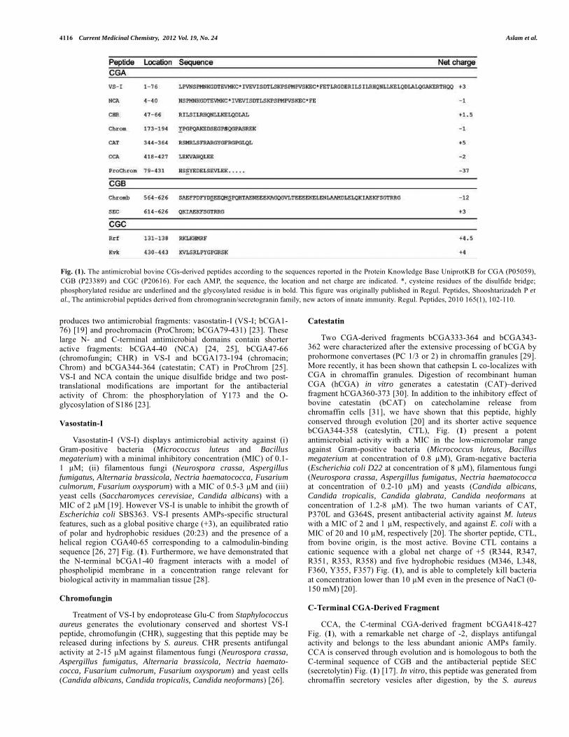
4116 Current Medicinal Chemistry, 2012 Vol. 19, No. 24 Aslam et al.
produces two antimicrobial fragments: vasostatin-I (VS-I; bCGA1-76) [19] and prochromacin (ProChrom; bCGA79-431) [23]. These large N- and C-terminal antimicrobial domains contain shorter active fragments: bCGA4-40 (NCA) [24, 25], bCGA47-66 (chromofungin; CHR) in VS-I and bCGA173-194 (chromacin; Chrom) and bCGA344-364 (catestatin; CAT) in ProChrom [25]. VS-I and NCA contain the unique disulfide bridge and two post-translational modifications are important for the antibacterial activity of Chrom: the phosphorylation of Y173 and the O-glycosylation of S186 [23].
Vasostatin-I
Vasostatin-I (VS-I) displays antimicrobial activity against (i) Gram-positive bacteria (Micrococcus luteus and Bacillus megaterium) with a minimal inhibitory concentration (MIC) of 0.1-1 �M; (ii) filamentous fungi (Neurospora crassa, Aspergillus fumigatus, Alternaria brassicola, Nectria haematococca, Fusarium culmorum, Fusarium oxysporum) with a MIC of 0.5-3 �M and (iii) yeast cells (Saccharomyces cerevisiae, Candida albicans) with a MIC of 2 �M [19]. However VS-I is unable to inhibit the growth of Escherichia coli SBS363. VS-I presents AMPs-specific structural features, such as a global positive charge (+3), an equilibrated ratio of polar and hydrophobic residues (20:23) and the presence of a helical region CGA40-65 corresponding to a calmodulin-binding sequence [26, 27] Fig. (1). Furthermore, we have demonstrated that the N-terminal bCGA1-40 fragment interacts with a model of phospholipid membrane in a concentration range relevant for biological activity in mammalian tissue [28].
Chromofungin
Treatment of VS-I by endoprotease Glu-C from Staphylococcus aureus generates the evolutionary conserved and shortest VS-I peptide, chromofungin (CHR), suggesting that this peptide may be released during infections by S. aureus. CHR presents antifungal activity at 2-15 �M against filamentous fungi (Neurospora crassa, Aspergillus fumigatus, Alternaria brassicola, Nectria haemato-cocca, Fusarium culmorum, Fusarium oxysporum) and yeast cells (Candida albicans, Candida tropicalis, Candida neoformans) [26].
Catestatin
Two CGA-derived fragments bCGA333-364 and bCGA343-362 were characterized after the extensive processing of bCGA by prohormone convertases (PC 1/3 or 2) in chromaffin granules [29]. More recently, it has been shown that cathepsin L co-localizes with CGA in chromaffin granules. Digestion of recombinant human CGA (hCGA) in vitro generates a catestatin (CAT)–derived fragment hCGA360-373 [30]. In addition to the inhibitory effect of bovine catestatin (bCAT) on catecholamine release from chromaffin cells [31], we have shown that this peptide, highly conserved through evolution [20] and its shorter active sequence bCGA344-358 (cateslytin, CTL), Fig. (1) present a potent antimicrobial activity with a MIC in the low-micromolar range against Gram-positive bacteria (Micrococcus luteus, Bacillus megaterium at concentration of 0.8 �M), Gram-negative bacteria (Escherichia coli D22 at concentration of 8 �M), filamentous fungi (Neurospora crassa, Aspergillus fumigatus, Nectria haematococca at concentration of 0.2-10 �M) and yeasts (Candida albicans, Candida tropicalis, Candida glabrata, Candida neoformans at concentration of 1.2-8 �M). The two human variants of CAT, P370L and G364S, present antibacterial activity against M. luteus with a MIC of 2 and 1 �M, respectively, and against E. coli with a MIC of 20 and 10 �M, respectively [20]. The shorter peptide, CTL, from bovine origin, is the most active. Bovine CTL contains a cationic sequence with a global net charge of +5 (R344, R347, R351, R353, R358) and five hydrophobic residues (M346, L348, F360, Y355, F357) Fig. (1), and is able to completely kill bacteria at concentration lower than 10 �M even in the presence of NaCl (0-150 mM) [20].
C-Terminal CGA-Derived Fragment
CCA, the C-terminal CGA-derived fragment bCGA418-427 Fig. (1), with a remarkable net charge of -2, displays antifungal activity and belongs to the less abundant anionic AMPs family. CCA is conserved through evolution and is homologous to both the C-terminal sequence of CGB and the antibacterial peptide SEC (secretolytin) Fig. (1) [17]. In vitro, this peptide was generated fromchromaffin secretory vesicles after digestion, by the S. aureus
Fig. (1). The antimicrobial bovine CGs-derived peptides according to the sequences reported in the Protein Knowledge Base UniprotKB for CGA (P05059), CGB (P23389) and CGC (P20616). For each AMP, the sequence, the location and net charge are indicated. *, cysteine residues of the disulfide bridge; phosphorylated residue are underlined and the glycosylated residue is in bold. This figure was originally published in Regul. Peptides, Shooshtarizadeh P et al., The antimicrobial peptides derived from chromogranin/secretogranin family, new actors of innate immunity. Regul. Peptides, 2010 165(1), 102-110.
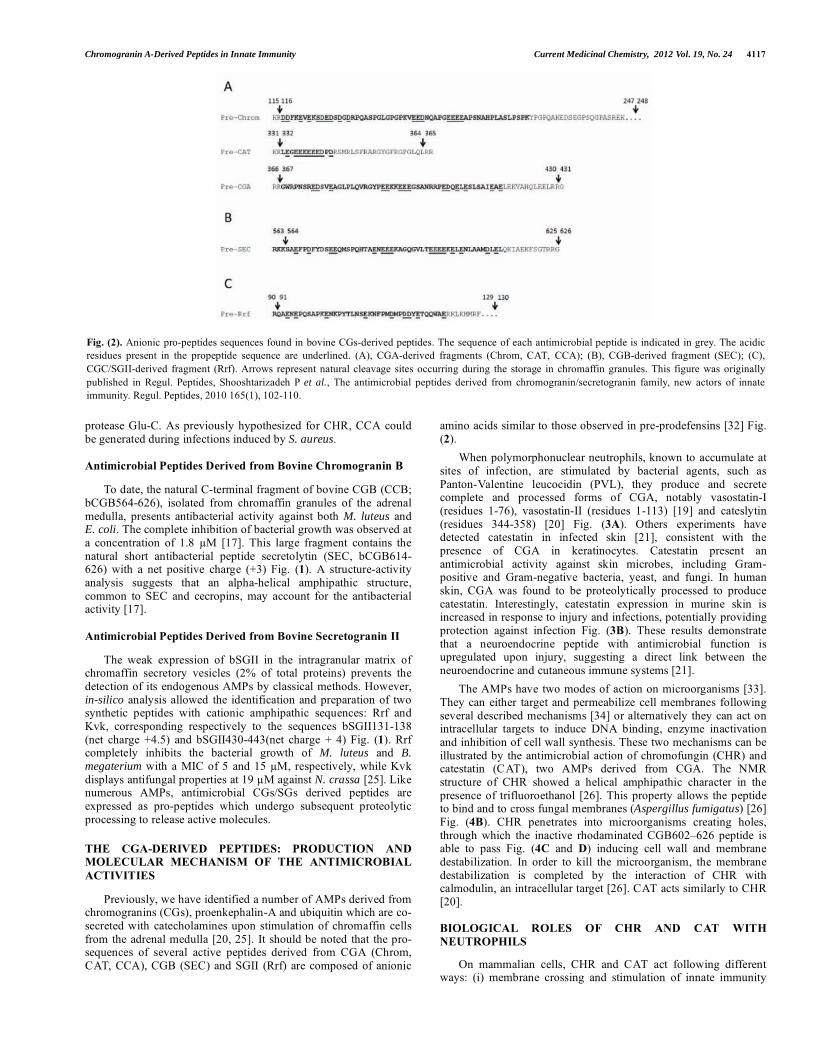
Chromogranin A-Derived Peptides in Innate Immunity Current Medicinal Chemistry, 2012 Vol. 19, No. 24 4117
protease Glu-C. As previously hypothesized for CHR, CCA could be generated during infections induced by S. aureus.
Antimicrobial Peptides Derived from Bovine Chromogranin B
To date, the natural C-terminal fragment of bovine CGB (CCB; bCGB564-626), isolated from chromaffin granules of the adrenal medulla, presents antibacterial activity against both M. luteus and E. coli. The complete inhibition of bacterial growth was observed at a concentration of 1.8 �M [17]. This large fragment contains the natural short antibacterial peptide secretolytin (SEC, bCGB614-626) with a net positive charge (+3) Fig. (1). A structure-activity analysis suggests that an alpha-helical amphipathic structure, common to SEC and cecropins, may account for the antibacterial activity [17].
Antimicrobial Peptides Derived from Bovine Secretogranin II
The weak expression of bSGII in the intragranular matrix of chromaffin secretory vesicles (2% of total proteins) prevents the detection of its endogenous AMPs by classical methods. However, in-silico analysis allowed the identification and preparation of two synthetic peptides with cationic amphipathic sequences: Rrf and Kvk, corresponding respectively to the sequences bSGII131-138 (net charge +4.5) and bSGII430-443(net charge + 4) Fig. (1). Rrf completely inhibits the bacterial growth of M. luteus and B. megaterium with a MIC of 5 and 15 �M, respectively, while Kvk displays antifungal properties at 19 �M against N. crassa [25]. Like numerous AMPs, antimicrobial CGs/SGs derived peptides are expressed as pro-peptides which undergo subsequent proteolytic processing to release active molecules.
THE CGA-DERIVED PEPTIDES: PRODUCTION AND MOLECULAR MECHANISM OF THE ANTIMICROBIAL ACTIVITIES
Previously, we have identified a number of AMPs derived from chromogranins (CGs), proenkephalin-A and ubiquitin which are co-secreted with catecholamines upon stimulation of chromaffin cells from the adrenal medulla [20, 25]. It should be noted that the pro-sequences of several active peptides derived from CGA (Chrom, CAT, CCA), CGB (SEC) and SGII (Rrf) are composed of anionic
amino acids similar to those observed in pre-prodefensins [32] Fig. (2).
When polymorphonuclear neutrophils, known to accumulate at sites of infection, are stimulated by bacterial agents, such as Panton-Valentine leucocidin (PVL), they produce and secrete complete and processed forms of CGA, notably vasostatin-I (residues 1-76), vasostatin-II (residues 1-113) [19] and cateslytin (residues 344-358) [20] Fig. (3A). Others experiments have detected catestatin in infected skin [21], consistent with the presence of CGA in keratinocytes. Catestatin present an antimicrobial activity against skin microbes, including Gram-positive and Gram-negative bacteria, yeast, and fungi. In human skin, CGA was found to be proteolytically processed to produce catestatin. Interestingly, catestatin expression in murine skin is increased in response to injury and infections, potentially providing protection against infection Fig. (3B). These results demonstrate that a neuroendocrine peptide with antimicrobial function is upregulated upon injury, suggesting a direct link between the neuroendocrine and cutaneous immune systems [21].
The AMPs have two modes of action on microorganisms [33]. They can either target and permeabilize cell membranes following several described mechanisms [34] or alternatively they can act on intracellular targets to induce DNA binding, enzyme inactivation and inhibition of cell wall synthesis. These two mechanisms can be illustrated by the antimicrobial action of chromofungin (CHR) and catestatin (CAT), two AMPs derived from CGA. The NMR structure of CHR showed a helical amphipathic character in the presence of trifluoroethanol [26]. This property allows the peptide to bind and to cross fungal membranes (Aspergillus fumigatus) [26] Fig. (4B). CHR penetrates into microorganisms creating holes, through which the inactive rhodaminated CGB602–626 peptide is able to pass Fig. (4C and D) inducing cell wall and membrane destabilization. In order to kill the microorganism, the membrane destabilization is completed by the interaction of CHR with calmodulin, an intracellular target [26]. CAT acts similarly to CHR [20].
BIOLOGICAL ROLES OF CHR AND CAT WITH NEUTROPHILS
On mammalian cells, CHR and CAT act following different ways: (i) membrane crossing and stimulation of innate immunity
Fig. (2). Anionic pro-peptides sequences found in bovine CGs-derived peptides. The sequence of each antimicrobial peptide is indicated in grey. The acidic residues present in the propeptide sequence are underlined. (A), CGA-derived fragments (Chrom, CAT, CCA); (B), CGB-derived fragment (SEC); (C), CGC/SGII-derived fragment (Rrf). Arrows represent natural cleavage sites occurring during the storage in chromaffin granules. This figure was originally published in Regul. Peptides, Shooshtarizadeh P et al., The antimicrobial peptides derived from chromogranin/secretogranin family, new actors of innate immunity. Regul. Peptides, 2010 165(1), 102-110.
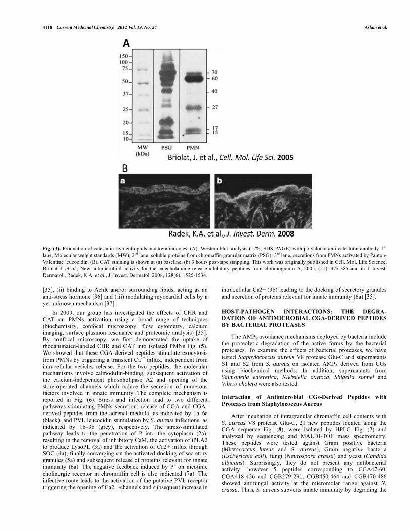
4118 Current Medicinal Chemistry, 2012 Vol. 19, No. 24 Aslam et al.
Fig. (3). Production of catestatin by neutrophils and keratinocytes. (A), Western blot analysis (12%, SDS-PAGE) with polyclonal anti-catestatin antibody. 1st
lane, Molecular weight standards (MW); 2nd lane, soluble proteins from chromaffin granular matrix (PSG); 3rd lane, secretions from PMNs activated by Panton-Valentine leucocidin. (B), CAT staining is shown at (a) baseline, (b) 3 hours post-tape stripping. This work was originally published in Cell. Mol. Life Science, Briolat J. et al., New antimicrobial activity for the catecholamine release-inhibitory peptides from chromogranin A, 2005, (21), 377-385 and in J. Invest. Dermatol., Radek, K.A. et al., J. Invest. Dermatol. 2008, 128(6), 1525-1534.
[35], (ii) binding to AchR and/or surrounding lipids, acting as an anti-stress hormone [36] and (iii) modulating myocardial cells by a yet unknown mechanism [37].
In 2009, our group has investigated the effects of CHR and CAT on PMNs activation using a broad range of techniques (biochemistry, confocal microscopy, flow cytometry, calcium imaging, surface plasmon resonance and proteomic analysis) [35]. By confocal microscopy, we first demonstrated the uptake of rhodaminated-labeled CHR and CAT into isolated PMNs Fig. (5).We showed that these CGA-derived peptides stimulate exocytosis from PMNs by triggering a transient Ca2+ influx, independent from intracellular vesicles release. For the two peptides, the molecular mechanisms involve calmodulin-binding, subsequent activation of the calcium-independent phospholipase A2 and opening of the store-operated channels which induce the secretion of numerous factors involved in innate immunity. The complete mechanism is reported in Fig. (6). Stress and infection lead to two different pathways stimulating PMNs secretion: release of CGA and CGA-derived peptides from the adrenal medulla, as indicated by 1a–6a (black), and PVL leucocidin stimulation by S. aureus infections, as indicated by 1b–3b (grey), respectively. The stress-stimulated pathway leads to the penetration of P into the cytoplasm (2a), resulting in the removal of inhibitory CaM, the activation of iPLA2 to produce LysoPL (3a) and the activation of Ca2+ influx through SOC (4a), finally converging on the activated docking of secretory granules (5a) and subsequent release of proteins relevant for innate immunity (6a). The negative feedback induced by P’ on nicotinic cholinergic receptor in chromaffin cell is also indicated (7a). The infective route leads to the activation of the putative PVL receptor triggering the opening of Ca2+-channels and subsequent increase in
intracellular Ca2+ (3b) leading to the docking of secretory granules and secretion of proteins relevant for innate immunity (6a) [35].
HOST-PATHOGEN INTERACTIONS: THE DEGRA-DATION OF ANTIMICROBIAL CGA-DERIVED PEPTIDES BY BACTERIAL PROTEASES
The AMPs avoidance mechanisms deployed by bacteria include the proteolytic degradation of the active forms by the bacterial proteases. To examine the effects of bacterial proteases, we have tested Staphylococcus aureus V8 protease Glu-C and supernatants S1 and S2 from S. aureus on isolated AMPs derived from CGs using biochemical methods. In addition, supernatants from Salmonella enteretica, Klebsiella oxytoca, Shigella sonnei and Vibrio cholera were also tested.
Interaction of Antimicrobial CGs-Derived Peptides with Proteases from Staphylococcus Aureus
After incubation of intragranular chromaffin cell contents with S. aureus V8 protease Glu-C, 21 new peptides located along the CGA sequence Fig. (8), were isolated by HPLC Fig. (7) and analyzed by sequencing and MALDI-TOF mass spectrometry. These peptides were tested against Gram positive bacteria (Micrococcus luteus and S. aureus), Gram negative bacteria (Escherichia coli), fungi (Neurospora crassa) and yeast (Candida albicans). Surprisingly, they do not present any antibacterial activity; however 5 peptides corresponding to CGA47-60, CGA418-426 and CGB279-291, CGB450-464 and CGB470-486 showed antifungal activity at the micromolar range against N. crassa. Thus, S. aureus subverts innate immunity by degrading the
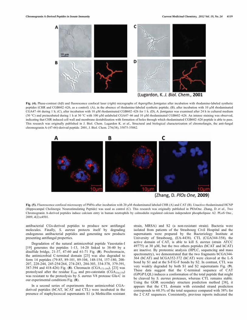
Chromogranin A-Derived Peptides in Innate Immunity Current Medicinal Chemistry, 2012 Vol. 19, No. 24 4119
Fig. (4). Phase-contrast (left) and fluorescence confocal laser (right) micrographs of Aspergillus fumigatus after incubation with rhodamine-labeled synthetic peptides (CHR and CGB602–626, as a control). (A), in the absence of rhodamine-labeled synthetic peptide; (B), after incubation with 10 �M rhodaminated CGA47–66 during 1 h; (C), after incubation with 10 �M rhodaminated CGB602–626 for 1 h. (D), A. fumigatus was examined after 24 h in cultured medium (30 °C) and preincubated during 1 h at 30 °C with 100 �M unlabeled CGA47–66 and 10 �M rhodaminated CGB602–626. An intense staining was observed, indicating that CHR induced cell wall and membrane destabilization with formation of holes through which rhodaminated CGB602–626 peptide is able to pass. This research was originally published in J. Biol. Chem. Lugardon K. et al., Structural and biological characterization of chromofungin, the anti-fungal chromogranin A-(47-66)-derived peptide. 2001, J. Biol. Chem. 276(38), 35875-35882.
Fig. (5). Fluorescence confocal microscopy of PMNs after incubation with 20 �M rhodaminated labeled CHR (A) and CAT (B). Unactive rhodaminated HCNP (Hippocampal Cholinergic Neurostimulating Peptide) was used as control (C). This research was originally published in PlOsOne. Zhang, D. et al., Two Chromogranin A-derived peptides induce calcium entry in human neutrophils by calmodulin regulated calcium independent phospholipase A2. PLoS One., 2009, 4(2):e4501.
antibacterial CGs-derived peptides to produce new antifungal molecules. Finally, S. aureus protects itself by degradingendogenous antibacterial peptides and generating new products presenting antifungal properties.
Degradation of the natural antimicrobial peptide Vasostatin-I [19] generates the peptides 1-13, 14-20 linked to 38-40 by a disulfide bridge, 21-37, 47-60 and 61-71 Fig. (8). Prochromacin, the antimicrobial C-terminal domain [23] was also degraded to form 14 peptides (79-85, 89-101, 89-104, 148-154, 157-180, 200-207, 228-244, 245-254/264, 274-283, 284-303, 334-378, 379-391, 387-394 and 418-426) Fig. (8). Chromacin (CGA173-194), [23] was proteolysed after the residue E180 and pro-catestatin (CGA334-378)was resistant to the proteolysis by S. aureus V8 protease Glu-C in our experimental conditions Fig. (8).
In a second series of experiments three antimicrobial CGA-derived peptides (bCAT, hCAT and CTL) were incubated in the presence of staphylococcal supernatants S1 (a Methicillin resistant
strain, MRSA) and S2 (a non-resistant strain). Bacteria were isolated from patients of the Strasbourg Civil Hospital and the supernatants were prepared by the Bacteriology Institute at University of Strasbourg, (EA-4438). CTL (CGA344-358), the active domain of CAT, is able to kill S. aureus (strain ATCC 49775) at 30 �M, but the two others peptides (bCAT and hCAT) are inactive. By proteomic analysis (HPLC, sequencing and mass spectrometry), we demonstrated that the two fragments bCGA344-364 (bCAT) and hCGA352-372 (hCAT) were cleaved at the L-S bond by S1 and at the S-F/G-F bonds by S2. In contrast, CTL was very weakly degraded by both S1 and S2 supernatants Fig. (9).These data suggest that the C-terminal sequence of CAT (GPGP/LQL) induces a conformation of the total peptide that might be cleaved by S. aureus proteases, whereas CTL remains stable. Using the GOR secondary structure prediction method [38], it appears that the CTL domain with extended strand prediction corresponds to 60.3% of the total sequence compared with 38% for the 2 CAT sequences. Consistently, previous reports indicated the
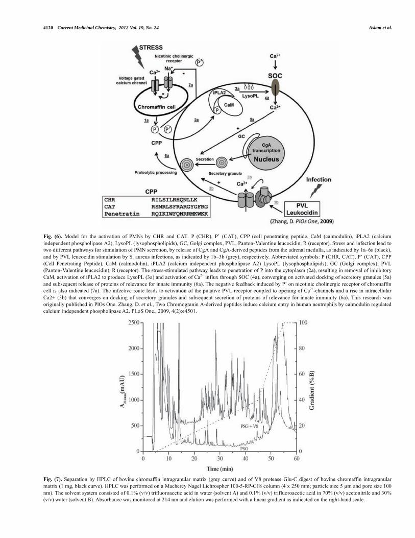
4120 Current Medicinal Chemistry, 2012 Vol. 19, No. 24 Aslam et al.
Fig. (6). Model for the activation of PMNs by CHR and CAT. P (CHR), P’ (CAT), CPP (cell penetrating peptide, CaM (calmodulin), iPLA2 (calcium independent phospholipase A2), LysoPL (lysophospholipids), GC, Golgi complex, PVL, Panton-Valentine leucocidin, R (receptor). Stress and infection lead to two different pathways for stimulation of PMN secretion, by release of CgA and CgA-derived peptides from the adrenal medulla, as indicated by 1a–6a (black), and by PVL leucocidin stimulation by S. aureus infections, as indicated by 1b–3b (grey), respectively. Abbreviated symbols: P (CHR, CAT), P’ (CAT), CPP (Cell Penetrating Peptide), CaM (calmodulin), iPLA2 (calcium independent phospholipase A2) LysoPL (lysophospholipids); GC (Golgi complex); PVL (Panton-Valentine leucocidin), R (receptor). The stress-stimulated pathway leads to penetration of P into the cytoplasm (2a), resulting in removal of inhibitory CaM, activation of iPLA2 to produce LysoPL (3a) and activation of Ca2+ influx through SOC (4a), converging on activated docking of secretory granules (5a) and subsequent release of proteins of relevance for innate immunity (6a). The negative feedback induced by P’ on nicotinic cholinergic receptor of chromaffin cell is also indicated (7a). The infective route leads to activation of the putative PVL receptor coupled to opening of Ca2+-channels and a rise in intracellular Ca2+ (3b) that converges on docking of secretory granules and subsequent secretion of proteins of relevance for innate immunity (6a). This research was originally published in PlOs One. Zhang, D. et al., Two Chromogranin A-derived peptides induce calcium entry in human neutrophils by calmodulin regulated calcium independent phospholipase A2. PLoS One., 2009, 4(2):e4501.
Fig. (7). Separation by HPLC of bovine chromaffin intragranular matrix (grey curve) and of V8 protease Glu-C digest of bovine chromaffin intragranular matrix (1 mg, black curve). HPLC was performed on a Macherey Nagel Lichrospher 100-5-RP-C18 column (4 x 250 mm; particle size 5 �m and pore size 100 nm). The solvent system consisted of 0.1% (v/v) trifluoroacetic acid in water (solvent A) and 0.1% (v/v) trifluoroacetic acid in 70% (v/v) acetonitrile and 30% (v/v) water (solvent B). Absorbance was monitored at 214 nm and elution was performed with a linear gradient as indicated on the right-hand scale.

Chromogranin A-Derived Peptides in Innate Immunity Current Medicinal Chemistry, 2012 Vol. 19, No. 24 4121
Fig. (8). Sequence alignment of the identified peptides produced after incubation of the stimulated chromaffin cell secretions with S. aureus V8 protease Glu-C. Antimicrobial regions Vasostatin and Prochromacin are marked by grey lines and the new generated peptides are in black. Arrows indicate cleavage site by Glu-C protease.
importance of an extended strand for several classes of protease inhibitors [39]. Our observation indicates that the structure of CTL might present a protease inhibitor activity.
Fig. (9). Degradation analysis of bCAT, hCAT and CTL with S1 and S2 two Staphylococcus aureus supernatants. Arrows indicate cleavage sites produced by proteases from S1 and S2. CTL resists to the proteolytic degradation.
Interaction of Antimicrobial CGs-Derived Peptides with Proteases from Diarrheogenic Bacteria
Bacteria were isolated from patients of the Strasbourg Civil Hospital by the Bacteriology Institute, University of Strasbourg, (EA-4438). Klebsiella oxytoca, Salmonella enterica, Shigella sonnei, and Vibrio cholera infections have a clinical interest as they cause other symptoms beside diarrhea. Klebsiella oxytoca may be commensal [42] and is involved in post-antibiotic diarrheas [40], as well as nosocomial infections in newborns and adults [41]. K. oxytoca has also been associated with hemorrhagic colitis [43] and intercurrent colitis in Crohn's disease [44]. Salmonella enterica destroys infected cells and causes gastrointestinal infections which propagates through blood (sepsis) or through lymphatic vessel (typhoid fever). Shigella sonnei and Vibrio cholera non O1 produce Shiga toxin which causes inflammation of the intestinal mucosa. Klebsiella oxytoca, Salmonella enterica, Shigella sonnei, and Vibrio cholera all develop an antibiotic resistance. Salmonella was reported to be resistant to the action of Ciprofloxacin [45] and Ceftriaxone [46].
We tested the antimicrobial activity of bovine (bCGA344-364), rat (rCGA344-364) and human (hCGA352-372) CAT, bovine CTL (bCGA344-358), two short fragments hCGA360-372 and the conserved tetrapeptide LSFR (bCGA348-351) against the four bacterial strains mentioned above. In addition, we tested a scrambled peptide relative to the sequence of bovine CAT and the procatestatin fragment bCGA332-364. We found antimicrobial activities only for bovine CAT and CTL, indicating that CTL, the
shorter fragment, corresponds to the active domain of CAT. Procatestatin was inactive in similar experimental conditions. Bovine CAT and CTL were active against Klebsiella oxytoca,Salmonella enterica and Vibrio cholera at 100 �M and 30 �Mrespectively and against Shigella Sonnei at 50 �M and 25 �M. In addition, CHR and the C-terminal fragment (CGA387-431) were inactive for concentration below 100 �M. We then compared the profiles of the peptides alone and with the inoculated medium, using HPLC.
These experiments showed that all the peptides were completely degraded by the bacteria, except for CTL. The degradation by bacterial proteases was observed by the HPLC profiles of proCAT (CGA332-364) after incubation with Salmonella enterica Fig. (10). The complete peptide and the processed forms were analyzed by sequencing and mass spectrometry (MALDI-TOF) Fig. (10).
SYNERGY OF THE COMBINATION OF ANTIMICROBIAL CGA-DERIVED PEPTIDES WITH ANTIBIOTICS
The high toxic effects of antibiotics have shifted the research focus towards the discovery of new combination of antimicrobial agents with broad spectrum of activity and less toxicity. Synergy is the combined activity of two agents which is higher than the added activity of each agent separately [47]. Considering the direct interaction of AMPs with membranes, they represent good candidates to potentiate antibiotics activity through synergistic actions. To demonstrate that antimicrobial peptides are able to reduce the doses of antibiotics used and to potentiate their activity, we evaluated the synergistic effects of three CGA-derived peptides (CAT, CTL and amidated CTL) together with Minocyclin and Voriconazole. Antimicrobial tests were carried out using the antimicrobial peptides (at concentration below MIC), the antibiotics or a combination of both, evaluating the fractional inhibitory concentration (FIC): FIC �0.5 corresponds to a synergistic effect, between 0.5 and 2 to an additive effect, and > 2 to an antagonistic effect [47]. Importantly, the combination of amidated CTL and Minocyclin led to a FIC of 0.37 against S. aureus. In addition, for CTL and Voriconazole, we obtained a FIC of 0.25 and 0.5 against Candida albicans and Candida tropicalis, respectively.
One could imagine a mechanism by which the AMPs could favor the destabilization of the membrane allowing the antibiotics to rapidly penetrate inside the bacterial cells and more efficiently
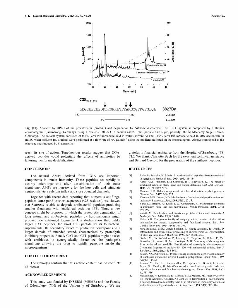
4122 Current Medicinal Chemistry, 2012 Vol. 19, No. 24 Aslam et al.
reach its site of action. Together our results suggest that CGA-derived peptides could potentiate the effects of antibiotics by favoring membrane destabilization.
CONCLUSIONS
The natural AMPs derived from CGA are important components in innate immunity. These peptides act rapidly to destroy microorganisms after destabilization of their outer membrane. AMPs are non-toxic for the host cells and stimulate neutrophils via a calcium influx and store operated channels.
Together with recent data reporting that numerous antifungal peptides correspond to short sequences (<25 residues), we showed that S.aureus is able to degrade antibacterial peptides producing smaller fragments with antifungal activities [48]. Thus, a new concept might be proposed in which the proteolytic degradation of long natural and antibacterial peptides by host pathogens might produce new antifungal fragments. Our studies show that, unlike larger CAT peptides, the short CTL peptide resists to bacterial supernatants. Its secondary structure prediction corresponds to a larger domain of extended strand, characterized by proteolytic inhibitory properties. Finally CAT and CTL peptides might be used with antibiotics to synergistically destabilize the pathogen's membrane allowing the drug to rapidly penetrate inside the microorganisms.
CONFLICT OF INTEREST
The author(s) confirm that this article content has no conflicts of interest.
ACKNOWLEDGEMENTS
This study was funded by INSERM (MHMB) and the Faculty of Odontology (YH) of the University of Strasbourg. We are
grateful to financial assistance from the Hospital of Strasbourg (FS, TL). We thank Charlotte Bach for the excellent technical assistance and Bernard Guérold for the preparation of the synthetic peptides.
REFERENCES
[1] Bulet, P.; Stocklin, R.; Menin, L. Anti-microbial peptides: from invertebrates to vertebrates. Immunol. Rev., 2004, 198, 169-184.
[2] Aerts, A.M.; François, I.E.; Cammue, B.P.; Thevissen, K. The mode of antifungal action of plant, insect and human defensins. Cell. Mol. Life Sci., 2008, 65(12), 2069-2079.
[3] Manners, J.M. Hidden weapons of microbial destruction in plant genomes.Genome Biol., 2007, 8(9), 225.
[4] Yeaman, M.R.; Yount, N.Y. Mechanisms of antimicrobial peptide action and resistance. Pharmacol. Rev., 2003, 55(1), 27-55.
[5] Yang, D.; Biragyn, A.; Kwak, L.W.; Oppenheim, J.J. Mammalian defensins in immunity: more than just microbicidal. Trends Immunol., 2002, 23(6), 291-296.
[6] Zanetti, M. Cathelicidins, multifunctional peptides of the innate immunity. J. Leukocyte Biol., 2004, 75(1), 39-48.
[7] Helle, K.B. The granin family of uniquely acidic proteins of the diffuse neuroendocrine system: comparative and functional aspects. Biol. Rev. Cambr. Philo. Soc., 2004, 79(4), 769-794.
[8] Metz-Boutigue, M.H.; Garcia-Sablone, P.; Hogue-Angeletti, R.; Aunis, D. Intracellular and extracellular processing of chromogranin A. Determination of cleavage sites. Eur. J. Biochem., 1993, 217(1), 247-257.
[9] Strub, J.M.; Garcia-Sablone, P.; Lonning, K.; Taupenot, L.; Hubert, P.; Van Dorsselaer, A.; Aunis, D.; Metz-Boutigue, M.H. Processing of chromogranin B in bovine adrenal medulla. Identification of secretolytin, the endogenous C-terminal fragment of residues 614-626 with antibacterial activity. Eur. J. Biochem., 1995, 229(2), 356-368.
[10] Seidah, N.G.; Chretien, M. Proprotein and prohormone convertases: a family of subtilases generating diverse bioactive polypeptides. Brain Res., 1999, 848(1-2), 45-62.
[11] Anouar, Y.; Yon, L.; Desmoucelles, C.; Leprince, J.; Breault, L.; Gallo-Payet, N.; Vaudry, H. Identification of a novel secretogranin II-derived peptide in the adult and fetal human adrenal gland. Endocr. Res. 1998, 24(3-4), 731-736.
[12] Marksteiner, J.; Kirchmair, R.; Mahata, S.K.; Mahata, M.; Fischer-Colbrie, R.; Hogue-Angeletti, R.; Saria, A.; Winkler, H. Distribution of secretoneurin, a peptide derived from secretogranin II, in rat brain: an immunocytochemical and radioimmunological study. Eur. J. Neurosci., 1993, 54(4), 923-944.
Fig. (10). Analysis by HPLC of the procatestatin (proCAT) and degradation by Salmonella enterica. The HPLC system is composed by a Dionex chromatogram, (Germerong, Germany), using a Nucleosil 300-5 C18 column (4�250 mm, particle size 5 �m, porosity 300 Å; Macherey Nagel, Düren, Germany). The solvent system consisted of 0.1% (v/v) trifluoroacetic acid in water (solvent A) and 0.09% (v/v) trifluoroacetic acid in 70% acetonitrile in milliQ water (solvent B). Elutions were performed at a flow rate of 700 �L min�1 using the gradient indicated on the chromatogram. Arrows correspond to the cleavage sites induced by S. enteretica.
Chromogranin A-Derived Peptides in Innate Immunity Current Medicinal Chemistry, 2012 Vol. 19, No. 24 4123
[13] Yajima, A.; Ikeda, M.; Miyazaki, K.; Maeshima, T.; Narita, N.; Narita, M.; Manserin, a novel peptide from secretogranin II in the neuroendocrine system. Neuroreport, 2004, 15(11), 1755-1759.
[14] Eiden, L.E. Is chromogranin a prohormone? Nature, 1987, 325(6102), 301. [15] Tatemoto, K.; Efendi�, S.; Mutt, V.; Makk, G.; Feistner, G.J; Barchas, J.D.
Pancreastatin, a novel pancreatic peptide that inhibits insulin secretion. Nature, 1986, 324(6096), 476-478.
[16] Helle, K.B.; Corti, A.; Metz-Boutigue, M.H.; Tota, B. The endocrine role for chromogranin A: a prohormone for peptides with regulatory properties. Cell. Mol. Life Sci., 2007, 64(22), 2863-2886.
[17] Strub, J.M.; Hubert, P.; Nullans, G.; Aunis, D.; Metz-Boutigue, M.H. Antibacterial activity of secretolytin, a chromogranin B-derived peptide (614-626), is correlated with peptide structure. FEBS Lett., 1996, 379(3), 273-278.
[18] Metz-Boutigue, M.H.; Goumon, Y.; Lugardon, K.; Strub, J.M.; Aunis, D. Antibacterial peptides are present in chromaffin cell secretory granules. Cell. Mol. Neurobiol., 1998, 18(2), 249-266.
[19] Lugardon, K.; Raffner, R.; Goumon, Y.; Corti, A.; Delmas, A.; Bulet, P.; Aunis, D.; Metz-Boutigue, M.H. Antibacterial and antifungal activities of vasostatin-1, the N-terminal fragment of chromogranin A. J. Biol. Chem.,2000, 275(15), 10745-10753.
[20] Briolat, J.; Wu, S.D.; Mahata, S.K.; Gonthier, B.; Bagnard, D.; Chasserot-Golaz, S.; Helle, K.B.; Aunis, D.; Metz-Boutigue, M.H. New antimicrobial activity for the catecholamine release-inhibitory peptide from chromogranin A. Cell. Mol. Life Sci., 2005, 62(3), 377-385.
[21] Radek, K.A.; Lopez-Garcia, B.; Hupe, M.; Niesman, I.R.; Elias, P.M.; Taupenot, L; Mahata, S.K.; O'Connor, D.T; Gallo, R.L. The neuroendocrine peptide catestatin is a cutaneous antimicrobial and induced in the skin after injury. J. Invest. Dermatol., 2008, 128(6), 1525-1534.
[22] Sternberg, E.M. Neural regulation of innate immunity: a coordinated nonspecific host response to pathogens. Nat. Rev. Immunol., 2006, 6(4), 318-328.
[23] Strub, J.M.; Goumon, Y.; Lugardon, K.; Capon, C.; Lopez, M.; Moniatte, M.; Van Dorsselaer, A.; Aunis, D.; Metz-Boutigue, M.H. Antibacterial activity of glycosylated and phosphorylated chromogranin A-derived peptide 173-194 from bovine adrenal medullary chromaffin granules. J. Biol. Chem., 1996, 271(45), 28533-28540.
[24] Maget-Dana, R.; Metz-Boutigue, M.H.; Helle, K.B. The N-terminal domain of chromogranin A (CgA1-40) interacts with monolayers of membrane lipids of fungal and mammalian compositions. Ann. N. Y. Acad. Sci., 2002, 971,352-354.
[25] Shooshtarizadeh, P.; Zhang, D.; Chich, J.F.; Gasnier, C.; Schneider, F.; Haïkel, Y.; Aunis, D.; Metz-Boutigue, M.H. The antimicrobial peptides derived from chromogranin/secretogranin family, new actors of innate immunity. Regul. Peptides, 2010, 165(1), 102-110.
[26] Lugardon, K.; Chasserot-Golaz, S.; Kieffer, A.E.; Maget-Dana, R.; Nullans, G.; Kieffer, B.; Aunis, D.; Metz-Boutigue, M.H. Structural and biological characterization of chromofungin, the antifungal chromogranin A-(47-66)-derived peptide. J. Biol. Chem., 2001, 276(38), 35875-35882.
[27] Yoo, S.H. Identification of the Ca(2+)-dependent calmodulin-binding region of chromogranin A. Biochemistry, 1992, 31(26), 6134-6140.
[28] Blois, A.; Holmsen, H.; Martino, G.; Corti, A.; Metz-Boutigue, M.H.; Helle, K.B. Interactions of chromogranin A-derived vasostatins and monolayers of phosphatidylserine, phosphatidylcholine and phosphatidylethanolamine. Regul. Peptides, 2006, 134(1), 30-37.
[29] Taylor, C.V.; Taupenot, L.; Mahata, S.K.; Mahata, M.; Wu, H.; Yasothornsrikul, S.; Toneff, T.; Caporale, C.; Jiang, Q.; Parmer, R.J.; Hook, V.Y.; O'Connor, D.T. Formation of the catecholamine release-inhibitory peptide catestatin from chromogranin A. Determination of proteolytic cleavage sites in hormone storage granules. J. Biol. Chem., 2000, 275(30), 22905-22915.
[30] Biswas, N.; Rodriguez-Flores, J.L.; Courel, M.; Gayen, J.; Vaingankar, S.M.; Mahata, M.; Torpey, J.W.; Taupenot, L.; O'Connor, D.T.; Mahata, S.K. Cathepsin L colocalizes with chromogranin A in chromaffin vesicles to generate active peptides. Endocrinology, 2009, 150(8), 3547-3557.
[31] Mahata, S.K.; O'Connor, D.T.; Mahata, M.; Yoo, S.H.; Taupenot, L.; Wu,
H;, Gill, B.M.; Parmer, R.J. Novel autocrine feedback control of catecholamine release. A discrete chromogranin a fragment is a noncompetitive nicotinic cholinergic antagonist. J. Clin. Invest., 1997,100(6), 1623-1633.
[32] Valore, E.V.; Ganz, T. Posttranslational processing of defensins in immature human myeloid cells. Blood, 1992, 79, 1538-1544.
[33] Brogden, K.A. Antimicrobial peptides: Pore formers or metabolic inhibitors in bacteria? Nat Rev. Microbiol., 2005, 3, 238-250.
[34] Jenssen, H.; Hamill, P.; Hancock, R.E. Peptide antimicrobial agents. Clin. Microbiol. Rev. 2006, 19, 491-511.
[35] Zhang, D.; Shooshtarizadeh, P.; Laventie, B.J.; Colin, D.A. ; Chich, J.F. ;Vidic, J. ; de Barry, J.; Chasserot-Golaz, S. ; Delalande, F. ; Van Dorsselaer, A. ; Schneider, F.; Helle, K.; Aunis, D.; Prévost, G. ; Metz-Boutigue, M.H. Two Chromogranin A-derived peptides induce calcium entry in human neutrophils by calmodulin regulated calcium independent phospholipase A2. PLoS One., 2009, 4(2), e4501.
[36] Herrero, C.J.; Ales, E.; Pintado, A.J.; Lopez, M.G.; Garcia-Palomero, E.; Mahata, S.K.; O'Connor, D.T.; Garcia, A.G.; Montiel, C. Modulatory mechanism of the endogenous peptide catestatin on neuronal nicotinic acetylcholine receptors and exocytosis. J. Neurosci. 2002, 22, 377-388.
[37] Angelone, T.; Quintieri, A.M.; Brar, B.K.; Limchaiyawat, P.T.; Tota, B.; Mahata, S.K.; Cerra, M.C. The antihypertensive chromogranin a peptide catestatin acts as a novel endocrine/paracrine modulator of cardiac inotropism and lusitropism. Endocrinology, 2008, 149, 4780-4793.
[38] Gorkiewicz, G. Nosocomial and antibiotic-associated diarrhea caused by organisms other than Clostridium difficile. Int. J. Antimicrob. Ag., 2009,33(Suppl 1), S37-41.
[39] Garnier, J.; Gibrat, J.F.; Robson, B.; GOR secondary structure prediction method version IV. Methods in Enzymology, 1996, 266, 540-553, Doolittle R. F. Ed.
[40] Fairlie, D.P.; Tyndall, J.D. ; Reid, R.C. ; Wong, A.K. ; Abbenante, G. ; Scanlon, M.J.; March, D.R. ; Bergman, D.A. ; Chai, C.L. ; Burkett, B.A. Confirmational selection of inhibitors and substrates by proteolytic enzymes: implications for drug design and polypeptide processing. J. Med. Chem.,2000, 43(7):1271-1281.
[41] Biran, V.; Gaudin, A.; Mariani-Kurdjian, P.; Doit, C.; Bingen, E.; Aujard, Y. Implication of extended-spectrum beta-lactamase enterobacteriaceae in nosocomial infections in neonates. Arch. Pediatrie, 2010, 17(Suppl 4), S150-153.
[42] Tsakris, A.; Poulou, A.; Markou, F.; Pitiriga, V.; Piperaki, E.T.; Kristo, I.; Pournaras, S. Dissemination of clinical isolates of Klebsiella oxytoca harboring CMY-31, VIM-1, and a New OXY-2-type variant in the community. Antimicrob. Agents Chemother., 2011, 55(7), 3164-3168.
[43] Hoffmann, K.M.; Deutschmann, A.; Weitzer, C.; Joainig, M.; Zechner, E.; Hogenauer, C.; Hauer, A.C. Antibiotic-associated hemorrhagic colitis caused by cytotoxin-producing Klebsiella oxytoca. Pediatrics, 2010, 125(4), e960-963.
[44] Plessier, A.; Cosnes, J.; Gendre, J.P.; Beaugerie, L. Intercurrent Klebsiella oxytoca colitis in a patient with Crohn's disease. Gastroenterol. Clin. Biol., 2002, 26(8-9), 799-800.
[45] Medalla, F.; Sjölund-Karlsson, M.; Shin, S.; Harvey, E.; Joyce, K.; Theobald, L.; Nygren, B.N.; Pecic, G.; Gay, K.; Austin, J.; Stuart, A.; Blanton, E.; Mintz, E. D.; Whichard, J. M.; Barzilay, E. J. Ciprofloxacin-resistant Salmonella enterica Serotype Typhi, United States, 1999-2008. Emerging Infectious Diseases, 2011, 17(6), 1095-1098.
[46] Su, L.H.; Teng, W.S.; Chen, C.L.; Lee, H.Y.; Li, H.C.; Wu, T.L.; Chiu, C.H. Increasing ceftriaxone resistance in Salmonellae, Taiwan. Emerg. Infect. Dis., 2011, 17(6), 1086-1090.
[47] Serra, P.; Brandimarte, C.; Martino, P.; Carlone, S.; Giunchi, G. Synergistic treatment of enterococcal endocarditis: in vitro and in vivo studies. Arch. Intern. Med., 1977, 137(11), 1562-1567.
[48] Desbois AP, Tschörner D, Coote PJ. Survey of small antifungal peptides with chemotherapeutic potential. Curr. Pharm. Biotechnol. 2011,12(8):1263-1291.
Received: January 05, 2012 Revised: April 04, 2012 Accepted: April 05, 2012

The Natural Antimicrobial Chromogranins/Secretograni
ns- Derived Peptides – Production, Lytic Activity and
Processing by Bacterial Proteases.
Atindehou M, Aslam R, Chich JF, Hai kel Y, Schneider F, Metz Boutigue MH. (2012) In: Bobbarala V, editor. Antimicrobial Agents: ISBN: 978-953-51-0723-1, InTech, DOI: 10.5772/33263.

9
The Natural Antimicrobial Chromogranins/Secretogranins-Derived
Peptides – Production, Lytic Activity and Processing by Bacterial Proteases
Ménonvè Atindehou*, Rizwan Aslam*, Jean-François Chich, Youssef Haïkel, Francis Schneider and Marie-Hélène Metz-Boutigue
Biomatériaux et Ingénierie Tissulaire, University of Strasbourg, Strasbourg France
1. Introduction
1.1 Multidrug antibiotic resistance and innate immunity
Multidrug-resistant organisms, such as methicillin-resistant Staphylococcus aureus (MRSA) and Vancomycin-resistant enterococci (VRE) have important infection control implications in all healthcare stings. Multidrug antibiotic resistance is a worldwide crucial health problem and the production of new potent antibiotics, acting alone or in combination is urgent. In addition, a major factor in the emergence of antibiotic resistant organisms is the overuse of antibiotics in the hospital or the community. To overcome this abuse, numerous efforts are undertaken to reduce antibiotics prescription and/or promote synergistic effects by others molecules.
Indeed, stimulating organism defense is a promising way to struggle against pathogens. The innate immune system is, since 2 billion years, the primary defense in most living organisms and antimicrobial peptides (AMPs) are fundamental components of the innate immune defense of multicellular organisms, either animal or vegetal (Bulet et al., 2004; Aerts et al. 2008; Manners, 2007).
1.2 The antimicrobial peptides
The antimicrobial peptides (AMPs) have been well conserved throughout the evolution and they ensure the organism’s defense against a large number of pathogens. They serve as endogenous antibiotics that are able to rapidly kill bacteria, fungi and viruses. Interestingly, they are not toxic for the host cells. Taking into consideration the diversity of the living beings, it is presumed that a large number of specific antibiotic peptides have been developed during evolution, allowing a protection of each organism in various conditions and the last years it has clearly appeared that many of these peptides, in addition to their direct antimicrobial activity, also have a wide range of functions in modulating both innate and adaptive immunity. Most of these are small molecules (less than 40 aminoacids) but some can be proteins. To date more than 1414 antibacterial, antifungal and 107 antiviral peptides have been * These authors contributed equally
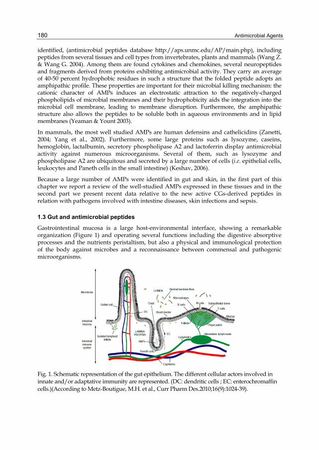
Antimicrobial Agents 180
identified, (antimicrobial peptides database http://aps.unmc.edu/AP/main.php), including peptides from several tissues and cell types from invertebrates, plants and mammals (Wang Z. & Wang G. 2004). Among them are found cytokines and chemokines, several neuropeptides and fragments derived from proteins exhibiting antimicrobial activity. They carry an average of 40-50 percent hydrophobic residues in such a structure that the folded peptide adopts an amphipathic profile. These properties are important for their microbial killing mechanism: the cationic character of AMPs induces an electrostatic attraction to the negatively-charged phospholipids of microbial membranes and their hydrophobicity aids the integration into the microbial cell membrane, leading to membrane disruption. Furthermore, the amphipathic structure also allows the peptides to be soluble both in aqueous environments and in lipid membranes (Yeaman & Yount 2003).
In mammals, the most well studied AMPs are human defensins and cathelicidins (Zanetti, 2004; Yang et al., 2002). Furthermore, some large proteins such as lysozyme, caseins, hemoglobin, lactalbumin, secretory phospholipase A2 and lactoferrin display antimicrobial activity against numerous microorganisms. Several of them, such as lysozyme and phospholipase A2 are ubiquitous and secreted by a large number of cells (i.e. epithelial cells, leukocytes and Paneth cells in the small intestine) (Keshav, 2006).
Because a large number of AMPs were identified in gut and skin, in the first part of this chapter we report a review of the well-studied AMPs expressed in these tissues and in the second part we present recent data relative to the new active CGs-derived peptides in relation with pathogens involved with intestine diseases, skin infections and sepsis.
1.3 Gut and antimicrobial peptides
Gastrointestinal mucosa is a large host-environmental interface, showing a remarkable organization (Figure 1) and operating several functions including the digestive absorptive processes and the nutrients peristaltism, but also a physical and immunological protection of the body against microbes and a reconnaissance between commensal and pathogenic microorganisms.
Fig. 1. Schematic representation of the gut epithelium. The different cellular actors involved in innate and/or adaptative immunity are represented. (DC: dendritic cells ; EC: enterochromaffin cells.)(According to Metz-Boutigue, M.H. et al., Curr Pharm Des.2010;16(9):1024-39).

The Natural Antimicrobial Chromogranins/Secretogranins-Derived Peptides – Production, Lytic Activity and Processing by Bacterial Proteases 181
Lamina propria is a conjunctive tissue composed of fibroblasts, immune cells and collagen. It also contains capillaries and lymphatic vessels. Epithelial cells, or enterocytes, are disposed on a single layer separating the lumen from the lamina propria. These cells are tightly bound by tight junctions forming an impermeable barrier to commensal flora and to pathogens. Brush-border microvilli are present on the apical surface of absorptive enterocytes, representing a large absorbing surface and allowing of microorganisms to the gut. In contrast, microfold cells are not present in microvilli (Figure 1); these cells express cathepsin E (a proteolytic enzyme) and Toll-like receptors able to secrete proinflammatory cytokines and chemokines. The main function of microfold cells is the transport of antigens from the lumen to the subepithelial lymphoid tissue and thus to the adaptive immune system.
Several anatomical structures are present along the gastrointestinal tractus. Peyer’s patches are lymphoid structures containing B and T cells, macrophages and dendritic cells (Figure 1). Lieberkühn crypts are found in the small intestine and they constitute the basis of the intestinal villi (Figure 1). They contain multipotent stem cells and cells, involved in gastrointestinal immunity. Two other cellular types are also present in intestine: i) goblet cells synthesize and secrete large quantities of mucin, ii) enterochromaffin cells that originate from neural crest synthesize serotonin (5-HT) and numerous neuropeptides (Figure 1).
In addition to humoral and cellular immunity, non-immunological defense mechanisms represent an important line of intestinal defenses. Some of these protective factors have been amply documented: pancreatic and gastric juices, intestinal motility and intestinal flora (Sarker, 1992).
Mucosal epithelial cells and Paneth cells produce a variety of AMPs (defensins, cathelicidins, cryptdin related peptides, bactericidal/permeability increasing protein (BPI), chemokine CCL20 and bacteriolytic enzymes such as lysozyme and group IIA phospholipase A2 (Müller et al., 2005). In addition to their direct role in killing pathogenic microorganisms, AMPs are involved in attraction of leukocytes, alarming the adaptive immune system and neutralizing the proinflammatory bacterial molecules (Müller et al., 2005).
Lysozyme
Lysozyme is synthesized and secreted by Paneth cells, macrophages, neutrophils and epithelial cells (Mason & Taylor, 1975; Satoh et al., 1988). Its role and selectivity towards microbes are the same as in skin.
Lactoferrin
Lactoferrin (LF) exhibits a wide spectrum of antimicrobial and immunotropic properties (Artym et al., 2005). In contrast to caseins, LF is particularly resistant to proteolytic degradation in alimentary tract. LF is absorbed from the intestine by means of specific receptors located on brush border cells. Orally administered LF stimulates both local and systemic immune responses. It suppresses the growth of pathogenic bacteria, while promoting the multiplication of non-pathogenic Lactobacillus sp. and Bifidobacterium sp. (Artym et al., 2005).
Studies on mice showed LF to be protective against bacteremia and endotoxemia. LF inhibits the activity of proinflammatory cytokines, nitric oxide and reactive forms of oxygen. Furthermore, LF promotes the differentiation of T and B cells from their immature precursors and increases the activity of NK and LAK (lymphokine activated killer) cells (Artym et al., 2005).

Antimicrobial Agents 182
Phospholipase A2
The hydrophobic layer of phosphatidylcholine (PC) overlies and protects the surface of the gastrointestinal (GI) tract, contributing to barrier integrity. In addition, phospholipase A2 is synthesized by Paneth cells and this enzyme hydrolyses bacterial membrane phospholipids to generate both free fatty acids and lysophospholipids. An important prerequisite for the action of phospholipase A2 is the successful binding to the phospholipids surface. In vitro studies utilizing recombinant enzymes and artificial phospholipids substrates have shown that phospholipases act on anionic phospholipids (phosphatidylglycerol, phosphatidylserine and phosphatidylethanolamine), but are inactive with phosphatidylcholine due to the lack of high affinity binding (Wu et al., 2010). During critical illness such as sepsis, gut barrier integrity may be compromised, which could be related to degradation of PC. Pretreatment with an orally active sPLA(2) inhibitor blocks the LPS-induced increase in GI permeability, and may suggest a new approach to reinforce the GI mucosal barrier and prevent complications from endotoxin in trauma in other septic conditions (Zayat et al., 2008).
Cathelicidins
Cathelicidin LL-37/hCAP18 is synthesized by neutrophils, where it was first identified (Romeo et al., 1988) and epithelial cells of the colon (Hase et al., 2002). In vitro, it has chemotactic properties for monocytes, macrophages and T cells (Koczulla et al., 2003). LL-37 is found in sites of inflammation where it modifies dendritic cells (DCs) differentiation, relying innate and adaptive immunity. In vitro, modified DCs had, among others characteristics induced by the peptide, enhanced secretion of Th-1 inducing cytokines and promoted Th1 responses (Davidson et al., 2004). LL-37 acts synergistically with IL-1-beta to increase the production of suppressive cytokines (IL-6 and IL-10) and chemokines MCP-1 and MCP-3 by macrophages (Yu et al., 2007). It acts via the transcription factor CREB and the activation of phosphorylation of the kinase Akt. In LPS-stimulated monocytes, LL-37 inhibits the release of TNF-alpha modulating inflammatory response induced by LPS, endotoxins and other agonists of TLRs (Mookherjee et al., 2006).
RNAses
Angiogenin-4 (Ang-4) is a member of the ribonucleases family. This protein is synthesized by Paneth cells and is similar to RNase 7 found in skin. Its secretion is stimulated by exposure to LPS. Ang-4 kills E. faecalis and L. monocytogenes at concentrations as low as 1 µM, whereas its concentration in crypts can be 1000 times greater (Hooper et al., 2003). Similarly to defensins, it is sensitive to salt concentration and is potentially cytotoxic to eukaryotic cells (Saxena et al., 1992).
C-type lectins
C-type lectins HIP/PAP are synthesized in human by enterocytes and Paneth cells. The same protein exists in mouse and is named RegIII gamma. These lectins bind Gram-positive peptidoglycan and act by direct killing. Several members of this family are found in gastrointestinal tissues (Dieckgraefe et al., 2002).
Defensins
As in skin, defensins have a direct antimicrobial role as well as immunomodulatory function. Alpha-defensins are synthesized by Paneth cell in the gastrointestinal tractus (Porter et al., 2002). Alpha-defensin expression does not require microbe induction since

The Natural Antimicrobial Chromogranins/Secretogranins-Derived Peptides – Production, Lytic Activity and Processing by Bacterial Proteases 183
they are synthesized in germ-free conditions (Putsep et al., 2000) and/or prenatally (Mallow et al., 1996). In transfected mouse, it was shown that the alpha-defensin hBD-5 protects efficiently against Salmonella typhimurium, demonstrating the direct antimicrobial effect of this peptide. In mouse, alpha-defensins are named cryptdins and several families of peptides related to cryptdins are regrouped under the term CRS (Cryptdin Related Sequences). Interestingly, these CRS can form homo- or heterodimers, thus allowing a combinatorial diversity to struggle against pathogens (Hornef et al., 2004).
Beta-defensins are expressed in enterocytes of the small and large intestines. 28 beta-defensin encoding genes have been identified in human genome, but only 8 were found to be expressed. hBD-1 is constitutively expressed in absence of stimulus or bacterial infection (O’Neil et al., 1999), while some nutrients can stimulate its production in cell lines (Sherman et al., 2006). In mouse, an infection by the Cryptosporidium parasite resulted in a down-regulation of mBD-1 (Zaalouk et al., 2004), while in vitro, sporozoites are killed by this defensin. Some authors conclude on an unique and important regulation of hBD-1, during small intestine infections (Dann et Eckmann, 2007). hBD-2 is not constitutively expressed, but is induced by an infection or by proinflammatory stimuli (O’Neil et al., 1999). hBD-3 and -4 are inducible and particularly expressed in crypt regions (Fahlgren et al., 2004). Defensins can also act as chemotactic agents for immune cells in a similar way to that described for the skin.
Bactericidal Permeability Increasing protein
Bactericidal/permeability-increasing protein (BPI), a constituent of primary neutrophil granules, is a potent natural antibiotic and anti-BPI antibodies are detected during infectious enteritis. In addition, BPI is a target antigen for anti-neutrophil cytoplasmic antoantibodies in inflammatory bowel diseases such as Crohn's disease and ulcerative colitis (Walmsley et al., 1997).
Neuropeptides
Enterochromaffin cells (EC) (Siddique et al., 2009) (Figure 1) are enteroendocrine cells present in the intestine, especially colon (Kuramoto et al., 2007) and containing large amounts of serotonin (5-HT). These cells can sense luminal content before its basolaterally release, and activate afferent neuron endings within lamina propria, allowing information exchange between gut and central nervous system (Hansen & Witte et al., 2008). Besides this important role, EC secrete also numerous other products, among which VIP (Zanner et al., 2004), Substance P (Heitz et al., 1976), CgA, CgB and secretogranin II/CgC (Cetin & Grube, 1991) and melatonin (Raikhlin et Kvetnoy, 1976).
Despite the crucial role of these cells, their sparse repartition and their low number did not allowed their extensive study. However, the BON cells were proposed as a model (Kim et al., 2001), that will enhance further research. When EC were stimulated by odors, they released serotonin, showing that these cells can also be stimulated by spices and fragrances (Braun et al., 2007). Moreover, a new method was proposed allowing isolating and purifying EC from biopsies (Modlin et al., 2006).
1.4 Skin and antimicrobial peptides
Mammal skin is an essential defense barrier against external aggressions, such as microbial pathogens, oxidant stress, chemical aggressions, mechanical insults, burns etc. For a long time,

Antimicrobial Agents 184
skin was considered as a simple physical barrier, but it is in a process of continual regeneration and has its own immunological, histological and nervous responses to environment.
Skin is composed of three layers, from inside to outside (Figure 2): i) hypodermis or subcutaneous tissue, ii) dermis, or corium, with a 3 to 5 mm thickness, iii) epidermis, with a thickness varying from 0.06 to 0.8 mm. Epidermis can be subdivided itself into four layers : stratum basale, stratum spinosum, stratum granulosum and stratum corneum (Figure 2). The deeper layers are composed of keratinocytes, melanocytes, Langerhans cells, Merkel cells and malpighian cells (Figure 2). Epidermis is composed as a gradient of differentiated keratinocytes, synthesizing keratine in stratum granulosum, and losing nuclei and organelles.
Skin, and more specifically stratum corneum, acts as a barrier in several ways (Elias, 2007). Corneocytes and extracellular matrix represent a physical barrier (“brick wall” model) (Figure 2).
Fig. 2. Schematic representation of the different skin layers, adapted from different sources. A: Epidermis, dermis and subcutaneous tissues are shown with the different structures composing them. (according to Metz-Boutigue, M.H. et al., Curr Pharm Des. 2010;16(9):1024-39).x
The slightly acidic surface (pH~ 5.0), as well as the low hydration level of the skin represents a hostile area for pathogens, such as Staphylococcus aureus. Lipids (ceramides, cholesterol, free fatty acids) and their metabolic products present in stratum corneum act also as antimicrobial defense. Last, constitutive (and/or inducible) expression of antimicrobial peptides and proteins helps to maintain skin integrity and to prevent pathogen colonization. On the contrary, the surface of healthy skin is ideal for the growth of the normal cutaneous microflora (Micrococcacea, i.e. Staphylococcus epidermidis and Corynebacteriacea) that colonizes skin, competes with pathogens for nutrients and synthesizes antimicrobial compounds. These evolutionary conserved components of the innate immune system can act as direct antimicrobial agents and exert a role as immunomodulatory molecules in normal skin and during skin diseases, such as atopic dermatitis or psoriasis.
Lysozyme
Lysozyme is the first antimicrobial protein found in human skin. It was located in cytoplasm of epidermal cells in granular layers and in malpighian cells present in the stratum spinosum layer (Ogawa et al., 1971; Papini et al., 1982). Lysozyme is mainly active against Gram-

The Natural Antimicrobial Chromogranins/Secretogranins-Derived Peptides – Production, Lytic Activity and Processing by Bacterial Proteases 185
positive bacteria (S. aureus), but is also active against Gram-negative bacteria, acting probably as a control of bacterial growth. Still recently, the contribution of lysozyme to cutaneaous defense was subjected to debate since it was not detected in stratum corneum as well as in washing fluid. However, it was recently detected in skin wash of adults, and lysozyme concentration was 5 times higher in newborn skin than in adult (Walker et al., 2008), confirming its status of antimicrobial molecule, as well as giving it an important role in preventing infections in newborn children.
Lactoferricin
Lactoferricin is an antimicrobial peptide originally produced by pepsin digestion of lactoferrin. It is active against Gram-positive, Gram-negative bacteria and also against Candida albicans (Bellamy et al., 1993). This molecule was also detected in skin wash of adult and newborn children (Walker et al., 2008). Synthesized by melanocytes, cutaneous lactoferrin is an iron-binding protein with antibacterial properties due to its ability to sequester iron in biological fluids or to destabilize bacterial membranes, limiting microorganism proliferation and adhesion. It has also immunomodulatory properties by up and down regulating immune cells involved in inflammatory processes (Legrand et al., 2005). The protective anti-inflammatory role of lactoferrin is due to its ability to bind free ferric ion acting as an anti-oxidant (Walker et al., 2008). It can bind to LPS and their receptors during an infection as well (Legrand et al., 2005). Expression of virulence factors of S. aureus is modulated by transferrin and lactoferrin (Kansal et al., 2005), demonstrating that these iron-binding proteins play an important role in the host-pathogen interaction in skin and in mucosal tissue probably by LPS or its receptors binding.
Dermcidin and its derived peptides
Dermcidin, is constituvely and specifically expressed in the eccrine sweat glands within the dermis of human skin, secreted into the sweat and transported via sweat to the epidermal surface (Schittek et al., 2001). It is a 47 amino acids peptide produced from hydrolysis of a 9.3 kDa precursor by cathepsin D (Baechle et al., 2006). It possesses antibacterial properties at low concentration against S. aureus, E. faecalis, E. coli and C. albicans. The in vivo importance of DCD in prevention of infections has been demonstrated by its low expression in patients with atopic dermatitis. It was shown that dermcidin induces the production by SepA of S. aureus, a proteolytic virulence factor that cleaves and inactivates dermcidin (Lai et al., 2007). In the eccrine sweat, several proteolytically generated DCD fragments (DCD-1, DCD-1L) have been identified. DCD-1L is the most abundant antimicrobial peptide present in sweat, but other peptides derived from dermcidin by proteolysis are also found (Baechle et al., 2006; Rieg et al., 2006). The distribution of these peptides was found to be different according to the individuals. Most of them have 2 to 4 of the major DCD-derived peptides with the constant presence of at least one of the following peptides: DCD-1L (63-110), LEK-45 (66-110) and SSL-29 (63-91). The authors also showed that the distribution of these peptides is dependent on the body sites, which correlates with the presence of eccrine sweat glands and not with apocrine glands. Body parts in contact with pathogens (arms, face etc.) produce high levels of DCD-derived peptides. The molecular analysis of the antimicrobial activity of dermcidin-derived peptides showed that peptides like DCD-1L or SSL-23 do not disrupt bacterial membranes, but kill bacteria by still unknown mechanisms (Steffen et al., 2006).
Recently, by using a proteomic approach, a dermcidin precursor was found in human cervico-vaginal fluid (Shaw et al., 2007), together with haptoglobin, neutrophil defensin,
Antimicrobial Agents 186
lysozyme and lactoferrin. Dermcidin precursor was also found in human gestational tissue (Lee Motoyama et al., 2007), where it is proposed to play a role in pregnancy by regulating trophoblastic functions.
Cathelicidin, LL-37, hCAP18
Cathelicidin is found in eccrine gland cells, but also released into circulation. The CAP18 precursor is produced in skin by keratinocytes and is processed within neutrophils, keratinocytes and mast cells by inflammation or injury. In circulation, the mature form is LL-37, after processing of CAP18 by neutrophil-derived elastase and proteinase-3, but other proteases can also produce KS-30 and RK-21, two peptides active against pathogenic bacteria. Cathelicidin expression is also regulated at the transcriptional level by bacterial LPS, cutaneous injury and pro-inflammatory mediators (IL-6, retinoic acid). The LL-37 is intensively studied and besides its wide antibacterial spectrum, it is considered as a mediator between innate and adaptive immunity (Kai-Larsen & Agerberth, 2008) and cell differentiation can also regulate its activity.
RNase A superfamily
Eight known functional RNase A ribonucleases genes are encoding small polypeptides of 15 kDa (Dyer & Rosenberg, 2006). Besides their well-documented ribonuclease activity, some of these proteins display unexpected antimicrobial activities unrelated to their primary function. Eosinophil-derived neurotoxin (EDN/RNase 2) and eosinophil cationic peptide (ECP/RNase 3) are proteins secreted by eosinophilic leukocytes and were primarily tested for their toxic role against parasites. In vitro, ECP has also an activity against Gram-positive and Gram-negative bacteria (Lehrer et al., 1989).
RNase A7 was identified as a major agent of the innate immune response of the skin acting on Gram-positive and Gram-negative bacteria and also on C. albicans (Harder & Schröder, 2002). RNase 7 transcripts were induced in keratinocyte culture by addition of TNF-alpha, interferon-gamma, interleukin-1 beta and in the presence of bacteria (Harder & Schröder, 2002). More recently, RNase 5 was added to the list of antimicrobial molecules present in skin (stratum corneum), and the same authors showed that skin proteases are involved in inhibition of RNases 5 and 7 (Abtin et al., 2009).
Psoriasin (S100A7)
Psoriasin belongs to the S100 family of calcium-binding proteins. This family is composed of 21 genes and 11 proteins that have been found to be expressed in human epidermis or in cultured keratinocytes. Langerhans cells and melanocytes (Boni et al., 1997; Broome et al., 2003) express S100B and Meissner’s corpuscules (sensorial receptors localized in the upper part of dermis) express S100P (Del Valle et al., 1994). These proteins possess two EF hands (helix-loop-helix calcium binding domains) and they act probably as calcium sensors. Several functions have been proposed for S100 proteins in keratinocytes, the main role being an implication in skin inflammatory processes (Jinquan et al., 1996). Another role could be keratinocytes membrane remodeling, that occurs during differentiation: psoriasin and another member of the S100 family, calgranulin-A (S100A9), have been shown to have their expression correlated with the degree of keratinocyte differentiation, suggesting that they are involved in this process (Martinsson et al., 2005]. A third role could be an involvement in the formation of calcium channels, in conjunction with annexins. Other postulated roles
The Natural Antimicrobial Chromogranins/Secretogranins-Derived Peptides – Production, Lytic Activity and Processing by Bacterial Proteases 187
concern S100 proteins as substrate for transglutaminase, resulting in an incorporation of S100 in the cornified envelope; a last role could be a response to exogenous agents that modulate S100 proteins distribution and consequently their function (Eckert et al., 2004). Psoriasin has been found to be overexpressed in psoriasis. It is produced in stratum corneum by keratinocytes (Martinsson et al., 2005) and its basal expression is influenced by extracellular calcium level. Its expression in normal adult tissue is low, but high expression levels were detected in fetal skin, as for transferrin, suggesting a protective role in innate immunity. Psoriasin was found to be the main E. coli-cidal agent in the skin. It is a chemotactic agent for neutrophils and CD4+ T cells (Jinquan et al., 1996). Moreover, psoriasin mediates the production of several inflammatory cytokines and chemokines from neutrophils via MAPK p38 and ERK activation. It also induces reactive oxygen species production and the exocytosis of alpha-defensins from neutrophils (Zheng et al., 2008).
Defensins
To date 4 defensins (hBD-1 to -4) in neutrophils and 2 defensins (hBD-5 and hBD-6) produced by Paneth cells were identified. The first inducible human defensin, hBD-2, was identified in psoriatic lesions as the most abundant AMP. It was found to be expressed in terminally differentiated keratinocytes, in a structure located in stratum corneum, lamellar bodies that contain lipid-rich secretory granules. It is probably released with lipid-like content of these lamellar bodies (Oren et al., 2003). hBD-2 is also up-regulated locally by infections (Radek & Gallo, 2007) or wounds (Butmarc et al., 2004). It has preferential bactericidal properties against Gram-negative bacteria (Harder et al., 1997) and like LL-37, its effect is sensitive to the concentration of NaCl. hBD-2 derived from neutrophils, promotes prostaglandins production and histamine release from mast cells, playing a role in allergic response (Bals et al., 1998). hBD-2 has also chemotactic properties for immature dendritic cells and memory T cells; it was described to bind to CCR-6, the receptor for macrophage inflammatory protein 3 alpha. In monocytes, hBD-2 expression is stimulated by several cytokines (Ganz, 2003; Kanda & Watanabe, 2008) and Il-1 seems to be the major inducer of hBD-2 production. Bacteria can also stimulate the expression of hBD-2 by epithelial cells, in a cytokine-independent pathway. P. aeruginosa is a powerful inducer of hBD-2 by primary keratinocytes (Schroeder & Harder, 2006).
hBD-1 was considered as a constitutively expressed antimicrobial peptide and in particularly not induced by proinflammatory cytokines. However, its production can be induced by peptidoglycan or LPS exposure (Sorensen et al., 2005). It is expressed in malpighian layer and in stratum corneum (Ali et al., 2001) and this expression is induced by increasing concentration of calcium (Harder et al., 2004), condition that provokes keratinocyte differentiation in vitro (Lichti et al., 2008).
hBD-3 has its expression induced by EGF that provokes keratinocytes proliferation in skin wounds (Sorensen et al., 2006).
It has chemotactic properties for monocytes (Garcia et al., 2001). While its expression is not induced by infection, hBD-3 displays a broad spectrum of antimicrobial activities against Gram-positive and Gram-negative bacteria, as well as against fungi (Harder et al., 2001). Regarding the adaptive immune system, hBD-2, 3 and 4 stimulate expression of proinflammatory cytokines, IL-10 and MCP-1 (Niyonsaba et al., 2007). They also stimulate the phosphorylation of STAT-1 and STAT-3 that induce keratinocytes migration and proliferation.

Antimicrobial Agents 188
Neuropeptides in skin immunity
It was reported that neuropeptides display antimicrobial activities, linking together nervous and immune system (Radek & Gallo, 2007; Sternberg, 2006). Both systems can influence each other: brain and peripheral nervous system directly influence the activity of innate and adaptive immune system. Immune system can relay signals to the nervous system via the production of growth factors and cytokines. For example, stress can induce alterations in the immune response (Webster et al. 2002), or can be elicited by infection or injury with release of neuropeptides (Brogden et al., 2005).
Exchange between both systems can occur at systemic, as well as at regional or local levels (Sternberg, 2006). The first, global level gathers sympathetic nervous system, the hypothalamic-pituitary-adrenal axis and circulating AMPs. The second, local level, is composed of nervous endings, neuropeptide-releasing cells and receptors-exhibiting cells.
At the skin level, important structures, such as Merkel cells (Lucarz & Brand, 2007) localized at the basement membrane, separating epidermis from dermis, are neuropeptide-producing cells, cutaneous nervous cells and target cells. Merkel cells have characteristics of both epidermal and neuroendocrine cells. They are connected to nervous system with terminal sensory synapses and dense-core granules contain CGRP (Calcitonin Gene Related Peptide), VIP (VasoIntestinal Peptide), and CgA (Chromogranin A)-derived peptides (Hartschuh et al., 1989a; Hartschuh et al., 1989b).
Alpha-melanocyte-stimulating hormone (alpha-MSH), a 13 amino-acid peptide, is synthesized by keratinocytes, melanocytes, monocytes and astrocytes (Wikberg et al., 2000). This peptide derives from the pro-opiomelanocortin (POMC) after a processing by a proteolytic cascade (Pritchard & White, 2007), producing also five other peptides. Alpha-MSH acts as an AMP by inhibiting S. aureus and C. albicans growth at picomolar concentration (Cutuli et al., 2000). Interestingly, the tripeptide KPV (alpha-MSH 11-13) exhibited similar antimicrobial properties (Hiltz & Lipton, 1990; Mandrika et al., 2001; Mugridge et al., 1991), without effect on melanocytes (Sawyer et al. 1990). Alpha-MSH acts in two ways; it has a direct antimicrobial effect at very low concentration and reduces inflammatory responses associated with UV induced epithelial injury (Radek & Gallo, 2007).
2. Structural and biological properties of the antimicrobial peptides derived from chromogranins/secretogranins
2.1 Introduction
Chromogranins/secretogranins (CGs/SGs) constitute the granin family of genetically distinct acidic proteins present in secretory vesicles of nervous, endocrine and immune cells (Helle, 2004). The natural processing of bovine CGs is well described in granules of sympathoadrenal medullary chromaffin cells, where the resulting peptides are co-secreted with the catecholamines (Metz-Boutigue et al., 1993). The numerous cleavage sites are consistent with the specificity of prohormone convertases (PC1/3 and PC2) and carboxypeptidase E (CPE), that reside within chromaffin granules (Metz-Boutigue et al., 1993; Seidah & Chretien 1999). Secretogranin II (SgII), the third member of the chromogranin family is also processed to generate several natural fragments (Metz-Boutigue et al., 1993; Anouar et al., 1998; Marksteiner et al., 1993; Yajima et al., 2004). The discovery
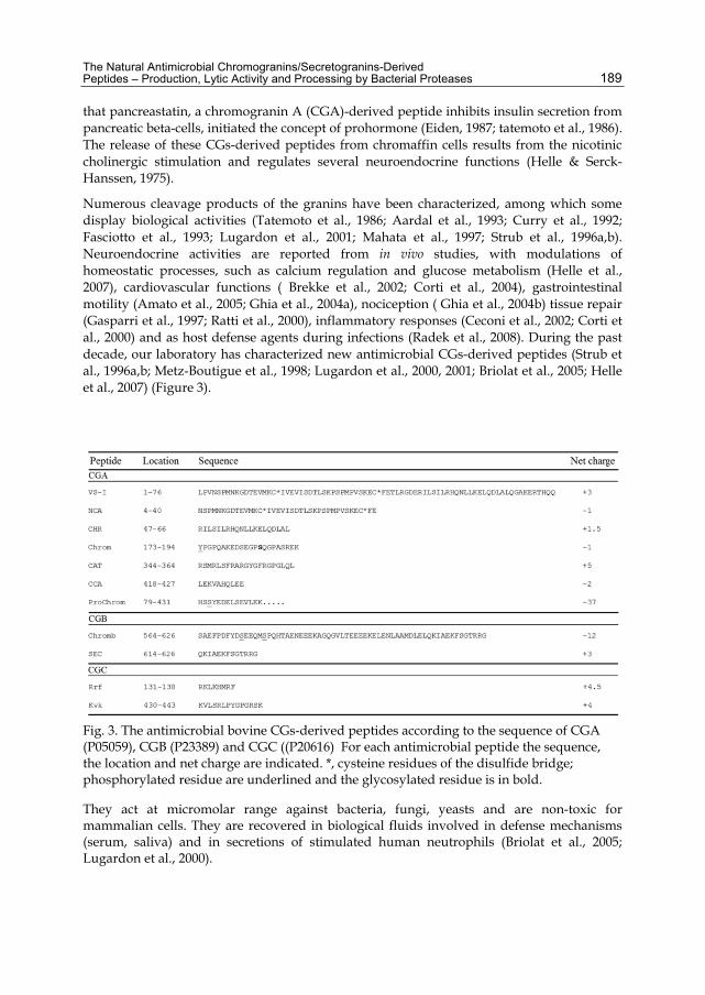
The Natural Antimicrobial Chromogranins/Secretogranins-Derived Peptides – Production, Lytic Activity and Processing by Bacterial Proteases 189
that pancreastatin, a chromogranin A (CGA)-derived peptide inhibits insulin secretion from pancreatic beta-cells, initiated the concept of prohormone (Eiden, 1987; tatemoto et al., 1986). The release of these CGs-derived peptides from chromaffin cells results from the nicotinic cholinergic stimulation and regulates several neuroendocrine functions (Helle & Serck-Hanssen, 1975).
Numerous cleavage products of the granins have been characterized, among which some display biological activities (Tatemoto et al., 1986; Aardal et al., 1993; Curry et al., 1992; Fasciotto et al., 1993; Lugardon et al., 2001; Mahata et al., 1997; Strub et al., 1996a,b). Neuroendocrine activities are reported from in vivo studies, with modulations of homeostatic processes, such as calcium regulation and glucose metabolism (Helle et al., 2007), cardiovascular functions ( Brekke et al., 2002; Corti et al., 2004), gastrointestinal motility (Amato et al., 2005; Ghia et al., 2004a), nociception ( Ghia et al., 2004b) tissue repair (Gasparri et al., 1997; Ratti et al., 2000), inflammatory responses (Ceconi et al., 2002; Corti et al., 2000) and as host defense agents during infections (Radek et al., 2008). During the past decade, our laboratory has characterized new antimicrobial CGs-derived peptides (Strub et al., 1996a,b; Metz-Boutigue et al., 1998; Lugardon et al., 2000, 2001; Briolat et al., 2005; Helle et al., 2007) (Figure 3).
Fig. 3. The antimicrobial bovine CGs-derived peptides according to the sequence of CGA (P05059), CGB (P23389) and CGC ((P20616) For each antimicrobial peptide the sequence, the location and net charge are indicated. *, cysteine residues of the disulfide bridge; phosphorylated residue are underlined and the glycosylated residue is in bold.
They act at micromolar range against bacteria, fungi, yeasts and are non-toxic for mammalian cells. They are recovered in biological fluids involved in defense mechanisms (serum, saliva) and in secretions of stimulated human neutrophils (Briolat et al., 2005; Lugardon et al., 2000).
Antimicrobial Agents 190
These new AMPs are integrated in the concept that highlights the key role of the adrenal medulla in the immunity (Sternberg, 2006) as previously reported for adrenaline and neuropeptide Y that regulate immunity systemically once released from the adrenal medulla. Furthermore, the adrenal medulla contains and releases large amounts of IL-6 and TNF-alpha in response to pro-inflammatory stimuli such as LPS, IL-1 alpha and IL-1 beta (Metz-Boutigue et al., 1998). The discovery of the presence of TLRs on the adrenal cortex cells raises the interesting possibility that the adrenal gland might have a direct role in the response to pathogens, activation of innate immune response and clearing of infectious agents (Sternberg, 2006).
2.2 Antimicrobial peptides derived from chromogranin A
Several new antimicrobial peptides isolated from the granules of chromaffin cells of the bovine adrenal medulla correspond to CGA-derived peptides (Figure 3). The corresponding sequences are highly conserved in human. Interestingly, the main cleavage site in position 78-79 of bCGA and the subsequent remove of the two basic residues K77 and K78 by the carboxipeptidase H (Metz-Boutigue et al., 1993) produces two antimicrobial fragments: vasostatin-I (VS-I; bCGA1-76) (Lugardon et al., 2000) and prochromacin (Prochrom; bCGA79-431) (Strub et al.,1996b). For these N- and C-terminal domains with antimicrobial activities several shorter active fragments were identified: for VS-I, bCGA1-40 (N CgA; NCA) (Shooshtarizadeh et al., 2010), bCGA47-66 (chromofungin; CHR) and for ProChrom, bCGA173-194 (Chromacin; Chrom) (Strub et al., 1996b) and bCGA344-364 (Catestatin; CAT) (Shooshtarizadeh et al., 2010). The unique disulfide bridge of bCGA is present in VS-I and NCA sequences. Two post-translational modifications are important for the expression of the antibacterial activity of Chrom: the phosphorylation of Y173 and the O-glycosylation of S186 [130] (Strub et al., 1996a). Furthermore, it is important to point out that a dimerization motif GXXXG similar to that reported for Glycophorin A (Brosig & Langosch, 1998) is present in the Chrom sequence (G184-G188).
Vasostatin-I
Vasostatin-I (VS-I) displays antimicrobial activity against (i) Gram-positive bacteria (Micrococcus luteus and Bacillus megaterium) with a minimal inhibitory concentration (MIC) in the range 0.1-1 µM; (ii) against filamentous fungi (Neurospora crassa, Aspergillus fumigatus, Alternaria brassicola, Nectria haematococca, Fusarium culmorum, Fusarium oxysporum) with a MIC of 0.5-3 µM and (iii) against yeast cells (Saccharomyces cerevisiae, Candida albicans) with a MIC of 2 µM (Lugardon et al., 2000). However VS-I is unable to inhibit the growth of Escherichia coli SBS363 and Escherichia coli D22. VS-I (Figure 3) possesses structural features specific for antimicrobial peptides, such as a global positive charge (+3), an equilibrated number of polar and hydrophobic residues (20:23) and the presence of a helical region CGA40-65 characterized to be a calmodulin-binding sequence (Lugardon et al., 2001; Yoo, 1992). The loss of the antibacterial activity of CGA7-57 suggests that the N- and C-terminal sequences are essential, nevertheless CGA7-57 is less efficient than VS-I against fungi. Besides, the disulfide bridge is essential for the antibacterial, but not the antifungal property. Altogether, these data suggest that antibacterial and antifungal activities of VS-I have different structural requirements (Lugardon et al., 2001). Interestingly, two helix-helix dimerization motifs important for the interaction with membranes such as LXXXXXXL,
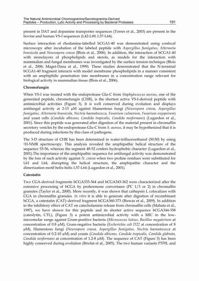
The Natural Antimicrobial Chromogranins/Secretogranins-Derived Peptides – Production, Lytic Activity and Processing by Bacterial Proteases 191
present in DAT and dopamine transporter sequences (Torres et al., 2003) are present in the bovine and human VS-I sequences (L42-L49; L57-L64).
Surface interaction of rhodamine-labelled bCGA1-40 was demonstrated using confocal microscopy after incubation of the labeled peptide with Aspergillus fumigatus, Alternaria brassicola and Neurospora crassa (Blois et al., 2006). In addition, the interaction of bCGA1-40 with monolayers of phospholipids and sterols, as models for the interaction with mammalian and fungal membranes was investigated by the surface tension technique (Blois et al., 2006; Maget-Dana et al., 1999). These studies demonstrated that the N-terminal bCGA1-40 fragment interacts with model membrane phospholipids in a manner consistent with an amphiphilic penetration into membranes in a concentration range relevant for biological activity in mammalian tissue (Blois et al., 2006).
Chromofungin
When VS-I was treated with the endoprotease Glu-C from Staphylococcus aureus, one of the generated peptide, chromofungin (CHR), is the shortest active VS-I-derived peptide with antimicrobial activities (Figure 3). It is well conserved during evolution and displays antifungal activity at 2-15 µM against filamentous fungi (Neurospora crassa, Aspergillus fumigatus, Alternaria brassicola, Nectria haematococca, Fusarium culmorum, Fusarium oxysporum) and yeast cells (Candida albicans, Candida tropicalis, Candida neoformans) (Lugardon et al., 2001). Since this peptide was generated after digestion of the material present in chromaffin secretory vesicles by the endoprotease Glu-C from S. aureus, it may be hypothesized that it is produced during infections by this class of pathogens.
The 3-D structure of CHR has been determined in water-trifluoroethanol (50:50) by using 1H-NMR spectroscopy. This analysis revealed the amphipathic helical structure of the sequence 53-56, whereas the segment 48-52 confers hydrophobic character (Lugardon et al., 2001).The importance of the amphipathic sequence for antifungal activity was demonstrated by the loss of such activity against N. crassa when two proline residues were substituted for L61 and L64, disrupting the helical structure, the amphipathic character and the dimerization motif helix-helix L57-L64 (Lugardon et al., 2001).
Catestatin
Two CGA-derived fragments bCGA333-364 and bCGA343-362 were characterized after the extensive processing of bCGA by prohormone convertases (PC 1/3 or 2) in chromaffin granules (Taylor et al., 2000). More recently, it was shown that cathepsin L colocalizes with CGA in chromaffin granules. In vitro it is able to generate after digestion of recombinant hCGA, a catestatin (CAT)–derived fragment hCGA360-373 (Biswas et al., 2009). In addition to the inhibitory effect of CAT on catecholamine release from chromaffin cells (Mahata et al., 1997), we have shown for this peptide and its shorter active sequence bCGA344-358 (cateslytin, CTL), (Figure 3) a potent antimicrobial activity with a MIC in the low-micromolar range against Gram-positive bacteria (Micrococcus luteus, Bacillus megaterium at concentration of 0.8 µM), Gram-negative bacteria (Escherichia coli D22 at concentration of 8 µM), filamentous fungi (Neurospora crassa, Aspergillus fumigatus, Nectria haematococca at concentration of 0.2-10 µM) and yeasts (Candida albicans, Candida tropicalis, Candida glabrata, Candida neoformans at concentration of 1.2-8 µM). The sequence of CAT (Figure 3) has been highly conserved during evolution (Briolat et al., 2005). The two human variants P370L and
Antimicrobial Agents 192
G364S display antibacterial activity against M. luteus with a MIC of 2 and 1 µM, respectively, and against E. coli with a MIC of 20 and 10 µM, respectively (Briolat et al., 2005). However, the most active peptide corresponds to the bovine sequence. Bovine CTL, a cationic sequence with a global net charge of +5 (R344, R347, R351, R353, R358) and five hydrophobic residues (M346, L348, F360, Y355, F357) (Figure 3), is able to completely kill bacteria at concentration lower than 10 µM even in the presence of NaCl (0-150 mM) (Briolat et al., 2005). The C-terminal sequence bCGA352-358 is inactive, whereas the N-terminal sequences bCGA344-351 and bCGA 348-358 are antibacterial at 20 µM.
C-terminal CGA-derived fragment
CCA, the C-terminal CGA-derived fragment bCGA418-427 (Figure 3), with a remarkable net charge of -2, displays antifungal activity and belongs to the less abundant anionic AMPs family. It is well conserved during evolution and is homologous to the C-terminal sequence of CGB and the antibacterial peptide SEC (secretolytin) [(Strub et al., 1996b). This peptide was generated in vitro after digestion, by the protease Glu-C from S. aureus, of the material present in chromaffin secretory vesicles. As previously postulated for CHR, CCA could be generated during infections induced by this pathogen.
2.3 Antimicrobial peptides derived from bovine chromogranin B
To date, the natural C-terminal fragment of bovine CGB (CCB; bCGB 564-626), isolated from chromaffin granules of the adrenal medulla, was found to display antibacterial activity against both M. luteus and E. coli. The complete inhibition of bacterial growth was observed at a concentration around 1.8 µM (Strub et al., 1996b). This large fragment contains the natural short antibacterial peptide secretolytin (SEC, bCGB614-626) with a net positive charge (+3) (Figure 3). We observed the natural formation of a pyrrolidone glutamic acid at the N-terminal end of SEC and both forms displayed antibacterial activity against M. luteus, reaching 100% of growth inhibition at 2 µM (Strub et al., 1996ab). A structure-activity analysis suggests that an alpha-helical amphipathic structure common to SEC and cecropins may account for the antibacterial activity (Strub et al., 1996b).
2.4 Antimicrobial peptides derived from bovine secretogranin II
Because bSGII is weakly expressed in the intragranular matrix of chromaffin secretory vesicles (2% of total proteins), the detection of endogenous AMPs by classical methods was unsuccessful. After in-silico analysis, two synthetic peptides with cationic amphipathic sequences were prepared: Rrf and Kvk, which correspond to the sequences bSGII131-138 and bSGII430-443 with respective net charges of +4.5 and 4 (Figure 3). Rrf, completely inhibits the bacterial growth of M. luteus and B. megaterium with a MIC of 5 and 15 µM, respectively, and Kvk displays antifungal properties at 19 µM against N. crassa (Shooshtarizadeh et al., 2010).
3. Interaction of antimicrobial chromogranins-derived peptides with bacterial proteases
The AMPs avoidance mechanisms deployed by bacteria include the proteolytic degradation of the active forms by the bacterial proteases. In order to examine the effects
The Natural Antimicrobial Chromogranins/Secretogranins-Derived Peptides – Production, Lytic Activity and Processing by Bacterial Proteases 193
of bacterial proteases on the isolated AMPs derived from CGs, we have tested the effects of Staphylococcus aureus V8 protease Glu-C and several supernatants from S aureus, Salmonella enteretica, Klebsiella oxytoca, Shigella sonnei and Vibrio cholera. By using biochemical methods we have analyzed the degradation of the peptide in presence of bacteria.
Interaction of antimicrobial CGs-derived peptides with proteases from diarrheogenic bacteria Bacteria were isolated from patients of the Strasbourg Civil Hospital by the Bacteriology Institute, University of Strasbourg, (EA-4438). The four strains have a clinical interest because apart from inducing diarrhea, they may cause other infections.
Thus, Klebsiella was involved in the occurrence of post-antibiotic diarrheas (Gorkiewicz 2009). Many studies show that Klebsiella oxytoca is also involved in nosocomial infections for newborns or adults (Biran et al., 2010) Klebsiella infections may also be commensal (Tsakris et al., 2011). Klebsiella oxytoca has also been associated with hemorrhagic colitis (Hoffmann et al., 2010) and intercurrent colitis in Crohn's disease (Plessier et al. 2002). Salmonella destroys infected cells and the infection continues through blood (sepsis) or through lymphatic vessel (typhoid fever). Salmonella cause also gastrointestinal infections. Shigella sonnei and Vibrio cholera non O1 cause inflammation of the intestinal mucosa by producing the Shiga toxin.
Klebsiella oxytoca, Salmonella enterica, Shigella sonnei, and Vibrio cholera develop phenomena of antibiotic resistance. Thus, Salmonella was reported to be resistant for the action of Ciprofloxacine (Medalla et al., 2011) and Ceftriaxone (Su et al., 2011).
Concerning CgA, we have tested bovine, rat and human CAT corresponding to the sequences bCgA344-364, rCgA6344-364 and hCgA352-372, bovine CTL located at bCgA344-358, two short fragments hCgA360-372 and the conserved tetrapeptide LSFR (bCgA348-351). In addition, we have tested a scrambled peptide relative to the sequence of bovine CAT and the procatestatin fragment bCgA332-364.
We have found antimicrobial activities only for the bovine CAT and CTL, showing that CTL is the shorter active fragment and that it corresponds to the active domain of CAT. Procatestatin was inactive in similar experimental conditions. Bovine CAT and CTL were active against Klebsiella oxytoca, Salmonella enterica and Vibrio cholera at 100 µM and 50 µM respectively and against Shigella Sonnei at 50 µM and 25 µM. In addition, CHR and the C-terminal fragment (CgA387-431) were inactive for concentration up to 100 µM. In contrast, CTL is active at 30 µM against the four pathogens.
Three CgB-derived peptides (CgB58-62, CgB279-291, and CgB547-560) and secretoneurin corresponding to SgII189-254 were examined against the four strains in order to analyse their degradation by bacterial proteases. By using HPLC we have compared the profiles of the peptide alone and the peptide with the inoculated medium.
These experiments show that except CTL all the peptides are completely degraded by the bacteria. To illustrate these data, we present on Figure 4, the profiles relative to CAT and CTL in presence of buffer with Salmonella enterica. The complete peptide and the processed form are analysed by sequencing and mass spectrometry (MALDI-TOF).
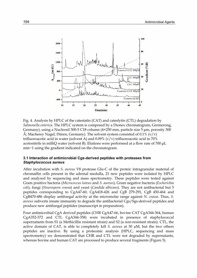
Antimicrobial Agents 194
Fig. 4. Analysis by HPLC of the catestatin (CAT) and cateslytin (CTL) degradation by Salmonella enterica. The HPLC system is composed by a Dionex chromatogram, Germerong, Germany), using a Nucleosil 300-5 C18 column (4×250 mm, particle size 5 μm, porosity 300 Å; Macherey Nagel, Düren, Germany). The solvent system consisted of 0.1% (v/v) trifluoroacetic acid in water (solvent A) and 0.09% (v/v) trifluoroacetic acid in 70% acetonitrile in milliQ water (solvent B). Elutions were performed at a flow rate of 700 μL min−1 using the gradient indicated on the chromatogram.
3.1 Interaction of antimicrobial Cgs-derived peptides with proteases from Staphylococcus aureus
After incubation with S. aureus V8 protease Glu-C of the proteic intragranular material of chromaffin cells present in the adrenal medulla, 21 new peptides were isolated by HPLC and analysed by sequencing and mass spectrometry. These peptides were tested against Gram positive bacteria (Micrococcus luteus and S. aureus), Gram negative bacteria (Escherichia coli), fungi (Neurospora crassa) and yeast (Candida albicans). They are not antibacterial but 5 peptides corresponding to CgA47-60, CgA418-426 and CgB 279-291, CgB 450-464 and CgB470-486 display antifungal activity at the micromolar range against N. crassa. Thus, S. aureus subverts innate immunity to degrade the antibacterial Cgs/Sgs-derived peptides and produce new antifungal peptides (manuscript in preparation).
Four antimicrobial CgA derived peptides (CHR CgA47-66, bovine CAT CgA344-364, human CgA352-372 and CTL CgA344-358) were incubated in presence of staphylococcal supernatants from S1 (a Methicillin resistant strain) and S2 (a non-resistant strain). CTL, the active domain of CAT, is able to completely kill S. aureus at 30 µM, but the two others peptides are inactive. By using a proteomic analysis (HPLC, sequencing and mass spectrometry) we demonstrated that CHR and CTL were not degraded by supernatants, whereas bovine and human CAT are processed to produce several fragments (Figure 5).

The Natural Antimicrobial Chromogranins/Secretogranins-Derived Peptides – Production, Lytic Activity and Processing by Bacterial Proteases 195
Fig. 5. Analysis by HPLC of the catestatin (CAT) degradation by a methicillin resistant Staphylococcus aureus (MRSA). The HPLC system is composed by a Dionex chromatograph, (Germerong, Germany), using a Nucleosil 300-5 C18 column (4×250 mm, particle size 5 μm, porosity 300 Å; Macherey Nagel, Düren, Germany). The solvent system consisted of 0.1% (v/v) trifluoroacetic acid in water (solvent A) and 0.09% (v/v) trifluoroacetic acid in 70% acetonitrile in milliQ water (solvent B). Elutions were performed at a flow rate of 700 μL min−1 using the gradient indicated on the chromatogram.
4. Synergy of the combination of antimicrobial peptides with antibiotics
The emergence of multi-drug resistant bacteria (MDR), with therapeutic failure against Staphylococcus aureus (MRSA), Klebsiella pneumonia, Acinebacter baumannii and Pseudomonas aeruginosa have paved the way to develop new therapeutic agents by the help of the synergism. In addition the highly toxic effects of antibiotics have shifted the research focus to discover new peptides with broad spectrum of activity and less toxicity. Synergy is the combined activity of two antimicrobial agents that can never be attained by any one of them singly (Serra et al., 1977). Numerous AMPs demonstrate broad spectrum of activity against pathogens, interacting directly with membranes or acting with a specific mode. They represent interesting candidates to synergistically act with antibiotics.
4.1 For Staphylococcus aureus MRSA
Most of the patients can prone to serious bacterial infections caused mainly by the multi-resistant microorganisms, Staphylococcus aureus coagulase negative spp are one of them. These coagulase negative strains (MRSA) have got approximately 90% of methicillin resistance due to β-lactam resistance (Silva et al., 2011). Story just not stopped here, but still it continues, some of the staphylococcal strains got resistance to the other drugs such as Vancomycin, which was previously widely used against the MRSA infections, and to treat
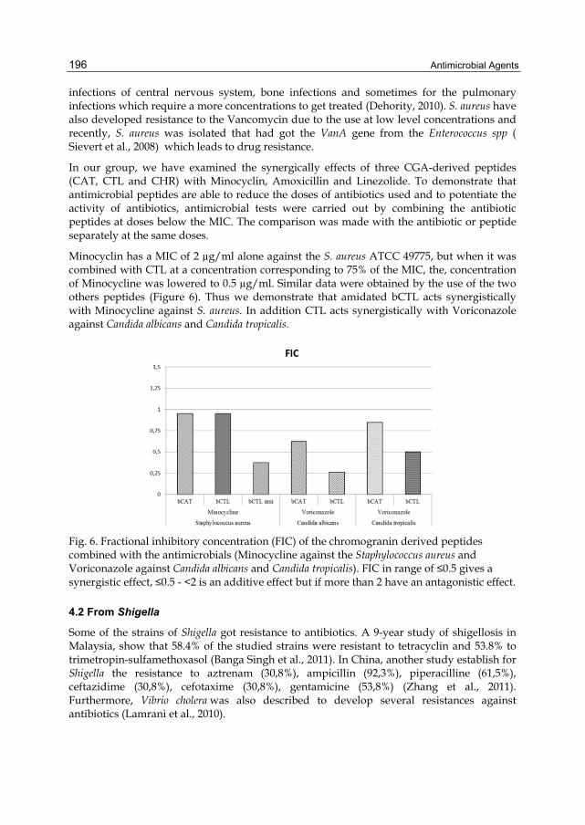
Antimicrobial Agents 196
infections of central nervous system, bone infections and sometimes for the pulmonary infections which require a more concentrations to get treated (Dehority, 2010). S. aureus have also developed resistance to the Vancomycin due to the use at low level concentrations and recently, S. aureus was isolated that had got the VanA gene from the Enterococcus spp ( Sievert et al., 2008) which leads to drug resistance.
In our group, we have examined the synergically effects of three CGA-derived peptides (CAT, CTL and CHR) with Minocyclin, Amoxicillin and Linezolide. To demonstrate that antimicrobial peptides are able to reduce the doses of antibiotics used and to potentiate the activity of antibiotics, antimicrobial tests were carried out by combining the antibiotic peptides at doses below the MIC. The comparison was made with the antibiotic or peptide separately at the same doses.
Minocyclin has a MIC of 2 µg/ml alone against the S. aureus ATCC 49775, but when it was combined with CTL at a concentration corresponding to 75% of the MIC, the, concentration of Minocycline was lowered to 0.5 µg/ml. Similar data were obtained by the use of the two others peptides (Figure 6). Thus we demonstrate that amidated bCTL acts synergistically with Minocycline against S. aureus. In addition CTL acts synergistically with Voriconazole against Candida albicans and Candida tropicalis.
Fig. 6. Fractional inhibitory concentration (FIC) of the chromogranin derived peptides combined with the antimicrobials (Minocycline against the Staphylococcus aureus and Voriconazole against Candida albicans and Candida tropicalis). FIC in range of ≤0.5 gives a synergistic effect, ≤0.5 - <2 is an additive effect but if more than 2 have an antagonistic effect.
4.2 From Shigella
Some of the strains of Shigella got resistance to antibiotics. A 9-year study of shigellosis in Malaysia, show that 58.4% of the studied strains were resistant to tetracyclin and 53.8% to trimetropin-sulfamethoxasol (Banga Singh et al., 2011). In China, another study establish for Shigella the resistance to aztrenam (30,8%), ampicillin (92,3%), piperacilline (61,5%), ceftazidime (30,8%), cefotaxime (30,8%), gentamicine (53,8%) (Zhang et al., 2011). Furthermore, Vibrio cholera was also described to develop several resistances against antibiotics (Lamrani et al., 2010).
The Natural Antimicrobial Chromogranins/Secretogranins-Derived Peptides – Production, Lytic Activity and Processing by Bacterial Proteases 197
In conclusion these studies show that CGA-derived AMPs potentiate the effects of antibiotic drugs. One could imagine a mechanism in which the peptide would favor the destabilization of the membrane allowing the antibiotic to rapidly penetrate inside the bacterial cells and thus to reach its site of action.
5. Chromogranin A, a new marker of severity
In clinical practice, CGA has been used as a marker of pheochromocytomas (O’Connor et al., 1984), carcinoid tumors (O’Connor & Deftos, 1986; Syversen et al., 1993), neuroblastomas (Hsiao et al., 1990), neuroendocrine tumors (Berruti et al., 2005), and neurodegenerative diseases (Rangon et al., 2003). Recent data have shown CGA to be a useful prognostic indicator in patients with chronic heart failure (Omland et al., 2003, suggesting that CGA may have some association with cardiovascular diseases. Furthermore, a pilot study has shown CGA to be a predictor of mortality in patients with acute myocardial infarction (Estensen et al., 2006). Characterization of the severity of organ failures and prediction of patient outcome are of major importance for physicians who care for critically ill patients. Multiple organ failure (MOF) remains the main problem in intensive care because of its impact on morbidity, mortality, and resources (Baue et al., 1998). MOF can develop as a consequence of multiple causes, such as infection, systemic inflammatory response syndrome (SIRS), myocardial infarction, septic shock, leading to the activation of various endogenous cascades, cellular dysfunction and death (Baue et al., 1998).
In a recent study we have evaluated whether unselected critically ill patients at ICU (Hautepierre Hospital, Strasbourg, France) admission demonstrate increased plasma CGA concentrations and whether CGA can be of any interest in the care of patients at high risk of death. Patients older than 18 years were recruited consecutively over 3 months during 2007. Exclusion criteria included: duration of stay >24 h and conditions known to increase CGA concentrations independently of acute stress [i.e., a history of documented neuroendocrine tumors (O’Connor & Deftos, 1986) or chronic treatment with proton pump inhibitors before admission (Giusti et al., 2004). Patients who required surgical interventions were also excluded. Of the 120 participants included in the study, 70 patients had a primary diagnosis severe infection, and 50 had a SIRS. Serum CGA concentrations were measured with a commercial sandwich RIA kit (a gift of Cisbio Bioassays, Marcoule, France. In the central 95% of the healthy population, serum CGA concentrations range from 19µg/L to 98µg/L. In neuroendocrine system tumors, the CGA serum concentration varies from the typical range up to 1200 µg/L, depending on the biological and structural characteristics of the tumor, as well as on the extent of tumor spread (Degorce et al., 1999). As a control Procalcitonin (PCT) concentrations were measured on the Kryptor system (Brahms Diagnostic) with the time-resolved amplified cryptate emission methodology in accordance with the manufacturer’s recommendations. The Simplified Acute Physiological Score II (SAPS II) and the Logistic Organ Dysfunction System (LODS) score were calculated at admission according to published standards (Levy et al., 2003; Le Gall et al., 1993). Our data show that CGA concentration was positively but weakly correlated with age, PCT concentration, creatinine concentration, SAPS II, and LODS score (P <0.001 for all variables) and was correlated with CRP concentration (Zhang et al., 2008). Thirty-three deaths occurred during the median follow- up time of 23 days. The death rates for CGA and PCT are shown by quartiles in Figure 7. Statistical analysis revealed a significant difference in death rates between CGA quartile 4 and CGA quartiles 1, 2, and 3 (P < 0.001, log-rank test). The death rate for CGA quartile 3 was also significantly different from that of CGA quartile 1 (P= 0.033).
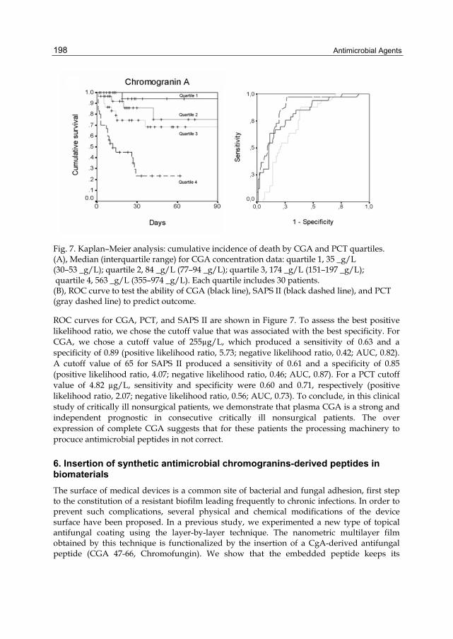
Antimicrobial Agents 198
Fig. 7. Kaplan–Meier analysis: cumulative incidence of death by CGA and PCT quartiles. (A), Median (interquartile range) for CGA concentration data: quartile 1, 35 _g/L (30–53 _g/L); quartile 2, 84 _g/L (77–94 _g/L); quartile 3, 174 _g/L (151–197 _g/L); quartile 4, 563 _g/L (355–974 _g/L). Each quartile includes 30 patients. (B), ROC curve to test the ability of CGA (black line), SAPS II (black dashed line), and PCT (gray dashed line) to predict outcome.
ROC curves for CGA, PCT, and SAPS II are shown in Figure 7. To assess the best positive likelihood ratio, we chose the cutoff value that was associated with the best specificity. For CGA, we chose a cutoff value of 255µg/L, which produced a sensitivity of 0.63 and a specificity of 0.89 (positive likelihood ratio, 5.73; negative likelihood ratio, 0.42; AUC, 0.82). A cutoff value of 65 for SAPS II produced a sensitivity of 0.61 and a specificity of 0.85 (positive likelihood ratio, 4.07; negative likelihood ratio, 0.46; AUC, 0.87). For a PCT cutoff value of 4.82 µg/L, sensitivity and specificity were 0.60 and 0.71, respectively (positive likelihood ratio, 2.07; negative likelihood ratio, 0.56; AUC, 0.73). To conclude, in this clinical study of critically ill nonsurgical patients, we demonstrate that plasma CGA is a strong and independent prognostic in consecutive critically ill nonsurgical patients. The over expression of complete CGA suggests that for these patients the processing machinery to procuce antimicrobial peptides in not correct.
6. Insertion of synthetic antimicrobial chromogranins-derived peptides in biomaterials
The surface of medical devices is a common site of bacterial and fungal adhesion, first step to the constitution of a resistant biofilm leading frequently to chronic infections. In order to prevent such complications, several physical and chemical modifications of the device surface have been proposed. In a previous study, we experimented a new type of topical antifungal coating using the layer-by-layer technique. The nanometric multilayer film obtained by this technique is functionalized by the insertion of a CgA-derived antifungal peptide (CGA 47-66, Chromofungin). We show that the embedded peptide keeps its
The Natural Antimicrobial Chromogranins/Secretogranins-Derived Peptides – Production, Lytic Activity and Processing by Bacterial Proteases 199
antifungal activity by interacting with the fungal membrane and penetrating into the cell. In vitro studies demonstrated that such an antifungal coating is able to inhibit the growth of yeast Candida albicans by 65% and completely stop the proliferation of filamentous fungus N.crassa. The cytotoxicity of such a coating was also assessed by growing human gingival fibroblasts at its surface. Finally, the antifungal coating of poly (methylmethacrylate), a widely used material for biomedical devices, is successfully tested in an in vivo oral candidiasis rat model (Etienne et al., 2005).
7. Conclusions
CGs family emerges as prohormones able to modulate homeostatic processes in response to excessive stimulations such as microbial infections. The studies concerning the expression of CGs and their antimicrobial peptides in patients with inflammatory diseases and the correlation with the proteolytic processes occurring in these pathologies vs. controls are crucial to understand the involvement of these prohormones and their derived peptides in innate and adaptive immunities. Calcium is a universal secondary messenger involved in many cellular signal transduction pathways, regulating crucial functions such as secretion, cell motility, proliferation and cell death. The calcium-dependent immunomodulatory properties of CHR and CAT are important for the understanding of their involvement in inflammatory mechanisms. In sum, these linear peptides may represent prototypic lead molecules useful for the development of new therapeutic agents and also biomaterials.
8. Acknowledgement
This study was funded by INSERM, the University of Strasbourg (UDS) and INRA (JFC). We are grateful to financial assistance from the Odontology Faculty of the University of Strasbourg (MA, RA). We thank Charlotte Bach for the excellent technical assistance, Bernard Guérold for the preparation of the synthetic peptides, Thomas Lavaux for fruitful discussions, Cisbio Bioassays Research and Development for providing the ELISA kit required for CGA dosages, EA44-38 for the isolation of bacteria and the preparation of bacterial supernatants and Martine Rivet for the preparation of the manuscript.
9. References
Aardal, S., Helle, K.B., Elsayed, S., Reed, R.K. & Serck-Hanssen, G. (1993). Vasostatins, comprising the N-terminal domain of chromogranin A, suppress tension in isolated human blood vessel segments. Journal of Neuroendocrinology, Vol. 5, No. 4, (August 1993), pp. 405-412, ISSN: 0953-8194.
Abtin, A., Eckhart, L., Mildner, M., Ghannadan, M., Harder, J., Schroder, J.M. & Tschachler, E. (2009). Degradation by stratum corneum proteases prevents endogenous RNase inhibitor from blocking antimicrobial activities of RNase 5 and RNase 7. Journal of Investigative Dermatology, Vol. 129, No. 9, (March 2009), pp. 2193-2201, ISSN: 0022-202X.
Aerts A.M., François, I.E., Cammue, B.P. & Thevissen, K. (2008). The mode of antifungal action of plant, insect and human defensins. Cellular and Molecular Life Science, Vol. 65, No. 12, (July 2008), pp. 2069-2079, ISSN: 1420-682X.
Antimicrobial Agents 200
Ali, R.S., Falconer, A., Ikram, M., Bissett, C.E., Cerio, R. & Quinn, A.G. (2001). Expression of the peptide antibiotics human beta defensin-1 and human beta defensin-2 in normal human skin. Journal of Investigative Dermatology, Vol. 117, No. 1, (July 2001), pp.106-111, ISSN: 0022-202X.
Amato, A., Corti, A., Serio, R. & Mule, F. (2005). Inhibitory influence of chromogranin A N-terminal fragment (vasostatin-1) on the spontaneous contractions of rat proximal colon. Regulatory Peptides, Vol. 130, No.1-2, (August 2005), pp. 42-47, ISSN: 0167-0115.
Anouar, Y., Yon, L., Desmoucelles, C., Leprince, J., Breault, L., Gallo-Payet, N. & Vaudry, H. (1998). Identification of a novel secretogranin II-derived peptide in the adult and fetal human adrenal gland. Endocrinology Research Vol. 24, No. 3-4, (August-November 1998), pp. 731-736, ISSN: 0743-5800.
Artym, J., Zimecki, M., Kuryszko, J. & Kruzel, M.L. (2005). Lactoferrin accelerates reconstitution of the humoral and cellular immune response during chemotherapy-induced immunosuppression and bone marrow transplant in mice. Stem Cells Development, Vol. 14, No.5, (October 2005), pp. 548-555, ISSN: 1547-3287.
Baechle, D., Flad, T., Cansier, A., Steffen, H., Schittek, B., Tolson, J., Herrmann, T., Dihazi, H., Beck, A., Mueller, G.A., Mueller, M., Stevanovic, S., Garbe, C., Mueller, C.A. & Kalbacher H. (2006). Cathepsin D is present in human eccrine sweat and involved in the postsecretory processing of the antimicrobial peptide DCD-1L. Journal of Biological Chemistry, Vol. 281, No.9, (March 2006), pp. 5406-5415, ISSN: 0021-9258.
Bals, R., Wang, X., Zasloff, M. & Wilson, J.M. (1998). The peptide antibiotic LL-37/hCAP-18 is expressed in epithelia of the human lung where it has broad antimicrobial activity at the airway surface. Proceedings of National Academy of Sciences of the United States of America, Vol. 95, No. 16, (August 1998), pp. 9541-9546, ISSN: 0027-8424.
Banga Singh, K.K., Ojha, S.C., Deris, Z.Z., & Rahman, R.A. (2011). A 9-year study of shigellosis in Northeast Malaysia: Antimicrobial susceptibility and shifting species dominance, Journal of Public Health, Vol. 19, No. 3, (June 2011), pp. 231-236, ISSN: 1613-2238.
Baue, A.E., Durham, R. & Faist, E. (1998). Systemic inflammatory response syndrome (SIRS), multiple organ dysfunction syndrome (MODS), multiple organ failure (MOF): are we winning the battle? Shock, Vol. 10, No. 2, (August 1998), pp. 79-89, ISSN: 1073-2322.
Bellamy, W., Wakabayashi, H., Takase, M., Kawase, K., Shimamura, S. & Tomita, M. (1993). Killing of Candida albicans by lactoferricin B, a potent antimicrobial peptide derived from the N-terminal region of bovine lactoferrin. Medical Microbiology and Immunology, Vol. 182, No .2, (May 1993), pp. 97-105, ISSN: 0300-8584.
Berruti, A., Mosca, A., Tucci, M., Terrone, C., Torta, M., Tarabuzzi, R., Russo, L., Cracco, C., Bollito, E., Scarpa, R.M., Angeli, A. & Dogliotti, L. (2005). Independent prognostic role of circulating chromogranin A in prostate cancer patients with hormone-refractory disease. Endocrine Related Cancer, Vol. 12, No. 1, (March 2005), pp.109-117, ISSN: 1351-0088.
Biran, V., Gaudin, A., Mariani-Kurdjian, P., Doit, C., Bingen, E. & Aujard, Y. (2010). Implication of extended-spectrum beta-lactamase enterobacteriaceae in nosocomial infections in neonates. Archives de Pédiatrie, Vol. 17, Suppl 4, (September 2010), pp. S150-153, ISSN: 0929-693X.

The Natural Antimicrobial Chromogranins/Secretogranins-Derived Peptides – Production, Lytic Activity and Processing by Bacterial Proteases 201
Biswas, N., Rodriguez-Flores, J.L., Courel, M., Gayen, J., Vaingankar, S.M., Mahata, M., Torpey, J.W., Taupenot, L., O'Connor, D.T. & Mahata, S.K. (2009). Cathepsin L colocalizes with chromogranin A in chromaffin vesicles to generate active peptides. Endocrinology, Vol. 150, No. 8, (August 2009), pp. 3547-3557, ISSN: 0013-7227.
Blois, A., Holmsen, H., Martino, G., Corti, A., Metz-Boutigue, M.H. & Helle, K.B. (2006). Interactions of chromogranin A-derived vasostatins and monolayers of phosphatidylserine, phosphatidylcholine and phosphatidylethanolamine. Regulatory Peptides, Vol. 134, No. 1, (March 2006), pp. 30-37, ISSN: 0167-0115.
Boni, R., Burg G., Doguoglu, A., Ilg, E.C., Schafer, B.W., Mulle, B. & Heizmann, C.W. (1997). Immunohistochemical localization of the Ca2+ binding S100 proteins in normal human skin and melanocytic lesions. The British Journal of Dermatology, Vol. 137, No. 1, (July 1997) pp. 39-43, ISSN: 0007-0963.
Braun, T., Voland, P., Kunz, L., Prinz, C. & Gratzl, M. (2007). Enterochromaffin cells of the human gut: sensors for spices and odorants. Gastroenterology, Vol. 132, No. 5), (May 2007), pp. 1890-1901, ISSN: 0016-5085.
Brekke, J.F., Osol, G.J. & Helle KB. (2002). N-terminal chromogranin-derived peptides as dilators of bovine coronary resistance arteries. Regulatory Peptides, Vol. 105, No. 2, (May 2002), pp. 93-100, ISSN: 0167-0115.
Briolat, J., Wu, S.D., Mahata, S.K., Gonthier, B., Bagnard, D., Chasserot-Golaz, S., Helle, K.B., Aunis D & Metz-Boutigue MH. (2005). New antimicrobial activity for the catecholamine release-inhibitory peptide from chromogranin A. Cellular and Molecular Life Science, Vol. 62, No. 3, (February 2005), pp. 377-385, ISSN: 1420-682X.
Broome, A.M., Ryan, D. & Eckert, R.L. (2003). S100 protein subcellular localization during epidermal differentiation and psoriasis. The Journal of Histochemistry and Cytochemistry, Vol. 51, No. 5, (May 2003), pp. 675-685, ISSN: 0022-1554.
Bulet, P., Stocklin, R. & Menin, L. (2004). Anti-microbial peptides: from invertebrates to vertebrates. Immunological reviews, Vol. 198, (April 2004), pp. 169-184, ISSN: 0105-2896.
Butmarc, J., Yufit, T., Carson, P. & Falanga, V. (2004). Human beta-defensin-2 expression is increased in chronic wounds. Wound repair and regeneration, Vol. 12, No. 4, (July-August 2004), pp. 439-443, ISSN: 1067-1927.
Ceconi, C., Ferrari, R., Bachetti, T., Opasich, C., Volterrani, M., Colombo, B., Parrinello, G; & Corti A. (2002). Chromogranin A in heart failure; a novel neurohumoral factor and a predictor for mortality. European heart journal, Vol. 23, No. 12, (June 2002), pp. 967-974, ISSN: 0195-668X.
Cetin, Y. & Grube, D. (1991). Immunoreactivities for chromogranin A and B, and secretogranin II in the guinea pig entero-endocrine system: cellular distributions and intercellular heterogeneities. Cell and tissue research, Vol. 264, No. 2, (May 1991), pp. 231-241, ISSN: 0302-766X.
Corti, A., Ferrari, R. & Ceconi, C. (2000). Chromogranin A and tumor necrosis factor-alpha (TNF) in chronic heart failure. Advances in experimental medicine and biology, Vol. 482, pp. 351-359, ISSN: 0065-2598.
Corti, A., Mannarino, C., Mazza, R., Angelone, T., Longhi, R. & Tota, B. (2004). Chromogranin A N-terminal fragments vasostatin-1 and the synthetic CGA 7-57 peptide act as cardiostatins on the isolated working frog heart. General and

Antimicrobial Agents 202
comparatative endocrinology, Vol. 136, No. 2, (April 2004), pp. 217-224, ISSN: 0016-6480.
Curry, W.J., Shaw, C., Johnston, C.F., Thim, L. & Buchanan, K.D. (1992). Isolation and primary structure of a novel chromogranin A-derived peptide, WE-14, from a human midgut carcinoid tumour. FEBS letters, Vol. 301, No. 3, (April 1992), pp. 319-321, ISSN: 0014-5793.
Cutuli, M., Cristiani, S., Lipton, J.M. & Catania, A. (2000). Antimicrobial effects of alpha-MSH peptides. Journal of leukocyte biology, Vol. 67, No. 2, (February 2000), pp. 233-239, ISSN: 0741-5400.
Dann, S.M. & Eckmann, L. (2007). Innate immune defenses in the intestinal tract. Current opinion in gastroenterology, Vol. 23, No. 2, (March 2007), pp. 115-120, ISSN: 0267-1379.
Davidson, D.J., Currie, A.J., Reid, G.S., Bowdish, D.M., MacDonald, K.L., Ma, R.C., Hancock, R.E. & Speert, D.P. (2004). The cationic antimicrobial peptide LL-37 modulates dendritic cell differentiation and dendritic cell-induced T cell polarization. The journal of immunology, Vol. 172, No. 2, (January 2004), pp. 1146-1156, ISSN: 0022-1767.
Degorce, F., Goumon, Y., Jacquemart, L., Vidaud, C., Bellanger, L., Pons-Anicet, D., Seguin, P., Metz-Boutigue, M.H. & Aunis, D. (1999). A new human chromogranin A (CgA) immunoradiometric assay involving monoclonal antibodies raised against the unprocessed central domain (145-245). British journal of cancer, Vol. 79, No.1, (january 1999), pp. 65-71, ISSN: 0007-0920.
Dehority, W. (2010). Use of vancomycin in pediatrics. The pediatric infectious disease, Vol. 29, No. 5, (may 2010), pp. 462-463, ISSN: 0891-3668.
Del Valle, M.E., Vazquez, E., Represa, J., Malinovsky, L. & Vega, J.A. (1994). Immunohistochemical localization of calcium-binding proteins in the human cutaneous sensory corpuscles. Neuroscience letters, Vol. 168, No. 1-2, (February 1994), pp. 247-250, ISSN: 0304-3940.
Dieckgraefe, B.K., Crimmins, D.L., Landt, V., Houchen, C., Anant, S., Porche-Sorbet, R. & Ladenson, J.H. (2002). Expression of the regenerating gene family in inflammatory bowel disease mucosa: Reg Ialpha upregulation, processing, and antiapoptotic activity. Journal of investigative medicine, Vol. 50, No. 6, (November 2002), pp. 421-434, ISSN: 1081-5589.
Dyer, K.D. & Rosenberg, H.F. (2006). The RNase a superfamily: generation of diversity and innate host defense. Molecular diversity Vol. 10, No. 4, (November 2006), pp. 585-597, ISSN: 1381-1991.
Eckert, R.L., Broome, A.M., Ruse, M., Robinson, N., Ryan, D. & Lee, K. (2004). S100 proteins in the epidermis. The journal of investigative dermatology, Vol. 123, No. 1, (July 2004), pp. 23-33, ISSN: 0022-202X.
Eiden, L.E. (1987). Is chromogranin a prohormone? Nature, Vol. 325, No. 6102, (January 1987), pp. 301, ISSN: 0028-0836.
Elias, P.M. (2007). The skin barrier as an innate immune element. Seminars in immunopathology Vol. 29, No. 1, (April 2007), pp. 3-14, ISSN: 1863-2297.
Estensen, M.E., Hognestad, A., Syversen, U., Squire, I., Nq, L., Kickshus, Dickstein, K., Omland, T. (2006). Prognostic value of plasma chromogranin A levels in patients
The Natural Antimicrobial Chromogranins/Secretogranins-Derived Peptides – Production, Lytic Activity and Processing by Bacterial Proteases 203
with complicated myocardial infarction. American heart journal, Vol. 152, No.5, (november 2006), pp. 927.e1-6, ISSN : 0002-8703.
Etienne, O., Gasnier, C., Taddei, C., Voegel, J.C., Aunis, D., Schaaf, P., Metz-Boutigue, M.H., Bolcato-Bellemin, A.L. & Egles, C. (2005). Antifungal coating by biofunctionalized polyelectrolyte multilayered films. Biomaterials, Vol. 26, No. 33, (November 2005), pp. 6704-6412, ISSN: 0142-9612.
Fahlgren, A., Hammarstrom, S., Danielsson, A. & Hammarstrom, M.L. (2004). beta-Defensin-3 and -4 in intestinal epithelial cells display increased mRNA expression in ulcerative colitis. Clinical and experimental immunology, Vol. 137, No. 2, (August 2004), pp. 379-385, ISSN: 0009-9104.
Fasciotto, B.H., Trauss, C.A., Greeley, G.H. & Cohn, D.V. (1993). Parastatin (porcine chromogranin A347-419), a novel chromogranin A-derived peptide, inhibits parathyroid cell secretion. Endocrinology, Vol. 133, No. 2, (August 1993), pp. 461-466, ISSN: 0013-7227.
Forrester, J.S., Diamond, G., Chatterjee, K. & Swan, H.J. (1976). Medical therapy of acute myocardial infarction by application of hemodynamic subsets (second of two parts). The New England journal of medicine, Vol. 295. No. 25, (December 1976), pp.1404-1413, ISSN: 0028-4793.
Ganz, T. (2003). Defensins: antimicrobial peptides of innate immunity. Nature Review Immunology, Vol. 3, No. 9, (September 2003), pp. 710-720, ISSN: 1474-1733.
Garcia, J.R., Jaumann, F., Schulz, S., Krause, A., Rodriguez-Jimenez, J., Forssmann, U., Adermann, K., Kluver, E., Vogelmeier, C., Becker, D., Hedrich, R., Forssmann, W.G, & Bals, R. (2001). Identification of a novel, multifunctional beta-defensin (human beta-defensin 3) with specific antimicrobial activity. Its interaction with plasma membranes of Xenopus oocytes and the induction of macrophage chemoattraction. Cell and tissue research, Vol. 306, No. 2, (November 2001), pp. 257-264, ISSN: 0302-766X.
Gasparri, A., Sidoli, A., Sanchez, L.P., Longhi, R., Siccardi, A.G., Marchisio, P.C. & Corti, A. (1997). Chromogranin A fragments modulate cell adhesion. Identification and characterization of a pro-adhesive domain. The journal of biological chemistry, Vol. 272, No. 33, (August 1997), pp. 20835-20843, ISSN: 0021-9258.
Ghia, J.E., Crenner, F., Metz-Boutigue, M.H., Aunis, D., Angel, F. (2004b). The effect of a chromogranin A-derived peptide (CgA4-16) in the writhing nociceptive response induced by acetic acid in rats. Life Science, Vol. 75, No.15, (August 2004), pp. 1787-1799, ISSN: 0024-3205.
Ghia, J.E., Crenner, F., Rohr, S., Meyer, C., Metz-Boutigue, M.H., Aunis, D. & Angel F. (2004a). A role for chromogranin A (4-16), a vasostatin-derived peptide, on human colonic motility. An in vitro study. Regulatory Peptides, Vol. 121, No. 1-3, (September 2004) pp. 31-39, ISSN: 0167-0115.
Giusti, M., Sidoti, M., Augeri, C., Rabitti, C. & Minuto, F. (2004). Effect of short-term treatment with low dosages of the proton-pump inhibitor omeprazole on serum chromogranin A levels in man. European journal of endocrinology, Vol. 150, No. 3, (March 2004), pp: 299-303, ISSN: 0804-4643.
Gorkiewicz, G. (2009). Nosocomial and antibiotic-associated diarrhoea caused by organisms other than Clostridium difficile. International journal of antimicrobial agents, Vol. 33, Suppl 1, (March 2009), pp. S37-41, ISSN: 0924-8579.
Antimicrobial Agents 204
Hansen, M.B. & Witte, A.B. (2008). The role of serotonin in intestinal luminal sensing and secretion. Acta physiologica Vol. 193, No. 4, (August 2008), pp. 311-323, ISSN: 1748-1708.
Harder, J., Bartels, J., Christophers, E. & Schroder, J.M. (1997). A peptide antibiotic from human skin. Nature, Vol. 387, No. 6636, (June 1997), pp. 861 ISSN: 0028-0836.
Harder, J., Bartels, J., Christophers, E. & Schroder, J.M. (2001). Isolation and characterization of human beta -defensin-3, a novel human inducible peptide antibiotic. The journal of biological chemistry, Vol. 276, No. 8, (February 2001), pp. 5707-5713, ISSN: 0021-9258.
Harder, J. & Schroder, J.M. (2002). RNase 7, a novel innate immune defense antimicrobial protein of healthy human skin. The journal of biological chemistry,Vol. 277, No. 48, (November 2002), pp. 46779-46784, ISSN: 0021-9258.
Harder, J., Meyer-Hoffert, U., Wehkamp, K., Schwichtenberg, L. & Schroder, J.M. (2004). Differential gene induction of human beta-defensins (hBD-1, -2, -3, and -4) in keratinocytes is inhibited by retinoic acid. The journal of investigative dermatology, Vol. 123, No. 3, (September 2004), pp. 522-529, ISSN: 0022-202X.
Hartschuh, W., Weihe, E. & Yanaihara, N. (1989a). Immunohistochemical analysis of chromogranin A and multiple peptides in the mammalian Merkel cell: further evidence for its paraneuronal function? Archives of histology and cytology, Vol. 52, Suppl., pp. 423-431, ISSN: 0914-9465.
Hartschuh,W., Weihe, E. & Egner, U. (1989b). Chromogranin A in the mammalian Merkel cell: cellular and subcellular distribution.The journal of investigative dermatology, Vol. 93, No. 5, (November 1989), pp. 641-648, ISSN: 0022-202X.
Hase, K., Eckmann, L., Leopard, J.D., Varki, N. & Kagnoff, M.F. (2002). Cell differentiation is a key determinant of cathelicidin LL-37/human cationic antimicrobial protein 18 expression by human colon epithelium. Infection and Immunity, Vol. 70, No. 2, (February 2002), pp. 953-963, ISSN: 0019-9567.
Heitz, P., Polak, J.M., Timson, D.M. & Pearse, A.G. (1976). Enterochromaffin cells as the endocrine source of gastrointestinal substance P. Histochemistry, Vol. 49, No. 4, (November 1976), pp. 343-347, ISSN: 0301-5564.
Helle, K.B. & Serck-Hanssen, G. (1975). The adrenal medulla: a model for studies of hormonal and neuronal storage and release mechanisms. Molecular and Cellular Biochemistry, Vol. 6, No. 2, (February 1975), pp. 127-146, ISSN: 0300-8177.
Helle, K.B. (2004). The granin family of uniquely acidic proteins of the diffuse neuroendocrine system: comparative and functional aspects. Biological review of the Cambridge Philosophical Society, Vol. 79, No. 4, (November 2004), pp. 769-794, ISSN: 1464-7931.
Helle, K.B., Corti, A., Metz-Boutigue, M.H., & Tota, B. (2007). The endocrine role for chromogranin A: a prohormone for peptides with regulatory properties. Cellular and molecular life science, Vol. 64, No. 22, (November 2007), pp. 2863-2886, ISSN 1420-682X.
Hiltz, M.E. & Lipton, J.M. (1990). Alpha-MSH peptides inhibit acute inflammation and contact sensitivity. Peptides, Vol. 11, No. 5, (September-October 1990), pp. 979-982, ISSN: 0196-9781.
Hoffmann, K.M., Deutschmann, A., Weitzer, C., Joainig, M., Zechner, E., Hogenauer, C. & Hauer, A.C. (2010). Antibiotic-associated hemorrhagic colitis caused by cytotoxin-

The Natural Antimicrobial Chromogranins/Secretogranins-Derived Peptides – Production, Lytic Activity and Processing by Bacterial Proteases 205
producing Klebsiella oxytoca. Pediatrics, Vol. 125, No. 4, (April 2010), pp. e960-963, ISSN: 0031-4005.
Hooper, L.V., Stappenbeck, T.S., Hong, C.V. & Gordon, J.I. (2003). Angiogenins: a new class of microbicidal proteins involved in innate immunity. Nature immunology, Vol. 4, No. 3, (March 2003), pp. 269-273, ISSN: 1529-2908.
Hornef, M.W., Putsep, K., Karlsson, J., Refai, E. & Andersson, M. (2004). Increased diversity of antimicrobial peptides by covalent dimer formation. Nature immunology Vol. 5, No. 8, (August 2004), pp. 836-843, ISSN: 1529-2908.
Jinquan, T., Vorum, H., Larsen, C.G., Madsen, P., Rasmussen, H.H., Gesser, B., Etzerodt, M., Honore, B., Celis, J.E. & Thestrup-Pedersen, K. (1996). Psoriasin: a novel chemotactic protein. The journal of investigative dermatology, Vol. 107, No. 1, (July 1996), pp. 5-10, ISSN: 0022-202X.
Kai-Larsen, Y. & Agerberth, B. (2008). The role of the multifunctional peptide LL-37 in host defense. Frontiers in bioscience, Vol. 1, No. 13, (May 2008), pp. 3760-3767, ISSN: 1093-4715.
Kanda, N. & Watanabe, S. (2008). IL-12, IL-23, and IL-27 enhance human beta-defensin-2 production in human keratinocytes. European journal of immunology, Vol. 38, No.5, (May 2008), pp. 1287-1296, ISSN: 0014-2980.
Kansal, R.G., Aziz, R.K. & Kotb, M. (2005). Modulation of expression of superantigens by human transferrin and lactoferrin: a novel mechanism in host-Streptococcus interactions. The Journal of infectious diseases, Vol. 191, No.12, (June 2005), pp. 2121-2129, ISSN: 0022-1899.
Keshav, S. (2006). Paneth cells: leukocyte-like mediators of innate immunity in the intestine. Journal of leukocyte biology, Vol. 80, No. 3, (September 2006), pp. 500-508, ISSN: 0741-5400.
Kim, M., Cooke, H.J., Javed, N.H., Carey, H.V., Christofi, F. & Raybould, H.E. (2001). D-glucose releases 5-hydroxytryptamine from human BON cells as a model of enterochromaffin cells. Gastroenterology, Vol. 121, No.6, (Dec 2001), pp.1400-1406, ISSN 0016-5085
Koczulla, R., von Degenfeld, G., Kupatt, C., Krotz, F., Zahler, S., Gloe, T., Issbrucker, K., Unterberger, P., Zaiou, M., Lebherz, C., Karl, A., Raake, P., Pfosser, A., Boekstegers, P., Welsch, U., Hiemstra, P.S., Vogelmeier, C., Gallo, R.L., Clauss, M. & Bals, R. (2003). An angiogenic role for the human peptide antibiotic LL-37/hCAP-18. The Journal of clinical investigation, Vol. 111, No.11, (June 2003), pp.1665-1672, ISSN: 0021-9738.
Kuramoto, H., Kadowaki, M., Sakamoto, H., Yuasa, K., Todo, A. & Shirai, R. (2007). Distinct morphology of serotonin-containing enterochromaffin (EC) cells in the rat distal colon. Archives of histology and cytology, Vol. 70, No.4, (November 2007), pp. 235-241, ISSN: 0914-9465.
Lai, Y., Villaruz, A.E., Li ,M., Cha, D.J., Sturdevant, D.E., & Otto, M. (2007). The human anionic antimicrobial peptide dermcidin induces proteolytic defence mechanisms in staphylococci. Molecular microbiology, Vol. 63, No.2, (January 2007), pp. 497-506, ISSN: 0950-382X.
Lamrani Alaoui, H., Oufdou, K., & Mezrioui, N.E. (2010). Determination of several potential virulence factors in non-o1 Vibrio cholerae, Pseudomonas aeruginosa, faecal
Antimicrobial Agents 206
coliforms and streptococci isolated from Marrakesh groundwater. Water science and technology, Vol. 61, No.7, pp. 1895-1905, ISSN: 0273-1223.
Lee Motoyama J.P., Kim-Motoyama, H., Kim, P., Nakagama, H., Miyagawa, K. & Suzuki, K. (2007). Identification of dermcidin in human gestational tissue and characterization of its proteolytic activity. Biochemical and biophysical research communications, Vol. 357, No.4, (June 2007), pp. 828-833, ISSN: 0006-291X.
Le Gall, J.R., Lemeshow, S. & Saulnier, F. (1993). A new simplified acute physiology score (SAPS II) based on a european/north american multicenter study. The journal of the American medical association. Vol. 27, No.17, (may 1994), pp.1321, ISSN: 0098-7484.
Legrand, D., Elass, E., Carpentier, M. & Mazurier, J. (2005). Lactoferrin: a modulator of immune and inflammatory responses. Cellular and molecular life science, Vol. 62, No.22, (November 2005), pp. 2549-2559, ISSN: 1420-682X.
Lehrer, R.I., Szklarek, D., Barton, A., Ganz, T., Hamann, K.J. & Gleich, G.J. (1989). Antibacterial properties of eosinophil major basic protein and eosinophil cationic protein. Journal of immunology, Vol. 142, No.2, (June 1989), pp. 4428-4434, ISSN: 0022-1767.
Levy, M.M., Fink, M.P., Marshall, J.C., Abraham, E., Angus, D. Cook, D., Cohen, J., Opal, S.M., Vincent, J.L. & Ramsay, G. (2003). International sepsis definitions conference. Intensive care, Vol. 29, No.4, (april 2003), pp.530-538, ISSN: 0342-4642.
Lichti, U., Anders, J. & Yuspa, S.H. (2008). Isolation and short-term culture of primary keratinocytes, hair follicle populations and dermal cells from newborn mice and keratinocytes from adult mice for in vitro analysis and for grafting to immunodeficient mice. Nature Protocols, Vol. 3, No.5, (April 2008), pp. 799-810, ISSN: 1750-2799.
Lucarz, A. & Brand, G. (2007). Current considerations about Merkel cells. European journal of cell biology, Vol. 86, No.5, (May 2007), pp. 243-251, ISSN: 0171-9335.
Lugardon, K., Raffner, R., Goumon, Y., Cort, A., Delmas, A., Bulet, P., Aunis, D. & Metz-Boutigue, M.H. (2000). Antibacterial and antifungal activities of vasostatin-1, the N-terminal fragment of chromogranin A. Journal of biological chemistry, Vol. 275, No. 15, (April 2000), pp. 10745-10753, ISSN: 0021-9258.
Lugardon, K., Chasserot-Golaz, S., Kieffer, A.E., Maget-Dana, R., Nullans, G., Kieffer, B., Aunis, D. & Metz-Boutigue M.H. (2001). Structural and biological characterization of chromofungin, the antifungal chromogranin A-(47-66)-derived peptide. Journal of biological chemistry, Vol. 276, No.38, (September 2001), pp. 35875-35882, ISSN: 0021-9258.
Maget-Dana, R. (1999). The monolayer technique: a potent tool for studying the interfacial properties of antimicrobial and membrane-lytic peptides and their interactions with lipid membranes. Biochemica and biophysica acta, Vol. 1462, No. 1-2, (December 1999), pp. 109-140, ISSN: 0006-3002.
Maget-Dana, R., Metz-Boutigue, M.H. & Helle, K.B. (2002). The N-terminal domain of chromogranin A (CgA1-40) interacts with monolayers of membrane lipids of fungal and mammalian compositions. Annals of New York Academy of Sciences, Vol. 971, (October 2002), pp. 352-354, ISSN: 0077-8923.
Mahata, S.K., O'Connor, D.T., Mahata, M., Yoo, S.H., Taupenot, L., Wu, H., Gill, B.M. & Parmer, R.J. (1997). Novel autocrine feedback control of catecholamine release. A discrete chromogranin a fragment is a noncompetitive nicotinic cholinergic
The Natural Antimicrobial Chromogranins/Secretogranins-Derived Peptides – Production, Lytic Activity and Processing by Bacterial Proteases 207
antagonist. Journal of clinical investigation, Vol. 100, No. 6, (September 1996), pp. 1623-1633, ISSN: 0021-9738.
Mallow, E.B., Harris, A., Salzman, N., Russell, J.P., DeBerardinis, R.J., Ruchelli, E. & Bevins, C.L. (1996). Human enteric defensins. Gene structure and developmental expression. The Journal of biological chemistry, Vol. 271, No.8, (February 1996), pp. 4038-4045, ISSN: 0021-9258.
Mandrika, I., Muceniece, R. & Wikberg, J.E. (2001). Effects of melanocortin peptides on lipopolysaccharide/interferon-gamma-induced NF-kappaB DNA binding and nitric oxide production in macrophage-like RAW 264.7 cells: evidence for dual mechanisms of action. Biochemical pharmacology, Vol. 61, No.5, (March 2001), pp. 613-621, ISSN: 0006-2952.
Manners, J.M. (2007). Hidden weapons of microbial destruction in plant genomes. Genome biology, Vol. 8, No.9, pp. 225, ISSN: 1465-6906.
Marksteiner, J., Kirchmair, R., Mahata, S.K., Mahata, M., Fischer-Colbrie, R., Hogue-Angeletti, R., Saria, A. & Winkler H. (1993). Distribution of secretoneurin, a peptide derived from secretogranin II, in rat brain: an immunocytochemical and radioimmunological study. The European journal of neuroscience, Vol. 54, No. 4, (June 1993), pp. 923-944, ISSN: 0306-4522.
Martinsson, H., Yhr, M. & Enerback, C. (2005). Expression patterns of S100A7 (psoriasin) and S100A9 (calgranulin-B) in keratinocyte differentiation. Experimental dermatology, Vol. 14, No. 3, (March 2005), pp. 161-168, ISSN: 0906-6705.
Mason, D.Y. & Taylor, C.R. (1975). The distribution of muramidase (lysozyme) in human tissues. Journal of clinical pathology, Vol. 28, No.2, (February 1975), pp. 124-132, ISSN: 0021-9746.
Medalla, F., Sjölund-Karlsson, M., Shin, S., Harvey, E., Joyce, K., Theobald, L., Nygren, B.N., Pecic, G., Gay, K., Austin, J., Stuart, A., Blanton, E., Mintz, E. D., Whichard, J. M. & Barzilay, E. J. (2011). Ciprofloxacin-resistant Salmonella enterica Serotype Typhi, United States, 1999-2008. Emerging Infectious Diseases, Vol. 17, No. 6, (June 2011), pp. 1095-1098, ISSN: 1080-6040.
Metz-Boutigue, M.H., Garcia-Sablone, P., Hogue-Angeletti, R. & Aunis D. (1993). Intracellular and extracellular processing of chromogranin A. Determination of cleavage sites. European journal of biochemistry, Vol. 217, No. 1, (October 1993), pp. 247-257, ISSN: 0014-2956.
Metz-Boutigue, M.H., Goumon, Y., Lugardon, K., Strub, J.M. & Aunis, D. (1998). Antibacterial peptides are present in chromaffin cell secretory granules. Cellular and molecular neurobiology, Vol. 18, No. 2, (April 1998), pp. 249-266, ISSN: 0272-4340.
Modlin, I.M., Kidd, M., Pfragner, R., Eick, G.N. & Champaneria, M.C. (2006). The functional characterization of normal and neoplastic human enterochromaffin cells. Journal of clinical endocrinology and metabolism, Vol. 91, No.6, (June 2006), pp. 2340-2348, ISSN: 0021-972X.
Mookherjee, N., Brown, K.L., Bowdish, D.M., Doria, S., Falsafi, R., Hokamp, K., Roche, F.M., Mu, R., Doho, G.H., Pistolic, J., Powers, J.P., Bryan, J., Brinkman, F.S. & Hancock, R.E. (2006). Modulation of the TLR-mediated inflammatory response by the human defense peptide LL-37. Journal of immunology, Vol. 176, No. 4, (February 2006), pp. 2455-2464, ISSN: 0022-1767.
Antimicrobial Agents 208
Mugridge, K.G., Perretti, M., Ghiara, P. & Parente, L. (1991). Alpha-melanocyte-stimulating hormone reduces interleukin-1 beta effects on rat stomach preparations possibly through interference with a type I receptor. European journal of pharmacology, Vol. 197, No. 2-3, (May 1991), pp. 151-155, ISSN: 0014-2999.
Müller, C.A., Autenrieth, I.B. & Peschel, A. (2005). Innate defenses of the intestinal epithelial barrier. Cellular and molecular life sciences, Vol. 62, No. 12, (June 2005), pp. 1297-1307, ISSN: 1420-682X.
Niyonsaba, F., Ushio, H., Nakano, N., Ng, W., Sayama, K., Hashimoto, K., Nagaoka, I., Okumura, K. & Ogawa, H. (2007). Antimicrobial peptides human beta-defensins stimulate epidermal keratinocyte migration, proliferation and production of proinflammatory cytokines and chemokines. Journal of investigative dermatology, Vol. 127, No. 3, (March 2007), pp. 594-604, ISSN: 0022-202X.
Ogawa, H., Miyazaki, H. & Kimura, M. (1971). Isolation and characterization of human skin lysozyme. Journal of investigative dermatology, Vol. 57, No. 2, (August 1971), pp. 111-116, ISSN: 0022-202X.
Omland, T., Dickstein, K. & Syversen, U. (2003). Association between plasma chromogranin A concentration and long-term mortality after myocardial infarction. The American journal of medicine, Vol. 114, No. 1, (January 2003), pp. 25-30, ISSN: 0002-9343.
O'Neil, D.A., Porter, E.M., Elewaut, D., Anderson, G.M., Eckmann, L., Ganz, T. & Kagnoff, M.F. (1999). Expression and regulation of human beta-defensins hBD-1 and hBD-2 in intestinal epithelium. Journal of immunology, Vol. 163, No. 12, (December 1999), pp. 6718-6724, ISSN: 0022-1767.
Oren, A., Ganz, T., Liu, L. & Meerloo, T. (2003). In human epidermis, beta-defensin 2 is packaged in lamellar bodies. Experimental and molecular pathology, Vol. 74, No.2, (April 2003), pp. 180-182, ISSN: 0014-4800.
Papini, M., Simonetti, S., Franceschini, S., Scaringi, L. & Binazzi, M. (1982). Lysozyme distribution in healthy human skin. Archives of dermatological research, Vol. 272, No. 1-2, pp. 167-170, ISSN: 0340-3696.
Plessier, A., Cosnes, J., Gendre, J.P. & Beaugerie, L. (2002). Intercurrent Klebsiella oxytoca colitis in a patient with Crohn's disease. Gastroenterologie clinique et biologique, Vol. 26, No. 8-9, (August-September 2002), pp. 799-800, ISSN: 0399-8320.
Porter, E.M., Bevins, C.L., Ghosh, D. & Ganz, T. (2002). The multifaceted Paneth cell. Cellular and molecular life sciences, Vol. 59, No. 1, (January 2002), pp. 156-170, ISSN: 1420-682X.
Pritchard, L.E. & White, A. (2007). Neuropeptide processing and its impact on melanocortin pathways. Endocrinology Vol. 148, No. 9, (September 2007), pp. 4201-4207, ISSN: 0013-7227.
Putsep, K., Axelsson, L.G., Boman, A., Midtvedt, T., Normark, S., Boman, H.G. & Andersson, M. (2000). Germ-free and colonized mice generate the same products from enteric prodefensins. Journal of biological chemistry, Vol. 275, No. 51, (December 2000), pp. 40478-40482, ISSN: 0021-9258.
Radek, K.A. & Gallo, R. (2007). Antimicrobial peptides: natural effectors of the innate immune system. Seminars in Immunopathology, Vol. 29, No. 1, (April 2007), pp. 27-43, ISSN: 1863-2297.
Radek, K.A., Lopez-Garcia, B., Hupe, M., Niesman, I.R., Elias, P.M., Taupenot, L, Mahata, S.K., O'Connor, D.T. & Gallo, R.L. (2008). The neuroendocrine peptide catestatin is
The Natural Antimicrobial Chromogranins/Secretogranins-Derived Peptides – Production, Lytic Activity and Processing by Bacterial Proteases 209
a cutaneous antimicrobial and induced in the skin after injury. The Journal of Investigative Dermatology, Vol. 128, No. 6, (June 2008) pp. 1525-1534, ISSN: 0022-202X.
Raikhlin, N.T. & Kvetnoy, I.M. (1976). Melatonin and enterochromaffin cells. Acta Histochemica, Vol. 55, No. 1, pp. 19-24, ISSN: 0065-1281.
Rangon, C.M., Haik, S., Faucheux, B.A., Metz-Boutigue, M.H., Fierville, F., Fuchs, J.P, Hauw, J.J. & Aunis, D. (2003). Different chromogranin immunoreactivity between prion and a-beta amyloid plaque. Neuroreport, Vol. 14, No. 5, (April 2003), pp.755-758, ISSN: 0959-4965.
Ratti, S., Curnis, F., Longhi, R., Colombo, B., Gasparri, A., Magni, F., Manera, E., Metz-Boutigue, M.H. & Corti A. (2000). Structure-activity relationships of chromogranin A in cell adhesion. Identification of an adhesion site for fibroblasts and smooth muscle cells. The Journal of biological chemistry, Vol. 275, No. 38, (September 2000), pp. 29257-29263, ISSN: 0021-9258.
Rieg, S., Seeber, S., Steffen, H., Humeny, A., Kalbacher, H., Stevanovic, S., Kimura, A., Garbe, C., Schittek, B. (2006). Generation of multiple stable dermcidin-derived antimicrobial peptides in sweat of different body sites. The Journal of investigative Dermatology, Vol. 126, No. 2, (February 2006), pp. 354-365, ISSN: 0022-202X.
Romeo, D., Skerlavaj, B., Bolognesi, M. & Gennaro, R. (1988). Structure and bactericidal activity of an antibiotic dodecapeptide purified from bovine neutrophils. The Journal of biological chemistry, Vol. 263, No. 20, (July 1988), pp. 9573-9575, ISSN: 0021-9258.
Sarker, S.A. & Gyr, K. (1992). Non-immunological defence mechanisms of the gut. Gut, Vol. 33, No. 7, (July 1992), pp. 987-993, ISSN: 0017-5749.
Satoh, Y., Ishikawa, K., Tanaka, H., Oomori, Y. & Ono, K. (1988). Immunohistochemical observations of lysozyme in the Paneth cells of specific-pathogen-free and germ-free mice. Acta Histochemical, Vol. 83, No. 2, pp. 185-188, ISSN: 0065-1281.
Sawyer, T.K., Staples, D.J., Castrucci, A.M., Hadley, M.E., al-Obeidi, F.A., Cody, W.L. & Hruby, V.J. (1990). Alpha-melanocyte stimulating hormone message and inhibitory sequences: comparative structure-activity studies on melanocytes. Peptides, Vol. 11, No. 2, (March 1990), pp. 351-357, ISSN: 0196-9781.
Saxena, S.K., Rybak, S.M., Davey, R.T., Jr., Youle, R.J. & Ackerman, E.J. (1992). Angiogenin is a cytotoxic, tRNA-specific ribonuclease in the RNase A superfamily. The Journal of biological chemistry, Vol. 267, No. 30, (October 1992), pp. 21982-21986, ISSN: 0021-9258.
Schittek, B., Hipfel, R., Sauer, B., Bauer, J., Kalbacher, H., Stevanovic, S., Schirle, M., Schroeder, K., Blin, N., Meier, F., Rassner, G. & Garbe, C. (2001). Dermcidin: a novel human antibiotic peptide secreted by sweat glands. Nature immunology, Vol. 2, No. 12, (December 2001), pp. 1133-1137, ISSN: 1529-2908.
Schroder, J.M. & Harder, J. (2006). Antimicrobial skin peptides and proteins. Cellular and molecular life sciences, Vol. 63, No. 4, (February 2006), pp. 469-486, ISSN: 1420-682X.
Seidah, N.G. & Chretien, M. (1999). Proprotein and prohormone convertases: a family of subtilases generating diverse bioactive polypeptides. Brain research, Vol. 848, No. 1-2, (November 1999), pp. 45-62, ISSN: 0006-8993.

Antimicrobial Agents 210
Serra, P., Brandimarte, C., Martino, P., Carlone, S., & Giunchi, G. (1977). Synergistic treatment of enterococcal endocarditis: in vitro and in vivo studies. Archives of internal medicine, Vol. 137, No. 11, (November 1977), pp. 1562-1567, ISSN: 0003-9926.
Shaw, J.L., Smith, C.R. & Diamandis, E.P. (2007). Proteomic analysis of human cervico-vaginal fluid. Journal of proteome research, Vol. 6, No. 7, (July 2007), pp. 2859-2865, ISSN: 1535-3893.
Sherman, H., Chapnik, N. & Froy, O. (2006). Albumin and amino acids upregulate the expression of human beta-defensin 1. Molecular immunology, Vol. 43, No. 10, (April 2006), pp.1617-1623, ISSN: 0161-5890.
Shooshtarizadeh, P., Zhang, D., Chich, J.F., Gasnier, C., Schneider, F., Haïkel, Y., Aunis, D. & Metz-Boutigue, M.H. (2010). The antimicrobial peptides derived from chromogranin/secretogranin family, new actors of innate immunity. Regulatory peptides, Vol. 165, No. 1, (November 2009), pp. 102-110, ISSN: 0167-0115.
Siddique, Z.L., Drozdov, I., Floch, J., Gustafsson, B.I., Stunes, K., Pfragner, R., Kidd, M. & Modlin, I.M. (2009). KRJ-I and BON cell lines: defining an appropriate enterochromaffin cell neuroendocrine tumor model. Neuroendocrinology Vol. 89, No. 4, (March 2009), pp. 458-470, ISSN: 0028-3835.
Sievert, D.M., Rudrik, J.T., Patel, J.B., McDonald, L.C., Wilkins, M.J. & Hageman, J.C. (2008). Vancomycin-resistant Staphylococcus aureus in the United States, 2002-2006. Clinical infectious diseases, Vol. 46, No. 5, (March 2008), pp. 668-674, ISSN: 1058-4838.
Silva, L.V., Araújo, M.T., Santos, K.R. & Nunes, A.P. (2011). Evaluation of the synergistic potential of vancomycin combined with other antimicrobial agents against methicillin-resistant Staphylococcus aureus and coagulase-negative Staphylococcus spp strains. Memorias do Instituto Oswaldo Cruz, Vol. 106, No. 1, (February 2011), pp. 44-50, ISSN: 0074-0276.
Sorensen, O.E, Thapa, D.R., Rosenthal, A., Liu, L., Roberts, A.A. & Ganz, T. (2005). Differential regulation of beta-defensin expression in human skin by microbial stimuli. Journal of immunology, Vol. 174, No. 8, (April 2005), pp. 4870-4879, ISSN: 0022-1767.
Sorensen, O.E., Thapa, D.R., Roupe, K.M., Valore, E.V., Sjobring, U., Roberts, A.A., Schmidtchen, A. & Ganz, T. (2006). Injury-induced innate immune response in human skin mediated by transactivation of the epidermal growth factor receptor. The Journal of clinical investigation, Vol. 116, No. 7, (July 2006), pp. 1878-1885, ISSN: 0021-9738.
Steffen, H., Rieg, S., Wiedemann, I., Kalbacher, H., Deeg, M., Sahl, H.G., Peschel, A., Gotz, F., Garbe, C. & Schittek, B. (2006). Naturally processed dermcidin-derived peptides do not permeabilize bacterial membranes and kill microorganisms irrespective of their charge. Antimicrobial agents and chemotherapy, Vol. 50, No. 8, (August 2006), pp. 2608-2620, ISSN: 0066-4804.
Sternberg, E.M. (2006). Neural regulation of innate immunity: a coordinated nonspecific host response to pathogens. Nature review immunology, Vol. 6, No. 4, (April 2006), pp. 318-328, ISSN: 1474-1733.
Strub, J.M., Goumon, Y., Lugardon, K., Capon, C., Lopez, M., Moniatte, M., Van Dorsselaer, A., Aunis, D. & Metz-Boutigue, MH. (1996a). Antibacterial activity of glycosylated and phosphorylated chromogranin A-derived peptide 173-194 from bovine adrenal
The Natural Antimicrobial Chromogranins/Secretogranins-Derived Peptides – Production, Lytic Activity and Processing by Bacterial Proteases 211
medullary chromaffin granules. The Journal of biological chemistry, Vol. 271, No. 45, (November 1996), pp. 28533-28540, ISSN: 0021-9258.
Strub, J.M., Hubert, P., Nullans, G., Aunis, D. & Metz-Boutigue, M.H. (1996b). Antibacterial activity of secretolytin, a chromogranin B-derived peptide (614-626), is correlated with peptide structure. Federation of European Biochemical Society Letters, Vol. 379, No. 3, (February 1996), pp. 273-278, ISSN: 0014-5793.
Su, L.H., Teng, W.S., Chen, C.L., Lee, H.Y., Li, H.C., Wu, T.L. & Chiu, C.H. (2011). Increasing ceftriaxone resistance in Salmonellae, Taiwan. Emerging infectious diseases, Vol. 17, No. 6, (June 2011), pp. 1086-1090, ISSN: 1080-6040.
Syversen, U., Mignon, M., Bonfils, S., Kristensen, A. & Waldum, H.L. (1993). Chromogranin A and pancreastatin-like immunoreactivity in serum of gastrinoma patients. Acta oncologica, Vol. 32, No. 2, pp. 161-165, ISSN: 0284-186X.
Tatemoto, K., Efendić, S., Mutt, V., Makk, G., Feistner, G.J. & Barchas, J.D. (1986). Pancreastatin, a novel pancreatic peptide that inhibits insulin secretion. Nature, Vol. 324, No. 6096, (December 1986), pp. 476-478, ISSN: 0028-0836.
Taylor, C.V., Taupenot, L., Mahata, S.K., Mahata, M., Wu, H., Yasothornsrikul, S., Toneff, T., Caporale, C., Jiang, Q., Parmer, R.J., Hook, V.Y. & O'Connor, D.T. (2000). Formation of the catecholamine release-inhibitory peptide catestatin from chromogranin A. Determination of proteolytic cleavage sites in hormone storage granules. The Journal of biological chemistry, Vol. 275, No. 30, (July 2000), pp. 22905-22915, ISSN: 0021-9258.
Torres, G.E., Carneiro, A., Seamans, K., Fiorentini, C., Sweeney, A., Yao, W.D. & Caron, M.G. (2003). Oligomerization and trafficking of the human dopamine transporter. Mutational analysis identifies critical domains important for the functional expression of the transporter. The Journal of biological chemistry, Vol. 278, No. 4, (January 2003), pp. 2731-2739, ISSN: 0021-9258.
Tsakris, A., Poulou, A., Markou, F., Pitiriga, V., Piperaki, E.T., Kristo, I. & Pournaras, S. (2011). Dissemination of clinical isolates of Klebsiella oxytoca harboring CMY-31, VIM-1, and a New OXY-2-type variant in the community. Antimicrobial agents and chemotherapy, Vol. 55, No. 7, (July, 2011), pp. 3164-3168, ISSN: 0066-4804.
Walker, V.P., Akinbi, H.T., Meinzen-Derr, J., Narendran, V., Visscher, M. & Hoath, S.B. (2008). Host defense proteins on the surface of neonatal skin: implications for innate immunity. The Journal of pediatrics, Vol. 152, No. 6, (July 2008), pp. 777-781, ISSN: 0022-3476.
Walmsley, R.S., Gillen, C.D. & Allan, R.N. (1997). Prognosis and management of Crohn's disease in the over-55 age group. Postgraduate medical journal, Vol. 73, No. 858, (April 1997), pp. 225-229, ISSN: 0032-5473.
Wang, Z. & Wang, G. (2004). APD: the Antimicrobial Peptide Database. Nucleic acids research, Vol. 32, (January 2004), pp. D590-592, ISSN: 0305-1048.
Webster, J.I., Tonelli, L. & Sternberg, E.M. (2002). Neuroendocrine regulation of immunity. Annual review of immunology, Vol. 20, (October 2001), pp. 125-163, ISSN: 0732-0582.
Wikberg, J.E., Muceniece, R., Mandrika, I., Prusis, P., Lindblom, J., Post, C. & Skottner, A. (2000). New aspects on the melanocortins and their receptors. Pharmacological research, Vol. 42, No. 5, (November 2000), pp. 393-420, ISSN: 1043-6618.
Antimicrobial Agents 212
Yajima, A., Ikeda, M., Miyazaki, K., Maeshima, T., Narita, N. & Narita, M. (2004). Manserin, a novel peptide from secretogranin II in the neuroendocrine system. Neuroreport., Vol. 15, No. 11, (August 2004), pp. 1755-1759, ISSN: 0959-4965.
Yang, D., Biragyn, A., Kwak, L.W. & Oppenheim, J.J. (2002). Mammalian defensins in immunity: more than just microbicidal. Trends in immunology, Vol. 23, No. 6, (June 2002), pp. 291-296, ISSN: 1471-4906.
Yeaman, M.R. & Yount, N.Y. (2003). Mechanisms of antimicrobial peptide action and resistance. Pharmacological reviews, Vol. 55, No. 1, (March 2003), pp. 27-55, ISSN: 0031-6997.
Yoo, S.H. (1992). Identification of the Ca(2+)-dependent calmodulin-binding region of chromogranin A. Biochemistry, Vol. 31, No. 26, (July 1992), pp. 6134-6140, ISSN: 0006-2960.
Yu, .J, Mookherjee, N., Wee, K., Bowdish, D.M., Pistolic, J., Li, Y., Rehaume, L. & Hancock R.E. (2007). Host defense peptide LL-37, in synergy with inflammatory mediator IL-1beta, augments immune responses by multiple pathways. Journal of immunology, Vol. 179, No.11, (December 2007), pp. 7684-7691, ISSN: 0022-1767.
Zaalouk, T.K., Bajaj-Elliott, M., George, J.T. & McDonald, V. (2004). Differential regulation of beta-defensin gene expression during Cryptosporidium parvum infection. Infection and immunity, Vol. 72, No.5, (May 2004), pp. 2772-2779, ISSN 0019-9567
Zanetti, M. (2004). Cathelicidins, multifunctional peptides of the innate immunity. Journal of leukocyte biology, Vol. 75, No. 1, (January 2004), pp. 39-48, ISSN: 0741-5400.
Zanner, R., Gratzl, M. & Prinz, C. (2004). Expression of the endocytic proteins dynamin and amphiphysin in rat gastric enterochromaffin-like cells. Journal of Cell Science, Vol. 117, No. pt11, (May 2004), pp. 2369-2376, ISSN: 0021-9533.
Zayat, M., Lichtenberger, L.M. & Dial, E.J. (2008). Pathophysiology of LPS-induced gastrointestinal injury in the rat: role of secretory phospholipase A2. Shock, Vol. 30, No. 2, (August 2008), pp. 206-211, ISSN: 1073-2322.
Zhang, D., Lavaux, T., Voegeli, A.C., Lavigne, T., Castelain, V., Meyer, N., Sapin, R., Aunis, D., Metz-Boutigue, M.H. & Schneider, F. (2008). Prognostic value of chromogranin A at admission in critically ill patients: a cohort study in a medical intensive care unit. Clinical chemistry, Vol. 54, No. 9, (September 2008), pp. 1497-1503, ISSN: 0009-9147.
Zhang, R., Zhou, H.W., Cai, J.C., Zhang, J., Chen, G.X., Nasu, M. & Xie, X.Y. (2011). Serotypes and extended-spectrum beta-lactamase types of clinical isolates of Shigella spp. from the Zhejiang province of China. Diagnostic Microbiology and infectious disease, Vol. 69, No. 1, (January 2011), pp. 98-104, ISSN: 0732-8893.
Zheng, Y., Niyonsaba, F., Ushio, H., Ikeda, S., Nagaoka, I., Okumura, K. & Ogawa, H. (2008). Microbicidal protein psoriasin is a multifunctional modulator of neutrophil activation. Immunology, Vol. 124, No. 3, (July 2008), pp. 357-367, ISSN: 0019-2805.

Supplementary data: chapter 1 results and discussion.
Presentation of
proteomic analysis of activated PMNs
secretion, by Staphylococcus aureus
leukotoxins LukE/D.

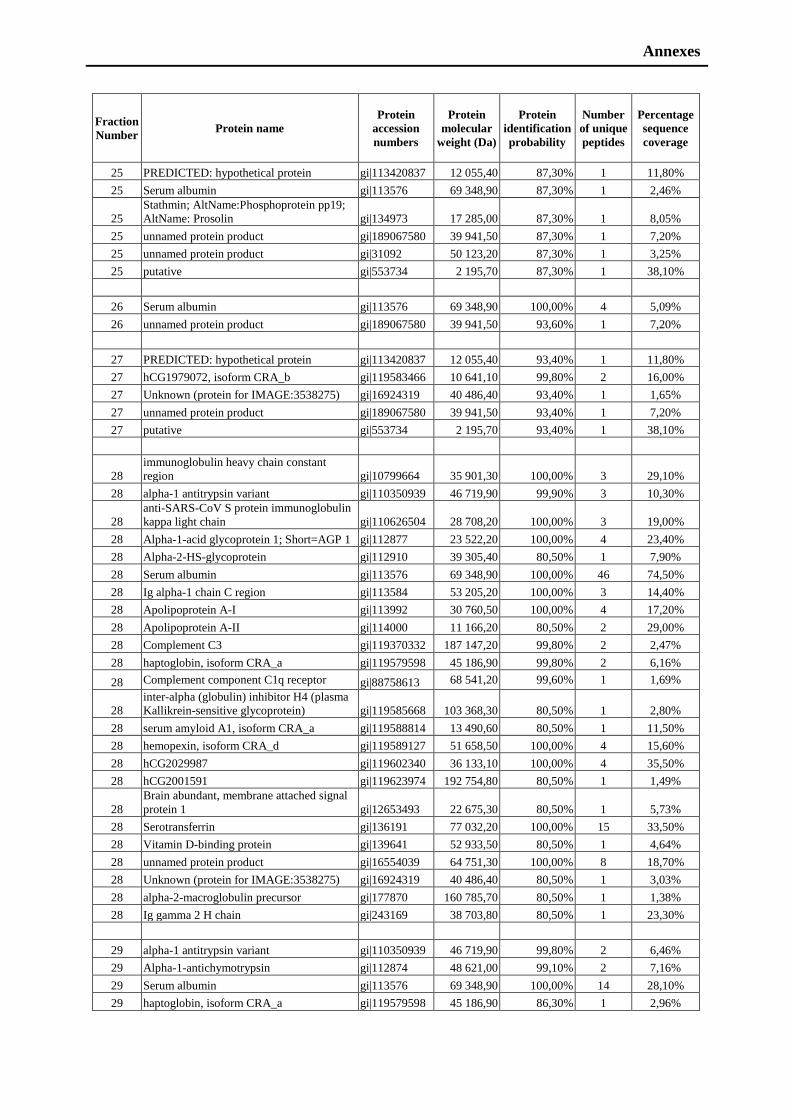
Annexes
Fraction
Number Protein name
Protein
accession
numbers
Protein
molecular
weight (Da)
Protein
identification
probability
Number
of unique
peptides
Percentage
sequence
coverage
25 PREDICTED: hypothetical protein gi|113420837 12 055,40 87,30% 1 11,80%
25 Serum albumin gi|113576 69 348,90 87,30% 1 2,46%
25
Stathmin; AltName:Phosphoprotein pp19;
AltName: Prosolin gi|134973 17 285,00 87,30% 1 8,05%
25 unnamed protein product gi|189067580 39 941,50 87,30% 1 7,20%
25 unnamed protein product gi|31092 50 123,20 87,30% 1 3,25%
25 putative gi|553734 2 195,70 87,30% 1 38,10%
26 Serum albumin gi|113576 69 348,90 100,00% 4 5,09%
26 unnamed protein product gi|189067580 39 941,50 93,60% 1 7,20%
27 PREDICTED: hypothetical protein gi|113420837 12 055,40 93,40% 1 11,80%
27 hCG1979072, isoform CRA_b gi|119583466 10 641,10 99,80% 2 16,00%
27 Unknown (protein for IMAGE:3538275) gi|16924319 40 486,40 93,40% 1 1,65%
27 unnamed protein product gi|189067580 39 941,50 93,40% 1 7,20%
27 putative gi|553734 2 195,70 93,40% 1 38,10%
28
immunoglobulin heavy chain constant
region gi|10799664 35 901,30 100,00% 3 29,10%
28 alpha-1 antitrypsin variant gi|110350939 46 719,90 99,90% 3 10,30%
28
anti-SARS-CoV S protein immunoglobulin
kappa light chain gi|110626504 28 708,20 100,00% 3 19,00%
28 Alpha-1-acid glycoprotein 1; Short=AGP 1 gi|112877 23 522,20 100,00% 4 23,40%
28 Alpha-2-HS-glycoprotein gi|112910 39 305,40 80,50% 1 7,90%
28 Serum albumin gi|113576 69 348,90 100,00% 46 74,50%
28 Ig alpha-1 chain C region gi|113584 53 205,20 100,00% 3 14,40%
28 Apolipoprotein A-I gi|113992 30 760,50 100,00% 4 17,20%
28 Apolipoprotein A-II gi|114000 11 166,20 80,50% 2 29,00%
28 Complement C3 gi|119370332 187 147,20 99,80% 2 2,47%
28 haptoglobin, isoform CRA_a gi|119579598 45 186,90 99,80% 2 6,16%
28 Complement component C1q receptor gi|88758613 68 541,20 99,60% 1 1,69%
28
inter-alpha (globulin) inhibitor H4 (plasma
Kallikrein-sensitive glycoprotein) gi|119585668 103 368,30 80,50% 1 2,80%
28 serum amyloid A1, isoform CRA_a gi|119588814 13 490,60 80,50% 1 11,50%
28 hemopexin, isoform CRA_d gi|119589127 51 658,50 100,00% 4 15,60%
28 hCG2029987 gi|119602340 36 133,10 100,00% 4 35,50%
28 hCG2001591 gi|119623974 192 754,80 80,50% 1 1,49%
28
Brain abundant, membrane attached signal
protein 1 gi|12653493 22 675,30 80,50% 1 5,73%
28 Serotransferrin gi|136191 77 032,20 100,00% 15 33,50%
28 Vitamin D-binding protein gi|139641 52 933,50 80,50% 1 4,64%
28 unnamed protein product gi|16554039 64 751,30 100,00% 8 18,70%
28 Unknown (protein for IMAGE:3538275) gi|16924319 40 486,40 80,50% 1 3,03%
28 alpha-2-macroglobulin precursor gi|177870 160 785,70 80,50% 1 1,38%
28 Ig gamma 2 H chain gi|243169 38 703,80 80,50% 1 23,30%
29 alpha-1 antitrypsin variant gi|110350939 46 719,90 99,80% 2 6,46%
29 Alpha-1-antichymotrypsin gi|112874 48 621,00 99,10% 2 7,16%
29 Serum albumin gi|113576 69 348,90 100,00% 14 28,10%
29 haptoglobin, isoform CRA_a gi|119579598 45 186,90 86,30% 1 2,96%

Annexes
29 unnamed protein product gi|16554039 64 751,30 99,80% 2 6,50%
29 Unknown (protein for IMAGE:3538275) gi|16924319 40 486,40 86,30% 1 3,03%
29 unnamed protein product gi|189067580 39 941,50 86,30% 1 7,20%
29 unnamed protein product gi|31092 50 123,20 86,30% 1 3,25%
30 Serum albumin gi|113576 69 348,90 100,00% 4 10,70%
30 desmoplakin I
gi|1147813,gi|
115502381 331 765,20 50,00% 1 1,04%
30
RCSD domain containing 1, isoform
CRA_a gi|119611202 47 131,10 50,00% 1 3,62%
30 JUP protein gi|15080189 81 727,90 50,00% 1 2,42%
30 Unknown (protein for IMAGE:3538275) gi|16924319 40 486,40 50,00% 1 3,03%
30 TALDO1 protein gi|48257056 37 523,70 50,00% 1 2,97%
30 Complement factor D gi|158515408 27 014,40 99,60% 1 10,30%
31
immunoglobulin heavy chain constant
region gi|10799664 35 901,30 88,60% 1 7,36%
31 alpha-1 antitrypsin variant gi|110350939 46 719,90 88,60% 1 2,39%
31
anti-SARS-CoV S protein immunoglobulin
kappa light chain gi|110626504 28 708,20 88,60% 1 5,95%
31 Serum albumin gi|113576 69 348,90 100,00% 27 50,10%
31 hCG2029987 gi|119602340 36 133,10 88,60% 1 16,70%
31 Serotransferrin gi|136191 77 032,20 99,80% 2 4,73%
31 unnamed protein product gi|16554039 64 751,30 100,00% 3 7,50%
31 Unknown (protein for IMAGE:3538275) gi|16924319 40 486,40 99,80% 2 5,79%
31
myristoylated alanine-rich C-kinase
substrate gi|187387 31 526,20 88,60% 1 4,52%
31 unnamed protein product gi|189067580 39 941,50 88,60% 1 7,20%
31 Histone H1.5; AltName:Histone H1a gi|19856407 22 563,70 88,60% 1 7,08%
31 protein Len,Bence-Jones gi|229528 24 203,90 81,70% 1 15,50%
31 Gox, cytosolic GTP-binding protein gi|459639 881,8 88,60% 1 100,00%
32 Serum albumin gi|113576 69 348,90 100,00% 5 11,70%
32 hCG1979072, isoform CRA_b gi|119583466 10 641,10 93,70% 1 13,00%
32
Brain abundant, membrane attached signal
protein 1 gi|12653493 22 675,30 93,70% 1 5,73%
32 Unknown (protein for IMAGE:3538275) gi|16924319 40 486,40 93,70% 1 3,03%
32 unnamed protein product gi|193787391 32 417,00 93,70% 1 4,07%
32 unnamed protein product gi|194382178 63 522,00 93,70% 1 1,52%
32 Histone H1.5; AltName:Histone H1a gi|19856407 22 563,70 93,70% 1 7,08%
33 Serum albumin gi|113576 69 348,90 91,30% 1 2,13%
33 vasodilator-stimulated phosphoprotein gi|11414808 39 592,80 99,80% 2 3,97%
33 Unknown (protein for IMAGE:3538275) gi|16924319 40 486,40 99,80% 2 5,79%
33 unnamed protein product gi|194382178 63 522,00 91,30% 1 1,52%
34 polyubiquitin gi|11024714 68 474,80 83,10% 2 2,63%
34 HIST1H4H protein gi|111309351 11 349,70 83,10% 1 7,77%
34 TMSB4X protein gi|112180539 5 034,90 99,20% 2 43,20%
34 Serum albumin gi|113576 69 348,90 100,00% 4 11,80%
34 Annexin A1 gi|113944 38 697,90 83,10% 1 3,76%
34 vasodilator-stimulated phosphoprotein gi|11414808 39 592,80 83,10% 1 3,17%
34 HIST1H2BC protein gi|114205460 13 918,60 83,10% 1 7,14%
34 Protein S100-A8, AltName:Calgranulin-A gi|115442 10 816,90 83,10% 1 11,80%
34 Protein S100-A9; AltName:Calgranulin-B gi|115444 13 224,30 83,10% 1 13,20%
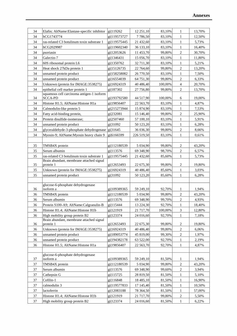
Annexes
34 Elafin; AltName:Elastase-specific inhibitor gi|119262 12 251,10 83,10% 1 13,70%
34 hCG1743774 gi|119573727 7 786,50 83,10% 1 12,50%
34 ras-related C3 botulinum toxin substrate 1 gi|119575445 21 432,60 83,10% 1 5,73%
34 hCG2029987 gi|119602340 36 133,10 83,10% 1 16,40%
34 psoriasin gi|12053626 11 453,70 99,80% 2 30,70%
34 Galectin-7 gi|1346431 15 056,70 83,10% 1 11,80%
34 60S ribosomal protein L6 gi|1350762 32 711,30 83,10% 1 5,21%
34 Heat shock 27kDa protein 1 gi|15126735 22 764,60 99,80% 2 13,20%
34 unnamed protein product gi|158258082 26 770,50 83,10% 1 7,50%
34 unnamed protein product gi|16554039 64 751,30 99,80% 2 6,33%
34 Unknown (protein for IMAGE:3538275) gi|16924319 40 486,40 100,00% 4 20,70%
34 epithelial cell marker protein 1 gi|187302 27 756,80 99,80% 2 13,70%
34
squamous cell carcinoma antigen-1 isoform
SCCA-PD gi|193792580 44 517,90 100,00% 6 19,00%
34 Histone H1.5; AltName:Histone H1a gi|19856407 22 563,70 83,10% 1 4,87%
34 Calmodulin-like protein 5 gi|215273944 15 874,90 83,10% 1 7,53%
34 Fatty acid-binding protein, gi|232081 15 146,40 99,80% 2 25,90%
34 Protein disulfide-isomerase; gi|2507460 57 100,10 83,10% 1 5,91%
34 unnamed protein product gi|31092 50 123,20 83,10% 1 6,28%
34 glyceraldehyde-3-phosphate dehydrogenase gi|31645 36 036,30 99,80% 2 8,66%
34 Myosin-9; AltName:Myosin heavy chain 9 gi|6166599 226 519,50 83,10% 1 0,61%
35 TMSB4X protein gi|112180539 5 034,90 99,80% 2 43,20%
35 Serum albumin gi|113576 69 348,90 98,70% 2 6,57%
35 ras-related C3 botulinum toxin substrate 1 gi|119575445 21 432,60 85,60% 1 5,73%
35
Brain abundant, membrane attached signal
protein 1 gi|12653493 22 675,30 99,80% 2 19,80%
35 Unknown (protein for IMAGE:3538275) gi|16924319 40 486,40 85,60% 1 3,03%
35 unnamed protein product gi|31092 50 123,20 85,60% 1 6,28%
36
glucose-6-phosphate dehydrogenase
isoform a gi|109389365 59 249,10 92,70% 1 1,94%
36 TMSB4X protein gi|112180539 5 034,90 99,80% 2 43,20%
36 Serum albumin gi|113576 69 348,90 99,70% 2 4,93%
36 Protein S100-A9; AltName:Calgranulin-B gi|115444 13 224,30 92,70% 1 18,40%
36 Histone H1.4; AltName:Histone H1b gi|121919 21 717,70 100,00% 3 12,80%
36 High mobility group protein B2 gi|123374 24 016,60 92,70% 1 7,18%
36
Brain abundant, membrane attached signal
protein 1 gi|12653493 22 675,30 99,80% 2 19,80%
36 Unknown (protein for IMAGE:3538275) gi|16924319 40 486,40 99,80% 2 6,06%
36 unnamed protein product gi|189053774 45 819,00 99,30% 2 1,97%
36 unnamed protein product gi|194382178 63 522,00 92,70% 1 2,19%
36 Histone H1.5; AltName:Histone H1a gi|19856407 22 563,70 92,70% 1 4,87%
37
glucose-6-phosphate dehydrogenase
isoform a gi|109389365 59 249,10 81,50% 1 1,94%
37 TMSB4X protein gi|112180539 5 034,90 99,80% 2 43,20%
37 Serum albumin gi|113576 69 348,90 99,60% 2 3,94%
37 Cathepsin G gi|115725 28 819,50 81,50% 1 5,10%
37 Cofilin-1 gi|116848 18 485,10 81,50% 1 16,90%
37 calmodulin 3 gi|119577833 17 145,40 81,50% 1 10,50%
37 lactoferrin gi|12083188 78 364,50 81,50% 1 57,00%
37 Histone H1.4; AltName:Histone H1b gi|121919 21 717,70 99,80% 2 5,50%
37 High mobility group protein B2 gi|123374 24 016,60 81,50% 1 6,22%
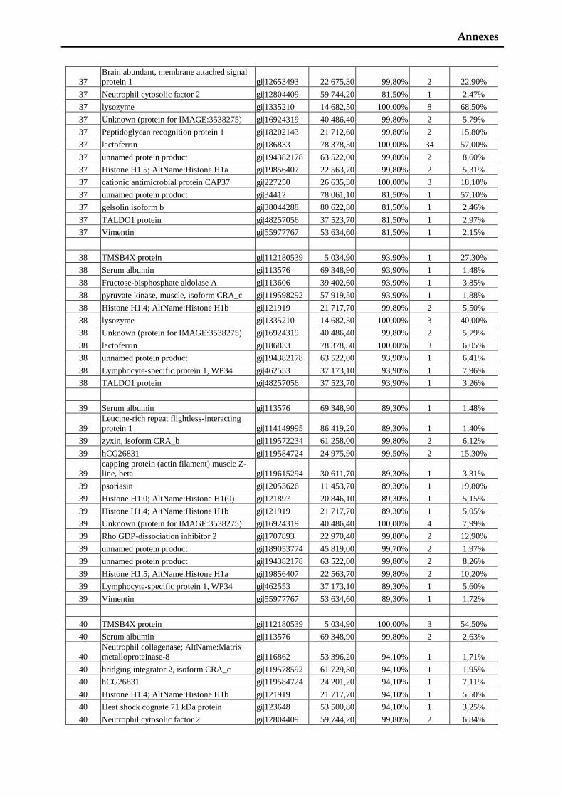
Annexes
37
Brain abundant, membrane attached signal
protein 1 gi|12653493 22 675,30 99,80% 2 22,90%
37 Neutrophil cytosolic factor 2 gi|12804409 59 744,20 81,50% 1 2,47%
37 lysozyme gi|1335210 14 682,50 100,00% 8 68,50%
37 Unknown (protein for IMAGE:3538275) gi|16924319 40 486,40 99,80% 2 5,79%
37 Peptidoglycan recognition protein 1 gi|18202143 21 712,60 99,80% 2 15,80%
37 lactoferrin gi|186833 78 378,50 100,00% 34 57,00%
37 unnamed protein product gi|194382178 63 522,00 99,80% 2 8,60%
37 Histone H1.5; AltName:Histone H1a gi|19856407 22 563,70 99,80% 2 5,31%
37 cationic antimicrobial protein CAP37 gi|227250 26 635,30 100,00% 3 18,10%
37 unnamed protein product gi|34412 78 061,10 81,50% 1 57,10%
37 gelsolin isoform b gi|38044288 80 622,80 81,50% 1 2,46%
37 TALDO1 protein gi|48257056 37 523,70 81,50% 1 2,97%
37 Vimentin gi|55977767 53 634,60 81,50% 1 2,15%
38 TMSB4X protein gi|112180539 5 034,90 93,90% 1 27,30%
38 Serum albumin gi|113576 69 348,90 93,90% 1 1,48%
38 Fructose-bisphosphate aldolase A gi|113606 39 402,60 93,90% 1 3,85%
38 pyruvate kinase, muscle, isoform CRA_c gi|119598292 57 919,50 93,90% 1 1,88%
38 Histone H1.4; AltName:Histone H1b gi|121919 21 717,70 99,80% 2 5,50%
38 lysozyme gi|1335210 14 682,50 100,00% 3 40,00%
38 Unknown (protein for IMAGE:3538275) gi|16924319 40 486,40 99,80% 2 5,79%
38 lactoferrin gi|186833 78 378,50 100,00% 3 6,05%
38 unnamed protein product gi|194382178 63 522,00 93,90% 1 6,41%
38 Lymphocyte-specific protein 1, WP34 gi|462553 37 173,10 93,90% 1 7,96%
38 TALDO1 protein gi|48257056 37 523,70 93,90% 1 3,26%
39 Serum albumin gi|113576 69 348,90 89,30% 1 1,48%
39
Leucine-rich repeat flightless-interacting
protein 1 gi|114149995 86 419,20 89,30% 1 1,40%
39 zyxin, isoform CRA_b gi|119572234 61 258,00 99,80% 2 6,12%
39 hCG26831 gi|119584724 24 975,90 99,50% 2 15,30%
39
capping protein (actin filament) muscle Z-
line, beta gi|119615294 30 611,70 89,30% 1 3,31%
39 psoriasin gi|12053626 11 453,70 89,30% 1 19,80%
39 Histone H1.0; AltName:Histone H1(0) gi|121897 20 846,10 89,30% 1 5,15%
39 Histone H1.4; AltName:Histone H1b gi|121919 21 717,70 89,30% 1 5,05%
39 Unknown (protein for IMAGE:3538275) gi|16924319 40 486,40 100,00% 4 7,99%
39 Rho GDP-dissociation inhibitor 2 gi|1707893 22 970,40 99,80% 2 12,90%
39 unnamed protein product gi|189053774 45 819,00 99,70% 2 1,97%
39 unnamed protein product gi|194382178 63 522,00 99,80% 2 8,26%
39 Histone H1.5; AltName:Histone H1a gi|19856407 22 563,70 99,80% 2 10,20%
39 Lymphocyte-specific protein 1, WP34 gi|462553 37 173,10 89,30% 1 5,60%
39 Vimentin gi|55977767 53 634,60 89,30% 1 1,72%
40 TMSB4X protein gi|112180539 5 034,90 100,00% 3 54,50%
40 Serum albumin gi|113576 69 348,90 99,80% 2 2,63%
40
Neutrophil collagenase; AltName:Matrix
metalloproteinase-8 gi|116862 53 396,20 94,10% 1 1,71%
40 bridging integrator 2, isoform CRA_c gi|119578592 61 729,30 94,10% 1 1,95%
40 hCG26831 gi|119584724 24 201,20 94,10% 1 7,11%
40 Histone H1.4; AltName:Histone H1b gi|121919 21 717,70 94,10% 1 5,50%
40 Heat shock cognate 71 kDa protein gi|123648 53 500,80 94,10% 1 3,25%
40 Neutrophil cytosolic factor 2 gi|12804409 59 744,20 99,80% 2 6,84%
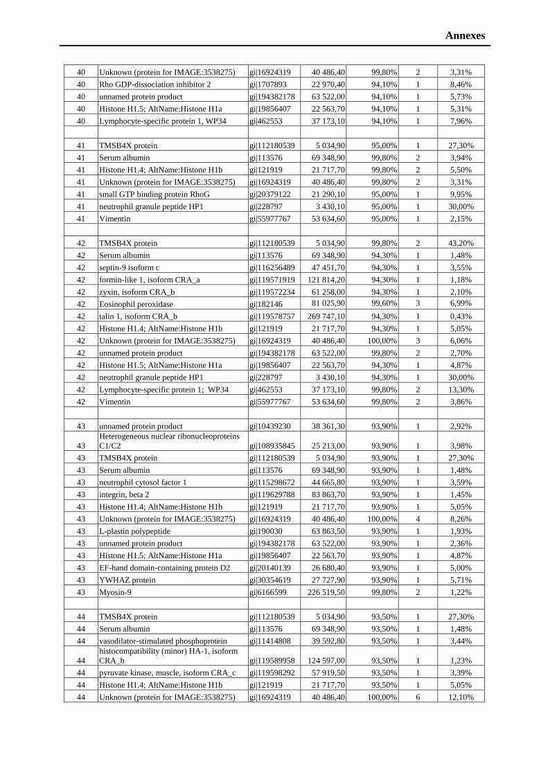
Annexes
40 Unknown (protein for IMAGE:3538275) gi|16924319 40 486,40 99,80% 2 3,31%
40 Rho GDP-dissociation inhibitor 2 gi|1707893 22 970,40 94,10% 1 8,46%
40 unnamed protein product gi|194382178 63 522,00 94,10% 1 5,73%
40 Histone H1.5; AltName:Histone H1a gi|19856407 22 563,70 94,10% 1 5,31%
40 Lymphocyte-specific protein 1, WP34 gi|462553 37 173,10 94,10% 1 7,96%
41 TMSB4X protein gi|112180539 5 034,90 95,00% 1 27,30%
41 Serum albumin gi|113576 69 348,90 99,80% 2 3,94%
41 Histone H1.4; AltName:Histone H1b gi|121919 21 717,70 99,80% 2 5,50%
41 Unknown (protein for IMAGE:3538275) gi|16924319 40 486,40 99,80% 2 3,31%
41 small GTP binding protein RhoG gi|20379122 21 290,10 95,00% 1 9,95%
41 neutrophil granule peptide HP1 gi|228797 3 430,10 95,00% 1 30,00%
41 Vimentin gi|55977767 53 634,60 95,00% 1 2,15%
42 TMSB4X protein gi|112180539 5 034,90 99,80% 2 43,20%
42 Serum albumin gi|113576 69 348,90 94,30% 1 1,48%
42 septin-9 isoform c gi|116256489 47 451,70 94,30% 1 3,55%
42 formin-like 1, isoform CRA_a gi|119571919 121 814,20 94,30% 1 1,18%
42 zyxin, isoform CRA_b gi|119572234 61 258,00 94,30% 1 2,10%
42 Eosinophil peroxidase gi|182146 81 025,90 99,60% 3 6,99%
42 talin 1, isoform CRA_b gi|119578757 269 747,10 94,30% 1 0,43%
42 Histone H1.4; AltName:Histone H1b gi|121919 21 717,70 94,30% 1 5,05%
42 Unknown (protein for IMAGE:3538275) gi|16924319 40 486,40 100,00% 3 6,06%
42 unnamed protein product gi|194382178 63 522,00 99,80% 2 2,70%
42 Histone H1.5; AltName:Histone H1a gi|19856407 22 563,70 94,30% 1 4,87%
42 neutrophil granule peptide HP1 gi|228797 3 430,10 94,30% 1 30,00%
42 Lymphocyte-specific protein 1; WP34 gi|462553 37 173,10 99,80% 2 13,30%
42 Vimentin gi|55977767 53 634,60 99,80% 2 3,86%
43 unnamed protein product gi|10439230 38 361,30 93,90% 1 2,92%
43
Heterogeneous nuclear ribonucleoproteins
C1/C2 gi|108935845 25 213,00 93,90% 1 3,98%
43 TMSB4X protein gi|112180539 5 034,90 93,90% 1 27,30%
43 Serum albumin gi|113576 69 348,90 93,90% 1 1,48%
43 neutrophil cytosol factor 1 gi|115298672 44 665,80 93,90% 1 3,59%
43 integrin, beta 2 gi|119629788 83 863,70 93,90% 1 1,45%
43 Histone H1.4; AltName:Histone H1b gi|121919 21 717,70 93,90% 1 5,05%
43 Unknown (protein for IMAGE:3538275) gi|16924319 40 486,40 100,00% 4 8,26%
43 L-plastin polypeptide gi|190030 63 863,50 93,90% 1 1,93%
43 unnamed protein product gi|194382178 63 522,00 93,90% 1 2,36%
43 Histone H1.5; AltName:Histone H1a gi|19856407 22 563,70 93,90% 1 4,87%
43 EF-hand domain-containing protein D2 gi|20140139 26 680,40 93,90% 1 5,00%
43 YWHAZ protein gi|30354619 27 727,90 93,90% 1 5,71%
43 Myosin-9 gi|6166599 226 519,50 99,80% 2 1,22%
44 TMSB4X protein gi|112180539 5 034,90 93,50% 1 27,30%
44 Serum albumin gi|113576 69 348,90 93,50% 1 1,48%
44 vasodilator-stimulated phosphoprotein gi|11414808 39 592,80 93,50% 1 3,44%
44
histocompatibility (minor) HA-1, isoform
CRA_b gi|119589958 124 597,00 93,50% 1 1,23%
44 pyruvate kinase, muscle, isoform CRA_c gi|119598292 57 919,50 93,50% 1 3,39%
44 Histone H1.4; AltName:Histone H1b gi|121919 21 717,70 93,50% 1 5,05%
44 Unknown (protein for IMAGE:3538275) gi|16924319 40 486,40 100,00% 6 12,10%
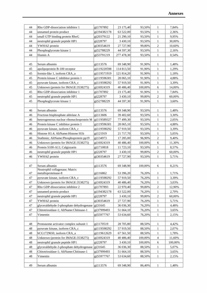
Annexes
44 Rho GDP-dissociation inhibitor 1 gi|1707892 23 175,40 93,50% 1 7,84%
44 unnamed protein product gi|194382178 63 522,00 93,50% 1 2,36%
44 small GTP binding protein RhoG gi|20379122 21 290,10 93,50% 1 9,95%
44 neutrophil granule peptide HP1 gi|228797 3 430,10 93,50% 1 30,00%
44 YWHAZ protein gi|30354619 27 727,90 99,80% 2 10,60%
44 Phosphoglycerate kinase 1 gi|52788229 44 597,30 93,50% 1 2,16%
44 filamin A gi|53791219 277 478,30 93,50% 1 0,54%
45 Serum albumin gi|113576 69 348,90 91,90% 1 1,48%
45 apolipoprotein B-100 receptor gi|119220598 114 813,50 91,90% 1 1,29%
45 formin-like 1, isoform CRA_a gi|119571919 121 814,20 91,90% 1 1,18%
45 Protein kinase C inhibitor protein 1 gi|119596301 28 065,10 91,90% 1 4,88%
45 pyruvate kinase, isoform CRA_c gi|119598292 57 919,50 91,90% 1 3,39%
45 Unknown (protein for IMAGE:3538275) gi|16924319 40 486,40 100,00% 6 14,00%
45 Rho GDP-dissociation inhibitor 1 gi|1707892 23 175,40 91,90% 1 7,84%
45 neutrophil granule peptide HP1 gi|228797 3 430,10 99,80% 2 60,00%
45 Phosphoglycerate kinase 1 gi|52788229 44 597,30 91,90% 1 3,60%
46 Serum albumin gi|113576 69 348,90 93,50% 1 1,48%
46 Fructose-bisphosphate aldolase A gi|113606 39 402,60 93,50% 1 3,30%
46 heterogeneous nuclear ribonucleoprotein M gi|119589327 77 499,30 93,50% 1 2,05%
46 Protein kinase C inhibitor protein 1 gi|119596301 28 065,10 93,50% 1 4,88%
46 pyruvate kinase, isoform CRA_c gi|119598292 57 919,50 93,50% 1 3,39%
46 Histone H1.4; AltName:Histone H1b gi|121919 21 717,70 93,50% 1 5,05%
46 Stathmin; AltName:Phosphoprotein pp19 gi|134973 17 285,00 96,90% 2 9,40%
46 Unknown (protein for IMAGE:3538275) gi|16924319 40 486,40 100,00% 6 11,30%
46 Protein S100-A11; Calgizzarin gi|1710818 11 723,10 93,50% 1 8,57%
46 neutrophil granule peptide HP1 gi|228797 3 430,10 99,80% 2 60,00%
46 YWHAZ protein gi|30354619 27 727,90 93,50% 1 5,71%
47 Serum albumin gi|113576 69 348,90 100,00% 6 8,21%
47
Neutrophil collagenase; Matrix
metalloproteinase-8 gi|116862 53 396,20 76,20% 1 1,71%
47 pyruvate kinase, isoform CRA_c gi|119598292 57 919,50 76,20% 1 3,39%
47 Unknown (protein for IMAGE:3538275) gi|16924319 40 486,40 76,20% 1 3,03%
47 Rho GDP-dissociation inhibitor 2 gi|1707893 22 970,40 99,80% 2 12,90%
47 unnamed protein product gi|194382178 63 522,00 76,20% 1 2,70%
47 neutrophil granule peptide HP1 gi|228797 3 430,10 99,80% 2 60,00%
47 YWHAZ protein gi|30354619 27 727,90 76,20% 1 5,71%
47 glyceraldehyde-3-phosphate dehydrogenase gi|31645 36 036,30 76,20% 1 4,48%
47 Chitotriosidase-1; AltName:Chitinase-1 gi|37999493 51 664,10 76,20% 1 3,65%
47 Vimentin gi|55977767 53 634,60 76,20% 1 2,15%
48 Proteasome activator complex subunit 1 gi|1170519 28 705,80 88,50% 1 4,42%
48 pyruvate kinase, isoform CRA_c gi|119598292 57 919,50 88,50% 1 2,07%
48 hCG1729650, isoform CRA_a gi|119612629 67 561,50 88,50% 1 1,78%
48 Unknown (protein for IMAGE:3538275) gi|16924319 40 486,40 100,00% 4 11,60%
48 neutrophil granule peptide HP1 gi|228797 3 430,10 100,00% 6 100,00%
48 glyceraldehyde-3-phosphate dehydrogenase gi|31645 36 036,30 88,50% 1 5,07%
48 Chitotriosidase-1; AltName:Chitinase-1 gi|37999493 51 664,10 88,50% 1 3,65%
48 Vimentin gi|55977767 53 634,60 88,50% 1 2,15%
49 Serum albumin gi|113576 69 348,90 86,40% 1 1,48%
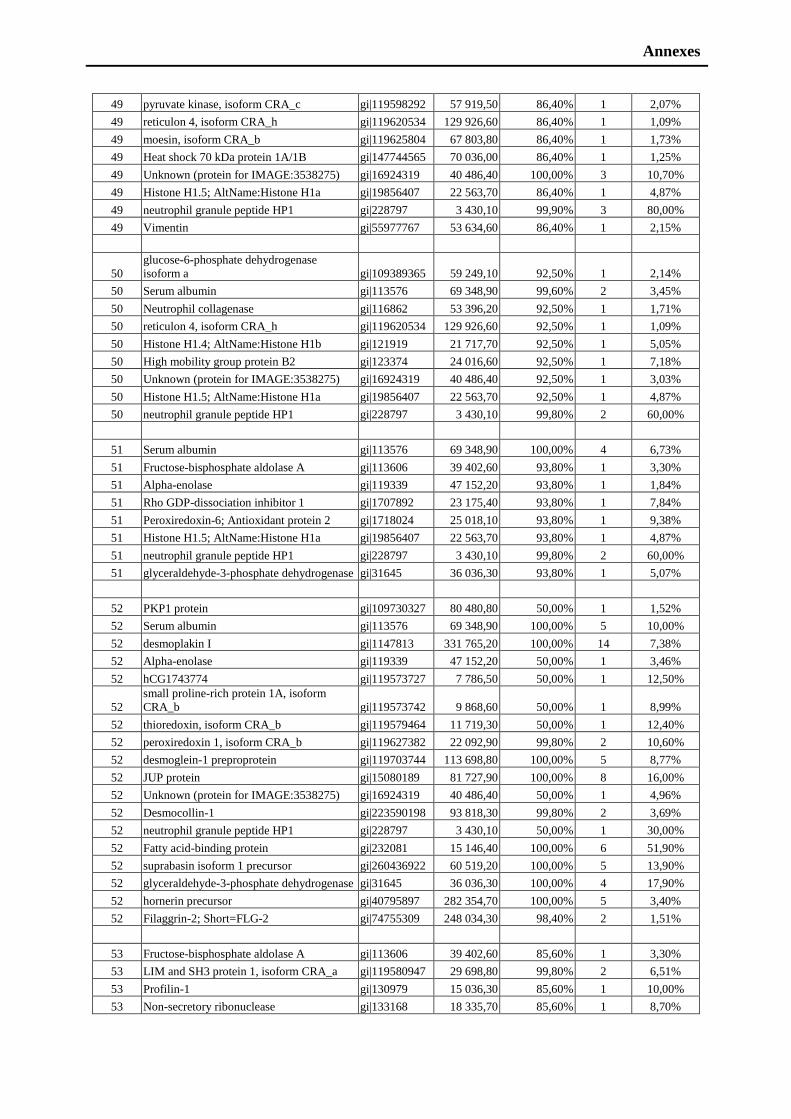
Annexes
49 pyruvate kinase, isoform CRA_c gi|119598292 57 919,50 86,40% 1 2,07%
49 reticulon 4, isoform CRA_h gi|119620534 129 926,60 86,40% 1 1,09%
49 moesin, isoform CRA_b gi|119625804 67 803,80 86,40% 1 1,73%
49 Heat shock 70 kDa protein 1A/1B gi|147744565 70 036,00 86,40% 1 1,25%
49 Unknown (protein for IMAGE:3538275) gi|16924319 40 486,40 100,00% 3 10,70%
49 Histone H1.5; AltName:Histone H1a gi|19856407 22 563,70 86,40% 1 4,87%
49 neutrophil granule peptide HP1 gi|228797 3 430,10 99,90% 3 80,00%
49 Vimentin gi|55977767 53 634,60 86,40% 1 2,15%
50
glucose-6-phosphate dehydrogenase
isoform a gi|109389365 59 249,10 92,50% 1 2,14%
50 Serum albumin gi|113576 69 348,90 99,60% 2 3,45%
50 Neutrophil collagenase gi|116862 53 396,20 92,50% 1 1,71%
50 reticulon 4, isoform CRA_h gi|119620534 129 926,60 92,50% 1 1,09%
50 Histone H1.4; AltName:Histone H1b gi|121919 21 717,70 92,50% 1 5,05%
50 High mobility group protein B2 gi|123374 24 016,60 92,50% 1 7,18%
50 Unknown (protein for IMAGE:3538275) gi|16924319 40 486,40 92,50% 1 3,03%
50 Histone H1.5; AltName:Histone H1a gi|19856407 22 563,70 92,50% 1 4,87%
50 neutrophil granule peptide HP1 gi|228797 3 430,10 99,80% 2 60,00%
51 Serum albumin gi|113576 69 348,90 100,00% 4 6,73%
51 Fructose-bisphosphate aldolase A gi|113606 39 402,60 93,80% 1 3,30%
51 Alpha-enolase gi|119339 47 152,20 93,80% 1 1,84%
51 Rho GDP-dissociation inhibitor 1 gi|1707892 23 175,40 93,80% 1 7,84%
51 Peroxiredoxin-6; Antioxidant protein 2 gi|1718024 25 018,10 93,80% 1 9,38%
51 Histone H1.5; AltName:Histone H1a gi|19856407 22 563,70 93,80% 1 4,87%
51 neutrophil granule peptide HP1 gi|228797 3 430,10 99,80% 2 60,00%
51 glyceraldehyde-3-phosphate dehydrogenase gi|31645 36 036,30 93,80% 1 5,07%
52 PKP1 protein gi|109730327 80 480,80 50,00% 1 1,52%
52 Serum albumin gi|113576 69 348,90 100,00% 5 10,00%
52 desmoplakin I gi|1147813 331 765,20 100,00% 14 7,38%
52 Alpha-enolase gi|119339 47 152,20 50,00% 1 3,46%
52 hCG1743774 gi|119573727 7 786,50 50,00% 1 12,50%
52
small proline-rich protein 1A, isoform
CRA_b gi|119573742 9 868,60 50,00% 1 8,99%
52 thioredoxin, isoform CRA_b gi|119579464 11 719,30 50,00% 1 12,40%
52 peroxiredoxin 1, isoform CRA_b gi|119627382 22 092,90 99,80% 2 10,60%
52 desmoglein-1 preproprotein gi|119703744 113 698,80 100,00% 5 8,77%
52 JUP protein gi|15080189 81 727,90 100,00% 8 16,00%
52 Unknown (protein for IMAGE:3538275) gi|16924319 40 486,40 50,00% 1 4,96%
52 Desmocollin-1 gi|223590198 93 818,30 99,80% 2 3,69%
52 neutrophil granule peptide HP1 gi|228797 3 430,10 50,00% 1 30,00%
52 Fatty acid-binding protein gi|232081 15 146,40 100,00% 6 51,90%
52 suprabasin isoform 1 precursor gi|260436922 60 519,20 100,00% 5 13,90%
52 glyceraldehyde-3-phosphate dehydrogenase gi|31645 36 036,30 100,00% 4 17,90%
52 hornerin precursor gi|40795897 282 354,70 100,00% 5 3,40%
52 Filaggrin-2; Short=FLG-2 gi|74755309 248 034,30 98,40% 2 1,51%
53 Fructose-bisphosphate aldolase A gi|113606 39 402,60 85,60% 1 3,30%
53 LIM and SH3 protein 1, isoform CRA_a gi|119580947 29 698,80 99,80% 2 6,51%
53 Profilin-1 gi|130979 15 036,30 85,60% 1 10,00%
53 Non-secretory ribonuclease gi|133168 18 335,70 85,60% 1 8,70%
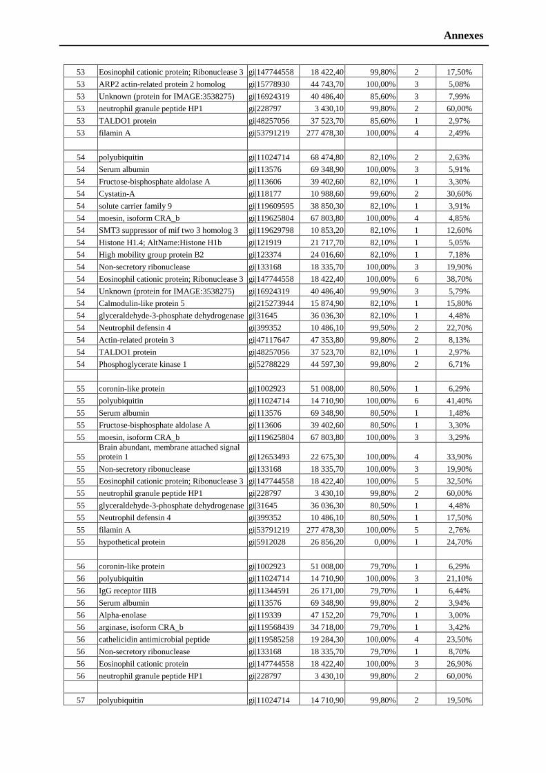
Annexes
53 Eosinophil cationic protein; Ribonuclease 3 gi|147744558 18 422,40 99,80% 2 17,50%
53 ARP2 actin-related protein 2 homolog gi|15778930 44 743,70 100,00% 3 5,08%
53 Unknown (protein for IMAGE:3538275) gi|16924319 40 486,40 85,60% 3 7,99%
53 neutrophil granule peptide HP1 gi|228797 3 430,10 99,80% 2 60,00%
53 TALDO1 protein gi|48257056 37 523,70 85,60% 1 2,97%
53 filamin A gi|53791219 277 478,30 100,00% 4 2,49%
54 polyubiquitin gi|11024714 68 474,80 82,10% 2 2,63%
54 Serum albumin gi|113576 69 348,90 100,00% 3 5,91%
54 Fructose-bisphosphate aldolase A gi|113606 39 402,60 82,10% 1 3,30%
54 Cystatin-A gi|118177 10 988,60 99,60% 2 30,60%
54 solute carrier family 9 gi|119609595 38 850,30 82,10% 1 3,91%
54 moesin, isoform CRA_b gi|119625804 67 803,80 100,00% 4 4,85%
54 SMT3 suppressor of mif two 3 homolog 3 gi|119629798 10 853,20 82,10% 1 12,60%
54 Histone H1.4; AltName:Histone H1b gi|121919 21 717,70 82,10% 1 5,05%
54 High mobility group protein B2 gi|123374 24 016,60 82,10% 1 7,18%
54 Non-secretory ribonuclease gi|133168 18 335,70 100,00% 3 19,90%
54 Eosinophil cationic protein; Ribonuclease 3 gi|147744558 18 422,40 100,00% 6 38,70%
54 Unknown (protein for IMAGE:3538275) gi|16924319 40 486,40 99,90% 3 5,79%
54 Calmodulin-like protein 5 gi|215273944 15 874,90 82,10% 1 15,80%
54 glyceraldehyde-3-phosphate dehydrogenase gi|31645 36 036,30 82,10% 1 4,48%
54 Neutrophil defensin 4 gi|399352 10 486,10 99,50% 2 22,70%
54 Actin-related protein 3 gi|47117647 47 353,80 99,80% 2 8,13%
54 TALDO1 protein gi|48257056 37 523,70 82,10% 1 2,97%
54 Phosphoglycerate kinase 1 gi|52788229 44 597,30 99,80% 2 6,71%
55 coronin-like protein gi|1002923 51 008,00 80,50% 1 6,29%
55 polyubiquitin gi|11024714 14 710,90 100,00% 6 41,40%
55 Serum albumin gi|113576 69 348,90 80,50% 1 1,48%
55 Fructose-bisphosphate aldolase A gi|113606 39 402,60 80,50% 1 3,30%
55 moesin, isoform CRA_b gi|119625804 67 803,80 100,00% 3 3,29%
55
Brain abundant, membrane attached signal
protein 1 gi|12653493 22 675,30 100,00% 4 33,90%
55 Non-secretory ribonuclease gi|133168 18 335,70 100,00% 3 19,90%
55 Eosinophil cationic protein; Ribonuclease 3 gi|147744558 18 422,40 100,00% 5 32,50%
55 neutrophil granule peptide HP1 gi|228797 3 430,10 99,80% 2 60,00%
55 glyceraldehyde-3-phosphate dehydrogenase gi|31645 36 036,30 80,50% 1 4,48%
55 Neutrophil defensin 4 gi|399352 10 486,10 80,50% 1 17,50%
55 filamin A gi|53791219 277 478,30 100,00% 5 2,76%
55 hypothetical protein gi|5912028 26 856,20 0,00% 1 24,70%
56 coronin-like protein gi|1002923 51 008,00 79,70% 1 6,29%
56 polyubiquitin gi|11024714 14 710,90 100,00% 3 21,10%
56 IgG receptor IIIB gi|11344591 26 171,00 79,70% 1 6,44%
56 Serum albumin gi|113576 69 348,90 99,80% 2 3,94%
56 Alpha-enolase gi|119339 47 152,20 79,70% 1 3,00%
56 arginase, isoform CRA_b gi|119568439 34 718,00 79,70% 1 3,42%
56 cathelicidin antimicrobial peptide gi|119585258 19 284,30 100,00% 4 23,50%
56 Non-secretory ribonuclease gi|133168 18 335,70 79,70% 1 8,70%
56 Eosinophil cationic protein gi|147744558 18 422,40 100,00% 3 26,90%
56 neutrophil granule peptide HP1 gi|228797 3 430,10 99,80% 2 60,00%
57 polyubiquitin gi|11024714 14 710,90 99,80% 2 19,50%
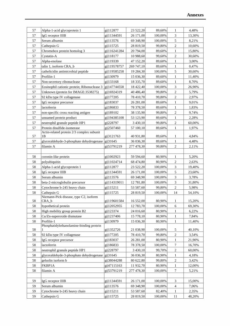
Annexes
57 Alpha-1-acid glycoprotein 1 gi|112877 23 522,20 89,60% 1 4,48%
57 IgG receptor IIIB gi|11344591 26 171,00 100,00% 3 13,30%
57 Serum albumin gi|113576 69 348,90 100,00% 5 8,21%
57 Cathepsin G gi|115725 28 819,50 99,80% 2 10,60%
57 Chromobox protein homolog 3 gi|116241284 20 794,00 89,60% 1 15,80%
57 Cystatin-A gi|118177 10 988,60 99,60% 2 30,60%
57 Alpha-enolase gi|119339 47 152,20 89,60% 1 3,00%
57 talin 1, isoform CRA_b gi|119578757 269 747,10 89,60% 1 0,47%
57 cathelicidin antimicrobial peptide gi|119585258 19 284,30 100,00% 5 30,60%
57 Profilin-1 gi|130979 15 036,30 89,60% 1 11,40%
57 Non-secretory ribonuclease gi|133168 18 335,70 89,60% 1 8,70%
57 Eosinophil cationic protein; Ribonuclease 3 gi|147744558 18 422,40 100,00% 3 26,90%
57 Unknown (protein for IMAGE:3538275) gi|16924319 40 486,40 99,80% 2 5,79%
57 92 kDa type IV collagenase gi|177205 78 410,70 99,80% 2 2,97%
57 IgG receptor precursor gi|183037 26 281,00 89,60% 1 9,01%
57 lactoferrin gi|186833 78 378,50 89,60% 1 1,83%
57 non-specific cross reacting antigen gi|189102 38 135,90 99,80% 2 9,74%
57 unnamed protein product gi|194385108 53 123,90 89,60% 1 2,28%
57 neutrophil granule peptide HP1 gi|228797 3 430,10 99,80% 2 60,00%
57 Protein disulfide-isomerase gi|2507460 57 100,10 89,60% 1 1,97%
57
Actin-related protein 2/3 complex subunit
1B gi|3121763 40 931,80 89,60% 1 4,84%
57 glyceraldehyde-3-phosphate dehydrogenase gi|31645 36 036,30 89,60% 1 4,48%
57 filamin A gi|53791219 277 478,30 99,80% 2 2,11%
58 coronin-like protein gi|1002923 59 594,60 80,90% 1 5,20%
58 polyubiquitin gi|11024714 68 474,80 80,90% 2 2,63%
58 Alpha-1-acid glycoprotein 1 gi|112877 23 522,20 100,00% 3 19,40%
58 IgG receptor IIIB gi|11344591 26 171,00 100,00% 5 23,60%
58 Serum albumin gi|113576 69 348,90 100,00% 3 3,78%
58 beta-2-microglobulin precursor gi|114319011 12 781,80 100,00% 3 31,80%
58 Cytochrome b-245 heavy chain gi|115211 53 587,60 99,80% 2 5,98%
58 Cathepsin G gi|115725 28 819,50 100,00% 14 54,10%
58
Niemann-Pick disease, type C2, isoform
CRA_b gi|119601584 16 552,00 80,90% 1 15,20%
58 hypothetical protein gi|12052955 12 783,70 100,00% 6 69,30%
58 High mobility group protein B2 gi|123374 24 016,60 80,90% 1 6,22%
58 Cu/Zn-superoxide dismutase gi|1237406 15 778,10 80,90% 1 7,84%
58 Profilin-1 gi|130979 15 036,30 80,90% 1 11,40%
58
Phosphatidylethanolamine-binding protein
1 gi|1352726 21 038,90 100,00% 5 40,10%
58 92 kDa type IV collagenase gi|177205 78 410,70 99,80% 2 3,54%
58 IgG receptor precursor gi|183037 26 281,00 80,90% 1 21,90%
58 lactoferrin gi|186833 78 378,50 100,00% 7 16,70%
58 neutrophil granule peptide HP1 gi|228797 3 430,10 99,70% 2 60,00%
58 glyceraldehyde-3-phosphate dehydrogenase gi|31645 36 036,30 80,90% 1 4,18%
58 gelsolin isoform b gi|38044288 80 622,80 99,80% 2 3,42%
58 FKBP1A gi|47115163 11 932,70 80,90% 1 12,00%
58 filamin A gi|53791219 277 478,30 100,00% 7 5,21%
59 IgG receptor IIIB gi|11344591 26 171,00 100,00% 3 15,00%
59 Serum albumin gi|113576 69 348,90 100,00% 4 7,06%
59 Cytochrome b-245 heavy chain gi|115211 53 587,60 82,40% 1 2,35%
59 Cathepsin G gi|115725 28 819,50 100,00% 11 48,20%

Annexes
59 Elongation factor 2 gi|119172 95 322,10 99,80% 2 2,56%
59 Alpha-enolase gi|119339 47 152,20 82,40% 1 3,00%
59 calmodulin 3 gi|119577833 17 145,40 82,40% 1 10,50%
59 haptoglobin, isoform CRA_a gi|119579598 45 186,90 100,00% 4 8,87%
59
Niemann-Pick disease, type C2, isoform
CRA_b gi|119601584 16 552,00 98,20% 2 21,90%
59 CD93 antigen gi|119630576 68 541,20 82,40% 1 2,45%
59 Profilin-1; gi|130979 15 036,30 100,00% 6 56,40%
59 Eosinophil cationic protein gi|147744558 18 422,40 82,40% 1 7,50%
59 dermcidin isoform 2
gi|148271063,
gi|20141302 11 266,10 82,40% 1 10,00%
59 Unknown (protein for IMAGE:3538275) gi|16924319 40 486,40 82,40% 1 4,96%
59 92 kDa type IV collagenase gi|177205 78 410,70 99,80% 2 2,97%
59 IgG receptor precursor gi|183037 26 281,00 99,80% 2 17,60%
59 lactoferrin gi|186833 78 378,50 100,00% 8 15,00%
59 unnamed protein product gi|189053774 45 819,00 99,80% 2 5,16%
59 unnamed protein product gi|194373815 38 499,90 99,80% 2 8,05%
59 neutrophil granule peptide HP1 gi|228797 3 430,10 99,80% 2 60,00%
59 suprabasin isoform 1 precursor gi|260436922 60 519,20 82,40% 1 3,05%
59
TIMP-2, CSC-21K=tissue inhibitor of
metalloproteinase gi|262883 21 630,00 99,80% 2 18,20%
59 gelsolin isoform b gi|38044288 80 622,80 99,80% 2 4,79%
59 FKBP1A gi|47115163 11 932,70 82,40% 1 12,00%
59 filamin A gi|53791219 277 478,30 100,00% 12 7,54%
60 IgG receptor IIIB gi|11344591 26 171,00 99,80% 2 11,60%
60 Serum albumin gi|113576 69 348,90 100,00% 5 8,21%
60 Cathepsin G gi|115725 28 819,50 100,00% 6 30,60%
60 ADP-ribosyl cyclase 2 gi|116241273 35 721,40 99,80% 2 8,81%
60 Elongation factor 2; gi|119172 95 322,10 79,90% 1 1,52%
60 calmodulin 3, isoform CRA_e gi|119577833 17 145,40 99,80% 2 10,50%
60 haptoglobin, isoform CRA_a gi|119579598 45 186,90 100,00% 7 16,70%
60
phosphohistidine phosphatase 1, isoform
CRA_b gi|119608696 13 804,60 79,90% 1 9,60%
60 Coronin-1C gi|12643898 53 232,30 79,90% 1 2,53%
60 Profilin-1 gi|130979 15 036,30 79,90% 1 11,40%
60
Heterogeneous nuclear ribonucleoproteins
A2/B1 gi|133257 37 412,30 79,90% 1 2,55%
60 lysozyme gi|1335210 14 682,50 100,00% 6 58,50%
60 Heat shock 70 kDa protein 1A/1B gi|147744565 70 036,00 79,90% 1 6,24%
60 SGP28 protein gi|1587691 27 622,10 100,00% 5 27,30%
60 Peptidoglycan recognition protein 1 gi|18202143 21 712,60 99,80% 2 15,80%
60 IgG receptor precursor gi|183037 26 281,00 79,90% 1 9,87%
60 lactoferrin gi|186833 78 378,50 100,00% 13 27,10%
60 unnamed protein product gi|189053774 45 819,00 99,70% 2 4,42%
60 unnamed protein product gi|194388072 42 725,80 79,90% 1 3,17%
60 cationic antimicrobial protein CAP37 gi|227250 26 635,30 99,80% 2 9,24%
60 neutrophil granule peptide HP1 gi|228797 3 430,10 79,90% 1 30,00%
60 Fatty acid-binding protein, gi|232081 15 146,40 79,90% 1 15,60%
60 gelsolin isoform b gi|38044288 80 622,80 79,90% 1 3,01%
60 filamin A gi|53791219 277 478,30 100,00% 10 6,01%
61 Serum albumin gi|113576 69 348,90 100,00% 5 8,21%
61 Fructose-bisphosphate aldolase A gi|113606 39 402,60 74,00% 1 3,85%
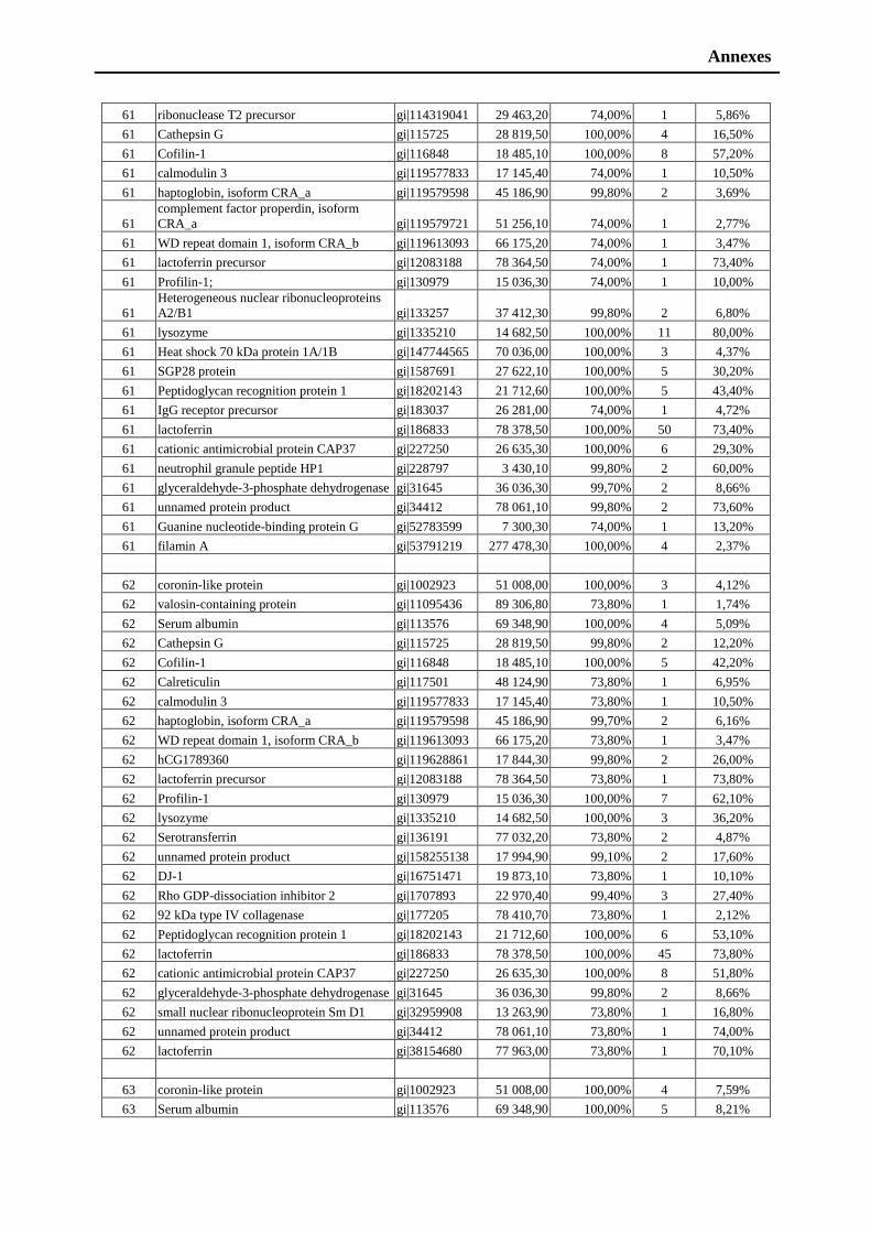
Annexes
61 ribonuclease T2 precursor gi|114319041 29 463,20 74,00% 1 5,86%
61 Cathepsin G gi|115725 28 819,50 100,00% 4 16,50%
61 Cofilin-1 gi|116848 18 485,10 100,00% 8 57,20%
61 calmodulin 3 gi|119577833 17 145,40 74,00% 1 10,50%
61 haptoglobin, isoform CRA_a gi|119579598 45 186,90 99,80% 2 3,69%
61
complement factor properdin, isoform
CRA_a gi|119579721 51 256,10 74,00% 1 2,77%
61 WD repeat domain 1, isoform CRA_b gi|119613093 66 175,20 74,00% 1 3,47%
61 lactoferrin precursor gi|12083188 78 364,50 74,00% 1 73,40%
61 Profilin-1; gi|130979 15 036,30 74,00% 1 10,00%
61
Heterogeneous nuclear ribonucleoproteins
A2/B1 gi|133257 37 412,30 99,80% 2 6,80%
61 lysozyme gi|1335210 14 682,50 100,00% 11 80,00%
61 Heat shock 70 kDa protein 1A/1B gi|147744565 70 036,00 100,00% 3 4,37%
61 SGP28 protein gi|1587691 27 622,10 100,00% 5 30,20%
61 Peptidoglycan recognition protein 1 gi|18202143 21 712,60 100,00% 5 43,40%
61 IgG receptor precursor gi|183037 26 281,00 74,00% 1 4,72%
61 lactoferrin gi|186833 78 378,50 100,00% 50 73,40%
61 cationic antimicrobial protein CAP37 gi|227250 26 635,30 100,00% 6 29,30%
61 neutrophil granule peptide HP1 gi|228797 3 430,10 99,80% 2 60,00%
61 glyceraldehyde-3-phosphate dehydrogenase gi|31645 36 036,30 99,70% 2 8,66%
61 unnamed protein product gi|34412 78 061,10 99,80% 2 73,60%
61 Guanine nucleotide-binding protein G gi|52783599 7 300,30 74,00% 1 13,20%
61 filamin A gi|53791219 277 478,30 100,00% 4 2,37%
62 coronin-like protein gi|1002923 51 008,00 100,00% 3 4,12%
62 valosin-containing protein gi|11095436 89 306,80 73,80% 1 1,74%
62 Serum albumin gi|113576 69 348,90 100,00% 4 5,09%
62 Cathepsin G gi|115725 28 819,50 99,80% 2 12,20%
62 Cofilin-1 gi|116848 18 485,10 100,00% 5 42,20%
62 Calreticulin gi|117501 48 124,90 73,80% 1 6,95%
62 calmodulin 3 gi|119577833 17 145,40 73,80% 1 10,50%
62 haptoglobin, isoform CRA_a gi|119579598 45 186,90 99,70% 2 6,16%
62 WD repeat domain 1, isoform CRA_b gi|119613093 66 175,20 73,80% 1 3,47%
62 hCG1789360 gi|119628861 17 844,30 99,80% 2 26,00%
62 lactoferrin precursor gi|12083188 78 364,50 73,80% 1 73,80%
62 Profilin-1 gi|130979 15 036,30 100,00% 7 62,10%
62 lysozyme gi|1335210 14 682,50 100,00% 3 36,20%
62 Serotransferrin gi|136191 77 032,20 73,80% 2 4,87%
62 unnamed protein product gi|158255138 17 994,90 99,10% 2 17,60%
62 DJ-1 gi|16751471 19 873,10 73,80% 1 10,10%
62 Rho GDP-dissociation inhibitor 2 gi|1707893 22 970,40 99,40% 3 27,40%
62 92 kDa type IV collagenase gi|177205 78 410,70 73,80% 1 2,12%
62 Peptidoglycan recognition protein 1 gi|18202143 21 712,60 100,00% 6 53,10%
62 lactoferrin gi|186833 78 378,50 100,00% 45 73,80%
62 cationic antimicrobial protein CAP37 gi|227250 26 635,30 100,00% 8 51,80%
62 glyceraldehyde-3-phosphate dehydrogenase gi|31645 36 036,30 99,80% 2 8,66%
62 small nuclear ribonucleoprotein Sm D1 gi|32959908 13 263,90 73,80% 1 16,80%
62 unnamed protein product gi|34412 78 061,10 73,80% 1 74,00%
62 lactoferrin gi|38154680 77 963,00 73,80% 1 70,10%
63 coronin-like protein gi|1002923 51 008,00 100,00% 4 7,59%
63 Serum albumin gi|113576 69 348,90 100,00% 5 8,21%
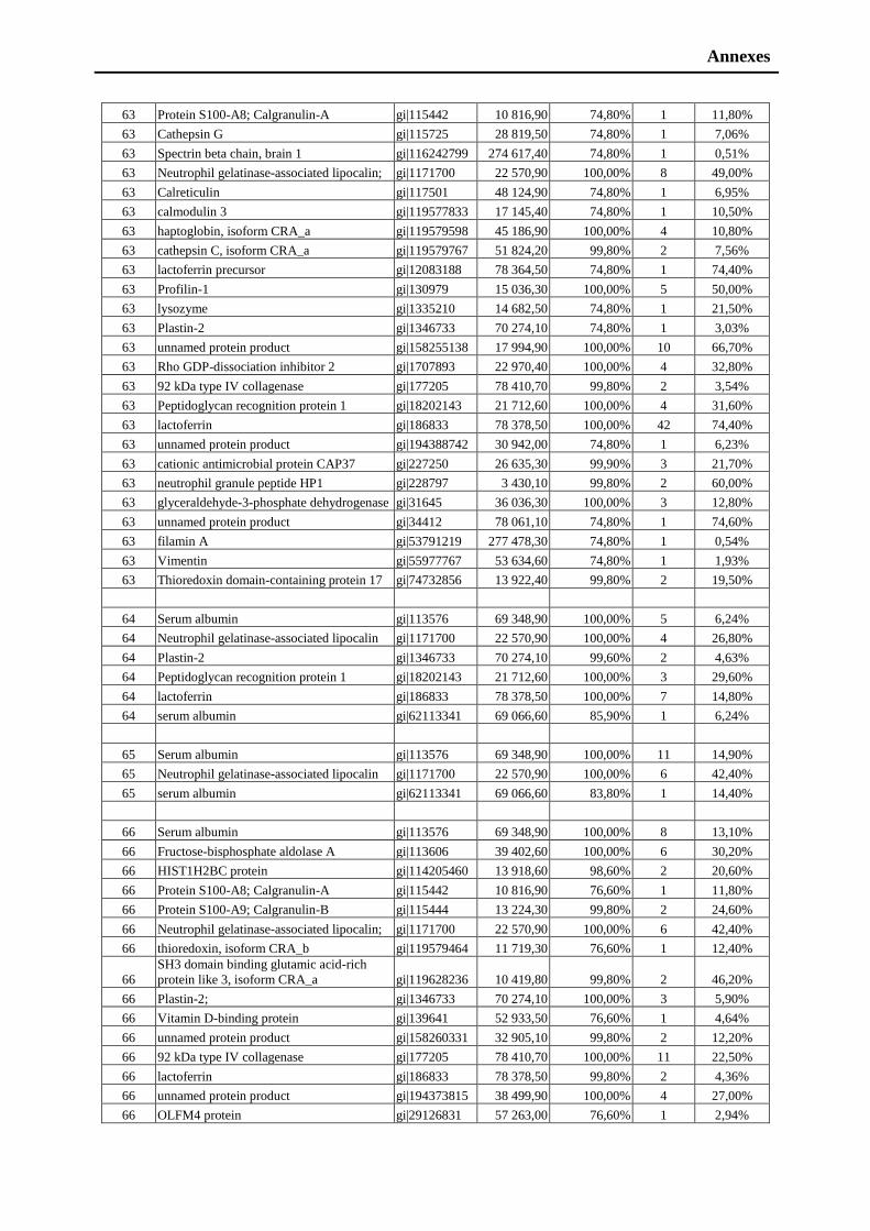
Annexes
63 Protein S100-A8; Calgranulin-A gi|115442 10 816,90 74,80% 1 11,80%
63 Cathepsin G gi|115725 28 819,50 74,80% 1 7,06%
63 Spectrin beta chain, brain 1 gi|116242799 274 617,40 74,80% 1 0,51%
63 Neutrophil gelatinase-associated lipocalin; gi|1171700 22 570,90 100,00% 8 49,00%
63 Calreticulin gi|117501 48 124,90 74,80% 1 6,95%
63 calmodulin 3 gi|119577833 17 145,40 74,80% 1 10,50%
63 haptoglobin, isoform CRA_a gi|119579598 45 186,90 100,00% 4 10,80%
63 cathepsin C, isoform CRA_a gi|119579767 51 824,20 99,80% 2 7,56%
63 lactoferrin precursor gi|12083188 78 364,50 74,80% 1 74,40%
63 Profilin-1 gi|130979 15 036,30 100,00% 5 50,00%
63 lysozyme gi|1335210 14 682,50 74,80% 1 21,50%
63 Plastin-2 gi|1346733 70 274,10 74,80% 1 3,03%
63 unnamed protein product gi|158255138 17 994,90 100,00% 10 66,70%
63 Rho GDP-dissociation inhibitor 2 gi|1707893 22 970,40 100,00% 4 32,80%
63 92 kDa type IV collagenase gi|177205 78 410,70 99,80% 2 3,54%
63 Peptidoglycan recognition protein 1 gi|18202143 21 712,60 100,00% 4 31,60%
63 lactoferrin gi|186833 78 378,50 100,00% 42 74,40%
63 unnamed protein product gi|194388742 30 942,00 74,80% 1 6,23%
63 cationic antimicrobial protein CAP37 gi|227250 26 635,30 99,90% 3 21,70%
63 neutrophil granule peptide HP1 gi|228797 3 430,10 99,80% 2 60,00%
63 glyceraldehyde-3-phosphate dehydrogenase gi|31645 36 036,30 100,00% 3 12,80%
63 unnamed protein product gi|34412 78 061,10 74,80% 1 74,60%
63 filamin A gi|53791219 277 478,30 74,80% 1 0,54%
63 Vimentin gi|55977767 53 634,60 74,80% 1 1,93%
63 Thioredoxin domain-containing protein 17 gi|74732856 13 922,40 99,80% 2 19,50%
64 Serum albumin gi|113576 69 348,90 100,00% 5 6,24%
64 Neutrophil gelatinase-associated lipocalin gi|1171700 22 570,90 100,00% 4 26,80%
64 Plastin-2 gi|1346733 70 274,10 99,60% 2 4,63%
64 Peptidoglycan recognition protein 1 gi|18202143 21 712,60 100,00% 3 29,60%
64 lactoferrin gi|186833 78 378,50 100,00% 7 14,80%
64 serum albumin gi|62113341 69 066,60 85,90% 1 6,24%
65 Serum albumin gi|113576 69 348,90 100,00% 11 14,90%
65 Neutrophil gelatinase-associated lipocalin gi|1171700 22 570,90 100,00% 6 42,40%
65 serum albumin gi|62113341 69 066,60 83,80% 1 14,40%
66 Serum albumin gi|113576 69 348,90 100,00% 8 13,10%
66 Fructose-bisphosphate aldolase A gi|113606 39 402,60 100,00% 6 30,20%
66 HIST1H2BC protein gi|114205460 13 918,60 98,60% 2 20,60%
66 Protein S100-A8; Calgranulin-A gi|115442 10 816,90 76,60% 1 11,80%
66 Protein S100-A9; Calgranulin-B gi|115444 13 224,30 99,80% 2 24,60%
66 Neutrophil gelatinase-associated lipocalin; gi|1171700 22 570,90 100,00% 6 42,40%
66 thioredoxin, isoform CRA_b gi|119579464 11 719,30 76,60% 1 12,40%
66
SH3 domain binding glutamic acid-rich
protein like 3, isoform CRA_a gi|119628236 10 419,80 99,80% 2 46,20%
66 Plastin-2; gi|1346733 70 274,10 100,00% 3 5,90%
66 Vitamin D-binding protein gi|139641 52 933,50 76,60% 1 4,64%
66 unnamed protein product gi|158260331 32 905,10 99,80% 2 12,20%
66 92 kDa type IV collagenase gi|177205 78 410,70 100,00% 11 22,50%
66 lactoferrin gi|186833 78 378,50 99,80% 2 4,36%
66 unnamed protein product gi|194373815 38 499,90 100,00% 4 27,00%
66 OLFM4 protein gi|29126831 57 263,00 76,60% 1 2,94%
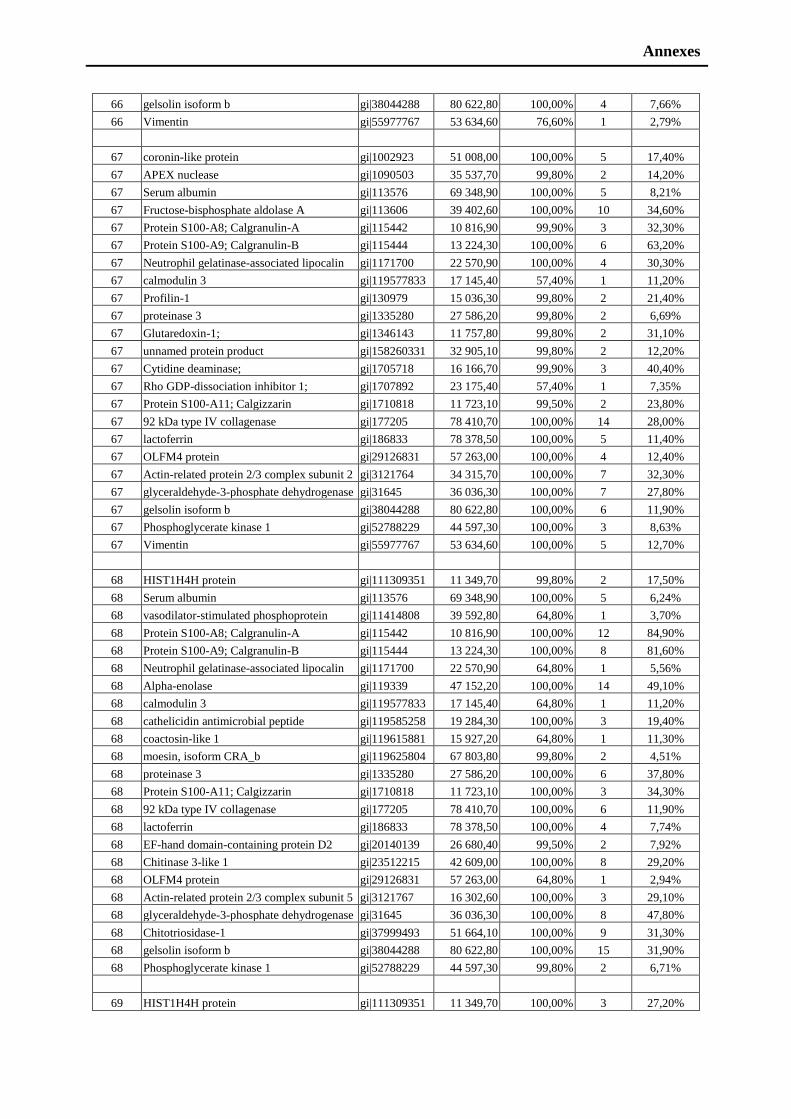
Annexes
66 gelsolin isoform b gi|38044288 80 622,80 100,00% 4 7,66%
66 Vimentin gi|55977767 53 634,60 76,60% 1 2,79%
67 coronin-like protein gi|1002923 51 008,00 100,00% 5 17,40%
67 APEX nuclease gi|1090503 35 537,70 99,80% 2 14,20%
67 Serum albumin gi|113576 69 348,90 100,00% 5 8,21%
67 Fructose-bisphosphate aldolase A gi|113606 39 402,60 100,00% 10 34,60%
67 Protein S100-A8; Calgranulin-A gi|115442 10 816,90 99,90% 3 32,30%
67 Protein S100-A9; Calgranulin-B gi|115444 13 224,30 100,00% 6 63,20%
67 Neutrophil gelatinase-associated lipocalin gi|1171700 22 570,90 100,00% 4 30,30%
67 calmodulin 3 gi|119577833 17 145,40 57,40% 1 11,20%
67 Profilin-1 gi|130979 15 036,30 99,80% 2 21,40%
67 proteinase 3 gi|1335280 27 586,20 99,80% 2 6,69%
67 Glutaredoxin-1; gi|1346143 11 757,80 99,80% 2 31,10%
67 unnamed protein product gi|158260331 32 905,10 99,80% 2 12,20%
67 Cytidine deaminase; gi|1705718 16 166,70 99,90% 3 40,40%
67 Rho GDP-dissociation inhibitor 1; gi|1707892 23 175,40 57,40% 1 7,35%
67 Protein S100-A11; Calgizzarin gi|1710818 11 723,10 99,50% 2 23,80%
67 92 kDa type IV collagenase gi|177205 78 410,70 100,00% 14 28,00%
67 lactoferrin gi|186833 78 378,50 100,00% 5 11,40%
67 OLFM4 protein gi|29126831 57 263,00 100,00% 4 12,40%
67 Actin-related protein 2/3 complex subunit 2 gi|3121764 34 315,70 100,00% 7 32,30%
67 glyceraldehyde-3-phosphate dehydrogenase gi|31645 36 036,30 100,00% 7 27,80%
67 gelsolin isoform b gi|38044288 80 622,80 100,00% 6 11,90%
67 Phosphoglycerate kinase 1 gi|52788229 44 597,30 100,00% 3 8,63%
67 Vimentin gi|55977767 53 634,60 100,00% 5 12,70%
68 HIST1H4H protein gi|111309351 11 349,70 99,80% 2 17,50%
68 Serum albumin gi|113576 69 348,90 100,00% 5 6,24%
68 vasodilator-stimulated phosphoprotein gi|11414808 39 592,80 64,80% 1 3,70%
68 Protein S100-A8; Calgranulin-A gi|115442 10 816,90 100,00% 12 84,90%
68 Protein S100-A9; Calgranulin-B gi|115444 13 224,30 100,00% 8 81,60%
68 Neutrophil gelatinase-associated lipocalin gi|1171700 22 570,90 64,80% 1 5,56%
68 Alpha-enolase gi|119339 47 152,20 100,00% 14 49,10%
68 calmodulin 3 gi|119577833 17 145,40 64,80% 1 11,20%
68 cathelicidin antimicrobial peptide gi|119585258 19 284,30 100,00% 3 19,40%
68 coactosin-like 1 gi|119615881 15 927,20 64,80% 1 11,30%
68 moesin, isoform CRA_b gi|119625804 67 803,80 99,80% 2 4,51%
68 proteinase 3 gi|1335280 27 586,20 100,00% 6 37,80%
68 Protein S100-A11; Calgizzarin gi|1710818 11 723,10 100,00% 3 34,30%
68 92 kDa type IV collagenase gi|177205 78 410,70 100,00% 6 11,90%
68 lactoferrin gi|186833 78 378,50 100,00% 4 7,74%
68 EF-hand domain-containing protein D2 gi|20140139 26 680,40 99,50% 2 7,92%
68 Chitinase 3-like 1 gi|23512215 42 609,00 100,00% 8 29,20%
68 OLFM4 protein gi|29126831 57 263,00 64,80% 1 2,94%
68 Actin-related protein 2/3 complex subunit 5 gi|3121767 16 302,60 100,00% 3 29,10%
68 glyceraldehyde-3-phosphate dehydrogenase gi|31645 36 036,30 100,00% 8 47,80%
68 Chitotriosidase-1 gi|37999493 51 664,10 100,00% 9 31,30%
68 gelsolin isoform b gi|38044288 80 622,80 100,00% 15 31,90%
68 Phosphoglycerate kinase 1 gi|52788229 44 597,30 99,80% 2 6,71%
69 HIST1H4H protein gi|111309351 11 349,70 100,00% 3 27,20%

Annexes
69 Serum albumin gi|113576 69 348,90 100,00% 6 8,21%
69 MYL6 protein gi|113812151 16 942,80 100,00% 4 25,80%
69 Protein S100-A8; Calgranulin-A gi|115442 10 816,90 100,00% 7 71,00%
69 Protein S100-A9; Calgranulin-B gi|115444 13 224,30 100,00% 10 86,80%
69 Alpha-enolase gi|119339 47 152,20 100,00% 4 13,60%
69 myeloperoxidase, isoform CRA_b gi|119614877 83 839,00 100,00% 5 8,32%
69 moesin, isoform CRA_b gi|119625804 67 803,80 100,00% 6 10,70%
69 Nucleoside diphosphate kinase A gi|127981 15 511,20 62,40% 1 12,40%
69 proteinase 3 gi|1335280 27 586,20 100,00% 8 51,20%
69 Protein S100-A11; Calgizzarin gi|1710818 11 723,10 99,80% 2 23,80%
69 Transketolase gi|1729976 67 861,40 62,40% 1 2,89%
69 beta-hemoglobin gi|183945 15 947,70 100,00% 10 76,90%
69 Protein S100-A12; Calgranulin-C gi|2507565 10 557,40 100,00% 4 65,20%
69 glyceraldehyde-3-phosphate dehydrogenase gi|31645 36 036,30 100,00% 5 24,80%
69 Chitotriosidase-1 gi|37999493 51 664,10 62,40% 1 4,51%
69 gelsolin isoform b gi|38044288 80 622,80 100,00% 6 11,60%
70 unnamed protein product gi|10439139 58 277,60 100,00% 7 23,70%
70 T200 leukocyte common antigen precursor gi|10999057 147 237,90 56,90% 1 0,84%
70 Serum albumin gi|113576 69 348,90 100,00% 4 7,06%
70 MYL6 protein gi|113812151 16 942,80 100,00% 6 41,70%
70 Protein S100-A8; Calgranulin-A gi|115442 10 816,90 100,00% 5 55,90%
70 Protein S100-A9; Calgranulin-B gi|115444 13 224,30 100,00% 9 81,60%
70 Neutrophil collagenase gi|116862 53 396,20 100,00% 8 26,80%
70 Alpha-enolase gi|119339 47 152,20 98,60% 2 7,83%
70 matrix metallopeptidase 8 gi|119587431 53 409,20 56,90% 1 28,50%
70 calponin 2, isoform CRA_e gi|119589975 33 679,60 56,90% 1 5,50%
70 spectrin, alpha, non-erythrocytic 1 gi|119608212 282 267,10 100,00% 5 3,43%
70 myeloperoxidase, gi|119614877 83 839,00 100,00% 18 30,50%
70 actin related protein 2/3 complex, subunit 3 gi|119618319 20 529,80 99,80% 2 10,10%
70 proteinase 3 gi|1335280 27 586,20 100,00% 6 37,80%
70 Serpin B10 gi|1345616 45 386,90 99,50% 2 13,40%
70 hemoglobin alpha-1 globin chain gi|13650074 15 239,60 56,90% 1 10,60%
70 Protein S100-A11; Calgizzarin gi|1710818 11 723,10 99,80% 2 23,80%
70 Transketolase gi|1729976 67 861,40 56,90% 1 1,93%
70 Resistin; Cysteine-rich secreted protein gi|18202962 11 401,00 100,00% 4 51,90%
70 beta-hemoglobin gi|183945 15 980,00 98,20% 3 23,10%
70 lactoferrin gi|186833 78 378,50 56,90% 1 1,55%
70 Protein S100-A12; Calgranulin-C gi|2507565 10 557,40 100,00% 5 67,40%
70 glyceraldehyde-3-phosphate dehydrogenase gi|31645 36 036,30 100,00% 5 26,30%
71 Serum albumin gi|113576 69 348,90 100,00% 4 6,24%
71 Protein S100-A8; Calgranulin-A gi|115442 10 816,90 100,00% 3 46,20%
71 Protein S100-A9; Calgranulin-B gi|115444 13 224,30 100,00% 8 63,20%
71 Adenylyl cyclase-associated protein 1 gi|116241280 51 655,20 100,00% 7 20,80%
71 Neutrophil collagenase gi|116862 53 396,20 100,00% 10 30,80%
71 RecName: Full=Grancalcin gi|1170014 23 992,60 50,00% 1 3,23%
71 Neutrophil elastase gi|119292 28 499,40 100,00% 3 27,00%
71 Alpha-enolase gi|119339 47 152,20 86,80% 2 8,53%
71 vinculin, isoform CRA_a gi|119574932 116 706,30 50,00% 1 1,03%
71 myeloperoxidase, isoform CRA_b gi|119614877 83 839,00 100,00% 20 33,30%
71 proteinase 3 gi|1335280 27 586,20 50,00% 1 12,60%
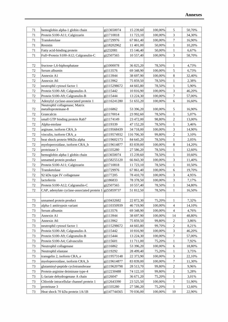
Annexes
71 hemoglobin alpha-1 globin chain gi|13650074 15 239,60 100,00% 5 50,70%
71 Protein S100-A11; Calgizzarin gi|1710818 11 723,10 100,00% 3 34,30%
71 Transketolase gi|1729976 67 861,40 100,00% 7 16,90%
71 Resistin gi|18202962 11 401,00 50,00% 1 10,20%
71 Fatty acid-binding protein gi|232081 15 146,40 50,00% 1 6,67%
71 Full=Protein S100-A12; Calgranulin-C gi|2507565 10 557,40 100,00% 3 58,70%
72 fructose-1,6-biphosphatase gi|1000078 36 825,20 78,50% 1 4,73%
72 Serum albumin gi|113576 69 348,90 100,00% 5 6,73%
72 Annexin A1 gi|113944 38 697,90 100,00% 8 32,40%
72 Annexin A6 gi|113962 75 859,50 78,50% 1 2,38%
72 neutrophil cytosol factor 1 gi|115298672 44 665,80 78,50% 1 5,90%
72 Protein S100-A8; Calgranulin-A gi|115442 10 816,90 100,00% 3 46,20%
72 Protein S100-A9; Calgranulin-B gi|115444 13 224,30 100,00% 7 57,00%
72 Adenylyl cyclase-associated protein 1 gi|116241280 51 655,20 100,00% 6 16,60%
72
Neutrophil collagenase; Matrix
metalloproteinase-8 gi|116862 53 396,20 100,00% 5 16,90%
72 Grancalcin gi|1170014 23 992,60 78,50% 1 5,07%
72 small GTP binding protein Rab7 gi|1174149 23 472,00 98,80% 2 13,00%
72 Alpha-enolase gi|119339 47 152,20 78,50% 1 3,46%
72 arginase, isoform CRA_b gi|119568439 34 718,00 100,00% 3 14,90%
72 vinculin, isoform CRA_a gi|119574932 116 706,30 99,80% 2 3,10%
72 heat shock protein 90kDa alpha gi|119602173 84 645,20 78,50% 1 2,05%
72 myeloperoxidase, isoform CRA_b gi|119614877 83 839,00 100,00% 8 14,20%
72 proteinase 3 gi|1335280 27 586,20 78,50% 1 12,60%
72 hemoglobin alpha-1 globin chain gi|13650074 15 239,60 78,50% 1 10,60%
72 unnamed protein product gi|158255120 66 843,30 100,00% 3 11,40%
72 Protein S100-A11; Calgizzarin gi|1710818 11 723,10 78,50% 1 10,50%
72 Transketolase gi|1729976 67 861,40 100,00% 6 19,70%
72 92 kDa type IV collagenase gi|177205 78 410,70 100,00% 3 4,95%
72 lactoferrin gi|186833 78 378,50 100,00% 3 7,17%
72 Protein S100-A12; Calgranulin-C gi|2507565 10 557,40 78,50% 1 34,80%
72 CAP, adenylate cyclase-associated protein 1 gi|55859737 51 812,50 78,50% 1 16,50%
73 unnamed protein product gi|10432682 22 872,30 75,20% 1 7,32%
73 alpha-1 antitrypsin variant gi|110350939 46 719,90 100,00% 4 14,10%
73 Serum albumin gi|113576 69 348,90 100,00% 4 4,76%
73 Annexin A1 gi|113944 38 697,90 100,00% 14 48,80%
73 Annexin A6 gi|113962 75 859,50 99,80% 2 3,86%
73 neutrophil cytosol factor 1 gi|115298672 44 665,80 99,70% 2 8,21%
73 Protein S100-A8; Calgranulin-A gi|115442 10 816,90 100,00% 3 46,20%
73 Protein S100-A9; Calgranulin-B gi|115444 13 224,30 100,00% 7 57,00%
73 Protein S100-A4; Calvasculin gi|115601 11 711,00 75,20% 1 7,92%
73 Neutrophil collagenase gi|116862 53 396,20 100,00% 6 18,80%
73 Neutrophil elastase gi|119292 28 499,40 75,20% 1 3,75%
73 transgelin 2, isoform CRA_e gi|119573148 22 373,90 100,00% 3 22,10%
73 myeloperoxidase, isoform CRA_b gi|119614877 83 839,00 100,00% 7 11,30%
73 glutaminyl-peptide cyclotransferase gi|119620798 28 513,70 99,80% 2 9,96%
73 Protein-arginine deiminase type-4 gi|12230488 74 122,10 99,80% 2 5,28%
73 L-lactate dehydrogenase A chain gi|126047 36 671,20 75,20% 1 3,01%
73 Chloride intracellular channel protein 1 gi|12643390 23 525,50 100,00% 7 51,90%
73 proteinase 3 gi|1335280 27 586,20 75,20% 1 12,60%
73 Heat shock 70 kDa protein 1A/1B gi|147744565 70 036,00 100,00% 10 22,90%
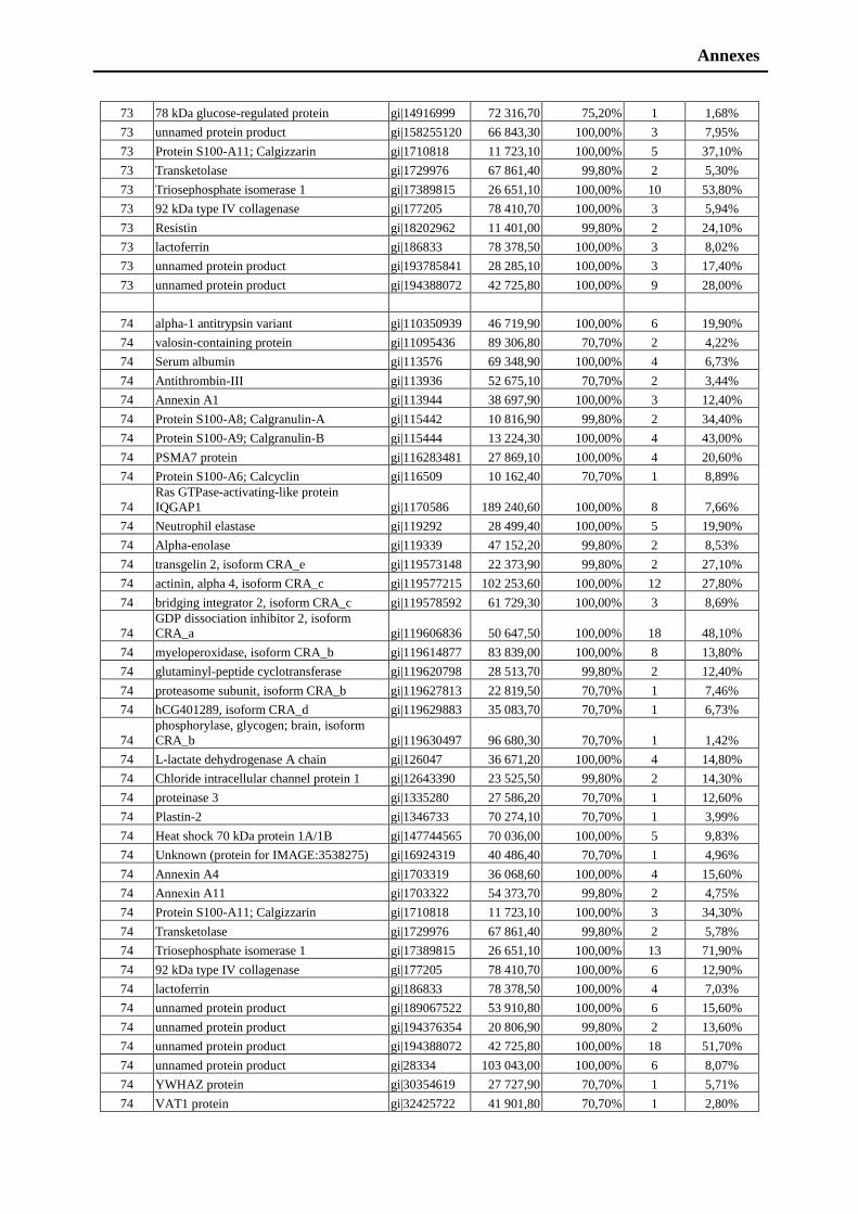
Annexes
73 78 kDa glucose-regulated protein gi|14916999 72 316,70 75,20% 1 1,68%
73 unnamed protein product gi|158255120 66 843,30 100,00% 3 7,95%
73 Protein S100-A11; Calgizzarin gi|1710818 11 723,10 100,00% 5 37,10%
73 Transketolase gi|1729976 67 861,40 99,80% 2 5,30%
73 Triosephosphate isomerase 1 gi|17389815 26 651,10 100,00% 10 53,80%
73 92 kDa type IV collagenase gi|177205 78 410,70 100,00% 3 5,94%
73 Resistin gi|18202962 11 401,00 99,80% 2 24,10%
73 lactoferrin gi|186833 78 378,50 100,00% 3 8,02%
73 unnamed protein product gi|193785841 28 285,10 100,00% 3 17,40%
73 unnamed protein product gi|194388072 42 725,80 100,00% 9 28,00%
74 alpha-1 antitrypsin variant gi|110350939 46 719,90 100,00% 6 19,90%
74 valosin-containing protein gi|11095436 89 306,80 70,70% 2 4,22%
74 Serum albumin gi|113576 69 348,90 100,00% 4 6,73%
74 Antithrombin-III gi|113936 52 675,10 70,70% 2 3,44%
74 Annexin A1 gi|113944 38 697,90 100,00% 3 12,40%
74 Protein S100-A8; Calgranulin-A gi|115442 10 816,90 99,80% 2 34,40%
74 Protein S100-A9; Calgranulin-B gi|115444 13 224,30 100,00% 4 43,00%
74 PSMA7 protein gi|116283481 27 869,10 100,00% 4 20,60%
74 Protein S100-A6; Calcyclin gi|116509 10 162,40 70,70% 1 8,89%
74
Ras GTPase-activating-like protein
IQGAP1 gi|1170586 189 240,60 100,00% 8 7,66%
74 Neutrophil elastase gi|119292 28 499,40 100,00% 5 19,90%
74 Alpha-enolase gi|119339 47 152,20 99,80% 2 8,53%
74 transgelin 2, isoform CRA_e gi|119573148 22 373,90 99,80% 2 27,10%
74 actinin, alpha 4, isoform CRA_c gi|119577215 102 253,60 100,00% 12 27,80%
74 bridging integrator 2, isoform CRA_c gi|119578592 61 729,30 100,00% 3 8,69%
74
GDP dissociation inhibitor 2, isoform
CRA_a gi|119606836 50 647,50 100,00% 18 48,10%
74 myeloperoxidase, isoform CRA_b gi|119614877 83 839,00 100,00% 8 13,80%
74 glutaminyl-peptide cyclotransferase gi|119620798 28 513,70 99,80% 2 12,40%
74 proteasome subunit, isoform CRA_b gi|119627813 22 819,50 70,70% 1 7,46%
74 hCG401289, isoform CRA_d gi|119629883 35 083,70 70,70% 1 6,73%
74
phosphorylase, glycogen; brain, isoform
CRA_b gi|119630497 96 680,30 70,70% 1 1,42%
74 L-lactate dehydrogenase A chain gi|126047 36 671,20 100,00% 4 14,80%
74 Chloride intracellular channel protein 1 gi|12643390 23 525,50 99,80% 2 14,30%
74 proteinase 3 gi|1335280 27 586,20 70,70% 1 12,60%
74 Plastin-2 gi|1346733 70 274,10 70,70% 1 3,99%
74 Heat shock 70 kDa protein 1A/1B gi|147744565 70 036,00 100,00% 5 9,83%
74 Unknown (protein for IMAGE:3538275) gi|16924319 40 486,40 70,70% 1 4,96%
74 Annexin A4 gi|1703319 36 068,60 100,00% 4 15,60%
74 Annexin A11 gi|1703322 54 373,70 99,80% 2 4,75%
74 Protein S100-A11; Calgizzarin gi|1710818 11 723,10 100,00% 3 34,30%
74 Transketolase gi|1729976 67 861,40 99,80% 2 5,78%
74 Triosephosphate isomerase 1 gi|17389815 26 651,10 100,00% 13 71,90%
74 92 kDa type IV collagenase gi|177205 78 410,70 100,00% 6 12,90%
74 lactoferrin gi|186833 78 378,50 100,00% 4 7,03%
74 unnamed protein product gi|189067522 53 910,80 100,00% 6 15,60%
74 unnamed protein product gi|194376354 20 806,90 99,80% 2 13,60%
74 unnamed protein product gi|194388072 42 725,80 100,00% 18 51,70%
74 unnamed protein product gi|28334 103 043,00 100,00% 6 8,07%
74 YWHAZ protein gi|30354619 27 727,90 70,70% 1 5,71%
74 VAT1 protein gi|32425722 41 901,80 70,70% 1 2,80%
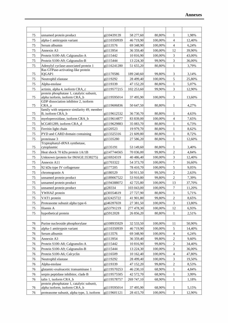
Annexes
75 unnamed protein product gi|10439139 58 277,60 80,80% 1 1,98%
75 alpha-1 antitrypsin variant gi|110350939 46 719,90 100,00% 4 12,40%
75 Serum albumin gi|113576 69 348,90 100,00% 4 6,24%
75 Annexin A3 gi|113954 36 359,40 100,00% 12 39,90%
75 Protein S100-A8; Calgranulin-A gi|115442 10 816,90 100,00% 3 43,00%
75 Protein S100-A9; Calgranulin-B gi|115444 13 224,30 99,90% 3 36,00%
75 Adenylyl cyclase-associated protein 1 gi|116241280 51 655,20 80,80% 1 3,79%
75
Ras GTPase-activating-like protein
IQGAP1 gi|1170586 189 240,60 99,80% 3 3,14%
75 Neutrophil elastase gi|119292 28 499,40 100,00% 5 25,80%
75 Alpha-enolase gi|119339 47 152,20 80,80% 1 5,07%
75 actinin, alpha 4, isoform CRA_c gi|119577215 102 253,60 99,90% 3 12,90%
75
protein phosphatase 1, catalytic subunit,
alpha isoform, isoform CRA_b gi|119595014 37 495,90 100,00% 3 13,60%
75
GDP dissociation inhibitor 2, isoform
CRA_a gi|119606836 50 647,50 80,80% 1 4,27%
75
family with sequence similarity 49, member
B, isoform CRA_b gi|119612532 36 730,70 80,80% 1 4,63%
75 myeloperoxidase, isoform CRA_b gi|119614877 83 839,00 100,00% 4 7,65%
75 hCG401289, isoform CRA_d gi|119629883 35 083,70 80,80% 1 6,73%
75 Ferritin light chain gi|120523 19 979,70 80,80% 1 8,62%
75 PYD and CARD domain containing gi|13325316 21 609,80 80,80% 1 8,72%
75 proteinase 3 gi|1335280 27 586,20 80,80% 1 12,60%
75
Tryptophanyl-tRNA synthetase,
cytoplasmic gi|135191 53 149,60 80,80% 1 3,40%
75 Heat shock 70 kDa protein 1A/1B gi|147744565 70 036,00 99,80% 2 4,84%
75 Unknown (protein for IMAGE:3538275) gi|16924319 40 486,40 100,00% 3 12,40%
75 Annexin A11 gi|1703322 54 373,70 100,00% 7 16,60%
75 92 kDa type IV collagenase gi|177205 78 410,70 100,00% 5 9,05%
75 chromogranin A gi|180529 50 911,50 99,50% 2 2,63%
75 unnamed protein product gi|189067522 53 910,80 99,80% 2 7,39%
75 unnamed protein product gi|194388072 42 725,80 100,00% 13 37,70%
75 unnamed protein product gi|28334 103 043,00 100,00% 7 11,20%
75 YWHAZ protein gi|30354619 27 727,90 80,80% 1 5,71%
75 VAT1 protein gi|32425722 41 901,80 99,80% 2 8,65%
75 Proteasome subunit alpha type-6 gi|46397659 27 381,50 100,00% 3 13,80%
75 filamin A gi|53791219 277 478,30 100,00% 12 6,93%
75 hypothetical protein gi|5912028 26 856,20 80,80% 1 2,51%
76 Purine nucleoside phosphorylase gi|108935929 32 533,50 100,00% 11 50,90%
76 alpha-1 antitrypsin variant gi|110350939 46 719,90 100,00% 5 14,40%
76 Serum albumin gi|113576 69 348,90 100,00% 4 6,24%
76 Annexin A3 gi|113954 36 359,40 99,80% 2 9,60%
76 Protein S100-A8; Calgranulin-A gi|115442 10 816,90 99,80% 2 34,40%
76 Protein S100-A9; Calgranulin-B gi|115444 13 224,30 100,00% 3 36,00%
76 Protein S100-A6; Calcyclin gi|116509 10 162,40 100,00% 4 47,80%
76 Neutrophil elastase gi|119292 28 499,40 100,00% 3 19,50%
76 Alpha-enolase gi|119339 47 152,20 99,80% 2 8,53%
76 glutamic-oxaloacetic transaminase 1 gi|119570253 46 230,10 68,90% 1 4,84%
76 serpin peptidase inhibitor, clade B gi|119575505 42 572,70 68,90% 1 3,99%
76 talin 1, isoform CRA_b gi|119578757 269 747,10 68,90% 1 1,18%
76
protein phosphatase 1, catalytic subunit,
alpha isoform, isoform CRA_b gi|119595014 37 495,90 68,90% 1 5,15%
76 proteasome subunit, alpha type, 3, isoform gi|119601121 28 415,70 100,00% 3 12,90%
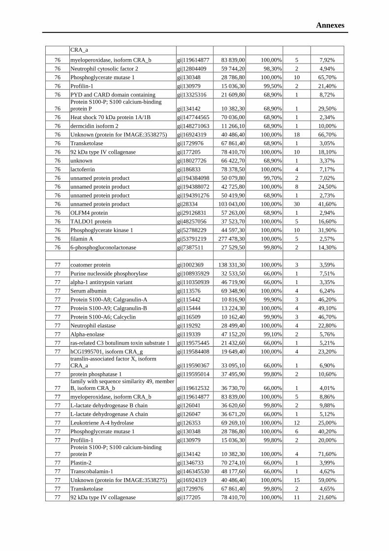
Annexes
CRA_a
76 myeloperoxidase, isoform CRA_b gi|119614877 83 839,00 100,00% 5 7,92%
76 Neutrophil cytosolic factor 2 gi|12804409 59 744,20 98,30% 2 4,94%
76 Phosphoglycerate mutase 1 gi|130348 28 786,80 100,00% 10 65,70%
76 Profilin-1 gi|130979 15 036,30 99,50% 2 21,40%
76 PYD and CARD domain containing gi|13325316 21 609,80 68,90% 1 8,72%
76
Protein S100-P; S100 calcium-binding
protein P gi|134142 10 382,30 68,90% 1 29,50%
76 Heat shock 70 kDa protein 1A/1B gi|147744565 70 036,00 68,90% 1 2,34%
76 dermcidin isoform 2 gi|148271063 11 266,10 68,90% 1 10,00%
76 Unknown (protein for IMAGE:3538275) gi|16924319 40 486,40 100,00% 18 66,70%
76 Transketolase gi|1729976 67 861,40 68,90% 1 3,05%
76 92 kDa type IV collagenase gi|177205 78 410,70 100,00% 10 18,10%
76 unknown gi|18027726 66 422,70 68,90% 1 3,37%
76 lactoferrin gi|186833 78 378,50 100,00% 4 7,17%
76 unnamed protein product gi|194384098 50 079,80 99,70% 2 7,02%
76 unnamed protein product gi|194388072 42 725,80 100,00% 8 24,50%
76 unnamed protein product gi|194391276 50 419,90 68,90% 1 2,73%
76 unnamed protein product gi|28334 103 043,00 100,00% 30 41,60%
76 OLFM4 protein gi|29126831 57 263,00 68,90% 1 2,94%
76 TALDO1 protein gi|48257056 37 523,70 100,00% 5 16,60%
76 Phosphoglycerate kinase 1 gi|52788229 44 597,30 100,00% 10 31,90%
76 filamin A gi|53791219 277 478,30 100,00% 5 2,57%
76 6-phosphogluconolactonase gi|7387511 27 529,50 99,80% 2 14,30%
77 coatomer protein gi|1002369 138 331,30 100,00% 3 3,59%
77 Purine nucleoside phosphorylase gi|108935929 32 533,50 66,00% 1 7,51%
77 alpha-1 antitrypsin variant gi|110350939 46 719,90 66,00% 1 3,35%
77 Serum albumin gi|113576 69 348,90 100,00% 4 6,24%
77 Protein S100-A8; Calgranulin-A gi|115442 10 816,90 99,90% 3 46,20%
77 Protein S100-A9; Calgranulin-B gi|115444 13 224,30 100,00% 4 49,10%
77 Protein S100-A6; Calcyclin gi|116509 10 162,40 99,90% 3 46,70%
77 Neutrophil elastase gi|119292 28 499,40 100,00% 4 22,80%
77 Alpha-enolase gi|119339 47 152,20 99,10% 2 5,76%
77 ras-related C3 botulinum toxin substrate 1 gi|119575445 21 432,60 66,00% 1 5,21%
77 hCG1995701, isoform CRA_g gi|119584408 19 649,40 100,00% 4 23,20%
77
translin-associated factor X, isoform
CRA_a gi|119590367 33 095,10 66,00% 1 6,90%
77 protein phosphatase 1 gi|119595014 37 495,90 99,80% 2 10,60%
77
family with sequence similarity 49, member
B, isoform CRA_b gi|119612532 36 730,70 66,00% 1 4,01%
77 myeloperoxidase, isoform CRA_b gi|119614877 83 839,00 100,00% 5 8,86%
77 L-lactate dehydrogenase B chain gi|126041 36 620,60 99,80% 2 9,88%
77 L-lactate dehydrogenase A chain gi|126047 36 671,20 66,00% 1 5,12%
77 Leukotriene A-4 hydrolase gi|126353 69 269,10 100,00% 12 25,00%
77 Phosphoglycerate mutase 1 gi|130348 28 786,80 100,00% 6 40,20%
77 Profilin-1 gi|130979 15 036,30 99,80% 2 20,00%
77
Protein S100-P; S100 calcium-binding
protein P gi|134142 10 382,30 100,00% 4 71,60%
77 Plastin-2 gi|1346733 70 274,10 66,00% 1 3,99%
77 Transcobalamin-1 gi|146345530 48 177,60 66,00% 1 4,62%
77 Unknown (protein for IMAGE:3538275) gi|16924319 40 486,40 100,00% 15 59,00%
77 Transketolase gi|1729976 67 861,40 99,80% 2 4,65%
77 92 kDa type IV collagenase gi|177205 78 410,70 100,00% 11 21,60%
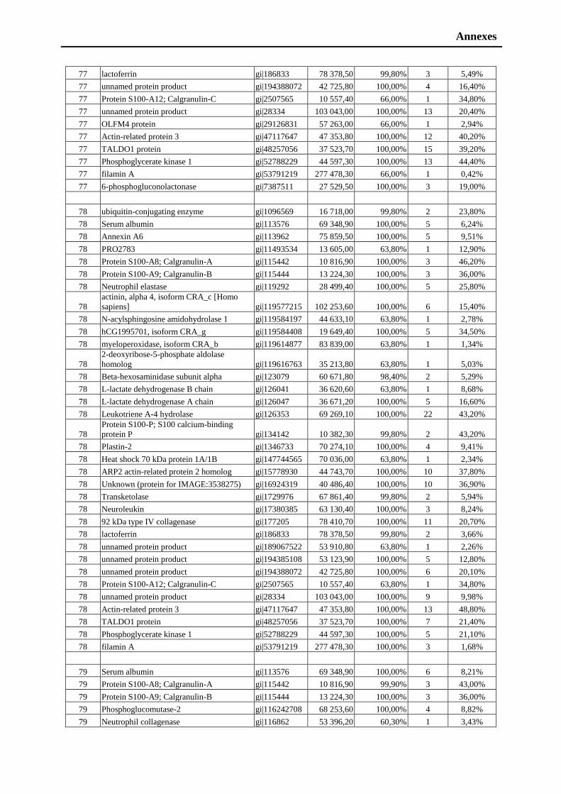
Annexes
77 lactoferrin gi|186833 78 378,50 99,80% 3 5,49%
77 unnamed protein product gi|194388072 42 725,80 100,00% 4 16,40%
77 Protein S100-A12; Calgranulin-C gi|2507565 10 557,40 66,00% 1 34,80%
77 unnamed protein product gi|28334 103 043,00 100,00% 13 20,40%
77 OLFM4 protein gi|29126831 57 263,00 66,00% 1 2,94%
77 Actin-related protein 3 gi|47117647 47 353,80 100,00% 12 40,20%
77 TALDO1 protein gi|48257056 37 523,70 100,00% 15 39,20%
77 Phosphoglycerate kinase 1 gi|52788229 44 597,30 100,00% 13 44,40%
77 filamin A gi|53791219 277 478,30 66,00% 1 0,42%
77 6-phosphogluconolactonase gi|7387511 27 529,50 100,00% 3 19,00%
78 ubiquitin-conjugating enzyme gi|1096569 16 718,00 99,80% 2 23,80%
78 Serum albumin gi|113576 69 348,90 100,00% 5 6,24%
78 Annexin A6 gi|113962 75 859,50 100,00% 5 9,51%
78 PRO2783 gi|11493534 13 605,00 63,80% 1 12,90%
78 Protein S100-A8; Calgranulin-A gi|115442 10 816,90 100,00% 3 46,20%
78 Protein S100-A9; Calgranulin-B gi|115444 13 224,30 100,00% 3 36,00%
78 Neutrophil elastase gi|119292 28 499,40 100,00% 5 25,80%
78
actinin, alpha 4, isoform CRA_c [Homo
sapiens] gi|119577215 102 253,60 100,00% 6 15,40%
78 N-acylsphingosine amidohydrolase 1 gi|119584197 44 633,10 63,80% 1 2,78%
78 hCG1995701, isoform CRA_g gi|119584408 19 649,40 100,00% 5 34,50%
78 myeloperoxidase, isoform CRA_b gi|119614877 83 839,00 63,80% 1 1,34%
78
2-deoxyribose-5-phosphate aldolase
homolog gi|119616763 35 213,80 63,80% 1 5,03%
78 Beta-hexosaminidase subunit alpha gi|123079 60 671,80 98,40% 2 5,29%
78 L-lactate dehydrogenase B chain gi|126041 36 620,60 63,80% 1 8,68%
78 L-lactate dehydrogenase A chain gi|126047 36 671,20 100,00% 5 16,60%
78 Leukotriene A-4 hydrolase gi|126353 69 269,10 100,00% 22 43,20%
78
Protein S100-P; S100 calcium-binding
protein P gi|134142 10 382,30 99,80% 2 43,20%
78 Plastin-2 gi|1346733 70 274,10 100,00% 4 9,41%
78 Heat shock 70 kDa protein 1A/1B gi|147744565 70 036,00 63,80% 1 2,34%
78 ARP2 actin-related protein 2 homolog gi|15778930 44 743,70 100,00% 10 37,80%
78 Unknown (protein for IMAGE:3538275) gi|16924319 40 486,40 100,00% 10 36,90%
78 Transketolase gi|1729976 67 861,40 99,80% 2 5,94%
78 Neuroleukin gi|17380385 63 130,40 100,00% 3 8,24%
78 92 kDa type IV collagenase gi|177205 78 410,70 100,00% 11 20,70%
78 lactoferrin gi|186833 78 378,50 99,80% 2 3,66%
78 unnamed protein product gi|189067522 53 910,80 63,80% 1 2,26%
78 unnamed protein product gi|194385108 53 123,90 100,00% 5 12,80%
78 unnamed protein product gi|194388072 42 725,80 100,00% 6 20,10%
78 Protein S100-A12; Calgranulin-C gi|2507565 10 557,40 63,80% 1 34,80%
78 unnamed protein product gi|28334 103 043,00 100,00% 9 9,98%
78 Actin-related protein 3 gi|47117647 47 353,80 100,00% 13 48,80%
78 TALDO1 protein gi|48257056 37 523,70 100,00% 7 21,40%
78 Phosphoglycerate kinase 1 gi|52788229 44 597,30 100,00% 5 21,10%
78 filamin A gi|53791219 277 478,30 100,00% 3 1,68%
79 Serum albumin gi|113576 69 348,90 100,00% 6 8,21%
79 Protein S100-A8; Calgranulin-A gi|115442 10 816,90 99,90% 3 43,00%
79 Protein S100-A9; Calgranulin-B gi|115444 13 224,30 100,00% 3 36,00%
79 Phosphoglucomutase-2 gi|116242708 68 253,60 100,00% 4 8,82%
79 Neutrophil collagenase gi|116862 53 396,20 60,30% 1 3,43%
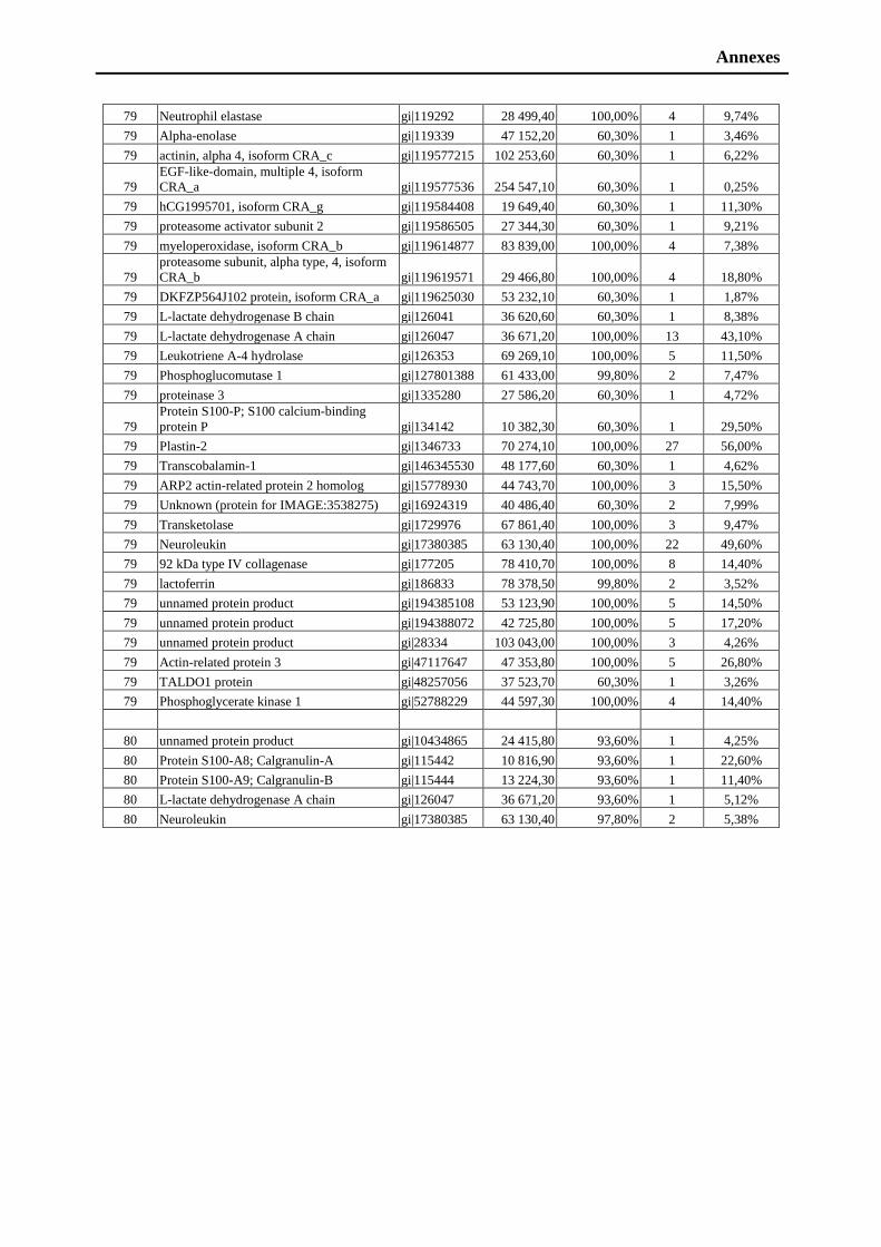
Annexes
79 Neutrophil elastase gi|119292 28 499,40 100,00% 4 9,74%
79 Alpha-enolase gi|119339 47 152,20 60,30% 1 3,46%
79 actinin, alpha 4, isoform CRA_c gi|119577215 102 253,60 60,30% 1 6,22%
79
EGF-like-domain, multiple 4, isoform
CRA_a gi|119577536 254 547,10 60,30% 1 0,25%
79 hCG1995701, isoform CRA_g gi|119584408 19 649,40 60,30% 1 11,30%
79 proteasome activator subunit 2 gi|119586505 27 344,30 60,30% 1 9,21%
79 myeloperoxidase, isoform CRA_b gi|119614877 83 839,00 100,00% 4 7,38%
79
proteasome subunit, alpha type, 4, isoform
CRA_b gi|119619571 29 466,80 100,00% 4 18,80%
79 DKFZP564J102 protein, isoform CRA_a gi|119625030 53 232,10 60,30% 1 1,87%
79 L-lactate dehydrogenase B chain gi|126041 36 620,60 60,30% 1 8,38%
79 L-lactate dehydrogenase A chain gi|126047 36 671,20 100,00% 13 43,10%
79 Leukotriene A-4 hydrolase gi|126353 69 269,10 100,00% 5 11,50%
79 Phosphoglucomutase 1 gi|127801388 61 433,00 99,80% 2 7,47%
79 proteinase 3 gi|1335280 27 586,20 60,30% 1 4,72%
79
Protein S100-P; S100 calcium-binding
protein P gi|134142 10 382,30 60,30% 1 29,50%
79 Plastin-2 gi|1346733 70 274,10 100,00% 27 56,00%
79 Transcobalamin-1 gi|146345530 48 177,60 60,30% 1 4,62%
79 ARP2 actin-related protein 2 homolog gi|15778930 44 743,70 100,00% 3 15,50%
79 Unknown (protein for IMAGE:3538275) gi|16924319 40 486,40 60,30% 2 7,99%
79 Transketolase gi|1729976 67 861,40 100,00% 3 9,47%
79 Neuroleukin gi|17380385 63 130,40 100,00% 22 49,60%
79 92 kDa type IV collagenase gi|177205 78 410,70 100,00% 8 14,40%
79 lactoferrin gi|186833 78 378,50 99,80% 2 3,52%
79 unnamed protein product gi|194385108 53 123,90 100,00% 5 14,50%
79 unnamed protein product gi|194388072 42 725,80 100,00% 5 17,20%
79 unnamed protein product gi|28334 103 043,00 100,00% 3 4,26%
79 Actin-related protein 3 gi|47117647 47 353,80 100,00% 5 26,80%
79 TALDO1 protein gi|48257056 37 523,70 60,30% 1 3,26%
79 Phosphoglycerate kinase 1 gi|52788229 44 597,30 100,00% 4 14,40%
80 unnamed protein product gi|10434865 24 415,80 93,60% 1 4,25%
80 Protein S100-A8; Calgranulin-A gi|115442 10 816,90 93,60% 1 22,60%
80 Protein S100-A9; Calgranulin-B gi|115444 13 224,30 93,60% 1 11,40%
80 L-lactate dehydrogenase A chain gi|126047 36 671,20 93,60% 1 5,12%
80 Neuroleukin gi|17380385 63 130,40 97,80% 2 5,38%

Rizwan ASLAM
Les peptides antimicrobiens dérivés de la chromogranine A et Staphylococcus aureus: de l’analyse
de l’interaction hôte-pathogène au développement de revêtement de polymère antimicrobien
Résumé
Les chromogranines (Cgs) sont une famille de protéines acides exprimées dans les granules des cellules neuroendocrines et immunitaires. Plusieurs peptides dérivés des Cgs présentent des activités antimicrobiennes. L’objectif de ma thèse est d’évaluer l’interaction hôte-pathogène et ensuite de développer un polymère antimicrobien avec insertion du peptide antimicrobien cateslytin (CTL).
Dans une première partie, nous avons évalué l’aptitude de la leukotoxine LukE/D à induire la sécrétion des neutrophiles et rôle des protéases bactériennes à dégrader les peptides dérivés de la CgA. Les neutrophiles activés sécrètent de nombreux composés que nous avons identifiés. De plus, la dégradation des PAMs dérivés de la CgA par les protéases de S. aureus a été déterminée. Sur tous les PAMs testés, CTL est le seul qui tue S. aureus et résister à dégradation. Par ailleurs, CgA et CgB sont dégradés par la protéase Glu-C pour produire de nouveaux fragments sans activité antibactérienne, mais d’activité antifongique.
Dans une deuxième partie, nous avons décidé de préparer un revêtement conjugué à CTL. CTL-C est utilisé pour préparer des films avec le dépôt alterné de CHI et HA-CTL-C. Par la suite nous avons synthétisé HAFITC-CTL-C and HAFITC pour analyser leur interaction. HAFITC-CTL-C est rapidement détectable dans le cytoplasme sans provoquer la lyse cellulaire. De plus, les films contenant CTL-C ne sont pas toxiques pour les fibroblastes gingivaux humains.
En conclusion, CTL est le seul peptide antimicrobien dérivé de la CgA qui peut tuer S. aureus et résiste à la dégradation protéolytique, ce qui est de bon augure pour de nouvelles études visant à développer des biomatériaux antimcrobiens.
Mots clés: Staphylococcus aureus, leucotoxine, LukE/D, l'immunité innée, les peptides antimicrobiens,
chromogranines, cateslytin, revêtement de polymère antimicrobien, biomatériaux antimcrobiens
Abstract
Chromogranins (Cgs) are a family of acidic proteins, expressed in secretory granules of neuro-endocrine and immune cells. Several Cgs derived peptides express antimicrobial activity. Current study was aimed to evaluate host-pathogen interaction and ultimately to develop antimicrobial polymer with insertion of cateslytine (CTL).
In first part, stimulatory ability of leukotoxin LukE/D to induce neutrophils secretions and role of bacterial proteases to degrade CgA-derived AMPs was evaluated. Activated neutrophils secrete various components which were identified. Later by using antimicrobial assays, several fractions were found active and later discussed with respect to proteomic analysis. Additionally, degradation of CgA derived AMPs by S. aureus proteases was demonstrated. Out of various AMPs tested, CTL was only that can kill S. aureus and resist protease degradation. Furthermore, CgA and CgB are processed by Glu-C protease to produce new fragments lacking antibacterial activity but presenting antifungal activity.
Secondly, we aimed to prepare CTL conjugated biomaterial coating. CTL-C was used to prepare PEM films with alternative deposition of CHI and HA-CTL-C and evaluated for antimicrobial activities. Later on, we synthesized HA
FITC-CTL-C and HA
FITC to analyze their interaction. HA
FITC-CTL-C was readily detectable in
cytoplasm without provoking cell lysis. Moreover CTL-C inserted PEM films are non-toxic to human gingival fibroblast cells.
In conclusion, CTL is the only CgA-derived AMP that can kill S. aureus and resistant to proteolytic degradation, which is a promising feature for further studies in order to develop antimicrobial biomaterials.
Key words: Staphylococcus aureus, leukotoxin, LukE/D, innate immunity, antimicrobial peptides,
chromogranins, cateslytin, antimicrobial polymer coating, antimicrobial biomaterial.