Embed Size (px)
Citation preview

Three-dimensional Reconstruction of Human Cystic FibrosisTransmembrane Conductance Regulator Chloride ChannelRevealed an Ellipsoidal Structure with Orifices beneath thePutative Transmembrane Domain*
Received for publication, April 25, 2008, and in revised form, August 5, 2008 Published, JBC Papers in Press, August 22, 2008, DOI 10.1074/jbc.M803185200
Kazuhiro Mio‡, Toshihiko Ogura‡§, Muneyo Mio‡, Hiroyasu Shimizu¶�, Tzyh-Chang Hwang¶, Chikara Sato‡1,and Yoshiro Sohma¶**‡‡2
From the ‡Neuroscience Research Institute, National Institute of Advanced Industrial Science and Technology (AIST), Umezono1-1-4, Tsukuba, Ibaraki 305-8568, Japan, §Precursory Research for Embryonic Science and Technology (PRESTO), Japan Scienceand Technology Agency, 4-1-8 Honcho Kawaguchi, Saitama 332-0012, Japan, ¶John M. Dalton Cardiovascular Research Center,University of Missouri, Columbia, Missouri 65211, Departments of �Hygiene and Public Health and **Physiology, Osaka MedicalCollege, Takatsuki, Osaka 569-8686, Japan, and ‡‡Department of Pharmacology, Keio University School of Medicine, Shinjuku,Tokyo 160-8582, Japan
The cystic fibrosis transmembrane conductance regulator(CFTR) chloride channel is a membrane-integral protein thatbelongs to an ATP-binding cassette superfamily. Mutations inthe CFTR gene cause cystic fibrosis in which salt, water, andprotein transports are defective in various tissues. Here weexpressed wild-type human CFTR as a FLAG-fused protein inHEK293 cells heterologously and purified it in three steps: anti-FLAGandwheat germagglutinin affinity chromatographies andsize exclusion chromatography. The stoichiometry of the pro-tein was analyzed using various biochemical approaches,including chemical cross-linking, blue-native PAGE, size exclu-sion chromatography, and electron microscopy (EM) observa-tion of antibody-decorated CFTR. All these data support adimeric assembly of CFTR. Using 5,039 automatically selectedparticles from negatively stained EM images, the three-dimen-sional structure of CFTR was reconstructed at 2-nm resolutionassuming a 2-fold symmetry. CFTR, presumably in a closedstate, was shown to be an ellipsoidal particle with dimensions of120 � 106 � 162 A. It comprises a small dome-shaped extracel-lular and membrane-spanning domain and a large cytoplasmicdomain with orifices beneath the putative transmembranedomain. EM observation of CFTR�anti-regulatory domain anti-body complex confirmed that two regulatory domains arelocated around the bottom end of the larger oval cytoplasmicdomain.
The cystic fibrosis transmembrane conductance regulator(CFTR)3 (also termedABCC7) is a uniquemember of the ATP-binding cassette (ABC) superfamily in that CFTR functions asan anion channel, whereas most other members function asactive transporters. CFTR is expressed in the luminal mem-branes of secreting and absorbing epithelia and plays a criticalrole in transepithelial salt and water transport. Dysfunction ofCFTR leads to cystic fibrosis, the most common lethal autoso-mal recessive disorder in Caucasians (1–3). On the other hand,extremely high activity of CFTR, usually caused by bacterialtoxins, results in secretory diarrhea (4, 5), killing millions ofinfants in developing countries every year (6). Understandingthe structure/function relationship and the underlying mecha-nisms of CFTR are essential for developing novel therapeuticsfor CFTR-mediated diseases.Like other ABC transporters, CFTR is formed by two
repeated motifs, each of which has a membrane-spanningdomain (MSD) and a cytoplasmic nucleotide-binding domain(NBD). However, a regulatory domain (R domain) locatedbetween the first NBD (NBD1) and the secondMSD (MSD2) isunique in CFTR among ABC transporters. This domain con-tains several phosphorylation sites for protein kinase A andprotein kinase C, and the level of their phosphorylation con-trols CFTR channel activity. Once they are phosphorylated,opening and closing (gating) of the CFTR channel is con-trolled by ATP binding and hydrolysis at its two NBDs (7).Each NBD contains the Walker A and Walker B nucleotide-binding motifs and the “signature sequence” LSGGQ, whichdefines the ABC superfamily. It is generally accepted that
* This work was supported, in whole or in part, by National Institutes of HealthGrants R01 DK55835-09 and R01 HL53445-11 (to T.-C. H.). This work wasalso supported by a grant-in-aid for scientific research on priority areas,structure of biological macromolecular assemblies (to K. M. and C. S.); by agrant from the Japan New Energy and Industrial Technology DevelopmentOrganization (NEDO) (to C. S.); by a grant from PRESTO of the Japan Scienceand Technology Agency (to T. O.); and by Japan Society for the Promotionof Science (JSPS) Grant 19590215 (to Y. S.). The costs of publication of thisarticle were defrayed in part by the payment of page charges. This articlemust therefore be hereby marked “advertisement” in accordance with 18U.S.C. Section 1734 solely to indicate this fact.
1 To whom correspondence may be addressed. Tel.: 81-29-861-5562; Fax:81-29-861-6478; E-mail: [email protected].
2 To whom correspondence may be addressed: Dept. of Pharmacology,Keio University School of Medicine, 35 Shinanomachi, Shinjuku-ku,Tokyo 160-8582, Japan. Tel.: 81-3-5363-3750; Fax: 81-3-3359-8889;E-mail: [email protected].
3 The abbreviations used are: CFTR, cystic fibrosis transmembrane conduct-ance regulator; ABC, ATP-binding cassette; bis-Tris, 2-[bis(2-hydroxyeth-yl)amino]-2-(hydroxymethyl)propane-1,3-diol; DDM, n-dodecyl-�-D-mal-toside; EM, electron microscopy; HEK, human embryonic kidney; MSD,membrane-spanning domain; NBD, nucleotide-binding domain; NMDG,N-methyl-D-glucamine; NN, neural network; PBS, phosphate-bufferedsaline; R domain, regulatory domain; RS, Stokes radius; SA, simulatedannealing; SEC, size exclusion chromatography; SPINNS, single particleimage analysis method using neural network and simulated annealing;TBS, Tris-buffered saline; Tricine, N-[2-hydroxy-1,1-bis(hydroxymethyl)-ethyl]glycine.
THE JOURNAL OF BIOLOGICAL CHEMISTRY VOL. 283, NO. 44, pp. 30300 –30310, October 31, 2008© 2008 by The American Society for Biochemistry and Molecular Biology, Inc. Printed in the U.S.A.
30300 JOURNAL OF BIOLOGICAL CHEMISTRY VOLUME 283 • NUMBER 44 • OCTOBER 31, 2008
by guest on March 23, 2019
http://ww
w.jbc.org/
Dow
nloaded from
NBD1 and -2 are dimerized in a head to tail configurationwith two ATP molecules sandwiched between the WalkerA/B motifs of one NBD and the signature sequences of thecounterpart NBD (8). There are lines of convincing evidencethat the opening of CFTR chloride channel is associated withthis NBD dimerization (9). The detailed dynamics of ATPbinding and NBD dimerization in CFTR gating has beenintensively investigated (10–12).Despite a plethora of biochemical and electrophysiological
results, structural information ofCFTR is limited because of thelack of abundant CFTR protein sources and the difficulties inprotein purification and crystallization. So far, only the struc-ture of the NBD1 domain of CFTR has been determined byx-ray crystallography using mouse (13) and human (14) pro-teins. Ford and co-workers (15, 16) described low resolutionstructures of wild-type human CFTR by two-dimensional elec-tron crystallography (15) and by the single particle reconstruc-tion technique (16). The two-dimensional crystallographyshowed two different monomeric structures for CFTR (15). Onthe other hand, the single particle analysis suggested a structurewhose particle size is compatible with a dimeric association bytwo CFTR molecules (16).In this study, we purified glycosylated mature CFTR and
reconstructed its three-dimensional structure from negativelystained EM images. Our data suggest that two CFTR proteinsform an ellipsoidal “tail to tail” dimeric configuration with sideorifices in the cytoplasm and that at least part of the R domain islocated around the bottom end of the larger cytoplasmicdomain.
EXPERIMENTAL PROCEDURES
Expression Constructs, Transfection of HEK293 Cells, andMembrane Preparation—Full-length human CFTR cDNA wassubcloned into pcDNA3.1 Zeo (Invitrogen) and tagged withFLAG sequence at either theN (abbreviatedN-FLAGCFTR) orC terminus (C-FLAG CFTR). The HEK293 cells were trans-fected with 4 �g of the DNA construct/1 � 106 cells for 6 husing the calcium phosphate precipitation method (17) toexpress FLAG-fused CFTR protein. After 48 h, cells werewashed twice in TBS (20 mM Tris-HCl, pH 7.4 at 4 °C, and 140mM NaCl) and harvested using Teflon cell scrapers. After thecells were collected by centrifugation, they were first homoge-nized with a Teflon homogenizer in 10 volumes (v/w) of TBSand then centrifuged at 1,500 � g for 10 min to remove debris.The supernatant was centrifuged at 100,000 � g for 60 min toobtain the membrane fraction.Immunofluorescence Experiments—HEK293 cells trans-
fected with CFTR constructs were cultured for 48 h and fixedwith 4% paraformaldehyde in phosphate-buffered saline (PBS;136 mM NaCl, 1.4 mM KCl, 10 mM Na2HPO4, and 1.7 mMKH2PO4, pH 7.4) at room temperature for 10 min. For mem-brane permeabilization, cells were incubatedwith PBS contain-ing 0.1% saponin and 2 mg/ml bovine serum albumin at roomtemperature for 30 min. Cells were incubated with Cy3-conju-gated anti-FLAGantibodies (Sigma) in PBS containing 2mg/mlbovine serum albumin and 0.1% saponin at 4 °C for 6 h. Theywere washed with PBS three times and incubated with PBScontaining 1 �g/ml of 4�,6-diamidino-2-phenylindole at room
temperature for 30 min. Samples were washed with PBS andmounted on glass slides with 50% glycerol. Control cells with-out membrane permeabilization were treated similarly exceptthat saponin treatment was not performed. Samples wereobserved using an Olympus IX 71 microscope with a DP50CCD camera (Olympus).Electrophysiological Measurement—Channel functions of
C-FLAG CFTR expressed in Chinese Hamster ovary orHEK293 cells were compared with those reported for wild-typeCFTR (10–12, 18, 19) by inside-out patch clamp recordingusing an EPC10 Patch Clamp Amplifier (HEKA Electronik).The pipette solution contained 140 mM NMDG-Cl, 2 mMMgCl2, 5 mM CaCl2, and 10 mM HEPES, pH 7.4 with NMDG.Cells were perfused with a bath solution containing 145 mMNaCl, 5 mM KCl, 2 mM MgCl2, 1 mM CaCl2, 5 mM glucose, 5mMHEPES, pH 7.4 with NaOH, and 20mM sucrose. After theestablishment of an inside-out configuration, the patch wasperfused with a standard perfusion solution (i.e. intracellularsolution) containing 150mMNMDG-Cl, 2 mMMgCl2, 10mMEGTA, and 8 mM Tris, pH 7.4 with NMDG. Patches wereheld at �50 mV in all experiments. CFTR channel currentswere recorded at room temperature. The currents were fil-tered at 100 Hz with an eight-pole Bessel filter (model LPF-8;Warner Instruments) and digitized on line at 500 Hz. CFTRchannels were activated by the combined application of 1mM MgATP and 25 units/ml cAMP-dependent proteinkinase. MgATP and cAMP-dependent protein kinase werepurchased from Sigma.Protein Purification—The membrane fraction was homog-
enized in 5 volumes (v/w) of buffer A (TBS containing 25 mMn-dodecyl �-D-maltoside (DDM) (Sigma), 300 mM MgCl2,10% glycerol, protease inhibitors, and 0.02% sodium azide).After centrifuging at 100,000 � g for 30 min, the supernatantcontaining solubilized FLAG-tagged CFTR was loaded ontoan anti-FLAG affinity column (Sigma) equilibrated inadvance. The column was then washed with 10 column vol-umes of buffer B (TBS containing 5 mM DDM, 300 mMMgCl2, 10% glycerol, protease inhibitors, and 0.02% sodiumazide), and the bound CFTR protein was eluted with buffer Bcontaining 100 �g/ml FLAG peptide (Sigma). The eluatesfrom the FLAG column were analyzed by SDS-PAGE fol-lowed by silver staining.The CFTR-rich fractions were pooled and loaded to the
wheat germ agglutinin (WGA)-agarose column (Seikagaku).Unbound protein was washed out by 5 column volumes ofbuffer B. The bound CFTR protein was eluted by the lineargradient ofN-acetylglucosamine (GlcNAc) from 20 to 100 mM.The elution of CFTR protein was monitored by SDS-PAGEanalysis. The CFTR-rich fractions were pooled, concentratedwith a Microcon filter unit (YM-100; Millipore) and furtherpurified by Superdex 200 size exclusion chromatography (SEC)in a SMART System (GE Healthcare) using buffer C (TBS con-taining 5 mM DDM, 500 mM MgCl2, 10% glycerol, and 0.02%sodium azide). The elution of protein from the SEC was moni-tored by its UV absorbance at 280 nm and also by SDS-PAGEanalysis.Estimation of Stokes Radius by SEC—The distribution coef-
ficient, Kav, was calculated from the equation Kav � (Ve � V0)/
Three-dimensional Reconstruction of CFTR Chloride Channel
OCTOBER 31, 2008 • VOLUME 283 • NUMBER 44 JOURNAL OF BIOLOGICAL CHEMISTRY 30301
by guest on March 23, 2019
http://ww
w.jbc.org/
Dow
nloaded from

(Vt � V0) where Ve is the elution volume of high molecularweight standards (GE Healthcare) or CFTR protein. Columnvoid volume (V0) was measured with blue dextran 2000, andVt represents total bed volume. The Stokes radius (RS) ofCFTR was determined using a calibration curve constructedby plotting the RS of each reference protein versus (�logKav)
1⁄2 according to the relationship (�log Kav)1⁄2 � �(� � RS)
(20, 21). All the standards and the CFTR were solubilized inthe same buffer used for CFTR purification. The standardproteins used were thyroglobulin (RS � 85.0 Å), ferritin (RS� 61.0 Å), catalase (RS � 52.2 Å), and aldolase (RS � 48.1 Å).The elution of CFTR was repeated five times; the data arepresented by average � S.D.SDS and Blue-Native PAGE—The standard method of Lae-
mmli (22) was applied for SDS-PAGE. Samples were mixedwith an equal volumeof sample buffer containing 62.5mMTris-HCl, pH 6.8, 2% SDS, 25% glycerol, 0.04 M dithiothreitol, and0.01% bromphenol blue and then incubated at 60 °C for 15min.Proteins were separated in a 5–20% polyacrylamide gel andvisualized by silver staining. For Western blots, electrophore-sed proteins in the gel were transferred to a polyvinylidenedifluoride membrane and analyzed with alkaline phosphatase-labeled anti-FLAG antibodies (Sigma). For blue-native PAGE,purified CFTR was mixed with an equal volume of samplebuffer (1% Coomassie G-250, 100 mM NaCl, 20% glycerol, and100mM bis-Tris, pH 7.2) and electrophoresed in a 4–16% poly-acrylamide gel using a cathode buffer containing 50 mM bis-Tris, 50 mM Tricine, pH 6.8, and 0.002% Coomassie G-250 andan anode buffer containing 50 mM bis-Tris and 50 mM Tricine,pH 6.8 (23, 24). Electrophoresis started at 50 V for 30 min andwas continued at 150 V for 90 min at room temperature. Afterdestaining the gel with a solution containing 50%methanol and10% acetic acid, the protein in the gel was visualized by silverstaining or analyzed by Western blotting. The molecular massof intact CFTRwas estimated from a calibration curve obtainedby plotting the mobility of standard proteins relative to the dyefront against the logarithm of their molecular masses. Thestandards used were thyroglobulin (669 kDa), ferritin (440kDa), catalase (232 kDa), lactate dehydrogenase (140 kDa), andbovine serum albumin (67 kDa).Chemical Cross-linking—The TBS component in the buffer
was substituted with PBS by dialysis. Glutaraldehyde or disuc-cinimidyl suberate was mixed with CFTR-containing solutionto the indicated final concentration at room temperature for 30min. Cross-linking was terminated by incubation with an equalvolume of SDS sample buffer at 60 °C for 15 min. The proteinswere separated in a 2–15% acrylamide gel and analyzed byWestern blotting using anti-FLAG antibody. A protein sampletreated similarly but without cross-linker was prepared as acontrol.Transmission Electron Microscopy—Purified CFTR proteins
of�50 �g/ml were adsorbed by thin carbon films supported bycopper mesh grids, which were rendered hydrophilic inadvance by glow discharge in low air pressure. Samples werewashed with five drops of double distilled water, negativelystained with 2% uranyl acetate solution for 30 s twice, blotted,and dried in air. Micrographs of negatively stained particleswere recorded in a JEOL 100CX transmission electron micro-
scope at �52,100 magnification with 100-kV acceleration volt-age and an electron dose of �30 electrons/Å2 at the specimenlevel. Themagnification calibration was performed by a gratingreplica (Nissin EM Co.). Images were recorded on SO-163image films (Eastman Kodak Co.) developed with a D19 devel-oper (Kodak) and digitized with a Scitex Leafscan 45 scanner(Leaf Systems) at a pixel size of 1.92 Å at the specimen level.Molecular complexes between CFTR and the antibodies
were generated by mixing the purified CFTR with an anti-FLAG M2 antibody (Sigma) or an anti-R domain MAB1660antibody (R&D Systems) at 4 °C for 30 min. After removingexcessive antibodies by SEC, the complexes were negativelystained and observed by EM. To obtain clearer images of theCFTR�anti-FLAG antibody complexes, Fab fragments weregenerated from the antibodies by papain digestion and con-jugated with colloidal gold (BBInternational). The conjugatewas isolated from non-reacted Fab molecules by 10–30%glycerol gradient centrifugation and then mixed with CFTRat 4 °C for 30 min. Protein G conjugated with colloidal gold(BBInternational) was used to obtain clearer images ofMAB1660 binding. The protein G-gold was mixed withCFTR�MAB1660 complexes on the WGA-agarose, and thenexcessive gold was washed out. The gold-labeled complexeswere eluted with 100 mM GlcNAc, negatively stained, andobserved by EM.Automated Particle Selection and Image Analysis—We have
developed a single particle image analysis method using neuralnetwork (25–27) and simulated annealing (28, 29) namedSPINNS (30). The following image analysis was performedusing our SPINNS and IMAGICV (31). CFTR projections werefirst picked up using the autoaccumulation method with simu-lated annealing (SA) (28). 291 particles in 160 � 160-pixel sub-frames were selected and used to train a three-layer pyramidal-type neural network (NN) (25, 26). Using the trained NN, 5,039particles were selected. After background subtraction, the par-ticles selected by the NN were aligned rotationally and transla-tionally (32, 33) by the reference-free method (26). The alignedimages were sorted into 150 classes by the modified growingneural gas network method (27). Their class averages wereadopted as new references, and this cycle, from alignment toaveraging, was repeated 11 times.The Euler angles of the class averages were automatically
determined by the echo-correlated three-dimensional recon-struction method with SA (29). In this step, C2 symmetry wasimposed because top and bottom images in averaged projec-tions exhibited 2-fold symmetry. These Euler angles were usedto calculate a primary three-dimensional structure by thesimultaneous iterative reconstruction technique (34). Thereprojections from the volumewere used as references formul-tireference alignment, and raw images in the library werealigned and further clustered, providing improved cluster aver-ages. From these averages, a new three-dimensional map wasgenerated by the reconstruction method using SA without athree-dimensional reference.The three-dimensional map was further refined by the pro-
jection matching method (35) followed by an optimizationusing echo-correlated reconstruction. This cycle was repeatedfor seven cycles. Particle images that correlated poorly with the
Three-dimensional Reconstruction of CFTR Chloride Channel
30302 JOURNAL OF BIOLOGICAL CHEMISTRY VOLUME 283 • NUMBER 44 • OCTOBER 31, 2008
by guest on March 23, 2019
http://ww
w.jbc.org/
Dow
nloaded from

projections from the three-dimensional structure were automati-cally rejectedusing thecross-correlation function.The final recon-struction includes 4,206 particles, 83.5% of all the selected images.The resolution of the final three-dimensional map was assessedusing the Fourier shell correlation function (36) at the thresholdof 0.5.
RESULTS
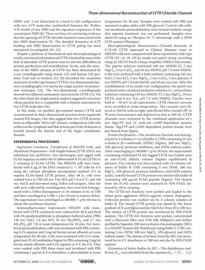
Heterologous Expression of CFTR Protein—We first con-structed vectors containing human CFTR cDNA tagged withFLAG sequence at either the N or C terminus and transfectedHEK293 cells with the vectors by the calcium phosphate pre-cipitationmethod. The expressions of CFTRwere compared byimmunofluorescence using Cy3-conjugated anti-FLAG anti-body. After the membrane permeabilization of the cells, theantibody reacted with �10% of cells expressing C- or N-FLAGCFTR (Fig. 1a, first and third rows). The level of Cy3 fluores-cence was higher in the cells transfected with C-FLAG CFTRthan those with N-FLAG CFTR. In contrast, the non-perme-abilized control cells were negative to the antibody (Fig. 1a,second and fourth rows), confirming that both N and C terminiare located inside the cells. The expressed CFTR proteins weresolubilized with DDM and analyzed by Western blotting usinganti-FLAG antibody. The antibody detected a broadened bandat the expected size for themature fully glycosylatedCFTR (Fig.1b, black arrowheads; estimated size, 177 kDa (169 kDa for themonomeric CFTR protein plus FLAG tag and associated gly-can)) (37). A sharp band beneath the mature CFTR is likely thecore-glycosylated immature CFTR (indicated by a small arrow)(37). The expression of C-FLAG CFTR increased during the30-h period after transfection and reached its plateau at 48 h.The expression of N-FLAG CFTR was faintly detected at thecorresponding position. We confirmed that both constructsretain ATP and cAMP-dependent protein kinase phosphoryla-tion-dependent channel functions (see below). However,because the expression level of N-FLAGCFTRwasmuch lowerthan that of C-FLAG CFTR, we adopted C-FLAG CFTR forfurther studies.Electrophysiological Measurement of C-FLAG CFTR—The
channel function of C-FLAG CFTR was examined using thepatch clamp technique. Both cAMP-dependent proteinkinase and ATP were applied to the cytoplasmic (bath) side ofthe inside-out patch obtained from HEK293 cell expressingC-FLAG CFTR (Fig. 2a). Once the C-FLAG CFTR was phos-phorylated, the channel was opened by ATP alone (Fig. 2a).Single channel conductance (�6 picosiemens) and the ATP-dependent gating behavior are similar to those in non-FLAG-tagged wild-type CFTR (10–12, 18, 19). The ATP dependenceof the channel activity of C-FLAG CFTR is almost identicalbetween the HEK293 and Chinese Hamster ovary expressionsystems (Fig. 2, b and c) and also comparable to that of non-FLAG-tagged wild-type CFTR (10–12, 18, 19). These data con-firmed that the mechanisms of cAMP-dependent proteinkinase phosphorylation-induced ATP-dependent gating andthe ion conducting functions were retained in C-FLAG CFTR.Purification of CFTR Proteins from Heterologously Expressed
HEK293 Cells—CFTR proteins were purified from the mem-brane fraction by a combination of anti-FLAG immunoaffinity
chromatography, lectin affinity chromatography, and SEC. TheCFTR bound to the FLAG affinity column was competitivelyeluted with 100 �g/ml FLAG peptides and analyzed by SDS-PAGE. Although CFTR was electrophoresed as a predominantband at 177 kDa, several contaminants including a sharp bandat 100 kDa and an immature CFTR moving slightly faster thanthe mature protein were also observed (Fig. 3a). To isolatemature CFTR, the eluate from the FLAG column was furthersubjected to WGA chromatography, which has been shown tobe effective for CFTR purification (15). The bound CFTR waseluted using a linear gradient of GlcNAc from 20 to 100 mM in
FIGURE 1. Expression of recombinant CFTR in HEK cells. a, immunofluores-cent images of HEK293 cells expressing FLAG-tagged CFTR. Top row, cellstransfected with C-FLAG CFTR and permeabilized with 0.1% saponin. CFTRprotein was detected using Cy3-conjugated anti-FLAG antibody (middlepanel, red). Cell nuclei were visualized by 4�,6-diamidino-2-phenylindole(DAPI) (right panel, blue). Second row, cells expressing C-FLAG CFTR wereexamined without saponin treatment. The cells were negative to the FLAGantibody, suggesting that the C-terminal epitope is inside the cell. Third row,cells transfected with N-FLAG CFTR and permeabilized by saponin. The levelof Cy3 immunofluorescence is lower than that of C-FLAG CFTR. Fourth row,the non-permeabilized cells transfected with N-FLAG CFTR were also nega-tive to the antibody. Cells were cultured for 48 h after transfection. The scalebar represents 50 �m. b, Western blot with anti-FLAG antibody. Left panel,cells were transfected with C-FLAG CFTR and cultured for the indicated peri-ods. Proteins were solubilized from the membrane using DDM and precipi-tated with anti-FLAG gel. Expression of CFTR increased with the cultureperiod up to 48 h. Right panel, the expression of N-FLAG CFTR was very lowbut was detected at the same size as C-FLAG CFTR. The broad bands at 177kDa are glycosylated mature CFTR (arrowheads), and the sharp bands thatmove faster than the mature protein are core-glycosylated but immatureCFTR (small arrow).
Three-dimensional Reconstruction of CFTR Chloride Channel
OCTOBER 31, 2008 • VOLUME 283 • NUMBER 44 JOURNAL OF BIOLOGICAL CHEMISTRY 30303
by guest on March 23, 2019
http://ww
w.jbc.org/
Dow
nloaded from
which the CFTR was mainly eluted between 40 and 60 mM(Fig. 3b).Although the CFTR was highly concentrated after this puri-
fication step, a broad band at 110 kDa still remained (Fig. 3b). Itwas recognized by anti-FLAG antibody (data not shown) andspeculated to be degraded CFTR. To remove this contaminant,the eluate from the WGA column was concentrated using aMicrocon YM-100 filter unit and further purified by Superdex200 SEC. The elution of CFTR from the columnwasmonitoredby UV absorption at 280 nm (Fig. 3c), and the aliquot of eachfraction was analyzed by SDS-PAGE followed by silver staining(Fig. 3d). A sharp peak at 1.04-ml elution in the SEC was con-firmed to be CFTR (Fig. 3d). The 110-kDa contamination wasfainter than the band of CFTR and mainly eluted at 1.12–1.16ml. A rise at 0.87-ml elution and large absorbance at 1.5 ml didnot accompany detectable proteins; these are speculated to beabsorptions due to micelles of lipids derived from the plasmamembrane. Absorbance of FLAG peptides was observed at2.00-ml elution. An aliquot at 1.04-ml elution (Fig. 3, c and d,arrow) was used for the EM study. This fraction containedhighly concentrated mature CFTR with a minimal contamina-tion of the 110-kDa proteins.Using the purified CFTR, the hydrated size (Stokes radius,
RS) of CFTRwas calculated from the elution volume in SEC (20,21) where CFTR was eluted between the thyroglobulin (elutedat 0.991 ml, RS is 85.0 Å) and ferritin (eluted at 1.129 ml, RS is61.0 Å). The RS of CFTR was determined to be 74.8 � 1.1 Å(mean � S.D., n � 5) from the calibration curve (Fig. 3e).Chemical cross-linking and blue-native PAGE were used to
determine the stoichiometry. Purified CFTR was treated with
various concentrations of glutaral-dehyde or disuccinimidyl suberateand analyzed by SDS-PAGE fol-lowed by Western blotting usinganti-FLAG antibody (Fig. 4a). TheTBS in the buffer composition wasreplaced with PBS by dialysis inadvance. The band of CFTR treatedwith either chemical was shifted to ahigher position with a molecularweight expected for a CFTR dimer.To more accurately estimate themolecular mass of the purifiedCFTR, we ran the blue-native PAGEand found the band of CFTRbetween the standards of 232 and440 kDa (Fig. 4b, indicated byarrow). From the calibration curveobtained from the standard pro-teins, the molecular mass of thepurified CFTR was determined tobe 353 kDa (Fig. 4c), nearly doublethe size of the CFTRmonomer (177kDa including tag and glycan).These results suggest that purifiedCFTR proteins may assume a dimerconfiguration.Electron Microscopy and Three-
dimensional Reconstruction of CFTR—Purified CFTR proteinswere blotted onto a glow-discharged carbon film supported bya copper mesh grid, negatively stained with 2% uranyl acetate,and imaged using an electron microscope at �52,100 magnifi-cation. Variously shaped particles of uniform size wereobserved (Fig. 5). Most particles were triangular or polygonalwith round corners. The variation in shapes is interpreted toreflect different orientations of the same molecule on the grid.The elliptical particles, although rarely observed, are postulatedto be top views of the dimeric CFTR, whereas the triangular orpolygonal shapes likely represent side views.Because the hydropathy plot of CFTRpredicted that 79.5% of
the CFTR sequence is in the cytoplasm (see Fig. 9a), the largerdomain of the protein is speculated to be cytoplasmic. To con-firm this hypothesis, the FLAG tag at the cytoplasmic C termi-nus (38) was decorated with the anti-FLAG antibody, and thecomplex was negatively stained and observed by EM. The anti-bodies indeed attached to the larger domain of CFTR, confirm-ing that this side of the molecule represents the cytoplasmicdomain (Fig. 6a). It should be noted that we frequentlyobserved CFTR particles bearing two antibodies (Fig. 6a, rightend column). This observation further supports the dimericassembly of CFTR. The bound antibodies were much moreclearly observed using the Fab fragments conjugated with col-loidal gold. Gold particles were again observed on the largerdomain of CFTR (Fig. 6b).The projections of CFTRwere picked up by a combination of
two automatic programs: the autoaccumulation method usingSA (28) and the three-layered neural network method (25, 26).The three-dimensional structure was reconstructed with echo-
FIGURE 2. Channel function of C-FLAG CFTR. a, activation of C-FLAG CFTR by cAMP-dependent protein kinase(PKA)-dependent phosphorylation and ATP. Single channel currents were obtained from an inside-out patchfrom HEK293 cells expressing C-FLAG CFTR. After phosphorylation, ATP alone can gate C-FLAG CFTR. Vm � �50mV. The dashed line indicates the closed level. b, ATP-dependent macroscopic currents of cAMP-dependentprotein kinase-phosphorylated C-FLAG CFTR in the inside-out configuration. The concentrations of ATP in thebath are indicated above. Vm � �50 mV. c, ATP concentration dependence of macroscopic currents of C-FLAGCFTR expressed in HEK293 (red) and Chinese Hamster ovary (green) cells. Vm � �50 mV. These results are alsocomparable to the channel function of non-FLAG-tagged wild-type CFTR (10 –12, 18, 19).
Three-dimensional Reconstruction of CFTR Chloride Channel
30304 JOURNAL OF BIOLOGICAL CHEMISTRY VOLUME 283 • NUMBER 44 • OCTOBER 31, 2008
by guest on March 23, 2019
http://ww
w.jbc.org/
Dow
nloaded from

correlated reconstructionmethods using SA assumingC2 sym-metry in our SPINNS (25–30) and other algorithms in theIMAGIC V software (31) (see “Experimental Procedures” fordetails). The final reconstruction included 4,206 particles,83.5% of all the selected images.Representative raw images are presented (Fig. 7a, first row)with
their corresponding class averages (second row) and with the sur-
face representations and the projec-tions of the reconstructed three-di-mensional structure (third and fourthrows). A high level of consistency wasobserved in size, shape, and innerstructure among these data sets (Fig.7a), indicating successful three-di-mensional reconstruction from theoriginal particle images. According tothe Fourier shell correlation function(36), the resolution limit is 2 nm bythe correlation coefficient0.5 crite-rion (Fig. 7b). A plot of the Eulerangles of the 94 adopted class aver-ages shows that CFTR is almost ran-domly oriented on the grid surface(Fig. 7c).Structural Features of the CFTR
Molecule—The surface representa-tion demonstrates that CFTR is anellipsoidal molecule of 162 Å inheight. The elliptical top view has amajor diameter of 120 Å and minordiameter of 106 Å (Fig. 8a). Thethree-dimensional map is con-toured at an isosurface containing avolume corresponding to 276 kDaassuming a protein density of 1.37g/cm3, which is 81.7% of the dimericCFTR mass (338 kDa) calculatedfrom the amino acid composition.The putative position of the mem-brane-spanning region is indicatedby a blue line in Fig. 8a (�30 Å inwidth) so that the ratio of eachdomain is close to the predictionfrom the hydropathy plot (i.e. thevolumes of extracellular, mem-brane-spanning, and cytoplasmicdomains are 3.8, 16.7, and 79.5%,respectively; Fig. 9a).The surface of the dome-shaped
extracellular domain and the MSDis seamless and smooth, whereas thecytoplasmic surface is rough andcontains orifices under the putativetransmembrane region. Large ori-fices of �50 � 30 Å are prominentin the center of the wider side views(Fig. 8b, panels 11 and 12), andsmall elliptical orifices of 20 � 25 Å
are prominent at the narrower side views (panel 15). Theseorifices might correspond to the region of intracellular loopsbetween MSDs (1).Sections normal to the symmetry axis demonstrate the
tightly packed internal density at extracellular and transmem-brane domains (Fig. 8c, panels 1–6). In contrast, the densityinside the cytoplasmic domain is low (arrows, panels 8–10)
FIGURE 3. Purification of the CFTR protein from transiently expressed HEK cells. Silver staining of aliquotsat each purification step from anti-FLAG (a) and from WGA columns (b). Proteins bound to the WGA columnwere eluted by a linear gradient of GlcNAc from 20 to 100 mM. Bands of fully glycosylated mature CFTR areindicated by arrowheads. c, CFTR-rich eluates from the WGA column were concentrated using the MicroconYM-100 filter unit and analyzed by Superdex 200 SEC. CFTR was eluted in a sharp peak at 1.04-ml elution(indicated by an arrow). d, SDS-PAGE analysis of the SEC fractions. The intensity of the CFTR band in the gelcorresponds to absorption in SEC. The fraction at 1.04 ml (arrow) was used for EM image analysis. e, from theelution volumes of CFTR and the standards, the RS of CFTR was calculated as 74.8 � 1.1 Å (mean � S.D., n � 5).T, thyroglobulin (RS, 85.0 Å); F, ferritin (RS, 61.0 Å); C, catalase (RS, 52.2 Å); A, aldolase (RS, 48.1 Å).
Three-dimensional Reconstruction of CFTR Chloride Channel
OCTOBER 31, 2008 • VOLUME 283 • NUMBER 44 JOURNAL OF BIOLOGICAL CHEMISTRY 30305
by guest on March 23, 2019
http://ww
w.jbc.org/
Dow
nloaded from

probably because of the stain permeation through the orifices(arrowheads).To determine the location of the R domain in the reconstruc-
tion, purified CFTR was mixed with an R domain-specific anti-body, MAB1660, and the complex was negatively stained andobserved by EM. The antibodies attached to the larger domainsuggesting that the R domain is located around the bottom endof the cytoplasmic domain (Fig. 9b). CFTRparticles bearing twoantibodies were frequently observed, and this again supportsthe dimeric stoichiometry of reconstructedCFTR (Fig. 9b, rightend). The MAB1660 bound to the CFTR molecule was clearlydemonstrated by the binding of gold-conjugated protein Gwhere one or two gold conjugates bound to the larger domain ofCFTR (Fig. 9c). When two gold conjugates were observed forone CFTR particle, these two gold conjugates were closelylocated next to each other at the cytoplasmic end of the particle,indicating that the CFTR particle assumes a tail to tail configu-ration. Perhaps because of the steric hindrance and increased
ion strength in the buffer composition, not all the epitopeswerebound with antibodies or gold conjugates.
DISCUSSION
Purification of CFTR Protein—The insect Sf9 expression sys-tem has been shown to express a large quantity of CFTR pro-teins (39).We adopted human embryonic kidney HEK293 cellsbecause, contrary to the insect cell system, mammalian cells
FIGURE 4. Chemical cross-linking and native PAGE analysis of CFTR.a, purified CFTR was treated with glutaraldehyde or disuccinimidyl suberate(DSS) at the indicated concentrations and separated by SDS-PAGE followedby Western blotting using anti-FLAG antibody. The band of CFTR (gray arrow-heads) was shifted upward at the corresponding position of a dimer (blackarrowheads) by treatments with both cross-linkers. b, blue-native PAGE ofCFTR. Purified CFTR was separated in a blue-native gel and visualized by silverstaining (Silver) or Western blotting using anti-FLAG antibody (anti-FLAG). TheCFTR protein was detected between the standards of 232 and 440 kDa(arrow). c, calculation of molecular mass from the blue-native PAGE. From themobility of proteins relative to the dye front (Rf), the molecular mass ofnative CFTR was calculated to be 353 kDa, suggesting dimeric assembly(i.e. 177 kDa � 2 � 354 kDa). T, thyroglobulin (669 kDa); F, ferritin (440kDa); C, catalase (232 kDa); L, lactate dehydrogenase (140 kDa); A, bovineserum albumin (66 kDa).
FIGURE 5. Electron microscopy of negatively stained CFTR. After adsorp-tion to hydrophilic carbon film, negatively stained samples were imaged on aJEOL 100CX electron microscope at a magnification of �52,100 with 100-kVacceleration voltage. The CFTR particles were observed as uniformly sizedprojections. For statistical analysis, 291 particles were automatically pickedup by the autoaccumulation method and utilized as training data for thethree-layer NN autopicking system. Using the trained NN, 5,039 particles werepicked up and used for the analysis. The scale bar represents 200 Å.
FIGURE 6. Assignment of the cytoplasmic C terminus of CFTR. a, gallery ofnegatively stained CFTR�anti-FLAG-antibody complexes. Schematic diagramsof CFTR (open particles) and antibodies (filled in gray) are presented below. Thelocation of the cytoplasmic C terminus was assigned to the larger end of theCFTR molecule. CFTR decorated with two FLAG antibodies was frequentlyobserved (right end column). b, gallery of CFTR�Fab-gold complexes. Fab frag-ments of anti-FLAG antibodies were conjugated with colloidal gold andthen mixed with purified CFTR. Similar to the observation in a, the goldconjugate binds to the periphery of the larger domain. CFTR particlesbearing two gold particles were also observed (right end column). Thescale bars represent 100 Å.
Three-dimensional Reconstruction of CFTR Chloride Channel
30306 JOURNAL OF BIOLOGICAL CHEMISTRY VOLUME 283 • NUMBER 44 • OCTOBER 31, 2008
by guest on March 23, 2019
http://ww
w.jbc.org/
Dow
nloaded from

allow production of mature CFTR proteins with full glycosyla-tion, which enables us to useWGAchromatography for proteinpurification. As the CFTR protein is reported to bind to variouschaperones and regulatory proteins in the cell and to form aglobal protein interaction network (40), we developed a purifi-cation protocol aimed at removing tightly bound associatedproteins from theCFTR.One successful approachwas the care-ful selection of buffer components. We increased the ionicstrength in the buffer to eliminate bound proteins. By adding300–500 mM magnesium chloride in the buffer, we couldobtain highly purified CFTR proteins without disrupting thesubunit assembly. Alkaline treatment of the plasmamembrane,a method that has been commonly applied to remove associ-ated proteins from the membrane-integral proteins (41), wasnot adopted in this study. Our experiments demonstrated thatCFTR purified using alkaline treatment was eluted at a veryearly fraction in SEC where aggregated proteins are frequentlyeluted (data not shown). Another important key wasWGA col-umn chromatography, which concentrates fully glycosylatedCFTR and excludes immature CFTR and associated proteins(15). To eliminate minor contaminants, we applied SEC as the
final step for CFTR purification; this step was not used in theprevious structural study (15). At the end of this purificationprocess, fully glycosylatedmature CFTR proteins were purifiedas a sharp single peak in SEC.Structure of the Reconstructed CFTR—In this study, using the
single particle analysis of the negatively stained EM images, wereconstructed the three-dimensional structure of unphospho-rylated CFTR under a nucleotide-free condition. We applied2-fold symmetry in the three-dimensional reconstruction.Because of a small number of symmetry axes, we used our echo-correlated reconstruction method. Because this method doesnot compress projections into sinograms for a posteriori angu-lar assignment of the projections, it enables accurate three-di-mensional reconstruction of the molecules without high pointsymmetries. The particles are also picked up using our pro-grams with SA and NN to avoid any human intention andpreference.The structure of wild-type human CFTR, reconstructed at
2-nm resolution, was an ellipsoidal molecule with 120 � 106 �162-Å dimensions. It was larger than the two different confor-mations of CFTR with 6–7-nm diameter obtained from two-dimensional crystallography (15). Because these reportedCFTR structures were suggested to be monomeric (15), thevertical dimension of 120 � 106 Å in our reconstruction iscomparable with that expected for a dimer.TheRdomain is unique inCFTRamong theABC transporter
family and important for the regulation of gating. TheRdomaincontains multiple phosphorylation sites, and the level of theirphosphorylation regulatesCFTRchannel activity (42). RecentlyBaker et al. (43) suggested that these phosphorylation sites areallocated in the multiple helical segments of the R domain andinteract with NBD1 when they are unphosphorylated. How-ever, once they are phosphorylated, the helicity around them isreduced, hence reducing their interaction with NBD1 (43). Inour experiments, the R domain should be mostly unphospho-rylated and presumably bound to NBD1 (and possibly to NBD2as well). Both monoclonal antibodies against the C-terminalFLAG and R domain bound to the cytoplasmic end of CFTR,indicating that the R domain/NBD system is organized in thelarger cytoplasmic domain (Figs. 6 and 9). The fact that theanti-R domain antibody can bind to theCFTRparticle indicatesthat at least the epitope of the R domain is located at a positionaccessible from the outside of the molecule under the unphos-phorylated condition.In contrast to the large cytoplasmic domains, the dome-
shaped extracellular andmembrane-spanning domains are rel-atively small. This is in good agreement with a homologymodelof CFTR using the crystal structure of Sav1866 as a template(44). The low resolution of our EM structure does not reveal theextracellular pore entrance of the CFTR channel. Thus, it isunclear whether the ion conduction pathway is formed by oneor two CFTR molecules.Orifices observed under the putative transmembrane region,
presumably in the intracellular loops region (44), were promi-nent (Fig. 8a). Similar side pores are also reported in thevoltage-sensitive sodium channel (45) and Kv1.2 potassiumchannel (46). They are thought to be part of the exit pathwayfor permeant ions into the cytosol. Whether these side pores
FIGURE 7. Three-dimensional reconstruction of CFTR. a, raw images ofCFTR with different Euler angles (row 1) are compared with the correspondingtwo-dimensional averages (row 2), the surface views of the three-dimensionalreconstruction (row 3), and the reprojections of the three-dimensional recon-struction (row 4) consistent through the reconstruction. Protein is displayedin bright shades. The scale bars represent 100 Å. b, Fourier shell correlationfunction indicates the resolution limit of 2 nm by the Fourier shell correlation0.5 criterion. c, plot of the Euler angles (�, �) of 94 adopted class averagesdemonstrates almost random orientations of CFTR molecules on the carbonsurface.
Three-dimensional Reconstruction of CFTR Chloride Channel
OCTOBER 31, 2008 • VOLUME 283 • NUMBER 44 JOURNAL OF BIOLOGICAL CHEMISTRY 30307
by guest on March 23, 2019
http://ww
w.jbc.org/
Dow
nloaded from

have equivalent function for CFTR remains unclear. Furtherimprovement in resolution using cryo-EM single particleanalysis may shed light on the functional role of these sidepores.Dimeric Stoichiometry of theCFTRMolecule—Analysis of the
quaternary structure of CFTR is essential to understand themechanisms of ion permeation and gating regulation of CFTR;however, the stoichiometry of functional CFTR has not beenclearly determined yet (47). Bear et al. (48) succeeded to recon-stitute a cAMP-dependent protein kinase-activated ATP-de-pendent low conductance chloride channel in lipid bilayerusing purified CFTR proteins. This study provided evidencethat the CFTRmolecule alone is sufficient to form a functionalchannel. Subsequent biochemical and physiological studiesindicated that CFTR behaves as a monomer (49), and the mon-omer has both channel function and ATPase activity whenexpressed in Sf9 cells (50). Zhang et al. (51) proposed, from
analyses of magnitudes and distri-butions of subconductance statesof CFTR mutations in the sixthtransmembrane segment, that asingle CFTR polypeptide can forman anion-conducting pore.On the other hand, many studies
suggested that themajority of CFTRmolecules in the plasma membraneexist and function as dimers.Freeze-fracture images of CFTRprotein expressed in Xenopusoocytes demonstrated that CFTRforms a dimer-like structure at themembrane-spanning region (52).Also atomic forcemicroscopic stud-ies of CFTR expressed in Xenopusoocytes showed that CFTR assumesa 2-fold symmetry to the centralpore (53). Tandemly linked het-erodimers of wild-type CFTR and aR mutant CFTR showed mixedgating properties between thesechannels, indicating that the twoCFTR molecules interact to form asingle ion conductance pore (54).Raghuram et al. (55) revealed thatthe activity of the CFTR channel isenhanced by intermolecular inter-actions via bivalent PDZ domains,supporting the hypothesis that biva-lent PDZ domains of sodium hydro-gen exchange regulatory factor reg-ulate CFTR by cross-linking twoCFTR molecules at the C-terminaltails. Ramjeesingh et al. (56) re-ported that CFTR exists as mono-mers, dimers, and multimers inmammalian cells. Among them,dimeric CFTR is the predominantform in the plasma membrane.
In the present study, both the chemical cross-linking exper-iments and blue-native PAGE revealed that CFTR has a molec-ular mass corresponding to a dimer. A diameter derived fromthe SEC (estimated at 149.6 Å, assuming it is a hydrated sphere)also agrees well with the dimensions of a CFTR dimer. Further-more two antibodies simultaneously bound to the large cyto-plasmic domain of a single CFTR particle in both experimentsusing anti-FLAG and anti-R domain antibodies, supporting thedimeric assembly of the purified CFTR.The dimeric structure in the current study contradicts the
previously reported structures of CFTR using two-dimensionalcrystals (15). However, our results corroborate a more recentreport that the EM structure of CFTR has a dimension that isconsistent with two copies of the homology model of CFTR(16). It is interesting to note that the thickness of the recon-structed molecule in Awayn et al. (16) was �5 nm, approxi-mately half of that in our reconstruction (�10-nm thickness),
FIGURE 8. Surface representation of CFTR. a, side and top views of the CFTR protein. CFTR presumably in aclosed state is shown to be an ellipsoidal particle with dimensions of 120 � 106 � 162 Å. The molecular massenclosed by the isosurface is 276 kDa, corresponding to 81.7% of the dimeric CFTR protein. The putativeposition of the plasma membrane is indicated on the side view by a blue line of 30 Å in thickness (M) so that theratio of each domain is close to the prediction from the amino acid sequence. b, surface views from 25 differentangles. c, sections normal to the symmetric axis at 9.5-Å intervals through the molecule. The number in eachpanel corresponds to the slice position indicated on the narrower side view (left). Slices included in the trans-membrane domain are marked by blue. Rifts in the cytoplasmic surface (arrowheads in panels 8 –10) correspondto the orifices in the surface representation. The inside of the molecule is mostly low density (arrows), suggest-ing that stain permeates into the molecule. The scale bars represent 100 Å.
Three-dimensional Reconstruction of CFTR Chloride Channel
30308 JOURNAL OF BIOLOGICAL CHEMISTRY VOLUME 283 • NUMBER 44 • OCTOBER 31, 2008
by guest on March 23, 2019
http://ww
w.jbc.org/
Dow
nloaded from

and the structure was labeled with only a single (rather thantwo) nickel-nitrilotriacetic acid-nanogold C-terminal decora-tor. These discrepancies may be due to differences in methodsfor protein purification.
REFERENCES1. Riordan, J. R., Rommens, J. M., Kerem, B., Alon, N., Rozmahel, R., Grzel-
czak, Z., Zielenski, J., Lok, S., Plavsic, N., Chou, J. L., Drumm, M. L., Ian-nuzzi, M. C., Collins, F. S., and Tsui, L. C. (1989) Science 245, 1066–1073
2. Welsh, M. J., and Smith, A. E. (1993) Cell 73, 1251–12543. Tsui, L. C., and Durie, P. (1997) Hosp. Pract. 32, 115–118, 123–129, 1344. Gabriel, S. E., Brigman, K. N., Koller, B. H., Boucher, R. C., and Stutts,M. J.
(1994) Science 266, 107–1095. Sears, C. L., and Kaper, J. B. (1996)Microbiol. Rev. 60, 167–2156. Bhattacharya, S. K. (1995) J. Indian Med. Assoc. 93, 237–2387. Chen, T. Y., and Hwang, T. C. (2008) Physiol. Rev. 88, 351–3878. Davidson, A. L., and Chen, J. (2004) Annu. Rev. Biochem. 73, 241–2689. Vergani, P., Lockless, S.W., Nairn, A. C., and Gadsby, D. C. (2005)Nature
433, 876–88010. Bompadre, S. G., Cho, J. H., Wang, X., Zou, X., Sohma, Y., Li, M., and
Hwang, T. C. (2005) J. Gen. Physiol. 125, 377–39411. Bompadre, S. G., Ai, T., Cho, J. H., Wang, X., Sohma, Y., Li, M., and
Hwang, T. C. (2005) J. Gen. Physiol. 125, 361–37512. Zhou, Z., Wang, X., Li, M., Sohma, Y., Zou, X., and Hwang, T. C. (2005)
J. Physiol. 569, 447–45713. Lewis, H. A., Buchanan, S. G., Burley, S. K., Conners, K., Dickey, M., Dor-
wart, M., Fowler, R., Gao, X., Guggino, W. B., Hendrickson, W. A., Hunt,J. F., Kearins, M. C., Lorimer, D., Maloney, P. C., Post, K.W., Rajashankar,K. R., Rutter, M. E., Sauder, J. M., Shriver, S., Thibodeau, P. H., Thomas,P. J., Zhang, M., Zhao, X., and Emtage, S. (2004) EMBO J. 23, 282–293
14. Lewis, H. A., Zhao, X., Wang, C., Sauder, J. M., Rooney, I., Noland, B. W.,Lorimer, D., Kearins, M. C., Conners, K., Condon, B., Maloney, P. C.,Guggino, W. B., Hunt, J. F., and Emtage, S. (2005) J. Biol. Chem. 280,1346–1353
15. Rosenberg, M. F., Kamis, A. B., Aleksandrov, L. A., Ford, R. C., and Rior-dan, J. R. (2004) J. Biol. Chem. 279, 39051–39057
16. Awayn, N. H., Rosenberg,M. F., Kamis, A. B., Aleksandrov, L. A., Riordan,J. R., and Ford, R. C. (2005) Biochem. Soc. Trans. 33, 996–999
17. Graham, F. L., and van der Eb, A. J. (1973) Virology 52, 456–46718. Bompadre, S.G., Sohma, Y., Li,M., andHwang, T.C. (2007) J. Gen. Physiol.
129, 285–29819. Zhou, Z., Wang, X., Liu, H. Y., Zou, X., Li, M., and Hwang, T. C. (2006)
J. Gen. Physiol. 128, 413–42220. Laurent, T., C., and Killander, J. (1964) J. Chromatogr. 14, 317–33021. Mio, K., Ogura, T., Hara, Y., Mori, Y., and Sato, C. (2005) Biochem. Bio-
phys. Res. Commun. 333, 768–77722. Laemmli, U. K. (1970) Nature 227, 680–68523. Schagger, H., and von Jagow, G. (1991) Anal. Biochem. 199, 223–23124. Schagger, H., Cramer, W. A., and von Jagow, G. (1994) Anal. Biochem.
217, 220–23025. Ogura, T., and Sato, C. (2001) J. Struct. Biol. 136, 227–23826. Ogura, T., and Sato, C. (2004) J. Struct. Biol. 145, 63–7527. Ogura, T., Iwasaki, K., and Sato, C. (2003) J. Struct. Biol. 143, 185–20028. Ogura, T., and Sato, C. (2004) J. Struct. Biol. 146, 344–35829. Ogura, T., and Sato, C. (2006) J. Struct. Biol. 156, 371–38630. Yazawa,M., Ferrante, C., Feng, J.,Mio, K., Ogura, T., Zhang,M., Lin, P. H.,
Pan, Z., Komazaki, S., Kato, K., Nishi,M., Zhao, X.,Weisleder, N., Sato, C.,Ma, J., and Takeshima, H. (2007) Nature 448, 78–82
31. van Heel, M., Harauz, G., Orlova, E. V., Schmidt, R., and Schatz, M. (1996)J. Struct. Biol. 116, 17–24
32. Frank, J. (2006) Three-Dimensional ElectronMicroscopy of Macromolecu-lar Assemblies: Visualization of BiologicalMolecules in Their Native State,Oxford University Press, New York
33. van Heel, M., Gowen, B., Matadeen, R., Orlova, E. V., Finn, R., Pape, T.,Cohen, D., Stark, H., Schmidt, R., Schatz, M., and Patwardhan, A. (2000)Q. Rev. Biophys. 33, 307–369
34. Penczek, P., Radermacher, M., and Frank, J. (1992) Ultramicroscopy 40,33–53
35. Penczek, P. A., Grassucci, R. A., and Frank, J. (1994) Ultramicroscopy 53,251–270
36. Harauz, G., and van Heel, M. (1986) Optik 73, 146–15637. Cheng, S. H., Gregory, R. J., Marshall, J., Paul, S., Souza, D. W., White,
G. A., O’Riordan, C. R., and Smith, A. E. (1990) Cell 63, 827–83438. Denning, G. M., Ostedgaard, L. S., Cheng, S. H., Smith, A. E., and Welsh,
M. J. (1992) J. Clin. Investig. 89, 339–34939. O’Riordan, C. R., Lachapelle, A. L., Marshall, J., Higgins, E. A., and Cheng,
S. H. (2000) Glycobiology 10, 1225–123340. Wang, X., Venable, J., LaPointe, P., Hutt, D. M., Koulov, A. V., Coppinger,
J., Gurkan, C., Kellner, W., Matteson, J., Plutner, H., Riordan, J. R., Kelly,J. W., Yates, J. R., III, and Balch, W. E. (2006) Cell 127, 803–815
41. Steck, T. L., and Yu, J. (1973) J. Supramol. Struct. 1, 220–23242. Gadsby, D. C., and Nairn, A. C. (1999) Physiol. Rev. 79, S77–S10743. Baker, J. M., Hudson, R. P., Kanelis, V., Choy, W. Y., Thibodeau, P. H.,
Thomas, P. J., and Forman-Kay, J. D. (2007) Nat. Struct. Mol. Biol. 14,738–745
44. Mendoza, J. L., and Thomas, P. J. (2007) J. Bioenerg. Biomembr. 39,499–505
45. Sato, C., Ueno, Y., Asai, K., Takahashi, K., Sato, M., Engel, A., and Fujiyo-shi, Y. (2001) Nature 409, 1047–1051
46. Long, S. B., Campbell, E. B., and Mackinnon, R. (2005) Science 309,897–903
47. Riordan, J. R. (2005) Annu. Rev. Physiol. 67, 701–718
FIGURE 9. Binding of anti-R domain antibody to CFTR. a, transmembranetopology predicted for CFTR. The subunit is composed of two repeatedmotifs, each of which comprises an MSD and a cytoplasmic NBD. An R domainis located between NBD1 and MSD2. The molecule is divided into 3.8% extra-cellular, 16.7% membrane-spanning, and 79.5% cytoplasmic domains, calcu-lated from the amino acid sequence. b, gallery of negatively stained CFTR�anti-Rdomain antibody (MAB1660) complexes. MAB1660 specifically binds to thelarger domain of CFTR. c, gallery of CFTR�MAB1660�protein G-gold complexes.Protein G-gold was mixed with CFTR�MAB1660 complexes on the WGA column,and then excessive gold was washed out. Similar to the observation in b, theprotein G-gold again binds to the larger domain. CFTR particles bearing two goldparticles were also observed. The scale bars represent 100 Å.
Three-dimensional Reconstruction of CFTR Chloride Channel
OCTOBER 31, 2008 • VOLUME 283 • NUMBER 44 JOURNAL OF BIOLOGICAL CHEMISTRY 30309
by guest on March 23, 2019
http://ww
w.jbc.org/
Dow
nloaded from

48. Bear, C. E., Li, C. H., Kartner, N., Bridges, R. J., Jensen, T. J., Ramjeesingh,M., and Riordan, J. R. (1992) Cell 68, 809–818
49. Chen, J. H., Chang, X. B., Aleksandrov, A. A., and Riordan, J. R. (2002) J.Membr. Biol. 188, 55–71
50. Ramjeesingh, M., Li, C., Kogan, I., Wang, Y., Huan, L. J., and Bear, C. E.(2001) Biochemistry 40, 10700–10706
51. Zhang, Z. R., Cui, G., Liu, X., Song, B., Dawson, D. C., andMcCarty, N. A.(2005) J. Biol. Chem. 280, 458–468
52. Eskandari, S., Wright, E. M., Kreman, M., Starace, D. M., and Zampighi,
G. A. (1998) Proc. Natl. Acad. Sci. U. S. A. 95, 11235–1124053. Schillers, H., Shahin, V., Albermann, L., Schafer, C., and Oberleithner, H.
(2004) Cell Physiol. Biochem. 14, 1–1054. Zerhusen, B., Zhao, J., Xie, J., Davis, P. B., and Ma, J. (1999) J. Biol. Chem.
274, 7627–763055. Raghuram, V., Mak, D. O., and Foskett, J. K. (2001) Proc. Natl. Acad. Sci.
U. S. A. 98, 1300–130556. Ramjeesingh, M., Kidd, J. F., Huan, L. J., Wang, Y., and Bear, C. E. (2003)
Biochem. J. 374, 793–797
Three-dimensional Reconstruction of CFTR Chloride Channel
30310 JOURNAL OF BIOLOGICAL CHEMISTRY VOLUME 283 • NUMBER 44 • OCTOBER 31, 2008
by guest on March 23, 2019
http://ww
w.jbc.org/
Dow
nloaded from

Chikara Sato and Yoshiro SohmaKazuhiro Mio, Toshihiko Ogura, Muneyo Mio, Hiroyasu Shimizu, Tzyh-Chang Hwang,
Orifices beneath the Putative Transmembrane DomainConductance Regulator Chloride Channel Revealed an Ellipsoidal Structure with
Three-dimensional Reconstruction of Human Cystic Fibrosis Transmembrane
doi: 10.1074/jbc.M803185200 originally published online August 22, 20082008, 283:30300-30310.J. Biol. Chem.
10.1074/jbc.M803185200Access the most updated version of this article at doi:
Alerts:
When a correction for this article is posted•
When this article is cited•
to choose from all of JBC's e-mail alertsClick here
http://www.jbc.org/content/283/44/30300.full.html#ref-list-1
This article cites 55 references, 15 of which can be accessed free at
by guest on March 23, 2019
http://ww
w.jbc.org/
Dow
nloaded from

![A Novel Three-phase Three-Switch Three-Level High Power ......In [l] a three-phase three-switch three-level boost-type PWM rectifier system (cf. Fig.1) has been proposed which shows](https://img.dokumen.tips/doc/110x75/5fac53daf3633c2f9b66b17f/a-novel-three-phase-three-switch-three-level-high-power-in-l-a-three-phase.jpg)