Embed Size (px)
Citation preview

Three-dimensional Elastic Image Registration Based on StrainEnergy Minimization: Application to Prostate Magnetic Resonance
Imaging
Bao Zhang,1,2 Dwayne D. Arola,2 Steve Roys,1 and Rao P. Gullapalli1
The use of magnetic resonance (MR) imaging in con-junction with an endorectal coil is currently the clinicalstandard for the diagnosis of prostate cancer because ofthe increased sensitivity and specificity of this approach.However, imaging in this manner provides images andspectra of the prostate in the deformed state because ofthe insertion of the endorectal coil. Such deformationmay lead to uncertainties in the localization of prostatecancer during therapy. We propose a novel 3-D elasticregistration procedure that is based on the minimizationof a physically motivated strain energy function thatrequires the identification of similar features (points,curves, or surfaces) in the source and target images. TheGauss–Seidel method was used in the numerical imple-mentation of the registration algorithm. The registrationprocedure was validated on synthetic digital images, MRimages from prostate phantom, and MR images obtainedon patients. The registration error, assessed by averag-ing the displacement of a fiducial landmark in the targetto its corresponding point in the registered image, was0.2 ± 0.1 pixels on synthetic images. On the prostatephantom and patient data, the registration errors were1.0 ± 0.6 pixels (0.6 ± 0.4 mm) and 1.8 ± 0.7 pixels(1.1 ± 0.4 mm), respectively. Registration also improvedimage similarity (normalized cross-correlation) from0.72 ± 0.10 to 0.96 ± 0.03 on patient data. Registrationresults on digital images, phantom, and prostate data invivo demonstrate that the registration procedure can beused to significantly improve both the accuracy oflocalized therapies such as brachytherapy or externalbeam therapy and can be valuable in the longitudinalfollow-up of patients after therapy.
KEY WORDS: Elastic registration, strain energyminimization, magnetic resonance imaging, prostate,endorectal coil
INTRODUCTION
T he use of magnetic resonance imaging (MRI)in the staging and follow-up of patients with
prostate cancer has been steadily increasing, andMRI has been used for preoperative evaluation,cancer staging, and image guidance for prostateinterventions.1–4 Magnetic resonance imaging inconjunction with endorectal coil and MR spectros-copy (MRS) has been shown to further improvethe sensitivity and specificity of cancer detection,providing high-quality images with nearly a ten-fold signal-to-noise improvement over bodyphased-array coils.5–7 Although rigid endorectalprobes are available, the most popular endorectalprobe uses a balloon. This device involves theinsertion of the endorectal coil in the rectumfollowed by inflation of the balloon that coversthe coil to about 40∼60 cc of air while ensuringthat the coil face is against the posterior portion ofthe prostate gland. This procedure secures theprostate gland between the coil and pubic bone andensures that the gland itself does not move duringthe imaging procedure. However, this techniqueresults in images with significant distortion. For
Journal of Digital Imaging, Vol 24, No 4 (August), 2011: pp 573Y585 573
Online publication 15 June 2010doi: 10.1007/s10278-010-9306-5
1From the Magnetic Resonance Research Center, Depart-ment of Diagnostic Radiology, University of Maryland Schoolof Medicine, 22 South Greene St., Baltimore, MD 21201, USA.
2From the Department of Mechanical Engineering, Univer-sity of Maryland Baltimore County, Catonsville, MD 21250,USA.
Correspondence to: Rao P. Gullapalli, Magnetic ResonanceResearch Center, Department of Diagnostic Radiology, Uni-versity of Maryland School of Medicine, 22 South Greene St.,Baltimore, MD 21201, USA; tel: +1-410-3282099; fax: +1-410-3285937; e-mail: [email protected]
Copyright * 2010 by Society for Imaging Informatics inMedicine

image-guided interventions such as external beamtherapy or brachytherapy, such distortions must betaken into account before an effective treatmentplan can be generated.Several investigators have proposed both rigid
and nonrigid registration to correct for prostateimage distortion introduced by the endorectal coil.Rigid body registration deals with the translationand rotation of images.8,9 The underlying globaltransformation is expressed in terms of a finitenumber of parameters (i.e., three parameters eachfor translation, rotation, scaling, and shear). Undercertain circumstances, however, these global trans-formations are inadequate to describe the trans-formation, especially in the case of highlylocalized deformation. This has led to the develop-ment of nonrigid registration techniques. The twogeneral transformation functions employed in non-rigid registration that delineate the local distortionare polynomials10 and thin-plate splines (TPS).11–13
The coefficients of the polynomial and the TPSfunctions are obtained by maximizing the costfunctions using normalized cross-correlation ormutual information.13
Other nonrigid registration methods providetransformations that are solutions to the governingequations based on the physical model used. Thesemethods include elastic registration,14–17 fluidregistration,18 diffusion-based registration,19 andoptical flow-based registration.20 Among theseregistration methods, elastic registration is ofparticular interest in prostate imaging because itappropriately models the prostate as a 3D, incom-pressible elastic object.
Here, we present a novel elastic registrationalgorithm that is based on strain energy mini-mization. Rather than computing the forces andsolving the force–balance equations for defor-mation as demonstrated by many investigators,the proposed registration algorithm derives thedeformation using the strain energy minimiza-tion technique. Results from this algorithm arepresented on simulated digital images, prostatephantom images, and images obtained frompatients who underwent prostate MR imagingusing the endorectal coil.
MATERIALS AND METHODS
Theory of Strain Energy Minimization
According to the principles of dynamics, thepotential energy function has a stationary value ifthe system is conservative and is in equilibrium.This implies that when the system is stable, thepotential energy function is at a minimum.21 Thistheorem has broad applications in engineering indetermining the deformation, stress, and straindistributions of a static system. This theoremforms the basis in deriving the transformationmatrix to align endorectal coil based images beforeand after distortion.Treating the prostate as an elastic body, the
potential energy of the system at static equilibriumis purely the strain energy U, as defined in thefollowing expression:22
U ¼ZZZ�
1
2le2 þ 2� "2x þ "2y þ "2z
� �þ � g2xy þ g2yz þ g2xz
� �h id� ð1Þ
where, λ and μ are two Lame constants characteriz-ing material constitutive properties, μ is the shearmodulus of the material characterizing the rigidity, λis an engineering constant that describes the effectsof dilatation based on tensile stress, εp (p=x, y, or z) isthe normal strain in p direction, γpq (pq=xy, yz, or zx)is the shear strain in the p plane pointing toward q, e
is the unit volume change, and Ω is the prostatevolume (Fig. 1).Krouskop et al.23 determined the Poisson’s ratio
of prostate tissue to be 0.495, this was alsoconfirmed by Read’s study.24 Given this, the firstterm λe2 in Eq. 1 can be ignored since it is twoorders of magnitude lower than the other two terms
574 ZHANG ET AL.

(see Appendix 1). Thus the above equation issimplified to:
U ¼ �
ZZZ�
"2x þ "2y þ "2z
� �þ 1
2g2xy þ g2yz þ g2xz
� �� �d�
ð2Þ
Based on the strain–displacement relationship,22
this equation can be rewritten in terms of displace-ment (position change of a point) in the Cartesiancoordinate system as:
U ¼ �RRR
@u@x
� �2 þ @v@y
� �2þ @w
@z
� �2� �dxdydz
þ 12 �RRR
@u@y þ @v
@x
� �2þ @v
@z þ @w@y
� �2þ @u
@z þ @w@x
� �2� �dxdydz
ð3Þ
where, u, v, and w are the displacements in x, y,and z directions, respectively.The strain energy minimization requires that
dU ¼ 0 ð4Þ
and when this is subjected to the Dirichletboundary conditions:
u @�j ¼ v @�j ¼ w @�j ¼ 0 ð5Þ
where, ∂Ω denotes the boundary of volume Ω (seeFig. 1).Equation 4 is also constrained by the corre-
spondences of image features (such as contours in2D or surfaces in 3D) between the source andtarget images. Please note that the source image isthe non-deformed image, the target image, or thereference image is the one obtained in the deformedstate, and the registered image is the source image
that has been transformed to the reference image.Suppose that we have a point p(x, y, z) in the sourceimage, and as a result of distortion correction itmoves to point P(X, Y, Z) (P is to be determinedthrough registration). If point p(x, y, z) belongs to Σ(the set of voxels of those features in the sourceimage that have corresponding features in the target;Fig. 1), it will move to its corresponding point P’(X’,Y’,Z’) in the target image for which a position ismeasured manually or automatically. Then theconstraint can be expressed in the form of the energyfunction: Z
�
P � P0
� �2d� ¼ 0 ð6Þ
The transformation between the source and targetimages can be obtained by solving Eqs. 4–6. Theobtained transformation, that is, the strain energyminimization transformation (SEMT), forms thebasis of the novel elastic registration on prostateendorectal coil MR imaging (erMRI).
Discretization
Suppose we have 2 images Is and It, where Isis the source image and It is the target image.The voxel pijk(xijk, yijk, zijk) in Is evolves intothe voxel Pijk(Xijk, Yijk, Z ijk) in It. Here i, j, andk are in the range of [1,L], [1,M], and [1,N],respectively, and (L,M,N) are the dimensions ofthe image volume. The displacement vector(u, v, w) of voxel pijk is:
u; v;wð Þ ¼ Pijk � pijk
¼ Xijk � xijk ; Yijk � yijk ; Zijk � zijk� � ð7Þ
Fig 1. Schematic of the registration model. Voxels in the source image are denoted p(x,y,z). Under deformation they evolve into P(X,Y,Z)or P’(X’,Y’,Z’) if the source voxel belongs to the set Σ. In this figure, Σ is a voxel set of interest as denoted by the red square. Also, Ωdenotes the image volume, and ∂Ω denotes the surfaces of volume Ω.
575IMAGE REGISTRATION USING STRAIN ENERGY MINIMIZATION
Plugging in Eqs. 3 and 7 to Eq. 4 and using forwarddifference formula, Eq. 4 can be discretized to a set of
linear equations. The new positions Pijk(Xijk, Yijk, Z ijk)are the solutions of the linear equation system:
Xijk ¼
2Xiþ1jk þ 2Xi�1jk þ Xijþ1k þ Xij�1k þ Xijkþ1 þ Xijk�1 þ Yiþ1jk � Yijk� �
� Yiþ1j�1k � Yij�1k
� �þ Ziþ1jk � Zijk� �� Ziþ1jk�1 � Zijk�1
� �" #
8
Yijk ¼
Yiþ1jk þ Yi�1jk þ 2Yijþ1k þ 2Yij�1k þ Yijkþ1 þ Yijk�1 þ Zijþ1k � Zijk� �
� Zijþ1k�1 � Zijk�1
� �þ Xijþ1k � Xijk
� �� Xi�1jþ1k � Xi�1jk
� �" #
8
Zijk ¼
Ziþ1jk þ Zi�1jk þ Zijþ1k þ Zij�1k þ 2Zijkþ1 þ 2Zijk�1 þ Xijkþ1 � Xijk
� �� Xi�1jkþ1 � Xi�1jk
� �þ Yijkþ1 � Yijk� �� Yij�1kþ1 � Yij�1k
� �" #
8
ð8Þ
for 2≤ i≤L – 1, 2≤ j≤M – 1, 2≤k≤N–1, and (i, j, k)u Σ.The boundary conditions as described by Eq. 5
and the constraints as expressed in Eq. 6, leads usto the following:
Xijk ¼ xijk ; Yijk ¼ yijk ; Zijk ¼ zijk
for i ¼ 1 or L; j ¼ 1 or M ; k ¼ 1 or Nð9Þ
Xijk ¼ X0ijk ; Yijk ¼ Y
0ijk ;
Zijk ¼ Z0ijk for i; j; kð Þ 2
X ð10Þ
The final registered image volume can becomputed from Eqs. 8–10.Note that there are no constitutive parameters
such as λ and μ in Eqs. 8–10, which makes thisalgorithm essentially parameter free. The newvoxel position Pijk(Xijk, Yijk, Zijk) is expressed asan explicit function of the positions of its closestneighbors.
Numerical Implementation
The Gauss–Seidel method was used to itera-tively compute the solutions of the linear equationsystem (Eqs. 8–10). Convergence for the numer-ical implementation was set such that the positiondifference (or displacement) of any voxel betweentwo consecutive iterations was less than ε (0.01
pixels in our case) or when the number of iterationreached s0. Figure 2 illustrates the procedure ofthis novel registration algorithm and the numericalimplementation of SEMT. This algorithm wasimplemented in the MATLAB environment (Math-works, Inc., Natick, MA, USA).To estimate the number of iterations required for
convergence, the above algorithm was applied to apair of digital images consisting of a rectangulargrid of correspondence points with a deformationsimilar to the one depicted in Figure 1. After eachiteration, the maximum difference between thesource and target correspondence points (in voxels)was found Figure 3 shows a typical relationshipbetween the difference of two consecutive iterationsand the iteration number. As can be seen, a differenceof 0.01 is approached when the number of iterationsreaches 300. For the purpose of our study we set themaximum number of iterations, s0, to 300. Thecomputation time needed for 300 iterations is about20 s for a 2D registration and 15∼30 min for 3Dregistration on a 3.4-GHz personal computer.
Validation
Validation of the algorithm was accomplishedon digital phantoms, a prostate phantom designedin house, and actual prostate MR images acquiredusing an endorectal coil. To assess the accuracy ofthe strain energy minimization transformation(SEMT) algorithm, the intensity difference, nor-malized cross-correlation coefficient (NCC), anddisplacement of known landmarks were computed.
57 ZHANG ET AL.6

Digital Phantom
A digital phantom was generated that consistedof a sphere (radius 10, arbitrary units) centered in a30×30×30 (arbitrary units) volume. A 3D charac-ter ‘F’ was embedded in the sphere as shown in
Figure 4. For simplicity, the sphere was assumedto be elastic, isotropic, and homogeneous. Aknown deformation was applied to this digitalphantom to test the efficacy of the algorithm inregistering the sphere to the ellipsoid. In our studythe sphere was elastically deformed to an ellipsoid
Fig 3. Semi-log plot depicting the number of iterations required for a given maximum difference.
Start
Discretize correspondences
Source image Target image
Boundary conditions
End
Correspondences (landmarks, contours,
surfaces)
Interpolation
Registered Images
Compute the transformation using the Strain Energy
Minimization Transformation (SEMT) algorithm
d<ε or s>s0?
New transformation Ts: Xs
ijk = … Ys
ijk = … (Eqs. 8-10) Zs
ijk = …
Output transformation
T
Set iteration number s = 0Set stopping criteria ε , s0
Initial guess of transformationTs(Xs, Ys, Zs)
s++
satisfying voxel correspondenceand boundary conditions
Compute differenced=max{Ts-Ts-1}
NO
YES
Subroutine – SEMT algorithm
Fig 2. Flowchart of the registration procedure and the SEMT algorithm using Gauss–Seidel method.
577IMAGE REGISTRATION USING STRAIN ENERGY MINIMIZATION

(three semiaxes: a=9, b=12, and c=10 in x, y, andz directions, respectively) along with the embed-ded 3D character ‘F’. In this case, the sphere is thesource image, which is deformed to the ellipsoid,the target or reference image. A total of 186predefined matching landmarks on the surfaces ofthe sphere and the ellipsoid were used to derive thetransformation matrix using the SEMT algorithm.
Prostate Phantom
The prostate phantom as shown in Figure 5 wasbuilt in-house and incorporates all the necessaryelements of a prostate gland, including tissueconsistency and the biochemicals contained withina normal prostate tissue.25 The gland was con-structed from 0.5% agarose (Type I Low EEO9012-36-6, Sigma Chemical Co., St. Louis, MO,USA) and was made in the shape of an ellipsoidwith dimensions of 50×40×30 mm3 in the x, y,and z directions. The gland was doped with0.1 mM gadolinium diethyltriaminepentaaceticacid, producing visible contrast between the glandand the background gel. The biochemicals choline(10 mM), creatine (30 mM), citrate (100 mM), andlactate (35 mM), which are predominantly seen inprostate MRS imaging, were also added. A thin
layer of lard was added surrounding the gland tomimic periprostatic fat. A piece of polycarbonatebar was placed above the gland to constraindeformation, much as the pubic bone would do inthe actual prostate. At the bottom of the phantom, atunnel allowed insertion of the endorectal coil.Sesame seeds were randomly embedded within theprostate phantom, and their displacement was usedin assessing the accuracy of the registration. Imagesof this prostate phantom were first obtained with theendorectal inserted but with no balloon inflation(source images). As in normal prostate erMRI, theballoon was then inflated with 100 cc of air, whichallowed the prostate phantom to distort in a mannersimilar to that of a real prostate gland during an invivo erMRI procedure. MR images were obtainedin this distorted position (target images).Forty matching landmark points along each of
the semi-automatically drawn contours were usedto correct for deformation. In total, there were 400landmark points for the entire prostate that wereused in determining the transformation matrix.
In vivo Data
The registration algorithm was also applied on tensets of human prostate data, each with two typical
Seed
Gland
Endorectal coil
ba
Lard
Pubic bone
Urethra
Fig 5. Prostate phantom: a picture of the prostate phantom, and b representative MR image. Sesame seeds are shown as dark dots in thegland.
z
xy
a b c
Fig 4. The 3D open look of a the source sphere, b the target ellipsoid, and c the registered volume with a character ’F’ inside. TheCartesian coordinate system is shown for later reference in Figure 6.
57 ZHANG ET AL.8
deformation states (one with 0 cc of air and the otherwith 40–60 cc of air inflated in the endorectal coilballoon). Once again, the source images are obtainedwith deflated coil and the target images are obtainedwith the balloon inflated. The matching landmarksalong the prostate surface were selected in the samefashion as stated in the section above. There were atotal of 200∼400 landmarks used to derive thetransformation depending on the size of each prostate.Approval from the University of Maryland Institu-tional Review Board was obtained for this study.
Registration Evaluation
The performance of the registration algorithmwas assessed using normalized NCC between thetarget and the registered images using the follow-ing relationship:
NCC ¼
Pi
Pj
Pk
I1ijk � I1� �
I2ijk � I2� �
ffiffiffiffiffiffiffiffiffiffiffiffiffiffiffiffiffiffiffiffiffiffiffiffiffiffiffiffiffiffiffiffiffiffiffiffiffiffiffiffiffiffiffiffiffiffiffiffiffiffiffiffiffiffiffiffiffiffiffiffiffiffiffiffiffiffiffiffiffiffiffiffiffiffiffiffiffiffiffiffiffiffiffiffiffiffiffiffiffiffiffiffiffiffiffiffiPi
Pj
Pk
I1ijk � I1� �2 ! P
i
Pj
Pk
I2ijk � I2� �2 !vuut
where, I1ijk and I2ijk are the image intensities atpoint (i,j,k) in image I1 and I2, respectively, and �I1and �I2 are the average intensities of I1 and I2,respectively. If NCC=1, then I1 and I2 areidentical, which means a perfect registration.Registration error was assessed by averaging the
displacement of a fiducial landmark in the targetimage to its corresponding point in the registeredimage. These fiducial landmarks were either geo-metric or anatomic, both of which were identifiedin the target image and have correspondences inthe registered image. A typical dataset usually usedtwo to six fiducial landmarks per slice dependingon the visible features inside the prostate. In thecase of the prostate phantom, the embeddedsesame seeds allowed us to measure the displace-ment of a point resulting from the inflation of thecoil balloon and then to assess the accuracy of theregistration algorithm.
Comparison to Other RegistrationTechniques
We compared the results of our SEMTregistration with established affine and B-splineregistration techniques. Both these techniquesare available on MIPAV (Ver 3.1.1, 2007, Center
for Information Technology, National Institute ofHealth, Rockville, MD, USA). We performed 12-parameter affine registration, which includes threetranslations, three rotations, three scaling, andthree shearing. For the B-spline registration, thedegree of the basic functions was set to three,which guarantees a smooth displacement field. Tri-linear interpolation for performing registrationoperations and correlation ratio as the cost functionwas used for both methods.
MRI Data Acquisitions
The prostate phantom and patient data wereacquired on a Philips Eclipse 1.5-T system (PhilipsMedical System, Cleveland, OH, USA). Anendorectal coil (Medrad, Inc., Indianola, PA,USA) was used to acquire the T2-weighted axialimages using a fast spin echo sequence.The T2-weighted images of the prostate phan-
tom were acquired while the inserted endorectalcoil was fully deflated or was inflated with about100 cc of air. The acquisition parameters were:TR/TE=3412/91 ms, matrix ¼ 256� 192, fieldof view (FOV)=16.0 cm, slice thickness=3.0 mm,and number of slices=29.Ten patients (ages, 61±9 years) with biopsy-
confirmed prostate malignancy underwent erMRI.T2-weighted axial images of the prostate glandwere obtained from patients with the insertedendorectal coil in its inflated state (40–60 cc airinflation) and completely deflated state (TE=91 ms,TR varied from 3,500 to 4,500 ms depending on thepatients, matrix ¼ 256� 192, FOV=16 cm, andslice thickness=3.0 mm). The slice number variedfrom 25 to 38, depending on the size of the prostate.
RESULTS
Simulated Phantom Data
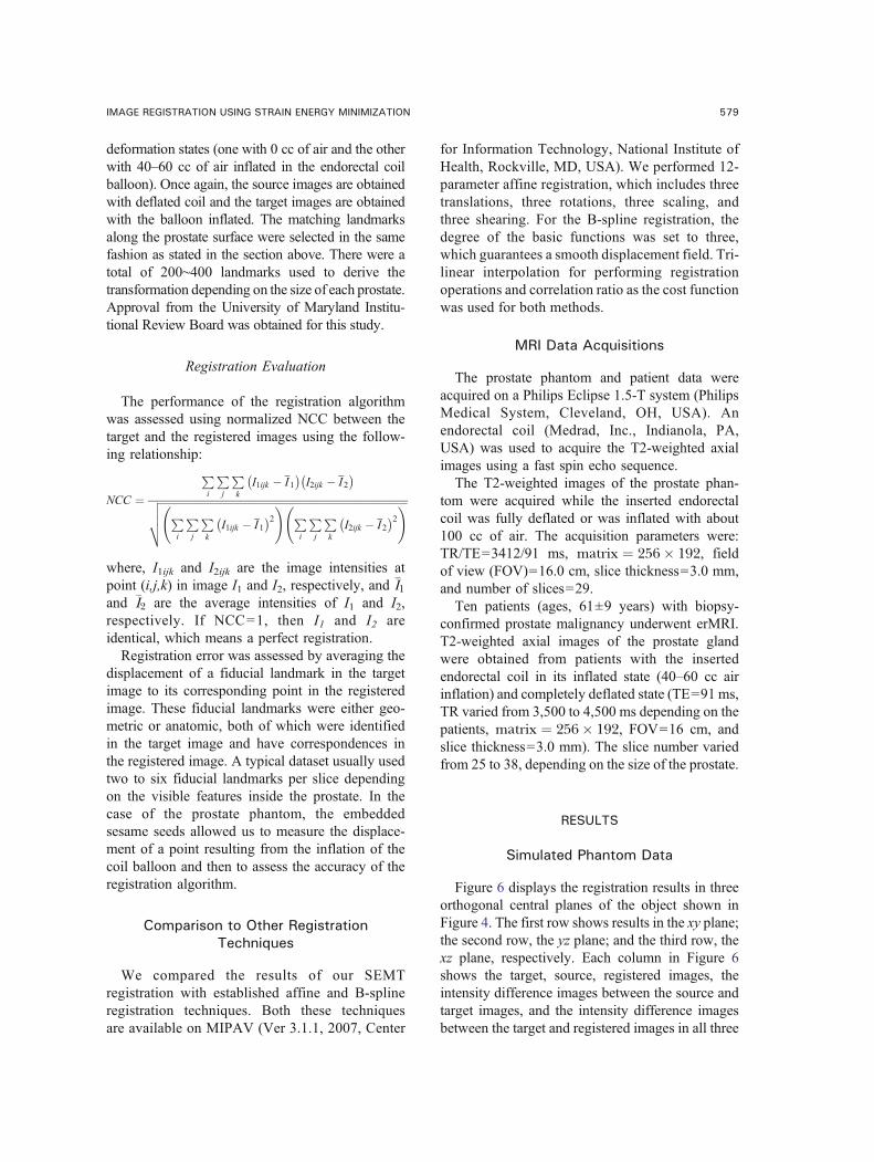
Figure 6 displays the registration results in threeorthogonal central planes of the object shown inFigure 4. The first row shows results in the xy plane;the second row, the yz plane; and the third row, thexz plane, respectively. Each column in Figure 6shows the target, source, registered images, theintensity difference images between the source andtarget images, and the intensity difference imagesbetween the target and registered images in all three
579IMAGE REGISTRATION USING STRAIN ENERGY MINIMIZATION

orthogonal planes. Also shown in Figure 6 is thedisplacement distribution along the three planes,which is superimposed on the source images in thesecond column. The blue arrows represent thedisplacement vectors with the length of the arrowsrepresenting their magnitude. To facilitate visualcomparison of the intensity difference between thepre- and post-registration images (Fig. 6, column 4and column 5), the same grayscale was adoptedbetween the two sets of images. As shown inFigure 6, the registration reduced the intensitydifference in the subtracted images between thetarget and the registered images. The NCCimproved from 0.91 to 1.0 after SEMT registration.
To assess the registration error, ten corners of thecharacter “F” (Fig. 6, xy plane and column 1) on thecentral slice were selected to serve as fiduciallandmarks. Table 1 summarizes the displacement ofthese landmarks between the source and target imagesand the registration error. The average displacementof the landmarks between the source and targetimages was 1.0±0.5 pixels. The algorithm attainedan average registration error of 0.2±0.1 pixels.
Prostate Phantom Data
Figure 7 shows the registration results from oneof the axial MR images taken through the center of
Table 1. Position, Displacement, and Registration Error of Ten Corners of Character F in a Digital Phantom
Corner
Position (x,y) (pixel)
Displacement (pixel) Error (pixel)Source Target Registered
C1 9.0, 7.0 10.0, 6.4 10.0, 6.1 1.3 0.3C2 18.3, 7.0 17.2, 6.4 17.4, 6.2 1.4 0.3C3 11.7, 9.9 11.8, 9.1 11.7, 9.1 0.9 0.1C4 18.3, 9.9 17.2, 9.1 17.3, 9.0 1.5 0.2C5 11.7, 12.0 11.8, 11.9 11.7, 11.9 0.2 0.1C6 17.4, 12.0 16.2, 11.9 16.5, 12.0 1.3 0.3C7 11.7, 14.7 11.8, 14.7 11.7, 14.7 0.1 0.1C8 17.4, 14.7 16.2, 14.7 16.5, 14.8 1.3 0.3C9 9.0, 20.4 10.0, 21.1 10.0, 21.3 1.3 0.2C10 11.7, 20.4 11.8, 21.1 11.7, 21.1 0.8 0.1
Target Source Registered Target-Source Target-Registered
xy
yz
xz
×
× ×
×× ×
× ×
× ×36
1 2
7 8
9 10
45
Fig 6. Registration results on the synthesized digital phantom. Each row shows images in each of the orthogonal planes. Columnsrepresent image type. The source image column is overlaid by the displacement vectors in three orthogonal planes. Also shown in targetimage (xy plane) are the landmarks (marked by red crosses) used to measure the accuracy of registration. NCC improved from 0.91 to 1.0.
5 ZHANG ET AL.80

the prostate phantom shown in Figure 5. Acomparison of the intensity difference betweenthe target and source (Fig. 7d) and the target andregistered images (Fig. 7e) qualitatively demon-strates the registration accuracy obtained using theSEMT algorithm. The NCC improved from 0.84 to0.99 after registration. Twelve sesame seeds wereused as features that were matched between thetarget and registered information to assess theregistration error. Table 2 provides a list ofmeasurements made using these 12 seeds. Theaverage displacement of the sesame seeds was 4.8±0.4 pixels (3.0±0.3 mm). The registration error wasfound to be 1.0±0.6 pixels (0.6±0.4 mm).
In vivo Prostate Data
Figure 8 shows an example registration from invivo prostate images. The blue arrows shown inFigure 8b show the direction and magnitude ofdisplacement. The comparison of intensity differ-ence images in Figure 8d, e before and afterregistration, qualitatively demonstrates the regis-tration accuracy as determined by the SEMTalgorithm. Five feature landmarks (see Fig. 8a)
depicting benign prostatic hyperplasia nodules ofthe prostate was chosen to evaluate the registrationerror for the slice shown. The average displace-ment of these five landmarks was 6.7±1.5 pixels(4.2±0.9 mm). The registration error was 1.7±1.3pixels (1.1±0.8 mm).The NCC was computed over the segmented
volume of the prostate and was found to improvefrom 0.69 to 0.97 for this particular case.The results on all ten sets of patient images were
summarized in Table 3. The NCC was improvedon average from 0.72±0.10 to 0.96±0.03 for thewhole group after SEMT registration (PG0.0001).The average displacement of fiducial landmarksinside the prostate gland from all the subjectswas found to be 6.1±1.9 pixels (3.8±1.2 mm)before registration, which was reduced to 1.8±0.7 pixels (1.1±0.4 mm) after registration. Alsoshown in Table 3 are the registration errorsmade by the 12-parameter affine registration andB-spline registration programs. The registrationerrors were 5.2±1.6 pixels (3.2±1.0 mm) whenusing the affine registration method and 2.9±1.4pixels (1.8±0.9 mm) when using the B-splinemethod.
Table 2. Position, Displacement, and Registration Error of 12 Sesame Seeds in the Prostate Phantom
Sesame seed
Position (pixel)
Displacement (pixel) Error (pixel)Source Target Registered
S1 130, 170 130, 174 130, 175 4.0 1.0S2 126, 157 126, 162 125, 161 5.0 1.4S3 119, 160 119, 165 120, 165 5.0 1.0S4 117, 161 115, 165 116, 165 4.5 1.0S5 140, 150 140, 155 140, 154 5.0 1.0S6 135, 139 135, 144 135, 144 5.0 0.0S7 130, 138 130, 143 130, 142 5.0 1.0S8 107, 141 107, 146 106, 146 5.0 1.0S9 141, 131 141, 136 139, 135 5.0 2.2S10 134, 127 133, 132 133, 132 5.1 0.0S11 125, 126 124, 131 123, 131 5.1 1.0S12 121, 128 120, 132 120, 133 4.1 1.0
Fig 7. Registration results of the prostate phantom in the xy plane: a target image, b source image overlaid with the displacement, cregistered image, d intensity difference image (a–b), and e intensity difference image (a–c). NCC improved from 0.84 to 0.99.
581IMAGE REGISTRATION USING STRAIN ENERGY MINIMIZATION

DISCUSSION
MR images of the prostate are typically obtainedwhen the prostate is in its distorted position.Advanced imaging techniques, such as diffusion-weighted imaging, perfusion-weighted imaging,and MRS, have been shown to increase specificityin the detection of cancer. However, all theinformation provided from these techniques isobtained with the prostate in its deformed state.For techniques such as external beam therapy andbrachytherapy to be effective, prostate imagesobtained in the distorted state must be registeredback to their original state. For example, in thecase of external beam brachytherapy, the imagesand the spectroscopic information obtained fromthe prostate with the endorectal coil (deformedimages) can be registered to the MR imagesobtained without the use of endorectal coil (non-deformed images). These images and spectra canthen be co-registered with the CT images fortreatment planning. This will ensure accurate
localization of the tumor in its undistorted stateand will allow for accurate treatment planning.We have developed a novel SEMT algorithm to
perform prostate image registration that registersimages to single-pixel accuracy. The algorithmrequires that the object undergoing deformation beelastic, incompressible, and in equilibrium whilescanning. Incompressible tissue, such as the breastand prostate, are suitable candidates for suchelastic registration.8,13 In a physically conservativesystem, the potential energy has a stationary valueif the system is in equilibrium. In the case of ourregistration problem, in which image volumes aretreated as elastic bodies, the potential energy ispurely the strain energy. The minimization of thisstrain energy allows for the transformation of thedeformed elastic, incompressible objects to beregistered.In most in vivo situations it is difficult to
quantitatively evaluate the performance of aregistration algorithm because of the lack of‘ground truth’. Our initial results on the simulated
Table 3. Normalized Correlation Coefficient, Maximum Pixel Displacement Before and After Registration Using the SEMT, Affine, and B-Spline Algorithms on In vivo Prostate Images
Patient
NCCa
Original displacement (Pixel)
Registration error (Pixel)
Before After SEMT Affine B-spline
P1 0.71 0.98 6.0 1.3 2.2 2.1P2 0.50 0.92 6.1 1.1 (fail) 4.8P3 0.75 0.99 6.7 1.6 6.5 2.7P4 0.73 0.90 6.4 3.1 5.0 1.9P5 0.72 0.98 8.5 1.5 7.4 4.6P6 0.74 0.98 3.0 1.4 4.6 3.6P7 0.74 0.98 5.6 2.9 5.3 1.8P8 0.69 0.97 6.7 2.0 6.3 1.9P9 0.75 0.96 3.0 1.7 3.9 0.9P10 0.62 0.98 8.8 1.2 5.6 4.8Mean±std 0.72±0.10 0.96±0.03 6.1±1.9 1.8±0.7 5.2±1.6 2.9±1.4
aNCC was computed after manual segmentation of prostate
2 3
1 54× × ×
××
a b c d e
Fig 8. Representative registration results for a prostate patient: a target image with five feature landmarks marked by red crosses, bsource image overlaid with the displacement, c registered image, d intensity difference image (a–b), and e intensity difference image (a–c).NCC improved from 0.69 to 0.97.
5 ZHANG ET AL.82

digital phantom and the development of a prostatephantom were essential in evaluating the robust-ness of the SEMT algorithm.25 This multipurposephantom mimicked the tissue property of theprostate and provided us a scenario that was asclose as possible to that routinely experienced inthe clinical setting. Because it has all the necessarytissue properties and biochemicals normally seenin a prostate gland, the phantom can be used to testthe robustness of new imaging and spectroscopictechniques. The periprostatic fat also allows us tomimic the difficulty associated with saturatingouter-volume chemical species that may alias intothe volume of interest, especially when performingMRS. The sesame seeds also may be used forpracticing image-guided biopsies. However, here,we used the seeds to determine the registrationerror of the SEMT algorithm.The transformation matrix obtained by SEMT
relies on feature correspondences between thesource and target images. Any mismatch of thecorrespondences may result in registration errors.We have chosen well populated, uniformly dis-tributed points along the whole surface of theprostate to derive the transformation between thesource and target images. We have shown that ouralgorithm is accurate up to 1.1 mm based on themeasurements made at corresponding featurepoints of the prostate. Other methods, such asactive contour26 and/or active surface models,27
may be adopted to generate the correspondencesautomatically to further reduce the resolution to asubpixel level.We compared our SEMT algorithm to two other
well-established methods of registration namelythe 12-parameter affine registration and B-splineregistration. Figure 9 shows the performance ofthese two techniques in comparison to our SEMTalgorithm. Given the range of displacements seenin ten prostate images as shown by the first metricin Figure 9, the SEMT algorithm outperformed theother two techniques by demonstrating nearly 38%reduction in registration errors over B-spline, andover 60% reduction in errors in comparison to theaffine registration technique, respectively.In the last decade, many applications of image
registration for prostate images have been reported.Dubois et al.8 implemented a rigid registration on aprostate phantom and obtained a registration errorof 1.6±0.7 mm. Because of anatomical variabilityin the prostate and because of the fairly nonrigid
characteristics of transformation, rigid registrationmay not yield accurate results for registration ofprostate images. Wu et al.10 adopted a polynomialtransformation to register prostate MRSI with anerror of 2 mm or less. Fei et al.13 applied TPS toprostate image registration, reducing the prostatecentroid displacement from 3.4 to 0.6 mm. Wanget al.28 implemented a ‘demons’ algorithm (agrayscale-based deformable image registrationalgorithm) on a phantom prostate and achieved atracking accuracy of better than 1.5 mm. Theresults from our method are either comparable orbetter than these reported methods. A notabledifference between our study and those mentionedhere is that our approach is based on the use ofcorrespondence points coupled with physical con-straints, whereas the cited studies used voxel-intensity based approaches for registration.The proposed SEMT approach is similar to
those reported in the literature in that they allidentify the feature correspondences (points, con-tours, or surfaces) in the source and target imagesand then use these correspondences as input toderive the underlying deformation transformation.The principal difference between our method andthese others is that our transformation is derivedfrom strain energy minimization using elastictheory. Broit14 was the first to use a model derivedfrom elasticity theory to match 2D and 3D images.He defined a cross-correlation coefficient betweenlocal regions in two images to derive forces thatdeform the source image to the target. The trans-formation is obtained by solving the Navier–Lame
0.0
1.0
2.0
3.0
4.0
5.0
6.0
Pre-Correction
Affine B-spline SEMT
Reg
istr
atio
n er
ror
(mm
)
Fig 9. Graph depicting the performance of SEMT, affine, and B-spline registration algorithms on prostate images. Minimumregistration errors are realized with the SEMT algorithm.
583IMAGE REGISTRATION USING STRAIN ENERGY MINIMIZATION

equations. Instead of using the local similarity asthe measure of external driving forces, Davatzikoset al.15 proposed a spring model in which theexternal force is obtained from mapping thecontours of structures in the source and targetimages. They obtained their transformation byempirically choosing the two Lame constants� ¼ � ¼ 1:5� 10��9� �
in the equilibrium equa-tions. Peckar et al.17 incorporated the knowndisplacements of some boundary structures in thesource and target images as hard constraints to theregistration to compute the transformation. In thatstudy, λ was intentionally set to zero and the otherconstant μ was eliminated. This makes the regis-tration model completely parameter free. Unliketraditional elastic registration methods, whichsolve the equilibrium equations for the trans-formation, the SEMT method obtains the trans-formation by minimizing the strain energy of theregistered image volume. Making a reasonableassumption of incompressibility (Krouskop etal.23), the registration model is parameter free.The advantages of the proposed approach includethe need for only a scalar energy function insteadof a set of equilibrium equations; the reasonableassumption of incompressibility, which makes thealgorithm parameter free; and the ability to handleboth rigid and nonrigid deformation equally well.It should be noted that we performed the
registration on the entire prostate image with asubtle assumption that the image volume has thesame mechanical property throughout. As weknow, many different structures and tissues areinside and outside the prostate gland. So thisassumption may lead to some systematic registra-tion error. For more accurate registration, it wouldbe necessary to take these structures into consid-eration. Some differences in the mechanical prop-erty of the tissue are expected, but when dealingwith tissues that have significantly differentmechanical properties, it should be noted that theSEMT algorithm no longer remains parameter free.Hence, a modification to Eq. 8 is required thattakes into consideration the surrounding tissuecharacteristics. Our future studies will characterizetissues from different regions of and surroundingthe prostate for an accurate assessment of dis-placement and strain during compression of theprostate.
CONCLUSION
We have implemented a novel registrationalgorithm based on strain energy minimizationand demonstrated its effectiveness on phantom andprostate images. Results from our study showregistration error of approximately 1 mm over theentire volume of the prostate. Accurate registrationof prostate images obtained in the deformed statecan be useful in treatment planning and also forlongitudinal evaluation of progression/regressionin patients with prostate cancer. In addition,although we have demonstrated the utility of ouralgorithm on MR images, we believe that thismethod can also be applied to images from anytwo different imaging modalities provided thatcorresponding features are available in both.
ACKNOWLEDGMENT
The work was supported by the U.S. Department of DefenseIDEA grant W81XWH-04-1-0249 (PC031042).
APPENDIX 1
The relationship between the two constants λ and μ is:
l ¼ 2u�1� 2u
ðA:1Þ
And the relationship between the unit volume changee and normal strain εx is:
e ¼ 1� 2uð Þ"x ðA:2Þ
So the first term λe2 of Eq. 1 can be rewritten as:
le2 ¼ 2u�1� 2u
1� 2uð Þ"xð Þ2 ¼ 2u� 1� 2uð Þ"2xðA:3Þ
Given Poisson’s ratio v=0.495,23 the above expres-sion is approximately
le2 ¼ 0:01�"2x ðA:4Þ
which makes it negligible as compared to the othertwo terms in Eq. 1.
5 4 ZHANG ET AL.8

REFERENCES
1. Sanchez-Chapado M, Angulo JC, Ibarburen C, Aguado F,Ruiz A, Viano J, Garcia-Segura JM, Gonzalez-Esteban J,Rodriquez-Vallejo JM: Comparison of digital rectal examina-tion, transrectal ultrasonography, and multicoil magnetic reso-nance imaging for preoperative evaluation of prostate cancer.Eur Urol 21:140–149, 1997
2. Futterer JJ: MR imaging in local staging of prostatecancer. Eur J Radiol 63(3):328–334, 2007
3. Susil RC, Camphausen K, Choyke P, McVeigh ER,Gustafson GS, Ning H, Miller RW, Atalar E, Coleman CN,Menard C: System for prostate brachytherapy and biopsy in astandard 1.5T MRI scanner. Magn Reson Med 52(3):683–687,2004
4. Nguyen PL, Chen MH, D’Amico AV, Tempany CM,Steele GS, Albert M, Cormack RA, Carr-Locke DL, Bleday R,Suh WW: Magnetic resonance image-guided salvage brachy-therapy after radiation in select men who initially presentedwith favorable-risk prostate cancer: a prospective phase 2 study.Cancer 110(7):1485–1492, 2007
5. Schnall MD, Imai Y, Tomaszewski J, Pollack HM,Lenkinski RE, Kressel HY: Prostate cancer: local staging withendorectal surface coil MR imaging. Radiology 178:797–802,1991
6. Husband JE, Padhani AR, Mac Vicar AD, Revell P:Magnetic resonance imaging of prostate cancer: comparison ofimage quality using endorectal and pelvic phased array coils.Clin Radiol 53:673–681, 1998
7. D'Amico AV, Schnall M, Whittington R, Malkowicz SB,Schultz D, Tomaszewski JE, Wein A: Endorectal coil magneticresonance imaging identifies locally advanced prostate cancer inselect patients with clinically localized disease. Urology51:449–454, 1998
8. Dubois DF, Bice Jr, WS, Prestige BR: CT and MRIderived source localization error in a custom prostate phantomusing automated image coregistration. Med Phys 28:2280–2284, 2001
9. Fei B, Wheaton A, Lee Z, Duerk JL, Wilson DL:Automatic MR volume registration and its evaluation for thepelvis and prostate. Phys Med Biol 47:823–838, 2002
10. Wu X, Dibiase SJ, Gullapalli R, Yu CX: Deformableimage registration for the use of magnetic resonance spectros-copy in prostate treatment planning. Int J Radiat Oncol BiolPhys 58:577–583, 2004
11. Venugopal N, McCurdy B, Hnatov A, Dubey A: Afeasibility study to investigate the use of thin-plate splines to accountfor prostate deformation. Phys Med Biol 50:2871–2885, 2005
12. Lian J, Xing L, Hunjan S, Dumoulin C, Levin J, Lo A,Watkins R, Rohling K, Giaquinto R, Kim D, Spielman D,Daniel B: Mapping of the prostate in endorectal coil-based
MRI/MRSI and CT: a deformable registration and validationstudy. Med Phys 31:3087–3094, 200413. Fei B, Kemper C, Wilson DL: A comparative study of
warping and rigid body registration for the prostate and pelvicMR volumes. Comput Med Imaging Graph 27:267–281, 200314. Broit C: Optimal Registration of Deformed Images, PhD
thesis. University of Pennsylvania, Philadelphia, 198115. Davatzikos C, Prince JL, Bryan RN: Image registration
based on boundary mapping. IEEE Trans Med Imaging 15:112–115, 199616. Gee J, Haynor D, Briquer L, Bajcsy R: Advances in
elastic matching theory and its implementation. In: Troccas J,Grimsom WEL, Mosges R Eds. Proceedings of the First JointConference on Computer Vision, Virtual Reality and Roboticsin Medicine and Medical Robotics and Computer-AssistedSurgery. Springer-Verlag, London, 1997, pp 63–7217. Peckar W, Schnorr C, Rohr K, Stiehl HS: Two step
parameter-free elastic image registration with prescribed pointdisplacements. J Math Imaging Vision 10:143–162, 199918. Christensen GE, Rabbit RD, Miller MI: Deformable
templates using large deformation kinematics. IEEE TransImage Process 5:1435–1447, 199619. Thirion JP: Image matching as a diffusion process: an
analogy with Maxwell’s demons. Med Image Anal 2:243–260,199820. Beuchemin SS, Barron JL: The computation of optical
flow. ACM Comput Surv 27:433–467, 199521. Greenwood DT: Classical Dynamics. Dover Publica-
tions, Dover, 199722. Ugural AC, Fenster SK: Advanced Strength and Applied
Elasticity, 3rd edition. Prentice-Hall, Inc, Upper Saddle River,199523. Krouskop TA, Wheeler TM, Kallel F, Garra BS, Hall T:
Elastic moduli of breast and prostate tissues under compression.Ultrason Imaging 20:260–274, 199824. Read K, Hosseinzadeh K, Dibiase S, Gullapalli R:
Characterization of prostate deformation during MR examina-tion using endorectal coil for accurate localization of tumorduring brachytherapy. Proc Intl Soc Mag Reson Med 2340, 200125. Zhang B, Gullapalli RP: Multipurpose prostate phantom.
Proc Intl Soc Mag Reson Med 2115, 200526. Kass M, Witkin A, Terzopoulos D: Snakes: active
contour models. Int J Comput Vision 1:321–331, 198827. Cohen L, Cohen I: Finite-element methods for active
contour models and balloons for 2D and 3D images. IEEETrans Pattern Anal Mach Intell 15:1131–1147, 199328. Wang H, Dong L, O'Daniel J, Mohan R, Garden AS,
Ang KK, Kuban DS, Bonnen M, Chang JY, Cheung R:Validation of an accelerated 'demons' algorithm for deformableimage registration in radiation therapy. Phys Med Biol 50:887–905, 2005
5 58IMAGE REGISTRATION USING STRAIN ENERGY MINIMIZATION