Embed Size (px)
Citation preview

This is the author version of an article published as: Kricker, Jennifer A. and Towne, Chris L. and Firth, Sue M. and Herington, Adrian C. and Upton, Zee (2003) Structural and Functional Evidence for the Interaction of Insulin-Like Growth Factors (IGFs) and IGF Binding Proteins with Vitronectin. Endocrinology 144(7):pp. 2807-2815. Copyright 2003 The Endocrine Society Accessed from http://eprints.qut.edu.au
1
Structural and functional evidence for the interaction of insulin-like growth factors
(IGFs) and IGF-binding proteins with vitronectin.
Jennifer A. Kricker#, Chris L. Towne, Sue M. Firth*, Adrian C. Herington and Zee
Upton
Tissue BioRegeneration and Integration Program, School of Life Sciences, Queensland
University of Technology, 2 George Street, Brisbane, Qld, 4001, Australia.
*Kolling Institute of Medical Research, University of Sydney, Royal North Shore Hospital,
Pacific Highway, St Leonards, NSW, 2065, Australia.
Running Title: IGFBPs modulate binding of IGFs to VN
Key words: IGFs, IGFBPs, vitronectin, binding, modulation, integrin receptor
#Author for correspondence:
Jennifer Kricker
Tissue BioRegeneration and Integration Program
School of Life Sciences
Queensland University of Technology
GPO Box 2434
Brisbane, Qld 4001
Australia
Tel: +61-7-3864-5177
Fax: +61-7-3864-1534
E-mail: [email protected].
Copyright (C) 2003 by The Endocrine Society
Endocrinology. First published March 27, 2003 as doi:10.1210/en.2002-221086
2
Abstract
Previous studies demonstrated that IGF-II binds directly to vitronectin (VN) while IGF-I
binds poorly. However, binding of VN to integrins has been demonstrated to be essential for
a range of IGF-I stimulated biological effects including IGF binding protein-5 (IGFBP-5)
production, IGF type-1 receptor autophosphorylation and cell migration. Thus we
hypothesised that a link between IGF-I and VN must occur and may be mediated through
IGFBPs. This was tested using competitive binding assays with VN and [125I]-labelled IGFs
in the absence and presence of IGFBPs. IGFBP-4, IGFBP-5 and non-glycosylated IGFBP-3
were shown to significantly enhance binding of IGF-I to VN, while IGFBP-2 and
glycosylated IGFBP-3 had a smaller effect. Furthermore binding studies with analogues
indicate that glycosylation status and the heparin-binding domain of IGFBP-3 are important
in this interaction. To examine the functional significance of IGFs binding to VN, cell
migration in MCF7 cells was measured and found to be enhanced when VN was pre-bound to
IGF-I in the presence of IGFBP-5. The effect required IGF:IGFBP:VN complex formation;
this was demonstrated by use of a non-IGFBP-binding IGF-I analogue. Together, these data
indicate the importance of IGFBPs in modulating IGF-I binding to VN and that this binding
has functional consequences in cells.
3
Introduction
The mitogenic effects of insulin-like growth factors (IGFs) are modulated by
members of the IGF-binding protein (IGFBP) family. These proteins have been
demonstrated to both inhibit and potentiate IGF action (1). In addition to the six IGFBPs,
another group of proteins termed IGFBP-related proteins (IGFBP-rPs), have also been shown
to bind the IGFs, however with a much lower affinity. Upton et al. (2) have reported
identification of another endogenous protein complex consisting of vitronectin (VN) and
IGF-II. This is particularly interesting as VN is structurally unrelated to both the IGFBPs and
IGFBP-rPs.
VN, a multifunctional protein found in plasma and extracellular matrix, is a
component of the urokinase system. A number of proteins bind to VN, including
glycosaminoglycans (3, 4), which bind via a heparin-binding domain in VN, and integrins,
which bind via an R-G-D sequence (5, 6). It is through the binding of various proteins to
these motifs, as well as other domains within VN, that diverse physiological processes such
as extracellular anchoring, cell spreading and migration are mediated (7-9).
IGF-II has been shown to bind directly to VN, whereas only minimal binding of IGF-I
to VN occurs (2). Nevertheless, it is intriguing that VN appears to be critical for a number of
IGF-I-related effects including cellular DNA synthesis, type-1 IGF receptor
autophosphorylation and cell migration (10-12). More specifically, Clemmons and co-
workers have shown that VN binding to the integrin αvβ3 is critical for IGF-I stimulated
smooth muscle cell migration (13). In addition, inhibition of IGFBP-5 binding to porcine
SMC extracellular matrix also reduces cellular responses to IGF-I (14). Furthermore, the
potentiating effects of IGFBPs on IGF action appear to require interaction with, as yet
unidentified, cell-surface associated proteins which may include VN (15). For example,
IGFBP-5 has been demonstrated to facilitate binding of IGF-I to bone independently of the
4
IGF receptors (16) and IGFBP-3 has also been shown to potentiate IGF action markedly
following binding to the cell surface (17).
Given the importance of IGFBPs and VN for regulation of IGF action, we
hypothesized that IGFBPs may mediate direct binding of IGFs to VN, and in particular, that
IGFBPs may be necessary for functional interaction of IGF-I with VN. We now provide
substantial evidence to support this hypothesis based upon studies examining binding of
labelled-IGF-I and -IGF-II to VN in the absence and presence of IGFBPs and enhanced cell
migration in the presence of these complexes.
5
Experimental Procedures
Materials
IGF-I, IGF-II, Des(1-3)IGF-I, Des(1-6)IGF-II, [Leu27]IGF-II, IGFBP-1, -2, and -4
were purchased from GroPep Pty Ltd (Adelaide, SA, Australia). IGFBP-5, glycosylated
IGFBP-3, heparin binding domain (HBD) mutant IGFBP-3 and mutant non-glycosylated
IGFBP-3 were produced as described previously by Firth et al. (18), while glycosylated
IGFBP-6 was kindly donated by Dr Leon Bach (Department of Medicine, University of
Melbourne, Vic, Australia). Human breast carcinoma (MCF-7) cells were obtained from the
American Type Culture Collection (ATCC # HTB-22). Human vitronectin was purchased
from Promega Corporation (Madison, WI, USA). RIA grade BSA, heparin, chloramine-T,
sodium metabisulphite and Sigmacote were purchased from Sigma Chemical Co. (St Louis,
MO, USA). Sodium [125-iodide], Sephadex G-50 and HiTrap heparin affinity columns were
obtained from Amersham Pharmacia Biotech UK Ltd (Buckinghamshire, England). Other
chromatography equipment for radiolabelling IGFs was purchased from BioRad Laboratories
Inc. (Hercules, CA, USA). Hanks’ Balanced Salt Solution (HBSS), Dulbecco’s Modified
Eagles Medium (DMEM), DMEM-Ham’s F12 (DMEM-F12), Trypsin, Penicillin-
Streptomycin and Gentamycin were purchased from Invitrogen Australia Pty Ltd (Mt.
Waverley, Vic, Australia) while Foetal Calf Serum was from Trace Scientific (Noble Park,
Vic, Australia). Removawell Immulon-4 HB wells were from Dynex Technologies Inc.
(Chantilly, VA, USA) while 80 cm2 culture flasks and 24-well plates were from Nagle Nunc
International (Roskilde, Denmark). Transwells were purchased from Costar (New York, NY,
USA). Autoradiographic film was purchased from Eastman Kodak (Rochester, NY, USA)
while low molecular weight protein markers were obtained from BioRad Laboratories Inc.
All other reagents were of analytical grade. General plastic-ware used in experiments
containing IGFBPs and VN was siliconised with Sigmacote and left to air-dry overnight.
6
Radiolabelling of Proteins
IGF-I and IGF-II were iodinated according to the chloramine-T method as described
by GroPep Pty Ltd for IGFs while IGFBP-3 (glycosylated and non-glycosylated) was
iodinated as per Dr Janet Martin (personal communication). The chloramine-T reactions
were performed for 1 minute for the IGFs (10 µg) and 15 seconds for IGFBP-3 (5 µg).
Labelled IGFs were purified using size exclusion on Sephadex G-50, with 50 mM sodium
phosphate, 150 mM NaCl, 0.25% w/v BSA, pH 6.5 as the elution buffer, while labelled
IGFBP-3 was purified using heparin affinity chromatography and 50 mM sodium phosphate,
0.1% w/v BSA, pH 6.5 as the equilibration buffer. The protein was eluted using elution
buffers 1-3, consisting of the equilibration buffer containing 1) 0.4 M NaCl, 2) 0.75 M NaCl
and 3) 1.0 M NaCl, all at pH 6.5 respectively. Confirmation that the [125I]-IGFBP-3 was the
correct molecular size and was not fragmented during the labelling procedure was obtained
by non-reducing SDS-polyacrylamide gel electrophoresis. Ten thousand cpm of [125I]-
IGFBP-3 fractions from peaks in the iodination elution profile were run on a 4% stacking
/10% separation Tris-glycine gel, dried and then exposed to autoradiographic film for 1-7
days.
Solid-Plate Binding Assay
IGF:VN:IGFBP binding assays were performed in removable Immulon wells coated
with or without 300 ng of vitronectin in 100 µL DMEM at 37°C, 5% CO2 for 2 - 4 hours.
Wells were rinsed twice with HEPES Binding Buffer (HBB: 0.1 M HEPES, 0.12 M NaCl, 5
mM KCl, 1.2 mM MgSO4, 8 mM glucose containing 0.5% w/v BSA, pH 7.6) to prevent non-
specific binding. [125I]-labelled protein (IGF-I, IGF-II, glycosylated IGFBP-3 or non-
glycosylated IGFBP-3) (10000 cpm) in HBB + 0.5% BSA in the absence or presence of
7
increasing concentrations of unlabelled IGFs (0.1 – 100 ng), IGF analogues (0.1 – 100 ng)
and/or IGFBPs (0.05 – 100 ng) were incubated overnight at 4°C in a final volume of 100 µL
(19). Unbound radiolabelled protein was then removed by aspiration and the wells were
washed three times with HBSS. Radioactivity remaining bound in each well was then
determined using a gamma counter. Each sample was measured in triplicate and the
experiment repeated at least three times. Student’s paired t-test was used to compare
amounts of labelled protein of test wells to the control (absence of VN and presence of
tracer). Differences were significant if the p value was less than 0.05.
Transwell Cell Migration Assay
Human breast carcinoma (MCF-7) cells were grown in DMEM-F12 media
supplemented with 10% foetal calf serum, penicillin (50 units/mL), streptomycin (0.1 µg/mL)
and gentamycin (1 µg/mL). Cells were grown to 70 – 80% confluence at 37°C in a
humidified environment with 5% CO2. Cell migrations assays using Transwells were
performed using cells from passages 24 to 34.
The lower chambers of 12 µm pore polycarbonate tissue culture treated Transwells
were pre-coated with 1 µg VN in serum-free DMEM-F12 and incubated at 37° C for 2 hours.
Media containing unbound VN was then removed and the lower chambers washed twice with
HBB containing 0.5% BSA. IGF-I or Des(1-3)IGF-I (1 – 100 ng) in DMEM-F12 + 0.05%
BSA was added to the lower chamber in the absence or presence of IGFBP-5 (1000 ng) and
allowed to bind to the pre-coated VN overnight at 4°C. The media containing unbound
growth factors was removed and the lower chambers washed twice with DMEM-F12 +
0.05% BSA. MCF-7 cells that had been serum-starved for 4 hours were trypsinised and
seeded on to the microporous membrane in the upper chamber of the Transwell inserts
(200,000 cells/well) and incubated at 37° C in 5% CO2 for 5 hours. Cells that had migrated
8
to the lower surface of the porous membrane were then fixed in 37% formaldehyde and
stained with 0.01% crystal violet in 0.1 mM borate buffer (pH 9). The number of cells that
had migrated to the lower side of the membrane was quantitated by extracting the crystal
violet stain in 10% acetic acid and determining the optical density of these extracts at 595
nm. Treatments were expressed as a percentage of the response observed on VN alone. Data
were pooled from duplicate samples from three experiments and significant differences in
responses compared to VN, or between treatments, were determined by Tukey’s analysis of
multiple means. Differences were significant if the p value was less than 0.05.
9
Results
Effects of IGFs and IGF-II analogues on binding of [125I]-IGF-II to VN
To demonstrate that the interaction of IGF-II with VN is specific, [125I]-IGF-II
binding assays were conducted in the presence of IGF-II analogues with varying affinities for
IGFBPs and/or IGF receptors. Des(1-6)IGF-II (which has a low affinity for IGFBPs) (20)
and [Leu27]IGF-II (which has low affinity for the type-1 IGF receptor and IGFBP-3) (21)
were equipotent with native IGF-II in their ability to displace [125I]-IGF-II bound to VN (data
not shown). Half-maximal competitive effects were observed at approximately 1 ng. IGF-I,
on the other hand, was much less effective at displacing [125I]-IGF-II, achieving
approximately a 20% reduction at 0.2 ng with no further reduction at higher doses up to 100
ng.
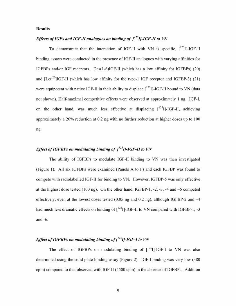
Effect of IGFBPs on modulating binding of [125I]-IGF-II to VN
The ability of IGFBPs to modulate IGF-II binding to VN was then investigated
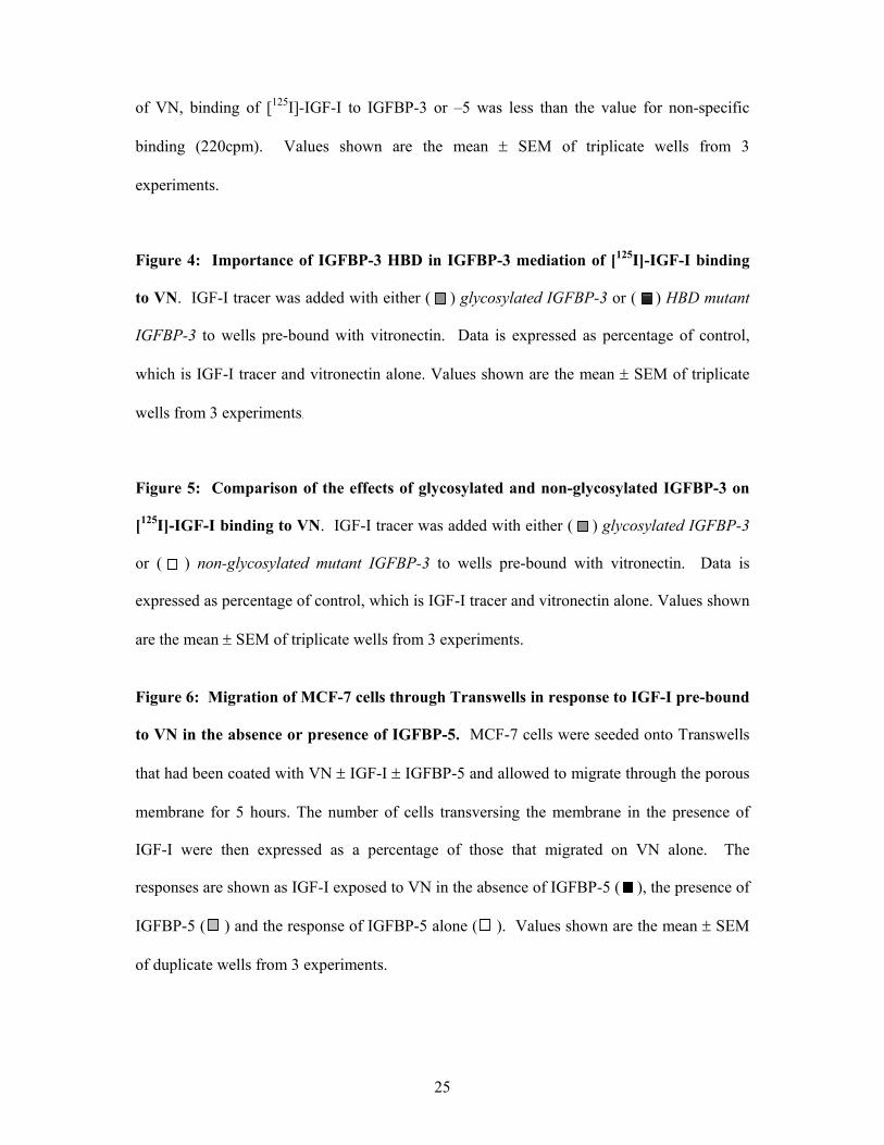
(Figure 1). All six IGFBPs were examined (Panels A to F) and each IGFBP was found to
compete with radiolabelled IGF-II for binding to VN. However, IGFBP-5 was only effective
at the highest dose tested (100 ng). On the other hand, IGFBP-1, -2, -3, -4 and –6 competed
effectively, even at the lowest doses tested (0.05 ng and 0.2 ng), although IGFBP-2 and –4
had much less dramatic effects on binding of [125I]-IGF-II to VN compared with IGFBP-1, -3
and -6.
Effect of IGFBPs on modulating binding of [125I]-IGF-I to VN
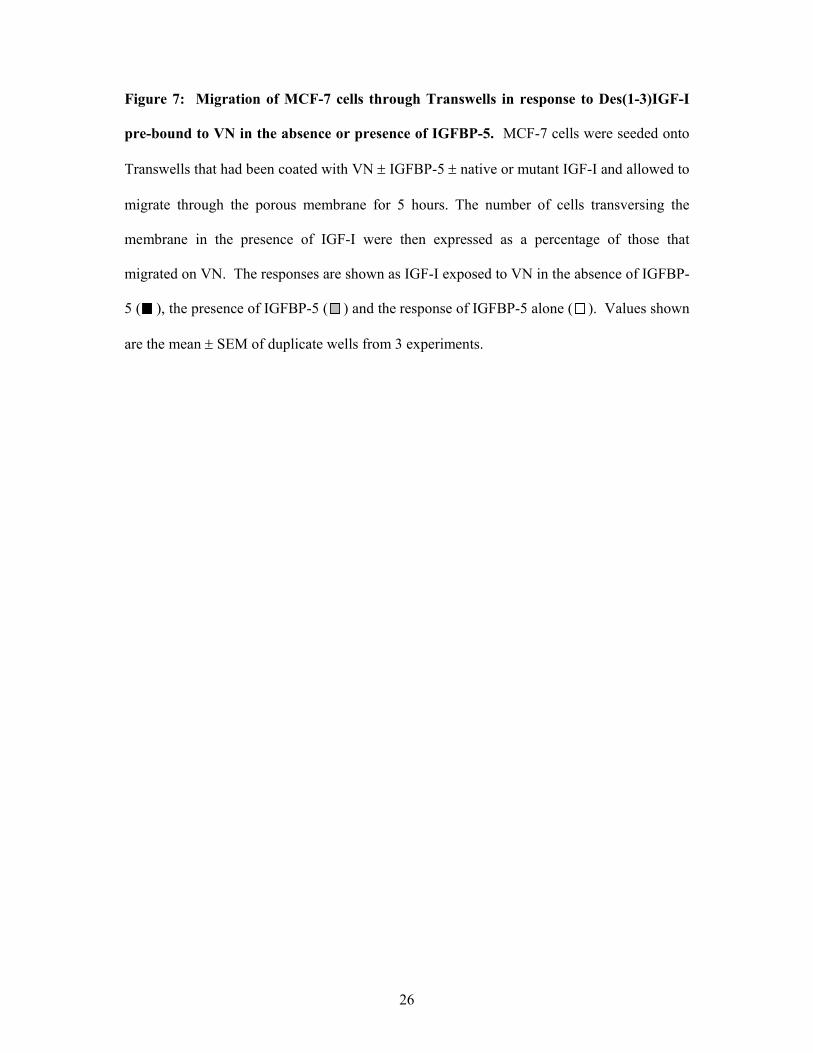
The effect of IGFBPs on modulating binding of [125I]-IGF-I to VN was also
determined using the solid plate-binding assay (Figure 2). IGF-I binding was very low (380
cpm) compared to that observed with IGF-II (4500 cpm) in the absence of IGFBPs. Addition
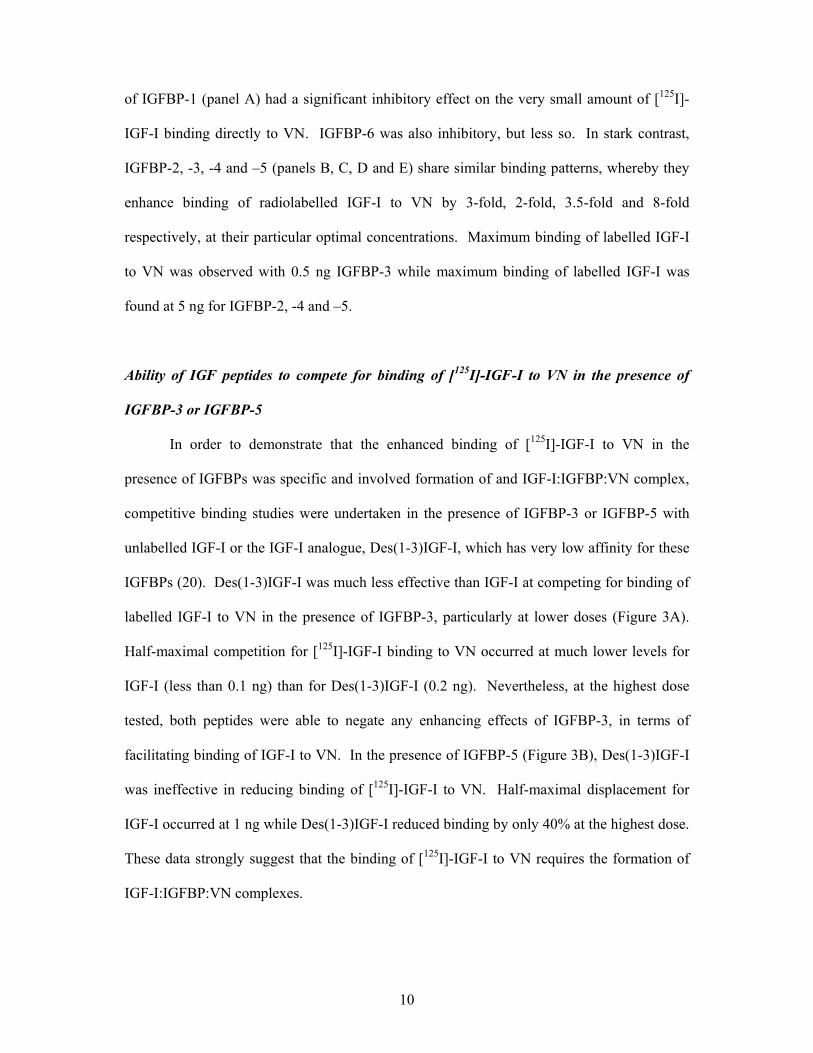
10
of IGFBP-1 (panel A) had a significant inhibitory effect on the very small amount of [125I]-
IGF-I binding directly to VN. IGFBP-6 was also inhibitory, but less so. In stark contrast,
IGFBP-2, -3, -4 and –5 (panels B, C, D and E) share similar binding patterns, whereby they
enhance binding of radiolabelled IGF-I to VN by 3-fold, 2-fold, 3.5-fold and 8-fold
respectively, at their particular optimal concentrations. Maximum binding of labelled IGF-I
to VN was observed with 0.5 ng IGFBP-3 while maximum binding of labelled IGF-I was
found at 5 ng for IGFBP-2, -4 and –5.
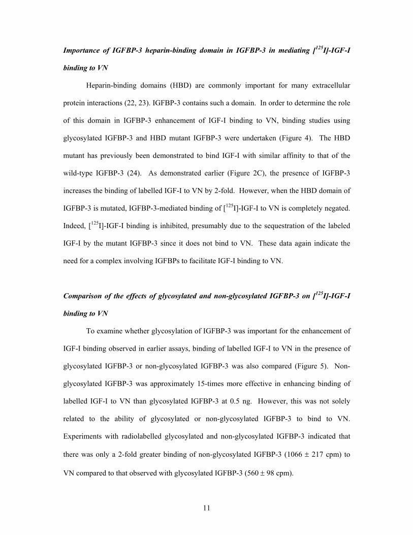
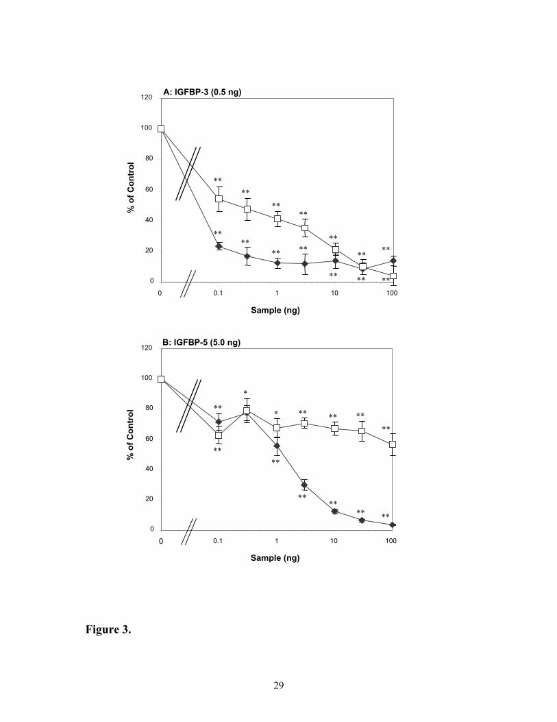
Ability of IGF peptides to compete for binding of [125I]-IGF-I to VN in the presence of
IGFBP-3 or IGFBP-5
In order to demonstrate that the enhanced binding of [125I]-IGF-I to VN in the
presence of IGFBPs was specific and involved formation of and IGF-I:IGFBP:VN complex,
competitive binding studies were undertaken in the presence of IGFBP-3 or IGFBP-5 with
unlabelled IGF-I or the IGF-I analogue, Des(1-3)IGF-I, which has very low affinity for these
IGFBPs (20). Des(1-3)IGF-I was much less effective than IGF-I at competing for binding of
labelled IGF-I to VN in the presence of IGFBP-3, particularly at lower doses (Figure 3A).
Half-maximal competition for [125I]-IGF-I binding to VN occurred at much lower levels for
IGF-I (less than 0.1 ng) than for Des(1-3)IGF-I (0.2 ng). Nevertheless, at the highest dose
tested, both peptides were able to negate any enhancing effects of IGFBP-3, in terms of
facilitating binding of IGF-I to VN. In the presence of IGFBP-5 (Figure 3B), Des(1-3)IGF-I
was ineffective in reducing binding of [125I]-IGF-I to VN. Half-maximal displacement for
IGF-I occurred at 1 ng while Des(1-3)IGF-I reduced binding by only 40% at the highest dose.
These data strongly suggest that the binding of [125I]-IGF-I to VN requires the formation of
IGF-I:IGFBP:VN complexes.
11
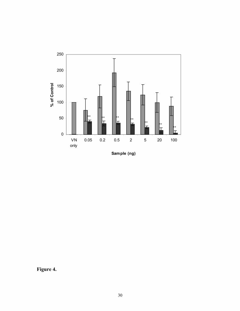
Importance of IGFBP-3 heparin-binding domain in IGFBP-3 in mediating [125I]-IGF-I
binding to VN
Heparin-binding domains (HBD) are commonly important for many extracellular
protein interactions (22, 23). IGFBP-3 contains such a domain. In order to determine the role
of this domain in IGFBP-3 enhancement of IGF-I binding to VN, binding studies using
glycosylated IGFBP-3 and HBD mutant IGFBP-3 were undertaken (Figure 4). The HBD
mutant has previously been demonstrated to bind IGF-I with similar affinity to that of the
wild-type IGFBP-3 (24). As demonstrated earlier (Figure 2C), the presence of IGFBP-3
increases the binding of labelled IGF-I to VN by 2-fold. However, when the HBD domain of
IGFBP-3 is mutated, IGFBP-3-mediated binding of [125I]-IGF-I to VN is completely negated.
Indeed, [125I]-IGF-I binding is inhibited, presumably due to the sequestration of the labeled
IGF-I by the mutant IGFBP-3 since it does not bind to VN. These data again indicate the
need for a complex involving IGFBPs to facilitate IGF-I binding to VN.
Comparison of the effects of glycosylated and non-glycosylated IGFBP-3 on [125I]-IGF-I
binding to VN
To examine whether glycosylation of IGFBP-3 was important for the enhancement of
IGF-I binding observed in earlier assays, binding of labelled IGF-I to VN in the presence of
glycosylated IGFBP-3 or non-glycosylated IGFBP-3 was also compared (Figure 5). Non-
glycosylated IGFBP-3 was approximately 15-times more effective in enhancing binding of
labelled IGF-I to VN than glycosylated IGFBP-3 at 0.5 ng. However, this was not solely
related to the ability of glycosylated or non-glycosylated IGFBP-3 to bind to VN.
Experiments with radiolabelled glycosylated and non-glycosylated IGFBP-3 indicated that
there was only a 2-fold greater binding of non-glycosylated IGFBP-3 (1066 ± 217 cpm) to
VN compared to that observed with glycosylated IGFBP-3 (560 ± 98 cpm).
12
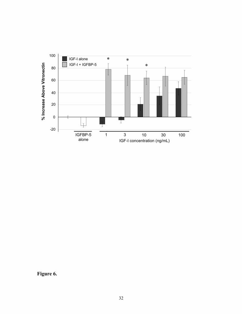
Effects of IGF-I and IGFBP-5 on cell migration in MCF-7 breast carcinoma cells
In order to determine if the enhanced binding of IGF-I to VN in the presence of
IGFBP-5 has functional consequences, the effects of the complexes on stimulating cells to
migrate were examined using the MCF-7 cell line. Minimal cell migration was observed in
the absence of VN, regardless of the presence or absence of IGF-I, IGFBP-5 or the
combination of both (data not shown). In the presence of VN, but in the absence of IGFBP-5,
1 and 3 ng of IGF-I exposed to VN resulted in non-significant decreases in cell migration on
VN by 11 ± 4 and 4 ± 6 % respectively, whilst 10, 30 and 100 ng of IGF-I exposed to VN
resulted in increased migration of 21 ± 10, 35 ± 14 and 47 ± 11 % (Figure 6). Striking
differences in responses were observed with the addition of 1 µg IGFBP-5, with 1, 3, 10, 30
and 100 ng IGF-I increasing cell migration on VN between 64 – 78 ± 17 %. These increased
responses in the presence of IGFBP-5 were significant at 1, 3 and 10 ng of IGF-I (p< 0.05).
Moreover, the responses were not due to IGF-independent effects of IGFBP-5, as the
presence of this binding protein alone resulted in a significant reduction in migration
compared to VN of 14 ± 4 % (p < 0.05).
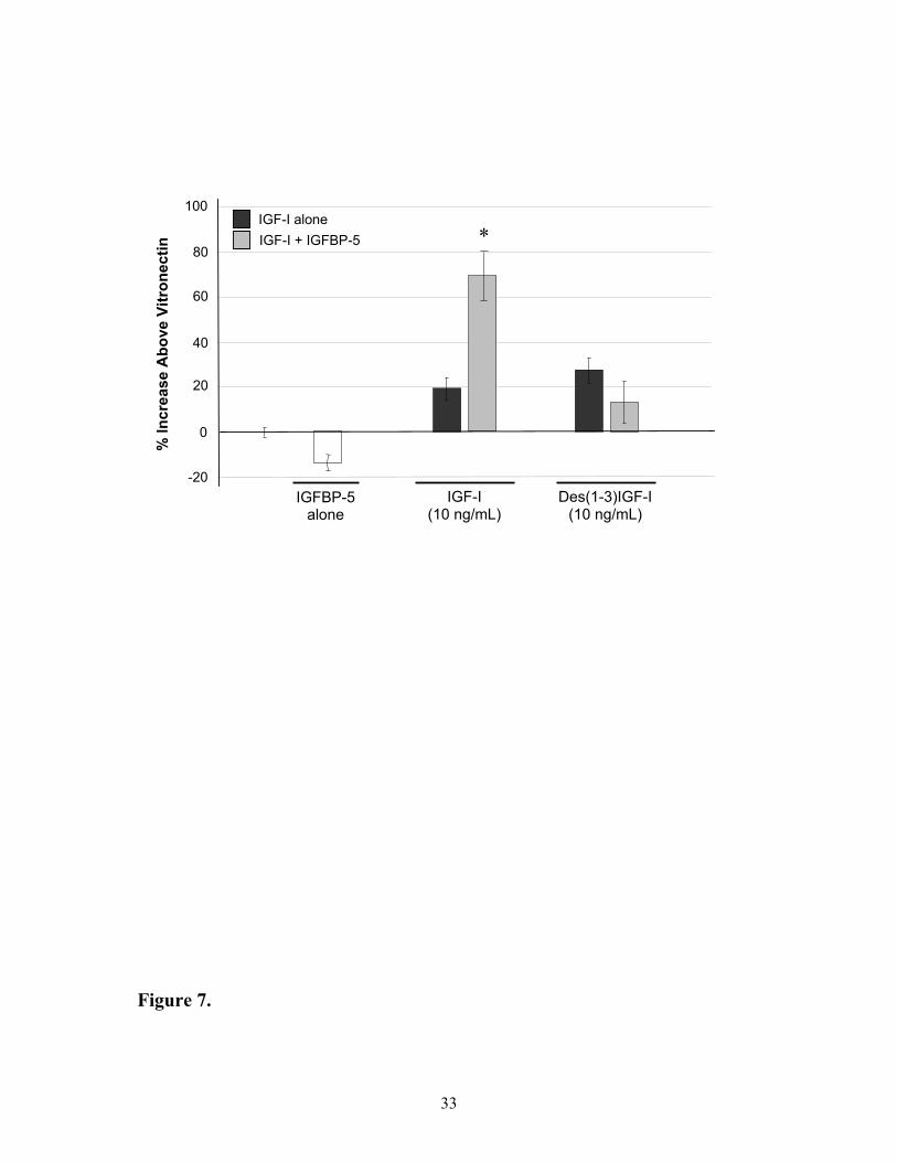
Comparison of the IGF-I peptides on MCF-7 cell migration in the absence and presence of
IGFBP-5
To examine whether the increase in cell migration following addition of IGF-I and
IGFBP-5 involved the formation of a ternary IGF-I:IGFBP-5:VN complex, responses were
compared between native IGF-I and the Des(1-3)IGF-I analogue that has reduced affinity for
IGFBPs while retaining its ability to activate the IGF-I receptor (20). In the absence of
IGFBP-5, assays with either 10 ng of native IGF-I or Des(1-3)IGF-I in the presence of VN
resulted in increased migration on VN of 19 ± 5 and 27 ± 7 % respectively (Figure 7).
13
However, only the migration of cells in the Transwells with native IGF-I treatment was
significantly increased by the addition of IGFBP-5 (an increase of 71 ± 10 % compared to
VN). Responses observed with the Des(1-3)IGF-I in the presence of IGFBP-5 remained
unchanged (an increase of 13 ± 9 % compared to VN).
14
Discussion
The studies reported here extend previous observations in which VN was identified as
a novel high-affinity IGF-II binding protein (2) that may be responsible for mediation of
many effects of IGF-II in the extracellular environment. The same earlier studies (2)
revealed that IGF-I did not bind directly to VN. This was somewhat surprising given the
increasing evidence suggesting a key role for VN in mediating a number of core cellular
effects of IGF-I such as cellular DNA synthesis, type-1 IGF receptor autophosphorylation
and cell migration (10-12, 25). To explain this, we proposed that IGFBPs may be specifically
required to mediate binding of IGF-I to VN.
The present study provides evidence to support our hypothesis by demonstrating that
IGF-I can only interact with VN via the intermediate involvement of IGFBPs. This
investigation has shown for the first time that: - i) direct binding of IGF-II to VN does not
require IGFBPs but is competitively inhibited by IGFBPs; ii) IGF-I binding to VN is
significantly enhanced by all IGFBPs except for IGFBP-1 and –6; iii) the role of IGFBPs is
specific since a) Des(1-3)IGF-I is a poor competitor for binding of labelled IGF-I to VN in
the presence of IGFBPs; and b) IGFBP-3 enhancement of IGF-I binding to VN requires an
intact IGFBP-3 HBD and is affected by the glycosylation state of IGFBP-3. In addition, we
have shown that the IGF:VN interaction is functionally significant (cell migration) through
the interaction of IGF-I indirectly (via IGFBP-5) with vitronectin, while the direct interaction
of IGF-II has been shown by others in our laboratory (Noble et al, unpublished data).
IGF-II binding to VN is independent of IGFBPs, this being demonstrated by two
means. First, by the equivalent competitive inhibition of [125I]-IGF-II binding by wild-type
IGF-II and by two analogues, Des(1-6)IGF-II and [Leu27]IGF-II, which have reduced affinity
for IGFBPs and the type-1 IGF receptor, respectively (20, 21). Second, all six IGFBPs were
shown to inhibit IGF-II binding to VN, at least at the higher levels tested. The most effective
15
IGFBPs were IGFBP-1, -3 and –6. Competition for binding of IGF-II to VN may occur
either by IGFBPs directly competing with IGF-II for the IGF-II binding site on VN or by the
IGFBPs binding and sequestering IGF-II in solution. Our studies to date cannot distinguish
between these possibilities for IGFBP-3. For IGFBP-1 and –6, however, since these IGFBPs
also inhibited IGF-I binding to VN, it is likely that these IGFBPs bind IGF-I and/or IGF-II in
solution and hence primarily sequester the IGFs away from VN. It is likely that the very
efficient inhibition of IGF-II:VN binding by IGFBP-6 reflects its high affinity for IGF-II
(26). Previous biochemical data (2) have demonstrated by 2-D gel electrophoresis that the
purified VN used for IGF-II binding studies was devoid of any traces of contaminating
IGFBPs. This is further substantiated by the inability of IGF-I to bind to VN.
The finding that IGF-I binding to VN is markedly enhanced in the presence of
IGFBPs is of particular interest. All IGFBPs, except for IGFBP-1 and –6, enhanced binding
of IGF-I to VN to varying degrees. The lack of effect observed with IGFBP-6 suggests it
does not bind to VN and/or may also be an indication of its low affinity for IGF-I (26). The
inhibitory effect of IGFBP-1 on the other hand suggests that the minor amount of binding of
IGF-I directly to VN was blocked by IGFBP-1, presumably via IGFBP-1 sequestration of
IGF-I in solution. These data, taken together with the presence of an RGD integrin-binding
motif in IGFBP-1 (27) and the finding that IGFBP-1 can bind directly to integrins to effect
cell migration and proliferation (28-30), indicate that these IGFBP-1-stimulated cellular
responses are unlikely to also involve VN.
The specificity and functional significance of the requirement for IGFBPs to facilitate
IGF-I binding to VN was demonstrated in several ways. First, through competitive inhibition
studies, it was shown that while unlabelled IGF-I was effective in reducing the enhancing
effects of both IGFBP-3 and –5 on binding of [125I]-IGF-I to VN, Des(1-3)IGF-I, which has a
16
much reduced affinity for IGFBPs, especially IGFBP-5 (20), was a great deal less effective.
These data reflect the necessity for IGFBP (-3 or –5) to mediate the binding of IGF-I to VN.
Second, we have demonstrated that the HBD motif in IGBFP-3 is a critical
determinant of the ability of IGFBP-3 to enhance IGF-I binding to VN. IGFBP-3-mediated
binding of IGF-I to VN was abolished when the IGFBP-3 HBD region was mutated even
though the affinity of IGF-I for this IGFBP-3 variant is similar to that of the wild-type
IGFBP-3 (24). The heparin-binding site in proteins such as IGFBP-3 and VN has been
previously implicated in cell association processes (7, 15, 31-34). This observation mirrors
other recent findings that the HBD of IGFBP-3 was required for binding to fibronectin, a
protein with similar functions to VN (35).
Likewise, IGFBP-5 has been shown to bind via its HBD to VN with high affinity (36)
and that functional effects of IGFBP-5 required trimeric complex formation with IGF-I. This
complex was found to effect IGF-I-mediated functional responses through the αvβ3 integrin
(36). Interestingly, these IGF-I-stimulated responses were decreased in the presence of
heparin, again highlighting the involvement of IGFBP basic amino acid residues in binding to
VN. Indeed, the study by Nam et al. (36), where labelled IGFBP-5 was used to examine
VN:IGFBP-5 complex formation independently validates our own findings, which
demonstrate IGF:IGFBP:VN complex formation using labelled IGF-I.
Third, we have shown that the ability of IGF-I to bind to VN is markedly influenced
by the glycosylation state of IGFBP-3. In various IGF/IGFBP studies, the effect of the
glycosylation state of the IGFBPs has received little attention. Indeed, glycosylation is
reported to play little role in IGFBP-3-mediated IGF effects (37-39). In contrast, we
demonstrate here that non-glycosylated IGFBP-3 markedly enhances binding of labelled IGF-
I to VN compared to glycosylated IGFBP-3. This was due, in part only, to a greater (two-
17
fold) ability of non-glycosylated to bind to VN, demonstrating that factors other than
glycosylation are important. Firth and Baxter have previously demonstrated that de-
glycosylated IGFBP-3 has a higher affinity for the cell surface (38). In view of the present
data this difference may reflect preferential binding of de-glycosylated IGFBP-3 to VN
associated with the cell surface. While non-glycosylated IGFBP-3 would not appear to be
especially relevant in the physiological context, the observations in this study suggest that use
of non-glycosylated IGFBP-3 in a trimeric protein complex with IGF-I and VN may well
prove to be a potent way to facilitate delivery of IGF-I to the cell surface - a phenomenon
which potentially could be used in therapeutic and industrial applications to manipulate cell
processes.
The findings presented here, along with those by others (25, 36, 40), provide
important new insights into the mechanism by which IGF-I mediates its effects via VN and
VN-binding integrins. Although it has not been specifically addressed in this study, these
findings also offer an explanation as to how IGF-II and IGF-I can exert different functions as
IGF-II appears to bind directly to VN, whereas IGF-I binds indirectly via select IGFBPs.
Thus, despite their structural similarity, the IGFs have clearly evolved different regulatory
mechanisms to provide the capacity for different cellular functional roles.
We propose that four of the IGFBPs, namely IGFBP-2, -3, -4 and -5, enhance IGF-I
binding to VN by forming a heterotrimeric complex comprised of IGF:IGFBP:VN, and that
this complex is required for cellular responses. We have shown here that the proposed
heterotrimeric complex involving IGFBP-5 enhances MCF-7 breast carcinoma cell migration
to a significantly greater extent than either VN:IGFBP or VN:IGF binary complexes. The
functional requirement for IGFBPs in the complex also has been demonstrated by showing
that Des(1-3)IGF-I, which binds poorly to IGFBP-3 or –5 (20), fails to stimulate MCF-7 cell
migration in this system. Together, these data confirm our hypothesis that IGFBPs are
18
directly involved in, and indeed required for, enhancing the cellular responsiveness of IGF-I
in the presence of VN.
Previous studies by Clemmons et al. also indicated there is a functional and specific
connection between IGF-I and VN, as blocking of the VN receptor, αvβ3, inhibited IGF-I
mediated cellular responses (13, 14). Grulich-Henn et al. (25) have also recently
demonstrated that transport of IGF-I across endothelial cell monolayers required IGF-I
interacting with VN. These investigations also suggested that VN was not likely to be a
primary binding site for IGF-I and that IGFBPs could be implicated. The results from the
study reported here, in which IGF-I is linked to VN via IGFBPs, can potentially explain the
observation that VN is critical in a number of IGF-I-stimulated cellular responses such as
those reported by Clemmons et al. (13) and Grulich-Henn et al. (25), despite there being only
minimal direct binding of IGF-I to VN (2). Together, these findings give insights as to how
IGF-I can mediate diverse effects such as cell migration and cellular DNA synthesis and,
moreover, suggest that VN may have a critical role in linking effects requiring both activation
of integrins and the type-1 IGF receptor as demonstrated by Maile et al. (40). Thus, the
IGF:IGFBP:VN complex appears to be important in normal growth and development and
further functional and structural investigation of this complex may provide mechanisms for
maintaining these physiologies in altered diseased states.
19
Acknowledgments
We acknowledge the technical advice of Dr Janet Martin (Kolling Institute for Medical
Research, Australia) in IGFBP-3 iodinations and the generosity of Dr Leon Bach (University
of Melbourne, Department of Medicine, Australia) in providing IGFBP-6 for these studies.
We would also like to acknowledge Dr D.R. Powell (Baylor College of Medicine, Houston,
TX, USA) for helpful discussion. This study was supported by the Queensland Cancer Fund
and the QUT Small Grants Scheme.
Abbreviations used in this paper: [125I], 125Iodine; IGF, insulin-like growth factor; IGFBP,
IGF-binding protein; IGFBP-rP, IGFBP-related protein; VN, vitronectin; R-G-D, Arg-Gly-
Asp; HBD, heparin binding domain; SMC, smooth muscle cell; DMEM, Dulbecco’s
Modified Eagles Medium; HBB, HEPES binding buffer; HBSS, Hanks’ balanced salt
solution; cpm, counts per minute; SEM, standard error of mean.
20
References
1. Jones JI, Clemmons DR 1995 Insulin-like growth factors and their binding proteins: biological actions. Endocr Rev 16:3-34.
2. Upton Z, Webb H, Hale K, et al. 1999 Identification of vitronectin as a novel insulin-like growth factor-II binding protein. Endocrinology 140:2928-31.
3. Kost C, Stuber W, Ehrlich HJ, Pannekoek H, Preissner KT 1992 Mapping of binding sites for heparin, plasminogen activator inhibitor-1, and plasminogen to vitronectin's heparin-binding region reveals a novel vitronectin-dependent feedback mechanism for the control of plasmin formation. J Biol Chem 267:12098-105.
4. Francois PP, Preissner KT, Herrmann M, et al. 1999 Vitronectin interaction with glycosaminoglycans. Kinetics, structural determinants, and role in binding to endothelial cells. J Biol Chem 274:37611-9.
5. Boettiger D, Lynch L, Blystone S, Huber F 2001 Distinct ligand-binding modes for integrin alpha(v)beta(3)-mediated adhesion to fibronectin versus vitronectin. J Biol Chem 276:31684-90.
6. Felding-Habermann D CD 1993 Vitronectin and its receptors. Current Opinion in Cell Biology 5:864-868.
7. de Boer HC, Preissner KT, Bouma BN, de Groot PG 1992 Binding of vitronectin-thrombin-antithrombin III complex to human endothelial cells is mediated by the heparin binding site of vitronectin. J Biol Chem 267:2264-8.
8. Wilkins-Port CE, McKeown-Longo PJ 1996 Heparan sulfate proteoglycans function in the binding and degradation of vitronectin by fibroblast monolayers. Biochem Cell Biol 74:887-97.
9. Deng G, Curriden SA, Hu G, Czekay RP, Loskutoff DJ 2001 Plasminogen activator inhibitor-1 regulates cell adhesion by binding to the somatomedin B domain of vitronectin. J Cell Physiol 189:23-33.
10. Jones JI, Doerr ME, Clemmons DR 1995 Cell migration: interactions among integrins, IGFs and IGFBPs. Prog Growth Factor Res 6:319-27.
11. Zheng B, Clemmons DR 1998 Blocking ligand occupancy of the alphaVbeta3 integrin inhibits insulin-like growth factor I signaling in vascular smooth muscle cells. Proc Natl Acad Sci U S A 95:11217-22.
12. Maile LA, Badley-Clarke J, Clemmons DR 2001 Structural analysis of the role of the beta 3 subunit of the alpha V beta 3 integrin in IGF-I signaling. J Cell Sci 114:1417-25.
13. Clemmons DR, Horvitz G, Engleman W, Nichols T, Moralez A, Nickols GA 1999 Synthetic alphaVbeta3 antagonists inhibit insulin-like growth factor-I-stimulated smooth muscle cell migration and replication. Endocrinology 140:4616-21.
21
14. Rees C, Clemmons DR 1998 Inhibition of IGFBP-5 binding to extracellular matrix and IGF-I-stimulated DNA synthesis by a peptide fragment of IGFBP-5. J Cell Biochem 71:375-81.
15. Forsten KE, Akers RM, San Antonio JD 2001 Insulin-like growth factor (IGF) binding protein-3 regulation of IGF-I is altered in an acidic extracellular environment. J Cell Physiol 189:356-365.
16. Mohan S, Nakao Y, Honda Y, et al. 1995 Studies on the mechanisms by which insulin-like growth factor (IGF) binding protein-4 (IGFBP-4) and IGFBP-5 modulate IGF actions in bone cells. J Biol Chem 270:20424-31.
17. Conover CA 1992 Potentiation of insulin-like growth factor (IGF) action by IGF-binding protein-3: studies of underlying mechanism. Endocrinology 130:3191-9.
18. Firth SM, Ganeshprasad U, Poronnik P, Cook DI, Baxter RC 1999 Adenoviral-mediated expression of human insulin-like growth factor-binding protein-3. Protein Expr Purif 16:202-11.
19. Ballard FJ, Read LC, Francis GL, Bagley CJ, Wallace JC 1986 Binding properties and biological potencies of insulin-like growth factors in L6 myoblasts. Biochem J 233:223-30.
20. Francis GL, Aplin SE, Milner SJ, McNeil KA, Ballard FJ, Wallace JC 1993 Insulin-like growth factor (IGF)-II binding to IGF-binding proteins and IGF receptors is modified by deletion of the N-terminal hexapeptide or substitution of arginine for glutamate-6 in IGF-II. Biochem J 293:713-9.
21. Roth BV, Burgisser DM, Luthi C, Humbel RE 1991 Mutants of human insulin-like growth factor II: expression and characterization of analogs with a substitution of TYR27 and/or a deletion of residues 62-67. Biochem Biophys Res Commun 181:907-14.
22. Booth BA, Boes M, Andress DL, et al. 1995 IGFBP-3 and IGFBP-5 association with endothelial cells: role of C-terminal heparin binding domain. Growth Regul 5:1-17.
23. Campbell PG, Andress DL 1997 Insulin-like growth factor (IGF)-binding protein-5-(201-218) region regulates hydroxyapatite and IGF-I binding. Am J Physiol 273:E1005-13.
24. Firth SM, Ganeshprasad U, Baxter RC 1998 Structural determinants of ligand and cell surface binding of insulin-like growth factor-binding protein-3. J Biol Chem 273:2631-8.
25. Grulich-Henn J, Ritter J, Mesewinkel S, Heinrich U, Bettendorf M, Preissner KT 2002 Transport of insulin-like growth factor-I across endothelial cell monolayers and its binding to the subendothelial matrix. Exp Clin Endocrinol Diabetes 110:67-73.
26. Bach LA, Hsieh S, Sakano K, Fujiwara H, Perdue JF, Rechler MM 1993 Binding of mutants of human insulin-like growth factor II to insulin-like growth factor binding proteins 1-6. J Biol Chem 268:9246-54.
22
27. Jones JI, Gockerman A, Busby WH, Jr., Wright G, Clemmons DR 1993 Insulin-like growth factor binding protein 1 stimulates cell migration and binds to the alpha 5 beta 1 integrin by means of its Arg-Gly-Asp sequence. Proc Natl Acad Sci U S A 90:10553-7.
28. Gleeson LM, Chakraborty C, McKinnon T, Lala PK 2001 Insulin-like growth factor-binding protein 1 stimulates human trophoblast migration by signaling through alpha 5 beta 1 integrin via mitogen-activated protein Kinase pathway. J Clin Endocrinol Metab 86:2484-93.
29. Gockerman A, Prevette T, Jones JI, Clemmons DR 1995 Insulin-like growth factor (IGF)-binding proteins inhibit the smooth muscle cell migration responses to IGF-I and IGF-II. Endocrinology 136:4168-73.
30. Chakraborty C, Gleeson LM, McKinnon T, Lala PK 2002 Regulation of human trophoblast migration and invasiveness. Can J Physiol Pharmacol 80:116-24.
31. Devi GR, Yang DH, Rosenfeld RG, Oh Y 2000 Differential effects of insulin-like growth factor (IGF)-binding protein-3 and its proteolytic fragments on ligand binding, cell surface association, and IGF-I receptor signaling. Endocrinology 141:4171-9.
32. Mazerbourg S, Zapf J, Bar RS, Brigstock DR, Monget P 2000 Insulin-like growth factor (IGF)-binding protein-4 proteolytic degradation in bovine, equine, and porcine preovulatory follicles: regulation by IGFs and heparin-binding domain-containing peptides. Biol Reprod 63:390-400.
33. Rosenblatt S, Bassuk JA, Alpers CE, Sage EH, Timpl R, Preissner KT 1997 Differential modulation of cell adhesion by interaction between adhesive and counter-adhesive proteins: characterization of the binding of vitronectin to osteonectin (BM40, SPARC). Biochem J 324:311-9.
34. Hocking DC, Sottile J, Reho T, Fassler R, McKeown-Longo PJ 1999 Inhibition of fibronectin matrix assembly by the heparin-binding domain of vitronectin. J Biol Chem 274:27257-64.
35. Gui Y, Murphy LJ 2001 Insulin-like growth factor (IGF)-binding protein-3 (IGFBP-3) binds to fibronectin (FN): demonstration of IGF-I/IGFBP-3/fn ternary complexes in human plasma. J Clin Endocrinol Metab 86:2104-10.
36. Nam T, Moralez A, Clemmons D 2002 Vitronectin Binding to IGF Binding Protein-5 (IGFBP-5) Alters IGFBP-5 Modulation of IGF-I Actions. Endocrinology 143:30-6.
37. Conover CA 1991 Glycosylation of insulin-like growth factor binding protein-3 (IGFBP-3) is not required for potentiation of IGF-I action: evidence for processing of cell-bound IGFBP-3. Endocrinology 129:3259-68.
38. Firth SM, Baxter RC 1999 Characterisation of recombinant glycosylation variants of insulin-like growth factor binding protein-3. J Endocrinol 160:379-87.
39. Sommer A, Spratt SK, Tatsuno GP, Tressel T, Lee R, Maack CA 1993 Properties of glycosylated and non-glycosylated human recombinant IGF binding protein-3 (IGFBP-3). Growth Regul 3:46-9.
23
40. Maile LA, Imai Y, Clarke JB, Clemmons DR 2002 Insulin-like growth factor I increases alpha Vbeta 3 affinity by increasing the amount of integrin-associated protein that is associated with non-raft domains of the cellular membrane. J Biol Chem 277:1800-5.
24
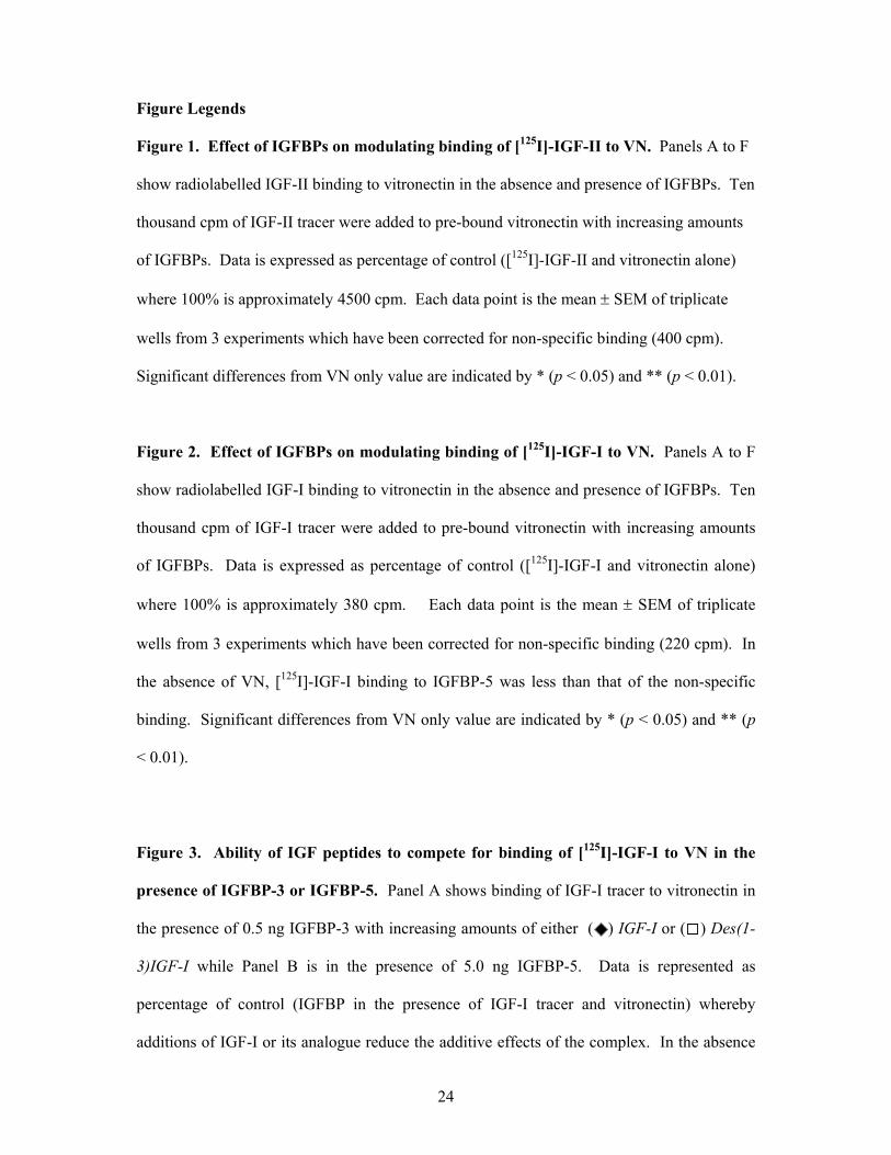
Figure Legends
Figure 1. Effect of IGFBPs on modulating binding of [125I]-IGF-II to VN. Panels A to F
show radiolabelled IGF-II binding to vitronectin in the absence and presence of IGFBPs. Ten
thousand cpm of IGF-II tracer were added to pre-bound vitronectin with increasing amounts
of IGFBPs. Data is expressed as percentage of control ([125I]-IGF-II and vitronectin alone)
where 100% is approximately 4500 cpm. Each data point is the mean ± SEM of triplicate
wells from 3 experiments which have been corrected for non-specific binding (400 cpm).
Significant differences from VN only value are indicated by * (p < 0.05) and ** (p < 0.01).
Figure 2. Effect of IGFBPs on modulating binding of [125I]-IGF-I to VN. Panels A to F
show radiolabelled IGF-I binding to vitronectin in the absence and presence of IGFBPs. Ten
thousand cpm of IGF-I tracer were added to pre-bound vitronectin with increasing amounts
of IGFBPs. Data is expressed as percentage of control ([125I]-IGF-I and vitronectin alone)
where 100% is approximately 380 cpm. Each data point is the mean ± SEM of triplicate
wells from 3 experiments which have been corrected for non-specific binding (220 cpm). In
the absence of VN, [125I]-IGF-I binding to IGFBP-5 was less than that of the non-specific
binding. Significant differences from VN only value are indicated by * (p < 0.05) and ** (p
< 0.01).
Figure 3. Ability of IGF peptides to compete for binding of [125I]-IGF-I to VN in the
presence of IGFBP-3 or IGFBP-5. Panel A shows binding of IGF-I tracer to vitronectin in
the presence of 0.5 ng IGFBP-3 with increasing amounts of either ( ) IGF-I or ( ) Des(1-
3)IGF-I while Panel B is in the presence of 5.0 ng IGFBP-5. Data is represented as
percentage of control (IGFBP in the presence of IGF-I tracer and vitronectin) whereby
additions of IGF-I or its analogue reduce the additive effects of the complex. In the absence
25
of VN, binding of [125I]-IGF-I to IGFBP-3 or –5 was less than the value for non-specific
binding (220cpm). Values shown are the mean ± SEM of triplicate wells from 3
experiments.
Figure 4: Importance of IGFBP-3 HBD in IGFBP-3 mediation of [125I]-IGF-I binding
to VN. IGF-I tracer was added with either ( ) glycosylated IGFBP-3 or ( ) HBD mutant
IGFBP-3 to wells pre-bound with vitronectin. Data is expressed as percentage of control,
which is IGF-I tracer and vitronectin alone. Values shown are the mean ± SEM of triplicate
wells from 3 experiments.
Figure 5: Comparison of the effects of glycosylated and non-glycosylated IGFBP-3 on
[125I]-IGF-I binding to VN. IGF-I tracer was added with either ( ) glycosylated IGFBP-3
or ( ) non-glycosylated mutant IGFBP-3 to wells pre-bound with vitronectin. Data is
expressed as percentage of control, which is IGF-I tracer and vitronectin alone. Values shown
are the mean ± SEM of triplicate wells from 3 experiments.
Figure 6: Migration of MCF-7 cells through Transwells in response to IGF-I pre-bound
to VN in the absence or presence of IGFBP-5. MCF-7 cells were seeded onto Transwells
that had been coated with VN ± IGF-I ± IGFBP-5 and allowed to migrate through the porous
membrane for 5 hours. The number of cells transversing the membrane in the presence of
IGF-I were then expressed as a percentage of those that migrated on VN alone. The
responses are shown as IGF-I exposed to VN in the absence of IGFBP-5 ( ), the presence of
IGFBP-5 ( ) and the response of IGFBP-5 alone ( ). Values shown are the mean ± SEM
of duplicate wells from 3 experiments.
26
Figure 7: Migration of MCF-7 cells through Transwells in response to Des(1-3)IGF-I
pre-bound to VN in the absence or presence of IGFBP-5. MCF-7 cells were seeded onto
Transwells that had been coated with VN ± IGFBP-5 ± native or mutant IGF-I and allowed to
migrate through the porous membrane for 5 hours. The number of cells transversing the
membrane in the presence of IGF-I were then expressed as a percentage of those that
migrated on VN. The responses are shown as IGF-I exposed to VN in the absence of IGFBP-
5 ( ), the presence of IGFBP-5 ( ) and the response of IGFBP-5 alone ( ). Values shown
are the mean ± SEM of duplicate wells from 3 experiments.
27
Figure 1.
A: IGFBP-1
0
20
40
60
80
100
120
VN only 0.05 0.2 0.5 2 5 20 100
% o
f Con
trol
****
****
****
**
A: IGFBP-1
0
20
40
60
80
100
120
VN only 0.05 0.2 0.5 2 5 20 100
% o
f Con
trol
****
****
****
** ****
****
****
**
B: IGFBP-2
0
20
40
60
80
100
120
VN only 0.05 0.2 0.5 2 5 20 100
% o
f Con
trol **
****
**
**
**
B: IGFBP-2
0
20
40
60
80
100
120
VN only 0.05 0.2 0.5 2 5 20 100
% o
f Con
trol **
****
**
**
****
****
**
**
**
C: IGFBP-3
0
20
40
60
80
100
120
VN only 0.05 0.2 0.5 2 5 20 100
% o
f Con
trol **
****
**
**
**
C: IGFBP-3
0
20
40
60
80
100
120
VN only 0.05 0.2 0.5 2 5 20 100
% o
f Con
trol **
****
**
**
**
**
****
**
**
**
D: IGFBP-4
0
20
40
60
80
100
120
VN only 0.05 0.2 0.5 2 5 20 100
% o
f Con
trol
**
***
D: IGFBP-4
0
20
40
60
80
100
120
VN only 0.05 0.2 0.5 2 5 20 100
% o
f Con
trol
**
***
**
***
E: IGFBP-5
0
20
40
60
80
100
120
VN only 0.05 0.2 0.5 2 5 20 100
Sample (ng)
% o
f Con
trol
**
E: IGFBP-5
0
20
40
60
80
100
120
VN only 0.05 0.2 0.5 2 5 20 100
Sample (ng)
% o
f Con
trol
**
F: IGFBP-6
0
20
40
60
80
100
120
VN only 0.05 0.2 0.5 2 5 20 100
Sample (ng)
% o
f Con
trol
**
**** **
****
*
F: IGFBP-6
0
20
40
60
80
100
120
VN only 0.05 0.2 0.5 2 5 20 100
Sample (ng)
% o
f Con
trol
**
**** **
****
***
**** **
****
*
28
Figure 2.
A: IGFBP-1
0
100
200
300
400
500
600
VN only 0.05 0.2 0.5 2 5 20 100
% o
f Con
trol
** ** ** ** ** **
A: IGFBP-1
0
100
200
300
400
500
600
VN only 0.05 0.2 0.5 2 5 20 100
% o
f Con
trol
** ** ** ** ** **** ** ** ** ** **
B: IGFBP-2
0
100
200
300
400
500
600
VN only 0.05 0.2 0.5 2 5 20 100
% o
f Con
trol
* ** **
B: IGFBP-2
0
100
200
300
400
500
600
VN only 0.05 0.2 0.5 2 5 20 100
% o
f Con
trol
* ** *** ** **
C: IGFBP-3
0
100
200
300
400
500
600
VN only 0.05 0.2 0.5 2 5 20 100
% o
f Con
trol
D: IGFBP-4
0
100
200
300
400
500
600
VN only 0.05 0.2 0.5 2 5 20 100
% o
f Con
trol * *
**
D: IGFBP-4
0
100
200
300
400
500
600
VN only 0.05 0.2 0.5 2 5 20 100
% o
f Con
trol * *
**
* *
**
E: IGFBP-5
0
100
200
300
400
500
600
VN only 0.05 0.2 0.5 2 5 20 100
Sample (ng)
% o
f Con
trol
***
**
*
808
E: IGFBP-5
0
100
200
300
400
500
600
VN only 0.05 0.2 0.5 2 5 20 100
Sample (ng)
% o
f Con
trol
***
**
*
808***
**
*
***
**
*
808808
F: IGFBP-6
0
100
200
300
400
500
600
VN only 0.05 0.2 0.5 2 5 20 100
Sample (ng)
% o
f Con
trol
* *
F: IGFBP-6
0
100
200
300
400
500
600
VN only 0.05 0.2 0.5 2 5 20 100
Sample (ng)
% o
f Con
trol
* ** *
29
Figure 3.
0
20
40
60
80
100
120
0.01 0.1 1 10 100
Sample (ng)
% o
f Con
trol
0
B: IGFBP-5 (5.0 ng)
****
**
****
**
*
**
*********
A: IGFBP-3 (0.5 ng)
0
20
40
60
80
100
120
0.01 0.1 1 10 100
Sample (ng)
% o
f Con
trol
0
**
****
** ****
**
**** **
** **
**
**
30
Figure 4.
0
50
100
150
200
250
VNonly
0.05 0.2 0.5 2 5 20 100
Sample (ng)
% o
f Con
trol
**
** ** **
** ** **

31
Figure 5.
0
500
1000
1500
2000
2500
3000
VNonly
0.05 0.2 0.5 2 5 20 100
Sample (ng)
% o
f Con
trol
*
*
*
32
Figure 6.
% In
crea
se A
bove
Vitr
onec
tin
40
60
80
20
0
100
-20 IGFBP-5
alone 1 100 30 10 3
IGF-I concentration (ng/mL)
** *IGF-I alone
IGF-I + IGFBP-5
33
Figure 7.
% In
crea
se A
bove
Vitr
onec
tin
40
60
80
20
0
100
-20 IGFBP-5
alone IGF-I
(10 ng/mL) Des(1-3)IGF-I
(10 ng/mL)
IGF-I alone IGF-I + IGFBP-5 *