Embed Size (px)
Citation preview

1
This is a post-print version of the following article: Fernández-Cabrera, MR ; Selvas, A. ;
Miguéns, M.; Higuera-Matas, A. ; Vale-Martínez, A. ; Ambrosio, E. ; Martí-Nicolovius, M. ;
Guillazo-Blanch, G. (2017) Parafascicular thalamic nucleus deep brain stimulation decreases
NMDA receptor GluN1 subunit gene expression in the prefrontal còrtex. Neuroscience, vol. 348,
p. 73-80. ISSN: 0306-4522 DOI: 10.1016/j.neuroscience.2017.02.009
Parafascicular thalamic nucleus deep brain stimulation decreases NMDA receptor
GluN1 subunit gene expression in the prefrontal cortex
Mónica R Fernández-Cabreraa*, Abraham Selvasa*, Miguel Miguénsb, Alejandro
Higuera-Matasa, Anna Vale-Martínezc, Emilio Ambrosioa, Margarita Martí-Nicoloviusc
and Gemma Guillazo-Blanchc.
a Departamento de Psicobiología, Facultad de Psicología, UNED, 28040 Madrid, Spain.
b Departamento de Psicología Básica I, Facultad de Psicología, UNED, 28040 Madrid,
Spain.
c Departament de Psicobiologia i Metodologia de les Ciències de la Salut, Institut de
Neurociències, Universitat Autònoma de Barcelona, Barcelona, Spain.
*These authors contributed equally to this work.
Corresponding author:
Miguel Miguéns,
Departamento de Psicología Básica I, Facultad de Psicología, Universidad Nacional de
Educación a Distancia (UNED)
C/ Juan del Rosal nº 10, 28040 Madrid, Spain
E-mail: [email protected]
Tel: +34 913987974, Fax: +34 913986287

2
Abstract
The rodent parafascicular nucleus (PFn) or the centromedian-parafascicular complex of
primates is a posterior intralaminar nucleus of the thalamus related to cortical activation
and maintenance of states of consciousness underlying attention, learning and memory.
Deep brain stimulation (DBS) of the PFn has been proved to restore arousal and
consciousness in humans and to enhance performance in learning and memory tasks in
rats. The primary expected effect of PFn DBS is to induce plastic changes in target
neurons of brain areas associated with cognitive function. In this study, Wistar rats were
stimulated for 20 mins in the PFn following a DBS protocol that had previously
facilitated memory in rats. NMDA and GABAB receptor binding, and gene expression
of the GluN1subunit of the NMDA receptor (NMDAR) were assessed in regions related
to cognitive functions, such as the prefrontal cortex and hippocampus. The results
showed that PFn DBS induced a decrease in NMDAR GluN1 subunit gene expression
in the cingulate and prelimbic cortices, but no significant statistical differences were
found in the density of NMDA or GABAB receptors in any of the analyzed regions.
Taken together, our findings suggest a possible role for the NMDAR GluN1 subunit in
the prefrontal cortex in the procognitive actions of the PFn DBS.
Keywords: electrical stimulation; glutamate; NMDA; GABAB; prelimbic cortex;
cingulated cortex.

3
Thalamic deep brain stimulation (DBS) has been proposed as a method for the
treatment of advanced Parkinson’s disease and primary dystonia, Gilles de la Tourette
Syndrome, epileptic seizures, pain diseases (Franzini et al., 2012), and also as a
potential treatment for cognitive and consciousness diseases (Baker 2016; Schiff et al.,
2012). Specifically, DBS of the intralaminar thalamic centromedian-parafascicular
(CM-PF) complex, mainly represented by the parafascicular nucleus (PFn) in rodents,
has been shown to be effective in both facilitating memory in animals and restoring
arousal and consciousness in humans (Baker et al., 2016; Schiff et al., 2012; Takamisu
Yamamoto et al. 2013).
The CM-PF complex links brainstem arousal systems to cerebral cortical and
basal ganglia networks crucial to the organization of wakeful behaviors (Smith et al.,
2014; Varela, 2014). Studies in rats have shown that PFn DBS enhanced active
avoidance conditioning retention (Vale-Martínez et al., 1998; Guillazo-Blanch et al.,
1999) and also reversed memory deficits caused by the lesion of the nucleus basalis
magnocellularis (Sos-Hinojosa et al., 2000). PFn is the major thalamic source of
glutamatergic projections to the striatum (Smith et al., 2004) and projects to prefrontal
regions such as the cingulate (Cg1) and prelimbic (PrL) cortices. Thus, PFn implication
in cognitive function may arise from its glutamatergic influence on such targets
(Quiroz-Padilla et al., 2010). Moreover, distinct nuclei of the thalamus, such as those of
the CM-PF complex, may be related to the hippocampus as a result of the direct
association of this structure with the basal ganglia-thalamo-cortical systems (Li et al.,
2014). However, despite the clinical benefits of DBS, the exact molecular and
pharmacological mechanisms underlying its effectiveness need to be clarified.
Glutamatergic synapses have been proposed as a core cellular mechanism for
memory encoding and processing, relying, in part, on their ability to dynamically adjust

4
the content of glutamate receptors in the postsynaptic membrane (Nong et al., 2003;
Han et al., 2013). N-methyl-D-aspartate receptors (NMDARs) are made up of two
obligatory GluN1 and two regulatory GluN2/3 subunits and play a key role in the
induction of long-term potentiation and depression (Malenka and Bear, 2004) and,
thereby, in learning and memory. Accordingly, cognitive deficits have been observed
following the selective deletion of the GluN1 subunit from the granule cells of the
dentate gyrus (Niewoehner et al., 2007).
There is also evidence that glutamate - gamma-aminobutyric acid (GABA)
interaction in dendritic spines is critical for the synchronized network oscillations
underlying cognitive processes (Kohl and Paulsen, 2010). In this regard, a cross-talk
between both neurotransmitters has been postulated inasmuch as the activation of
glutamate receptors decreased activity in GABAB receptors (Chalifoux and Carter,
2010; Kleschevnikov et al., 2012). Blocking NMDA receptors in prefrontal cortex
(PFC) with infusions of 2-amino-5-phosphonopentanoic acid (APV), an NMDA
receptor antagonist, impaired memory in a recognition memory task (Barker and
Warburton, 2008), contextual fear conditioning (Gilmartin and Helmstetter, 2010) and
trace eye-blink conditioning (Takehara-Nishiuchi et al., 2005). GABAB receptor
antagonists improved performance in a number of different cognitive tests, such as
hippocampal-dependent spatial learning and passive avoidance conditioning
(Kleschevnikov et al., 2012; Gillani et al., 2014). By contrast, GABAB receptor
agonists generally impair learning and memory in these tasks, although at times such
deficits are isoform-specific (Kasten et al., 2015; Zarrindast et al., 2002)
In the present study, we assessed the effects of PFn DBS on NMDA and
GABAB receptor binding and NMDAR GluN1subunit gene expression within several
brain regions related to learning and memory processes, such as the PFC [prelimbic

5
(PrL), infralimbic (IL), and cingulate (Cg1) cortices], hippocampus [cornus ammonis 1
(CA1), cornus ammnonis 3 (CA3), and dentate gyrus (DG)], and the primary auditory
cortex (Au) and primary motor cortex (M1) as control areas.
The DBS protocol applied was the same that had facilitated learning and
memory in previous studies (Vale-Martínez et al., 1998; Guillazo-Blanch et al., 1999).
Experimental Procedures
Subjects
Twenty naive male Wistar rats belonging to our laboratory’s breeding stock
were used (mean age= 96.21 days, SD=4.5; mean weight=408.53 g, SD = 40.14 at the
beginning of the experiment). All procedures were carried out in compliance with the
European Community Council Directive for the care and use of laboratory animals
(86/609/European Community Council) and authorized by the Generalitat de Catalunya
(Diari Oficial de la Generalitat de Catalunya 2450 7/8/1997, protocol number 5959).
Surgery
The animals were anesthetized (isofluorane; FORANE®, Abbott Laboratories, S.A.
Madrid) and underwent stereotaxic implantation of a monopolar stainless steel electrode
(Plastics One, Bilaney; 150 μm in diameter) into the PFn nucleus [AP, -4.10 mm from
bregma; ML,±0.70 mm from midline; and DV, -7.00 mm from skull surface according
to the Paxinos and Watson (1998) rat brain atlas, following procedures explained in
detail elsewhere (Sos-Hinojosa et al., 2000). All the rats were implanted in the right or
left hemisphere, in a balanced way for each group (DBS and Control). The electrode,
electrically insulated except at the tip, was soldered to a plastic connector anchored to
the skull with jeweler screws and dental cement (Vertex self-curing, Dentimex, Zeist,

6
Holland)”. The grounding electrode was a copper wire (200μm in diameter) with one
end soldered to the electrode connector and the other to a screw attached to the skull.
Following surgery, the skin was sutured and antiseptic (Topionic, Almirall
Prodesfarma) and rats were administered an antibiotic (Panolog, Novartis) and were
returned to their home cages for 10 days.
DBS treatment
After post-surgical recovery DBS experiments were performed. The alternating
electrical current was adjusted during several habituation sessions, and consisted of 1-
Hz cathodic square pulse trains of 500 ms delivered by an electrical stimulator (Model
CS-20, Cibertec, Madrid, Spain). Each train contained fifty 0.5 ms pulses. The current
intensity ranged from 60 to 100μA depending on the rats’ behavior (agitation, motor
stereotypies or other abnormal behavior were avoided). Such parameters were similar to
those in other studies reporting large increases in acetylcholine release (Rasmusson,
2000), cortical electroencephalographic activation (McLin et al., 2002, 2003; Golmayo
et al., 2003) and facilitation of learning and memory (Guillazo-Blanch et al., 1995,
1999; Vale-Martínez et al., 1998; Montero-Pastor et al., 2001, 2004). Twenty-four hours
prior to the DBS session, animals were allowed to acclimatize to the experimental box
for one hour with the electrode connected, with no current administered. After the
habituation sessions, rats in the DBS group received a single 20-min stimulation session
during which they were free to move. The treatment was applied in a stimulation cage
(26.5x30.5x35 cm) made of Plexiglass. Control rats were placed in the same cage for 20
min with the electrode clip connected, but with no stimulation. The duration of the DBS
treatment was based on previous studies in rats reporting enhanced cognitive effects
after applying similar time periods of stimulation (Boix-Trelis et al. 2009; Guillazo-

7
Blanch et al., 1995, 1999; Vale-Martínez et al., 1998; Montero-Pastor et al., 2001, 2004;
Shirvalkar et al., 2006). The animals’ behavior was monitored during the stimulation
session and no striking alterations were observed.
In order to obtain our measurements in a peak receptor synthesis and/or
trafficking to the membrane, animals were sacrificed by decapitation four hours after
treatment and their brains were rapidly removed and stored at -80ºC until slicing.
Histology
Brain coronal sections (40 µm) were cut on a freezing stage microtome
(Shandom Cryotome FSE, Thermo Electron Corporation, Massachusetts, USA)
according to the atlas of Paxinos and Watson (1998). The sections were mounted onto
slides (Superfrost™ Plus Microscope Slides; Thermo Fisher Scientific Inc.) and stored
at - 80 ºC until the day of the assays. A set of sections were mounted and stained with
Cresyl violet to check the correct implantation of the electrodes in the PFn. The sections
were then examined under a light microscope by two independent observers to verify
electrode placements (Olympus BX 41; Olympus Optical CO, LTD. Japan).
Microphotographs of the electrode placements were taken with a digital camera
(Olympus DP70). Electrode tip locations were reconstructed on plates according to the
Paxinos and Watson (1998) rat brain atlas.
NMDA and GABAB receptor autoradiography
Protocols for NMDA (Sakurai et al., 1991) and GABAB (Cremer et al., 2009)
receptors were carried out in a similar manner to previous studies (Higuera-Matas et al.,
2012). In short, for the NMDAR, slide-mounted brain sections were prewashed for 30
min in 50 mM Tris-acetate buffer and subsequently incubated in 50 mM Tris-acetate

8
buffer (pH 7.4) containing 5 nM of 3H-MK-801 (27.5 Ci/mmol; Perkin Elmer, Spain)
for 120 min at room temperature. Non-specific binding was determined in the presence
of 100 µM of non-radioactive MK-801 (Sigma, Spain). Following incubation, the
sections were washed in 50 mM of Tris-acetate buffer (pH 7.4, 4ºC) for 80 min in 250
ml of cold buffer. The slides were then washed in distilled water and desiccated with
cold air. Slides were exposed to desiccant (Sigma, Spain) overnight and were then
exposed to tritium-sensitive films (Biomax MR, Kodak, U.S.A). After 6-8 weeks at 0-
4ºC, the films were developed with Kodak-D19 fluid and subjected to image analysis.
Regarding the GABAB receptor, triplicate tissue sections were pre-washed three
times for 5 min at 4ºC in 50 mM Tris-HCl buffer containing 2.5 mM CaCl2 (pH 7.2).
The samples were then incubated for 60 min at 4ºC in the same buffer, containing 2 nM
3[H]-CGP 54626 (30 Ci/mmol: American Radiolabelled Chemicals Inc., Saint Louis,
MO, USA) in the presence or absence of 100 µM of unlabelled CGP 54626 (Tocris,
UK), to evaluate non-specific and total binding to GABAB receptors, respectively. After
three washes in the same cold buffer, the slides were dipped in distilled water and dried
with cold dry air. Slides were exposed to desiccant (Sigma) overnight and then exposed
to tritium-sensitive films (Biomax MR, Kodak, U.S.A). After 4 weeks at 0-4ºC, the
films were developed with Kodak-D19 fluid and subjected to image analysis.
In situ hybridization histochemistry for the GluN1 subunit
Duplicate tissue sections were fixed in 4% paraformaldehyde for 5 min and then
rinsed twice in phosphate-buffered saline (PBS). The sections were then acetylated for
10 min with 0.25% acetic anhydride in 0.1 M triethanolamine and 0.15 M sodium
chloride [pH 8.0], washed in 0.3 M sodium chloride with 0.03 M sodium citrate [pH
7.0], and dehydrated and delipidated through an ethanol–chloroform series. Following

9
previous procedures (Higuera-Matas et al., 2012), the tissue sections were hybridized
with [35S]-dATP (Perkin Elmer, Spain) terminal deoxynucleotidyl-transferase
(Promega, Spain) and end-labelled (100,000 c.p.m. per section) with the oligonucleotide
probe 5′-GAA CAG GTC ACC CGT GGT CAC CAG ATC GCA CTT CTG TGA
AGC CTC-3′ (Sigma, Spain), corresponding to nucleotides 975–1019 of the rats’
GluN1 subunit of the GRIN1 cDNA (Moriyoshi et al., 1991). Hybridization was carried
out overnight at 44°C in a humidified chamber and was completed by washes in a
graded series of saline-sodium citrate solutions (four at 55 °C and two at room
temperature). Slides were exposed to Kodak Biomax MR for 10 days and developed
(Kodak D-19) for image analysis.
Analysis of the autorradiograms
Once the film had been developed, densitometric analyses were performed using
an image processing and analysis program (Scion Image, Scion Corporation, Frederick,
MA, USA). The regions of interest are delineated in Figure 1. For NMDA and GABAB
autoradiography density measurements were calculated for each animal from two slides
per region (three slices/slide; two measurements/slice in consecutive brain sections) in
both hemispheres, and they were transformed to concentrations (nCi/mg of tissue
equivalent) using tritium-labelled microscale standards (Amersham Biosciences/GE
Helathcare, Spain). Finally, the fmol/mg tissue equivalent values were calculated. In the
case of NMDAR GluN1 subunit optical density (O.D.) arbitrary units were reported.
Measurements were calculated for each animal from one slide per region (two
slices/slide; one measurement/slice) in both hemispheres.
Statistical analysis
10
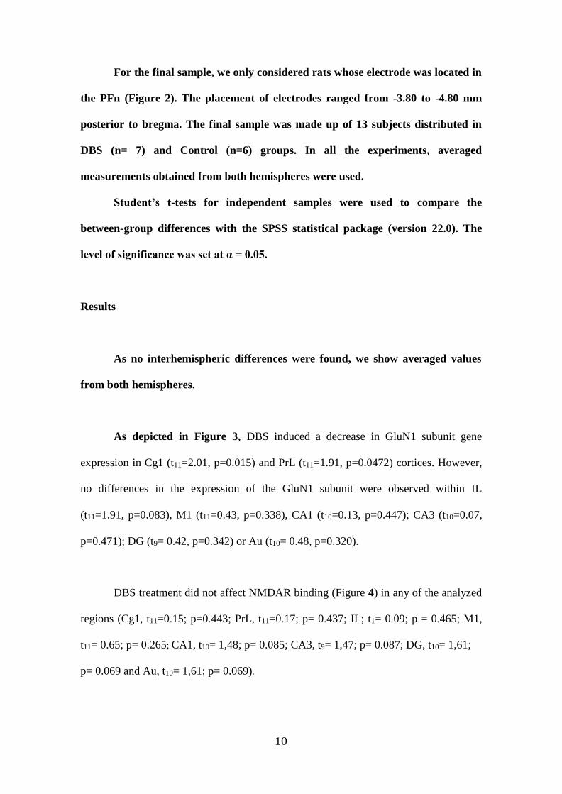
For the final sample, we only considered rats whose electrode was located in
the PFn (Figure 2). The placement of electrodes ranged from -3.80 to -4.80 mm
posterior to bregma. The final sample was made up of 13 subjects distributed in
DBS (n= 7) and Control (n=6) groups. In all the experiments, averaged
measurements obtained from both hemispheres were used.
Student’s t-tests for independent samples were used to compare the
between-group differences with the SPSS statistical package (version 22.0). The
level of significance was set at α = 0.05.
Results
As no interhemispheric differences were found, we show averaged values
from both hemispheres.
As depicted in Figure 3, DBS induced a decrease in GluN1 subunit gene
expression in Cg1 (t11=2.01, p=0.015) and PrL (t11=1.91, p=0.0472) cortices. However,
no differences in the expression of the GluN1 subunit were observed within IL
(t11=1.91, p=0.083), M1 (t11=0.43, p=0.338), CA1 (t10=0.13, p=0.447); CA3 (t10=0.07,
p=0.471); DG (t9= 0.42, p=0.342) or Au (t10= 0.48, p=0.320).
DBS treatment did not affect NMDAR binding (Figure 4) in any of the analyzed
regions (Cg1, t11=0.15; p=0.443; PrL, t11=0.17; p= 0.437; IL; t1= 0.09; p = 0.465; M1,
t11= 0.65; p= 0.265; CA1, t10= 1,48; p= 0.085; CA3, t9= 1,47; p= 0.087; DG, t10= 1,61;
p= 0.069 and Au, t10= 1,61; p= 0.069).
11
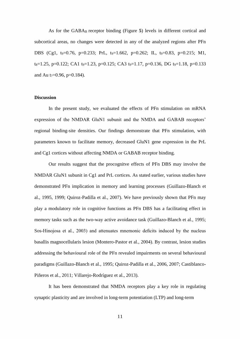
As for the GABAB receptor binding (Figure 5) levels in different cortical and
subcortical areas, no changes were detected in any of the analyzed regions after PFn
DBS (Cg1, t9=0.76, p=0.233; PrL, t9=1.662, p=0.262; IL, t9=0.83, p=0.215; M1,
t8=1.25, p=0.122; CA1 t9=1.23, p=0.125; CA3 t9=1.17, p=0.136, DG t9=1.18, p=0.133
and Au t7=0.96, p=0.184).
Discussion
In the present study, we evaluated the effects of PFn stimulation on mRNA
expression of the NMDAR GluN1 subunit and the NMDA and GABAB receptors’
regional binding-site densities. Our findings demonstrate that PFn stimulation, with
parameters known to facilitate memory, decreased GluN1 gene expression in the PrL
and Cg1 cortices without affecting NMDA or GABAB receptor binding.
Our results suggest that the procognitve effects of PFn DBS may involve the
NMDAR GluN1 subunit in Cg1 and PrL cortices. As stated earlier, various studies have
demonstrated PFn implication in memory and learning processes (Guillazo-Blanch et
al., 1995, 1999; Quiroz-Padilla et al., 2007). We have previously shown that PFn may
play a modulatory role in cognitive functions as PFn DBS has a facilitating effect in
memory tasks such as the two-way active avoidance task (Guillazo-Blanch et al., 1995;
Sos-Hinojosa et al., 2003) and attenuates mnemonic deficits induced by the nucleus
basallis magnocellularis lesion (Montero-Pastor et al., 2004). By contrast, lesion studies
addressing the behavioural role of the PFn revealed impairments on several behavioural
paradigms (Guillazo-Blanch et al., 1995; Quiroz-Padilla et al., 2006, 2007; Castiblanco-
Piñeros et al., 2011; Villarejo-Rodríguez et al., 2013).
It has been demonstrated that NMDA receptors play a key role in regulating
synaptic plasticity and are involved in long-term potentiation (LTP) and long-term
12
depression (LTD) processes (Dudek and Bear, 1992; Bliss and Collingridge, 1993;
Heynen et al., 1996). It has also been proposed that the NMDAR GluN1 subunit
regulates memory-related synaptic plasticity (Scott et al., 2004; Pérez-Otaño and Ehlers,
2005; Lau and Zukin, 2007). Changes in both NMDAR density and subunits would
seem to be critical in the LTP/LTD neurophysiological mechanisms underlying memory
processes. It has been argued that NMDAR stimulation might not always be equalled to
LTP and memory, but that it might induce other forms of synaptic plasticity such as
short-term potentiation, depotentiation and LTD which are also believed to contribute to
memory (Volianskis et al., 2015). In this sense, LTD expression in hippocampal
neurons was associated with a down-regulation of postsynaptic NMDARs (Heynen et
al., 2000). Moreover, immunocytochemical and electrophysiological studies analysing
the involvement of the different subunits in NMDAR internalization prompted by
glutamate, have shown that the selective activation of the glycine binding site in the
GLUN1 subunit induced a dramatic reduction in NMDAR cell-surface levels in the
presence of glutamate (Nong et al., 2003; Han et al., 2013). However, the regulation of
NMDAR surface trafficking is a complex and still not very well understood process
(Ladépêche et al 2013), and regarding the role of NMDA receptors in LTP it has been
suggested that as synapses maturate they could lose some of their NMDA-type
receptors, while no such trend was observed for AMPA-type receptors (Vardinon
Friedman et al 2000). Thus, decreased GluN1 mRNA could also be compatible with a
LTP effect.
The fact that, in our study, the NMDAR levels were not affected by DBS may be
explained by the dynamics of protein regulation, which take longer than messenger
ribonucleic acid (mRNA) production to become evident. Indeed, it may be suggested
that down-regulated NMDAR GluN1 subunit mRNAs are the first step for a complete

13
receptor down-regulation, which may well have become evident later. Given that
NMDARs are highly involved in the modulation of functional plasticity (Grosshans et
al., 2002; Malinow and Malenka, 2002; Malenka and Bear, 2004), a proper coupling of
synaptic glutamate to NMDARs in a certain area is crucial to guarantee that the
expected enhanced levels of glutamate induced by PFn DBS will result in improved
cognition rather than causing excitotoxicity or epilepsy (Werner and Coveñas, 2011).
As for the effects of PFn DBS in GABAB receptor density, the present
experiment showed that it was not affected by DBS. A number of studies have shown
the existence of pre- and postsynaptic GABAB receptors in glutamatergic synapses,
with GABAB receptor activation controlling many aspects of excitatory synaptic
transmission (Villalba et al., 2006; Chalifoux and Carter, 2010). There is also evidence
that glutamatergic activity may affect GABA receptor expression (Vargas et al., 2008).
However, this glutamatergic control over the GABA receptors involves the sustained
activation of AMPA receptors, which triggers the opening of NMDARs and L-type
calcium channels (Maier et al., 2010), and relies upon NMDAR activation in a time-
dependent manner (Terunuma et al., 2010). Considering that most PFn projections to
the PFC cortex are glutamatergic, it might therefore be assumed that the release of
glutamate at prefrontal level takes place time-dependently in the activation of NMDARs
(Terunuma et al., 2010) thus explaining why we did not see any significant changes in
GABAB receptor density. There is the possibility that changes might have occurred at
the level of GABAB gene transcription. Although this possibility might have been
tested by in situ hybridization, the probes available do not provide a good signal in
prefrontal areas, as suggested by others (Serrats et al., 2003). It could be an interesting
possibility for the future to look for other more sensitive approaches such as laser
14
capture microdissection followed by qPCR to precisely ascertain the putative
modulations at the mRNA level induced by our manipulations.
Nevertheless, further circumstances need to be considered, as we cannot rule out
other factors that may have influenced the results obtained in our study. The stimulation
of adjacent regions of PFn or passing fibres could indirectly modulate prefrontal
neuronal activity, and different pathways may operate independently to regulate PFC
activity through distinct mechanisms. However, in previous reports evaluating the effect
of PFn DBS on striatal neurons (Baldi et al., 1995), it has been shown that stimulation
of thalamic subregions and fibre tracts bordering the PFn did not affect extracellular
acetilcoline content of the dorsal striatum, in contrast to direct stimulation of the PFn.
Our findings cannot be directly compared to previous results obtained with PFn
DBS since, to our knowledge, there are no existing studies aimed at evaluating these
specific effects using precise stimulation parameters previously known to enhance
cognitive function. The positive effects on cognition have been related to DBS effects
on the expression of neurotrophic factors, immediately-early genes and markers of
synaptic plasticity (Arrieta-Cruz et al., 2010; Encinas et al., 2011; Gondard et al., 2015;
Kadar et al., 2011; Shirlvakar et al. 2006). However, it is difficult to compare the results
of these studies as DBS effects are highly dependent on the precise brain region of
delivery and the stimulation parameters applied (Logothetis et al., 2010), the specific
phase of information processing and the nature of tasks used to measure cognitive
function (Suthana and Fried, 2014). Furthermore, DBS may affect neuronal discharge
patterns not only locally but also in distant uni- or bidirectional brain areas (Alhourani
et al., 2015; Hardenacke et al., 2013).
Data obtained from thalamic DBS studies is diffuse due to the large number of
diseases treated and the number of thalamic nuclei targeted. Nevertheless, it is already
15
known that the continuous unilateral high frequency stimulation (100 Hz) of the central
lateral nucleus in the rat’s rostral intralaminar thalamus enhances cognition and
immediately-early gene expression of c-fos and zif268 in cerebral cortex and
hippocampus (Shirlvakar et al. 2006). Other studies have evaluated the effects of rostral
intralaminar thalamic DBS in rats and monkeys, showing high variability in the results
(Hardenacke et al., 2013). In this regard, Mair and Hembrook (2008) have demonstrated
and inverted -U relationship between thalamic activity and behavioural performance.
To date, the effects of DBS on the nervous system are generated at ionic,
synaptic, cellular and network levels to produce changes in behavior (McIntyre and
Andersson, 2016). Altogether, it is very difficult to integrate the different effects
observed into a single theory explaining the effects of DBS. Additional studies are
needed to define the effects of DBS on individual thalamic nuclei by evaluating
different stimulation parameters at different time-window periods.
Conclusions
Our results, together with the existing literature, suggest that the observed effects of the
PFn DBS on cognitive functioning may be linked to its role in the modulation of critical
regions such as the PrL and cingulated cortices inducing change in the expression of the
GluN1 subunit of NMDA receptors.

16
Acknowledgements
The authors thank Gerald-Patrick Fannon for his support with the English-language
editing and Rosa Ferrado for her technical assistance.

17
Funding Source
Funding for this study was provided by Ministerio de Ciencia e Innovación Grant
PSI2014-52660-R, the Spanish Ministries of Economy and Competitiveness (Reference
SAF2013-47520-P) and Health, Social Services and Equality (Carlos III Health
Institute, Spanish Network on Addictive Disorders, Reference RTA-RD12/028/0020,
and National Plan on Drug Abuse, Reference PNSD-2012I057); General Directorate
for Scientific Research of Community of Madrid (Reference: S-2011/BMD-2308); the
UNED Plan for Research Promotion; and the European Union (Reference:
JUST/2013/DPIP/AG/4823-EU MADNESS). The funders had no role in study design,
data collection and analysis, decision to publish or preparation of the manuscript.
18
REFERENCES
Baker JL, Ryou J-W, Wei XF, Butson CR, Schiff ND, Purpura KP (2016) Robust
modulation of arousal regulation, performance and frontostriatal activity through
central thalamic deep brain stimulation in healthy non-human primates. J
Neurophysiol. In press.
Baldi G, Russi G, Nannini L, Vezzani A, Consolo S (1995) Trans-synaptic Modulation
of Striatal ACh Release In Vivo by the Parafascicular Thalamic Nucleus. Eur J
Neurosci 7:1117–1120.
Barker GRI, Warburton EC (2008) NMDA receptor plasticity in the perirhinal and
prefrontal cortices is crucial for the acquisition of long-term object-in-place
associative memory. J Neurosci 28:2837–2844.
Bliss T V, Collingridge GL (1993) A synaptic model of memory: long-term potentiation
in the hippocampus. Nature 361:31–39.
Castiblanco-Piñeros E, Quiroz-Padilla MF, Cardenas-Palacio CA, Cardenas FP (2011)
Contribution of the parafascicular nucleus in the spontaneous object recognition
task. Neurobiol Learn Mem 96:272–279.
Chalifoux JR, Carter AG (2010) GABAB receptors modulate NMDA receptor calcium
signals in dendritic spines. Neuron 66:101–113.
Cremer CM, Palomero-Gallagher N, Bidmon H-J, Schleicher A, Speckmann E-J, Zilles
K (2009) Pentylenetetrazole-induced seizures affect binding site densities for
GABA, glutamate and adenosine receptors in the rat brain. Neuroscience 163:490–
499.
Dudek SM, Bear MF (1992) Homosynaptic long-term depression in area CA1 of
hippocampus and effects of N-methyl-D-aspartate receptor blockade. Proc Natl
19
Acad Sci U S A 89:4363–4367.
Gillani Q, Iqbal S, Arfa F, Khakwani S, Akbar A, Ullah A, Ali M, Iqbal F (2014) Effect
of GABA B Receptor Antagonist ( CGP35348 ) on Learning and Memory in
Albino Mice. 2014.
Gilmartin MR, Helmstetter FJ (2010) Trace and contextual fear conditioning require
neural activity and NMDA receptor-dependent transmission in the medial
prefrontal cortex. Learn Mem 17:289–296.
Grosshans DR, Clayton DA, Coultrap SJ, Browning MD (2002) LTP leads to rapid
surface expression of NMDA but not AMPA receptors in adult rat CA1. Nat
Neurosci 5:27–33.
Guillazo-Blanch G, Martí-Nicolovius M, Vale-Martínez A, Gruart-Massó A, Segura-
Torres P, Morgado-Bernal I (1995) Effects of parafascicular electrical stimulation
and lesion upon two-way active avoidance in rats. Neurobiol Learn Mem 64:215–
225.
Guillazo-Blanch G, Vale-Martínez AM, Martí-Nicolovius M, Coll-Andreu M,
Morgado-Bernal I (1999) The parafascicular nucleus and two-way active
avoidance: effects of electrical stimulation and electrode implantation. Exp brain
Res 129:605–614.
Han L, Campanucci VA, Cooke J, Salter MW (2013) Identification of a single amino
acid in GluN1 that is critical for glycine-primed internalization of NMDA
receptors. Mol Brain 6:36
Heynen AJ, Abraham WC, Bear MF (1996) Bidirectional modification of CA1 synapses
in the adult hippocampus in vivo. Nature 381:163–166.
Heynen AJ, Quinlan EM, Bae DC, Bear MF (2000) Bidirectional, Activity-Dependent
Regulation of Glutamate Receptors in the Adult Hippocampus In Vivo. Neuron
20
28:527–536.
Higuera-Matas A, Miguéns M, Coria SM, Assis MA, Borcel E, del Olmo N, Ambrosio
E (2012) Sex-specific disturbances of the glutamate/GABA balance in the
hippocampus of adult rats subjected to adolescent cannabinoid exposure.
Neuropharmacology 62:1975–1984.
Kasten CR, Blasingame SN, Boehm SL (2015) Bidirectional enantioselective effects of
the GABAB receptor agonist baclofen in two mouse models of excessive ethanol
consumption. Alcohol 49:37–46.
Kleschevnikov AM, Belichenko P V, Faizi M, Jacobs LF, Htun K, Shamloo M, Mobley
WC (2012) Deficits in cognition and synaptic plasticity in a mouse model of Down
syndrome ameliorated by GABAB receptor antagonists. J Neurosci 32:9217–9227.
Kohl MM, Paulsen O (2010) The roles of GABAB receptors in cortical network
activity. Adv Pharmacol 58:205–229.
Lau CG, Zukin RS (2007) NMDA receptor trafficking in synaptic plasticity and
neuropsychiatric disorders. Nat Rev Neurosci 8:413–426.
Maier PJ, Marin I, Grampp T, Sommer A, Benke D (2010) Sustained glutamate receptor
activation down-regulates GABAB receptors by shifting the balance from
recycling to lysosomal degradation. J Biol Chem 285:35606–35614.
Malenka RC, Bear MF (2004) LTP and LTD: an embarrassment of riches. Neuron
44:5–21.
Malinow R, Malenka RC (2002) AMPA receptor trafficking and synaptic plasticity.
Annu Rev Neurosci 25:103–126.
Moriyoshi K, Masu M, Ishii T, Shigemoto R, Mizuno N, Nakanishi S (1991) Molecular
cloning and characterization of the rat NMDA receptor. Nature 354:31–37.
Niewoehner B, Single FN, Hvalby Ø, Jensen V, Meyer zum Alten Borgloh S, Seeburg
21
PH, Rawlins JNP, Sprengel R, Bannerman DM (2007) Impaired spatial working
memory but spared spatial reference memory following functional loss of NMDA
receptors in the dentate gyrus. Eur J Neurosci 25:837–846.
Nong Y, Huang Y-Q, Ju W, Kalia L V, Ahmadian G, Wang YT, Salter MW (2003)
Glycine binding primes NMDA receptor internalization. Nature 422:302–307.
Paxinos G, Watson C (1998) The rat brain in stereotaxic coordinates. Vol. Acad Press
San Diego.
Pérez-Otaño I, Ehlers MD (2005) Homeostatic plasticity and NMDA receptor
trafficking. Trends Neurosci 28:229–238.
Quiroz-Padilla MF, Guillazo-Blanch G, Vale-Martínez A, Martí-Nicolovius M (2006)
Excitotoxic lesions of the parafascicular nucleus produce deficits in a socially
transmitted food preference. Neurobiol Learn Mem 86:256–263.
Quiroz-Padilla MF, Guillazo-Blanch G, Vale-Martínez A, Torras-García M, Martí-
Nicolovius M (2007) Effects of parafascicular excitotoxic lesions on two-way
active avoidance and odor-discrimination. Neurobiol Learn Mem 88:198–207.
Quiroz-Padilla MF, Martí-Nicolovius M, Guillazo-Blanch G (2010) [Posterior
intralaminar nuclei of the thalamus and cognitive processes]. Rev Neurol 51:217–
225.
Sakurai SY, Cha J-HJ, Penney JB, Young AB (1991) Regional distribution and
properties of [3H]MK-801 binding sites determined by quantitative
autoradiography in rat brain. Neuroscience 40:533–543.
Scott DB, Michailidis I, Mu Y, Logothetis D, Ehlers MD (2004) Endocytosis and
degradative sorting of NMDA receptors by conserved membrane-proximal signals.
J Neurosci 24:7096–7109.
Smith Y, Galvan A, Ellender TJ, Doig N, Villalba RM, Huerta-Ocampo I, Wichmann T,

22
Bolam JP (2014) The thalamostriatal system in normal and diseased states. Front
Syst Neurosci 8:5..
Smith Y, Raju D V, Pare J-F, Sidibe M (2004) The thalamostriatal system: a highly
specific network of the basal ganglia circuitry. Trends Neurosci 27:520–527.
Sos-Hinojosa H, Guillazo-Blanch G, Vale-Martínez A, Nadal R, Morgado-Bernal I,
Martí-Nicolovius M (2003) Parafascicular electrical stimulation attenuates nucleus
basalis magnocellularis lesion-induced active avoidance retention deficit. Behav
Brain Res 144:37–48.
Sos-Hinojosa H, Vale-Martínez A, Guillazo-Blanch G, Martí-Nicolovius M, Nadal-
Alemany R, Morgado-Bernal I (2000) Differential effects of parafascicular
electrical stimulation on active avoidance depending on the retention time, in rats.
Brain Res Bull 52:419–426.
Takehara-Nishiuchi K, Kawahara S, Kirino Y (2005) NMDA receptor-dependent
processes in the medial prefrontal cortex are important for acquisition and the early
stage of consolidation during trace, but not delay eyeblink conditioning. Learn
Mem 12:606–614.
Terunuma M, Vargas KJ, Wilkins ME, Ramírez OA, Jaureguiberry-Bravo M, Pangalos
MN, Smart TG, Moss SJ, Couve A (2010) Prolonged activation of NMDA
receptors promotes dephosphorylation and alters postendocytic sorting of GABAB
receptors. Proc Natl Acad Sci U S A 107:13918–13923.
Vale-Martínez A, Martí-Nicolovius M, Guillazo-Blanch G, Morgado-Bernal I (1998)
Differential site-specific effects of parafascicular stimulation on active avoidance
in rats. Behav Brain Res 93:107–118.
Varela C (2014) Thalamic neuromodulation and its implications for executive networks.
Front Neural Circuits 8:69.
23
Vargas KJ, Terunuma M, Tello JA, Pangalos MN, Moss SJ, Couve A (2008) The
availability of surface GABA B receptors is independent of gamma-aminobutyric
acid but controlled by glutamate in central neurons. J Biol Chem 283:24641–
24648.
Villalba RM, Raju D V, Hall R a, Smith Y (2006) GABA(B) receptors in the
centromedian/parafascicular thalamic nuclear complex: an ultrastructural analysis
of GABA(B)R1 and GABA(B)R2 in the monkey thalamus. J Comp Neurol
496:269–287.
Villarejo-Rodríguez I, Boadas-Vaello P, Portero-Tresserra M, Vale-Martínez A, Martí-
Nicolovius M, Guillazo-Blanch G (2013) Learning deficits in an odor reward-task
induced by parafascicular thalamic lesions are ameliorated by pretraining D-
cycloserine in the prelimbic cortex. Behav Brain Res 238:289–292.
Werner F-M, Coveñas R (2011) Classical neurotransmitters and neuropeptides involved
in generalized epilepsy: a focus on antiepileptic drugs. Curr Med Chem 18:4933–
4948.
Zarrindast MR, Bakhsha A, Rostami P, Shafaghi B (2002) Effects of intrahippocampal
injection of GABAergic drugs on memory retention of passive avoidance learning
in rats. J Psychopharmacol 16(4):313-9.

24
Legends to figures
Figure 1. Illustration of the regions of interest used for analysis shown on schematic rat
brain atlas diagrams (Paxinos and Watson, 2007).
Figure 2. Location of electrode tips for control (open circles) and DBS (filled circles)
rats throughout the rostral-caudal extent on schematic rat brain atlas diagrams (Paxinos
and Watson, 2007) at the level of PFn. For the antero-posterior section -4.30mm, a
representative electrode tip photomicrograph (10x) at the level of PF nucleus is shown.
Figure 3. Gene expression of the NMDA receptor GluN1 subunit after parafascicular
nucleus deep brain stimulation. mRNA gene expression of the NMDAR GluN1 subunit
revealed by in situ hybridization is shown as optical density arbitrary units. Data
represent the mean ± s.e.m. (*p<0.05, Student t test). Representative images of
NMDAR GLUN1 subunit gene expression are depicted at the foot of the figure at two
encephalic levels: forebrain (Bregma 3.00) and dorsal hippocampus (Bregma -3.48) in
control and experimental groups respectively. Regions: Cingulate cortex (Cg1),
Prelimbic cortex (PrL), Infralimbic cortex (IL), Motor cortex (M1), Hippocampus
(CA1, CA3), Dentate Gyrus (DG) and Auditive cortex (Au).
Figure 4. NMDA receptor levels after parafascicular nucleus deep brain stimulation.
Specific binding of [3H] MK-801 is represented as fm/mg of equivalent tissue as
revealed by quantitative receptor autoradiography. Data represent the mean ± s.e.m.
The images illustrate [3H] MK-801 total binding at two encephalic levels: forebrain
(Bregma 3.00) and dorsal hippocampus (Bregma -3.48) in control and experimental
groups respectively. Regions: Cingulate cortex (Cg1), Prelimbic cortex (PrL),
25
Infralimbic cortex (IL), Motor cortex (M1), Hippocampus (CA1, CA3), Dentate Gyrus
(DG) and Auditive cortex (Au).
Figure 5. GABAB receptor levels after parafascicular nucleus deep brain stimulation.
Specific binding of [3H] CGP 54626 is represented as fm/mg of equivalent tissue as
revealed by quantitative receptor autoradiography. No statistical differences were
observed in any of the analysed regions. Data represent the mean ± s.e.m. The images
below the figure represent [3H] CGP 54626 total binding at two encephalic levels:
forebrain (Bregma 3.00) and dorsal hippocampus (Bregma -3.48) in control and
experimental groups respectively. Regions: Cingulate cortex (Cg1), Prelimbic cortex
(PrL), Infralimbic cortex (IL), Motor cortex (M1), Hippocampus (CA1, CA3), Dentate
Gyrus (DG) and Auditive cortex (Au).