Embed Size (px)
Citation preview

Thickness of Hydroxyapatite Nanocrystal Controls MechanicalProperties of the Collagen−Hydroxyapatite InterfaceZhao Qin,† Alfonso Gautieri,†,‡ Arun K. Nair,† Hadass Inbar,† and Markus J. Buehler*,†,§,∥
†Laboratory for Atomistic and Molecular Mechanics (LAMM), Department of Civil and Environmental Engineering, MassachusettsInstitute of Technology, 77 Massachusetts Avenue, Room 1-235 A&B, Cambridge, Massachusetts 02139, United States‡Biomechanics Research Group, Department of Bioengineering, Politecnico di Milano, Via Golgi 39, 20133 Milan, Italy§Center for Computational Engineering, Massachusetts Institute of Technology, 77 Massachusetts Avenue, Cambridge, Massachusetts02139, United States∥Center for Materials Science and Engineering, Massachusetts Institute of Technology, 77 Massachusetts Avenue, Cambridge,Massachusetts 02139, United States
ABSTRACT: Collagen−hydroxyapatite interfaces compose an important building block of bone structures. While it is knownthat the nanoscale structure of this elementary building block can affect the mechanical properties of bone, a systematicunderstanding of the effect of the geometry on the mechanical properties of this interface between protein and mineral is lacking.Here we study the effect of geometry, different crystal surfaces, and hydration on the mechanical properties of collagen−hydroxyapatite interfaces from an atomistic perspective, and discuss underlying deformation mechanisms. We find that thepresence of hydroxyapatite significantly enhances the tensile modulus and strength compared with a tropocollagen moleculealone. The stiffening effect is strongly dependent on the thickness of the mineral crystal until a plateau is reached at 2 nm crystalthickness. We observe no significant differences due to the mineral surface (Ca surface vs OH surface) or due to the presence ofwater. Our result shows that the hydroxyapatite crystal with its thickness confined to the nanometer size efficiently increases thetensile modulus and strength of the collagen−hydroxyapatite composite, agreeing well with experimental observations thatconsistently show the existence of extremely thin mineral flakes in various types of bones. We also show that the collagen−hydroxyapatite interface can be modeled with an elastic network model which, based on the results of atomistic simulations,provides a good estimate of the surface energy and other mechanical features.
1. INTRODUCTIONBone serves a variety of mechanical, synthetic, and metabolicfunctions in the body.1 This tough, lightweight, elastic, andhighly dissipative material acts as a protective load-bearingframework and shows remarkable mechanical properties. Bonehas two main constituents: collagen protein and apatitemineral.2,3 Collagen molecules assemble into fibrils, whichmineralize by the formation of mineral crystals in the gapregions that exist due to the staggered arrangement, as shownin Figure 1a. Mineralized collagen fibrils form higher-levelhierarchical structures of bone by assembling with extrafibrilarmatrix. While larger structures differ depending on bone type,structures of mineralized fibrils are highly conserved across
species and different types of bone, and act as bone’s universalelementary building block.4−7
There are several forms of apatites, with the general formulaCa10(PO4)6X2 where X can be F, Cl, Br, or OH. The latter isthe case of hydroxyapatite (HA), the most relevant apatite inbone. Bone hydroxyapatite is in the form of nanosized mineralplatelets, where the thickness of the platelets can range from 1to 7 nm, the length from 15 to 200 nm, and the width from 10
Special Issue: Bioinspired Assemblies and Interfaces
Received: October 16, 2011Revised: December 26, 2011Published: December 31, 2011
Article
pubs.acs.org/Langmuir
© 2011 American Chemical Society 1982 dx.doi.org/10.1021/la204052a | Langmuir 2012, 28, 1982−1992

to 80 nm.4,5,7,8 The differences of geometric and mechanicalproperties of the collagen fibrils and hydroxyapatite crystals aredistinct. Collagen fibrils are soft and ductile, while the bulkhydroxyapatite is stiff and brittle, as summarized in Table 1.The geometric characteristic of a typical model of these twomaterials in human bone structures are as summarized in Figure1.Collagen−hydroxyapatite composites are not only the basic
building blocks of human bone, but they are among the mostabundant class of biomineralized materials in the animalkingdom. This composite exhibits properties of both hard andsoft matter, combining the toughness of inorganic material andthe flexibility of biological tissues. Although the structure ofbone and its mechanical properties are well-studied, theknowledge about how collagen fibrils and hydroxyapatitecrystals interact and deform as an integrated system underexternal stress are not well understood. Indeed, despite collagenbeing a soft material and hydroxyapatite being brittle, combinedthey form a composite of high-mechanical strength and fracturetoughness.8 A deep understanding of the properties of bonebuilding blocks requires a thorough investigation of theinterplay of the organic molecules with the mineral crystals.
Therefore, how the mechanical property of collagen−hydroxyapatite interface follows the morphology of the crystalsbecomes critical in understanding the mechanism of the bone’ssuperior mechanical property at the most fundamental level.Understanding of this mechanism is important to model thecomposites from the bottom up. This, in turn, requires anatomistic-level investigation of the chemo-mechanical proper-ties of the organic−inorganic interfaces and its correlation withthe overall mechanical behavior of the composite materials.Molecular simulation approaches may achieve the atomistic
resolution needed for such detailed mechanistic analyses andinvestigate biomineral interfaces at a level that is still difficult orimpossible to achieve with experimental techniques alone.Nonetheless, atomistic simulation studies of hydroxyapatite arerelatively recent, initially concentrating on the pure material.9,10
Subsequently, atomistic simulations have also been used to gaininsights into the mechanical properties of hydroxyapatite11−13
and its interface with proteins and peptides.14−16 Of particularinterest for the present study is the interaction ofhydroxyapatite with tropocollagen molecules, which is themajor protein phase in bone and thus understanding itsinteraction with hydroxyapatite is critical. Previous works have
Figure 1. Schematic of the molecular systems studied here. Panel a shows a schematic arrangement of the assembly of tropocollagen molecule andhydroxyapatite crystals in the bone. We focus on the region selected by black rectangular as shown. Panel b shows a lateral view and boundaryconditions of the systems with collagen molecule lying on the Ca surface. A lateral total force F that is parallel to the surface in the x-direction isapplied to the three α carbon atoms at the right end of the tropocollagen segment, while a single layer of atoms at the left end of the hydroxyapatiteis fixed as shown by the transparent glue. The OH surface is the bottom surface of the hydroxyapatite crystal in this panel. We zoom in to show theprojection of a unit cell of the hydroxyapatite with its two lattice constant a and b. Panel c shows a top view of the system. We zoom in to show theprojection of a unit cell of the hydroxyapatite with its two lattice constant a and c. Panel d shows the electric potential across the hydroxyapatitecrystal in the thickness direction (in y-direction) showing the negative nature of the OH surface and the positive nature of Ca surface.
Table 1. Basic Geometric and Mechanical Properties of Tropocollagen Molecule and Hydroxyapatite
material crystal typegeometricparameters basic unit
method ofmechanical test
tensile modulus(GPa) extensibility
tensile strength(GPa)
tropocollagenmolecule
protein triplehelix
d = 1.5 nm GPO experiment45 0.35−12 30%l = 9.3 nm simulation27 1.8−4 50% 13
hydroxyapatite hexagonalcrystal
h = 0.7−4.2 nm Ca10(PO4)6(OH)2 experiment46 114b = 2.5 nm simulation12 120.6 10%∼16% 7.4−9.6L = 10.5 nm
Langmuir Article
dx.doi.org/10.1021/la204052a | Langmuir 2012, 28, 1982−19921983

used atomistic simulations to investigate the load-deformationbehavior of tropocollagen molecules with hydroxyapatite in theproximity of their terminals,17,18 the adsorption energy oftropocollagen molecules such as the common Gly-Pro-Hypsegment,19,20 and the modeling of nanosystems that mimicmineralized fibrils21,22 or enamel.23 In atomistic modeling, allthe interactions between tropocollagen molecule and hydrox-yapatite at interfaces are decomposed into basic terms includingvan der Waals interactions, Coulombic interactions, andhydrogen bonds.24 The van der Waals interaction is pair-wised for all the atom pairs in the tropocollagen molecule andhydroxyapatite, while the Coulombic interactions mainly existbetween charged side-chains (e.g., NH3+ and COO−) andsurface ions of hydroxyapatite (Ca2+, PO4
3−, OH−). Hydrogenbonds were formed between the uncharged polar side-chains(e.g., OH in Hyp) and OH groups in hydroxyapatite. Althoughthose studies are helpful in revealing the adhesive properties atthe mineral surface, none of the earlier works provided directevidence to reveal the morphology−function relationship of thecollagen−hydroxyapatite composite at the nanoscopic scale.Research Design. In order to investigate the nano-
mechanics of the collagen−hydroxyapatite interface, in thiswork we examine the mechanical properties (such as the tensilemodulus and the tensile strength) of a tropocollagen moleculeat the interface of hydroxyapatite. For this purpose, we performa series of in silico mechanical tests by loading a tropocollagenmolecule lying on a hydroxyapatite substrate and assessing theforces and investigating the details of deformation mechanisms.A further aim of the work is to investigate how the interfaceproperties, such as the thickness of the hydroxyapatitesubstrate, the hydroxyapatite surface type (Ca surface versusOH surface), and the hydration state, influence the nano-mechanics of the collagen−hydroxyapatite interface. Ourmolecular simulation enables the visualization of the stressdistribution within tropocollagen molecules and hydroxyapatiteunder mechanical loading, which helps us to identify themolecular mechanism that governs the deformation oftropocollagen molecule at the collagen-hydroxyapatite interface.This allows for a deeper understanding of the structure−function relationship of the bone and can facilitate bottom-upmaterial design of synthetic bone materials with optimizedmaterial properties or potentially an improved understanding ofbone disease. The solvent condition is very important inconserving the protein conformation by stabilizing itssecondary structure. However, it is unclear whether thehydration condition can alter the thickness effect and howmuch difference it can cause. To explore this issue, we include acomparison between the simulations in dry and in solvatedconditions.
2. METHODSAtomic Model of Tropocollagen Molecule and Hydroxya-
patite. We build the model of the tropocollagen molecule by usingthe software THeBuScr (Triple Helical Collagen Building Script),25
which has been applied before in modeling the tropocollagenmolecule.26 We choose the simplest model of collagen, with onlyGly-Pro-Hyp triplets on each of the three chains. The tropocollagenmolecule model we use, [(Gly-Pro-Hyp)10]3, is truncated to 30 aminoacids per chain in order to reduce computational costs, since peptidesof comparable length have been used in previous studies.27,28 Becausethe tropocollagen length (9 nm) is much shorter than its persistencelength (10−20 nm), entropic effects are not considered here. Thecomputational model is developed with the aim of elucidating thegeneric behavior and deformation of a system containing a single
tropocollagen molecule and a crystal of hydroxyapatite, and as such isdesigned to be simple and not indented to be a direct representation ofthe actual bone nanostructure. However, it enables us to perform asystematic study of bone nanomechanics from a fundamental point ofview. The geometry and characteristics of the simulation system areshown in Figure 1.
Tropocollagen Parametrization. Collagen is the sole protein thatfeatures hydroxyproline (HYP), a nonstandard amino acid, resultingfrom hydroxylation of proline. Since it is rarely found, HYP is notparametrized in common biomolecular force fields such asCHARMM.29 However, a set for HYP has been developed by usingquantum mechanics simulations and subsequently deriving theatomistic parameters that best match the quantum-mechanicscalculation,30 with particular focus on the correct modeling of thepucker of the HYP ring. These parameters have been then successfullyused for collagen modeling in previous works.28,31,32
Hydroxyapatite Force Field Parametrization. Most force fields forbiomolecular simulation, like CHARMM,29 do not include parametersfor mineral crystal such as hydroxyapatite. Therefore, in order tomodel biomolecular systems including hydroxyapatite, we extendedthe CHARMM force field. We use bond, angle, and dihedralparameters as those reported earlier,10 which are based on bothquantum-mechanics calculations and empirical data. For nonbondedparameters, the authors adopted a Born−Mayer−Huggins model,which is not available in the CHARMM force field. Therefore, fornonbonded terms, we use data from ref 33 in which the authors fittedthe Born−Mayer−Huggins potential10 with a simpler Lennard-Jonespotential. The parametrization of force field consisted of threeconsecutive stages:10
• Fitting of the site charges as to represent the quantum-chemically computed electrostatic field within the three-dimensional (3D) periodic crystal environment;
• Derivation of intramolecular phosphate parameters fromgas-phase quantum-chemical calculations;
• Optimization of the remaining nonbonded parameterswith respect to minimal deviations between computedand experimental crystal structures via moleculardynamics simulations.
Crystal Geometry. We generated the hexagonal hydroxyapatitecrystal unit cell by using the Material Studio 4.4 (Accelrys, Inc.), whichpresents the following lattice parameters: a = 9.4214 Å, b = 9.4214 Å, c= 6.8814 Å, α = 90°, β = 90°, and γ = 120°. On the basis of this unitcell (44 atoms per unit cell), hydroxyapatite crystals of varyingthicknesses of 0.7 nm, 1.4 nm, 3.5 nm, and 4.2 nm are generated. Wefocus on the interaction on the (010) plane because this plane isdominant in the morphology of the biological material, due to thegrowth-directing effect of the collagen matrix.24 Another reason westudy this plane is that the electrostatic characteristic for the twoopposite surfaces of this plane is not neutral but negative of the OHsurface and positive of the Ca surface, as shown in Figure 1d. Since theinteraction between the tropocollagen molecule and mineral part isbasically composed of van der Waals interactions, electrostaticinteractions, and hydrogen bonds, those two surfaces provide twoextreme cases for validating our conclusions. Because the (100) planehas similar geometric and chemical properties as the (010) plane andthe (001) plane is neutral, the conclusions of the thickness effect in thepaper are valid for both those surfaces.
Molecular Dynamics Simulations. Molecular dynamics calcu-lations are performed using LAMMPS code34 and the modifiedCHARMM force field. Lennard-Jones and Coulomb interactions arecomputed with a switching function that ramps the energy and forcesmoothly to zero starting with 8 Å and cutting off at 10 Å. This cutoffrange is selected to include at least one complete lattice in thethickness direction. We have tested cutoff lengths longer than thisvalue and the tensile modulus and strength of the collagen-hydroxyapatite model are not affected by this increment. Theconstructed collagen-hydroxyapatite model is first geometricallyoptimized through energy minimization, then an NVT equilibration
Langmuir Article
dx.doi.org/10.1021/la204052a | Langmuir 2012, 28, 1982−19921984

is performed for 200 ps. The system temperature is set to 300 K withLangevin thermostat. After the equilibration, we observe that the root-mean-square deviation of the crystal is stable, and that no majorchanges occur in the crystal structure, confirming the reliability of theextended CHARMM force field. We use steered molecular dynamics(SMD) for loading, constraining the left-end part of the hydrox-yapatite crystal, and pulling the center of mass of the terminal α-carbon on the tropocollagen molecule right-end (see Figure 1b). Thepulling velocity is set to 0.01 Å/ps (1 m/s), similar to that used inprevious studies of the proteins’ tensile properties.28,35−37
Visualization and Data Analysis. We use visual moleculardynamics (VMD)38 to show the snapshots of simulation and computehydrogen bonds. We count the hydrogen bonds within a cutoffdistance of 3.5 Å and an angle range of 30°. We use MATLAB(Mathworks, Inc.) to analyze the results of the mechanical responseunder tension. We keep a record of the applying force F as a functionof the displacement of the SMD loading point dx. In thepostprocessing stage, we calculate the applied stress to the end ofthe tropocollagen molecule via σ = F/A where A = πD2/4 is the cross-section area of the tropocollagen molecule, where D is the averagediameter. Here A only counts the cross-section of the tropocollagenmolecule because the force in our model is directly applied to thetropocollagen molecule end for the simulation set up. The tensiledeformation is measured by the engineering strain calculated via ε =dx/l where l is the length of the tropocollagen molecule. We fit theσ−ε curve for deformation up to 5% strain against a linear function tofind the slope of the σ−ε curve at the zero deformation, which yieldsthe value of the tensile modulus E = dσ/dε|ε=0 . We repeat the linear fitprocedure for the σ−ε curve for deformation up to 8% strain to findthe tensile modulus as E′ and then obtain the height of the error barsas |E − E′| for the tensile modulus.Elastic Network Model. The deformation of the network can be
obtained by solving the equation
=K X Fij j i (1)
where X is the vector of the displacement of each discrete point in themodel, F is the force vector of the external force at each point, and thestiffness matrix K of the system is given by
=× ×
× × ×
⎡⎣⎢⎢
⎤⎦⎥⎥K
K K
K K
n n n n
n n n n n n
11( ) 12( )
21( ) 22( ) (2 2 ) (2)
where K11 K12 K21 and K22 are submatrixes of the size n × n . Here n isthe number of nodes in the tensile direction, with a value of n = l/Δl,where Δl is the unit length between the two nodes. Each of thematrices is of the expression as
=
+ −
− + −
− + −
− +
×
×
⎡
⎣
⎢⎢⎢⎢⎢⎢
⎤
⎦
⎥⎥⎥⎥⎥⎥
K
k k k
k k k k
k k k k
k k k
0 ... 0 0
2 0 ... 0 0...
0 0 0 ... 2
0 0 0 ... 0
n n
n n
11( )
1 3 1
1 1 3 1
1 1 3 1
1 1 3 ( ) (3)
=
=
−
−
−
−
× ×
×
⎡
⎣
⎢⎢⎢⎢⎢⎢
⎤
⎦
⎥⎥⎥⎥⎥⎥
K K
k
k
k
k
0 0 ... 0 0
0 0 ... 0 0...
0 0 0 ... 0
0 0 0 ... 0
n n n n
n n
12( ) 21( )
3
3
3
3 ( ) (4)
=
+ −
− + −
− + −
− ∞
×
×
⎡
⎣
⎢⎢⎢⎢⎢⎢
⎤
⎦
⎥⎥⎥⎥⎥⎥
K
k k k
k k k k
k k k k
k
0 ... 0 0
2 0 ... 0 0...
0 0 0 ... 2
0 0 0 ... 0
n n
n n
22( )
2 3 2
2 2 3 2
2 2 3 2
2 ( ) (5)
where K2n,2n =∞ corresponds to the fixed boundary condition. Each ofthe elastic parameters k1, k2, and k3 represents the tensile modulus ofthe tropocollagen molecule, hydroxyapatite, and the interfacialinteraction (including all nonbonded interactions, i.e., the van derWaals interactions, the Coulomb interactions, and hydrogen bonds),respectively. We have k1 = ECOLACOL/Δl and k2 = EHAAHA/Δl, whereECOL and EHA are the tensile modulus of the tropocollagen moleculeand hydroxyapatite, respectively and ACOL and AHA are the cross-section area of the tropocollagen molecule and hydroxyapatite,respectively. For our model, we have ACOL = πD2/4 and AHA = bh,which is a function of the hydroxyapatite thickness. The values ofthose parameters are as summarized in Table 3. The value of theparameter k3 is unknown in the model, but we find its value bycomparing the theoretical result to the simulation results.
The force boundary condition is included by using F = [Fapp 0 0 ... 00 0](2n × 1)
T . On the basis of these conditions and parameters, we obtainthe deformation of each node in the model as Xj, we find thedeformation exponentially decreases from the point under tension topoints further away. The tensile modulus of the entire model iscalculated via E = Fappl/(ACOLXj=1) in the same way as it was done forthe atomic simulations. It is noted that the value of Fapp used here doesnot affect the result because of each component in this tensile modulusis ideally elastic. Thereby, for any k3 value, we can obtain acorresponding curve for the tensile modulus−thickness relationship.
Table 2. Values of the Parameters of the Tensile Modulus and Strength of Collagen−Hydroxyapatite Interfaces as a Function ofthe Thickness of the Hydroxyapatite, Given by Eqs 6 and 7a
surface 1 (calcium) surface 2 (hydroxy)
parameter no water with water no water with water
E∞ (GPa) 30.16 31.72 31.72 31.87h0(nm) 0.86 0.51 0.02 0.36σ∞ (GPa) 20.79 21.41 19.37 20.62hσ(nm) 0.84 0.68 0.02 0.60H-bonds at collagen−hydroxyapatite interface of ε < 0.75 (/nm) 0.6 ± 0.08 0.5 ± 0.1 1.04 ± 0.13 1.02 ± 0.2H-bonds at collagen−hydroxyapatite interface of ε > 0.75 until rupture (/nm) 0.6 ± 0.2 0.2 ± 0.13 0.94 ± 0.1 0.8 ± 0.35
aThe hydrogen bond density at the collagen−hydroxyapatite interfaces (for h = 1.4 nm) are summarized here.
Langmuir Article
dx.doi.org/10.1021/la204052a | Langmuir 2012, 28, 1982−19921985

3. RESULTS AND DISCUSSION
Using the models of tropocollagen molecule and hydroxyapa-tite as shown in Figures 1b−d, we study the effect ofhydroxyapatite thickness h on the mechanical response of themodel under applied force. The force−extension curves in oursimulations corresponding to various thickness and chemicalsurfaces of the hydroxyapatite are summarized in Figure 2. Allcurves display a linear region from the beginning followed by a“bumpy” region before failure of the material occurs. Weobserve that the deformation of the model in the first regionoriginates from the direct extension of the atomic interactionsin the molecules, and rarely the sliding between thetropocollagen molecule and hydroxyapatite. For the “bumpy”region, we observe that the collagen chains, after fullystretching, starts to slide on top of the hydroxyapatite surface.Such a sliding mechanism occurs in a discontinuous waybecause of the atomically rough surface of the electrostaticinteractions and hydrogen-bond-forming patterns. Figure 3captures those characteristics by keeping a detailed record ofthe number of hydrogen bonds within the tropocollagenmolecule as well as between the collagen and hydroxyapatite asa function of the deformation, measured by strain. We observe
an overall decreasing number of hydrogen bonds within thetropocollagen molecule from a number around 1.1 H-bond/nmto that around 0.7 H-bond/nm as summarized in Figure 3a−d(normalized by the equilibrated length of tropocollagenmolecule which is 9.0 nm). We find that this trend is causedby the unfolding of the tropocollagen molecule, mediated bybreaking of the interchain hydrogen bonds and increasedalignment in the tension direction. From Figure 3e−h wefurther observe that the variation of the number of hydrogenbonds between tropocollagen and hydroxyapatite becomessignificantly larger for the OH-terminated surface (the bottomsurface of the crystal in Figure 1d). For example, for thickness h= 1.4 nm on the Ca-terminated surface, we have a hydrogenbond number of 0.6 ± 0.08 H-bond/nm for the linear regionbefore the strain of 0.75, while the number becomes 0.6 ± 0.2H-bond/nm for the bumpy region beyond the strain of 0.75.On the OH surface, the average value is 1.04 ± 0.13 H-bond/nm in the linear region, while it becomes 0.94 ± 0.1 H-bond/nm in the second region. We observe that this trend is alsofound in the presence of water, where the number of hydrogenbonds between tropocollagen and hydroxyapatite is 0.5 ± 0.1H-bond/nm (linear region) and 0.2 ± 0.13 H-bond/nm(bumpy region) for the Ca surface, whereas we find 1.02 ± 0.2H-bond/nm (linear region) and 0.8 ± 0.35 H-bond/nm(bumpy region) for the OH surface. Such a difference in theaverage value and standard deviation agrees with what we canread from the vibrating force−extension curve and curve ofhydrogen bond number between tropocollagen molecule andhydroxyapatite, which implies that the number of hydrogenbonds between tropocollagen molecule and hydroxyapatitecontributes to the interaction force. All those results are assummarized in Table 2.We now investigate the thickness effect on the tensile
modulus of the system for different surfaces and solvent
Table 3. Values of the Parameters Used in the ElasticNetwork Model in This Study
parameters values
tropocollagen molecule diameter D (in nm) 1.5length of tropocollagen molecule l (in nm) 9.0width of HA b (in nm) 2.5unit length between nodes Δl (in nm) 1.0rupture distance of nonbonded interaction Δx (in nm) 1.0tensile modulus of tropocollagen molecule ECOL (in GPa) 4.64tensile modulus of hydroxyapatite nanocrystal EHA (in GPa) 120.6
Figure 2. Stress−strain curves for collagen−hydroxyapatite composites. Stress−strain curves for Ca surface (a) and OH surface (b) in dryconditions, and Ca surface (c) and OH surface (d) in the presence of water.
Langmuir Article
dx.doi.org/10.1021/la204052a | Langmuir 2012, 28, 1982−19921986

conditions as summarized in Figure 4. It is observed that theexistence of hydroxyapatite significantly increases the effectivestiffness of the system by comparing the tensile modulus to thatof the pure collagen triple helix (corresponding to the case h =0). The increasing tensile modulus as a function of thehydroxyapatite thickness decreases exponentially for h ≠ 0, asshown in Figure 4. We fit those results mathematically by usingthe function
= − − −∞ ∞E E E E h h( ) exp( / )COL 0 (6)
where E∞ is the converged value of the tensile modulus forlarger h, ECOL (4.64 GPa for pure tropocollagen moleculestretching test in our simulation, which agrees with the result ofan earlier simulation study28) is the tensile modulus of puretropocollagen molecule, and h0 is a length constant as thecharacteristic thickness. It shows that for hydroxyapatite crystals
Figure 3. Number of hydrogen bonds. Panels a−d show the number of collagen intramolecular hydrogen bonds as a function of strain for the Casurface in vacuum (a), the OH surface in vacuum (b), the Ca surface in water (c), and the OH surface in water (d). Panels e−h show the number ofcollagen−hydroxyapatite hydrogen bonds as a function of strain for the Ca surface in vacuum (e), the OH surface in vacuum (f), the Ca surface inwater (g), and the OH surface in water (h).
Langmuir Article
dx.doi.org/10.1021/la204052a | Langmuir 2012, 28, 1982−19921987

with a thickness beyond h0, increasing the thickness can onlyincrease the stiffness of the system up to 50%. We summarizeE∞ and h0 for each of the two surfaces with and without wateraround the interface in Table 2. It can be seen that E∞ is rather
insensitive on the surface or hydration state, reaching a value of∼31 GPa. On the other hand, the tensile modulus is shown tobe dependent on the crystal thickness, in particular for the Casurface in dry conditions, where the tensile modulus in the case
Figure 4. Effect of hydroxyapatite thickness on the tensile modulus. The tensile modulus of the collagen−hydroxyapatite system is shown as afunction of the crystal thickness for the Ca surface in vacuum (a), the OH surface in vacuum (b), the Ca surface in water (c), and the OH surface inwater (d).
Figure 5. Effect of hydroxyapatite thickness on the tensile strength. The tensile strength, which corresponds to the maximum stress for the stress−strain curves as summarized in Figure 2, of the collagen−hydroxyapatite system is shown as a function of the crystal thickness for the Ca surface invacuum (a), the OH surface in vacuum (b), the Ca surface in water (c), and the OH surface in water (d).
Langmuir Article
dx.doi.org/10.1021/la204052a | Langmuir 2012, 28, 1982−19921988

of the thinnest crystal (0.7 nm) is found to be about half of theconvergence value. Also, in the case of the OH surface (dryconditions), the thickness has little or no influence, possiblydue to the higher number of collagen−hydroxyapatite hydrogenbonds formed with respect to the Ca surface (see Figure 3). Inthe wet cases, the difference in the thickness effect (shown byE∞ and h0) on the two surfaces is less strong, possibly due tothe presence of water-mediated hydrogen bonds at theinterface. This finding is supported by the evidence thatwater has a very strong attractive interaction with both thecollagen and hydroxyapatite, especially for hydroxyapatite.39
However, the existence of water can also have a function ofdecreasing direct mechanical connection between the tropo-collagen molecule and hydroxyapatite. This has been shown inprevious experimental works that investigated the effect ofhydration in mineralized fibrils.5 By comparing the deformationof the tissue, fibril, and mineral crystal level, it has been foundthat these three hierarchical levels take up successively lowerlevels of strain, in a ratio of 12:5:2. The reduced strain in themineral particles relative to the fibril (5:2) means that theremaining three-fifths of the strain are likely transferred byshearing of the ductile collagen matrix. It is shown that thedegree of hydration of the collagen matrix influences theamount of load on the mineral particles: dry collagen strainsless, and hence the brittle mineral phase bears relatively moreload. This result highlights the fact that in mineralized fibrilswater has a lubricant effect, enhancing the shearing of collagenover hydroxyapatite and thus decreasing the direct couplingbetween collagen and mineral phase, which is consistent withour findings. Thus, by combining the analysis shown above toour simulation results of E∞ for each surface, we can concludethat the existence of water mediates the collagen−hydrox-yapatite interface, leading to a more uniform E−h relationshipfor different surfaces as summarized in Table 2.Using the similar technique as shown above for the tensile
modulus, we investigate the tensile strength of the system fordifferent surfaces and solvent conditions, which are summarizedin Figure 5. We fit the maximum stress reached by the strain−stress curve as a function of the thickness by using
σ = σ − σ − σ −∞ ∞ σh h( ) exp( / )C COL (7)
where σ∞ is the converged maximum stress, σCOL (13 GPa froman earlier simulation study27) is the maximum stress of a puretropocollagen molecule, and hσ is the characteristic thickness. Itshows that for hydroxyapatite crystals with a thickness beyondhσ, increasing the thickness can only increase the strength of thesystem up to 15%. We summarize σ∞ and hσ for each of the twosurfaces with and without water around the interface in Table 2.It can be seen that σ∞ is rather insensitive on the surface orhydration state, reaching a value of ∼20 GPa. Similarly, we finda significant dependence on σC on h (shown for large hσ) for theCa surface in dry condition. Also, in the case of OH surface(dry conditions), the thickness has little or no influence. In thewet cases, the difference in the thickness effect (shown by σ∞and hσ) is less strong for these two difference surfaces. Thisresult agrees with the conclusion that the existence of watermediates the collagen−hydroxyapatite interface and leads to amore uniform σC−h relationship for different surfaces. Theresult also indicates that since the tensile strength of thetropocollagen molecule is weaker than the collagen−hydrox-yapatite composite, most of the rupture event happens bybreaking the tropocollagen molecule instead of by breaking theinterface, which is helpful for understanding the rupturemechanism in this composite material.Our result shows that thin flakes of hydroxyapatite crystals
with less than 1 nm can significantly improve the tensilemodulus of the pure tropocollagen molecule. This observationis likely due to the fact that the tensile modulus of thehydroxyapatite is much higher than that of the tropocollagenmolecule, making the composite much stiffer than purecollagen. This result agrees with experimental observations inwhich mineralized tendon shows a much higher tensilemodulus (it can reach a factor of 10 times) than theunmineralized tendon.7 However, the increment of the tensilemodulus as a function of the hydroxyapatite thickness vanishesat less than 2 nm, which implies that thicker hydroxyapatitecrystals do not further increase the tensile modulus beyond acharacteristic size. However, further increasing the size of themineral crystal may increase the brittleness of the collagen−hydroxyapatite composite. The convergence of the elasticproperties is caused by the fact that increasing stiffness of thehydroxyapatite platelet with increasing thickness needs toconform with the stiffness of the tropocollagen molecule under
Figure 6. Elastic network model of tropocollagen molecule−hydroxyapatite interface. Panel a shows the tensile modulus−thickness relationship as afunction of k3, which represents the collagen-hydroxyapatite interfacial interaction. It shows that the tensile modulus of the system increase with thethickness, and it converges to a value that depends on k3 . Using the value of k3, we can calculate the surface energy by using the linear assumptionbefore bond breaking as γsf = k3Δx2/(2bΔl). Panel b shows the relationship between the converged tensile modulus and the surface energy. On thebasis of our results that show a converged tensile modulus is 30.16−31.87 GPa, and thereby from the result based on the elastic model, we can obtainthe surface energy as 512.7−568.6 mJ/m2 as indicated by the two highlighted points in the figure.
Langmuir Article
dx.doi.org/10.1021/la204052a | Langmuir 2012, 28, 1982−19921989
deformation through the collagen−hydroxyapatite interfacethat has its own effective stiffness. This result agrees to theobservation that mineralized collagen structures in most formsof the natural morphology of bones are composed of collagenand thin hydroxyapatite flakes of a uniform size that variesbetween 1 and 4 nm in different bone types.7,40 Such anagreement between simulations and experiments, even thoughonly qualitative, given the difference in the actual structure ofbone at the nanoscale, shows that the molecular structure of thecollagen−hydroxyapatite interface is important in defining themechanical and physiological properties of bone.We ask the question: How can we explain the mechanical
mechanism of this tensile modulus−thickness relationship, andhow could we use our simulation results of the tensile modulusto quantitatively investigate the mechanical properties of thecollagen−hydroxyapatite interface? We address the question bysetting up an elastic network model of the collagen−hydroxyapatite interaction as illustrated in Figure 6b.41
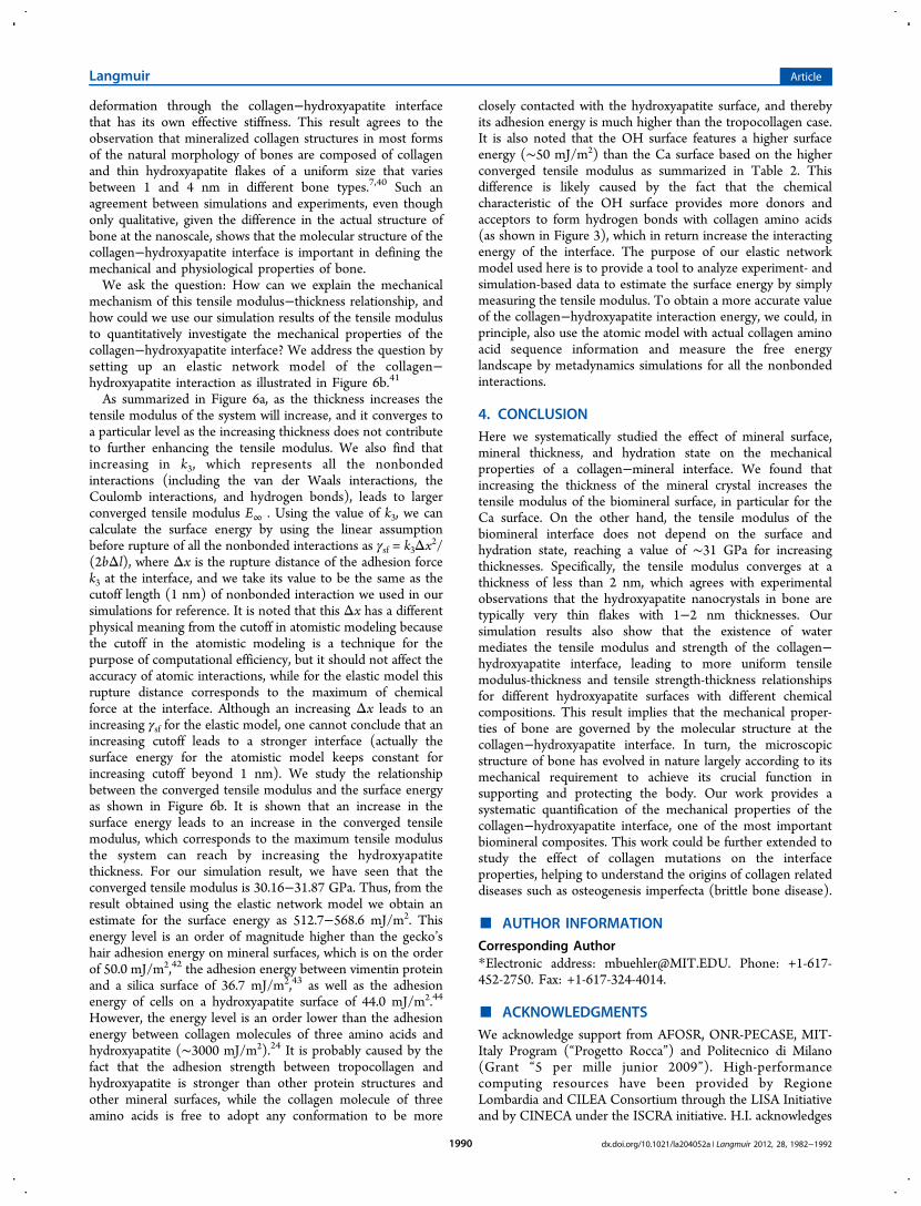
As summarized in Figure 6a, as the thickness increases thetensile modulus of the system will increase, and it converges toa particular level as the increasing thickness does not contributeto further enhancing the tensile modulus. We also find thatincreasing in k3, which represents all the nonbondedinteractions (including the van der Waals interactions, theCoulomb interactions, and hydrogen bonds), leads to largerconverged tensile modulus E∞ . Using the value of k3, we cancalculate the surface energy by using the linear assumptionbefore rupture of all the nonbonded interactions as γsf = k3Δx2/(2bΔl), where Δx is the rupture distance of the adhesion forcek3 at the interface, and we take its value to be the same as thecutoff length (1 nm) of nonbonded interaction we used in oursimulations for reference. It is noted that this Δx has a differentphysical meaning from the cutoff in atomistic modeling becausethe cutoff in the atomistic modeling is a technique for thepurpose of computational efficiency, but it should not affect theaccuracy of atomic interactions, while for the elastic model thisrupture distance corresponds to the maximum of chemicalforce at the interface. Although an increasing Δx leads to anincreasing γsf for the elastic model, one cannot conclude that anincreasing cutoff leads to a stronger interface (actually thesurface energy for the atomistic model keeps constant forincreasing cutoff beyond 1 nm). We study the relationshipbetween the converged tensile modulus and the surface energyas shown in Figure 6b. It is shown that an increase in thesurface energy leads to an increase in the converged tensilemodulus, which corresponds to the maximum tensile modulusthe system can reach by increasing the hydroxyapatitethickness. For our simulation result, we have seen that theconverged tensile modulus is 30.16−31.87 GPa. Thus, from theresult obtained using the elastic network model we obtain anestimate for the surface energy as 512.7−568.6 mJ/m2. Thisenergy level is an order of magnitude higher than the gecko’shair adhesion energy on mineral surfaces, which is on the orderof 50.0 mJ/m2,42 the adhesion energy between vimentin proteinand a silica surface of 36.7 mJ/m2,43 as well as the adhesionenergy of cells on a hydroxyapatite surface of 44.0 mJ/m2.44
However, the energy level is an order lower than the adhesionenergy between collagen molecules of three amino acids andhydroxyapatite (∼3000 mJ/m2).24 It is probably caused by thefact that the adhesion strength between tropocollagen andhydroxyapatite is stronger than other protein structures andother mineral surfaces, while the collagen molecule of threeamino acids is free to adopt any conformation to be more
closely contacted with the hydroxyapatite surface, and therebyits adhesion energy is much higher than the tropocollagen case.It is also noted that the OH surface features a higher surfaceenergy (∼50 mJ/m2) than the Ca surface based on the higherconverged tensile modulus as summarized in Table 2. Thisdifference is likely caused by the fact that the chemicalcharacteristic of the OH surface provides more donors andacceptors to form hydrogen bonds with collagen amino acids(as shown in Figure 3), which in return increase the interactingenergy of the interface. The purpose of our elastic networkmodel used here is to provide a tool to analyze experiment- andsimulation-based data to estimate the surface energy by simplymeasuring the tensile modulus. To obtain a more accurate valueof the collagen−hydroxyapatite interaction energy, we could, inprinciple, also use the atomic model with actual collagen aminoacid sequence information and measure the free energylandscape by metadynamics simulations for all the nonbondedinteractions.
4. CONCLUSIONHere we systematically studied the effect of mineral surface,mineral thickness, and hydration state on the mechanicalproperties of a collagen−mineral interface. We found thatincreasing the thickness of the mineral crystal increases thetensile modulus of the biomineral surface, in particular for theCa surface. On the other hand, the tensile modulus of thebiomineral interface does not depend on the surface andhydration state, reaching a value of ∼31 GPa for increasingthicknesses. Specifically, the tensile modulus converges at athickness of less than 2 nm, which agrees with experimentalobservations that the hydroxyapatite nanocrystals in bone aretypically very thin flakes with 1−2 nm thicknesses. Oursimulation results also show that the existence of watermediates the tensile modulus and strength of the collagen−hydroxyapatite interface, leading to more uniform tensilemodulus-thickness and tensile strength-thickness relationshipsfor different hydroxyapatite surfaces with different chemicalcompositions. This result implies that the mechanical proper-ties of bone are governed by the molecular structure at thecollagen−hydroxyapatite interface. In turn, the microscopicstructure of bone has evolved in nature largely according to itsmechanical requirement to achieve its crucial function insupporting and protecting the body. Our work provides asystematic quantification of the mechanical properties of thecollagen−hydroxyapatite interface, one of the most importantbiomineral composites. This work could be further extended tostudy the effect of collagen mutations on the interfaceproperties, helping to understand the origins of collagen relateddiseases such as osteogenesis imperfecta (brittle bone disease).
■ AUTHOR INFORMATION
Corresponding Author*Electronic address: [email protected]. Phone: +1-617-452-2750. Fax: +1-617-324-4014.
■ ACKNOWLEDGMENTSWe acknowledge support from AFOSR, ONR-PECASE, MIT-Italy Program (“Progetto Rocca”) and Politecnico di Milano(Grant “5 per mille junior 2009”). High-performancecomputing resources have been provided by RegioneLombardia and CILEA Consortium through the LISA Initiativeand by CINECA under the ISCRA initiative. H.I. acknowledges
Langmuir Article
dx.doi.org/10.1021/la204052a | Langmuir 2012, 28, 1982−19921990
support from Weizmann Institute of Science (Israel), supportfrom the Center for Excellence in Education, as well as theResearch Science Institute (RSI).
■ REFERENCES(1) Currey, J. D. Bones: Structure and Mechanics; Princeton UniversityPress: Princeton, NJ, 2002.(2) Fratzl, P.; Weinkamer, R. Nature’s hierarchical materials. Prog.Mater. Sci. 2007, 52, 1263−1334.(3) Fratzl, P. Collagen: Structure and Mechanics; Springer: New York,2008.(4) Fratzl, P.; Gupta, H. S.; Paschalis, E. P.; Roschger, P. Structureand mechanical quality of the collagen-mineral nano-composite inbone. J. Mater. Chem. 2004, 14 (14), 2115−2123.(5) Gupta, H. S.; Seto, J.; Wagermaier, W.; Zaslansky, P.; Boesecke,P.; Fratzl, P. Cooperative deformation of mineral and collagen in boneat the nanoscale. Proc. Natl. Acad. Sci. U.S.A. 2006, 103 (47), 17741−17746.(6) Jager, I.; Fratzl, P. Mineralized collagen fibrils: A mechanicalmodel with a staggered arrangement of mineral particles. Biophys. J.2000, 79 (4), 1737−1746.(7) Weiner, S.; Wagner, H. D. The material bone: Structuremechanical function relations. Annu. Rev. Mater. Sci. 1998, 28, 271−298.(8) Ji, B. H.; Gao, H. J. Mechanical properties of nanostructure ofbiological materials. J. Mech. Phys. Solids 2004, 52 (9), 1963−1990.(9) de Leeuw, N. H. Local ordering of hydroxy groups inhydroxyapatite. Chem. Commun. 2001, 17, 1646−1647.(10) Hauptmann, S.; Dufner, H.; Brickmann, J.; Kast, S. M.; Berry, R.S. Potential energy function for apatites. Phys. Chem. Chem. Phys. 2003,5 (3), 635−639.(11) Snyders, R.; Music, D.; Sigumonrong, D.; Schelnberger, B.;Jensen, J.; Schneider, J. M. Experimental and ab initio study of themechanical properties of hydroxyapatite. Appl. Phys. Lett. 2007, 90, 19.(12) Ching, W. Y.; Rulis, P.; Misra, A. Ab initio elastic properties andtensile strength of crystalline hydroxyapatite. Acta Biomater. 2009, 5(8), 3067−3075.(13) Cruz, F. J. A. L.; Canongia Lopes, J. N.; Calado, J. C. G.; Minasda Piedade, M. E. A molecular dynamics study of the thermodynamicproperties of calcium apatites. 1. Hexagonal phases. J. Phys. Chem. B2005, 109 (51), 24473−24479.(14) Zhang, H.-p.; Lu, X.; Leng, Y.; Fang, L.; Qu, S.; Feng, B.; Weng,J.; Wang, J. Molecular dynamics simulations on the interactionbetween polymers and hydroxyapatite with and without couplingagents. Acta Biomater. 2009, 5 (4), 1169−1181.(15) Zhou, H.; Wu, T.; Dong, X.; Wang, Q.; Shen, J. Adsorptionmechanism of BMP-7 on hydroxyapatite (001) surfaces. Biochem.Biophys. Res. Commun. 2007, 361 (1), 91−96.(16) Shen, J.-W.; Wu, T.; Wang, Q.; Pan, H.-H. Molecular simulationof protein adsorption and desorption on hydroxyapatite surfaces.Biomaterials 2008, 29 (5), 513−532.(17) Bhowmik, R.; Katti, K. S.; Katti, D. R. Mechanisms of load-deformation behavior of molecular collagen in hydroxyapatite-tropocollagen molecular system: Steered molecular dynamics study.J. Eng. Mech. 2009, 135 (5), 413−421.(18) Bhowmik, R.; Katti, K. S.; Katti, D. R. Mechanics of molecularcollagen is influenced by hydroxyapatite in natural bone. J. Mater. Sci.2007, 42 (21), 8795−8803.(19) Almora-Barrios, N.; de Leeuw, N. H. Modelling the interactionof a Hyp-Pro-Gly peptide with hydroxyapatite surfaces in aqueousenvironment. CrystEngComm 2010, 12 (3), 960−967.(20) de Leeuw, N. H.; Almora-Barrios, N. A density functional theorystudy of the interaction of collagen peptides with hydroxyapatitesurfaces. Langmuir 2010, 26 (18), 14535−14542.(21) Dubey, D. K.; Tomar, V. Role of the nanoscale interfacialarrangement in mechanical strength of tropocollagen−hydroxyapatite-based hard biomaterials. Acta Biomater. 2009, 5 (7), 2704−2716.
(22) Dubey, D. K.; Tomar, V. Role of hydroxyapatite crystal shape innanoscale mechanical behavior of model tropocollagen−hydroxyapa-tite hard biomaterials. Mater. Sci. Eng. C: Mater. Biol. Appl. 2009, 29(7), 2133−2140.(23) Zahn, D.; Duchstein, P. Atomistic modeling of apatite-collagencomposites from molecular dynamics simulations extended tohyperspace. J. Mol. Model. 2011, 17 (1), 73−79.(24) Almora-Barrios, N.; de Leeuw, N. H. A density functional theorystudy of the interaction of collagen peptides with hydroxyapatitesurfaces. Langmuir 2010, 26 (18), 14535−14542.(25) Rainey, J. K.; Goh, M. C. An interactive triple-helical collagenbuilder. Bioinformatics 2004, 20 (15), 2458−9.(26) Gautieri, A.; Buehler, M. J.; Redaelli, A. Deformation ratecontrols elasticity and unfolding pathway of single tropocollagenmolecules. J. Mech. Behav. Biomed. Mater. 2009, 2 (2), 130−7.(27) Buehler, M. J. Atomistic and continuum modeling of mechanicalproperties of collagen: Elasticity, fracture and self-assembly. J. Mater.Res. 2006, 21 (8), 1947−1961.(28) Gautieri, A.; Buehler, M. J.; Redaelli, A. Deformation ratecontrols elasticity and unfolding pathway of single tropocollagenmolecules. J. Mech. Behav. Biomed. Mater. 2009, 2 (2), 130−137.(29) Brooks, B. R.; Bruccoleri, R. E.; Olafson, B. D.; States, D. J.;Swaminathan, S.; Karplus, M. CHARMM - A program for macro-molecular energy, minimization, and dynamics calculations. J. Comput.Chem. 1983, 4 (2), 187−217.(30) Park, S.; Radmer, R. J.; Klein, T. E.; Pande, V. S. A new set ofmolecular mechanics parameters for hydroxyproline and its use inmolecular dynamics simulations of collagen-like peptides. J. Comput.Chem. 2005, 26 (15), 1612−1616.(31) Gautieri, A.; Uzel, S.; Vesentini, S.; Redaelli, A.; Buehler, M. J.Molecular and mesoscale mechanisms of osteogenesis imperfectadisease in collagen fibrils. Biophys. J. 2009, 97 (3), 857−865.(32) Gautieri, A.; Vesentini, S.; Redaelli, A.; Buehler, M. J.Hierarchical structure and nanomechanics of collagen microfibrilsfrom the atomistic scale up. Nano Lett. 2011, 11 (2), 757−766.(33) Bhowmik, R.; Katti, K. S.; Katti, D. Molecular dynamicssimulation of hydroxyapatite−polyacrylic acid interfaces. Polymer2007, 48 (2), 664−674.(34) Plimpton, S. Fast parallel algorithms for short-range molecular-dynamics. J. Comput. Phys. 1995, 117 (1), 1−19.(35) Gautieri, A.; Vesentini, S.; Redaelli, A.; Buehler, M. J.Intermolecular slip mechanism in tropocollagen nanofibrils. Int. J.Mater. Res. 2009, 100 (7), 921−925.(36) Srinivasan, M.; Uzel, S. G. M.; Gautieri, A.; Keten, S.; Buehler,M. J. Alport Syndrome mutations in type IV tropocollagen altermolecular structure and nanomechanical properties. J. Struct. Biol.2009, 168 (3), 503−510.(37) Qin, Z.; Kreplak, L.; Buehler, M. J. Hierarchical structurecontrols nanomechanical properties of vimentin intermediatefilaments. PLoS ONE 2009, 4 (10), e7294.(38) Humphrey, W.; Dalke, A.; Schulten, K. VMD: Visual moleculardynamics. J. Mol. Graph. 1996, 14 (1), 33−38.(39) Katti, D. R.; Pradhan, S. M.; Katti, K. S. Directional dependenceof hydroxyapatite−collagen interactions on mechanics of collagen. J.Biomech. 2010, 43 (9), 1723−1730.(40) Hu, Y. Y.; Rawal, A.; Schmidt-Rohr, K. Strongly bound citratestabilizes the apatite nanocrystals in bone. Proc. Natl. Acad. Sci. U.S.A.2010, 107 (52), 22425−22429.(41) Qin, Z.; Buehler, M. J. Cooperative deformation of hydrogenbonds in beta-strands and beta-sheet nanocrystals. Phys. Rev. E 2010,82, 6.(42) Prowse, M. S.; Wilkinson, M.; Puthoff, J. B.; Mayer, G.; Autumn,K. Effects of humidity on the mechanical properties of gecko setae.Acta Biomater. 2011, 7 (2), 733−738.(43) Kirmse, R.; Qin, Z. A.; Weinert, C. M.; Hoenger, A.; Buehler, M.J.; Kreplak, L. Plasticity of intermediate filament subunits. PLoS ONE2010, 5, 8.(44) Redey, S. A.; Razzouk, S.; Rey, C.; Bernache-Assollant, D.;Leroy, G.; Nardin, M.; Cournot, G. Osteoclast adhesion and activity
Langmuir Article
dx.doi.org/10.1021/la204052a | Langmuir 2012, 28, 1982−19921991

on synthetic hydroxyapatite, carbonated hydroxyapatite, and naturalcalcium carbonate: relationship to surface energies. J. Biomed. Mater.Res. 1999, 45 (2), 140−7.(45) Sun, Y. L.; Luo, Z. P.; Fertala, A.; An, K. N. Stretching type IIcollagen with optical tweezers. J. Biomech. 2004, 37 (11), 1665−1669.(46) Gilmore, R. S.; Katz, J. L. Elastic properties of apatites. J. Mater.Sci. 1982, 17 (4), 1131−1141.
Langmuir Article
dx.doi.org/10.1021/la204052a | Langmuir 2012, 28, 1982−19921992