Embed Size (px)
Citation preview

Research Collection
Doctoral Thesis
Forebrain neuronal glycine transporter 1 AS a neurocognitivemodulatorbehavioural analysis of an engineered mouse model
Author(s): Dubroqua, Sylvain
Publication Date: 2011
Permanent Link: https://doi.org/10.3929/ethz-a-006651458
Rights / License: In Copyright - Non-Commercial Use Permitted
This page was generated automatically upon download from the ETH Zurich Research Collection. For moreinformation please consult the Terms of use.
ETH Library

DISS. ETH NO. 19686
FOREBRAIN NEURONAL GLYCINE TRANSPORTER 1 AS A NEUROCOGNITIVE
MODULATOR: BEHAVIOURAL ANALYSIS OF AN ENGINEERED MOUSE MODEL
A dissertation submitted to
ETH ZURICH
for the degree of
Doctor of Sciences
presented by
SYLVAIN DUBROQUA
Master in Cognitive Science, Université Bordeaux 2, France
21th May, 1981
citizen of France
accepted on the recommendation of
Prof. dr. Joram Feldon, examiner
Prof. dr. Jacques Micheau, co-examiner
Dr. Benjamin K. Yee, co-examiner
2011

1
TABLE OF CONTENT
Summary 2
Résumé 4
Chapter 1: General Introduction 6
Chapter 2: Impacts of forebrain neuronal glycine transporter 1
disruption in the senescent brain: Evidence for age-dependent
phenotypes in Pavlovian learning 28
Chapter 3: Examining the sex- and circadian dependency
of a learning phenotype in mice with glycine transporter 1
deletion in two Pavlovian conditioning paradigms 52
Chapter 4: Glycine transporter 1 deletion enhances sensitivity
to CS-US contiguity and CS-US contingency:
Two phenotypes, one psychology? 81
Chapter 5: Working memory performance is unaffected by
glycine transporter 1 deletion from forebrain neurons 102
Chapter 6: General Discussion 124
Curriculum vitae 135
Acknowledgements 136

SUMMARY
Efficient N-methyl-D-aspartate receptor (NMDAR) signalling requires binding of the co-agonist
glycine at the NR1 subunit glycine-B site. The availability of glycine at the synaptic cleft is tightly
regulated by the glycine transporter-1 (GlyT1), which is expressed in both neurons and
astrocytes. Pharmacological or genetic disruption of GlyT1-mediated glycine-reuptake near
excitatory synapses is believed to enhance NMDAR function and may consequently enhance
cognitive performance. GlyT1 is therefore regarded as a potential pharmacological target to
ameliorate memory deficits in humans, including the schizophrenia-related cognitive symptoms
believed to stem from NMDAR hypofunction. Genetic deletion of GlyT1 in forebrain neurons of
mice appears to be associated with enhanced memory performance across multiple cognitive
tasks. However, the interpretation of these findings is open to question on several accounts,
which have not been satisfactorily addressed until now. This thesis therefore aims to provide an
in-depth comparative analysis of the behavioural phenotypes expressed by these forebrain
neuronal specific GlyT1 disrupted mutant mice and to test the stability and robustness of the
enhanced cognitive phenotype previously described. The first series of experiments provides
novel qualifications on the sex-dependent nature of the Pavlovian phenotype shown by the
mutant mice (Chapter 3). More specifically, gender critically determines the magnitude and
direction of Pavlovian conditioning in both aversive and appetitive conditioning paradigms.
These findings are relevant to psychiatric illnesses, such as schizophrenia, that show notable sex
differences with regard to symptomatology, onset, and course of the disease. In Chapter 2 it was
investigated whether the enhanced Pavlovian (aversive) conditioning phenotype observed
previously might be modified by age, since lifelong elevation of glycine synaptic levels might
result in excitotoxic effects and consequently cell death. Although, both adult and aged mutants
exhibited enhanced Pavlovian conditioning, the temporal expression of this phenotype (in terms
of within-session extinction) was notably absent in the aged mutants. Furthermore, a noticeable
increase in adult hippocampus neurogenesis was seen in the mutant mice, suggesting a possible
neuroanatomical contribution to the modulation of the cognitive phenotype. The experiments
carried out in a third study (Chapter 4) examined the notion that enhanced Pavlovian
conditioning exhibited by the forebrain neuronal specific GlyT1 mutant mice stemmed from
alterations in the selectivity of learning as opposed to a broad strengthening of the associative
strength between the conditioned and unconditioned stimulus. The outcome suggests a
bidirectional effect of forebrain neuronal GlyT1 deletion on Pavlovian associative learning: the
conditioned response was stronger in the mutant mice when the conditioned stimulus was a
good predictor of the unconditioned stimulus. Conversely, the conditioned response was

3
weaker when the conditioned stimulus-unconditioned stimulus (CS-US) contiguity or
contingency was degraded. It appears that alterations in the selectivity of learning may account
for the diverse phenotypes in the regulation of Pavlovian learning. These findings bear clear
relevance to the proposed therapeutic potential of GlyT1-inhibiting drugs for schizophrenia-
related cognitive deficits that might stem from an underlying lack of selectivity (or inflexibility)
in learning. Finally, a last series of experiments examined the impact of forebrain neuronal
GlyT1 deletion on working memory function by using a within-subject multi-paradigm
approach (Chapter 5). A null effect of the manipulation was seen across three different working
memory paradigms. The behavioural effects of the forebrain neuronal GlyT1 deletion thus
appear to be restricted to associative learning. These negative outcomes clearly contrasted the
improved working performance shown previously by mutant mice with GlyT1 deletion
extending into cortical glial cells. These complementary results might thus point to a potential
functional divergence between neuronal and glial GlyT1 populations in mediating cognitive
function, with a more prominent role for glial-based GlyT1 in regulating working memory
function. Hence, the present thesis has provided valuable novel insights into neuronal forebrain
specific GlyT1 function and its contribution to memory. Furthermore, the data presented here
demonstrate that an in-depth comprehension of the phenotypic expression of forebrain and
neuronal GlyT1 deletion, or of any genetic manipulation, is an important step forward in the
proof of concept, and constitute a critical component in the pre-clinical evaluation of potential
drug targets.

Résumé
La bonne transduction du message nerveux via le récepteur N-methyl-D-aspartate (NMDAR)
nécessite la fixation d’un co-agoniste, la glycine, sur le site glycine-B de la sous-unité NR1. La
disponibilité de la glycine au niveau de la fente synaptique est régulée par le transporteur 1 de
la glycine (GlyT1), exprimé par les neurones et les astrocytes. Un blocage de la recapture de la
glycine via GlyT1 par une approche génétique ou pharmacologique est donc supposé augmenter
la fonction des NMDAR et ainsi induire une amélioration des fonctions cognitives. Par
conséquent GlyT1 est une cible pharmacologique potentielle pour l’amélioration des troubles
mnésiques, comme par exemple ceux liés à la schizophrénie et supposés provenir d’une hypo-
fonction des NMDAR. Ainsi une délétion génétique de GlyT1 dans les neurones du
prosencephale de souris est associée à une amélioration des performances mnésiques dans de
nombreuses taches cognitives. Cependant l’origine de ces effets reste encore actuellement
sujette à interprétation sur de nombreux points et nécessite des études plus approfondies. Les
travaux présentés dans cette thèse avaient pour but de fournir une analyse poussée du
phénotype comportemental exprimé par des souris mutantes ayant une perturbation des GlyT1
exprimés par les neurones du prosencéphales en testant la stabilité et la fiabilité des résultats
précédemment obtenus. La première série d’expériences présentée ici met en évidence que le
phénotype observé chez les souris mutant dans une tache de conditionnement de type
"Pavlovien" est différent entre le male et la femelle (chapitre 2). Plus précisément, le genre est
un déterminant majeur du sens des effets induits par la mutation ainsi que de leur ampleur, lors
d’apprentissages Pavloviens aversifs mais aussi appétitifs. Cette découverte est
particulièrement pertinente dans le cadre de l’étude de certaines pathologies psychiatriques
comme la schizophrénie qui présentent des différences notables en fonction du sexe du patient
comme par exemple, la symptomatologie, le moment d’apparition et le développement de la
maladie. Les séries d’expériences présentées dans le chapitre 3 visent à déterminer si
l’amélioration des performances lors d’un apprentissage Pavlovien (aversif) est modifiée par
l’âge, puisque l’augmentation de la concentration en glycine au niveau synaptique tout au long
de la vie de l’animal pourrait avoir un effet excito-toxique entrainant une perte neuronale.
Cependant, les souris adultes et les souris âgées présentent une amélioration de
conditionnement Pavlovien comparable, l’expression temporelle de ce phénotype (en termes
d’extinction au cours d’une session) est notamment absente chez les souris mutantes âgées. De
plus une augmentation de neurogénèse hippocampique est observable chez les souris mutantes,
ce qui suggère une contribution possible de ces nouvelles cellules dans la modulation des
phénotypes observé dans les taches d’apprentissage et de mémoire. Les expériences menées

5
lors d’une troisième étude (chapitre 4) visent à déterminer si l’amélioration de
conditionnement pavlovien développée par les souris mutantes ne repose pas en fait sur une
altération de la sélectivité de l’apprentissage plutôt que sur un renforcement de la force
associative entre le stimulus conditionnel et le stimulus inconditionnel. Les résultats obtenus
suggèrent un effet bidirectionnel de la délétion de GlyT1 dans les neurones du prosencéphale
sur l’apprentissage associatif Pavlovien: la réponse conditionnée étant plus forte chez les souris
mutantes lorsque le stimulus conditionnel prédit bien l’occurrence du stimulus inconditionnel,
alors qu’à l’inverse la réponse conditionnée de ces même souris est plus faible quand la
contigüité temporelle ou la contingence entre stimulus conditionnel et stimulus inconditionnel
sont dégradées. Il apparait donc qu’une altération de la sélectivité de l’apprentissage pourrait
être en partie responsable des modifications des phénotypes Pavlovien observées. Ces travaux
apportent donc des preuves claires vis-à-vis du potentiel thérapeutique de drogues permettant
d’inhiber sélectivement les GlyT1 dans le cadre du traitement de certains des déficits cognitifs
observables chez les patients schizophrènes pouvant résulter d’un déficit de flexibilité (ou
rigidité comportementale) lors d’apprentissages. Enfin, une dernière série d’expérience
examine l’effet sur la mémoire de travail de la délétion de GlyT1 dans les neurones du
prosencéphale en utilisant pour cela une approche intra-sujets dans plusieurs paradigmes
expérimentaux (chapitre 5). Ces expériences mettent en évidence une absence d’effet de la
manipulation génétique dans trois paradigmes différents testant la mémoire de travail. Les
effets comportementaux induits par la manipulation génétique étudiée semblent de plus
restreints au domaine non spatial. Cette absence d’effet contraste avec l’amélioration de
mémoire de travail observée précédemment chez des souris mutantes pour lesquelles la
délétion de GlyT1 s’étendait aux cellules gliales corticales. Ces résultats complémentaires
suggèrent donc qu’il pourrait exister une divergence fonctionnelle entre les GlyT1 exprimés par
les neurones et ceux exprimés par les cellules gliales dans le contrôle des fonctions cognitives,
les GlyT1 des cellules gliales pouvant être plus fortement impliqué dans la modulation des
processus de mémoire de travail. Ainsi, la présente thèse fournie de nouvelles connaissances
quand aux fonctions spécifiques des GlyT1 exprimés par les neurones du prosencéphale ainsi
que sur leur rôle dans les processus mnésique. De plus, les données présentées démontrent
qu’une compréhension approfondie, de l’expression phénotypique de la délétion en GlyT1 au
niveau des neurones du prosencéphale, mais aussi pour n’importe quelle manipulation
génétique, est un important pas en avant dans la validation d’un concept, et constitue un
éléments majeur de l’évaluation préclinique d’une potentielle cible pharmacologique.

CHAPTER 1
General Introduction

Chapter 1: General Introduction
This chapter chronicles the milestones in research that led to discovery that facilitated N-
methyl-D-aspartate receptor (NMDAR) signalling and enhanced cognitive function could
be achieved through increased glycine occupation on the NMDAR glycine-B site (See table
1). More specifically, it aims to give an overview of the findings showing that this could be
accomplished via Glycine Transporter 1 (GlyT1) inhibition or genetic disruption. The
significance of glycine’s modulation of NMDAR function is highlighted by studies utilizing
glycine inhibitor drugs. In addition, the availability of GlyT1 conditional knockout mouse
models has provided further understanding of GlyT disruption and its impact on
cognition. In particular, the behavioural screening of a mouse model with a conditional
GlyT1 knockout in forebrain neurons has yielded promising findings. However, at the
same time it revealed several limitations that have not been satisfactorily addressed
until present. Comprehensive phenotyping of this mouse line within an integrated
behavioural framework is a prerequisite for understanding the cognitive aspects
involved and/or affected by this manipulation. This would also provide the basis towards
translating the experimental findings into therapeutic targets aimed specifically at these
changes. The studies presented in this dissertation were designed towards such an
approach, and this is elaborated further in the Thesis objectives and Thesis outline at the
end of this Chapter.
Glycine modulation of N-methyl-D-aspartate receptor (NMDAR) function
Due to its strong implication in several brain disorders and its predominant role in various
forms of synaptic plasticity, the NMDAR has been a prime candidate for extensive study (e.g.
(Collingridge and Bliss, 1995; Malenka et al., 1989; Tonkiss et al., 1988); see Box 1). Facilitation
of NMDAR function is considered a viable strategy to overcome cognitive deficiency associated
with a large number of psychiatric disorders. Ever since the pioneer study by Johnson and
Ascher (1987) showing that glycine could enhance NMDAR-mediated electrophysiological
responses, the glycinergic modulation of NMDAR function has received considerable interest
(for review see Johnson and Ascher, 1987; Monaghan et al., 1988; Thomson, 1989). Their
discovery followed previous indications that glycine might play an additional neuromodulatory
function besides its well-documented role as an inhibitory neurotransmitter, due to the
distribution of glycine binding sites in mammalian brain (e.g. Bristow et al., 1986; Kishimoto et
al., 1981). It was observed that inhibitory glycine receptors (radioactively labelled with [3H]
strychnine) were strongly expressed in the pons and spinal cord. In contrast, [3H] glycine not
only labelled these receptors, but also identified the existence of high affinity binding sites in the
forebrain (Bristow et al., 1986). Shortly after, Bowery (1987) noted that the distribution pattern
of high affinity glycine binding sites corresponded to the NMDA binding profile in the brain. For
simplification purposes, the strychnine-sensitive binding site is referred to as the GlyA-site,
whereas the strychnine-insensitive glycine recognition site is referred to as the GlyB-site
hereafter.
Chapter 1: General Introduction
8
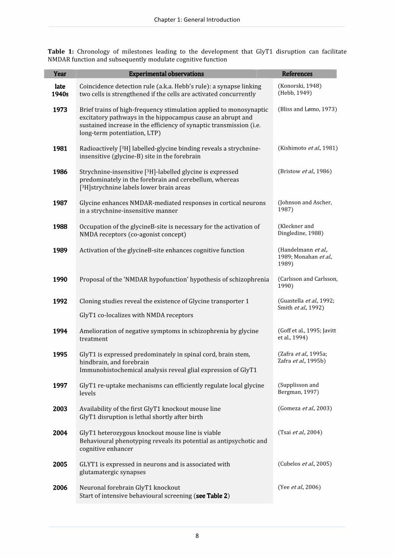
Table 1: Chronology of milestones leading to the development that GlyT1 disruption can facilitate
NMDAR function and subsequently modulate cognitive function
YearYearYearYear Experimental observationsExperimental observationsExperimental observationsExperimental observations ReferencesReferencesReferencesReferences
late late late late
1940s1940s1940s1940s
Coincidence detection rule (a.k.a. Hebb's rule): a synapse linking
two cells is strengthened if the cells are activated concurrently
(Konorski, 1948)
(Hebb, 1949)
1973197319731973 Brief trains of high-frequency stimulation applied to monosynaptic
excitatory pathways in the hippocampus cause an abrupt and
sustained increase in the efficiency of synaptic transmission (i.e.
long-term potentiation, LTP)
(Bliss and Lømo, 1973)
1981198119811981 Radioactively [3H] labelled-glycine binding reveals a strychnine-
insensitive (glycine-B) site in the forebrain
(Kishimoto et al., 1981)
1986198619861986 Strychnine-insensitive [3H]-labelled glycine is expressed
predominately in the forebrain and cerebellum, whereas
[3H]strychnine labels lower brain areas
(Bristow et al., 1986)
1987198719871987 Glycine enhances NMDAR-mediated responses in cortical neurons
in a strychnine-insensitive manner
(Johnson and Ascher,
1987)
1988198819881988 Occupation of the glycineB-site is necessary for the activation of
NMDA receptors (co-agonist concept)
(Kleckner and
Dingledine, 1988)
1989198919891989 Activation of the glycineB-site enhances cognitive function (Handelmann et al., 1989; Monahan et al., 1989)
1990199019901990 Proposal of the 'NMDAR hypofunction' hypothesis of schizophrenia (Carlsson and Carlsson,
1990)
1992199219921992 Cloning studies reveal the existence of Glycine transporter 1 (Guastella et al., 1992; Smith et al., 1992)
GlyT1 co-localizes with NMDA receptors
1994199419941994 Amelioration of negative symptoms in schizophrenia by glycine
treatment
(Goff et al., 1995; Javitt
et al., 1994)
1995199519951995 GlyT1 is expressed predominately in spinal cord, brain stem,
hindbrain, and forebrain
Immunohistochemical analysis reveal glial expression of GlyT1
(Zafra et al., 1995a;
Zafra et al., 1995b)
1997199719971997 GlyT1 re-uptake mechanisms can efficiently regulate local glycine
levels
(Supplisson and
Bergman, 1997)
2003200320032003 Availability of the first GlyT1 knockout mouse line (Gomeza et al., 2003)
GlyT1 disruption is lethal shortly after birth
2004200420042004 GlyT1 heterozygous knockout mouse line is viable (Tsai et al., 2004)
Behavioural phenotyping reveals its potential as antipsychotic and
cognitive enhancer
2005200520052005 GLYT1 is expressed in neurons and is associated with
glutamatergic synapses
(Cubelos et al., 2005)
2006200620062006 Neuronal forebrain GlyT1 knockout (Yee et al., 2006)
Start of intensive behavioural screening (see Table 2see Table 2see Table 2see Table 2)

Chapter 1: General Introduction
9
In 1988, Kleckner and Dingledine revealed that binding of glycine to glycine-B site is indeed a
prerequisite for the activation of the NMDAR and proposed the term “co-agonist”. This opened
alternative avenues to facilitate NMDAR neurotransmission, which could: (i) potentially
alleviate NMDAR-associated brain pathological conditions, (ii) augment synaptic plasticity and
subsequently enhance cognitive functions under normal conditions. Such an approach could
potentially circumvent the severe side-effects associated with direct stimulation of NMDARs,
such as neurotoxicity and seizures (Mayer and Westbrook, 1987; Rothman and Olney, 1995).
Indeed, one year later, evidence emerged that activation of the glycine regulatory site on the
NMDA receptor could enhance cognitive function (Handelmann et al., 1989; Monahan et al.,
1989).
One major limitation associated with glycine´s modulatory role on NMDAR function was the
apparent lack of sufficient knowledge in the regulatory mechanisms (re-uptake, release, and
metabolism) of glycine. It was, for instance, assumed that high affinity (in nM) NMDA-associated
glycine sites were saturated due to higher micromolar (µM) concentrations of glycine in the
extracellular fluid (Fletcher and Lodge, 1988; Matsiu et al., 1995). This limitation in the
knowledge was overcome following the discovery of sodium/chloride (Na2+/Cl-)-dependent
transporters for glycine in the brain (Fedele and Foster, 1992). Furthermore, cloning studies
uncovered the existence of two families of glycine transporters: glycine transporter 1 (GlyT1,
Guastella et al., 1992; Smith et al., 1992) and glycine transporter 2 (GlyT2, Liu et al., 1992). Both
glycine transporters differ in terms of their expression profile. GlyT2 is expressed in areas
where glycine acts as an inhibitory neurotransmitter, i.e. mainly in the spinal cord, brain stem
and hindbrain (Jursky and Nelson, 1996; Zafra et al., 1995a). GlyT1 shares overlapping regional
expression patterns with GlyT2 (Jursky and Nelson, 1996; Zafra et al., 1995a), but is
furthermore expressed in some regions with low levels of GlyA receptors, such as the forebrain
(Zafra et al., 1995a), where it co-localizes with the NMDAR (Smith et al., 1992). Although GlyT1
mRNA was detected in glia as well as in neurons, initial immunohistochemical analysis failed to
confirm the presence of GlyT1 in neurons (Zafra et al., 1995a). Therefore, GlyT1 was considered
to be localized only on astrocytes (Zafra et al., 1995b), until Cubelos and co-workers (2005)
confirmed pre-and post-synaptic GlyT1 expression in a sub-set of glutamatergic neurons closely
associated with NMDARs.

Chapter 1: General Introduction
10
Box 1: The NMDA receptor: its properties, role in synaptic plasticity and learning and memory
Characteristics of the NMDAR
The NMDAR belongs to the ionotropic glutamate
receptor sub-group, and exists as a heterotetrameric
complex consisting of two NR1 and two NR2 (A-D)
subunits. Combinations of different NR2 subunits
determine the pharmacological and physiological
profile of the receptor in terms of channel kinetics,
and its affinity for agonists and antagonists (Cull-
Candy et al., 2001). Besides glutamate binding at the
NR2 subunit, glycine co-agonist binding at the NR1
glycine-B site is essential for effective channel
opening (see Danysz and Parsons, 1998).
Furthermore, the receptor complex harbours several
endogenous allosteric ligand binding sites that modify
its physiological properties (See Figure 1).
Figure 1: Schematic illustration of the NMDAR complex. The receptor
complex comprises various binding sites, which are responsible for
interactions with agonists, antagonists, or modulatory ligands. These
sites, aid the facilitatory (polyamine) or inhibitory (Zn+) actions of
the receptor. The glutamate/ligand recognition site is critical for
receptor activation. Yet, glycine co-agonist binding at the strychnine
sensitive glycine-B site of the NR1 subunit is necessary to facilitate
channel opening. The receptor channel contains a voltage-dependent
Mg2+ binding site, as well as binding sites for dissociative
anaesthetics such as PCP, ketamine and MK-801. (Image obtained
from www.frca.co.uk/images/NMDA.jpg)
NMDARs are principally neuronal, and whilst they are
diffusely distributed throughout the brain, the highest
density is detected in the CA1 hippocampal subfield and in
layers I and III of the cerebral cortex (Monaghan and
Cotman, 1985). The expression and distribution of NMDARs
in the CNS is highly dependent on the combination of
subunits that constitute the complex as well as the
developmental stage of the organism (reviewed by Danysz
and Parsons, 1998; Nakanishi, 1992). Consequently, the
receptor is known to play a prominent role in a number of
neurobiological events during brain development (Ewald
and Cline, 2009; Haberny et al., 2002).
The notion that learning and memory depends upon changes
in the efficacy of chemical synapses (as discussed elsewhere)
has led to a primary focus on the NMDAR as the molecular
correlate of synaptic plasticity- the neurochemical substrate
presumed to underpin some forms of learning and memory.
In this regard, the NMDAR has several unique properties
that make it the ideal candidate for synaptic plasticity.
Besides being a ligand-gated ion channel, the receptor is
subject to a voltage-dependent block by magnesium (Mg2+),
which can be removed via AMPA receptor-mediated
membrane depolarization (Dingledine et al., 1999). Thus, it
acts as a coincident detector for pre-synaptic activity
(glutamate release) and post-synaptic activity (adequate
depolarization of the post-synaptic membrane). Second,
once activated, the NMDAR channel permits ion flow (Ca2+,
Na+ and K+), which can give rise to an excitatory
postsynaptic current (EPSC). The high Ca2+ permeability is a
key feature of the NMDAR given that it is accompanied by a
cascade of intracellular events that lead to long-term
synaptic changes (Long-term potentiation; Collingridge and
Bliss, 1995) and gene transcription (Bading et al., 1993).
Its role in synaptic modifications and memory
Cajal (1893 ) originally hypothesized that changes in the
strength of synaptic connections between active neurons are
necessary for information storage. This was subsequently
supported by Hebb (1949), who proposed that changes in
the strength of synaptic connections require concurrent
activation of the pre-and post-synaptic neurons. In
accordance to the Hebbian principle, it was later shown that
activity-dependent synaptic changes play a vital role in some
forms learning and memory (e.g. Lynch, 2004).
Consequently, studies aimed to understand the mechanism
for synaptic changes led to the discovery that bi-directional
activity-dependent forms of synaptic plasticity, such as long-
term potentiation (LTP), required NMDA glutamate receptor
channel opening (Bliss and Collingridge, 1993; Bliss and
Lømo, 1973; Collingridge et al., 1983; Zalutsk and Nicoll,
1990). As mentioned above, the NMDAR is an ideal ‘Hebbian’
candidate for synaptic changes due to its voltage-dependent
magnesium block (Mayer and Westbrook, 1987) and
“coincidence detection” mechanism. Notably, NMDAR
activation is critical for the induction of LTP, whereas
maintenance/expression of LTP is dependent on the
recruitment and expression of AMPA receptors at the
synapse (Malinow and Malenka, 2002; Martin et al., 2000).
NMDAR-dependent LTP is universally considered relevant to
the formation of learning and memory (Lynch, 2004; Martin
et al., 2000). This is supported by studies showing that
selective pharmacological blockade of NMDARs disrupt the
induction of LTP and impair learning and memory function
in a variety of paradigms (Bolhuis and Reid, 1992; Kawabe et
al., 1998; McHugh et al., 2008; Morris, 1989; Morris et al.,
1986). Furthermore, it has been demonstrated that specific
hippocampal NMDAR subunits, as well as their expression in
different hippocampal subfields, make a unique and
differential contribution to learning and memory
(Bannerman et al., 2008; Nakazawa et al., 2002; Nakazawa et
al., 2003; Niewoehner et al., 2007; Sakimura et al., 1995;
Tsien et al., 1996).

Chapter 1: General Introduction
11
The discovery that glycine transporters are expressed in the brain (Guastella et al., 1992; Liu et
al., 1992; Smith et al., 1992) suggests that these transporters regulate and maintain glycine
concentrations below saturation levels in the synaptic cleft. Indeed, Supplisson and Bergman
(1997) using an experimental model co-expressing GlyT1 and NMDAR in Xenopus oocytes
showed that GlyT1 efficiently reduces glycine near the membrane to concentrations lower than
the saturating concentration (nM range) for neuronal NMDARs when the bath concentration
(µM range) is similar to that in the cerebral spinal fluid. One year later, an in vitro study
confirmed GlyT1´s efficacy in maintaining unsaturated glycine concentration at the synaptic cleft
(Bergeron et al., 1998). The study revealed that blockade of GlyT1 by the inhibitor NFPS (N [3-
(4'-fluorophenyl)-3-(4'-phenylphenoxy) propel] sarcosine) led to increased glycine
concentrations and a subsequent boost of NMDAR function by nearly a 50% increase in evoked
EPSCs. These findings were replicated by others in vitro (e.g. Chen et al., 2003; Martina et al.,
2004), and in vivo (e.g. Kinney et al., 2003; Whitehead et al., 2004). Hence, since Johnson and
Ascher’s (1987) initial hypothesis of glycinergic modulation of NMDAR function, nearly two
decades of research has corroborated this notion, with further revelations that GlyT1 constitutes
an optimal target to regulate glycine availability in the vicinity of NMDARs.
GlyT1 function and schizophrenia
In parallel to the findings on NMDAR function, the ‘NMDAR hypofunction’ hypothesis of
schizophrenia emerged as an additional concept to the widely accepted hyperdopaminergic
theory on schizophrenia pathophysiology (Carlsson and Carlsson, 1990; also see Box 2; Olney
and Farber, 1995). Consequently, it has been suggested that enhanced glutamatergic
transmission could potentially have beneficial effects in schizophrenia (Carlsson and Carlsson,
1990). However, given that direct NMDAR activation is associated with neurotoxic and
convulsive effects, stimulation of the GlyB-site on the NMDAR might be considered an alternative
and favourable approach (Danysz and Parsons, 1998). Studies have indeed shown that glycine,
or D-cycloserine (a partial agonist at the GlyB-site that more readily crosses the blood-brain
barrier) significantly reduces the negative and cognitive symptoms in schizophrenic patients
who are only partially responsive to neuroleptics (Goff et al., 1995; Javitt, 2004). However,
exogenous glycine administration may not be an optimal approach towards increasing NMDAR
glycine B-site occupancy due to poor blood-brain barrier penetration, extensive metabolism
(enzymatic degradation) and potent regulatory mechanisms (glycine transporter mediated re-
uptake). Hence, elevating local concentrations of glycine by blocking its re-uptake might be a
better alternative to normalize NMDAR function (Javitt et al., 1997). For instance, the GlyT1
inhibitior, glycyldodecylamide (GDA), antagonizes the hyperactivity induced by phencyclidine
(PCP) more efficiently that glycine itself (Javitt et al., 1997). This has led to the production of
various selective and high-affinity GlyT1 inhibitors and has prompted the behavioural

Chapter 1: General Introduction
12
assessment of the impact of GlyT1 inhibition. These inhibitors have been shown to up-regulate
extracellular glycine concentrations (e.g. Boulay et al., 2008; Depoortere et al., 2005; Pinard et
al., 2010). In addition, based on their ability to facilitate NMDAR-mediated transmission and
enhance LTP/synaptic plasticity (e.g. Bergeron et al., 1998; Chen et al., 2003; Kinney et al., 2003;
Martina et al., 2004), GlyT1 inhibitors have been associated with improved mnemonic function
across multiple cognitive domains. For instance, NFPS and SSR504734 enhance working
memory and social recognition function (Manahan-Vaughan et al., 2008; Möhler et al., 2011;
Shimazaki et al., 2010; Singer et al., 2009b), suggesting that GlyT1 inhibitors may act as potential
cognitive enhancers. Furthermore, GlyT1 inhibitors normalize the behavioural effects induced
by NMDAR antagonists (such as MK-801 and PCP; Black et al., 2009; Boulay et al., 2008;
Depoortere et al., 2005; Hashimoto et al., 2008). The fact that several compounds can reverse
several cognitive deficits induced by NMDAR antagonists (Black et al., 2009; Boulay et al., 2008;
Depoortere et al., 2005), suggests that GlyT1 inhibition might be relevant and useful to the
cognitive deficiency associated with a number of psychiatric disorders, such as schizophrenia.
Box 2: Box 2: Box 2: Box 2: Schizophrenia and the NMDAR hypofunction hypothesisSchizophrenia and the NMDAR hypofunction hypothesisSchizophrenia and the NMDAR hypofunction hypothesisSchizophrenia and the NMDAR hypofunction hypothesis
Schizophrenia is a major psychotic disorder, which is
characterized by profound disturbances in cognitive
function, emotion and behaviour. Its symptomatology
is divided into three categories: (i) positive
(hallucinations, delusions and thought disturbances),
(ii) negative (flat affect, social withdrawal and
anhedonia) and (iii) cognitive symptoms (working
memory deficits, inability to sustain attention and
poor executive function). Schizophrenia is suggested
to have a developmental origin, whereby genetic
susceptibility and environmental factors both
contribute to changes in the developing neural
circuitry. In adulthood the disease is manifested in the
form of altered brain neurochemistry and aberrant
behaviour.
Initially, dopaminergic dysfunction was primarily
implicated in schizophrenia pathology due to the
capacity of amphetamine (an indirect dopamine
agonist) to induce psychoses (Snyder, 1976). In
addition, typical and atypical antipsychotics were
shown to act by blocking D2/D3 receptors (Creese et
al., 1976; Seeman et al., 1975). Moreover, the brains of
schizophrenic patients showed increased D2/D3
receptor expression and elevated dopamine levels
(Abi-Dargham et al., 2000; Abi-Dargham et al., 2009).
Whereas antipsychotic medication is effective in
normalizing the positive symptoms, the negative and
cognitive symptoms are poorly responsive (Kapur and
Remington, 2001; Tamminga, 1999), suggesting that
perhaps other neurotransmitter systems contribute to
schizophrenia pathology.
Inquiries into the potential role of the NMDA receptor
in schizophrenia emerged after the discovery that
phencyclidine (PCP) led to a schizophrenic-like
psychotic state in humans (Luby et al., 1959). This
finding was validated by showing that NMDAR
antagonism precipitated schizophrenic-like symptoms
in normal subjects and aggravated the symptoms in
schizophrenics (Adler et al., 1999; Jentsch and Roth,
1999; Krystal et al., 1994; Lahti et al., 1995a; Lahti et
al., 1995b). In animal studies, administration of
NMDAR antagonists (Gainetdinov et al., 2001) and
decreased NMDAR subunit expression (Mohn et al.,
1999) leads to multiple behavioural abnormalities,
which highly resemble schizophrenia symptoms.
These findings led to the formation of the NMDAR
hypofunction hypothesis (Carlsson and Carlsson,
1990; Olney and Farber, 1995; Olney et al., 1999),
which suggests that schizophrenia pathology stems
from decreased NMDAR function. In fact,
dopaminergic alterations might be secondary to
NMDA receptor dysfunction in schizophrenia, given
that these receptors are located in brain circuits
regulating dopamine release (Carlsson and Carlsson,
1990). Hence, the NMDAR offers an alternative
opportunity for new therapeutic strategies, which can
offer a wider range of action, especially with regards
to normalizing the negative and cognitive symptoms
of the disorder.

Chapter 1: General Introduction
13
Genetic knockout mouse models for GlyT1 function
Advances in molecular biology and genetic engineering techniques have provided a unique and
powerful tool to discern GlyT1 function at the behavioural level, which cannot be readily
achieved by pharmacological manipulation alone. Gene-targeted GlyT1 deletion corroborates
the rationale behind GlyT1 disruption and affords several advantages over the conventional
systemic drug studies, such as the capacity to achieve GlyT1 deletion restricted to a particular
brain region, or a particular cell type.
In 2003, the first GlyT1 knockout mouse line was generated (Gomeza et al., 2003). GlyT1 was
inactivated by homologous recombination disrupting the Slc6a9 gene (previously known as
GlyT1). Although these mice were anatomically normal, complete GlyT1 inactivation proved
fatal soon after birth, probably due to disturbances in the respiratory system arising from
excessive inhibitory glycinergic neurotransmission (Gomeza et al., 2003). The lethality of
homozygous mice precluded any behavioural assessment. This was achievable, however, in
viable heterozygous knockout (GlyT1 +/-) mice, which showed a 50% reduction of GlyT1 mRNA
expression (Tsai et al., 2004). For instance, by using a one-day protocol in the water maze task,
they showed that GlyT1 +/- mice performed comparably to controls in locating an escape
platform during acquisition. However, upon removal of the platform GlyT1 +/- mice showed a
tendency to search more in the former platform location compared to controls, leading the
authors to conclude that they had improved spatial retention (Tsai et al., 2004). This effect
could be considered relatively weak, given that this effect was only seen during the retention
probe test.
The discovery of pre-and post-synaptic GlyT1 expression in a sub-set of glutamatergic neurons
closely associated with NMDARs (Cubelos et al., 2005), provided the rationale to create a mouse
line bearing a forebrain- and neuronal-specific GlyT1 knockout (Yee et al., 2006). Due to the
nature of GlyT1 knockout, this mouse line is free from developmental respiratory complications
linked with excessive glycinergic inhibition. Accordingly, a conditional GlyT1 transgenic
(GlyT1tm1.2fl/fl (Slc6a9tm1.1Bois)) mouse line, which harbours two loxP sites flanking the
sequence between exons 5 to 11 of the GlyT1 gene was created (Gabernet et al., 2005). This
mouse line was backcrossed and maintained on a C57BL/6 background, was viable and devoid
of any known phenotypic abnormalities. In order to circumvent developmental compensatory
changes and to achieve forebrain-specific neuronal conditional deletion, excision of the GlyT1
coding sequence was accomplished by crossing GlyT1tm1.2fl/fl mice with another C57BL/6
mouse line that expressed a transgene encoding Cre recombinase under the control of the α-
CamKII subunit CamKIIαCre2834[Tg(Camk2a-cre)2834Lusc/0)]. This promoter is known to

Chapter 1: General Introduction
14
drive expression in excitatory forebrain neurons beginning at approximately 20 days after birth
(Dragatsis and Zeitlin, 2000). The resulting progeny (CamKIIαCre2834:GlyT1tm1.2fl/fl) were
characterized by a neuron-selective disruption of GlyT1 restricted to the forebrain (and are
henceforth designated as “GlyT1∆FB-neuron mice”). These mice exhibit a 30% reduction of GlyT1
expression in the forebrain, which is accompanied by a 35% reduction of GlyT1-dependent [3H]
glycine uptake. Notably, NMDAR expression levels remains unchanged, although GlyT1∆FB-neuron
mice display enhanced NMDAR-mediated synaptic currents. Furthermore, electrophysiological
recordings from GlyT1∆FB-neuron mice brain slices revealed a significant increase (+120%) in the
NMDA/AMPA response ratio relative to controls (Yee et al., 2006). Unlike the heterozygous
knockout mice (Tsai et al., 2004), this mouse line has been subjected to comprehensive
behavioural phenotyping, which is summarised in Table 2. The mice were initially tested for
possible physical and neurological impairments, which could potentially act as confounding
factors in subsequent behavioural tests. These results revealed no abnormalities in locomotor
activity or anxiety-related behaviour. Subsequently, the mice were assessed for their
responsiveness to psychomotor stimulant drugs and a battery of cognitive behavioural tests
relevant for schizophrenia-related cognitive deficiency (Table 2).

Chapter 1: General Introduction
15
Table 2: Summary of the behavioural phenotypes exhibited by the GlyT1 forebrain- and neuronal-specific
knockout mice across multiple experimental paradigms.
Forebrain neurons
Behavioural Function GlyT1∆FB-neuron
Associative Learning:
Conditioned freezing enhanced
Active avoidance learning enhanced
Conditioned taste aversion enhanced
Spatial Learning:
Working memory ambiguous
Reference memory null effect
Recognition Memory:
Object recognition enhanced
Attentional Processes:
Latent inhibition (learned inattention) enhanced
Prepulse inhibition (early sensory gating) impaired
MK-801 induced PPI disruption null effect
Amphetamine induced PPI disruption (not tested)
Behavioural Flexibility:
Two-choice discrimination reversal enhanced
Spatial reversal learning enhanced
Motor effects of psychostimulant drugs:
Motor stimulation by PCP or MK-801 resistant
Motor stimulation by amphetamine delayed
Other behaviour:
Accelerating rotarod null effect
Elevated plus maze test of anxiety null effect
Spontaneous open field activity and exploration null effect
Pro-cognitive phenotype in GlyT1∆FB-neuron mice?
Forebrain neuronal GlyT1 knockout mice were examined for changes in non-associative
learning by studying habituation in an object recognition task (Singer et al., 2007). The authors
used three spontaneous exploration tasks, in which memory for a stimulus was examined by
measuring the preference for novel over familiar (previously experienced) stimuli. These tests
capitalize on the preference of mice to explore novel stimuli, which is sensitive to habituation

Chapter 1: General Introduction
16
when the same stimulus is repeatedly presented. The procedure comprises two consecutive
phases: (i) a sample phase- in which a stimulus (i.e. an object) is presented and (ii) a test phase,
which is conducted following a defined retention interval. Here, a novel stimulus is introduced
along with the stimulus presented during the sample phase. In the object recognition memory
test, Singer et al. (2007) observed that both mutants and controls showed a preference for the
novel over the familiar object in a 2 min delay interval. In contrast, only the GlyT1∆FB-neuron
mutant mice demonstrated a strong preference for the novel object when the retention interval
between the sample and test phase was increased to 2 hours. The same study also
demonstrated that only the mutant mice were responsive to a change in the spatial
configuration of four objects. When examined for contextual novelty, the mutants and controls
showed equivalent preference for a novel over a familiar spatial environment. The authors
argued that delay-dependent enhanced recognition memory in GlyT1∆FB-neuron mice was
suggestive of a stronger memory trace and/or resistance to memory decay. Hence, they referred
to this phenotype as “pro-cognitive”. As discussed later by the authors (Singer et al., 2009c),
cognitive enhancement should be put in perspective to the context in which it is observed (e.g.
Sandberg and Bostrom, 2006). Thus, more generally speaking, any manipulation that can alter a
given cognitive function may possibly lead to beneficial or harmful effects depending on the
precise circumstances. For example, a stronger memory trace might be beneficial in the context
of a Ph.D. examination, yet detrimental in the context of a traumatic event. This issue was also
addressed by Sanderson and Bannerman (2007) when they questioned why evolutionary
pressures had not produced a mouse with clear selective advantages such as better learning
capacities by now. They argued that perhaps there is a price to pay for better recognition
memory. Interestingly, the emergence a cognitive enhancing phenotype in GlyT1∆FB-neuron mice,
such as enhanced latent inhibition effect (Yee et al., 2006) or enhanced recognition memory
(Singer et al., 2007) was demonstrated against the background of poor performance in the
controls. Therefore, as an alternative interpretation to the “pro-cognitive” phenotype, it seems
that the aforementioned effects might reflect inappropriate responding in the mutants under
conditions where a response is absent in the controls (see also Sanderson and Bannerman,
2007). For instance, the fact that only the mutants show a novelty preference at the 2 hr delay
interval may indicate impaired dishabituation. The control mice are considered to dishabituate
because they show renewed response to the previously presented object, probably due to a
decrease in familiarity of the stimulus over time. On the contrary, in the mutants the familiarity
of the previously presented object does not appear to decay over time. This notion thus
disagrees with the stronger memory trace and/or resistance to memory decay view suggested
by Singer et al. (2007).
Chapter 1: General Introduction
17
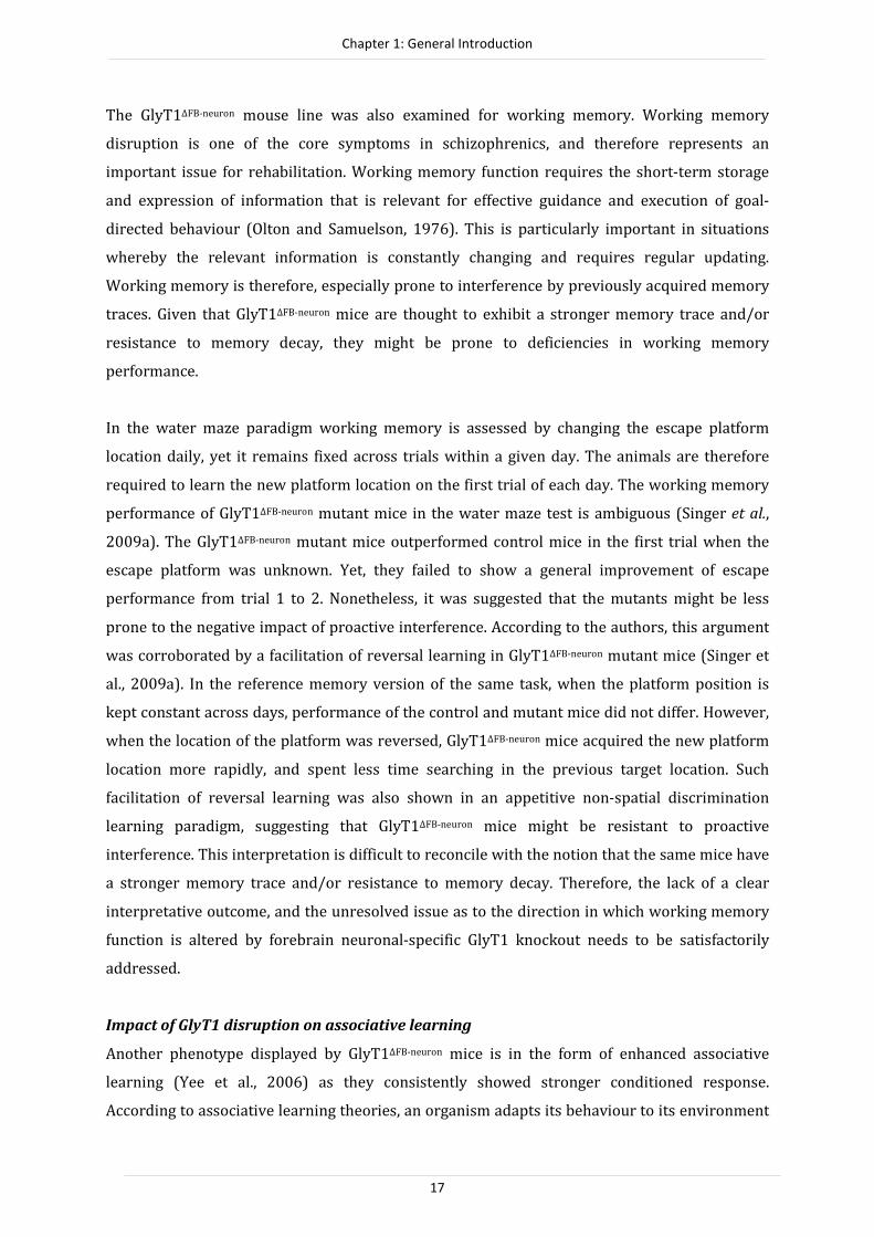
The GlyT1∆FB-neuron mouse line was also examined for working memory. Working memory
disruption is one of the core symptoms in schizophrenics, and therefore represents an
important issue for rehabilitation. Working memory function requires the short-term storage
and expression of information that is relevant for effective guidance and execution of goal-
directed behaviour (Olton and Samuelson, 1976). This is particularly important in situations
whereby the relevant information is constantly changing and requires regular updating.
Working memory is therefore, especially prone to interference by previously acquired memory
traces. Given that GlyT1∆FB-neuron mice are thought to exhibit a stronger memory trace and/or
resistance to memory decay, they might be prone to deficiencies in working memory
performance.
In the water maze paradigm working memory is assessed by changing the escape platform
location daily, yet it remains fixed across trials within a given day. The animals are therefore
required to learn the new platform location on the first trial of each day. The working memory
performance of GlyT1∆FB-neuron mutant mice in the water maze test is ambiguous (Singer et al.,
2009a). The GlyT1∆FB-neuron mutant mice outperformed control mice in the first trial when the
escape platform was unknown. Yet, they failed to show a general improvement of escape
performance from trial 1 to 2. Nonetheless, it was suggested that the mutants might be less
prone to the negative impact of proactive interference. According to the authors, this argument
was corroborated by a facilitation of reversal learning in GlyT1∆FB-neuron mutant mice (Singer et
al., 2009a). In the reference memory version of the same task, when the platform position is
kept constant across days, performance of the control and mutant mice did not differ. However,
when the location of the platform was reversed, GlyT1∆FB-neuron mice acquired the new platform
location more rapidly, and spent less time searching in the previous target location. Such
facilitation of reversal learning was also shown in an appetitive non-spatial discrimination
learning paradigm, suggesting that GlyT1∆FB-neuron mice might be resistant to proactive
interference. This interpretation is difficult to reconcile with the notion that the same mice have
a stronger memory trace and/or resistance to memory decay. Therefore, the lack of a clear
interpretative outcome, and the unresolved issue as to the direction in which working memory
function is altered by forebrain neuronal-specific GlyT1 knockout needs to be satisfactorily
addressed.
Impact of GlyT1 disruption on associative learning
Another phenotype displayed by GlyT1∆FB-neuron mice is in the form of enhanced associative
learning (Yee et al., 2006) as they consistently showed stronger conditioned response.
According to associative learning theories, an organism adapts its behaviour to its environment

Chapter 1: General Introduction
18
by exploiting environmental contingency amongst external stimuli and learn about the
predictive significance of a stimulus. This can be directly related to Hebb’s rule of coincidence
detection where the animals perceive two coincident events and learn to form an association
between them (Hebb, 1949) such that presentation of one can lead to the anticipation of the
other. This can be readily demonstrated in Pavlovian conditioning, whereby a neutral
conditioned stimulus (CS) is paired with a biologically significant unconditioned stimulus (US).
Successful associative learning results in the CS eliciting a conditioned response (CR). Evidence
for enhanced associative learning in GlyT1∆FB-neuron mice, in the form of stronger CR to the CS was
shown across multiple paradigms, including conditioned freezing, conditioned taste aversion,
and conditioned active avoidance learning (Yee et al., 2006). The authors suggested that
neuronal forebrain GlyT1 knockout might lead to a stronger memory trace because of a stronger
association between the CS and the US. This phenotype was observed in aversive paradigms
involving either foot shock or gastric malaise (illness). However, it remains to be determined
whether a similar phenotype might emerge in appetitively motivated paradigms. This is
addressed experimentally in Chapter 2.
Conditioning leads to the formation of an association between representations of two events.
Experimental findings in the field of animal learning have demonstrated that the formation of
associations cannot solely be explained by the principle of contiguity, i.e. conditioning is
effective when two events are paired in time and space. Consequently, learning theorists have
tried to identify circumstances which determine the effectiveness of a stimulus to enter an
association (i.e. its “associability”). The latent inhibition (LI) effect represents an important
demonstration of changes in the associability of a stimulus, which is caused by an animal’s past
experience of the predictive value of that stimulus for a reinforcer. The LI effect refers to the
observation that prior non-reinforced (without consequence) exposures of a stimulus can
retard that stimulus’ ability to enter into association with a significant event that is presented
subsequently (see Figure 2, Lubow and Moore, 1959). In associative learning, some theories
proposed that the CS pre-exposure (Phase 1) retards subsequent development of the
conditioned response (CR), which follows pairings between the same CS and a significant event
(the US, Phase 2). Different theoretical accounts for LI have been proposed. The dominant
concept is that LI reflects a loss of attention to the CS. However, these theories differ in the rules
that are believed to govern changes in attention. For instance, attention to the CS is diminished
because the animal learns from the non-reinforced CS pre-exposures (in Phase 1) that (i) the
stimulus is a relatively poor predictor of reinforcement (Mackintosh, 1975), (ii) that the
stimulus is followed by a predicted consequence (Pearce and Hall, 1980), i.e. there was no
surprise subsequent to the stimulus presentation, or (iii) because of habituation to the CS

Chapter 1: General Introduction
19
(Wagner, 1981). The loss of attention leads to slower processing of the CS during subsequent
conditioning (Phase 2). Alternatively, LI can be considered as a form of proactive interference,
in which the association between the CS and the absence of significant consequence (noUS)
during Phase 1 compete or interfere with subsequent expression (Weiner, 1990) or retrieval
(Bouton, 1993) of the CS-US association that occurs during Phase 2. Consequently, the two
inconsistent associations (CS-noUS and CS-US) will compete for the behavioural control in
response to the CS.
Figure 2: Schematic representation of an experimental procedure designed to assess the latent inhibition
effect.
Yee et al. (2006) showed that GlyT1∆FB-neuron mice exhibited LI under experimental conditions in
which the controls failed to do so, thereby reflecting an enhanced LI effect. The authors, thus,
argued that the impact of the molecular deletion could be understood as bi-directional because
the CR developed in non-pre-exposed conditions was enhanced, whereas the CR in pre-exposed
conditions was reduced. Two possible psychological mechanisms could explain these results:
first, stronger associative learning (as seen before), and/or secondly, facilitation in selectivity of
learning. The latter could be interpreted as a form of enhanced learned inattention (Gray et al.,
1991) or suppression of behavioural switching (Weiner, 1990). No matter which position one
may take, it appears that disruption of GlyT1 does not indiscriminately strengthen all

Chapter 1: General Introduction
20
associations at the cost of selectivity in learning. Bannerman and Sanderson (2007) offered an
alternative account for the findings of Yee et al. (2006) by extending their suggestion that the
mutant mice might be impaired in dishabituation. This was developed in their critique of the
observation of enhanced recognition memory reported in these mice by Singer et al. (2007).
Bannerman and Sanderson (2007) reckons that the lack of latent inhibition in the control mice
could be due to dishabituation to the pre-exposed stimulus (thus attention to it is renewed
when it was presented in conjunction to a US after pre-exposure). Accordingly, the presence of
LI in the mutants could be understood as stemming from an impairment in dishabituation; the
animals remained habituated to the pre-exposed CS. Habituation to the CS extended into Phase
2 when the CS became predictive of the US, which, therefore yielded the latent inhibition effect.
Finally, Singer et al. (2009c) offered another account for the LI effect in GlyT1∆FB-neuron mice by
disregarding the possible involvement of the two different mechanisms proposed by Yee et al.
(2006). They suggested, based on the “switching” account (Weiner, 1990), that because
GlyT1∆FB-neuron mice learned both CS-nothing link (during Phase 1) and CS-US link (during Phase
2) more effectively, the stronger CS-nothing link might imply a stronger impact on the
subsequent expression of the CR, commanded by the CS-US link. In contrast, the test
parameters were insufficient to generate latent inhibition in the control mice, because the
impact of the learned CS-nothing association on the expression of the CR was weaker compared
to the mutant mice. Hence, given the divergence of possible explanations offered in the
literature, the possibility that GlyT1 disruption can result in general facilitation in selectivity of
learning warrants further examination. In Chapter 4, this possibility is addressed by introducing
additional tests that might be similarly conceived as examples demonstrative of selectivity (not
all potential CSs and USs invariably enter into association) in Pavlovian learning.
Further considerations
Although GlyT1 might be beneficial for cognitive functions in the short-term, chronically
elevated glycine concentration levels in the vicinity of NMDARs might be detrimental in the long
run (see Danysz and Parsons, 1998). It has been argued that this approach might exacerbate,
rather than ameliorate, the pathophysiology of conditions like Alzheimer’s disease (Javitt,
2004). Hence, it is worth addressing the impact of enhanced NMDAR excitability, and potential
excitotoxicitity and neurodegeneration in aged GlyT1∆FB-neuron mice.
Chapter 1: General Introduction
21
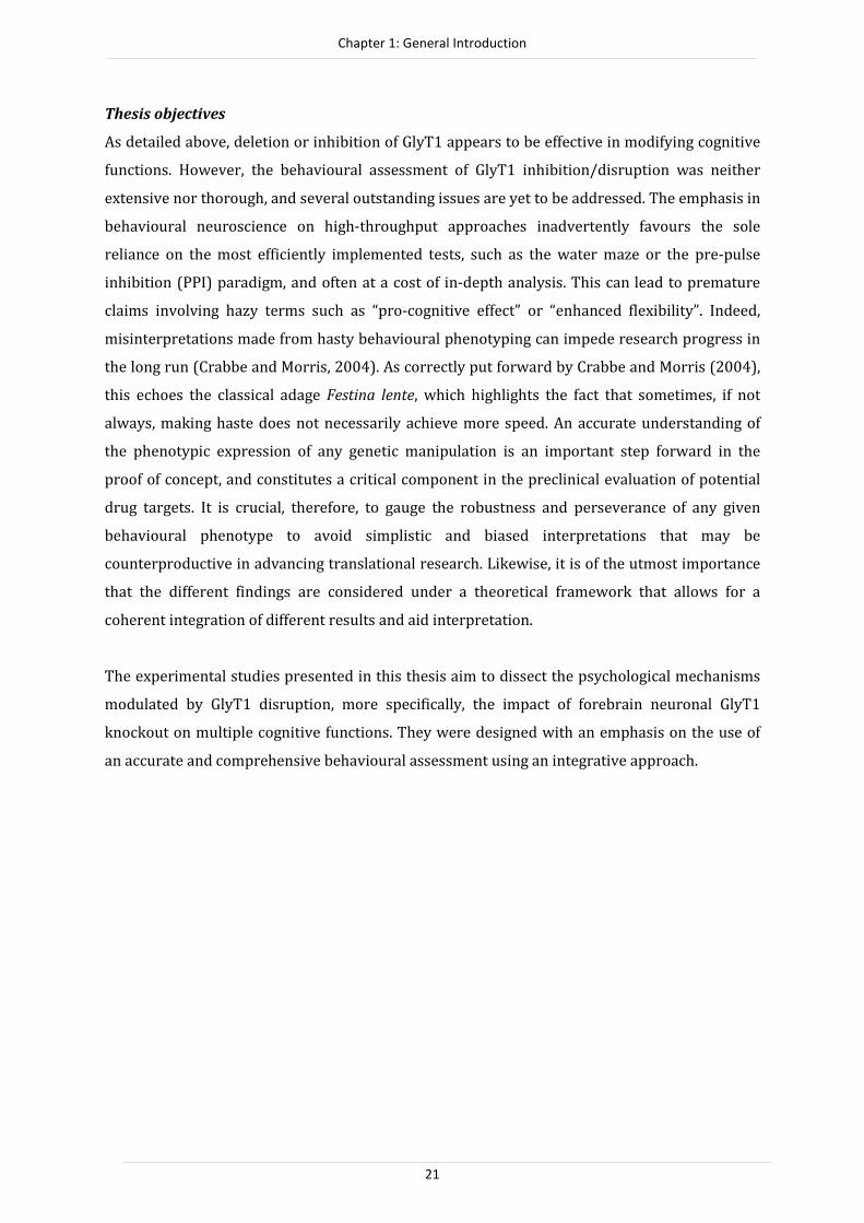
Thesis objectives
As detailed above, deletion or inhibition of GlyT1 appears to be effective in modifying cognitive
functions. However, the behavioural assessment of GlyT1 inhibition/disruption was neither
extensive nor thorough, and several outstanding issues are yet to be addressed. The emphasis in
behavioural neuroscience on high-throughput approaches inadvertently favours the sole
reliance on the most efficiently implemented tests, such as the water maze or the pre-pulse
inhibition (PPI) paradigm, and often at a cost of in-depth analysis. This can lead to premature
claims involving hazy terms such as “pro-cognitive effect” or “enhanced flexibility”. Indeed,
misinterpretations made from hasty behavioural phenotyping can impede research progress in
the long run (Crabbe and Morris, 2004). As correctly put forward by Crabbe and Morris (2004),
this echoes the classical adage Festina lente, which highlights the fact that sometimes, if not
always, making haste does not necessarily achieve more speed. An accurate understanding of
the phenotypic expression of any genetic manipulation is an important step forward in the
proof of concept, and constitutes a critical component in the preclinical evaluation of potential
drug targets. It is crucial, therefore, to gauge the robustness and perseverance of any given
behavioural phenotype to avoid simplistic and biased interpretations that may be
counterproductive in advancing translational research. Likewise, it is of the utmost importance
that the different findings are considered under a theoretical framework that allows for a
coherent integration of different results and aid interpretation.
The experimental studies presented in this thesis aim to dissect the psychological mechanisms
modulated by GlyT1 disruption, more specifically, the impact of forebrain neuronal GlyT1
knockout on multiple cognitive functions. They were designed with an emphasis on the use of
an accurate and comprehensive behavioural assessment using an integrative approach.
Chapter 1: General Introduction
22
Thesis outline
The first part of this thesis investigates the generality of the key phenotypes identified
previously in the GlyT1∆FB-neuron mouse line. The experiments described here aimed at extending
and building on previous findings in two main directions. First, a study was conducted to
examine whether one of the key robust phenotypes in GlyT1∆FB-neuron mice, namely enhanced
Pavlovian conditioning, is compromised by aging. Concurrently, it was investigated whether the
genetic manipulation in the aging brain might be associated with neuroanatomical and
morphological changes (Chapter 2). Both experimental questions were based on concerns over
possible harmful excitotoxic effects linked to chronic elevation of synaptic glycine levels arising
from GlyT1 deletion, such as accelerated aging process, weakened and/or reversal of the pro-
cognitive phenotypes identified in adulthood. Second, a study was designed (Chapter 3) to
include two critical confounds, which had been omitted in the original study by Yee et al.
(2006): Pavlovian conditioning was only investigated using (i) aversive paradigms and (ii) only
in female mice. Here, the consistency of the enhanced Pavlovian conditioning phenotype across
sexes, and the occurrence of this phenotype in an appetitive (non-aversive) paradigm was
investigated. This study offered further opportunity to examine the stability of the phenotype
across both the light and dark phases of the circadian rhythm. This comparison between the two
diurnal phases represents a critical consideration that is often overlooked in the majority of
phenotypic assays of genetically modified animals.
The second part of this thesis addressed the psychological nature of the Pavlovian phenotype
induced by GlyT1 deletion by focusing on its possible impact on the selectivity of learning. The
impression that GlyT1∆FB-neuron mice might exhibit aberrant selective learning during the
formation of associative memory was initially based on their phenotype in the latent inhibition
paradigm (Yee et al., 2006). Accordingly, a study was conducted (Chapter 4) which focused on
the selectivity of learning by incorporating two major determinants of Pavlovian associative
learning: (i) Contiguity, and (ii) Contingency. In light of the hitherto outstanding issues
concerning the potential resistance to proactive interference shown by the GlyT1∆FB-neuron mice
(Singer et al., 2009a), Chapter 5 investigates the impact of this mutation across multiple
behavioural paradigms designed to assess working memory function.
To conclude, the general discussion in Chapter 6 integrates the findings presented here with the
current literature on GlyT1 function, with the aim of providing a detailed understanding of the
potential, as well as limitation, of forebrain- and neuronal- specific GlyT1 inhibition in
modifying cognitive functions.

Chapter 1: General Introduction
23
REFERENCES
Abi-Dargham A, Martinez D, Mawlawi O, Simpson N, Hwang DR, Slifstein M, Anjilvel S, Pidcock J, Guo NN,
Lombardo I and others. 2000. Measurement of striatal and extrastriatal dopamine D1 receptor
binding potential with [11C]NNC 112 in humans: validation and reproducibility. J Cereb Blood
Flow Metab 20(2):225-43.
Abi-Dargham A, van de Giessen E, Slifstein M, Kegeles LS, Laruelle M. 2009. Baseline and amphetamine-
stimulated dopamine activity are related in drug-naive schizophrenic subjects. Biol Psychiatry
65(12):1091-3.
Adler CM, Malhotra AK, Elman I, Goldberg T, Egan M, Pickar D, Breier A. 1999. Comparison of ketamine-
induced thought disorder in healthy volunteers and thought disorder in schizophrenia. Am J
Psychiatry 156(10):1646-9.
Bading H, Ginty DD, Greenberg ME. 1993. Regulation of gene expression in hippocampal neurons by
distinct calcium signaling pathways. Science 260:181-186.
Bannerman DM, Niewoehner B, Lyon L, Romberg C, Schmitt WB, Taylor A, Sanderson DJ, Cottam J,
Sprengel R, Seeburg PH and others. 2008. NMDA receptor subunit NR2A is required for rapidly
acquired spatial working memory but not incremental spatial reference memory. J Neurosci
28(14):3623-30.
Bergeron R, Meyer TM, Coyle JT, Greene RW. 1998. Modulation of N-methyl-D-aspartate receptor function
by glycine transport. Proc Natl Acad Sci U S A 95(26):15730-4.
Black MD, Varty GB, Arad M, Barak S, De Levie A, Boulay D, Pichat P, Griebel G, Weiner I. 2009.
Procognitive and antipsychotic efficacy of glycine transport 1 inhibitors (GlyT1) in acute and
neurodevelopmental models of schizophrenia: latent inhibition studies in the rat.
Psychopharmacology (Berl) 202(1-3):385-96.
Bliss TV, Collingridge GL. 1993. A synaptic model of memory: long-term potentiation in the hippocampus.
Nature 361(6407):31-9.
Bliss TV, Lømo T. 1973. Long-lasting potentiation of synaptic transmission in the dentate area of the
anaesthetized rabbit following stimulation of the perforant path. J Physiol 232(2):331-56.
Bolhuis JJ, Reid IC. 1992. Effects of intraventricular infusion of the N-methyl-D-aspartate (NMDA)
receptor antagonist AP5 on spatial memory of rats in a radial arm maze. Behav Brain Res
47(2):151-7.
Boulay D, Pichat P, Dargazanli G, Estenne-Bouhtou G, Terranova JP, Rogacki N, Stemmelin J, Coste A,
Lanneau C, Desvignes C and others. 2008. Characterization of SSR103800, a selective inhibitor of
the glycine transporter-1 in models predictive of therapeutic activity in schizophrenia.
Pharmacol Biochem Behav 91(1):47-58.
Bouton ME. 1993. Context, time, and memory retrieval in the interference paradigms of Pavlovian
learning. Psychol Bull 114(1):80-99.
Bowery NG. 1987. Glycine-binding sites and NMDA receptors in brain. Nature 326(6111):338.
Bristow DR, Bowery NG, Woodruff GN. 1986. Light microscopic autoradiographic localisation of
[3H]glycine and [3H]strychnine binding sites in rat brain. Eur J Pharmacol 126(3):303-307.
Carlsson M, Carlsson A. 1990. Interaction between glutamatergic and monoaminergic systems within the
basal ganglia: Implications for schizophrenia and Parkinson's disease. Trends Neurosci 13:272-
276.
Chen L, Muhlhauser M, Yang CR. 2003. Glycine tranporter-1 blockade potentiates NMDA-mediated
responses in rat prefrontal cortical neurons in vitro and in vivo. J Neurophysiol 89(2):691-703.
Collingridge GL, Bliss TV. 1995. Memories of NMDA receptors and LTP. Trends Neurosci 18(2):54-6.
Collingridge GL, Kehl SJ, McLennan H. 1983. Excitatory amino acids in synaptic transmission in the
Schaffer collateral-commissural pathway of the rat hippocampus. J. Physiol. 334:33-46.
Crabbe JC, Morris RG. 2004. Festina lente: late-night thoughts on high-throughput screening of mouse
behavior. Nat Neurosci 7(11):1175-9.
Creese I, Burt DR, Snyder SH. 1976. Dopamine receptor binding predicts clinical and pharmacological
potencies of antischizophrenic drugs. Science 192(4238):481-3.
Cubelos B, Giménez C, Zafra F. 2005. Localization of the GLYT1 glycine transporter at glutamatergic
synapses in the rat brain. Cereb Cortex 15(4):448-459.
Cull-Candy S, Brickley S, Farrant M. 2001. NMDA receptor subunits: diversity, development and disease.
Curr Opin Neurobiol 11(3):327-35.
Danysz W, Parsons CG. 1998. Glycine and N-methyl-D-aspartate receptors: physiological significance and
possible therapeutic applications. Pharmacol Rev 50(4):597-664.

Chapter 1: General Introduction
24
Depoortere R, Dargazanli G, Estenne-Bouhtou G, Coste A, Lanneau C, Desvignes C, Poncelet M, Heaulme M,
Santucci V, Decobert M and others. 2005. Neurochemical, electrophysiological and
pharmacological profiles of the selective inhibitor of the glycine transporter-1 SSR504734, a
potential new type of antipsychotic. Neuropsychopharmacology 30(11):1963-85.
Dingledine R, Borges K, Bowie D, Traynelis SF. 1999. The glutamate receptor ion channels. Pharmacol Rev
51(1):7-61.
Dragatsis I, Zeitlin S. 2000. CaMKIIalpha-Cre transgene expression and recombination patterns in the
mouse brain. Genesis 26(2):133-5.
Ewald RC, Cline HT. 2009. NMDA Receptors and Brain Development. In: Van Dongen AM, editor. Biology
of the NMDA Receptor. Boca Raton (FL: CRC Press.
Fedele E, Foster AC. 1992. [3H]glycine uptake in rat hippocampus: kinetic analysis and autoradiographic
localization. Brain Res. 572(1-2):154-163.
Fletcher EJ, Lodge D. 1988. Glycine reverses antagonism of N-methyl-D-aspartate (NMDA) by 1-hydroxy-
3-aminopyrrolidone-2 (HA-966) but not by D-2-amino-5-phosphonovalerate (D-AP5) on rat
cortical slices. Eur J Pharmacol. 151(1):161-162.
Gabernet L, Pauly-Evers M, Schwerdel C, Lentz M, Bluethmann H, Vogt K, Alberati D, Mohler H, Boison D.
2005. Enhancement of the NMDA receptor function by reduction of glycine transporter-1
expression. Neurosci Lett 373(1):79-84.
Gainetdinov RR, Mohn AR, Caron MG. 2001. Genetic animal models: focus on schizophrenia. Trends
Neurosci 24(9):527-33.
Goff DC, Tsai GC, Monoach DS, Coyle JT. 1995. Dose-finding trial of D-cycloserine added to neuroleptics for
negative symptoms in schizophrenia. Am J Psychiatry 152:1213-1215.
Gomeza J, Ohno K, Hulsmann S, Armsen W, Eulenburg V, Richter DW, Laube B, Betz H. 2003. Deletion of
the mouse glycine transporter 2 results in a hyperekplexia phenotype and postnatal lethality.
Neuron 40(4):797-806.
Gray JA, Feldon J, Rawlins JNP, Smith AD, Hemsley DR. 1991. The Neuropsychology of Schizophrenia.
Behavioral and Brain Sciences 14(1):1-19.
Guastella J, Brecha N, Weigmann C, Lester HA, Davidson N. 1992. Cloning, expression, and localization of a
rat brain high-affinity glycine transporter. PNAS 89(15):7189-7193.
Haberny KA, Paule MG, Scallet AC, Sistare FD, Lester DS, Hanig JP, Slikker WJ. 2002. Ontogeny of the N-
methyl-D-aspartate (NMDA) receptor system and susceptibility to neurotoxicity. Toxicol. Sci.
68:9-17.
Handelmann GE, Nervins ME, Mueller LL, Arnolde SM, Cordi AA. 1989. Milacemide, a glycine prodrug,
enhances performance of learning tasks in nromal and amnesic rodents. Pharmacol. Biochem.
Behav. 34:823-828.
Hashimoto K, Fujita Y, Ishima T, Chaki S, Iyo M. 2008. Phencyclidine-induced cognitive deficits in mice are
improved by subsequent subchronic administration of the glycine transporter-1 inhibitor NFPS
and D-serine. Eur Neuropsychopharmacol 18(6):414-21.
Hebb DO. 1949. The Organization of Behavior: A neuropsychological theory. New York: Wiley.
Javitt DC. 2004. Glutamate as a therapeutic target in psychiatric disorders. Mol Psychiatry 9(11):984-97,
979.
Javitt DC, Sershen H, Hashim A, Lajtha A. 1997. Reversal of phencyclidine-induced hyperactivity by glycine
and the glycine uptake inhibitor glycyldodecylamide. Neuropsychopharmacology 17(3):202-4.
Javitt DC, Zylberman I, Zukin SR, Herescolevy U, Lindenmayer JP. 1994. Amerlioration of negative
sympomts in schizophrenia by glycine. Am J Psychiatry 151:1234-1236.
Jentsch JD, Roth RH. 1999. The neuropsychopharmacology of phencyclidine: from NMDA receptor
hypofunction to the dopamine hypothesis of schizophrenia. Neuropsychopharmacology
20(3):201-25.
Johnson JW, Ascher P. 1987. Glycine potentiates the NMDA response in cultured mouse brain neurons.
Nature 325(6104):529-31.
Jursky F, Nelson N. 1996. Developmental expression of the glycine transporters GLYT1 and GLYT2 in
mouse brain. J. Neurochem. 67(1):336-344.
Kapur S, Remington G. 2001. Atypical antipsychotics: new directions and new challenges in the treatment
of schizophrenia. Annu Rev Med 52:503-17.
Kawabe K, Yoshihara T, Ichitani Y, Iwasaki T. 1998. Intrahippocampal D-cycloserine improves MK-801-
induced memory deficits: radial-arm maze performance in rats. Brain Res 814(1-2):226-30.
Kinney GG, Sur C, Burno M, Mallorga PJ, Williams JB, Figueroa DJ, Wittmann M, Lemaire W, Conn PJ. 2003.
The glycine transporter type 1 inhibitor N-[3-(4 '-fluorophenyl)-3-(4 '-phenylphenoxy)propyl]

Chapter 1: General Introduction
25
sarcosine potentiates NMDA receptor-mediated responses in vivo and produces an antipsychotic
profile in rodent behavior. Journal of Neuroscience 23(20):7586-7591.
Kishimoto H, Simon JR, Aprison MH. 1981. Determination of the equilibrium dissociation constants and
number of glycine binding sites in several areas of the rat central nervous system, using a
sodium-independent system. J Neurochem 37(4):1015-1024.
Kleckner NW, Dingledine R. 1988. Requirement for glycine in activation of NMDA receptors expresses in
Xenopus oocytes. Science 214:835-837.
Konorski J. 1948. Conditioned Reflexes and Neuronal Organization: London: Cambridge University Press.
Krystal JH, Karper LP, Seibyl JP, Freeman GK, Delaney R, Bremner JD, Heninger GR, Bowers MB, Jr.,
Charney DS. 1994. Subanesthetic effects of the noncompetitive NMDA antagonist, ketamine, in
humans. Psychotomimetic, perceptual, cognitive, and neuroendocrine responses. Arch Gen
Psychiatry 51(3):199-214.
Lahti AC, Holcomb HH, Medoff DR, Tamminga CA. 1995a. Ketamine activates psychosis and alters limbic
blood flow in schizophrenia. Neuroreport 6(6):869-72.
Lahti AC, Koffel B, LaPorte D, Tamminga CA. 1995b. Subanesthetic doses of ketamine stimulate psychosis
in schizophrenia. Neuropsychopharmacology 13(1):9-19.
Liu QR, Nelson H, Mandiyan S, López-Corcuera B, Nelson N. 1992. Cloning and expression of a glycine
transporter from mouse brain. FEBS Lett. 305(2):110-114.
Lubow RE, Moore AU. 1959. Latent inhibition: the effect of nonreinforced pre-exposure to the conditional
stimulus. J Comp Physiol Psychol 52:415-9.
Luby ED, Cohen BD, Rosenbaum G, Gottlieb JS, Kelley R. 1959. Study of a new schizophrenomimetic drug;
sernyl. AMA Arch Neurol Psychiatry 81(3):363-9.
Lynch MA. 2004. Long-term potentiation and memory. Psychiol. Rev 84(1):87-136.
Mackintosh NJ. 1975. Theory of Attention - Variations in Associability of Stimuli with Reinforcement.
Psychological Review 82(4):276-298.
Malenka RC, Kauer JA, Perkel DJ, Mauk MD, Kelly PT, Nicoll RA, Waxham MN. 1989. An essential role for
postsynaptic calmodulin and protein kinase activity in long-term potentiation. Nature
340(6234):554-557.
Malinow R, Malenka RC. 2002. AMPA receptor trafficking and synaptic plasticity. Ann. Rev. Neurosci.
25:103-126.
Manahan-Vaughan D, Wildforster V, Thomsen C. 2008. Rescue of hippocampal LTP and learning deficits in
a rat model of psychosis by inhibition of glycine transporter-1 (GlyT1). European Journal of
Neuroscience 28(7):1342-1350.
Martin SJ, Grimwood PD, Morris RG. 2000. Synaptic plasticity and memory: an evaluation of the
hypothesis. Annu Rev Neurosci 23:649-711.
Martina M, Gorfinkel Y, Halman S, Lowe JA, Periyalwar P, Schmidt CJ, Bergeron R. 2004. Glycine
transporter type 1 blockade changes NMDA receptor-mediated responses and LTP in
hippocampal CA1 pyramidal cells by altering extracellular glycine levels. J Physiol 557(Pt 2):489-
500.
Matsiu T, Sekiguchi M, Hashimoto A, Tomita U, Nishikawa T, Wada K. 1995. Functional comparison of D-
serine and glycine in rodents: The effects on cloned NMDA recepteors and the extracellular
concentration. J. Neurochem. 65:454-458.
Mayer ML, Westbrook GL. 1987. Permeation and block of N-methyl-D-aspartic acid receptor channels by
divalent cations in mouse cultured central neurones. J. Physiol. 394:501-527.
McHugh SB, Niewoehner B, Rawlins JN, Bannerman DM. 2008. Dorsal hippocampal N-methyl-D-aspartate
receptors underlie spatial working memory performance during non-matching to place testing
on the T-maze. Behav Brain Res 186(1):41-7.
Möhler H, Boison D, Singer P, Feldon J, Pauly-Evers M, Yee BK. 2011. Glycine transporter 1 as a potential
therapeutic target for schizophrenia-related symptoms: Evidence from genetically modified
mouse models and pharmacological inhibition. Biochem Pharmacol.
Mohn AR, Gainetdinov RR, Caron MG, Koller BH. 1999. Mice with reduced NMDA receptor expression
display behaviors related to schizophrenia. Cell 98(4):427-36.
Monaghan DT, Cotman CW. 1985. Distribution of N-methyl-D-aspartate-sensitive L-[3H]glutamate-
binding sites in rat brain. J Neurosci 5(11):2909-2919.
Monaghan DT, Olverman HJ, Nguyen L, Watkins JC, Cotman CW. 1988. Two classes of N-methyl-D-
aspartate recognition sites: differential distribution and differential regulation by glycine. PNAS
85(24):9836-9840.

Chapter 1: General Introduction
26
Monahan JB, Handelmann GE, Hood WF, Cordi AA. 1989. d-Cycloserine, a positive modulatior of the N-
methyl-D-asparate receptor, enhances performance of learning tasks in rats. Pharmacol Biochem
Behav 34:647-653.
Morris RG. 1989. Synaptic plasticity and learning: selective impairment of learning rats and blockade of
long-term potentiation in vivo by the N-methyl-D-aspartate receptor antagonist AP5. J Neurosci
9(9):3040-57.
Morris RG, Anderson E, Lynch GS, Baudry M. 1986. Selective impairment of learning and blockade of long-
term potentiation by an N-methyl-D-aspartate receptor antagonist, AP5. Nature 319(6056):774-
6.
Nakanishi S. 1992. Science. Molecular diversity of glutamate receptors and implications for brain function.
258(5082):597-603.
Nakazawa K, Quirk MC, Chitwood RA, Watanabe M, Yeckel MF, Sun LD, Kato A, Carr CA, Johnston D,
Wilson MA and others. 2002. Requirement for hippocampal CA3 NMDA receptors in associative
memory recall. Science 297(5579):211-8.
Nakazawa K, Sun LD, Quirk MC, Rondi-Reig L, Wilson MA, Tonegawa S. 2003. Hippocampal CA3 NMDA
receptors are crucial for memory acquisition of one-time experience. Neuron 38(2):305-15.
Niewoehner B, Single FN, Hvalby O, Jensen V, Meyer zum Alten Borgloh S, Seeburg PH, Rawlins JN,
Sprengel R, Bannerman DM. 2007. Impaired spatial working memory but spared spatial
reference memory following functional loss of NMDA receptors in the dentate gyrus. Eur J
Neurosci 25(3):837-46.
Olney JW, Farber NB. 1995. Glutamate receptor dysfunction and schizophrenia. Arch Gen Psychiatry
52(12):998-1007.
Olney JW, Newcomer JW, Farber NB. 1999. NMDA receptor hypofunction model of schizophrenia. J
Psychiatr Res 33(6):523-33.
Olton DS, Samuelson RJ. 1976. Remembrance of Places Passed - Spatial Memory in Rats. Journal of
Experimental Psychology-Animal Behavior Processes 2(2):97-116.
Pearce JM, Hall G. 1980. A model for Pavlovian learning: variations in the effectiveness of conditioned but
not of unconditioned stimuli. Psychological Review 87(6):532-52.
Pinard E, Alanine A, Alberati D, Bender M, Borroni E, Bourdeaux P, Brom V, Burner S, Fischer H, Hainzl D
and others. 2010. Selective GlyT1 inhibitors: discovery of [4-(3-fluoro-5-trifluoromethylpyridin-
2-yl)piperazin-1-yl][5-methanesulfon yl-2-((S)-2,2,2-trifluoro-1-
methylethoxy)phenyl]methanone (RG1678), a promising novel medicine to treat schizophrenia. J
Med Chem 53(12):4603-14.
Ramón y Cajal S. 1893 Neue Darstellung vom histologischen Bau des Zentralnervensystem. Arch. Anat.
Entwick. :319–428.
Rescorla RA. 2004. Spontaneous recovery. Learn Mem 11(5):501-9.
Rothman SM, Olney JW. 1995. Excitotoxicity and the NMDA receptor--still lethal after eight years. Trends
Neurosci 18(2):57-8.
Sakimura K, Kutsuwada T, Ito I, Manabe T, Takayama C, Kushiya E, Yagi T, Aizawa S, Inoue Y, Sugiyama H
and others. 1995. Reduced hippocampal LTP and spatial learning in mice lacking NMDA receptor
epsilon 1 subunit. Nature 373(6510):151-5.
Sandberg A, Bostrom N. 2006. Converging cognitive enhancements. Ann NY Acad. Sci. 1093:201-227.
Sanderson DJ, Bannerman DM. 2007. Supersmart mice: surprising or surprised? Theoretical comment on
Singer, Boison, Mohler, Feldon, and Yee (2007). Behav Neurosci 121(5):1137-9.
Seeman P, Chau-Wong M, Tedesco J, Wong K. 1975. Brain receptors for antipsychotic drugs and
dopamine: direct binding assays. Proc Natl Acad Sci U S A 72(11):4376-80.
Shimazaki T, Kaku A, Chaki S. 2010. d-Serine and a glycine transporter-1 inhibitor enhance social memory
in rats. Psychopharmacology 209(3):263-270.
Singer P, Boison D, Mohler H, Feldon J, Yee BK. 2007. Enhanced recognition memory following glycine
transporter 1 deletion in forebrain neurons. Behav Neurosci 121(5):815-25.
Singer P, Boison D, Mohler H, Feldon J, Yee BK. 2009a. Deletion of glycine transporter 1 (GlyT1) in
forebrain neurons facilitates reversal learning: enhanced cognitive adaptability? Behav Neurosci
123(5):1012-27.
Singer P, Feldon J, Yee BK. 2009b. The glycine transporter 1 inhibitor SSR504734 enhances working
memory performance in a continuous delayed alternation task in C57BL/6 mice.
Psychopharmacology (Berl) 202(1-3):371-84.
Singer P, Yee BK, Feldon J, Iwasato T, Itohara S, Grampp T, Prenosil G, Benke D, Mohler H, Boison D. 2009c.
Altered mnemonic functions and resistance to N-METHYL-d-Aspartate receptor antagonism by
forebrain conditional knockout of glycine transporter 1. Neuroscience 161(2):635-54.

Chapter 1: General Introduction
27
Smith KE, Borden LA, Hartig PR, Branchek T, Weinshank RL. 1992. Cloning and expression of a glycine
transporter reveal colocalization with NMDA receptors. Neuron 8(5):927-35.
Snyder SH. 1976. The dopamine hypothesis of schizophrenia: focus on the dopamine receptor. Am J
Psychiatry 133(2):197-202.
Supplisson S, Bergman C. 1997. Control of NMDA receptor activation by a glycine transporter co-
expressed in Xenopus oocytes. J Neurosci 17:4580-4590.
Tamminga C. 1999. Glutamatergic aspects of schizophrenia. Br J Psychiatry Suppl(37):12-5.
Thomson AM. 1989. Glycine modulation of the NMDA receptor/channel complex. Trends Neurosci.
12(9):349-353.
Tonkiss J, Morris RG, Rawlins JN. 1988. Intra-ventricular infusion of the NMDA antagonist AP5 impairs
performance on a non-spatial operant DRL task in the rat. Exp Brain Res 73(1):181-188.
Tsai G, Ralph-Williams RJ, Martina M, Bergeron R, Berger-Sweeney J, Dunham KS, Jiang Z, Caine SB, Coyle
JT. 2004. Gene knockout of glycine transporter 1: characterization of the behavioral phenotype.
Proc Natl Acad Sci U S A 101(22):8485-90.
Tsien JZ, Huerta PT, Tonegawa S. 1996. The essential role of hippocampal CA1 NMDA receptor-dependent
synaptic plasticity in spatial memory. Cell 87(7):1327-38.
Wagner AR. 1981. SOP: A model of automatic memory processing in animal behavior. In: (Eds.) NESRRM,
editor. Information processing in animals: Memory mechanisms: Hillsdale, NJ: Erlbaum. p 5-47.
Weiner I. 1990. Neural substrates of latent inhibition: the switching model. Psychol Bull 108(3):442-61.
Whitehead KJ, Pearce SM, Walker G, Sundaram H, Hill D, Bowery NG. 2004. Positive N-methyl-D-aspartate
receptor modulation by selective glycine transporter-1 inhibition in the rat dorsal spinal cord in
vivo. Neuroscience 126(2):381-90.
Yee BK, Balic E, Singer P, Schwerdel C, Grampp T, Gabernet L, Knuesel I, Benke D, Feldon J, Mohler H and
others. 2006. Disruption of glycine transporter 1 restricted to forebrain neurons is associated
with a procognitive and antipsychotic phenotypic profile. J Neurosci 26(12):3169-81.
Zafra F, Aragón C, Olivares L, Danbolt NC, Giménez C, Storm-Mathisen J. 1995a. Glycine transporters are
differentially expressed among CNS cells. J Neurosci 15(5 Pt 2)):3952-3969.
Zafra F, Gomeza J, Olivares L, Aragón C, Giménez C. 1995b. Regional distribution and developmental
variation of the glycine transporters GLYT1 and GLYT2 in the rat CNS. Eur J Neurosci 7(6):1342-
1352.
Zalutsky RA, Nicoll RA. 1990. Comparison of two forms of long-term potentiation in single hippocampal
neurons. Science 248(4963):1619-1624.

28
CHAPTER 2
Impacts of forebrain neuronal glycine transporter 1
disruption in the senescent brain:
Evidence for age-dependent phenotypes in Pavlovian
learning
With Philipp Singer, Detlev Boison, Joram Feldon, Hanns Möhler, and Benjamin K. Yee
As published in Behavioral Neuroscience, vol. 124, no. 6, 839–850 (2010)

Chapter 2: GlyT1 in the aging brain
29
ABSTRACT
Genetic deletion of glycine transporter 1 (GlyT1) in forebrain neurons gives rise to
multiple pro-cognitive phenotypes, presumably due to enhanced N-methyl-D-aspartate
receptor (NMDAR) functions. However, concerns over possible harmful excitotoxic
effects under life-long elevation of synaptic glycine have been raised. Such effects might
accelerate the aging process, weakening or even reversing the pro-cognitive phenotypes
identified in adulthood. Here, we examined if one of the most robust phenotypes in the
mutant mouse line (CamKIIααααCre;GlyT1tm1.2fl/fI), namely, enhanced aversive Pavlovian
conditioning, might be modified by age. Comparison between 3-month-old (adult) and
22-month-old (aged) mutants confirmed the presence of this phenotype at both ages.
However, the temporal expression of the Pavlovian phenotype was modified in
senescence; while adult mutants showed a pronounced within-session extinction, aged
mutants did not. Expression of NR2B subunits of NMDAR and neural proliferation were
examined in the same animals by immunohistochemistry. These were reduced in the
aged mice as expected, but not exacerbated by the mutation. Thus, our results do not
substantiate the concerns of neurotoxic effects through life-long GlyT1 disruption in
forebrain neurons, but provide evidence for a modification of phenotypic expression as a
function of age. The latter points raise the need to further investigate other pro-cognitive
phenotypes identified at adulthood in this mutant line. In addition, we revealed here for
the first time a clear increase in the number of immature neurons in the hippocampus of
the mutants, although the behavioural significance of this phenotype remains to be
determined.
INTRODUCTION
N-methyl-d-aspartate receptor (NMDAR) -dependent synaptic plasticity has been implicated in
various forms of learning and memory processes (Morris, 1989; Morris, Anderson, Lynch, &
Baudry, 1986). Its hypofunction may be associated with the cognitive deficiency characteristic
of a number of psychiatric disorders, such as schizophrenia, as well as with the course of normal
aging (Rosenzweig & Barnes, 2003). Enhancing NMDAR function therefore represents a possible
strategy to ameliorate such deficits (Martin, Grimwood, & Morris, 2000). While direct activation
of NMDARs is prone to severe side effects including neurotoxicity and seizures (Rothman &
Olney, 1995), modulation of NMDAR function via allosteric sites offers an opportunity for
alternative pharmacological targets (Yang & Svensson, 2008). One strategy is to increase the
availability of glycine in the vicinity of NMDARs, and therefore the occupancy of the co-agonist
glycine-B site of the NMDAR complex. This can be effectively achieved by pharmacological
inhibition of glycine re-uptake via glycine transporter 1 (GlyT1) which co-localizes with
NMDARs in neurons. Such inhibition has been reported to enhance NMDAR excitability, confer
resistance to behavioural deficits induced by NMDAR antagonists, and lead to some forms of
enhanced cognitive performance (Black et al., 2009; Depoortere et al., 2005; Singer, Boison,
Mohler, Feldon, & Yee, 2009).

Chapter 2: GlyT1 in the aging brain
30
We have recently shown that disruption of GlyT1 restricted to forebrain neurons is sufficient to
facilitate performance on various cognitive processes, including Pavlovian conditioning,
reversal learning, latent inhibition and memory for object identity as well as object location
(Singer, Boison, Mohler, Feldon, & Yee, 2007; Singer et al., 2009; Yee et al., 2006). When GlyT1 is
knocked out in the entire forebrain, working memory function is also enhanced (Singer et al.,
2009), extending an earlier report of improved reference memory retention in constitutive
heterozygous GlyT1 knockout mice (Tsai et al., 2004). Although direct demonstration of GlyT1
deletion or blockade alone being sufficient to induce excitotoxicity is lacking, Sanderson and
Bannerman (2007) have raised concern over possible negative impacts of this manipulation in
the long term. The deletion of the GlyT1 gene, in spite of its potential cognitive gain, may carry a
putative ‘cost’. More specifically, GlyT1 deletion may carry a greater risk of neurodegeneration
or excitotoxicity, especially in aged animals, outweighing any potential cognitive gain from
enhanced NMDAR activation (Javitt, 2004). Increasing Ca2+ influx through NMDARs and
perhaps AMPA receptors may lead to excitotoxicity and neuronal death implicated in
neurodegenerative disorders such as Alzheimer's disease (Javitt, 2004). A life-long elevation of
glycine concentration in the vicinity of NMDARs might be apoptotic. This hypothesis is highly
relevant to the potential clinical application of GlyT1-inhibitors as cognitive enhancers or
antipsychotic drugs. The present study is designed to address empirically two critical
implications raised by Sanderson and Bannerman (2007).
In the present study we first examined if the cognitive enhancing effects of GlyT1
disruption demonstrated in adult animals might be modified in senescence, as would be
expected if the genetic disruption did carry long-term negative impact. A diminution, absence or
even reversal of such an effect in aged subjects would lend some credence to Sanderson and
Bannerman's (2007) concern. To this end, we compared the behavioural effects seen in mutant
mice with forebrain neuronal GlyT1 knockout to those observed in control mice at two ages: 3
months (“adult”) vs. 22 months old (“aged”). We focused on one of the most robust pro-
cognitive traits of this conditional genetic knockout, namely enhanced associative learning as
exemplified by Pavlovian conditioned freezing (Yee et al., 2006). Prior to the conditioning
experiment, possible relevant confounding effects that might interfere with the freezing
response were assessed by the elevated-plus maze test of anxiety and the open field test of
spontaneous locomotors activity.
Following behavioural assessments, selected anatomical markers were assessed to
gauge the putative negative impacts of long-term GlyT1 disruption in the brain. First, apoptosis
related to neurotoxicity or accelerated neurodegeneration in the hippocampus was evaluated
by activated caspase-3 immunohistochemistry (Budihardjo, Oliver, Lutter, Luo, & Wang, 1999).
Second, density of the immuno-signals against the NR1 and NR2B subunits of the NMDAR in

Chapter 2: GlyT1 in the aging brain
31
amygdala and prefrontal cortices in addition to hippocampus was measured. NMDARs are
pertinent to the normal functioning of these brain regions, which are implicated in the
acquisition and extinction of fear conditioning (Maren, 2001). Given that binding to glycine-B
site primes the internalization and degradation of NMDARs (Nong et al., 2003), persistent and
excessive glycine-B site activation might reduce NMDAR expression. Finally, the immature
neuronal marker doublecortin (DCX) was used to index neural proliferation in the dentate
gyrus, which is known to be regulated by NMDAR activity, and down-regulated in senescence
(Bizon & Gallagher, 2003).
METHODS
Subjects
The subjects were offspring obtained by crossing CamKIIαCre:Glyt1tm1.2fl/fl mice with
Glyt1tm1.2fl/fl mice (both maintained on a pure C57BL/6J background). This breeding strategy
yielded litters with a 1:1 mixture of CamKIIαCre:Glyt1tm1.2fl/fl (hereafter simply referred to as
“mutant”) and Glyt1tm1.2fl/fl (“control”) mice. Breeding took place in a specific-pathogen free
(SPF) breeding facility (Laboratory of Behavioural Neurobiology, ETH Zurich, Schwerzenbach,
Switzerland). Litters were weaned at postnatal day 21. Genotypes were determined by standard
PCR on tail biopsies obtained within 10 days after weaning on postnatal day 21 as previously
described (Yee et al., 2006). At the age of 11 weeks, the animals were transferred to a separate
climatized animal vivarium (21±1°C, relative humidity at 55±5%) under a reversed light-dark
cycle (lights off: 0800–2000). They were kept in groups of 3–5 in Makrolon® Type-III cages
(Techniplast, Milan, Italy), and maintained under constant ad libitum water and food (Kliba
3430, Klibamuhlen, Kaiseraugst, Switzerland). The ‘adult’ mice were approximately three-
months old at the time of testing, comprising 5 male and 6 female mutant mice, and 4 male and
5 female littermate controls. The ‘aged’ mice were approximately 22-24 months of age at the
time of testing, and comprised 5 male and 4 female mutant mice, and 7 male and 6 female
littermate controls. Animals from both age groups were tested together in a single experiment.
All tests were conducted in the dark phase of the light-dark cycle. The experimental
manipulations and procedures described here had been previously approved by the Swiss
Cantonal Veterinary Office, in accordance to the ethical standards required by the Swiss Act and
Ordinance on Animal Protection and the European Council Directive 86/609/EEC, which are in
accordance with the NIH Guide for the Care and Use of Laboratory Animals (1996).
The animals were subjected to the three behavioural tests as described below in the
order of their implementation.

Chapter 2: GlyT1 in the aging brain
32
Elevated plus maze test of anxiety
General anxiety can be affected by senescence and can therefore be a potential confounding
factor in the assessment of learned fear. In order to evaluate this, the elevated plus maze test
was used as an ethologically relevant test of anxiety, induced by exposed spaces and height. The
apparatus has been fully described elsewhere (Yee et al., 2004). Briefly, it consisted of a set of
four equally spaced arms ( 30cm long and 5 cm wide) joined to a central square platform (5 cm
× 5 cm); two opposing arms were exposed and unprotected whilst the other two arms were
protected by opaque side walls and an end wall at the distal end of the arm. The test began
when the animal was released into the central area of the maze with its head facing an open
arm, and allowed to explore freely for 5 min before being removed and returned to the home
cage. A digital camera mounted above the maze captured and transmitted images at a rate of
5Hz to a PC running the Ethovision tracking system (Noldus Technology Wageningen, The
Netherlands). This allowed the computation of two standard anxiety-related measures: (i)
percent time spent in the open arms = time in open arms / time in all four arms × 100%, and (ii)
percent number of entries into open arms = number of open arm entries / number of all arm
entries × 100%. In addition, locomotor activity was indexed by the total distance travelled on
the entire maze surface during the test period. A rest period of 48 h was allowed before the
assessment of the next test.
Open field test of spontaneous locomotor activity
Spontaneous activity can also be altered with senescence and become another potential
confound in the measurement of immobility in the subsequent conditioned freezing experiment.
This was assessed next in an open field. Four identical 40 x 40cm2 white open field arenas with
35cm high sidewalls and a water-proof inlay were used. The animals were tested in squads of
four, under diffused dim lighting at 30 lux. Allocation to the four test arenas was
counterbalanced. Each mouse was gently placed in the centre of the appropriate open field and
allowed to explore undisturbed for 1 h. The open fields were cleansed with water and dried
after each animal. Locomotor activity was indexed by spatial displacement tracked by the
EthoVision® tracking system (Version 3.1, Noldus Technology, Wageningen, The Netherlands),
expressed as distance travelled (in m) across successive 10-min bins. The conditioned freezing
was assessed 4 days after the open field test.
Conditioned freezing paradigm
The apparatus comprised two distinct sets of test chambers (4 per set), as fully described
elsewhere (Meyer, Feldon, Schedlowski, & Yee, 2005). All chambers were equipped with a grid
floor composed of stainless steel rods spaced at 10-mm intervals, and through which scrambled

Chapter 2: GlyT1 in the aging brain
33
electric shocks (unconditioned stimulus, US) could be delivered (Model E13-14, Coulbourn
Instruments). C57BL/6 mice are known however to suffer from a progressive deterioration in
hearing that begins at about 2 months of age and significant hearing impairment is expected by
the age of 18 months (Walton, Barsz, & Wilson, 2008). A clear hearing loss in the aged mutant
and control mice was confirmed by the presence of a pronounced deficit in the acoustic startle
response in comparison to the adult mice (data not shown). Hence, the use of an auditory
conditioned stimulus would have been inappropriate in this test, and instead a tactile stimulus
in the form of a high-frequency vibration of the grid floor was used as the conditioned stimulus
(CS). This was achieved by fixing a commercial vibrating unit (MiniVibrator Model 558095,
Orion Versand AG, Buchs, Switzerland) to the grid floor (Yee, Singer, Chen, Feldon, & Boison,
2007). Transmission of the sinusoidal vibration beyond the grid floor was dampened by
insulating the grid from the test chamber floor on four pieces of shock-absorbing sponge. A
constant background white noise (60 dB) was provided via a loudspeaker mounted inside each
chamber to mask the weak noise emitted from the vibrating unit. A digital camera was mounted
30 cm directly above the area of interest in each chamber. Digital images were captured at a rate
of 1 Hz and transmitted to a PC running the Windows XP (SP3) operating system via a PicoloTM
frame grabber (Euresys s.a., Liège, Belgium). An image analysis algorithm originally described
by Richmond et al (1998) was used to determine freezing (immobility) in real-time. The image
analysis was performed using Open eVision 1.1 (Euresys s.a., Liège, Belgium) under the control
of a customized Microsoft Visual Basic (version 6) script.
On day 1, the animals received two discrete trials of CS-US pairing. Each trial comprised
a 30-s CS followed immediately by a 1-s foot-shock set at 0.25mA. Each trial was preceded and
followed by a 180s interval. On day 2, the animals were returned to the same chambers and
observed for a period 480s in the absence of any discrete stimulus to assess conditioned
freezing to the training context. On day 3, the animals were placed in a novel and distinct
conditioning chamber to measure the conditioned freezing response specific to the CS.
Following an initial 120-s acclimatization period, the CS was presented continuously for 480s.
Freezing behaviour during the pre-CS and CS periods were separately evaluated. A second CS
test was repeated 24h later (i.e., on day 4). After successful demonstration of the retention of
extinction from the first to the second CS tests (i.e., between-days extinction), we went on to
examine spontaneous recovery (re-emergence of an extinguished CR) by conducting a third CS
test, 7 days later. Given evidence for an extinction phenotype in our mutant mice, the test of
spontaneous recovery allowed the examination of the dissipation of the inhibitory mechanism
implicated in extinction learning.

Chapter 2: GlyT1 in the aging brain
34
Neuroanatomy
Due to advanced age 3 animals (1 female control and 2 male mutant aged mice) died in the week
following completion of behavioural testing. The remaining animals were deeply anesthetized
with sodium pentobarbital (Nembutal®; 40 mg/kg, i.p.) and then perfused transcardially: first
with 0.9% NaCl saline, followed by a cold 0.15M phosphate buffered (pH 7.4) fixative solution
containing 4% paraformaldehyde and 15% saturated picric acid solution. Perfusion of one
animal (an adult female mutant) was not satisfactory and as a result the brain was not further
processed. The brains extracted from 19 adult mice (control male n=4, control female n=5,
mutant male n=5, mutant female n=5) and 19 aged mice (control male n=7, control female n=5,
mutant male n=3, mutant female n=4) were post-fixed for 24h before being subjected to
microwave-assisted fixation as previously described (Fritschy, Weinmann, Wenzel, & Benke,
1998). The brains were then immersed in 30% sucrose solution for cryoprotection until
sectioning. Eight series of free-floating 30µm thick coronal sections, spanning approximately
from 2.22mm to 7.08mm anterior to bregma, were cut using a freezing microtome. The sections
were then kept at -20°C until immunohistochemical processing.
Immunohistochemistry for each marker was performed on one series of sections per
animal. The following primary antibodies were used: goat anti-doublecortin (DCX) (1:1000;
Santa Cruz Biotechnology, Santa Cruz, USA), rabbit anti-cleaved Caspase-3 (1:600; Cell Signaling
Technology, Beverly, MA), rabbit anti-NMDA Receptor subunit type 1 (NR1) (1:4000; ABR, USA),
rabbit anti-NMDA Receptor subunit type 2B (NR2B) (1:1000; ABR, USA). Sections destined for
NR1 and NR2B immunostaining were first rinsed in Tris buffer for 10 min × 3, whereas sections
destined for Caspase-3 and DCX immunostaining were similarly washed in phosphate buffered
saline. Endogenous peroxidases were blocked via incubation in a phosphate buffer containing
10% (v/v) of 33% H2O2 for 10 min. Next, the sections were incubated in 0.3% Triton X-100
containing 10% normal serum for 1h at room temperature (RT). The appropriate primary
antibodies were then diluted in a buffer containing 0.3% Triton X-100 and 2% normal serum.
The sections were incubated overnight at RT (DCX, caspase3) or 4°C (NR1 and NR2B). After
three washes in buffer solution (10 min each), the sections were incubated for 1h with the
biotinylated secondary antibody diluted 1:500 in a buffer containing 2% normal serum and
0.3% Triton X-100. Sections were washed three times for 10 min, and then incubated with the
Vectastain kit (Vector Laboratories, Burligame, CA) diluted in the same buffer for 1h. After three
rinses in 0.1M Tris-HCl buffer (pH 7.4), the sections were stained with 1.25% 3,3-
diaminobenzidine (DAB, Fluka, Buchs, Switzerland) and 0.08% H2O2 in 0.1 M Tris-HCl (pH 7.6),
for 2–15 min. The DAB reaction was terminated by three washes in Tris-HCl. The sections were
then mounted onto gelatine-coated slides, air dried overnight, dehydrated and coverslipped
with Eukitt™ (Kindler GmbH & Co, Freiburg, Germany).

Chapter 2: GlyT1 in the aging brain
35
All subsequent quantifications were performed blind to the genotype and age of the
animals.
Stereological quantification of DCX-ir and Caspase 3-ir cells
Immunoreactive (ir) cells, identified by the presence of DAB staining in the cell nucleus, were
counted in one randomly chosen hemisphere using the optical fractionator method (Gundersen
et al., 1988). Every section within a randomly chosen 1:8 series was counted, allowing an
average of five sections per animal spanning approximately from 1.22 to 2.30 mm posterior to
bregma encompassing the dorsal hippocampus. Sampling was performed with a fixed counting
frame with a width of 30 μm and a length of 30 μm; and a sampling grid size of 200 × 200 μm2
in the x-y plane. Counting was performed using a 40× oil lens (N.A. 1.30). Delineation of the
region of interest (ROI) was performed under live microscopy (Zeiss Axioplan microscope) with
a 2.5× objective (N.A. 0.075). Volume estimates were performed with the assistance of
StereoInvestigator (version 6.50.1, Microbrightfield, Colchester, VT, USA), and application of the
Cavalieri method with the formula
V = (ΣA × tnom ) / ssf
where ΣA = the summed areas of ROIs across sections (computed by StereoInvestigator), tnom
= the nominal section thickness of 30 µm, and ssf = the section sampling fraction (1/8). The ROI
for the quantification of doublecortin-ir cells was restricted to the subgranular zone (SGZ) of the
dentate gyrus, whereas the ROI for caspase-3-ir cells included both dentate gyrus and Ammon’s
horn (subfields CA1/2 and CA3). All counts were expressed in units of cell per mm3.
Densitometry
Four to six coronal sections, extending across the prefrontal cortex, amygdala or the dorsal
hippocampus, per animal were analyzed using a Zeiss Axioplan microscope equipped with a
2.5× objective. Digital images of NR1 or NR2B immuno-stained sections were acquired
(Axiocam Zeiss, Germany), with the exposure times held constant during acquisition such that
pixel brightness was never saturated. Densitometry measurement was then obtained in one
randomly selected hemisphere by ImageJ software (National Institutes of Health, Bethesda, MD,
USA). For the amygdala, pixel brightness in the basolateral nucleus (BLA) and the central
nucleus (CeA) was separately measured. For the prefrontal cortex, cingulate cortex (Cg1), the
prelimbic cortex (PrL) and the infralimbic cortex (IL) were separately analyzed. For the dorsal
hippocampus, pixel brightness was measured in CA1/2 stratum radiatum and CA3 stratum
lucidum (see Figure 5). In each case, two to three non-overlapping 100 × 100 μm2 square grids

Chapter 2: GlyT1 in the aging brain
36
were placed randomly in the area of interest, and the background-corrected optical densities
(pixel brightness in the region of interest minus pixel brightness of the background) were
computed and averaged.
Statistical analysis
All data were analyzed by parametric analysis of variance (ANOVA) using the between-subject
factors genotype and age. Initial preliminary analyses also included the between-subject factor
sex, but there was no evidence for any interaction effect involving sex. This factor was therefore
dropped in the final analysis to increase statistical power. Additional within-subjects factors
were included as appropriated by the dependent variables in question. Supplementary
restricted analyses were also conducted to assist interpretation of statistically significant
effects. All statistical analyses were carried out using SPSS for Windows (version 13, SPSS Inc.
Chicago IL, USA) implemented on a PC running the Windows XP (SP3) operating system.
Because the doublecortin data set deviated substantially from the homoscedasticity assumption
of parametric ANOVA, a non-parametric ANOVA was performed on the rank-transformed data
(Conover & Iman, 1981).
RESULTS
Anxiety and spontaneous activity
Anxiety-like behaviour in the elevated plus maze was indexed by the animals’ reluctance to
venture into the open arms relative to the enclosed arms. This was expressed by the percent
time spent in the open arms, or the proportion of arms entries into the open arms (Table 1).
Separate 2 × 2 (age × genotype) ANOVAs of the two measures did not yield any significant effect
[F’s<1]. Thus, neither genotype nor age significantly modified the expression of anxiety.
Similarly, separate analysis of the total distance travelled in the maze was analysed, which also
did not yield any significant effects [F’s<1] (see Table 1), thus confirming that the test was not
confounded by possible group differences in spontaneous locomotor activity.

Chapter 2: GlyT1 in the aging brain
37
Table 1: Anxiety-like behaviour in the elevated plus maze (EPM) as indexed by (i) percentage of entries
made into the open arms, and (ii) percentage of time spent in the open arms. Spontaneous locomotor
activity was indexed by total distance travelled in the elevated plus maze. All values refer to mean ± SEM.
Measure
Control Mutant
Adult Aged Adult Aged
Anxiety in the EPM
Percent open arms entries 31.1±3.4 35.2±4.6 38.05±5.2 37.8±5.5
Percent time in open arms 21.5±6.0 32.2±7.0 27.3±8.8 32.5±8.0
Percent distance in open arms 14.6±4.0 24.6±5.1 22.8±8.4 24.6±5.1
Locomotor activity in EPM
Distance moved [m] 6.9±0.7 6.5±0.6 5.9±0.3 6.1±0.8
Locomotor activity was further evaluated in the open field for a longer period of time sufficient
for the assessment of locomotor habituation effect. Habituation was evident from the monotonic
reduction of activity over the course of the 60 min period – an effect that was equivalently seen
in all groups (see Figure 1). The overall level of activity was highly comparable across genotype
as well as age groups. A 2 × 2 × 6 (age × genotype × 10-min bins) ANOVA of distance travelled
only yielded a significant effect of bins [F(5,190)=101.3, p<0.001].
Potential difference in general anxiety or baseline locomotor activity might confound the
measure of conditioned fear in the form of freezing in the subsequent conditioned freezing
experiment. These were excluded by these two separate tests.

Chapter 2: GlyT1 in the aging brain
38
Figure 1: Spontaneous locomotor activity in the open field (OF) shown as total distance travelled per 10
min bins. All values refer to mean ± SEM.
Conditioned freezing
Here, we focused on the conditioned freezing paradigm since this test revealed the most robust
promnesic effect in adult mutants (Yee et al., 2006). On the day of conditioning, the level of
freezing generally increased over successive periods of inter-trial intervals (ITIs, Figures 2A). A
similar increase from the first to the second CS presentation in each CU-US pairing was
observed in the adult but not the aged mice (Figure 2A). Separate split-plot ANOVAs of freezing
recorded during ITIs and during CS presentations yielded only a main effect of ITI periods
[F(2,76)=55.07, p<0.001].
Conditioned freezing to the context was tested 24h later, and the levels of freezing
obtained did not reveal any effect of genotype [F(1,38)=1.78, p=0.20]. However, there was an
increase in freezing level over the course of the 8-min test that was apparent only in the adult
but not aged mice (Figure 2B). A 2 × 2 × 2 (genotype × age × half-session) ANOVA of percent
freezing revealed a significant age by half-session interaction [F(1,38)=4.39, p<0.05].

Chapter 2: GlyT1 in the aging brain
39
Figure 2: The acquisition of conditioned freezing to the tactile CS on day 1 (A) and context freezing on
day 2 (B) are depicted. All values refer to mean ± SEM.
Next, conditioned freezing to the CS was assessed in two extinction tests conducted across
consecutive days in a neutral context. Each test began with a 2-min pre-CS period allowing the
evaluation of baseline freezing levels. Analysis of Pre-CS freezing by a 2 × 2 × 2 (age × genotype
× days) ANOVA did not reveal any clear significant difference between the two ages
[F(1,38)=1.66, p=0.20] or genotypes [F(1,38)=4.07, p=0.05]. Next, the tactile CS was presented
for 8 min. The overall freezing response to the CS was stronger in the mutant mice compared
with the moderate freezing levels seen in the controls (Figures 3G, 3J). However, the time course
of the freezing response to the CS markedly differed between adult and aged mutant subjects
(Figure 3A). Adult mutants reacted to the initiation of the CS with a pronounced freezing
response which then quickly subsided in the course of the 8 min test period as a result of
within-session extinction (Figure 3A) – a profile that was similarly seen on both test days. In
contrast, the temporal profile of the aged mutants’ freezing response remained relatively stable
over time (Figure 3A) – suggesting weaker extinction within a test session compared with adult
mutants. The contrasting profile is more readily discernable in Figure 3B, which shows the
within-session profile averaged across the two consecutive test days. While within-session
extinction appeared weak in aged mutants, between-day extinction was still evident in all
groups.
A 2 × 2 × 2 × 4 (genotype × age × days × 2-min bins) ANOVA of percent freezing revealed
a significant effect of genotype [F(1,38)=7.06, p<0.05], supporting the overall presence of
enhanced conditioned freezing to the CS in the mutants regardless of age. Additionally, the
presence of a significant bins [F(3,114)=7.00, p<0.001] and days effects [F(1,38)=21.35,

Chapter 2: GlyT1 in the aging brain
40
p<0.001] reflected the overall presence of both within- and between-day extinction,
respectively. Lastly, evidence for age-dependent phenotypes was revealed when within-session
freezing over bins was examined, as supported by the presence of a significant genotype × age ×
bins interaction [F(3,114)=3.85, p<0.05] (Figure 3H and 3K). Orthogonal contrast analysis
indicated that this interaction was predominantly attributable to the linear component of bins
[F(1,38)=5.10 p<0.05], which accounted for 80% of the variance explained by the three-way
interaction. We therefore calculated the linear component for each group for additional post-
hoc comparisons. A downward sloping trend was detectable in all groups, and their mean
(±SEM) magnitudes are as follows, adult control = 0.63±0.98, adult mutant = 4.17±0.89, aged
control = 0.99±0.82, aged mutant-=0.38± 0.98. The significantly higher value for the adult
mutant [maximum p=0.01] than the other three groups (Figure 3C and 3F or 3I and 3L), which
did not differ from each other, suggested that the linear rate of extinction within-session was
the highest in the adult mutants.
Spontaneous recovery of conditioned CS freezing was evaluated by a 2 × 2 × 2 × 4
(genotype × age × days × 2-min bins) ANOVA that allowed a direct comparison of the animals’
freezing response exhibited on the CS tests conducted before and after the 7-day retention
period. As shown in Figure 4A, the CR recovered to a somewhat higher level, and mutant mice
regardless of age, again, exhibited a stronger response than controls. The analysis yielded a
significant effect of days suggestive of a spontaneous recovery effect [F(1,38)=8.36, p<0.01] as
well as a significant genotype effect [F(1, 38)=8.02, p<0.01] (Figure 4B). Although aged mutant
mice once again appeared to exhibit a weaker within-session extinction profile on this test,
there was no evidence for a significant genotype by bins interaction [F<1], which might be due
to the lack of a strong overall within-session extinction effect in this test, as suggested by the
lack of a statistically significant bins effect [F(3,114)=1.63, p>0.05].
Figure 3 (next page): Freezing to the vibration CS in the first and second CS test sessions (conducted 48 h
and 72 h after conditioning, respectively) is depicted in two perspectives: (a) comparison of age of each
phenotype, and (b) comparison of genotypes at each age. They are demarcated by the horizontal line in
the middle. A, D, G, and J depict the percent time freezing across successive 2-min bins (including the pre-
CS bin denoted as “P*” in the x-axis) on the two separate CS tests. B, E, H, and K represent the same data
when collapsed across the two test sessions. Following the emergence of the significant Genotype × Age ×
Bins interaction which was predominantly attributable to the linear trend, the linear components of the
Bins effect (collapsed across test sessions) in each group was calculated and depicted in C, F, I, and L.
Post-hoc comparisons based on the appropriate error mean square terms taken from the overall ANOVA
were performed at successive bins and significant pair-wise differences are denoted by asterisks in the
appropriate graphs. All values refer to mean ± SEM.

Chapter 2: GlyT1 in the aging brain
41

Chapter 2: GlyT1 in the aging brain
42
Figure 4: Freezing to the CS 7 days after the last CS-test (A) and overall freezing for the 3 CS-test (B) are
depicted. Percent time freezing is indexed as a function of 2-min bins including the 2 minutes of pre-CS.
Sp refer to the CS 7 days after the last CS-test. All values refer to mean ± SEM.
Immunohistochemistry
Visualization of apoptosis by Caspase-3 immunohistochemistry
The possible effect of forebrain neuronal GlyT1 knockout on apoptotic cell death was assessed
by caspase-3 immunostaining. Caspase-3 is a cysteine protease and a key degradative enzyme
involved in apoptosis (Rami, Jansen, Giesser, & Winckler, 2003). Quantification of caspase-3-ir
cells did not reveal any difference between groups. The mean numbers of cells per unit volume
(±SEM) were: control: adult=7.67 ± 2.7, aged=7.55 ± 2.36; mutant: adult=4.42 ± 2.46, aged=8.95
± 3.09. A 2 × 2 (age × genotype) ANOVA of the number of the caspase-3-ir cells in the granular
cell layer of the DG in the dHPC revealed neither a significant main effect of genotype and age
nor its interaction.
NR1 and NR2B immunohistochemistry
None of the measurements revealed any genotype effect (see Table 2). Separate 2 × 2 (age ×
genotype) ANOVAs (per marker per region) only yielded a main effect of age in the analysis of
CA3 NR2B immunodensity [F(1,35)=7.74, p<0.05]. Representative photographs taken from each
group are illustrated in Figure 5.

Chapter 2: GlyT1 in the aging brain
43
Table 2: Summary of the relative optical densities of NMDA receptor subunit 1 (NR1) and NMDA receptor
subunit 2B (NR2B) in the subregions of the dHPC (CA1/2 and CA3), Amygdala (BLA and CeA) and
Prefrontal cortex (Cg1, PrL and IL) of adult and aged mutant and control mice. Values refer to mean ±
SEM.
Control Mutant
Adult Aged Adult Aged
NR1
Hippocampus
CA1/2
61.9±3.6
63.8±3.1
63.76±3.30
66.25±4.08
CA3 60.8±3.3 59.0±2.8 60.49±2.94 61.93±3.68
Amygdala
BLA
29.62±2.11
25.39±1.83
27.41±2.0
27.68±2.39
CeA 33.20±2.29 29.38±1.98 30.74±2.17 32.23±2.60
PFC
Cg1
22.84±2.34
17.34±2.02
21.67±2.22
20.55±2.65
PrL 30.11±2.22 26.92±1.92 31.26±2.10 26.70±2.51
IL 33.06±2.84 30.24±2.46 34.67±2.69 28.39±3.22
NR2B
Hippocampus
CA1/2
64.9±2.0
62.5±1.7
67.2±1.8
62.0±2.3
CA3 59.9±2.0 55.3±1.8 61.1±1.8 54.5±2.3
Amygdala
BLA
44.0±2.9
45.0±2.5
50.4±2.8
46.0±3.3
CeA 56.2±2.7 55.5±2.4 57.5±2.6 55.4±3.1
PFC
Cg1
32.8±2.4
29.7±2.1
33.9±2.3
34.1±2.7
PrL 37.0±2.4 37.5±2.0 40.9±2.2 39.2±2.7
IL 40.5±2.9 41.6±2.5 46.0±2.7 41.2±3.3

Chapter 2: GlyT1 in the aging brain
44
Figure 5: Representative photomicrographs of NR2B immunostaining in the dHPC. (scale bar = 100 µm)
Visualization of immature neurons by doublecortin immunohistochemistry
Evaluation of the stereological estimates by a non-parametric ANOVA revealed a clear reduction
in the number of DCX-positive cells in the aged animals as expected [F(1,34)=107.07; p<0.05] in
comparison with the adults (Figure 6). Importantly, a significant genotype effect was also
detected [F(1,34)=4.47; p<0.05] suggesting an increase in DCX-positive cells in the mutant mice.
This effect did not appear to be significantly affected in senescence [Genotype x Age interaction:
F(1,34)=0.36, p=0.55] in spite of the substantial age-related decrease.

Chapter 2: GlyT1 in the aging brain
45
Figure 6: Representative photomicrographs of doublecortin-positive (DCX) immunostaining (scale bar =
200 µm). Separate box-plots showing the stereological estimates of DCX-positive cells per unit volume in
the dentate gyrus granule cell layers. Each box plot captures the data within the inter-quartile range in
the box. The group median is represented by the horizontal line inside each box. The upper and lower
whiskers represent the boundaries of the largest and lowest values within each group. All data refers to
the original untransformed data set, and their depiction is appropriate for the non-parametric (Genotype
× Age) ANOVA performed, which yielded a significant main effect of Genotype and of Age.
DISCUSSION
The present study replicated the robust effect of enhanced CS-freezing in young adult mice by
GlyT1 deletion in forebrain neurons, and confirmed that this phenotype could also be detected
in senescent age. As previously shown, this phenotype cannot be attributed to confounding
changes in general anxiety or locomotor activity at either age (Yee et al., 2006). However,
examination of the temporal expression of the conditioned freezing response revealed clear
differences between adult and aged mutants, while the freezing response was hardly modified
by age in the control mice (Figures 3D and 3E). Thus, senescence had modified the behavioural
effects of forebrain neuronal GlyT1 deletion. Parallel immunohistological examinations did not
reveal any obvious changes in apoptosis in the hippocampus or NR1 and NR2 expression in
multiple limbic cortices in the mutant mice. Surprisingly, we observed a pronounced

Chapter 2: GlyT1 in the aging brain
46
enhancement of neural proliferation in the mutants which seemed to persist into senescence
despite the drastic reduction due to aging.
The impact of aging in the mutants
Across the first two CS-test sessions, the temporal profile of CS-freezing in adult mutants is
marked by (i) pronounced elevation of freezing close to CS-onset, and (ii) rapid within-session
extinction across the continuous 8-min presentation of the CS, with freezing levels returning to
adult controls’ levels by session’s end (see Figure 3G). The former was most clearly seen on the
first CS-test day, whereas the latter persisted into the second and third test sessions albeit with
a weaker impression (see Figures 3G & 4A). In comparison with adult mutants, the initial
response near CS-onset was weaker in aged mutant mice, which also lacked the within-session
extinction phenotype seen in the adult mutants, such that aged mutants exhibited stronger
freezing than the other groups by session’s end on the first two CS-test sessions (see Figure 3J).
However, the lack of within-session extinction in the aged mutants might not be considered as a
phenotype since within-session extinction was weak in control mice at both ages (see Figure
3D). The latter might be attributed to the reduced number of CS-US pairings adopted in this
experiment, but it did not undermine the comparison between adult and aged mutants (Figures
3A, 3B and 3C), which indicated the loss of an important aspect of the adult CS-freezing
phenotype, suggesting a (qualitative) diminution of the adult phenotype.
While there was statistical evidence for a within-session extinction phenotype that was
affected by age, between-day extinction did not seem to be affected by GlyT1 deletion or age.
Long-term extinction learning is believed to involve new learning, which acts to inhibit
expression of the conditioned response (CR; Wagner, 1981; Wagner & Brandon, 2001).
Dissipation of such inhibition in time revealed the spontaneous recovery effect (Rescorla, 2004)
and confirmed that extinction did not lead to the erasure of the original CS-US memory trace.
Intact retention of extinction learning from the first to the second CS-tests sessions (separated
by 24h), and the re-emergence of the CR seven days later, suggest that the mechanism
underlying the control of new learning that underlies long-term extinction was relatively
unaffected by GlyT1 deletion. What might be unique to within-session extinction in our
paradigm?
Within-session extinction
Apart from new learning that directly competes or impedes CR during extinction, an
independent process has been proposed by Wagner and Brandon (2001). This could be
especially relevant to our test paradigm here, whereby the CS was presented continuously for a
protracted period (Haselgrove & Pearce, 2003). According to Wagner & Brandon’s model,

Chapter 2: GlyT1 in the aging brain
47
prolonged exposure of a CS is expected to reduce its efficacy to evoke a CR, because perception
of a stimulus steady in time and intensity is expected to first intensify (temporal integration)
and then decline (adaptation). The model attributes this to a progressive shift of the stimulus
representation from a primary active (A1) state to a secondary refractory (A2) state. When the
CS representation is at the A2 state, its ability to activate the US representation is weakened and
so is the evocation of the associated CR. However, when the CS representation departs from the
A2 state and returns to its inactive baseline state, it can be readily re-activated to the A1 state
when the stimulus reappears, thus again capable of recalling the US and therefore expression of
the CR. This process therefore does not contribute significantly to the cessation of CR between-
days. Thus, when the CS was presented in a test session the transition of the CS representation
from A1 to A2 states might be facilitated in our adult mutant mice, but this facilitation might be
selectively lost when they reached senescence.
This theoretical discourse would lead to the interesting prediction that associative
learning might be weaker rather than stronger in the adult mutants (compared with adult
controls) when the CS is extended in time during acquisition. It would predict that their CR
would be weaker than controls when conditioned with an extended CS, but stronger with
shorter CS. Thus far, we have obtained evidence that when CS and US are separated in time (i.e.,
trace conditioning) our mutant mice exhibited weaker freezing than control mice, which might
be supportive of this interpretation (Singer et al., unpublished data). The apparent enhanced
sensitivity to CS-US temporal discontinuity might again stem from a facilitated transition of the
CS representation from A1 to A2 state.
The account based on Wagner’s model shares some similarity with the attentional
theory by Mackintosh (1974, 1975), which predicts that attention to a CS depends on its
predictiveness of significant events (US's). Thus, attention to the CS would grow during
acquisition but should decay during extinction. The decline in CR during extinction may then be
attributed also to inattention to the CS. Indeed, such inattention to the CS is critical to the
expression of latent inhibition (Lubow & Moore, 1959), which is facilitated also by forebrain
neuronal GlyT1 deletion (Yee et al., 2006). The present results might predict that the latent
inhibition phenotype may be similarly attenuated as a function of age. This certainly warrants
verification as it might bear implication on the proposed use of GlyT1 inhibitors as antipsychotic
drugs (Javitt, 2009).
Effects in the brain
Consistent with our previous western blot analysis (Yee et al., 2006), NR1 expression in the
hippocampus of our mutant mice remained unaltered. We extended this null effect here to other

Chapter 2: GlyT1 in the aging brain
48
brain structures, namely, the amygdala and prefrontal cortices. Likewise, NR2 expression was
unaffected, including hippocampal NR2 expression which was particularly sensitive to aging
(Magnusson, Nelson, & Young, 2002). These null findings collectively not only speak against
compensatory changes in the NMDAR system and increases in NMDAR internalization resulting
from excessive priming by extracellular glycine, but also agree with the lack of changes in
apoptosis observed in the hippocampus. Hence, there is little evidence here in support of the
concern that forebrain neuronal GlyT1 deletion might induce neurotoxicity and/or undermine
the integrity of NMDAR transmission in senescent mice. Indeed, increased activation of
neuronal hippocampal glycine receptors (Xu and Gong, 2010) by elevated glycine might exert
neuroprotective effects. Further investigations within and beyond the glutamatergic
neurotransmission system are warranted to ascertain if loss of forebrain neuronal GlyT1 might
accelerate the aging process in the brain. Similarly, examinations of caspase-independent
apoptotic pathways would also be instructive (Abraham & Shaham, 2004).
Without any significant impact on NR1 or NR2 expression, the novel finding of enhanced
hippocampal neural proliferation was revealed in our mutant mice. This was an unexpected
finding since enhanced neurogenesis is linked to NMDAR blockade, and NMDAR activation to
decreased neurogenesis (Cameron, McEwen, & Gould, 1995). This novel finding revealed here
using doublecortin immunohistochemistry might be suggestive of an increase in the rate of
neurogenesis, cell differentiation or survival/integration of new neurons (Li, Mu, & Gage, 2009),
and additional markers (e.g., BrdU) are required to distinguish between these possibilities.
Nonetheless, we have since replicated this finding in behaviourally naïve mice (S. Dubroqua, O.
Raineteau, J. Feldon & B.K. Yee, unpublished data), thus excluding the possibility that prior
behavioural testing was responsible for this new finding.
Although outside the scope of the present study, one may question the possible
functional relevance of the neurogenesis phenotype. It might be tempting to link this to the
conditioned freezing phenotype reported here as well as other learning-related phenotypes
identified in this mutant mouse line thus far (Singer et al., 2007, 2009; Yee et al., 2006). Such
speculations however would be premature given that the cause-and-effect relationship between
neurogenesis and learning remains uncertain (Leuner, Gould, & Shors, 2006). Instead, this
transgenic mouse model might be useful in assessing the impact of enhanced neurogenesis on
learning, by identifying those phenotypes that might be eliminated specifically by neurogenesis
suppressing treatments (e.g. Deng, Saxe, Gallina, & Gage, 2009; Winocur, Wojtowicz, Sekeres,
Snyder, & Wang, 2006).

Chapter 2: GlyT1 in the aging brain
49
Conclusions
The present study has dispelled some concerns over possible negative side effects of long-term
GlyT1 down-regulation, but also identified clear modification of a robust behavioural phenotype
previously documented in the adult mutants with forebrain neuronal GlyT1 deletion. These are
especially relevant to the potential use of GlyT1 inhibiting drugs in the context of age-dependent
cognitive decline. The general importance of assessing genetic effects over the course of life is
also highlighted by the present study.
REFERENCES
Abraham, M. C., & Shaham, S. (2004). Death without caspases, caspases without death. Trends in Cell
Biology, 14, 184-193.
Bizon, J. L., & Gallagher, M. (2003). Production of new cells in the rat dentate gyrus over the lifespan:
relation to cognitive decline. European Journal of Neuroscience, 18, 215-219.
Black, M. D., Varty, G. B., Arad, M., Barak, S., De Levie, A., Boulay, D., Pichat, P., Griebel, G., & Weiner, I.
(2009). Procognitive and antipsychotic efficacy of glycine transport 1 inhibitors (GlyT1) in acute
and neurodevelopmental models of schizophrenia: latent inhibition studies in the rat.
Psychopharmacology (Berl), 202, 385-396.
Budihardjo, I., Oliver, H., Lutter, M., Luo, X., & Wang, X. (1999). Biochemical pathways of caspase
activation during apoptosis. Annual Review of Cell and Developmental Biology, 15, 269-290.
Cameron, H. A., McEwen, B. S., & Gould, E. (1995). Regulation of adult neurogenesis by excitatory input
and NMDA receptor activation in the dentate gyrus. Journal of Neuroscience, 15, 4687-4692.
Conover, W. J., & Iman, R. L. (1981). Rank transformation as a bridge between parametric and
nonparametric statistics. American Statistician, 35, 124-133.
Deng, W., Saxe, M. D., Gallina, I. S., & Gage, F. H. (2009). Adult-born hippocampal dentate granule cells
undergoing maturation modulate learning and memory in the brain. Journal of Neuroscience, 29,
13532-13542.
Depoortere, R., Dargazanli, G., Estenne-Bouhtou, G., Coste, A., Lanneau, C., Desvignes, C., Poncelet, M.,
Heaulme, M., Santucci, V., Decobert, M., Cudennec, A., Voltz, C., Boulay, D., Terranova, J. P.,
Stemmelin, J., Roger, P., Marabout, B., Sevrin, M., Vige, X., Biton, B., Steinberg, R., Francon, D., Alonso,
R., Avenet, P., Oury-Donat, F., Perrault, G., Griebel, G., George, P., Soubrie, P., & Scatton, B. (2005).
Neurochemical, electrophysiological and pharmacological profiles of the selective inhibitor of the
glycine transporter-1 SSR504734, a potential new type of antipsychotic.
Neuropsychopharmacology, 30, 1963-1985.
Fritschy, J. M., Weinmann, O., Wenzel, A., & Benke, D. (1998). Synapse-specific localization of NMDA and
GABA(A) receptor subunits revealed by antigen-retrieval immunohistochemistry. Journal of
Comparative Neurology, 390, 194-210.
Gundersen, H. J., Bagger, P., Bendtsen, T. F., Evans, S. M., Korbo, L., Marcussen, N., Moller, A., Nielsen, K.,
Nyengaard, J. R., Pakkenberg, B., & et al. (1988). The new stereological tools: disector, fractionator,
nucleator and point sampled intercepts and their use in pathological research and diagnosis.
Apmis: Acta Pathologica, Microbiologica et Immunologica Scandinavica, 96, 857-881.
Haselgrove, M., & Pearce, J. M. (2003). Facilitation of extinction by an increase or a decrease in trial
duration. Journal of Experimemtal Psychology: Animal Behavior Processes, 29, 153-166.
Javitt, D. C. (2004). Glutamate as a therapeutic target in psychiatric disorders. Molecular Psychiatry, 9,
984-997, 979.
Javitt, D. C. (2009). Glycine transport inhibitors for the treatment of schizophrenia: symptom and disease
modification. Current Opinion in Drug Discovery & Development, 12, 468-478.
Leuner, B., Gould, E., & Shors, T. J. (2006). Is there a link between adult neurogenesis and learning?
Hippocampus, 16, 216-224.
Li, Y., Mu, Y., & Gage, F. H. (2009). Development of neural circuits in the adult hippocampus. Current Topics
in Developmental Biology, 87, 149-174.
Lubow, R. E., & Moore, A. U. (1959). Latent inhibition: the effect of nonreinforced pre-exposure to the
conditional stimulus. Journal of Comparative & Physiological Psychology, 52, 415-419.

Chapter 2: GlyT1 in the aging brain
50
Magnusson, K. R., Nelson, S. E., & Young, A. B. (2002). Age-related changes in the protein expression of
subunits of the NMDA receptor. Brain Research Molecular Brain Research, 99, 40-45.
Mackintosh, N. J. (1974) The psychology of animal learning, Academic press inc., London
Mackintosh, N.J. (1975) A theory of attention: Variations in the associability of stimuli with reinforcement.
Psychological Review, 82, 276-298
Maren, S. (2001). Neurobiology of Pavlovian fear conditioning. Annual Review of Neuroscience, 24, 897-
931.
Martin, S. J., Grimwood, P. D., & Morris, R. G. (2000). Synaptic plasticity and memory: an evaluation of the
hypothesis. Annual Review of Neuroscience, 23, 649-711.
Meyer, U., Feldon, J., Schedlowski, M., & Yee, B. K. (2005). Towards an immuno-precipitated
neurodevelopmental animal model of schizophrenia. Neuroscience & Biobehavioral Reviews, 29,
913-947.
Morris, R. G. (1989). Synaptic plasticity and learning: selective impairment of learning rats and blockade
of long-term potentiation in vivo by the N-methyl-D-aspartate receptor antagonist AP5. Journal of
Neuroscience, 9, 3040-3057.
Morris, R. G., Anderson, E., Lynch, G. S., & Baudry, M. (1986). Selective impairment of learning and
blockade of long-term potentiation by an N-methyl-D-aspartate receptor antagonist, AP5. Nature,
319, 774-776.
Nong, Y., Huang, Y. Q., Ju, W., Kalia, L. V., Ahmadian, G., Wang, Y. T., & Salter, M. W. (2003). Glycine binding
primes NMDA receptor internalization. Nature, 422, 302-307.
Rami, A., Jansen, S., Giesser, I., & Winckler, J. (2003). Post-ischemic activation of caspase-3 in the rat
hippocampus: evidence of an axonal and dendritic localisation. Neurochemistry International, 43,
211-223.
Rescorla, R. A. (2004). Spontaneous recovery. Learning & Memory, 11, 501-509.
Richmond, M. A., Murphy, C. A., Pouzet, B., Schmid, P., Rawlins, J. N., & Feldon, J. (1998). A computer
controlled analysis of freezing behaviour. Journal of Neuroscience Methods, 86, 91-99.
Rosenzweig, E. S., & Barnes, C. A. (2003). Impact of aging on hippocampal function: plasticity, network
dynamics, and cognition. Progress in Neurobiology, 69, 143-179.
Rothman, S. M., & Olney, J. W. (1995). Excitotoxicity and the NMDA receptor--still lethal after eight years.
Trends in Neuroscience, 18, 57-58.
Sanderson, D. J., & Bannerman, D. M. (2007). Supersmart mice: surprising or surprised? Theoretical
comment on Singer, Boison, Mohler, Feldon, and Yee (2007). Behavioral Neuroscience, 121, 1137-
1139.
Singer, P., Boison, D., Mohler, H., Feldon, J., & Yee, B. K. (2007). Enhanced recognition memory following
glycine transporter 1 deletion in forebrain neurons. Behavioral Neuroscience, 121, 815-825.
Singer, P., Boison, D., Mohler, H., Feldon, J., & Yee, B. K. (2009). Deletion of glycine transporter 1 (GlyT1) in
forebrain neurons facilitates reversal learning: enhanced cognitive adaptability? Behavioral
Neuroscience, 123, 1012-1027.
Tsai, G., Ralph-Williams, R. J., Martina, M., Bergeron, R., Berger-Sweeney, J., Dunham, K. S., Jiang, Z., Caine,
S. B., & Coyle, J. T. (2004). Gene knockout of glycine transporter 1: characterization of the
behavioral phenotype. Proceedings of the National Academy of Sciences U S A, 101, 8485-8490.
Wagner, A. R. (1981). SOP: A model of automatic memory processing in animal behavior. In Spear N. E.,
Miller R. R. (Eds.), Information processing in animals: Memory mechanisms (pp. 5-47). Hillsdale,
NJ: Erlbaum
Wagner, A. R., Brandon, S. E. (2001). A componential theory of Pavlovian conditioning. In Mowrer R. R.,
Klein S. B. (Eds.), Handbook of contemporary learning theories (pp. 23-64). Mahwah, NJ: Erlbaum.
Walton, J. P., Barsz, K., & Wilson, W. W. (2008). Sensorineural hearing loss and neural correlates of
temporal acuity in the inferior colliculus of the C57BL/6 mouse. Journal of the Association for
Research in Otolaryngology, 9, 90-101.
Winocur, G., Wojtowicz, J. M., Sekeres, M., Snyder, J. S., & Wang, S. (2006). Inhibition of neurogenesis
interferes with hippocampus-dependent memory function. Hippocampus, 16, 296-304.
Yang, C. R., & Svensson, K. A. (2008). Allosteric modulation of NMDA receptor via elevation of brain
glycine and D-serine: the therapeutic potentials for schizophrenia. Pharmacology & Therapeutics,
120, 317-332.
Yee, B. K., Balic, E., Singer, P., Schwerdel, C., Grampp, T., Gabernet, L., Knuesel, I., Benke, D., Feldon, J.,
Mohler, H., & Boison, D. (2006). Disruption of glycine transporter 1 restricted to forebrain neurons
is associated with a procognitive and antipsychotic phenotypic profile. Journal of Neuroscience, 26,
3169-3181.

Chapter 2: GlyT1 in the aging brain
51
Yee, B. K., Hauser, J., Dolgov, V. V., Keist, R., Mohler, H., Rudolph, U., & Feldon, J. (2004). GABA receptors
containing the alpha5 subunit mediate the trace effect in aversive and appetitive conditioning and
extinction of conditioned fear. European Journal of Neuroscience, 20, 1928-1936.
Yee, B. K., Singer, P., Chen, J. F., Feldon, J., & Boison, D. (2007). Transgenic overexpression of adenosine
kinase in brain leads to multiple learning impairments and altered sensitivity to psychomimetic
drugs. European Journal of Neuroscience, 26, 3237-3252.
Xu, T.L., & Gong, N. (2010). Glycine and glycine receptor signalling in hippocampal neurons: diversity,
function and regulation. Progress in Neurobiology, 91, 349-361.

52
CHAPTER 3
Examining the sex- and circadian dependency of a
learning phenotype in mice with glycine transporter 1
deletion in two Pavlovian conditioning paradigms
With Detlev Boison , Joram Feldon , Hanns Möhler and Benjamin K. Yee
Submitted to Neurobiology of Learning and Memory

Chapter 3: Pavlovian learning & GlyT1 deletion
53
ABSTRACT
Behavioural characterisation of transgenic mice has been instrumental in search of
therapeutic targets for the modulation of cognitive function. However, little effort has
been devoted to phenotypic characterisation across environmental conditions and
genomic differences such as sex and strain, which is essential to translational research.
The present study is an effort in this direction. It scrutinised the stability and robustness
of the phenotype of enhanced Pavlovian conditioning reported in mice with forebrain
neuronal deletion of glycine transporter 1 by evaluating the possible presence of sex and
circadian dependency, and its consistency across aversive and appetitive conditioning
paradigms. The Pavlovian phenotype was essentially unaffected by the time of testing
between the two circadian phases, but it was modified by sex in both conditioning
paradigms. We observed that the effect size of the phenotype was strongest in female
mice tested during the dark phase in the aversive paradigm. Critically, the presence of
the phenotype in female mutants was accompanied by an increase in resistance to
extinction. Similarly, enhanced conditioned responding once again emerged solely in
female mutants in the appetitive conditioning experiment, which was again associated
with an increased resistance to extinction across days, but male mutants exhibited an
opposite trend towards facilitation of extinction. The present study has thus added
hitherto unknown qualifications and specifications of a previously reported memory
enhancing phenotype in this mouse line by identifying the determinants of the
magnitude and direction of the expressed phenotype. This in-depth comparative
approach is of value to the interpretation of behavioural findings in general.
INTRODUCTION
The search for genetic control and regulation of cognitive functions has extensively relied on the
use of engineered mouse models in which selected genes and their products are modified. These
are valuable and powerful translational tools that are widely available. Characterization of the
behavioural and cognitive phenotypes associated with engineered genes deletions or mutations
represents an important step in the proof of concept, and is an essential component in the
preclinical evaluation of potential drug targets. It is of utmost importance therefore to gauge the
robustness and generality of any specific behavioural phenotypes. Often, initial findings are
based on a single test paradigm and interpreted without reference to possible sex dependency
or environmental modulation. However, phenotypic expression can be labile and dependent on
sex (Caldarone et al., 2010; Wiltgen et al., 2005; Yee et al., 2004) or circadian phase (Hossain et
al., 2004; Marques & Waterhouse, 1994) even within the same mouse strain. Ignoring such
factors can result in a simplistic and biased interpretation which may be counterproductive to
translational research. Zucker & Beery (2010) have pointed out the limitations associated with
the predominant use of males in animal studies, and several studies have emphasised the
ethological inappropriateness of conducting behavioural tests on rodents during the light phase,

Chapter 3: Pavlovian learning & GlyT1 deletion
54
which corresponds to their inactive phase (Beeler et al., 2006; Hossain et al., 2004; Marques &
Waterhouse, 1994; Roedel et al., 2006).
Generalization across test paradigms is often overlooked. The emphasis on high-
throughput approaches inadvertently favours the sole reliance on the most efficiently
implemented tests, such as the preferential use of conditioned freezing for the assessment of
Pavlovian associative learning. Indeed, the origin of Pavlovian associative learning was
predominantly based on appetitive procedures, which obviously shares common associative
learning mechanisms with aversive procedures (Pavlov, 1927). Yet, they also diverge
significantly in terms of the motivational significance of the unconditioned stimulus (Konorski,
1976). In the past, the extension from aversive to appetitive Pavlovian paradigms has enabled
us to clarify the nature of a sex-dependent learning phenotype in genetically modified mice (Yee
et al., 2004). Such between-paradigm comparison may also be instructive in revealing
underlying mechanisms (Austin & Duka, 2010) and brain regions involved (Knapska et al.,
2006).
The present study provided the necessary scrutiny of a procognitive phenotype in
Pavlovian learning identified in mice carrying a genetic deletion of glycine transporter 1 in
forebrain neurons (GlyT1∆FBNeuron). This phenotype is presumably attributed to enhanced N-
methyl-d-aspartate receptor (NMDAR) function due to elevated synaptic availability of glycine,
an obligatory co-agonist of NMDAR activation. This has been taken as support of the general
thesis that NMDAR-dependent neural plasticity is central to learning and memory (Morris,
Anderson, Lynch, & Baudry, 1986; Morris, 1989), and the suggestion that its augmentation
could be beneficial for cognitive deficiency in a number of psychiatric disorders (Black et al.,
2009; Depoortere et al., 2005; Ingram et al., 1996; Martin, Grimwood, & Morris, 2000; Singer et
al., 2009). However, although the initial report of an enhanced Pavlovian conditioning
phenotype in GlyT1∆FBNeuron mice has been demonstrated across three conditioning
paradigms, the paradigms were all aversive in nature and the reported data were exclusively
derived from female subjects maintained in a reversed light-dark cycle to allow testing in the
dark phase (Yee et al., 2006). Although there is evidence that this phenotype is present in both
sexes and persisted into old age (Dubroqua et al., 2010), some data suggested that the
phenotype might be somewhat weaker statistically in the male sex (Philipp Singer, personal
communication).
The present study therefore explicitly examined the possibility of a sex-dependent
phenotypic expression and further incorporated the time of testing (light vs. dark phases) as an
additional factor. This design was repeated across two separate experiments for comparison
between aversive (conditioned freezing) and appetitive (conditioned approach response)
conditioning paradigms. Spontaneous locomotor activity, anxiety-related behaviour, and shock

Chapter 3: Pavlovian learning & GlyT1 deletion
55
sensitivity were assessed in order to control for potential confounding effects to assist
interpretation of the conditioning results.
METHODS
Subjects
All mice were derived from a pure C57BL/6J back ground as fully described before (see Yee et
al., 2006). The experimental subjects were bred by pairing CamKIIαCre:Glyt1tm1.2fl/fl mice
with Glyt1tm1.2fl/fl mice to yield litters of an expected 1:1 ratio of CamKIIαCre:Glyt1tm1.2fl/fl
(carrying one copy of CamKIIα-driven Cre-expression with homozygously floxed GlyT1 gene)
and Glyt1tm1.2fl/fl (with only homozygously floxed GlyT1 gene and no Cre expression)
genotypes. The former CamKIIαCre:Glyt1tm1.2fl/fl genotype is referred to as GlyT1∆FBNeuron and
denoted as the “mutant”, with the latter Glyt1tm1.2fl/fl genotype serving as comparison
“control” littermates. Breeding took place in a specific-pathogen free (SPF) breeding facility
(Laboratory of Behavioural Neurobiology, ETH Zurich, Schwerzenbach, Switzerland), and litters
were weaned and sexed on postnatal day 21. Genotypes were determined by standard PCR on
tail biopsies obtained within 10 days after weaning as previously described (Yee et al., 2006).
All manipulations and procedures described here had been previously approved by the
Swiss Cantonal Veterinary Office as required by the Swiss Act and Ordinance on Animal
Protection, which conforms with the ethical standards stipulated in the European Council
Directive 86/609/EEC and the NIH publication no. 86-23 (revised 1985) on animal
experimentation.
Housing under different circadian cycle
At the age of 11 weeks, a cohort of 87 mice (cohort A in Table 1) was subdivided into two
balanced groups, with respect to genotype and sex, for separate housing between two identical
climatized (21±1°C, relative humidity at 55±5%) vivaria differing only in their circadian rhythm.
One vivarium was maintained under a “normal” 12/12h light-dark cycle (lights off: 1900–
0700h), while the other was under a “reversed” light-dark cycle (lights off: 0700–1900hrs). It
thus allowed the direct behavioural comparison between light and dark phases in a between-
subjects manner. All behavioural experiments were conducted between 0800 and 1800 h, thus
falling within the light phase of animals kept in the “normal” cycle and the dark phase of those
kept under the “reversed” cycle. Henceforth, we refer to this between-subject factor as “light-
dark phase”. Mice of the same sex were housed in groups of 4 to 6 in Makrolon® Type-III cages
(Techniplast, Milan, Italy) with constant provision of water and food (Kliba 3430, Klibamuhlen,

Chapter 3: Pavlovian learning & GlyT1 deletion
56
Kaiseraugst, Switzerland) unless otherwise specified. The mice were approximately 12-16
weeks old at the time of testing.
Four cages (one for each genotype/sex group) each housing four mice of the same sex
and genotype, were arranged in each animal vivarium; and a miniature digital camera operating
in the visible and infrared spectrum was mounted above the grid cage to allow monitoring of
home cage activities. Video outputs of the four cameras in each animal vivarium were fed to a
multiplexer (YSQ-430, Sony, Japan) before being transmitted to a PC for digital storage and
cage-by-cage analysis. Five-minute 8-bit gray-scale video footages were sampled at 30 min past
every o’clock over the 11-day acclimatization period. An image analysis algorithm calculated the
number of pixels changed after appropriate thresholding (Open eVision 1.1, Euresys s.a.,
Belgium) between successive (360 x 288 pixels) binary images 1s apart. The number of pixels
changed was expressed as percentage of total pixels per frame, averaged across each hourly 5-
min sample. This number was then normalized and expressed as z-scores with respect to each
cage’s average second-by-second percent pixels changed across the 11-day period). For each
cage the 5-h running average time series was calculated and depicted in Figure 1.
Because our mice were bred under a reversed light-dark cycle, animals allocated to the
vivarium with the same circadian rhythm adapted quickly and exhibited the expected diurnal
variation in activity, namely, higher in the dark phase and lower in the light phase (Figure 1A,C).
In contrast, animals switched to the normal light-dark cycle experienced a shift of circadian
rhythm, and therefore their previous diurnal activity pattern persisted in the first 48h.
Thereafter, they underwent a period of adjustment for about two days before the emergence of
a new stable diurnal activity rhythm matching the light-dark rhythm (Figure 1B,C). As shown in
the average activity profiles obtained from the two rooms (Figure 1C), their activity cycles were
12-h out-of-phase with each other for the last 7-8 days prior to commencement of experiments.
Because all behavioural tests were conducted between 0800 to 1800h, mice kept in the
reversed cycle would be tested in the dark phase, and those in the normal cycle in the light
phase. The comparison between cycles effectively served as a contrast between light and dark
phases while keeping the time and conditions of testing identical.

Chapter 3: Pavlovian learning & GlyT1 deletion
57
Figure 1: Acclimatization to separate housing conditions with opposite light-dark (12h/12h) cycle.
Activity levels were determined by imaging analysis per cage. The hourly time series data obtained from
each cage lasted for 11 days. The data from each cage were normalized and expressed as z-scores (with
respect to the cage’s overall average and standard deviation) and then smoothed by a moving average
operator with a 5-h window (-2h to +2h). (A) shows the four cages kept under reversed cycle, and (B) the
four cages in the normal cycle. The lighting conditions in each room are indicated by appropriate shading
of the background (gray = dark phase, white = light phase). (C) illustrates the average profile obtained in
the two vivaria. The time-shift between light and dark phases are indicated by the vertical gray lines. By
the end of the acclimatization period, the diurnal activity patterns between the two housing conditions
were perfectly out-of-phase with each other.
Experimental Plan
The cohort of 87 mice, prepared as described above, was first evaluated in an elevated plus
maze test of anxiety and an open field test of locomotor activity before being split randomly into
two subsets: one destined for the aversive conditioning experiment, and the other for the
appetitive conditioning experiment (see Table 1). A separate cohort of naive mice (cohort B),
which comprised only female mice kept in reversed light-dark cycle, was prepared especially for
test of shock sensitivity.

Chapter 3: Pavlovian learning & GlyT1 deletion
58
Table 1: Number and distribution of the separate cohorts of animals used in the present study. Animals in
cohort A were all tested initially in the elevated plus maze (EPM) test of anxiety and in the open field test
of locomotor activity. The animals were then subdivided for Pavlovian conditioning using either an
aversive (conditioned freezing) or appetitive (conditioned approach) paradigm. Cohort B was
behaviourally naive at the time of testing.
Mutant Control
Cohorts
Experiments
Light Dark Light Dark
♂ ♀ ♂ ♀ ♂ ♀ ♂ ♀
A EPM & Open field 8 10 11 8 11 14 12 13
A Aversive conditioning 5 5 5 5 5 5 5 5
A Appetitive conditioning 3 5 6 3 6 9 7 8
B Shock sensitivity ─ ─ ─ 8 ─ ─ ─ 7
Elevated plus maze
The elevated plus maze test of anxiety has been fully described before (Hagenbuch et al., 2006).
Briefly, the maze was made of acrylic and consisted of four equally spaced arms radiating from a
central square measuring 5 × 5 cm. Each arm was 30 cm long and 5 cm wide. A gray removable
plastic floor in-lay was placed in the entire maze surface. One pair of opposing arms was
enclosed with opaque walls 14 cm high. The remaining two arms were exposed with a 3-mm-
high perimeter border along the outer edges. The maze was elevated 70 cm above floor level,
and positioned in the middle of a testing room with diffuse dim lighting (25 lux in the centre of
the maze). A digital camera was mounted above the maze and images were captured at a rate of
5 Hz and transmitted to a PC running the Ethovision (Version 3.1, Noldus Technology, The
Netherlands) tracking system. A test trial began with the mouse being placed in the central
square with its head facing one of the open arms. It was allowed to explore freely and
undisturbed for 5 min. The percent time in open arms = [time in open arms/time in all arms] ×
100% was used to index anxiety. In addition, the total distance travelled in the entire maze (i.e.,
arms and central platform) was recorded.

Chapter 3: Pavlovian learning & GlyT1 deletion
59
Open Field Test for spontaneous locomotor activity
This was conducted 48 h after the elevated plus maze experiment. The apparatus consisted of
four identical square arenas (40 × 40 cm) surrounded by 25 cm high walls. They were made of
wood with a white waterproof plastic surface. The four arenas were arranged in a 2-by-2
configuration, located in a testing room under diffused dim lighting (25 lux). A digital camera
was mounted directly above the four arenas, transmitting images to a PC running the Ethovision
(Version 3.1, Noldus Technology, The Netherlands) software which tracked the animals with a
temporal resolution of 5Hz. Four mice were tested simultaneously. They were placed in the
centre of the appropriate arena and allowed to explore undisturbed for 60 min. Afterwards,
they were returned to their home cage and the arenas were cleansed with water and dried
before the next squad of four mice. Locomotor activity was indexed by distance travelled (in
meter) across consecutive 10-min bins.
Pavlovian aversive conditioning
Five animals from each of the eight experimental groups were selected for this experiment (see
Table 1), which commenced 48h after the open field test.
Two sets of chambers were used to provide two distinct contexts. The first set of
chambers (context A) comprised four operant chambers (30 × 25 × 29 (high) cm, model E10-10,
Coulbourn Instruments, Allentown, PA) individually installed in a ventilated, sound insulated
chest. Each chamber was equipped with a grid floor made of stainless steel rods (4 mm in
diameter) spaced at an interval of 10 mm centre to centre, and through which scrambled
electric shock could be delivered by a shock generator (Model E13-14). The animal was
confined to a rectangular region (17.5 × 13 cm) in the centre by a clear Plexiglas enclosure.
Illumination inside the chamber was provided by a house light (2.8 W) positioned on the right
panel wall, 21 cm above the grid floor. The second set of chambers (context B) comprised four
cylindrical (19 cm in diameter) enclosures made of clear Plexiglas, resting on a metal mesh
floor, and located in ventilated, sound-insulated, wooden cabinets. They were illuminated by an
infrared light source instead of visible light. Each chamber also contained a sonalert tone
module (Model SC628, Mallory), which provided an 86 dBA tone, and a miniature digital camera
(sensitive to visible and infrared range) mounted 30 cm directly above the centre of the area of
interest. The output of the camera was fed to a multiplexer (YSQ-430, Sony, Japan) before being
transmitted to a computer running the Open eVision 1.1 (Euresys, Liège, Belgium) software
under the control of a customized Microsoft Visual Basic (version 6) script. The algorithm of the
freezing response detection procedure has been validated and fully described before (Richmond
et al., 1998). In brief, successive digitized images (192 × 144, at 8-bit gray scale) obtained at a
rate of 1 Hz were compared. The difference in number of pixels between adjacent frames was

Chapter 3: Pavlovian learning & GlyT1 deletion
60
then computed. If this was less than 0.05% of the total number of pixels in a frame, the animal
was considered to be freezing in that 1-s interval. The procedures comprised three distinct
phases: (i) conditioning, (ii) test of conditioned context freezing, and (iii) test of conditioned
tone freezing across days.
On Day 1, all animals were given three conditioning trials in context A. Each trial
consisted of a 30s tone stimulus (conditioned stimulus, CS) followed immediately by a 1-s 0.25
mA foot shock (unconditioned stimulus, US). The first trial was administered 3 minutes after the
animals were placed into the chambers. Successive trials were administered every 3 minutes.
The conditioning session was concluded with a final 3-min interval.
On Day 2, the animals were returned to context A. They were placed in the test chamber
for a period of 8 min.
On Days 3 to 10, conditioned freezing to the tone stimulus was assessed in context B.
The tone stimulus was administered 2 min after the animals were placed into the test chamber.
The tone remained on for a period of 8 minutes. The expression of freezing or immobility was
expressed as percent time freezing.
The three phases were separately analyzed.
Appetitive Pavlovian conditioning
The animals from cohort A not included in the aversive conditioning experiment (see Table 1)
were used in the appetitive conditioning experiment, which commenced 12 days after
conclusion of the open field experiment. Five days prior to conditioning, the animals were
gradually introduced and acclimatized to a food-restricted diet until they were finally
maintained on free feeding 2h per day throughout the experimental period. Their body weight
was closely monitored and prevented from falling below 85% of their ad libitum weight.
The apparatus consisted of eight Habitest System operant chambers (29 × 25.5 × 28 high
cm, Model E10-10, Coulbourn Instruments Allentown, PA), each located in a ventilated, sound-
insulated chamber. Illumination inside the chamber was provided by a house light (2.8 W)
positioned on the right panel wall, 21 cm above the grid floor. A partition wall (parallel to the
panel wall) was installed to reduce the floor area to 17 × 25.5 cm. A magazine tray was
positioned in the middle of the panel wall and 2 cm above floor level. Nose-poke responses to
the magazine were detected by an infrared photocell beam (Model H14-01M) placed at the
entrance. Liquid reward (0.01 ml of a 20% dilution of a commercial condensed milk, Milch
Lait®, Switzerland) was delivered in the magazine by an automated dipper (Model H14-05R)
attached to it. Each chamber also contained a sonalert (Model SC628, Mallory), which provided
an identical tone (86 dBA) to that used in the Pavlovian aversive conditioning experiment. Each
set of four chambers were connected via an interface to a PC running the Graphic State software

Chapter 3: Pavlovian learning & GlyT1 deletion
61
(Version 1.013) which provided independent control of each chamber and collected all response
data.
The animals were first familiarized with consuming the milk reward from the liquid
dispenser in a 15-min session, during which the liquid dipper was programmed to be raised (for
5s at a time) at random intervals (mean=30s, range = 5 to 60s). All animals learned to access the
magazine and to consume the liquid reward. Conditioning commenced the next day and
continued for 14 consecutive days. On each daily conditioning session, the animals were placed
in the same test chamber with house light on throughout. Ten discrete conditioning trials, each
consisting of a 5-s tone stimulus (the conditioned stimulus, CS) followed immediately by the
delivery of the liquid food reward available for 5s (the unconditioned stimulus, US), were
presented at random intervals (mean=120s, range=60–180s).
Eight days of extinction immediately followed the last conditioning session. An
extinction session was identical to that of the conditioning session except that no reward was
available (although the dipper still underwent the motion of delivery).
The approach response was measured by magazine nose pokes. Conditioned approach
response was indexed by comparing the frequency of nose pokes during CS presentation with
the 5-s pre CS period to control for baseline difference using the ratio: CS nose pokes / (CS nose
pokes + Pre-CS nose pokes), calculated daily. A value of 0.5 refers to chance performance such
that the nose poking frequency does not differ between the pre-CS and the CS periods. Data
collected in the conditioning and extinction phases were separately analysed.
Shock sensitivity
To evaluate if differences in sensitivity to shock between mutant and controls might contribute
to the conditioning phenotypes observed in the aversive Pavlovian conditioning experiment, a
separate cohort of naive mice (mutant: n=8, control: n=7) was prepared. Only female mice kept
in the reversed cycle (therefore tested in the dark phase) were used here since it was the
combination exhibiting the strongest effect size of enhanced conditioned freezing (see
Discussion). Four acoustic startle chambers for mice (SR-LAB, San Diego Instruments, San
Diego, CA, USA) were used to measure the direct whole body motor response to electric foot
shock. Each startle chamber comprised a cylindrical enclosure made of clear Plexiglas attached
horizontally on a lightweight mobile platform, which in turn was resting on a solid base inside a
sound-attenuated isolation cubicle. A foot shock grid was located along the length of the animal
enclosure and was remotely controlled by a programmable shocker. Whole body motion was
converted into analogue signals by a piezoelectric unit attached underneath the platform. These
signals (in arbitrary units proportional to the force produced) were digitized and stored by a
computer. Testing was performed under a constant background noise of 65 dBA produced by a

Chapter 3: Pavlovian learning & GlyT1 deletion
62
high-frequency loudspeaker mounted directly above the animal enclosure inside each test
chamber. The mice were first acclimatized to the apparatus for 2 min after being placed inside
the enclosure; 0.5-s foot shocks were then administered in either ascending-descending or
descending-ascending sequence (counter-balanced across animals) of the following intensity: 0
(baseline), 0.045, 0.075, 0.105, 0.135, 0.165, 0.195, 0.225 and 0.255 mA. A total of 18 readings
were obtained. A 0.5-s response window was defined starting from the shock onset. The force
(in arbitrary units) produced by whole body movements was sampled at a rate of 1kHz. The
peak response and the latency to peak response were taken to index the reaction to the shock.
Statistical analysis
All data were analysed by parametric analysis of variance (ANOVA) using the between-subject
factors genotype, light-dark phase and sex. Additional within-subject factors were included as
determined by the experimental design, such as days, bins, blocks and shock intensity, with
polynomial orthogonal contrasts. Statistical significant main effects and interaction terms were
further examined by post-hoc pair-wise analysis (based on the pooled error variance) and
supplementary restricted analyses to assist interpretation. All statistical analyses were carried
out using SPSS for Windows (version 18, SPSS Inc. Chicago IL, USA) implemented on a PC
running the Microsoft Windows 7 operating system.
RESULTS
Elevated plus maze test of anxiety
All animals in cohort A were first evaluated in the elevated plus maze (see Table 1). This
revealed that the presence or absence of a genotype effect depended on sex as much as on light-
dark phase (Figure 1). First of all, a sex difference across light and dark phase was seen in
control mice; male controls were more anxious than female controls. Against this background, a
pronounced anxiolytic phenotype was observed in male mice tested in the light phase, which
was in contrast to the opposite trend (i.e., anxiogenic effect) observed in the female tested also
in the light phase. These impressions were supported by a 2 × 2 × 2 (genotype × sex × light-dark
phase) ANOVA of percentage time spent in the open arms, which yielded a significant 3-way
interaction [F(1,79)=5.20, p<0.05]. Post-hoc pair-wise comparisons revealed a significant
genotype difference in male mice tested in the light phase [p=0.03], but the opposite trend
between female mutant and controls housed in the same condition did not achieve statistical
significance [p=0.09]. Nonetheless, the appearance of a sex-dependent phenotype in the
opposite direction gave rise to a significant genotype × sex interaction [F(1,39)=6.35, p<0.05] in a
restricted ANOVA confined to animals tested in the light phase. No evidence for such an

Chapter 3: Pavlovian learning & GlyT1 deletion
63
interaction was found in the ANOVA restricted to mice tested in the dark phase [F<1], which
revealed only a significant sex effect [F(1,40)=4.85, p<0.05]. The main effect of sex, however, did
not achieve significance in the overall ANOVA.
Separate analysis of locomotor activity confirmed that the observed effect on anxiety-
related behaviour described above was not confounded by any group differences in
spontaneous locomotor activity. Parallel analysis of the total distance travelled on the entire
maze surface during the test did not reveal any significant effect. The animals on average
(±SEM) had covered a total distance of 9.01±0.19 m within the 5-min test period.
Figure 2: The elevated plus maze test. Anxiety-like behaviour was indexed by the percentage of time
spent in the open arms. A significant anxiolytic effect was observed in male mutant mice compared with
male controls tested during the light phase (*p<0.005). No genotypic difference was apparent in animals
tested in the dark phase, although a sex effect was revealed in a restricted ANOVA performed in these
animals. � = light phase, � = dark phase, ♂ = male, ♀ = female. Data refer to mean ± SEM.
Locomotor activity in the open field
Locomotor activity was further evaluated in the open field for an extended period of time to
allow for the assessment of locomotor habituation effect. Habituation was evident by the
monotonic reduction of activity over the course of the 60 min period which resulted in a highly
significant bins effect [F(5,395)=336.08, p<0.001] in a 2 × 2 × 2 × 6 (genotype × sex × light-dark
phase × 10-min bins) ANOVA of distance travelled. Open field activity was independently
affected by light-dark phase (Figure 3A) and sex (Figure 3B). These were confirmed by the
presence of a significant sex effect [F(1,79)=14.54, p<0.001], its interaction with bins [F(5,395)=5.01,
p<0.001], and the light-dark phase by bins interaction [F(5,395)=2.55, p<0.05]. Post-hoc pair-wise
comparisons at successive bins revealed that testing in the dark phase facilitated habituation
transiently, leading to lower activity in bins 2-4 when compared to mice tested in the light
phase. Thus, the phase by bins interaction was predominantly explained by its quadratic
component [F(1,79)=5.83, p<0.02]. On the other hand, female mice were consistently more active
than male mice. But this effect was visually most pronounced towards the session’s end,
suggesting that habituation was stronger in male mice. Consistent with this impression, the sex

Chapter 3: Pavlovian learning & GlyT1 deletion
64
by bins interaction was strongest in its linear component [F(1,79)=8.95, p<0.005]. However, there
was no evidence of any genotype effect [all F’s <1] (Figure 3C).
Figure 3: Locomotor activity in the open field test. Activity was indexed by distance travelled across
successive 10 min-bins. The reduction of activity over time constituted locomotor habituation. The effects
of light-dark phase (A) and sex (B) are shown with data collapsed across the other factors. * = p<0.05
based on pair-wise comparisons between phases (in A) or sexes (in B). The genotype effect is separately plotted according to sex and light-dark phase (C). � = light phase, � = dark phase, ♂ = male, ♀ = female.
Data refer to mean ± SEM.
Pavlovian aversive conditioning
Conditioning session (day 1):
● CS-freezing: The amount of freezing observed in the presence of the tone
increased across the three tone-shock pairings (Figure 4). This was not affected by genotype or
light-dark phase (Figures 4A,C). However, female mice exhibited a stronger freezing response
by the last trial (Figure 4B), which was independent of the other factors. A 2 × 2 × 2 × 3
(genotype × sex × light-dark phase × conditioning trials) ANOVA revealed a significant trials
effect [F(2,64)=57.3, p<0.001], and its interaction with sex [F(2,64)=4.70, p<0.05]. Post-hoc pair-
wise comparisons at successive trials indicated that this interaction stemmed from a sex
difference specific to the last CS presentation [t(64)=4.10; p<0.01].
● ITI-freezing: A similar pattern emerged in the analysis of between-trials (ITI)
freezing (Figure 4D-F), such that female mice again showed stronger freezing, which emerged
over successive ITI-periods (Figure 4E). In addition, an effect of genotype was observed with
mutant mice displaying a stronger progressive increase in freezing across ITI's (Figure 4F).
These impressions were confirmed by a 2 × 2 × 2 × 4 (genotype × sex × light-dark phase × ITI-
periods) ANOVA, which yielded an effect of ITI-periods [F(3,96)=49.99, p<0.001] and of sex
[F(1,32)=5.94, p<0.05], with the genotype effect just missing the criterion for significance
[F(1,32)=4.09, p=0.05]. The temporal dependency of the sex and genotype effects was evident by

Chapter 3: Pavlovian learning & GlyT1 deletion
65
the presence of the highly significant sex by ITI-periods interaction [F(3,96)=8.47, p<0.001] and
genotype by ITI-periods interaction [F(3,96)=4.54, p=0.005]. Post-hoc pair-wise comparisons at
successive ITI’s indicated that the sex and genotype effects were essentially restricted to the last
two ITI periods. A significant sex difference was only detected in the last two ITI's [t(96)=3.56
and 5.97, respectively; both p's<0.01]; and a significant genotype difference was likewise
detectable only in the last two ITI's [t(96)=2.67 and 4.68, respectively; p's<0.01].
Figure 4: Conditioning session. Freezing is quantified as percent time during tone presentation across
three tone-shock trials (A-B), or during the inter-trials intervals (ITIs) period (D-F). * refers to significant
differences (p<0.01) indicated by post-hoc comparison at specific trial or ITIs following emergence of the
corresponding interaction effect depicted. Data refer to mean ± SEM.

Chapter 3: Pavlovian learning & GlyT1 deletion
66
Test of context freezing (day 2):
Conditioned fear to the context acquired on the first day was assessed by re-exposing the
animals to the same context 24h later. The levels of freezing were generally low during the 8-
min test and were not affected by sex, light-dark phases or genotype (Figure 5A,B,C). A 2 × 2 × 2
(genotype × sex × light-dark phase) ANOVA of percent time freezing did not yield any significant
effects.
Figure 5: Test of context freezing. Overall levels of conditioned freezing to the context across the entire
8-min test were not affected by sex (A), light-dark phase (B) or genotype (C). Data refer to mean ± SEM.
CS freezing (days 3 – 10):
Expression of conditioned freezing to the tone CS was assessed across the next 8 days in a
neutral context. Each daily test began with a 2-min “pre-CS” period followed by the presentation
of the CS continuously for 8 min. The weakening of the conditioned response (CR) over time
(within and across days) further provided a measure of extinction learning, since the CS was
now no longer followed by any US.
● Pre-CS freezing: The levels of freezing observed in the pre-CS period
remained low and stable over days (Figure 6). Animals tested in the light phase exhibited a
significantly higher level of Pre-CS freezing (2.02±0.31%) than those tested in the dark phase
(0.87±0.31%). A 2 × 2 × 2 (genotype × sex × light-dark phase) ANOVA of percent time freezing
during the 2-min pre-CS period yielded a significant light-dark phase effect [F(1,32)=7.56, p<0.01],
and a marginal sex effect [F(1,32)=3.79, p=0.06] with female mice also showing higher levels of
Pre-CS freezing (1.98±0.31%) compared with male mice (0.98±0.31%). The analysis revealed
no other significant effects.

Chapter 3: Pavlovian learning & GlyT1 deletion
67
● CS freezing: Presentation of the CS induced a conditioned freezing response due to its
previous pairing with the shock US. Because the shock no longer followed the CS, the level of
freezing fell across days, as well as across bins (within-day); and the latter effect was
progressively less pronounced across days. A 5-way 2 × 2 × 2 × 8 × 4 (genotype × sex × light-
dark phase × days × 2-min bins) ANOVA of percent time freezing revealed highly significant
effects of days [F(7,224)=51.18, p<0.001], bins [F(3,96)=32.83, p<0.001] as well as their interaction
[F(21,672)=10.40, p<0.001].
The overall level of conditioned freezing to the CS was significantly modified by
genotype [F(1,32)=4.69, p<0.05], sex [F(1,32)=10.33, p<0.005] and light-dark phase [F(1,32)=7.60,
p<0.01]. Freezing was generally higher in the mutants, amongst female mice tested in their light
phase (see Figure 6). The genotype and sex effects were visibly stronger in the early phase of
the test in terms of days, as well as time bins, leading to the emergence of these factors'
interactions with days [genotype × days: F(7,224)=2.07, p<0.05; sex × days: F(7,224)=7.79, p<0.001],
with bins [genotype × bins: F(3,96)=3.56, p<0.05; sex × bins: F(3,96)=7.47, p<0.005], and with bins
across days [genotype × days × bins: F(21,672)=1.68, p<0.05; sex × days × bins: F(21,672)=2.99,
p<0.001].
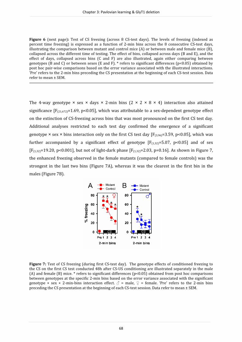
Chapter 3: Pavlovian learning & GlyT1 deletion
68
Figure 6 (next page): Test of CS freezing (across 8 CS-test days). The levels of freezing (indexed as
percent time freezing) is expressed as a function of 2-min bins across the 8 consecutive CS-test days,
illustrating the comparison between mutant and control mice (A) or between male and female mice (B),
collapsed across the different time of testing. The effect of bins, collapsed across days (B and E), and the
effect of days, collapsed across bins (C and F) are also illustrated, again either comparing between
genotypes (B and C) or between sexes (E and F). * refers to significant differences (p<0.05) obtained by
post hoc pair-wise comparisons based on the error variance associated with the illustrated interactions.
‘Pre’ refers to the 2-min bins preceding the CS presentation at the beginning of each CS-test session. Data
refer to mean ± SEM.
The 4-way genotype × sex × days × 2-min bins (2 × 2 × 8 × 4) interaction also attained
significance [F(21,672)=1.69, p<0.05], which was attributable to a sex-dependent genotype effect
on the extinction of CS-freezing across bins that was most pronounced on the first CS test day.
Additional analyses restricted to each test day confirmed the emergence of a significant
genotype × sex × bins interaction only on the first CS test day [F(3,96)=3.59, p<0.05], which was
further accompanied by a significant effect of genotype [F(1,32)=5.07, p<0.05] and of sex
[F(1,32)=19.20, p<0.001], but not of light-dark phase [F(1,32)=2.03, p=0.16]. As shown in Figure 7,
the enhanced freezing observed in the female mutants (compared to female controls) was the
strongest in the last two bins (Figure 7A), whereas it was the clearest in the first bin in the
males (Figure 7B).
Figure 7: Test of CS freezing (during first CS-test day). The genotype effects of conditioned freezing to
the CS on the first CS test conducted 48h after CS-US conditioning are illustrated separately in the male
(A) and female (B) mice. * refers to significant differences (p<0.05) obtained from post hoc comparisons
between genotypes at the specific 2-min bins based on the error variance associated with the significant genotype × sex × 2-min-bins interaction effect. ♂ = male, ♀ = female. ‘Pre’ refers to the 2-min bins
preceding the CS presentation at the beginning of each CS-test session. Data refer to mean ± SEM.

Chapter 3: Pavlovian learning & GlyT1 deletion
69
Figure 8: Modification of CS freezing by sex and light-dark phase across the 8 CS-test days. The effects of
sex, collapsed across other factors, was illustrated separately for animals tested in the light phase (A) and
in the dark phase (B). * refers to significant differences (p<0.05) based on the error variance associated
with the significant light-dark phase × sex × days interaction. � = light phase, � = dark phase. Data refer
to mean ± SEM.
Evidence for an interaction between sex and light-dark phase was supported by a significant
light-dark phase × sex × days interaction [F(7,224)=2.10, p<0.05], which was attributed to the sex
effect across days being stronger in animals tested in the dark phase than those tested in the
light phase (Figure 8). Consistent with this interpretation, a significant sex × days interaction
was only detected in a supplementary analysis restricted to the dark phase [F(7,112)=12.65
p<0.001], but not when restricted to the light phase [p=0.2].
● Spontaneous Recovery of CS freezing between consecutive CS sessions: The
freezing response to the CS in the initial bin tended to be higher to that seen in the last bin of the
previous CS test (see Figure 6A). This effect represents a partial recovery from the previous
day’s extinction learning, and is commonly referred to as ‘spontaneous recovery’. As extinction
was progressively consolidated over test days, the magnitude of spontaneous recovery also
weakened. However, it appeared that the spontaneous recovery effect was somewhat stronger
and more persistent in mutant mice. To specifically gauge spontaneous recovery between days,
a difference score was computed contrasting the magnitude of freezing of the first CS bin of a
given test day to the last CS bin of the preceding day. The difference scores were then subjected
to a 2 × 2 × 2 × 7 (genotype × sex × light-dark phase × days) ANOVA, which yielded a main effect
of days [F(1,32)=12.04, p<0.01] (see Figure 9A) and of genotype [F(1,32)=4.66, p<0.05] (see Figure
9B), thus confirming the initial visual impression derived from Figure 6A. No other effects
attained or approached statistical significance.

Chapter 3: Pavlovian learning & GlyT1 deletion
70
Figure 9: Spontaneous Recovery effect from the second to the last CS-test day. Comparison between the
spontaneous recovery of the mutant and control mice from the second CS-test and onwards, collapsed
across the other factors as a function of days (A), or averaged across CS-test days 2 to 8 (B). Spontaneous
recovery between successive CS-tests was indexed by a difference score = (levels of freezing during the first 2-min bins of dayN ─ levels of freezing during last 2-min bin of dayN-1). A positive value refers to the
spontaneous recovery effect. Data refer to mean± SEM.
Pavlovian appetitive conditioning
● Acquisition: The animals were subjected to CS-US conditioning for 14 days.
Conditioning was evident in all groups as indicated by the progressive increase in magazine
approaches during the CS presentation relative to the pre-CS period (Figure 10). A 2 × 2 × 2 × 7
(genotype × sex × light-dark phase × 2-day blocks) revealed only a significant effect of blocks
[F(6,234)=37.05, p<0.001].
Figure 10: The acquisition and extinction of Pavlovian appetitive conditioning. Conditioned approach
response was indexed by comparing the frequency of nose pokes during each 5-s CS presentation with
the 5-s pre-CS period using the ratio: CS nose pokes / (CS nose pokes + Pre-CS nose pokes). The genotype
effect across 2-day blocks is separately illustrated for the factor sex and light-dark phase (A-D). The 7
blocks of acquisition are demarcated by a vertical line from the last 4 blocks of extinction. The dotted line represents the level of chance performance. � = light phase, � = dark phase, ♂ = male, ♀ = female. Data
refer to mean ± SEM.

Chapter 3: Pavlovian learning & GlyT1 deletion
71
● Extinction: Next, the animals were subjected to eight daily extinction sessions
when the tone CS was no longer followed by the reward US. There was a progressive reduction
in the conditioned approach response, constituting extinction learning (Figure 10). A 2 × 2 × 2 ×
4 (genotype × sex × light-dark phase × 2-day blocks) ANOVA revealed a significant blocks effect
[F(3,117)=31.42, p<0.001]. A sex-dependent genotype effect was evident by the emergence of a
genotype × sex × blocks interaction [F(3,117)=3.10, p<0.05]. As shown in Figures 11A-B, female
mutants showed a trend towards resistance to extinction compared with female controls, while
a weak and opposite trend was observed in the male mutants. Given that the critical 3-way
interaction was solely attributed to the linear component of blocks [F(1,39)=10.37, p<0.005],
accounting for 97% of the variance explained by the interaction, further post hoc comparisons
of the linear rate of extinction were conducted, which confirmed the impression that the
mutation resulted in opposite effects on the linear rate of extinction across days between sexes
(see Figure 11C).
Figure 11: The acquisition and extinction of Pavlovian appetitive conditioning. The genotypic difference
over the course of the appetitive experiment (acquisition and extinction) is illustrated separately for male
(A) and female (B) mice, collapsed across the other factors. Emergence of a significant genotype × sex × 2-
day block interaction over the four extinction days was explained predominately by the linear trend in
blocks. The linear component was calculated and the (genotype by sex) group average is illustrated in (C),
which presents the original three-way interaction. ♂ = male, ♀ = female. Data refer to mean ± SEM.
Shock sensitivity
A separate cohort of female mice maintained under reversed cycle was used for the assessment
of shock sensitivity. This was chosen because this particular sex/light-dark cycle combination
resulted in the largest effect size of genotype in the conditioned freezing experiment (see
Discussion later). Analysis of peak response (Figure 12A) as well as time to reach the peak
response (Figure 12B) recorded in the startle chambers did not yield any genotype effect across

Chapter 3: Pavlovian learning & GlyT1 deletion
72
the entire range of electric shocks (0.045 – 0.255mA) examined. A separate 2 × 9 (Genotype ×
Shock intensity) ANOVA only revealed a main effect of shock intensity [peak response
magnitude: F(8,104)=17.37, p<0.001; time to peak response: F(8,104)=12.20, p<0.001].
Figure 12: Reactivity to shock. Reactivity to electric foot shock measured by peak response of whole body
movement (A) and time to reach the peak response (B) as a function of the shock intensity in a separate
naive cohort of female mutant and control mice tested during the dark phase. Data refer to mean± SEM.
DISCUSSION
In our attempt to scrutinize the reported enhanced Pavlovian learning phenotype following
forebrain neuronal GlyT1 deletion, we uncovered that the phenotypic expression was
distinguishable between the sexes as demonstrated in the aversive as well as in the appetitive
paradigms of Pavlovian conditioning employed here. These findings were free from any
confounding genotypic effect on pain sensitivity (Figure 12) and spontaneous activity measured
in the open field (Figure 3) or in the elevated plus maze. In the elevated plus maze test, we
further identified a hitherto unknown phenotype in anxiety-related behaviour through
extending the test to the light phase. This finding agrees with the anxiolytic profile of the GlyT1
inhibitor SSR504734 which was obtained in male animals tested during the light phase
(Depoortère et al., 2005). At the same time, the lack of such anxiety effects in the dark phase
agrees with previous reports based solely on results obtained in this phase (Yee et al., 2006),
and therefore anxiety did not confound the previously established phenotypes (Dubroqua et al.,
2010; Singer et al., 2007, 2009; Yee et al., 2006). This new finding in the elevated plus maze may
be interpreted as a state-dependent genotypic effect, revealed here by the explicit comparison
between mice kept in opposite light-dark cycles but tested at the same time under identical test
conditions. In contrast, the Pavlovian phenotypes were essentially unaffected by the light-dark
cycle, and against which evidence for sex-dependency was obtained.

Chapter 3: Pavlovian learning & GlyT1 deletion
73
Crossing the Sex Boundary
Jazin and Cahill (2010) have summarized many sex differences in the phenotypic expression of
a host of genetically manipulated mice. Here, the sex comparison allowed us to specify the form
and the extent to which the Pavlovian phenotype associated with forebrain neuronal GlyT1
deletion might differ between sexes. In the aversive paradigm, the phenotypic dependency on
sex was primarily one of magnitude: the phenotype of enhanced conditioned freezing appeared
stronger in the female mutants (Figure 7). As reported before (Yee et al., 2006), this phenotype
was specific to CS-freezing, without any significant impact on context-freezing. Indeed, neither
sex nor light-dark cycle exerted significant effect (Figure 5). However, given that the CS was a
good predictor of shock US here, context-freezing was predictably low in the range of 12%,
hence interpretation of these null effects should be exercised with caution due to possible floor
effects. Lack of statistical power was unlikely of a concern here because the effective sample size
in each of these contrasts was large (with sample size around 20 per sex, genotype or light-dark
cycle). By contrast, sex critically determined the direction of the Pavlovian learning phenotype
In the appetitive paradigm. The conditioned approach response was similarly enhanced in the
mutant females, but was surprisingly somewhat weakened in male mutants (Figure 11C).
Overall our results suggest that the enhanced Pavlovian learning phenotype appeared more
consistent and robust in females than males.
Evidence for sex-dependent phenotypic expression was obtained regardless of whether
sex exerted an effect by itself on performance. Across the different stages (acquisition, context-
test and extinction) of aversive conditioning, female mice were consistently showing a stronger
freezing response than male mice (Figure 6D; in keeping with previous mice studies e.g.,
Wiltgen et al., 2005; Yee et al., 2004; but also see Dalla & Shors, 2009). Our data therefore
concurred with the faster extinction rate in the female rats in fear conditioning reported by
Milad et al. (2009), but did not replicate their observation of weaker conditioning in female rats
during acquisition. Notably, the latter effect was weak and indeed non-significant. We observed
instead stronger conditioning in females although this emerged only on the 3rd CS presentation
and this minor divergence might stem from species differences. By contrast, no such sex
difference was apparent throughout the appetitive conditioning experiment. The apparent sex
effect in conditioned freezing experiment (i.e. generally stronger freezing in female mice)
cannot be attributed to sex differences in spontaneous activity given that the female mice were
more active than the male mice (Figure 3B). If anything, this sex difference in spontaneous
activity would under- rather than over-estimate freezing performance.
Another possible confound that might be expected to influence expression of the
conditioned freezing response, but not the conditioned approach responses was general

Chapter 3: Pavlovian learning & GlyT1 deletion
74
anxiety. The anxiety phenotype demonstrated in the elevated plus maze test was clearly
determined by the combined influence of sex and light-dark cycle. Forebrain neuronal GlyT1
deletion appeared to be anxiolytic in males but anxiogenic in females, thus effectively reversing
the sex-difference seen in control mice (Figure 2); and this cross interaction was only evident in
the light phase. To what extent could this pattern of results in anxiety-related behaviour explain
the sex-dependent outcomes seen in the Pavlovian aversive conditioning experiment?
Based on the premise that the expression of conditioned freezing depends on the
development of conditioned fear in response to the CS, anxiousness to threatening stimuli in
general might promote aversive associative learning. Hence, an anxious phenotype in female
mutants might be linked to the conditioned freezing phenotype in the same animals. However,
this account was undermined by two considerations. First, the conditioned freezing phenotype
did not significantly depend on light-dark cycle; and if anything, it was the strongest in female
mice tested in the dark phase of the cycle (kept in reversed light-dark cycle). Second, this
account should also predict an opposite conditioned freezing phenotype in the male mutants,
which was obviously not the case even though the magnitude of the enhanced freezing
phenotype was visually weaker in the male than the female mutants. As summarized in Table 2,
the critical effect sizes associated with the relevant genotype effect in the aversive conditioning
experiment was highest in the female/dark-phase combination, when the comparison between
mutants and controls yielded no difference in elevated plus maze behaviour (Figure 2). Indeed,
the combination of a null effect in anxiety and enhanced conditioned freezing replicated our
previous report (Yee et al., 2006). Re-calculation of the effect size from the original data
reported by Yee et al. (2006) yielded a value that is closer to that obtained here in animals of the
same sex and tested in the same phase (see Table 2).

Chapter 3: Pavlovian learning & GlyT1 deletion
75
Table 2. Effect size (ηp2) of the genotype derived from the restricted analysis for each combination of sex
and light-dark phase in the aversive Pavlovian paradigms and from the non pre-exposed group of the
previous aversive conditioning experiment (Yee et al., 2006). Effect size provides a measure of the
magnitude of a treatment effect, this index is independent of sample size and allowed us to directly
compare each of the sex and phase of testing combination used. The largest effect size was found in the
female mice tested during the dark phase of the light-dark cycle. The effect size of the previously reported
genotype effect on aversive conditioning, with female mice tested during the dark phase was also, larger
than the other sex/phase combinations in the present study.
Experiments
♀ ♂
� � � �
Aversive conditioning 0.09 0.45 0.03 0.16
Aversive conditioning (Yee et al., 2006) n/a 0.29 n/a n/a
The evidence thus did not support an emotional account of the conditioned freezing phenotype,
but favoured instead a cognitive interpretation. Importantly, the cognitive perspective can
readily accommodate the enhanced conditioned approach phenotype seen in the female
mutants in the appetitive Pavlovian paradigm, which shared a similar associative learning
mechanism (CS-US association), but did not involve any fear-related emotion.
Lost and Found in Translation across Paradigms
The inclusion of the appetitive conditioning experiment was highly instructive and meaningful,
because it provided an alternative test with similar cognitive demand but differed in several
psychological as well as procedural aspects. Appetitive conditioning is based on a motivational
system antagonistic to that in aversive conditioning (Konorski, 1976) and is therefore ideally
suited to test the generality of the reported Pavlovian phenotype that, so far, has been
demonstrated solely in associative learning paradigms using aversive unconditional stimulus
(Yee et al., 2006).
While this phenotype of enhanced conditioning was translatable to the current
appetitive paradigm in the female sex, the results obtained in the male sex were surprising, as
they yielded somewhat opposite effects between paradigms. The latter does defy an
interpretation simply based on enhanced associative strength. Indeed, such a simple account is
already insufficient to explain the original finding that forebrain neuronal GlyT1 deletion
Chapter 3: Pavlovian learning & GlyT1 deletion
76
enhanced the latent inhibition effect (i.e., the sensitivity to a CS’s history of non-reinforcement
prior to conditioning; Yee et al., 2006). Thus, there are situations in which the mutant mice were
more sensitive to the negative modulation of learning and/or expression of learned behaviour.
There was no evidence here to suggest that acquisition of the CS-US association was
facilitated across the three tone-shock pairing trials in the aversive conditioning experiment
(Figure 4C). However, freezing recorded during the inter-trial intervals was enhanced in the
mutants, with a notable difference emerging in the last two intervals – i.e., after the second and
third tone-shock pairing (Figure 4F). Unlike the development of conditioned freezing, the
acquisition of the (appetitively motivated) conditioned approach response is a relatively
protracted process allowing a closer examination of its development over many CS-US trials
conducted across days. The appetitive conditioning experiment here consolidated the lack of an
effect on conditioned responding throughout the entire acquisition phase (Figures 10 & 11).
The present study also showed that, irrespective of conditioning paradigm, the
Pavlovian phenotype was confined to the extinction phase – when the animals were confronted
with extended/repeated CS’s that was no longer followed by any US. This result suggests that
forebrain neuronal GlyT1 deletion robustly modified the expression rather than the acquisition
of the critical CS-US association. In the appetitive paradigm, evidence for enhanced conditioned
responding seen in the female mutants only emerged across daily extinction tests (Figure
11B,C). Although the direction of the weaker effect seen in male mutants was in the opposite
direction here (Figure 11A,C), the phenotypic expression was also confined to the extinction
phase. In the conditioned freezing experiment, the phenotype remained visible across the eight
daily extinction tests (Figure 6A), and the sex-dependent phenotypic expression was a
quantitative one as shown in the first extinction test (Figure 7). Hence, the unique phenotypic
element that is translatable across paradigms refers to the increased resistance to extinction in
female mutants. This element was detected across days in the appetitive conditioning
experiment, but solely on the first test day in the aversive conditioning experiment. Although
not seemingly a sex-dependent effect, evidence that extinction learning across extinction days
was also modified by GlyT1 deletion was obtained when we examined spontaneous recovery of
the conditioned freezing response between successive tests (Figure 9). Overall, spontaneous
recovery, attributed to the dissipation of inhibitory mechanisms responsible for extinction
(Rescorla, 2004), was stronger in the mutants – the daily re-emergence of the CS evoked a
stronger initial response in the mutants.
Taken together, extinction learning is most robustly affected by GlyT1 deletion in the
female sex. Although we cannot explain why this phenotype might be weaker or somewhat
reversed in the male sex when switched to the appetitive paradigm, this conclusion may be
considered as being consistent with the previous finding in the female mutants that latent

Chapter 3: Pavlovian learning & GlyT1 deletion
77
inhibition (LI) was enhanced (Yee et al., 2006). The demonstration of LI requires two distinct
phases with contrasting CS-US relationships: the pre-exposure phase in which CS is not followed
by any significant event [CS → nothing] precedes the conditioning phase. LI refers to the
observation that CS pre-exposed subjects show weaker conditioned response (CR) to the CS
after CS-US pairing compared to non-pre-exposed subjects. Several theories suggest that the
weaker CR does not stem from a failure to acquire the critical CS-US association but reflects the
[CS → nothing] memory trace competing over the control of behaviour during testing (Bouton,
1993; Kraemer and Spear, 1991, 1993; Weiner, 1990). Thus, a stronger influence of the prior
experience of non-reinforced CS exposures leads to stronger LI. Procedurally, extinction is
exactly the reverse of LI, whereby [CS → nothing] follows [CS → US]. Thus, the phenotype of
increased resistance to extinction could be similarly understood as a stronger influence of the
prior experience of [CS → US] over current control of responding. The mutant mice appeared to
be biased towards what they have learned first about the CS, and this might be described as a
stronger primacy effect or proactive interference in memory terms. This contradicts however
the finding that forebrain neuronal GlyT1 disruption enhanced rather than impaired reversal
learning (Singer et al., 2009), as it has been taken to suggest resistance rather than vulnerability
to proactive interference.
In discrimination learning, animals not only learn the opposing reward valence
associated with the discriminanda provided by the experimenter, but they also learn to
dichotomize their responses (approach vs avoidance) efficiently by focusing attention on one or
few distinguishable dimensions/features separating the discriminanda. Achieving the latter
would facilitate reversal learning, because the animals need only to reverse the contingency
related to the few attended dimensions (Sutherland and Mackintosh, 1971). This has been
offered as an explanation of the over-training reversal effect, whereby over-training
paradoxically enhances subsequent reversal learning (Reid, 1953). Hence, it is conceivable that
the reversal phenotype reported by Singer et al. (2009) reflects primarily enhanced selective
attention learning, whereas the extinction phenotype here reflects a stronger primacy effect in
memory recall.
The hypothesized impact on memory expression and selective attention are not
mutually exclusive, even though they might be expected to yield opposing effects on specific
paradigms, e.g., in discrimination reversal. When the separate effects act in the same direction
then the observed behavioural effect would be substantial. The LI effect which is expected to be
potentiated by a susceptibility to the primacy effect (Postman and Phillips, 1965) as well as
enhanced attentional learning (e.g. Sutherland and Mackintosh, 1971), was highly sensitive to
forebrain neuronal GlyT1 deletion (Yee et al., 2006). However, the possible sex and paradigm
dependency of the LI phenotype awaits further evaluation.

Chapter 3: Pavlovian learning & GlyT1 deletion
78
Separating the light from the dark
Sex is strictly a between-subject factor. The factor referring to the contrast between light and
dark phases, on the other hand, ought to be interpreted as a potential within-subject factor
referring to the different states (of wakefulness) phase-locked with the diurnal cycle. For
practical reasons, it was evaluated here in a between-subject design by shifting the light-dark
cycles between two animal keeping rooms by 12h, thus enabling us to conduct all tests at the
same absolute time. This approach allowed a more effective approximation to the contrast
between laboratories that systematically keep their animals either in one or the other cycle.
This contrast did not yield any substantial impact on the measures of conditioning behaviour
here. Notably, even when it was effective in eliciting a sex-dependent phenotype in the elevated
plus maze in the mutant mice, it was without an effect in the controls (Figure 2). Contrary to
some previous studies, we did not observe an increase in spontaneous locomotor activity in the
open field in mice tested in the dark phase in comparison with those tested in the light phase. If
anything, a transient effect in the opposite direction was recorded (Figure 3A). However,
increased activity was evident in the animals tested in the dark phase during the CS test of the
aversive conditioning experiment in the form of reduced freezing, and this was already evident
in the pre-CS phase. When the subsequent CS-freezing score on each day was corrected for
baseline pre-CS freezing, the main effect of light-dark phases on CS-freezing remained
significant [F(1,32)=6.94, p<0.02], without altering any other significant effects in the original
analysis. Therefore, the contrast between light-dark phases did appear to exert an effect on
conditioned freezing rather than affecting the general propensity to freeze as such. Finally, the
contrast between light-dark phases did not reveal any impact on behaviour in the appetitive
conditioning test. Thus, Pavlovian learning of specific CS-US association in general seems to be
relatively stable across the light-dark phases, with limited impact on the magnitude (effect size)
of the conditioned freezing phenotype identified in our mutant mice. Our results do not readily
support the general assertion that behavioural testing in the dark phase is generally more
appropriate than in the light phase solely on the basis that the former represents the active
phase in rodents (Marques & Waterhouse, 1994). Yet, caution should be exercised when
interpreting ethologically based behavioural tests, such as the elevated plus maze test of
anxiety, and other such tests that might be highly sensitive to wakefulness or circadian
fluctuation of hormonal activity.

Chapter 3: Pavlovian learning & GlyT1 deletion
79
Conclusions
In our attempt to test the generality and the robustness of a phenotype on conditioned freezing
behaviour previously reported in mice with forebrain neuronal GlyT1 deletion, the present
study has revealed (i) an hitherto unknown anxiety-related phenotype, and (ii) determinants
that may influence the magnitude and perhaps even the direction of a Pavlovian learning
phenotype in this particular genetic mouse model. Our results suggest the interaction between
genomic (sex difference) and environmental (light-dark cycle) factors critically determined the
expression of the anxiety phenotype, but sex-dependency predominates over the Pavlovian
phenotype. A satisfactory neurobiological account of the emergence of these phenotypes must
therefore take into account such specifications revealed here.
REFERENCES
Austin A. J., & Duka T. (2010). Mechanisms of attention for appetitive and aversive outcomes in Pavlovian
conditioning. Behav. Brain Res., 213, 19-26
Beeler, J. A., Prendergast, B., & Zhuang, X. (2006). Low amplitude entrainment of mice and the impact of
circadian phase on behaviour tests. Physiol. Behav. 87, 870–880.
Black, M. D., Varty, G. B., Arad, M., Barak, S., De Levie, A., Boulay, D., et al. (2009). Procognitive and
antipsychotic efficacy of glycine transport 1 inhibitors (GlyT1) in acute and neurodevelopmental
models of schizophrenia: latent inhibition studies in the rat. Psychopharmacology (Berl), 202, 385-
396.
Bouton M. E. (1993). Context, time, and memory retrieval in the interference paradigms of Pavlovian
learning. Psychol Bull, 114(1), 80-99. Caldarone B. J., Duman C. H., & Picciotto M. R. (2000). Fear
conditioning and latent inhibition in mice lacking the high affinity subclass of nicotinic
acetylcholine receptors in the brain. Neuropharmacology, 39, 2779-2784.
Dalla C., & Shors T. J. (2009). Sex differences in learning processes of classical and operant conditioning.
Physiol. Behav., 97, 229-238. Depoortere, R., Dargazanli, G., Estenne-Bouhtou, G., Coste, A.,
Lanneau, C., Desvignes, C., et al. (2005). Neurochemical, electrophysiological and pharmacological
profiles of the selective inhibitor of the glycine transporter-1 SSR504734, a potential new type of
antipsychotic. Neuropsychopharmacology, 30, 1963-1985.
Dubroqua S., Singer P., Boison D., Feldon J., Möhler H., & Yee B.K. (2010). Impacts of forebrain neuronal
glycine transporter 1 disruption in the senescent brain: Evidence for age-dependent phenotypes in
Pavlovian learning. Behav. Neurosci., 124, 839-850.
Hossain S. M., Wong B. K., & Simpson E. M. (2004). The dark phase improves genetic discrimination for
some high throughput mouse behavioral phenotyping. Genes Brain Behav., 3, 167-177.
Ingram D. K., Shimada A., Spangler E. L., Ikari H., Hengemihle J., Kuo H., et al. (1996) Cognitive
enhancement. New strategies for stimulating cholinergic, glutamatergic, and nitric oxide systems.
Ann. N. Y. Acad. Sci.,786,348-361
Jazin E., Cahill L. (2010) Sex differences in molecular neuroscience: from fruit flies to humans. Nat. Rev.
Neurosci., 11:9-17. Knapska E., Walasek G., Nikolaev E., Neuhäusser-Wespy F., Lipp H. P.,
Kaczmarek L., et al. (2006). Differential involvement of the central amygdala in appetitive versus
aversive learning. Learn. Mem., 13, 192-200.
Konorski, J. (1967). Integrative activity of the brain. An interdisciplinary approach, The University of
Chicago Press, Chicago and London.
Kraemer P. J., & Spear N. E. (1991). The effect of nonreinforced stimulus exposure on the strength of a
conditioned taste aversion as a function of retention interval: Do latent inhibition and extinction
involve a shared process. Animal Learning and Behavior, 20, 1-7.
Kraemer P. J., & Spear N. E. (1993). Retrieval Processes and Conditioning. In: T.R. Zentall (Ed), Animal
Cognition. A tribute to Donald A. Riley (pp. 87-110), New Jersey: Lawrence Erlbaum.
Marques M. D., & Waterhouse J. M. (1994). Masking and the evolution of circadian rhythmicity.
Chronobiol. Int., 11, 146-155.

Chapter 3: Pavlovian learning & GlyT1 deletion
80
Martin, S. J., Grimwood, P. D., & Morris, R. G. (2000). Synaptic plasticity and memory: an evaluation of the
hypothesis. Annu. Rev. Neurosci., 23, 649-711.
Milad, M. R., Igoe, S. A., Lebron-Milad, K., Novales, J. E. (2009) Estrous cycle phase and gonadal hormones
influence conditioned fear extinction. Milad MR, Igoe SA, Lebron-Milad K, Novales JE. Neuroscience,
164, 887-895.
Morris, R. G. (1989). Synaptic plasticity and learning: selective impairment of learning rats and blockade
of long-term potentiation in vivo by the N-methyl-D-aspartate receptor antagonist AP5. J.
Neurosci., 9, 3040-3057.
Morris, R. G., Anderson, E., Lynch, G. S., & Baudry, M. (1986). Selective impairment of learning and
blockade of long-term potentiation by an N-methyl-D-aspartate receptor antagonist, AP5. Nature,
319, 774-776.
Pavlov, I. P. (1927). Conditioned reflexes. London: Oxford University Press.
Postman, L. & Phillips, L. W. (1965). Short-term temporal changes in free recall. Quarterly journal of
experimental psychology, 17, 132-138
Reid L. S. (1953). The development of noncontinuity behavior through continuity learning. J. Exp.
Psychol., 46, 107-112.
Rescorla R. A. (2004). Spontaneous recovery. Learn. Mem., 11, 501-509.
Richmond, M. A., Murphy, C. A., Pouzet, B., Schmid, P., Rawlins, J. N. P., & Feldon, J. (1998). A computer
controlled analysis of freezing behaviour. J. Neurosci. Methods, 86, 91-99.
Roedel, A., Storch, C., Holsboer, F., & Ohl, F. (2006). Effects of light or dark phase testing on behavioural
and cognitive performance in DBA mice. Lab. Anim. 40, 371–381.
Singer, P., Boison, D., Mohler, H., Feldon, J., & Yee, B. K. (2007). Enhanced recognition memory following
glycine transporter 1 deletion in forebrain neurons. Behav. Neurosci., 121, 815-825.
Singer, P., Boison, D., Mohler, H., Feldon, J., & Yee, B. K. (2009). Deletion of glycine transporter 1 (GlyT1) in
forebrain neurons facilitates reversal learning: enhanced cognitive adaptability? Behav. Neurosci.,
123, 1012-1027.
Sutherland, N. S., & Mackintosh, N. J. (1971). Mechanisms of animal discrimination learning. New York:
Academic Press.
Weiner I. (1990). Neural substrates of latent inhibition: the switching model. Psychol. Bull., 108, 442-461.
Wiltgen B. J., Sanders M. J., Ferguson C., Homanics G. E., & Fanselow M. S. (2005). Trace fear conditioning
is enhanced in mice lacking the delta subunit of the GABAA receptor. Learn. Mem., 12, 327-333.
Yee, B. K., Balic, E., Singer, P., Schwerdel, C., Grampp, T., Gabernet, L., et al. (2006). Disruption of glycine
transporter 1 restricted to forebrain neurons is associated with a procognitive and antipsychotic
phenotypic profile. J. Neurosci., 26, 3169-3181.
Yee, B. K., Hauser, J., Dolgov, V. V., Keist, R., Mohler, H., Rudolph, U., et al. (2004). GABA receptors
containing the alpha5 subunit mediate the trace effect in aversive and appetitive conditioning and
extinction of conditioned fear. Eur. J. Neurosci., 20, 1928-1936.
Zucker I, & Beery A. K. (2010). Males still dominate animal studies. Nature, 465, 690.
81
CHAPTER 4
Glycine transporter 1 deletion enhances sensitivity to CS-
US contiguity and CS-US contingency:
Two phenotypes, one psychology?
With Philipp Singer, Detlev Boison, Hanns Möhler, Joram Feldon and Benjamin K. Yee
Submitted to Learning & Memory

Chapter 4: GlyT1 & Associative Learning
82
ABSTRACT
Mutant mice with conditional deletion of glycine transporter 1 (GlyT1) in forebrain
neurons exhibit enhanced Pavlovian conditioning. This phenotype seems to reflect an
alteration in the selectivity of learning rather than a broad strengthening of CS-US
associative strength. Evidence against the latter is derived from the observation that the
latent inhibition effect was potentiated in this mutant mouse line, suggesting that these
mice were more sensitive to a history of non-reinforcement of a potential CS. Here, we
extended this hypothesis by examining the response of these engineered mice to two
other major determinants of Pavlovian associative learning. To compare with the
conditioning procedure in which the US always followed the CS immediately, we either (i)
inserted a 'trace' interval between CS and US presentations to reduce the temporal
contiguity between CS and US, or (ii) added CS-only presentations that were not followed
by US to degrade CS-US contingency. While the impact of these manipulations on the
subsequent conditioned response (CR) to the CS was weak or absent in the control mice,
they strongly reduced CR in the mutant mice. These outcomes are attributable to the
bidirectional effects of the GlyT1 deletion in forebrain neurons on Pavlovian associative
learning: the CR was stronger than in controls when CS was a good predictor of US, but
weaker than in controls when the CS-US contiguity or contingency was degraded. Hence,
an interpretation based on simple modulation of associative strength again failed to
account for these new data sets. Instead, a stronger bias in the selectivity of learning may
offer a unifying account of these diverse phenotypes in the regulation of Pavlovian
learning. These results bear clear relevance to the proposed therapeutic potential of
GlyT1-inhibiting drugs for schizophrenia-related cognitive deficits that might stem from
an underlying lack of selectivity (or inflexibility) in learning.
INTRODUCTION
The N-methyl-d-aspartate receptor (NMDAR) is believed to be central to neuroplastic events
underlying at least some forms of learning and memory (e.g. Collingridge & Bliss, 1995; Morris,
1989; Morris, Anderson, Lynch, & Baudry, 1986), including Pavlovian associative learning.
Molecular interventions designed to boost NMDAR function have been reported to enhance
simple Pavlovian conditioning. A selective increase in NMDAR-mediated (as opposed to AMPA
receptor-mediated) currents can be achieved by altering directly the molecular composition of
the NMDAR (e.g. Tang et al., 1999), or indirectly by disrupting glycine transporter 1 (GlyT1) and
thereby increasing the availability of glycine, an obligatory NMDAR co-agonist, in glutamatergic
synapses (Yee et al., 2006).
Evidence for enhanced Pavlovian learning by GlyT1 disruption restricted to forebrain
neurons is supported by stronger conditioned responding (CR) to a conditioned stimulus (CS)
following its pairing with an unconditioned stimulus (US) across a variety of Pavlovian learning
paradigms, including conditioned freezing, conditioned taste aversion and conditioned active

Chapter 4: GlyT1 & Associative Learning
83
avoidance learning (Yee et al., 2006). However, in each case this effect was coupled with an
increased sensitivity to the CS pre-exposure effect. Prior non-reinforced exposure to the CS is
known to reduce the associability of the CS, reducing the effectiveness of subsequent CS-US
conditioning – a phenomenon known as latent inhibition (LI). Mutant mice with forebrain
neuronal GlyT1 disruption exhibited strong LI under a pre-exposure condition that was
insufficient to generate clear LI in controls. Thus, the impact of the molecular deletion appeared
to be bi-directional. The CR developed under the non-pre-exposed condition was somewhat
enhanced, whereas it was reduced under the pre-exposed condition. Hence, it is apparent that
the phenotype could not satisfactorily be understood as a broad (uni-directional) modulation of
associative strength.
The LI effect represents an important demonstration of selectivity in associative
learning, in the sense that not all potential CSs have an equivalent capacity to form an effective
CS→US association. Learning selectively avoids the acquisition of potential spurious
associations that might not be adaptive, thus affording animals with the ability to focus on the
more informative environmental cues relevant in predicting future outcomes. Learning to
anticipate and to be prepared for future events is the key adaptive value of learning. The
possibility that GlyT1 disruption might generally facilitate selectivity in learning thus warrants
further examination, since this might underlie the anti-psychotic potential of GlyT1 inhibition
against the cognitive inflexibility and deficiency characteristic of schizophrenia.
While LI reflects a modulation of CS associability due to its prior non-reinforced history,
the efficacy of CS-US association is also regulated by other factors. Here, we examined two such
critical determinants by manipulating the contiguity (Experiment 1) or contingency (Experiment
2) between CS and US during conditioning. Contiguity refers to the temporal proximity between
the antecedent CS and the to-be-predicted US. When they are interspersed by a "trace" interval,
and are therefore discontiguous, the subsequent CR is weakened (Pavlov, 1927). According to
the Rescorla-Wagner model (R.A. Rescorla & Wagner, 1972), the trace conditioning effect is
partly attributable to competition from context-US association promoted during the trace
intervals. Alternatively, the trace intervals may further render the CS to be perceived as a safety
signal (Seligman & Binik, 1977). The CS effectively serves as a signal predicting the temporary
absence of US, fostering inhibitory CS-US association that counters the excitatory associative
link that underlies the generation of the CR.
Similarly, CS-US contingency also emphasizes the informativeness of CS in predicting the
US, and it is typically formulated in probabilistic terms (Rescorla, 1968). CS-US contingency is
strongly positive when the probability of US occurring following the CS [P(US|CS)] exceeds the
probability of US occurrence in the absence of an antecedent CS [P(US|~CS)]. Under such
conditions, the CS-US association would be excitatory. Lowering P(US|CS), ceteris paribus,

Chapter 4: GlyT1 & Associative Learning
84
should degrade the CS-US contingency, for instance by embedding CS-alone trials amongst CS-
US trials (without altering the number or the density of US occurrence in time, see Figure 1).
The interspersed CS-alone trials constitute omissions of anticipated US, which according to the
Rescorla-Wagner model would produce a negative change to the CS-US associative strength by
promoting inhibitory CS-US association (Konorski, 1948), thus weakening the overall
associative strength and the intensity of the CR.
The present study examined the impact of forebrain neuronal GlyT1 deletion on the
expression of the trace conditioning effect and contingency degradation in two separate
conditioned freezing experiments. The trace intervals and the degree of CS-US contingency
degradation were deliberately chosen here to yield relatively weak effects in control mice, so as
to allow the demonstration of the enhanced sensitivity to CS-US discontiguity and contingency
anticipated in the mutant mice. In each case, the effect was gauged in comparison to a standard
procedure adopted from Yee et al. (2006) employing three discrete tone-shock pairings in
which the shock US always immediately followed the cessation of the tone CS. Finally, additional
experiments of foreground contextual conditioned freezing (i.e., in the absence of any
competing discrete CS) were also conducted (Experiments 3A and 3B) to allow comparison with
contextual freezing obtained in Experiments 1 and 2 when the context (in the background) was
competing with a discrete CS for potential associative strength to the US.
Figure 1: Schematic representation of the arrangements of tone CS and shock US in the conditioning
phase of the different experiments. The "no-trace/full contingency” conditioning was the control
condition, with which the "trace conditioning" (Experiment 1) and the "partial contingency" conditioning
(Experiment 2) was compared. In "foreground contextual conditioning", no discrete CS was presented,
and either a three-shock (Experiment 3A) or one-shock (Experiment 3B) was investigated here. The
horizontal line illustrated is not to scale. See methods for full descriptions.

Chapter 4: GlyT1 & Associative Learning
85
METHODS
Subjects
A homozygous Glyt1tm1.2fl/fl colony was established and maintained on a pure C57BL/6
background as described before (Gabernet et al., 2005). Forebrain neuron specific deletion of
GlyT1 (hereafter designated as “GlyT1∆fb-neuron”) was achieved by CamKIIαCre-mediated
recombination (see Yee et al., 2006). Appropriate heterozygous Cre mice were mated with
Glyt1tm1.2fl/fl mice to generate the desired mutant and control littermates. Animals of both
sexes were employed in the present study. The mice were weaned at 21 days old, and
littermates of the same sex were kept in groups of four to six in Makrolon Type-III cages
(Techniplast, Milan, Italy). The subjects were housed in a temperature- and humidity-controlled
(at 22°C and 55% R.H.) animal vivarium under a reversed light-dark cycle with lights off from
0700-1900hrs. Testing was always conducted in the dark phase of the cycle. The animals were
maintained under ad libitum water and food (Kliba 3430, Klibamuhlen, Kaiseraugst,
Switzerland) throughout the study. Separate cohorts of naïve mice were used for each
experiment according to a 2 × 2 factorial design (genotype × sex) (see Table 1). All experimental
procedures described had previously been approved by the Zurich Veterinary Office; they also
conformed to the ethical standards stipulated by the Swiss Act and Ordinance on Animal
Protection and were in accordance to the European Council Directive 86/609/EEC.
Table 1: Allocation and number of subjects for each experiment. Experiments were conducted separately,
with controls littermates derived from the same breeding cycle for comparison. Both male and female
mice were included. Experiment 1 and 2 involved an additional between-subject factor referring to the
test condition (see Procedures below).
Experiment Condition
1 Trace conditioning no-trace 12 (6♀ + 6♂) 16 (8♀ + 8♂)
trace 13 (7♀ + 6♂) 17 (10♀ + 7♂)
2 Contingency degradation full contingency 17 (12♀ + 5♂) 17 (12♀ + 5♂)
partial contingency 18 (13♀ + 5♂) 18 (13♀ + 5♂)
3A 3-shock contextual conditioning 12 (6♀ + 6♂) 12 (6♀ + 6♂)
3B 1-shock contextual conditioning 16 (8♀ + 8♂) 15 (7♀ + 8♂)
Mutants Controls

Chapter 4: GlyT1 & Associative Learning
86
Apparatus
The apparatus consisted of two sets of four conditioning chambers. The two sets were distinct
from each other, and were installed in separate testing rooms, providing two distinct contexts
as fully described before (Pietropaolo, Mintz, Feldon, & Yee, 2007; Yee et al., 2006). The first set
of chambers (context ‘A’) comprised four Coulbourn Instruments (Allentown, PA, USA) operant
chambers (Model E10-10), each equipped with a grid floor made of stainless steel rods (4 mm in
diameter) spaced at an interval of 10 mm centre to centre, and through which scrambled
electric shocks (the unconditioned stimulus, US, set at 0.3 mA) could be delivered (model E13–
14; Coulbourn Instruments). A transparent Plexiglas enclosure confined the animals to a
rectangular region (17.5 × 13 cm). The inside of the chambers was illuminated by a house light
(2.8 W) positioned on the panel wall, 21 cm above the grid floor. The second set of chambers
(context ‘B’) comprised four cylindrical (19 cm in diameter) enclosures made of clear Plexiglas
resting on a metal mesh floor. Illumination inside the chamber was provided by an infrared light
source instead of visible light. The conditioned stimulus (CS) in all the experiments was an 86-
dBA tone provided by a sonalert (model SC628; Mallory, Indianapolis, IN, USA). Each of the eight
chambers contained a miniature digital camera mounted 30 cm directly above the centre of the
area of interest. The algorithm of the freezing response detection procedure has been validated
and fully described before (Richmond et al., 1998).
Experiment 1: Trace conditioning
The procedure for trace conditioning followed that described earlier (Yee et al., 2004). Subjects
of each of the four groups (genotype × sex) were randomly allocated into one of two conditions
of CS-US pairing differing in the (trace) interval between CS offset and US-onset: either 0 or 20 s
(‘no-trace’ and ‘trace’ conditions, respectively; Figure 1). The animals were given three trials of
CS–US pairings (CS: 30 sec, US: 1 sec, 0.3mA) in context A on day 1. Each trial was preceded and
followed by a 120 s interval. Context test was performed on the next day by returning the
animals to the training context for a period of 8min. CS freezing to the tone stimulus was
evaluated on the next day (day 3) in the neutral context B where the CS was presented for 8 min
following a 2 min acclimatization period.
Experiment 2: Contingency degradation
Animals of each of the four groups (genotype × sex) were randomly allocated into one of two
conditions of CS-US pairing differing in overall CS-US contingency (full contingency vs. partial
contingency). In the full contingency condition, the shock US always followed immediately at the
end of tone CS, and three such CS-US pairings were administered. Thus, the probability of US
given CS, P(US|CS), is one. In the partial contingency condition, three additional CS-only

Chapter 4: GlyT1 & Associative Learning
87
presentations were intermixed so as to reduce P(US|CS) to 0.5. Figure 1 compares schematized
the contrast between these two CS-US arrangements.
On the day of conditioning (Day 1), three discrete trials of CS-US pairings were
administered at 3, 6.3 and 10 min into the session that lasted for a total of 13 min 33 s. In each
such trial, the CS and US were serially arranged with the termination of the 30-s CS coinciding
with the onset of the 1-s US. Animals in the partial contingency condition received in addition
three CS-alone trials at 4.3, 9 and 11.3 min into the session. On the next day (Day 2), a context-
freezing test was conducted by returning the animals to the training context A for a period of 8
min in the absence of any discrete stimuli. On Day 3, the freezing to the tone was evaluated in
the neutral context B during when the CS was presented for 8 min following an initial
acclimatization period of 2 min without CS.
Experiments 3A and 3B: Context foreground conditioning
The procedures have been fully described by Pietropaolo et al. (2007). Following conditioning
in context A on Day 1, test of context freezing was assessed by placing the animals in contexts A
and B on alternate days (in the order of A-B-A-B across Days 2 to 5), thus allowing the context-
specificity of the freezing response to be evaluated. Each context test lasted for 4 min.
Experiments 3A and 3B differed only with respect to the number of un-signalled shock (0.3mA
for 1s) employed on the conditioning day (see Figure 1). Three shocks were delivered in
Experiment 3A, and only one shock in Experiment 3B. Each shock delivery was preceded and
followed by a 3-min interval.
Statistical analysis
All data were analysed by parametric analysis of variance (ANOVA) with the between-subject
factor Genotype (mutant vs. littermate control). Preliminary analyses also included the
between-subject factor sex; this was dropped from the final analysis to increase statistical
power given that there was no evidence for any interaction effect involving sex. An additional
factor between subject of Trace interval (0s vs. 20s) and Contingency (full contingency vs.
partial contingency) was introduced in the analysis of Experiments 1 and 2, respectively.
Appropriate within-subject factors (time bins, trials, and days) were included according to the
nature of the considered dependent variables. To assist interpretation of the statistical
outcomes, significant effects were further investigated by pair-wise comparisons based on the
associated error terms taken from the overall ANOVA or supplementary restricted analyses
applied to a subset of data. All statistical analyses were carried out using SPSS for Windows
(version 18, SPSS Inc. Chicago IL, USA) implemented on a PC running the Windows 7 operating
system.

Chapter 4: GlyT1 & Associative Learning
88
RESULTS
Experiment 1: Trace conditioning
� CS-freezing (Day 1): The level of freezing during the tone-CS
presentation increased progressively across trials and this increment was noticeably weaker in
the ‘trace’ relative to the ‘no-trace’ condition regardless of the genotype (Figure 2A). These
impressions were confirmed by a 2 × 2 × 3 (genotype × trace × conditioning trials) ANOVA of
percent freezing, which revealed a significant main effect of trace [F(1,54)=10.21; p<0.01], trials
[F(2,108)=34.86; p < 0.01], and their interaction [F(2,108)=8.28; p<0.01]. Post hoc pair-wise
comparison (collapsed across genotype) indicated that the contrast between the trace and the
no-trace condition attained statistical significance in the last two CS presentations [at the
second CS-presentation: t(108)=2.75, p<.001; at the third CS-presentation: t(108)=8.16, p<.0001].
� Pre-CS freezing (Day 1): The freezing observed during the 30 s preceding
the presentation of the CS consistently increased across the successive CS-US pairings (Figure
2B). This was not affected by either genotype or trace condition. Consistent with these
impressions, a 2 × 2 × 3 (genotype × trace × Pre-CS periods) ANOVA of percent freezing only
yielded a significant effect of PreCS-periods [F(2,108)=71.11; p<0.01].
� Trace periods (Day 1): Freezing during the 20-s trace intervals (in-
between CS-US) which were only relevant for the animals in the trace condition was examined
separately (Figure 2C). This variable likewise showed a consistent increase across trials. A 2 × 3
(genotype × trials) ANOVA of percent time freezing revealed only a significant effect of trials
[F(2,56)=36.75; p<0.01]. There was no evidence for a genotype effect [F<1].
Figure 2. Acquisition of the conditioned response across the three CS-US pairing in Experiment 1. Levels
of freezing were expressed as percent time during the 30 s presentation of the CS and are depicted for the
no-trace and the trace conditions (A). Freezing observed during the 30 s preceding each CS presentation
(B) and during the 20 s trace interval for the trace condition only (C) are separately depicted. All group
showed an increase in the freezing response during acquisition of the conditioned response. Error bars
refer to ±SEM.

Chapter 4: GlyT1 & Associative Learning
89
� Context freezing (Day 2): Expression of conditioned fear developed towards the
training context was evaluated 24h after conditioning by re-exposing the animals to the context.
No difference was apparent amongst the four groups of mice (Figure 3). A 2 × 2 (genotype ×
trace) ANOVA of percent time freezing observed in the 8-min period did not reveal any
significant effects [all F’s<1].
Figure 3: Test of context freezing in Experiment 1. The animals were returned to the conditioning context
in the absence of the CS one day after conditioning. The average levels of freezing observed over the 8 min
context-test session were largely comparable amongst the four groups regardless of genotype and trace
condition. Error bars refer to ±SEM.
� Pre-CS & CS freezing (Day 3): Conditioned responding to the tone CS was
evaluated in a novel context 48 h after the initial conditioning session. Freezing was relatively
low and highly comparable across groups in the first 2 min baseline period prior to the
presentation of the CS (Figure 4A). A 2-way (genotype × trace) ANOVA of percent freezing
recorded during the 2min Pre-CS period did not yield any significant effect.
Onset of the CS led to a notable rise in freezing that was substantially weaker in the trace
condition than in the no-trace condition, constituting the trace conditioning effect, and this
effect was visibly more pronounced in the mutant mice (Figures 4A & 4B). The mutant mice
exhibited marginally stronger CS-freezing than controls in the no-trace condition. On the other
hand, the mutant mice exhibited marginally weaker CS-freezing than controls in the trace
condition. The combination of these contrasting impacts of genotype on conditioned freezing
(Figure 4B), led to the emergence of a near-significant genotype by trace interaction
[F(1,54)=3.81; p=0.056] in a 2 × 2 × 4 (genotype × trace × 2-min bins) ANOVA of percent time
freezing across successive 2-min bins during the 8-min CS period. This interaction term was
accompanied by a significant main effect of trace [F(1,54)=33.80; p<0.01], bins [F(3,162)=10.32;
p<0.01], and the three-way interaction [F(3,162)=3.01; p<0.05]. Post hoc pair-wise comparisons

Chapter 4: GlyT1 & Associative Learning
90
were then performed to assess genotypic difference within either the trace or no-trace
condition at each successive bins. These indicated that the mutant mice exhibited stronger
freezing than controls in the no-trace condition across bins 2 and 3 [p<0.05], while the mutant
mice showed weaker freezing than controls in the trace condition during the same two bins
[p<0.05]. These confirmed the graphical impression (Figure 4A) that the trace conditioning
effect was exacerbated in the mutants solely in bins 2 and 3. Supplementary analyses restricted
to each of the four CS bins further revealed the presence of a significant genotype × trace
interaction only in bins 2 and 3 [at bin 2: F(1,54)=5.88; p=0.02; at bin 3: F(1,54)=6.69, p=0.01]
Figure 4: Test of CS freezing in Experiment 1. The levels of freezing observed in the CS test (day 3) are
expressed as a function of 2-min bins. The first bin referred to the initial baseline period prior to CS-onset.
The CS was presented throughout the subsequent four bins in A. Baseline freezing behaviour in the pre-
CS bin did not differ between groups. Next, the trace conditioning effect was evident across all four bins:
animals in the trace condition (red lines) showed weaker freezing than those trained under the no-trace
condition (black lines). However, the mutants (filled symbols) exhibited clearly a stronger trace
conditioning effect than controls (open symbols) during bins 2 to 3 as indicated by post-hoc comparisons
between mutants and controls of either the trace (red # : p<0.05) or no-trace condition (black # : p<0.05).
The interpretation of a stronger trace conditioning effect was also evident in the average freezing level
across all four CS-bins as depicted in B, which was associated with a significant genotype × trace
interaction [p<0.05]. Pair-wise comparison confirmed the overall presence of the trace conditioning effect
in both mutant and control mice [*: p<0.05]. Error bars refer to ±SEM.
Experiment 2: Contingency degradation
� CS-freezing (Day 1): Freezing levels during CS presentation obtained in
the fully contingent and partial contingent training conditions were separately analysed
because the full contingency condition involved three discrete CS presentations (each followed
by a US, i.e., they were reinforced) whereas the partial contingency condition involved
additional three CS presentations (that were not followed by a US). Separate genotype × CS-
presentations ANOVAs indicated that freezing generally increased across successive CS
presentations regardless of training condition and genotype [full contingency condition:
F(2,64)=33.33, p<0.001; partial contingency condition: F(5,170)=14.93, p<0.001] (Figures 5A). As

Chapter 4: GlyT1 & Associative Learning
91
expected, the CS-freezing achieved under the partial contingency training condition was lower
than the full contingency condition as training progressed. This impression was confirmed by an
additional ANOVA comparing directly the two training conditions by focusing on the three-
shared reinforced CS-presentations. This 2 × 2 × 3 (genotype × contingency × CS-presentations)
ANOVA yielded a significant main effect of contingency [F(1,66)=11.49, p<0.01]. This was also
accompanied by a significant main effect of CS-presentations [F(2,132)=49.52, p<0.001], and its
interaction with contingency [F(2,132)=12.30, p<0.001]. Post hoc pair-wise comparison indicated
that the contrast between full and partial contingency only attained statistical significance in the
third reinforced CS-presentation [t(132)=6.49, p<.0001].
Figure 5: Acquisition of the conditioned response across the CS-US pairing in Experiment 2:Percent time
freezing at successive CS presentations is illustrated in A. In the full contingency condition (black lines),
animals experienced three CS presentations, and they were all followed by a US. These CS presentations
are indicated as "CS+". In the partial contingency condition (blue lines), animals experienced six CS
presentations: three were followed by shock US (CS+) which were interspersed by CS-alone presentation
(CS-). The recorded percent time freezing in the 30-s period preceding each CS presentation is depicted in
B. Error bars refer to ±SEM.
� Pre-CS freezing (Day 1): The freezing during the 30 sec preceding
successive CS presentations steadily increased regardless of training conditions and genotype
(Figure 5B). The two training conditions were again separately analysed by using a 2-way
(genotype x PreCS periods) ANOVA differing in the levels of the repeated measures factor. The
main effect of PreCS periods achieved statistical significant in both analyses [full contingency
condition: F(2,64)=6.78, p<0.01; partial contingency condition: F(5,170)=12.76, p<0.01]. As
expected, contingency degradation also had an impact on Pre-CS freezing but in the opposite
direction to that observed in terms of CS-freezing. Thus, animals trained under the partial
contingency condition exhibited stronger Pre-CS freezing as training progressed. This
interpretation was examined by a 2 × 2 × 3 (genotype × contingency × Pre-CS periods) ANOVA
comparing directly the two training conditions focusing on the three Pre-CS periods preceding
the reinforced CS's, which yielded a significant main effect of contingency [F(1,66)=4.87,

Chapter 4: GlyT1 & Associative Learning
92
p<0.05], Pre-CS periods [F(2,132)=35.19, p<0.001], and their interaction [F(2,132)=7.45,
p<0.001]. Post hoc pair-wise comparison indicated that the increase Pre-CS freezing observed in
the partial contingency condition only achieved statistical significance in the Pre-CS period
preceding the third reinforced CS-presentation [t(132)=4.55, p<.0001].
� Context freezing (Day 2): Expression of conditioned fear developed towards
the training context was evaluated 24h after conditioning by re-exposing the animals to the
context. No difference was apparent amongst the four groups of mice (Figure 6) as shown by a 2
× 2 (genotype × contingency) ANOVA of average percent time freezing observed in the 8-min
test period which did not yield any significant effect [all F’s<1].
Figure 6: Test of context freezing in Experiment 2. The animals were returned to the conditioning context
in the absence of the CS one day after conditioning. The average levels of freezing observed over the 8 min
context-test session were largely comparable amongst the four groups (Control/full contingency,
Control/partial contingency, Mutant/ full contingency, and Mutant/ partial contingency). Error bars refer
to ±SEM.
� Pre-CS & CS freezing (Day 3): Expression of conditioned freezing to the tone CS
was assessed on the next day in a neutral context, consisting of an initial 2-min pre-CS period
followed by a period of 8-min CS presentation. Freezing was low and highly comparable across
the four groups in the pre-CS period (Figure 7A). A 2-way (genotype × contingency) ANOVA of
percent freezing recorded during the 2min Pre-CS period did not yield any significant effect.
Onset of the CS led to a clear rise in freezing. The increase was substantially weaker in
mice trained under the partial contingency condition than those trained under the full
contingency condition (Figure 7A). However, this effect of contingency degradation was only
visible in the mutant mice (Figure 7B). The mutant mice were freezing more strongly than
controls in the full contingency condition, whilst they exhibited marginally weaker CS-freezing
than controls in the partial contingency condition (Figure 7B). These bidirectional effects of the
mutation contributed to the emergence of a significant genotype by contingency interaction
[F(1,66)=5.15, p<0.05] in a 2 × 2 × 4 (genotype × contingency × 2-min bins) ANOVA of percent
time freezing across successive 2-min bins during the 8-min CS period. Post-hoc pairwise
comparisons of the average freezing levels across the 8-min test indicated a significant effect of
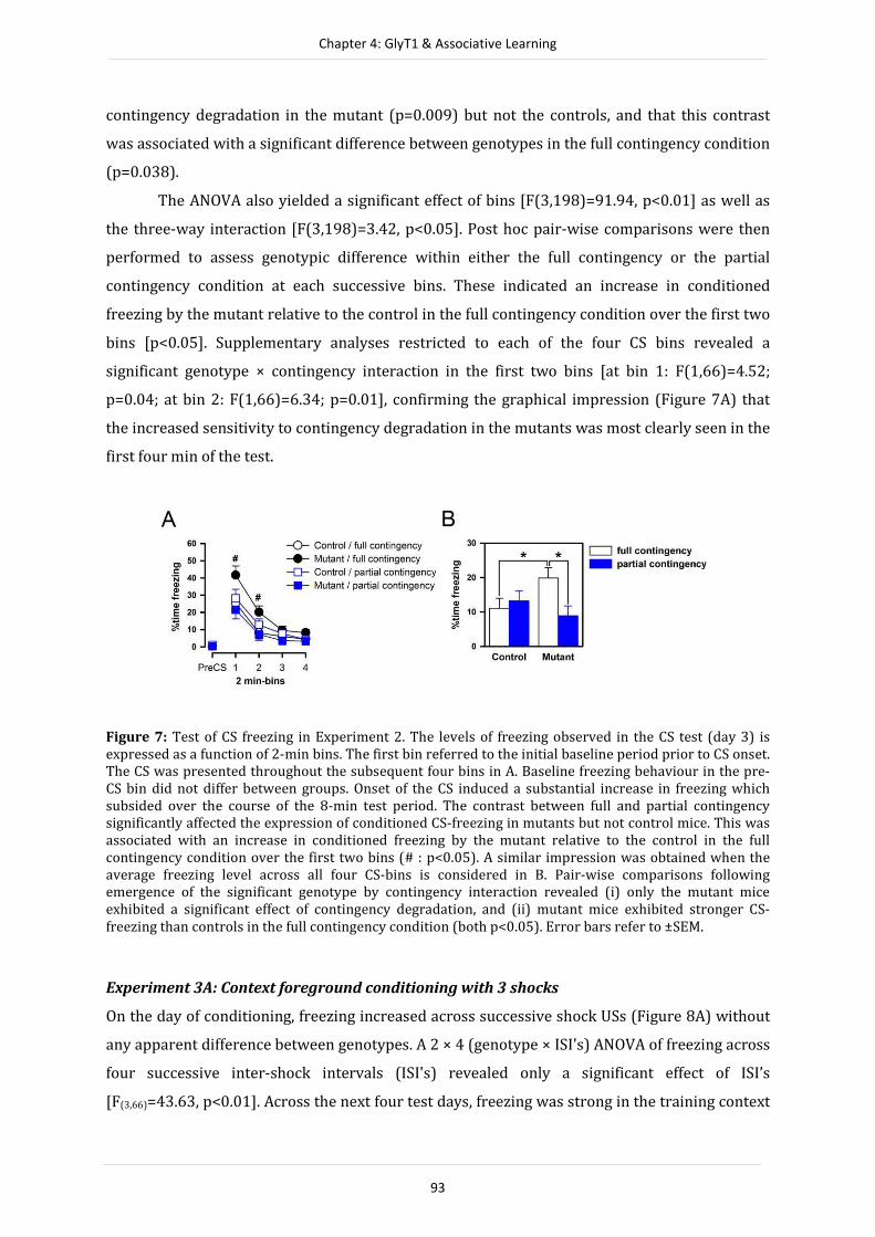
Chapter 4: GlyT1 & Associative Learning
93
contingency degradation in the mutant (p=0.009) but not the controls, and that this contrast
was associated with a significant difference between genotypes in the full contingency condition
(p=0.038).
The ANOVA also yielded a significant effect of bins [F(3,198)=91.94, p<0.01] as well as
the three-way interaction [F(3,198)=3.42, p<0.05]. Post hoc pair-wise comparisons were then
performed to assess genotypic difference within either the full contingency or the partial
contingency condition at each successive bins. These indicated an increase in conditioned
freezing by the mutant relative to the control in the full contingency condition over the first two
bins [p<0.05]. Supplementary analyses restricted to each of the four CS bins revealed a
significant genotype × contingency interaction in the first two bins [at bin 1: F(1,66)=4.52;
p=0.04; at bin 2: F(1,66)=6.34; p=0.01], confirming the graphical impression (Figure 7A) that
the increased sensitivity to contingency degradation in the mutants was most clearly seen in the
first four min of the test.
Figure 7: Test of CS freezing in Experiment 2. The levels of freezing observed in the CS test (day 3) is
expressed as a function of 2-min bins. The first bin referred to the initial baseline period prior to CS onset.
The CS was presented throughout the subsequent four bins in A. Baseline freezing behaviour in the pre-
CS bin did not differ between groups. Onset of the CS induced a substantial increase in freezing which
subsided over the course of the 8-min test period. The contrast between full and partial contingency
significantly affected the expression of conditioned CS-freezing in mutants but not control mice. This was
associated with an increase in conditioned freezing by the mutant relative to the control in the full
contingency condition over the first two bins (# : p<0.05). A similar impression was obtained when the
average freezing level across all four CS-bins is considered in B. Pair-wise comparisons following
emergence of the significant genotype by contingency interaction revealed (i) only the mutant mice
exhibited a significant effect of contingency degradation, and (ii) mutant mice exhibited stronger CS-
freezing than controls in the full contingency condition (both p<0.05). Error bars refer to ±SEM.
Experiment 3A: Context foreground conditioning with 3 shocks
On the day of conditioning, freezing increased across successive shock USs (Figure 8A) without
any apparent difference between genotypes. A 2 × 4 (genotype × ISI's) ANOVA of freezing across
four successive inter-shock intervals (ISI's) revealed only a significant effect of ISI’s
[F(3,66)=43.63, p<0.01]. Across the next four test days, freezing was strong in the training context

Chapter 4: GlyT1 & Associative Learning
94
'A' but remained low in the neutral context 'B' (Figure 8B), confirming the context-specificity of
the conditioned freezing response. A 2 × 2 × 2 (genotype × contexts × days) ANOVA supported
this conclusion with a significant effect of contexts [F(1,22)=47.47, p<0.001]. Freezing on the
second exposure to the training context was visibly lower than the first, reflecting extinction of
this conditioned response. A 2 × 2 (genotype × days) ANOVA restricted to context 'A' yielded a
significant effect of days [F(1,22)=7..25, p<0.05]. In all analyses, there was no evidence for any
genotype effects.
Figure 8: Acquisition and Test of context foreground conditioning in Experiment 3A. Percent time
freezing showed a monotonic increase across successive ISI-periods on day 1 as illustrated in A, with the
arrows representing the three shock US's. As shown in B, context freezing was assessed by returning the
animals to the shocked context (A) on days 2 and 4, in comparison to exposure to a neutral context (B) on
days 3 and 5. Higher levels of freezing in context A relative to that in context B constitutes conditioned
freezing specific to the shocked context. Mutant and control mice remained indistinguishable throughout
the entire experiment. All values refer to mean ± S.E.M.
Experiment 3B: Context foreground conditioning with 1 shock
There was a clear increase in freezing before and after the shock delivery on conditioning day
(Figure 9A) without any difference between mutants and controls. A 2 × 2 (genotype × ISI’s)
ANOVA only revealed a significant ISI's effect [F(1,29)=34.55, p<0.01]. Across the next four test
days, freezing was stronger in the training context 'A' but remained low in the neutral context
'B' (Figure 9B). This confirmed again the context-specificity of the conditioned freezing
response, which is supported by a 2 × 2 × 2 (genotype × contexts × days) ANOVA yielding a
highly significant effect of contexts [F(1,29)=59.29, p<0.001]. The genotype by contexts
interaction also emerged significantly [F(1,29)=5.51, p<0.05] due to stronger freezing exhibited by
the mutant mice in context 'A' [t(29)=2.66, p<0.05] but not context 'B' [t(29)=0.13, p=0.89] (Figure

Chapter 4: GlyT1 & Associative Learning
95
9C) as indicated by post hoc pair-wise comparisons. A 2 × 2 (genotype × days) ANOVA restricted
to context 'A' reaffirmed this conclusion, yielding an overall effect of genotype [F(1,29)=5.29,
p<0.05]. The absence of a significant effect of days here was consistent with the graphical
impression of the lack of extinction across the two tests in context 'A'.
Figure 9. Acquisition and Test of context foreground conditioning in Experiment 3B. A. The animals
showed a sharp increase in percent time freezing after the delivery of the shock US (indicated by the
arrow) in day 1. B. context freezing was assessed by returning the animals to the shocked context (A) on
days 2 and 4, in comparison to exposure to a neutral context (B) on days 3 and 5. C. Higher levels of
freezing in context A relative to that in context B constitutes conditioned freezing specific to the shocked
context. Mutant mice exhibited stronger freezing specifically to context A, although this effect only
achieved statistical significance on the first re-exposure test on day 2. * indicates p<0.05 based on
appropriate post hoc comparison. Values refer to mean ± S.E.M.
DISCUSSION
The present study has extended the previous finding of enhanced latent inhibition (LI) reported
in GlyT1∆fb-neuron mice (Yee et al., 2006) by showing that this conditional GlyT1 deletion model
was further associated with enhanced sensitivity to CS-US trace (Experiment 1, see Figure 4B)
and degradation of CS-US contingency (Experiment 2, see Figure 7B). At the same time, we
replicated the phenotype of enhanced conditioned freezing under the "no-trace" or "contingent"
test condition, which is operationally identical to the "non-pre-exposed" condition in the
original LI experiment reported by Yee et al. (2006). Furthermore, we showed that this
phenotype of enhanced conditioned freezing can be extended to foreground contextual
conditioning (Experiment 3B, see Figure 7B), but this effect seems fragile by comparison.
The joint presence of phenotypes in trace conditioning and contingency degradation in
GlyT1∆fb-neuron mice is of significant relevance to the interpretation of the mutation’s overall
impact on associative learning. As mentioned in the Introduction, a description based on
stronger conditioning as such is untenable. Such an account is particularly problematic in

Chapter 4: GlyT1 & Associative Learning
96
dealing with the trace conditioning results here (Experiment 1) whereby GlyT1∆fb-neuron mice
showed weaker conditioning under the 20-s trace condition. Similarly, there was no evidence
for enhanced conditioning either in the partial contingency condition (Experiment 2) or the CS
pre-exposed conditioning (see Yee et al., 2006). Hence, the test condition decides whether one
may observe an enhancement or conversely a diminution in the intensity of the conditioned
response. We propose here that perceptual learning mechanisms underlying the modulation of
the associability of a potential CS may offer a possible unifying account.
The dynamic of associability
CS associability refers to the perceived properties of a potential CS that determines its readiness
to enter into association with an impending US (Hall, 1991). One critical determinant of CS
associability is the CS’s salience, which refers to its ability to occupy the subject’s focus of
attention. Essentially, formal theories developed in the past accounting for the phenomena of LI,
trace conditioning and contingency degradation are, to varying extent, related to attention-
mediated selectivity in learning under differing Pavlovian learning situations.
All associative learning theories emphasize that attention to the CS critically governs the
rate of CS-US learning. Thus, stronger attention (i.e., CS being more salient) facilitates the
formation and growth of CS-US association. In its simplest form, the Rescorla and Wagner
(1972) model specifies the change of associative strength after successive CS-US pairing based
on a simple error correction rule, and the change in associative strength (ΔVn) after the nth trial
is formally described as:
ΔVn = α (λ ─ V),
where α denotes the associability of the CS, λ refers to the maximum strength that the CS-US
association can achieve and is set by the magnitude of the US, and V stands for the current CS-US
associative strength. The resulting (cumulative) associative strength after the nth trial (Vn) is
therefore:
Vn = Vn-1 + ΔVn
The parameter α is what we are interested in here, and it reflects the intensity,
discriminability, or salience of the CS. α is considered as a fixed property of the CS in the
Rescorla-Wagner’s initial model (1972), but mutant and control might perceived the same
stimulus with different effective α. Figure 10 depicts the consequence of this difference with
αcontrol = 0.5 and αmutant = 0.75. By the end of three CS-US pairings, the cumulative associative

Chapter 4: GlyT1 & Associative Learning
97
strength is higher in the mutant than in the control mice. Most interestingly, application of the
same parameters yielded the opposite outcome in our partial contingency design according to
the Rescorla-Wagner model (1972; Experiment 2): the final associative strength is lower in the
mutant than in the control mice.
Figure 10. The impact of CS Salience on CS-US associative strength under the partial and Fully contingent
conditions in Experiment 2 as predicted by the Rescorla-Wagner model. We applied the Rescorla-Wagner
model to calculate the final associative strength according to the partial or full contingency conditions
employed in Experiment 2 as a function of CS salience, α (in the range of 0.01 to 0.99). λ is set to 1, and the
initial value of CS-US associative strength is set to 0 in these simulations. Increasing α leads to a
monotonic increase in the final associative strength in the full contingency condition, but increasing α is
associated with an inverted U-shape in the partial contingency condition. Hence, beyond a critical value (α = ⅓ in this specific example), any further increase in α result in opposite impact on the final associative
strength. We therefore speculate that the outcome in Experiment 2 might be explicable in similar terms,
namely αmutant > αcontrol. To illustrate this, we assign αmutant = 0.75, and αcontrol = 0.5, and highlighted the corresponding theoretical values for mutant (□) and controls (○) of final associative strength in the graph.
The blue arrow refers to the increase in associative strength when comparing mutant/full contingency
with controls/full contingency condition. The red arrows refers to the decrease in associative strength
when comparing mutant/partial contingency with controls/partial contingency condition.
Chapter 4: GlyT1 & Associative Learning
98
The impact of CS-US discontiguity, as captured by the trace conditioning procedure (Experiment
1), is not easily conceptualized within the Rescorla-Wagner’s model (1972) which does not
readily incorporate the temporal dynamic between CS and US. On the other hand, the
“Sometimes Opponent Process” (SOP) model developed by Wagner (1981) can readily deal with
trace conditioning, and critically, here, offers an interpretation of the trace conditioning effect as
a consequence of the modulation of the subject’s attention to the CS. The model specifies that
the representation of a stimulus can assume different states of activation and this bears direct
consequence on the stimulus’ associability. Briefly, when a CS is presented, its representation is,
at first, strongly activated (A1 state), and can readily form an association with other stimuli at
the same state of activation. However, the primary A1 activated state is transient and the
representation will decay into a secondary state (A2 state) with low associability. The decay
from A1 to A2 states can be understood as a shift from the focus to the periphery of attention.
The A1→A2 decay can take place at any time and proceeds particularly rapidly as soon as the
stimulus is removed. Hence, imposition of a trace interval between CS and US hinders the
formation of effective CS-US association, and weakens subsequent CR expression. Enhanced
sensitivity to CS-US trace may imply that the A1→A2 decay is facilitated in the mutant (see
Figure 11). As depicted in Figure 11, application of Wagner’s model (1981) provides a temporal
dimension that incorporates the suggested difference in the perceived salience of the CS, i.e.,
αmutant > αcontrol. This translates into a stronger initial primary activation of CS representation,
which is responsible for the stronger CR in the mutant trained under the contiguous condition
(same as the contingent condition above). Faster A1→A2 decay is responsible for the mutant
mice's weaker CR trained in the "trace" condition relative to controls.

Chapter 4: GlyT1 & Associative Learning
99
Figure 11: Analysis of the phenotype revealed in experiment 1 on trace conditioning based on Wagner’s
SOP model (1981). According to Wagner’s SOP model (1981), the internal representation of a stimulus
(CS or US) may assume different states of activation, A1 and A2. It is activated at the primary state (A1)
upon presentation of the stimulus at a time when its representation is initially inactive (I state). The A1
state can be considered as a stimulus in the centre of attention. However, the activation state will decay in
time into a secondary state (A2) when the stimulus is occupying the periphery of attention. Eventually, it
would return to the inactive state. The model posits that associations are formed between stimuli that are
both in the A1 state, for instance, when CS and US are contiguously presented. When CS and US are
separated in time, the ensuing partial decay of CS representation into A2 state during the CS-US trace
interval will render the formation of CS-US association more difficult. According to this description, faster A1→A2 decay (as emphasized by the thicker red A1→A2 arrow in A) will give rise to a stronger
sensitivity to the trace conditioning effect, as seen in our mutant mice. This mouse line also responded
more strongly when CS-US contingency was degraded (Experiment 1), which we proposed may be
explicable in terms of the CS as being perceived as more salient as predicted by the Resorla-Wagner
model (i.e., αmutant > αcontrol, see Figure 10). This translates into a stronger initial A1 activation in Wagner’s SOP model and is represented by the red thick I→A1 arrow in A. Since only a limited amount of
information held active in A1 at any given time, an initial stronger activation in A1 state may be linked to the faster A1→A2 decay, as depicted in B. The illustrated comparison of the hypothetical temporal
function of the CS’s A1-state activation over time between mutant and control mice provides a theoretical
account for the bi-direction effect seen in Experiment 1 within the framework of Wagner’s SOP model.
The key characteristics of the mutant’s profile (red) in comparison with that of the controls (blue) are: (i)
stronger activation of the representation of the CS in A1 upon presentation of the CS, leading to stronger
CR in the contiguous training condition; and (ii) a faster transition from A1 to A2 state, leading to weaker
CR in the discontiguous training condition.
And, it may not be surprising that the initial elevation of perceived salience is accompanied by a
more rapid decay when one takes into account that only a limited amount of stimuli (or their
elements) can be maintained under A1 activation. Taken together, the phenotypes emerged in
the trace conditioning and the contingency degradation experiments could be attributed to
specific changes in the dynamic of attention modulation as described by existing (and
compatible) formal theories of associative learning (Mackintosh, 1975; Pearce & Hall, 1980;
Wagner, 1981), rather the differences in the overall capacity of information processing. Thus,
Figure 11 further emphasizes that the amount (area under the curve) of focal processing as
approximated by A1 activation summed across time needs not differ substantially between
genotypes.

Chapter 4: GlyT1 & Associative Learning
100
Learning not to attend
Thus, learning can be facilitated or hindered by appropriate changes in CS associability alone.
Rescorla-Wagner’s initial model (1972) considers CS associability (α) as a fixed property of the
external CS. Mackintosh (1975) and Pearce and Hall (1980) have developed separate theories
formally specifying how α may be modified depending on the signal-relation between the CS
and US, e.g., as learning progresses. They similarly predict that non-reinforced CS pre-exposures
(prior to CS-US pairing) would diminish the effective salience of CS and therefore impede
subsequent conditioning, giving rise to LI. GlyT1∆fb-neuron mice were more responsive to the CS
pre-exposures effect — these mutant mice thus expressed LI when the pre-exposure condition
was insufficient to yield LI in controls (Yee et al., 2006), suggesting a more rapid decay of the
pre-exposed CS’s effective salience when the CS had been presented repeatedly without being
followed by any significant events. According to Wagner’s SOP model (1981), this once again
involves a shift of CS representation into the A2 state, thus rendering it harder to form
association with any potential US. The model specifies that pre-exposure of the CS in a given
context endows to the context the ability to cue or prime the representation of the CS directly
into the A2 state when the animals are subsequently returned to it. The mechanism thus
depends on the formation of context-CS association. GlyT1∆fb-neuron mice might learn the context-
CS association more rapidly, for reasons similar to those underlying stronger CS-US association
explained above, and thus facilitated the expression of LI later.
In Experiments 3A and 3B, we directly tested if contextual conditioning might be
facilitated in GlyT1∆fb-neuron mice. The evidence was, however, tentative at best because (i)
stronger contextual conditioning was only observed in the one-shock but not the three-shock
paradigm, and (ii) its presence in the one-shock paradigm was restricted to the first re-exposure
test conducted 24 h following conditioning, when control mice were exhibiting relatively low
levels of freezing, but not on the second re-exposure test (Figure 9B). However, it should be
noted that learning to associate a given context with a significant event such as foot shocks may
not faithfully reflect the processes underlying association with a neutral stimulus. Hence, it is
worth noting that the novelty response induced by displacement of objects was stronger in
GlyT1∆fb-neuron mice than controls (Singer, Boison, Mohler, Feldon, & Yee, 2007), and this
phenotype might be interpreted as evidence for enhanced association between spatial context
and discrete object stimuli.
Conclusions
Instead of a broad strengthening of memory trace, we conclude here that the associative
learning phenotypes resulting from the selective deletion of forebrain neuronal GlyT1 might
stem from an effect on attentional modulation related to the processing of the critical CS. What

Chapter 4: GlyT1 & Associative Learning
101
is common to these apparently disparate phenotypes is that they all reflect a stronger selectivity
in learning, and that they can be attributed to attentional mechanisms specified in existing
learning theories. Although the precise physiological mechanisms whereby enhanced NMDAR
function (induced by GlyT1 deletion specifically) may bring about such impacts on behaviour
and cognition is unknown, our mouse model highlights neuronal GlyT1 in the forebrain as a
potential target for diseases with characteristic dysfunction in attentional rigidity, including
schizophrenia.
REFERENCES
Collingridge, G. L., & Bliss, T. V. 1995. Memories of NMDA receptors and LTP. Trends Neurosci, 18(2), 54-
56.
Gabernet, L., Pauly-Evers, M., Schwerdel, C., Lentz, M., Bluethmann, H., Vogt, K., Alberati, D., Mohler, H., &
Boison, D. (2005). Enhancement of the NMDA receptor function by reduction of glycine
transporter-1 expression. Neurosci Lett, 373(1), 79-84.
Hall, G. 1991. Perceptual and associative learning: Clarendon Press, Oxford.
Konorski, J. 1948. Conditioned Reflexes and Neuronal Organization: London: Cambridge University Press.
Mackintosh, N. J. 1975. Theory of Attention - Variations in Associability of Stimuli with Reinforcement.
Psychological Review, 82(4), 276-298.
Morris, R. G. 1989. Synaptic plasticity and learning: selective impairment of learning rats and blockade of
long-term potentiation in vivo by the N-methyl-D-aspartate receptor antagonist AP5. J Neurosci,
9(9), 3040-3057.
Morris, R. G., Anderson, E., Lynch, G. S., & Baudry, M. 1986. Selective impairment of learning and blockade
of long-term potentiation by an N-methyl-D-aspartate receptor antagonist, AP5. Nature, 319(6056),
774-776.
Pavlov, I. P. 1927. Conditioned Reflexes: An Investigation of the Physiological Activity of the Cerebral Cortex:
London: Oxford University Press.
Pearce, J. M., & Hall, G. 1980. A model for Pavlovian learning: variations in the effectiveness of conditioned
but not of unconditioned stimuli. Psychological Review, 87(6), 532-552.
Pietropaolo, S., Mintz, M., Feldon, J., & Yee, B. K. 2007. The behavioral sequela following the prevention of
home-cage grid-climbing activity in C57BL/6 mice. Behav Neurosci, 121(2), 345-355.
Rescorla, R. A. 1968. Probability of shock in the presence and absence of CS in fear conditioning. J Comp
Physiol Psychol, 66(1), 1-5.
Rescorla, R. A., & Wagner, A. R. 1972. A theory of Pavlovian conditioning: Variations in the effectiveness
of reinforcement and nonreinforcement. In A. H. B. W. F. Prokasy (Ed.), Classical conditioning II:
Current theory and research (pp. 64-99): New York: Appleton-Century-Crofts.
Richmond, M. A., Murphy, C. A., Pouzet, B., Schmid, P., Rawlins, J. N., & Feldon, J. 1998. A computer
controlled analysis of freezing behaviour. J Neurosci Methods, 86(1), 91-99.
Seligman, M. E. P., & Binik, Y. M. 1977. The safety signal hypothesis. In H. D. a. H. Hurwitz (Ed.), Pavlovian-
Operant Interactions (pp. 165-187): Hillsdale, N.J.: Lawrence Erlbaum Associates.
Singer, P., Boison, D., Mohler, H., Feldon, J., & Yee, B. K. 2007. Enhanced recognition memory following
glycine transporter 1 deletion in forebrain neurons. Behav Neurosci, 121(5), 815-825.
Tang, Y. P., Shimizu, E., Dube, G. R., Rampon, C., Kerchner, G. A., Zhuo, M., Liu, G., & Tsien, J. Z. 1999. Genetic
enhancement of learning and memory in mice. Nature, 401(6748), 63-69.
Wagner, A. R. 1981. SOP: A model of automatic memory processing in animal behavior. In N. E. S. R. R. M.
(Eds.) (Ed.), Information processing in animals: Memory mechanisms (pp. 5-47): Hillsdale, NJ:
Erlbaum.
Yee, B. K., Balic, E., Singer, P., Schwerdel, C., Grampp, T., Gabernet, L., Knuesel, I., Benke, D., Feldon, J.,
Mohler, H., & Boison, D. 2006. Disruption of glycine transporter 1 restricted to forebrain neurons is
associated with a procognitive and antipsychotic phenotypic profile. J Neurosci, 26(12), 3169-3181.
Yee, B. K., Hauser, J., Dolgov, V. V., Keist, R., Mohler, H., Rudolph, U., & Feldon, J. 2004. GABA receptors
containing the alpha5 subunit mediate the trace effect in aversive and appetitive conditioning and
extinction of conditioned fear. Eur J Neurosci, 20(7), 1928-1936.
102
CHAPTER 5
Working memory performance is unaffected by glycine
transporter 1 deletion from forebrain neurons
With Lucas Serrano, Detlev Boison, Joram Feldon, Pascual A. Gargiulo, Hanns Möhler, and
Benjamin K. Yee
Submitted to Behavioural Neuroscience

Chapter 5: GlyT1 & Spatial working memory
103
ABSTRACT
Glycine transporter 1 (GlyT1) is a potential pharmacological target to ameliorate
memory deficits, including schizophrenia-related cognitive symptoms believed to stem
from N-methyl-D-aspartate receptor (NMDAR) hypofunction. Disruption of glycine-
reuptake near excitatory synapses is expected to enhance NMDAR function by increasing
glycine-B site occupancy. Although genetic models with conditional GlyT1 deletion
restricted to forebrain neurons have yielded several promising promnesic effects, its
impact on working memory function remains unclear. Since working memory deficiency
represents a prominent cognitive symptom of schizophrenia, the present study aims to
clarify this important outstanding lacuna using a within-subject multi-paradigm
approach. Here, a consistent lack of effects was convincingly demonstrated across three
working memory test paradigms – the radial arm maze, the cheeseboard maze, and the
water maze, thus providing valuable specification to the regulation of forebrain neuronal
GlyT1 over learning and memory. These null outcomes contrasted with the enhancement
in working memory seen in mutant mice with GlyT1 deletion extended to cortical glial
cells suggesting that glial-based GlyT1 may be more closely linked to modulation of
working memory function. The present study thus points to a possible functional
divergence between neuronal and glial GlyT1 populations in the brain amidst evidence
that they are both cognitively relevant, but perhaps mediated by dissociable
mechanisms.
INTRODUCTION
The N-methyl-d-aspartate receptor (NMDAR) is critical for long-term synaptic modifications
believed to underlie various forms of learning and memory (Morris, Anderson, Lynch, & Baudry,
1986; Morris, 1989). Its hypofunction is implicated in cognitive deficiency that is characteristic
of a number of psychiatric disorders, including schizophrenia (Javitt, 2004). Hence, enhancing
NMDAR function might ameliorate such deficits (Martin, Grimwood, & Morris, 2000). However,
stimulation by direct NMDAR agonists can lead to severe side effects including neurotoxicity
and seizures, and therefore alternative approaches focusing on the modulatory sites on NMDAR
have received increasing attention. One such strategy is to increase occupancy of the glycine-B
site (located on NR1 subunit). Binding to the glycine-B site is mandatory for receptor activation
by the neurotransmitter glutamate, but extracellular glycine levels near glycine-B sites are
governed by glycine transporter 1 (GlyT1) co-localized with NMDAR in neurons and also
expressed in adjacent glial cells. GlyT1 mediates the re-uptake of glycine from the relevant
synaptic clefts thus maintaining levels of glycine at glycine-B sites at sub-saturated levels
(Wood, 1995; Danysz & Parsons, 1998; Aragon & Lopez-Corcuera, 2005; Eulenburg, Armsen,
Betz, & Gomeza, 2005). Disruption of GlyT1-mediated reuptake can therefore facilitate NMDAR
function due to the resulting elevation of glycine levels in extracellular space.

Chapter 5: GlyT1 & Spatial working memory
104
Systemic pharmacological blockade (Bergeron, Meyer, Coyle, & Greene, 1998;
Depoortere, et al., 2005) or genetic deletion (Tsai, et al., 2004; Gabernet, et al., 2005; Yee, et al.,
2006) of the GlyT1 gene enhances NMDAR excitability, reverses behavioural deficits produced
by NMDAR antagonists, and confers pro-cognitive effects relevant for the functional deficiency
in schizophrenia. In particular, working memory (the deficiency of which is a cardinal feature of
cognitive dysfunction in schizophrenia) is effectively enhanced in a delay-dependent manner by
the specific GlyT1 inhibitor, SSR504734 (Singer, Feldon, & Yee, 2009b). However, systemic drug
action does not differentiate between GlyT1 near NMDARs versus those expressed near
inhibitory glycinergic synapses. Hence, the use of genetically engineered mouse models is
necessary to consolidate the proof of principle underlying the rationale of GlyT1 inhibition as an
effective strategy to boost cognitive functions.
A genetic mouse model has confirmed that GlyT1 disruption restricted to the
telencephalon is sufficient to enhance working memory function (Singer, Boison, Mohler,
Feldon, & Yee, 2009a). However, this genetic model did not discriminate between the neuronal
and glial components of GlyT1. The latter would affect inhibitory glycinergic neurotransmission
via the strychnine-sensitive glycine-A site located on glycinergic receptors (Eulenburg, et al.,
2005; Betz, Gomeza, Armsen, Scholze, & Eulenburg, 2006). The genetic model based on the
CamKIIαCre loxP conditional gene deletion system is selective for forebrain neuronal GlyT1 and
offers a more specific model. Our previous attempt to evaluate working memory in this mouse
line (CamKIIα/GlyT1-KO mouse) using the water maze paradigm however failed to yield a
clearly interpretable outcome (Singer, et al., 2009a). In this test, the hidden escape platform
assumes a new position in the water maze on each day, and indexation of working memory
relies on within-day performance improvement seen from trial 1 to trial 2. However,
CamKII/GlyT1-KO already outperformed controls in trial 1 while not differing from controls in
trial 2. This divergence in baseline performance (when the platform location was essentially
unknown to the animals) has precluded a simple interpretation in terms of enhancement or
impairment.
The present study aimed to clarify this outstanding issue in order to identify the critical
cellular/regional specificity of the working memory-enhancing effect of GlyT1 down-regulation.
Here, a within-subject multi-paradigm approach was adopted. The animals were first evaluated
in the radial arm maze and cheeseboard paradigms of working memory, before being subjected
to a replication of the water maze working memory test based on Singer et al. (2009a). We
report here a convincing set of null effects indicating that cell-specificity and/or the magnitude
of deletion could be critical determinants on the efficacy of GlyT1 inhibition on working
memory function.

Chapter 5: GlyT1 & Spatial working memory
105
METHODS
Subjects
A full description of the generation of the CamKIIαCre; GlyT1tm1.2fl/fl mouse line has been
provided previously (see Yee, et al., 2006). The experimental subjects were generated by
crossing CamKIIαCre:Glyt1tm1.2fl/fl mice with Glyt1tm1.2fl/fl mice (both maintained on a pure
C57BL/6J background) to yield a 1:1 mixture of offspring comprising the
CamKIIαCre:Glyt1tm1.2fl/fl (“mutant”) and Glyt1tm1.2fl/fl (“control”) genotype. This breeding
strategy maintained the CamKIIαCre allele in a heterozygous state for the mutant, whereas all
mice were homozygous for the Glyt1tm1.2fl/fl allele. Breeding was performed in a specific-
pathogen free breeding facility (Laboratory of Behavioural Neurobiology, Swiss Federal
Institute of Technology Zurich, Schwerzenbach, Switzerland). Genotype was determined by
standard polymerase chain reaction on postnatal Days 21–30 as described by Yee et al. (2006).
Litters were weaned at postnatal day 21, and a mixture of mutant and control littermates of the
same sex were housed in groups of 4–6 in Macrolon Type-III cages (Techniplast, Milan, Italy). At
the age of 12 weeks, the subjects were transferred to a separate mouse vivarium (21 ± 1 C°;
relative humidity, 55 ± 5%) and single caged in Macrolon Type-II cages (Techniplast, Milan,
Italy) for the duration of behavioural testing. Mice had ad libitum access to water and food
(Kliba 3430, Klibamuhlen, Kaiseraugst, Switzerland), unless otherwise specified, and kept under
a reversed light–dark cycle with lights off from 0700 to 1900 hrs. All behavioural experiments
were conducted in the dark phase.
A cohort of male mice consisting of 10 mutant and 10 littermates controls was used in
the present study. The animals were approximately 24 weeks old at the beginning of
behavioural testing, and were subjected to working memory assessment in the radial arm maze,
cheeseboard maze and water maze. A separate cohort of naive animals comprising 7 mutant and
7 control male mice was used for the Y-maze test of spatial familiarity judgement.
All experimental procedures described had been previously approved by the Zürich
Veterinary Office. All manipulations conformed to the ethical standards stipulated by the Swiss
Act and Ordinance on Animal Protection, and were in accordance with the European Council
Directive 86/609/EEC as well as the National Institutes of Health publication no. 86-23 (revised
1985) on animal experimentation.

Chapter 5: GlyT1 & Spatial working memory
106
Apparatus & Procedures
Experiment 1: Radial Arm Maze
The apparatus consisted of an automated maze made of grey acrylic (Imetronic, Pessac, France)
position in the middle of a well-lit room enriched with extra-maze cues. The maze comprised
eight identical arms radiating from a common central platform. The maze was elevated 73 cm
above floor level. Each arm measured 55.5 cm long and 12 cm wide, and each was guided by
transparent Plexiglas walls (16 cm long and 11 cm high) mounted on either side to prevent mice
from jumping across adjacent arms. The entrance to each arm from the central platform was
guided by a movable transparent Plexiglas door (12cm × 16cm), which could be retracted into
the floor of the maze to allow access to specific arms. A food pellet dispenser was located at the
far end of each arm delivering 20 mg Noyes pellets (Noyes Precision Pellets PJPPP-0020,
Research Diets Inc., New Brunswick, NJ, USA). The entrance doors and pellet dispensers were
individually controlled by a PC that was connected to a digital camera mounted directly above
the maze for continuously tracking the animal’s position. The test procedures consisted of four
consecutive phases as specified below.
● Habituation: The animals were gradually introduced to a restricted food diet,
reducing access to food in the home cage from 12 h per day (day 1) to 2 h per day (from day 5
onwards). Body weights were monitored daily and prevented from falling below 86% of their
ad lib weight with additional feeding whenever necessary. In the same period, habituation to the
maze was conducted over six consecutive days. On the first two days, mice were placed in the
maze in pairs and allowed to explore freely the entire maze surface for 5 min where food pellets
were scattered along the eight arms. On the next two days, mice were placed individually on the
maze for 5 min with food pellets scattered only towards the end of each arm. On the last two
days, access was restricted to 4 arms forming a “+” configuration to be used in the next stage.
The two possible configurations were balanced between mutant and control animals. They were
allowed to collect one food pellet at the end of each arm within 5 min. Two mutant mice (out of
the initial ten) failed to explore the maze or did not consume any reward, and were dropped
from further testing.
● Working memory test (4-arm): Working memory performance was first assessed in a
4-arm design with the arms spaced 90° apart. A trial began by confining the mouse in the centre
of the maze with all doors closed. Ten seconds later, the doors to the four designated arms were
open and the animals were allowed to enter in any one arm. A choice was deemed to be made
when the body of the mouse reached 4 cm into the chosen arm. All doors were then closed and
they remained so except the door to the chosen arm was re-opened as the mouse reached the
food dispenser. Only the first visit to a given arm was rewarded by the delivery of one reward
pellet. The mouse returned to the central platform and again confined there for 10 s before the

Chapter 5: GlyT1 & Spatial working memory
107
next choice. The mice were allowed a maximum of 10 min to collect all four rewards, and all
mice succeeded in doing so across the 8 days of test.
● Working memory test (8-arm): The procedures were identical to the previous phase
except that all eight arms were employed and the maximum session time was extended to 30
min. The animals were now required to collect a total of 8 rewards, one from the end of each
arm. Re-entries like before were never rewarded. This phase include 5 consecutive daily
sessions.
● Working memory test (8-arms) with an intra-session time-out: Memory load was
increased from the 4-arm to the 8-arm procedures. Here, the retention load was taxed by
introducing a time-out following the collection of the fourth reward in a session. When the
mouse returned to the central platform after consuming the fourth reward, it was removed from
the maze and placed inside an opaque box in the experimental room for the time-out period,
which lasted 2 min in the first two sessions, and 15 min in the next two sessions. At the end of
the time out period, the mouse was returned to the central platform and allowed to collect the
remaining four pellets from the unvisited arms.
Working memory performance was indexed by scoring the number of working memory
errors defined as re-entries to any arms. The session time was also recorded.
Experiment 2: Cheeseboard Maze
The apparatus has been fully described previously (Llano Lopez, Hauser, Feldon, Gargiulo, &
Yee, 2010). It was made from a 3-cm thick circular wooden table measuring 1.1 m in diameter.
The maze was elevated 123cm above the floor and positioned in the same testing room used for
the radial arm experiments without any alteration to the extra maze cues. One side of the board
was smooth and was used in the habituation phase. The other was prepared with 32 food wells,
arranged in a radial pattern resembling the arrangement of arms in the radial arm maze (see
Llano Lopez, et al., 2010). There were thus eight rows of food wells radiating from the board
centre, each comprising four food wells spaced equally apart, spanning 20 to 45cm from the
board centre. Each food well measured 3.1cm in diameter and 1.3cm deep, into which a
removable plastic food cup could be fitted. A reward of 75µl condensed milk solution (25%
dilution of the commercial condensed milk manufactured by Alicommerce SAS, Liebefeld-Bern,
Switzerland) could be placed in the centre of the removable food cup. A digital camera mounted
directly above the maze was connected to a PC running the Ethovision tracking system (Noldus
Information Technology, Wageningen, The Netherlands) to record distance travelled at a
sampling rate of 5Hz.

Chapter 5: GlyT1 & Spatial working memory
108
● Habituation: The animals were already familiar with the testing room, and they were
therefore only habituated to the cheeseboard maze using the smooth side of the cheeseboard.
To begin, the mice were individually placed in the centre confined under a semitransparent
plastic beaker (diameter: 15cm, height: 25cm) for 5 s before being released and allowed two
minutes to explore the maze surface.
● Working Memory test: The test procedures resembled that of the water maze working
memory protocol and consisted of discrete visits (or trials) to the maze within a day. A different
food well was baited every day, but the same food well was repeatedly rewarded across visits
within a day. The baited well positions were counterbalanced across days, delays and animals
but only the wells located at 37.5 cm from the centre were selected. Each visit began with the
mouse placed in the board centre and confined under a beaker for 5 s. If an animal failed to
collect the reward within 120s, it was guided to it by the experimenter. After locating the
reward, the animals were allowed 20s to consume the reward. Over the first four days of test,
animals were given four trials/visits per day with an inter-trial interval (ITI) of 2 min. The ITI
delay was extended to 15 min over the next four days and the number of trials reduced to two.
The animals were kept in an opaque box during the ITIs.
Working memory performance was indexed by the improvement in efficiency of
collecting the food reward from trial 1 to trial 2 on each day. Further improvement from trials 3
onwards (in the first 4 days) might incorporate elements of reference memory and was not
included in the analysis, but they were effective in instilling the matching or ‘win-stay’ rule
implicit in this working memory procedure, which in contrast to the ‘win-shift’ strategy intrinsic
to the radial arm maze procedure.
Experiment 3: Water Maze
The water maze apparatus, procedures and testing room were essentially similar to those
employed by Singer et al. (2009a, b) to facilitate comparison with the previous studies. Briefly,
the maze was a white circular fibreglass tank, 102 cm in diameter and 36 cm deep. It was filled
to a depth of 19 cm with tap water which temperature was strictly maintained at 24±1°C. A
transparent Plexiglas cylinder (diameter 7 cm, height 18.5 cm) served as the escape platform,
with its surface submerged 0.5 cm below the water surface and therefore invisible to the
animals. A local cue (a black wooden arrow measuring 10 × 2 × 2 cm) could be mounted 7 cm
directly above the platform to allow visual detection of its location during the cued test. A digital
camera was installed above the water maze and was connected to a PC running the Ethovision
(version 3.1, Noldus Information Technology, Wageningen, The Netherlands), which tracked the
animal’s position at a sampling rate of 5 Hz and calculated the escape latency, distance moved
and average swim speed on each trial. Four points, spaced equally along the circumference of

Chapter 5: GlyT1 & Spatial working memory
109
the WM, served as the cardinal points: N, E, S and W, dividing the maze surface into four
quadrants (NE, SE, NW and SW) of equal size.
● Cued task: After the completion of the cheeseboard experiment, the mice were
returned to an ad libitum diet and left undisturbed for one day before commencement of the
water maze experiment. On the first day, the mice were evaluated on a ‘cued task’ in which a
local cue indicated the platform’s location. This served to familiarize the animals with the
escape response. The platform was positioned in the maze centre and each animal underwent
two consecutive trials, with the starting positions randomly selected from four release points
(N, E, S, and W). In the first trial, the mouse was released from the starting point with its head
facing the platform location. In the second trial, it was released from the starting point facing the
wall of the maze. The two trials were separated by 15s. When an animal failed to locate the
platform within the 60s limit, an escape latency of 60s was assigned and the animal was guided
to the platform by the experimenter and allowed to stay on it 15 s.
● Working memory test: The platform now always remained hidden underneath the
water surface, and assumed a new position on each test day but remained in the same position
across trials within the same day. The animals were first trained on a 4-trial-per-day procedure
to facilitate acquisition of the matching rule over the first four days with an ITI of 2 min, which
matched the design adopted in Experiment 2 with the cheeseboard maze. Over the next days,
the ITI was extended to 15 min, and returned to a 2-trial-per-day procedure. The mice were
kept in a waiting cage during the ITI located in the same experimental room. Working memory
was indexed by the reduction in time spent and path length in trial 2 in comparison with trial 1
when the platform location was unknown. As in the cheeseboard experiment, the third and
fourth trials included in the first four days of training were not included in the analysis. Twelve
platform positions were defined: six located at a distance of 35cm (in the N, S, NE, SE, SW, and
NW directions), and six more at a distance of 15cm from the centre (in the N, E, S, W, NW and SE
directions). These locations were counterbalanced across days and genotype factor. The start
position differed between consecutive trials, and was randomly selected from eight possible
release points (N, E, S, W, NE, NW, SE and SW) for each mouse.
Experiment 4: Y-maze
This test was performed in a separate cohort of naïve mice and provided an independent
evaluation of spatial recognition by testing the animals’ ability to distinguish between familiar
and novel locations as defined by extra maze cues. The Y-maze consisted of a wooden floor
surface, elevated 80 cm from the ground level, with three identical stems spaced 120° extending
from a triangular central area. Each arm measured 50 cm long and 10.5 cm wide, and the entire
maze area was surrounded from all sides by transparent Plexiglas walls 10 cm high. The floor of

Chapter 5: GlyT1 & Spatial working memory
110
the maze was covered with fresh sawdust bedding. A digital camera mounted above the maze
was connected to a PC running the Ethovision tracking system (version 3.1, Noldus Information
Technology, Wageningen, The Netherlands), which tracked the animal’s position at a sample
rate of 5 Hz.
Each mouse was randomly assigned two arms to serve as the ‘start’ and ‘familiar’ arms
used in the first (sample) phase of the test. The remaining third arm served as the ‘novel’ arm in
the second (test) phase. Allocation of arms (start, familiar, and novel) was counterbalanced
between genotype. At the start of the sample phase, access to the novel arm was blocked by an
opaque Plexiglas door (12 cm high, 7.5 cm wide and 0.5 cm thick). The mouse was then
introduced to the distal end of the start arm and allowed to explore freely both the start and the
unblocked (familiar) arm for 5 min before being removed from the maze. Timing of the 5 min
sample phase period began when the mouse had left the start arm. At the end of a specified
delay period, the animals were returned to the maze with free access to all three arms. The
mouse was once again placed in the start arm and allowed to explore the maze for 3 min
starting from the moment it left the start arm. The test was performed three times, each
conducted in a novel testing room with distinct extra maze cues, with increasing duration of
delay between the sample and test phase: 2 min, 2 h and 24 h. Except for the limited delay of 2
min, when the animals were placed inside separate waiting cages kept in the same testing room,
the animals were returned to their home cages during the longer delays of 2 and 24 h. To
prevent possible use of odour trail, the floor bedding was replaced with fresh sawdust after
every single exposure. Time spent as well as distance travelled within each arm of the maze in
the sample and test phases of the experiment was separately analysed. Performance in the test
phase was indexed by the preferential exploration of the ‘novel’ arm relative to the other two
arms.
Statistical analysis
All data were analysed by parametric analysis of variance (ANOVA) with the between-subject
factor Genotype (mutant vs. littermate control) and any within-subject factors deemed
appropriate for the specific experiment. To assist interpretation of the statistical outcomes,
significant effects were further investigated by pair-wise comparisons based on the associated
error terms taken from the overall ANOVA or supplementary restricted analyses applied to a
subset of data. All statistical analyses were carried out using SPSS for Windows (version 18,
SPSS Inc. Chicago IL, USA) implemented on a PC running the Windows 7 operating system.

Chapter 5: GlyT1 & Spatial working memory
111
RESULTS
Experiment 1: Working memory in the Radial arm maze
The experiment comprised four separate stages spanning across 17 days. These were separately
analyzed. Performance was indexed by working memory errors throughout as defined by any
re-entry.
● Four-arm protocol: Learning was reflected by the decrease of working memory errors
progressively across days, which was similarly observed in both mutant and control mice
(Figure 1A). A 2 × 8 (Genotype × Days) ANOVA of total errors only yielded a significant effect of
days [F(7,112)=2.59; p<0.05]. Neither the effect of genotype nor its interaction was close to
statistical significance [F’s < 1].
● Eight-arm protocol: As shown in Figure 1B, working memory errors increased when
the test was extended to the 8-arm protocol. This was followed by the gradual reduction of
working memory across the five test days. Performance was again largely comparable between
mutants and controls. A 2 × 5 (Genotype × Days) ANOVA of working memory errors yielded a
significant main effect of days [F(4,64)=10.24; p<0.01] without any statistical evidence for a
genotype effect [F(1,16)=1.76; p=0.20] or its interaction [F(4,64)=1.32; p=0.27].
● Eight-arm protocol with 2 min intra-session timeout: Consistent with the high level of
performance already established by the end of the previous stage, the animals committed very
few working memory error in collecting the first four rewards in the first half of the test session.
Next, there was as expected an increase in errors when the animals were returned to the maze
following the 2-min timeout (Figure 1C). This pattern of outcome was comparably observed in
mutant and control mice. A 2 × 2 × 2 (Genotype × Half-session × Days) ANOVA of working
memory errors only yielded a significant main effect of Half-session [F(1,16)=34.42; p<0.001].
● Eight-arm protocol with 15 min intra-session timeout: A similar pattern of results was
obtained with the intra-session timeout extended to 15 min (Figure 1D), again without any
evidence for any genotypic difference. A 2 × 2 × 2 (Genotype × Half-session × Days) ANOVA of
working memory errors again only revealed a significant effect of half-session [F(1,16)=14.78;
p<0.01] without any statistical significance for any genotype effect [all F’s <1].
Figure 1 (next page): Working memory performance on the Radial Arm Maze. The total number of errors
(re-entry into a previously visited arm) is shown across days for both groups in the four arms and the
eight arms configuration (A&B). There is a reduction of the total number of errors across days for both
genotypes reflecting learning of the tasks and improvement of working memory performance. In (C&D),
working memory performance on the eight arms baited configuration when a time-out is imposed after
four collected rewards. For each delay is depicted the total number of errors per half-sessions on each
day. In all the tasks (A, B, C & D) the mutation did not affect the performance. Error bars refer to ±SEM,
mutant (n=8), control (n=10).
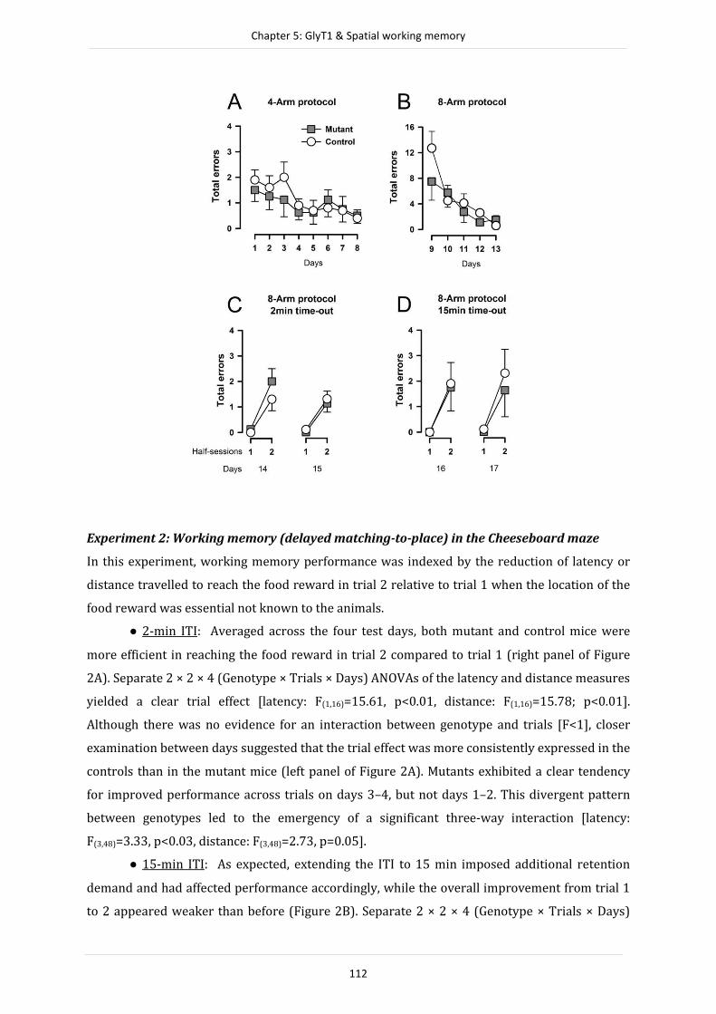
Chapter 5: GlyT1 & Spatial working memory
112
Experiment 2: Working memory (delayed matching-to-place) in the Cheeseboard maze
In this experiment, working memory performance was indexed by the reduction of latency or
distance travelled to reach the food reward in trial 2 relative to trial 1 when the location of the
food reward was essential not known to the animals.
● 2-min ITI: Averaged across the four test days, both mutant and control mice were
more efficient in reaching the food reward in trial 2 compared to trial 1 (right panel of Figure
2A). Separate 2 × 2 × 4 (Genotype × Trials × Days) ANOVAs of the latency and distance measures
yielded a clear trial effect [latency: F(1,16)=15.61, p<0.01, distance: F(1,16)=15.78; p<0.01].
Although there was no evidence for an interaction between genotype and trials [F<1], closer
examination between days suggested that the trial effect was more consistently expressed in the
controls than in the mutant mice (left panel of Figure 2A). Mutants exhibited a clear tendency
for improved performance across trials on days 3–4, but not days 1–2. This divergent pattern
between genotypes led to the emergency of a significant three-way interaction [latency:
F(3,48)=3.33, p<0.03, distance: F(3,48)=2.73, p=0.05].
● 15-min ITI: As expected, extending the ITI to 15 min imposed additional retention
demand and had affected performance accordingly, while the overall improvement from trial 1
to 2 appeared weaker than before (Figure 2B). Separate 2 × 2 × 4 (Genotype × Trials × Days)

Chapter 5: GlyT1 & Spatial working memory
113
ANOVAs distance and time to reach the reward location only yielded near-significant effects of
trials [latency: F(1,16)=4.20; p=0.06, distance: F(1,16)=4.45; p=0.05]. The analysis yielded no
support for any genotype effect or its interaction [All F’s<1].
The consistency of the latency and distance measures across the two ITI conditions was
in agreement with the lack of any genotypic effect on the separate analysis of speed of
movement (data not shown).
Figure 2: Working memory performance was assessed in the cheeseboard and the watermaze.
Performance in the working memory task in the cheeseboard maze indexed by latency to reach the
reward location, across days at each trial is depicted in A when the ITI was 2min and B when the ITI was
15 min. Latency averaged across days as a function of trials are depicted at the right of each graphs A and
B. The latency to reach the reward was significantly reduced from Trial 1 to Trial 2 when the delay was
2min, but failed to achieve significance when the delay was 15min. The performance never statistically
differed between genotypes (A and B). Working memory performance in the water maze indexed by
latency to reach the platform location, across days, for each trial is depicted in A when the ITI was 2min
and B when the ITI was 15 min. The latency averaged across days as a function of trials is depicted for
each ITI in the right of the graphs (C and D). In both delays, mice escaping performance statistically
progressed from Trial 1 to Trial 2 without any differences between genotypes. Error bars refer to ±SEM. *
refers to statistical significant difference (p<0.05) obtained from the pair-wise comparison after
significant genotype × trial × day interaction.

Chapter 5: GlyT1 & Spatial working memory
114
Experiment 3: Working memory in the Water maze
● Cued task: This was uneventful and all mice acquiring the escape response (data not
shown) without any suggestion of any genotypic difference, which is consistent with the
previous study by Singer et al. (2009b). The outcomes of the subsequent working memory tests
approximated closely to those obtained using the cheeseboard maze (Experiment 2) described
above.
● 2-min ITI: Over the first four test days when the ITI was limited to 2 min, both mutant
and control mice showed a similar magnitude of overall reduction in escape latency and
associated path length from trial 1 to 2 (right panel of Figure 2C). Separate 2 × 2 × 4 (Genotype ×
Trials × Days) ANOVAs of both performance measures yielded a significant effect of trials
[escape latency: F(1,16)=45.49; p<0.01; path length: F(1,16)=41.10; p<0.01]. Evidence for a
genotype effect was supported by the three-way interaction in escape latency which was near-
significant [F(3,48)=2.69, p=0.06] and reaching clear significance in escape latency [F(3,48)=3.25,
p<0.03]. The emergence of the three-way interaction once again stemmed from the variable
expression of the trials effect in the mutants across days in contrast to the stable expression
observed in controls (left panel of Figure 2C). Although the mutant mice’s performance
approximated the controls on the last two days, there was hardly any suggestion of improved
performance from trial 1 to 2 in the mutant mice over the first two test days. The latter, albeit
transient, effect was sufficient to produce also a significant genotype by trials interaction in the
path length measure [F(1,16)=4.89; p<0.05] (data not shown), though not in the parallel
analysis of escape latency [F(1,16)=2.73; p=0.12].
● 15-min ITI: When the ITI was extended to 15 min over the next four days,
performance had been stabilized in both groups. A consistent trial-dependent reduction in
escape latency and path length was detected across all days (Figure 2D), yielding a highly
significant trials effect in both performance measures [latency: F(1,16)=77.51; p<0.0001, path
length: F(1,16)=26.73; p<0.0001]. There was no longer any indication for a genotype effect or its
interactions [All F’s<1].
Similar to the cheeseboard experiment, there was no evidence for any significant group
difference in speed of movement (swim speed) across all stages of the watermaze experiment
(data not shown).
Experiment 4: Spatial familiarity judgement
Mutant and controls mice both exhibited similar preferential exploration of the novel arm in the
test phase in all three Y-maze tests of spatial familiarity judgement with increasing delay being
imposed between sample and test phase (Figure 3). Separate 2 × 3 (genotype × arms) ANOVA of
time spent per arm yielded a significant arm effect in all delay conditions [2-min: F(2, 24)= 12.96;

Chapter 5: GlyT1 & Spatial working memory
115
p<0.01, 2-h: F(2, 22)= 5.77; p<0.05, 24-h: F(2, 22)= 7.52; p<0.05]. There was no statistical indication
for any genotype effect whatsoever. The null results obtained in the test phase could not be
attributable to any difference in sample phase performance as suggested by separate analysis of
exploration time in the familiar arm recorded in the sample phase (data not shown).
Figure 3: Time spent in each arm during the Test phase. Mice show a higher exploration time on the
novel arm (Nv) compared to the familiar (Fm) and the start (St) arm, independently of their genotype.
Error bars refer to ±SEM.
DISCUSSION
The present study has succeeded in clarifying that forebrain neuronal GlyT1 disruption does not
produce appreciable impact on working memory functions. We demonstrated a consistent null
effect of this mutation on working memory function across three different paradigms
(Experiments 1–3), and showed that the mutation was free of any confound in spatial familiarity
judgement and related spatial perception (Experiment 4). The findings here thus constitute an
important qualification to the reported efficacy to enhance working memory functions by other
means of GlyT1 deletion or inhibition (Singer, et al., 2009b; Singer, et al., 2009c). The
comparison with the null effect of the present conditional GlyT1 deletion model might shed light
on how to maximize the therapeutic potential of GlyT1 inhibition against working memory
deficiency.
Meeting previous interpretative challenge
One impetus of the present study was to clarify the impact of forebrain neuronal GlyT1 deletion
on working memory because the data reported by Singer et al. (2009a) did not allow a clear
answer. The first challenge of interpretation stems from divergent performance on trial 1
observed between mutant and control mice in the original water maze working memory test
(Singer, et al., 2009a). It is hard to explain how the mutants outperformed the controls on trial 1

Chapter 5: GlyT1 & Spatial working memory
116
when the platform location was unbeknownst to the animals. Both groups were expected to
locate the platform solely by chance, and thus the reported trial-1 effect is counterintuitive. The
second interpretative challenge arose when trial 2 performance was taken into consideration,
because the pattern emerged appears (see Figure 2 in Singer, et al., 2009a) to be open to
conflicting interpretations. On the one hand, the lack of improvement from trial 1 to 2 in the
mutants against the presence of a significant improvement in the controls might speak for
impairment in the mutants. On the other hand, as argued by Singer et al. (2009a), the overall
escape performance of the mutant mice across the two trials was actually better than that of
controls even though overall performance in itself is not a measure of working memory.
Acknowledging the latter, Singer et al. (2009a) argued that the superior trial 1 performance in
the mutants could be linked to the enhanced reversal learning phenotype observed in the same
animals, and therefore the mutants would be better able to cope with conflicting memory. In the
working memory test, memory of the previous day’s platform location potentially might harm
trial 1 performance. Being able to overcome such negative interference allowed the mutants to
outperform controls. Singer et al. (2009a) speculated that this might be promoted by a non-
mnemonic search strategy that effectively minimized the negative impact of such proactive
interference in mutants, which incidentally might also be responsible for the limited
improvement from trial 1 to trial 2 observed in the mutant mice. This in fact could undermine
the validity of the task (in the hands of Singer, et al., 2009a) to assess working memory function
(see Crabbe & Morris, 2004). As a remedy for this shortcoming, Singer et al. (2009a) proposed
that increasing the number of trials within a day (without changing platform location) should
encourage the mutants to adopt a working memory strategy operating according to a "win-stay"
rule, since this would greatly benefit performance in all trials subsequent to the first. An
effective assessment of working memory function can then be realized. This prediction has been
partially fulfilled in Experiment 3 here, which allowed us to overcome the interpretative
difficulties in Singer et al.'s original report (2009a).
Now you see it, now you don’t
We were able to eliminate the trial-1 effect reported by Singer et al. (2009a) and thus overcame
a critical interpretative issue in the original study by the end of the first four days test of
working memory test when the ITI’s were 2 min. Thus, extension to a 4-trial-per-day protocol
had been effective in instilling an adherence to the matching rule underlying the watermaze
working memory paradigm. However, it has not escaped our notice that the precise pattern of
outcome reported by Singer et al. (2009a) was replicated here also on the first two test days
(Figure 2C) even though the critical 3-way interaction term just failed to attain outright
statistical significance [p=0.06]. Transient as it might be, its re-appearance is sufficient for us

Chapter 5: GlyT1 & Spatial working memory
117
not to dismiss Singer et al.’s (2009a) original outcome merely as a sampling (Type I) error. Yet,
the fact that this transient pattern emerged at the beginning (when the source of proactive
interference was low) rather than later into the experiment (when proactive interference would
be higher) is at odd with the account offered by Singer et al. (2009a) explained above. Their
speculation would have predicted the opposite. Thus, attributing the (transient) divergent trial-
1 performance to a resistance to proactive interference enjoyed by the mutants would seem
untenable here. The need for an alternative explanation would seem inescapable.
In the parallel evaluation of working memory extended to the cheeseboard maze
(Experiment 2), we obtained very comparable outcomes. The experiment again yielded an
overall pattern of result free from any significant divergence in trial-1 performance (right panel
of Figure 2B), and a similar dependency on days that differentiated mutants from controls (left
panel of Figure 2B). Hence, the key findings here including the transient phenotype
preferentially expressed at the beginning of training (as originally reported by Singer, et al.,
2009a) are not unique to the watermaze paradigm. They are demonstrably extendable to the
non-aversive alternative test that relies on positive (as opposed to negative) reinforcement but
carried essentially the same spatial and cognitive demand – viz., both the cheeseboard maze and
watermaze paradigms followed a matching-to-place (or “win-stay”) rule. When the Olton’s
(1976) radial arm maze test (Experiment 1) is also taken into consideration, we can further
conclude that the null effect in working memory function also generalized to a task that relied
instead on a non-matching-to-place rule, that emphasizes a "shift" rather than a "stay" strategy.
Interference & forgetting
The radial arm maze not only provided us a working memory paradigm based on a win-shift
rule, its application in the solution of the task further enables us to examine the capacity of the
working memory buffer. In the watermaze and cheeseboard experiments, the task simply
required the animals to return to one specific location made known to them a short while ago
(hence, 'win-stay'). In the radial arm maze task, however, efficient performance depends on the
recollection of multiple locations - i.e., the ability to judge the familiarity of many locations at the
same time, so the animals might effectively distinguish novel arms from familiar (visited) arms
so as to avoid the latter. A reduction in the capacity of the working memory buffer and/or
persistence of the memory trace of the visited arms may therefore impair performance. The
ability to avoid entering an arm that the animal has just visited would be less demanding than
avoiding an arm visit earlier, and therefore fewer errors of the former kind should be made.
This relative advantage can be readily understood as a form of the "recency effect" (Lund,
1925), because more recent experiences have less time for decay (forgetting) and suffers less
from retroactive interference (e.g., intervening arm-visits). We attempted to examine this by

Chapter 5: GlyT1 & Spatial working memory
118
classifying working memory errors (i.e., re-entries into already visited arms) by the number of
intervening choices preceding the error in question, i.e., the degree of disparity between two
visits into the same arm. The frequency of errors is expected to increase with increasing
disparity. This was equally observed in mutant and controls mice (Figure 4) based on the data
taken from the 8-arm protocol (i.e., those illustrated in Figure 1B). This is consistent with the Y-
maze experiment (Experiment 4) examining the persistence of memory trace across increasing
delays in familiarity judgement. Taken together, the radial arm maze convincingly showed that
forebrain neuronal GlyT1 disruption had no significant impact on the operation of working
memory. Given that this was the first test examined in this cohort, one can also be confident that
the results were free from any possible confounds due to transfer effects between tests.
Figure 4: Frequency of errors classified as a function of the degree of disparity between two visits in the
same arm for control and mutant mice based on the data obtained across the five days of test with the 8-
arm protocol (Fig. 1B). The working memory errors were classified depending on the number of arms
visited (disparity) between two visits into the same arm. For example, in the sequence of arm entries:
�→�→�→�→�→�→�→→�→ (where re-entry or working memory errors are underlined),
the re-entry into arm � is an error with disparity = 0 whereas the re-entry into arm � is an error with
disparity = 2. With increasing disparity, the frequency of error rises. This monotonic increase of error
frequency as a function of the disparity did not differ between genotype. Error bars refer to ±SEM.
What’s wrong with your mouse but not my mouse?
In contrast to previous demonstrations of a working memory enhancing effect by other forms of
GlyT1 inhibition or deletion (Singer, et al., 2009b,c), the resounding null effect here in mice
lacking specifically GlyT1 in forebrain neurons is intriguing. Our two previous positive
demonstrations involved: (i) disruption of GlyT1 from neurons and glial cells in the
telencephalon (EXM/GyT1-KO mice, Singer, et al., 2009c), and (ii) systemic injection of the
Chapter 5: GlyT1 & Spatial working memory
119
potent GlyT1 inhibitor, SSR504734 (Singer, et al., 2009b). Comparing the similarities and
differences amongst the three GlyT1 manipulations may lead to the identification of the critical
determinants allowing the pro-cognitive impact of GlyT1 inhibition in working memory to be
maximized. The critical elements should be common to the two efficacious GlyT1 manipulations,
but at the same time absent from the current CamKIIα/GlyT1 KO model examined here. Given
that Cre recombination is restricted to forebrain neurons in the CamKIIα/GlyT1 KO mice, but
extended to cells of both neuronal and glial lineages, this deduction would place an emphasis on
telencephalonic glial GlyT1 component.
The emphasis on this specific subpopulation of GlyT1 may first imply the importance of
achieving a sufficiently strong disruption of GlyT1 in the telencephalon. Because GlyT1 is mainly
expressed in glial cells (Gomeza, Ohno, & Betz, 2003a) their inclusion would produce a stronger
disruption of glycine uptake. Extending the deletion to glial cells (as in the EXM/GyT1-KO
model) doubled the reduction of GlyT1-mediated glycine uptake in the brain in comparison with
the current CamKIIα/GlyT1 KO model in which the deletion was confined to the forebrain
neuronal component (see Mohler, et al., 2011). At glutamatergic synapses where neuronal
GlyT1’s and NMDARs are co-localized, neighbouring glial-GlyT1s provide additional clearance of
glycine from the synaptic cleft. Hence, the additional loss of glial-GlyT1 would be expected to
further increase glycine-B site occupancy and consequently NMDAR-dependent neuronal
activity than that achieved in CamKIIα/GlyT1 mice. However, glial-GlyT1 also assumes a major
role in the uptake of glycine in forebrain glycinergic synapses (Eulenburg, et al., 2005; Gomeza,
Armsen, Betz, & Eulenburg, 2006). One may therefore expect that deletion of brain glial-GlyT1
might additionally potentiate inhibitory glycinergic transmission that depends on the binding of
glycine to strychnine-sensitive glycine-A site located on glycinergic receptors (GlyRs). This
consideration thus raises the interesting possibility that enhanced glycinergic inhibitory activity
might be partly responsible for enhancement in working memory reported before (Singer, et al.,
2009b,c). This not only accounts for the positive effect of systemic SSR504734, which likewise
may be expected to influence glycinergic neurotransmission, but also the lack of an effect in the
present CamKIIα/GlyT1 KO model in which the selective loss of neuronal GlyT1 should not
result in any appreciable direct impact on glycinergicneurostransmission.
Disrupting glial-GlyT1 may therefore lead to complementary changes on neuronal
excitation specifically mediated by NMDARs and network excitability via glycinergic inhibition
(Zhang, Gong, Fei, Xu, & Xu, 2008). The importance of inhibitory regulation in the maintenance
of network stability is well recognized (e.g. Mohler, et al., 2008). For instance, the relevance of
GABAergic inhibition to learning and memory has been most extensively examined (e.g.
Collinson, et al., 2002; Yee, et al., 2004). Glycinergic inhibition may assume a similar regulatory
role (Zhang, et al., 2008; Xu & Gong, 2010), and this certainly warrants direct investigation. The

Chapter 5: GlyT1 & Spatial working memory
120
two mechanisms (NMDAR-based and GlyR-based) are therefore not mutually exclusive. It is
possible that both contribute to the observed effects on working memory reported by Singer et
al. (2009b; 2009c). This hypothesis can be directly tested by mouse models with selective
disruption of glial-GlyT1. Although glial-GlyT1 function is essential for early life survival, its
deletion in adult mice is not associated with any gross abnormality (Eulenburg, Retiounskaia,
Papadopoulos, Gomeza, & Betz, 2010) and would be congenial for cognitive assays. It would also
be instructive, when one day becomes feasible, to target separately glial-GlyT1 associated with
NMDAR versus those near glycinergic synapses. The impact of selective glycinergic
manipulations in the brain on working memory is presently unknown. However, this possibility
may deserve further consideration even though glycine re-uptake at glycinergic synapses
depends on GlyT2 as well as glial-GlyT1 (Eulenburg, et al., 2005; Gomeza, et al., 2006).
The multiple yet dissociable effects of GlyT1 disruption
Comparison between the two conditional GlyT1 knockout models suggests that although both
EXM/GlyT1-KO and CamKIIα/GlyT1 KO mice showed functional resistance to a systemic
challenge to NMDAR antagonists (Yee, et al., 2006; Singer, et al., 2009c), only the latter has
produced a clear physiological phenotype in the form of enhanced NMDAR-mediated current.
Possibly due to glycine-primed NMDAR internalization (see Nong et al., 2003) or other
functional compensatory mechanisms, EXM/GlyT1-KO mice did not express this phenotype. Yet,
the reported enhanced NMDAR function in CamKIIα/GlyT1 KO mice did not translate into
enhanced working memory function as shown here, even though these mice have been shown
to exhibit other forms of cognitive enhancement (for a review, see Mohler, et al., 2011). Taken
together, these findings not only highlight the possible relevance of glycinergic
neurotransmission, but may also question the reliability or even validity of the link between
NMDAR and memory (see also Bannerman & Sprengel, 2010).
The timing of GlyT1 deletion is another major difference between the two mutant lines.
GlyT1 was deleted during embryonic development around gestational day 10 in EXM/GlyT1-KO
mice (Iwasato, et al., 2004). This is considerably earlier than in CamKIIα/GlyT1 KO mice, in
which GlyT1 was knocked out near postnatal day 21 at a time when brain development was
almost complete (Dragatsis & Zeitlin, 2000). One may reason therefore that EXM/GlyT1-KO
mice might be more prone to developmental compensation. Although this cannot be ruled out,
we can safely conclude that such compensatory changes were unlikely by themselves be
responsible for the working memory phenotype. It is because systemic exposure to the GlyT1
inhibiting drug, SSR504734, was able to facilitate working memory in adult wild type mice
when given acutely and therefore free from any developmental compensation (Singer, et al.,

Chapter 5: GlyT1 & Spatial working memory
121
2009b). However, the drug’s efficacy seems to be critically dependent on dosage in a manner
that could be relevant here.
Singer et al. (2009b) showed that the positive effect on working memory by SSR504734
only became apparent at high doses (30mg/kg) with lower doses (1mg/kg) completely
ineffective. However, such lower doses were sufficient to enhance a form of selective attention,
known as latent inhibition (LI, Black, et al., 2009). Such LI-enhancing action is shared by known
antipsychotic drugs (Moser, Hitchcock, Lister, & Moran, 2000), and the LI deficit is considered a
relevant endophenotype of schizophrenia (Amann, et al., 2010) central to the positive
symptomatology of schizophrenia on theoretical grounds (Gray, Feldon, Rawlins, Smith, &
Hemsley, 1991). This contrasts with the present emphasis on working memory, the deficiency
of which is of cardinal significance to schizophrenia cognitive symptoms. Interestingly, the LI-
enhancing effect of low doses of systemic GlyT1 inhibiting drugs (Black, et al., 2009) is captured
by CamKIIα/GlyT1 KO but not EXM/GlyT1-KO mice. It remains to be tested if GlyT1 inhibitors at
high doses that are effective in enhancing working memory (Singer, et al., 2009b) are devoid of
a latent inhibition enhancing effect. If so, then the double dissociation between CamKIIα/GlyT1-
KO and EXM/GlyT1-KO mice with respect to their facilitatory effects on LI and working memory
would be effectively mimicked by the contrast between low and high doses of GlyT1 inhibiting
drugs, respectively.
The divergent effect on LI between the two conditional knockout mouse lines might be
further related to their respective regional specificity of the gene deletion. Critically, the striatal
GlyT1 was spared in EXM/GlyT1-KO mice, but striatal GlyT1 in principal neurons were
disrupted in CamKIIα/GlyT1-KO mice. Hence, striatal GlyT1 disruption might be central to the
LI phenotype seen in CamKIIα/GlyT1-KO mice or produced by low doses of GlyT1 inhibitors in
wild type mice. This possibility warrants serious consideration because LI expression is known
to depend on striatal function (Weiner, 1990; Gray, et al., 1991). Our current attempt to
generate striatal specific GlyT1 disruption has generated some support for this hypothesis (Yee,
et al., 2010). This may strengthen glutamatergic inputs from limbic cortices to the ventral
striatum at relevant NMDARs (Gray, et al., 1991; Yee, Feldon, & Rawlins, 1995).
Postscript
The present discourse highlights that multiple molecular and pharmacological models are
necessary to appreciate fully the potential and limitations of a possible therapeutic target. This
approach enables the full and effective exploit of any potential drug target. Here, we show that
divergences as much as convergences are instructive towards the building of a more complete
mechanistic picture, and the synthesis of further hypotheses taking into account relevant cell
biology, anatomy and physiology.

Chapter 5: GlyT1 & Spatial working memory
122
REFERENCES
Amann, L. C., Gandal, M. J., Halene, T. B., Ehrlichman, R. S., White, S. L., McCarren, H. S., et al. (2010). Mouse
behavioral endophenotypes for schizophrenia. Brain Res Bull, 83(3-4), 147-161.
Aragon, C., & Lopez-Corcuera, B. (2005). Glycine transporters: crucial roles of pharmacological interest
revealed by gene deletion. Trends Pharmacol Sci, 26(6), 283-286.
Bannerman, D. M., & Sprengel, R. (2010). Multiple memory mechanisms? The long and the short of it.
EMBO J, 29(11), 1790-1791.
Bergeron, R., Meyer, T. M., Coyle, J. T., & Greene, R. W. (1998). Modulation of N-methyl-D-aspartate
receptor function by glycine transport. Proc Natl Acad Sci U S A, 95(26), 15730-15734.
Betz, H., Gomeza, J., Armsen, W., Scholze, P., & Eulenburg, V. (2006). Glycine transporters: essential
regulators of synaptic transmission. Biochem Soc Trans, 34(Pt 1), 55-58.
Black, M. D., Varty, G. B., Arad, M., Barak, S., De Levie, A., Boulay, D., et al. (2009). Procognitive and
antipsychotic efficacy of glycine transport 1 inhibitors (GlyT1) in acute and neurodevelopmental
models of schizophrenia: latent inhibition studies in the rat. Psychopharmacology (Berl), 202(1-3),
385-396.
Bradaia, A., Schlichter, R., & Trouslard, J. (2004). Role of glial and neuronal glycine transporters in the
control of glycinergic and glutamatergic synaptic transmission in lamina X of the rat spinal cord. J
Physiol, 559(Pt 1), 169-186.
Collinson, N., Kuenzi, F. M., Jarolimek, W., Maubach, K. A., Cothliff, R., Sur, C., et al. (2002). Enhanced
learning and memory and altered GABAergic synaptic transmission in mice lacking the alpha 5
subunit of the GABAA receptor. J Neurosci, 22(13), 5572-5580.
Crabbe, J. C., & Morris, R. G. (2004). Festina lente: late-night thoughts on high-throughput screening of
mouse behavior. Nat Neurosci, 7(11), 1175-1179.
Danysz, W., & Parsons, C. G. (1998). Glycine and N-methyl-D-aspartate receptors: physiological
significance and possible therapeutic applications. Pharmacol Rev, 50(4), 597-664.
Depoortère, R., Dargazanli, G., Estenne-Bouhtou, G., Coste, A., Lanneau, C., Desvignes, C., et al. (2005).
Neurochemical, electrophysiological and pharmacological profiles of the selective inhibitor of the
glycine transporter-1 SSR504734, a potential new type of antipsychotic.
Neuropsychopharmacology, 30(11), 1963-1985.
Dragatsis, I., & Zeitlin, S. (2000). CaMKIIalpha-Cre transgene expression and recombination patterns in
the mouse brain. Genesis, 26(2), 133-135.
Eulenburg, V., Armsen, W., Betz, H., & Gomeza, J. (2005). Glycine transporters: essential regulators of
neurotransmission. Trends Biochem Sci, 30(6), 325-333.
Eulenburg, V., Retiounskaia, M., Papadopoulos, T., Gomeza, J., & Betz, H. (2010). Glial glycine transporter 1
function is essential for early postnatal survival but dispensable in adult mice. Glia, 58(9), 1066-
1073.
Gabernet, L., Pauly-Evers, M., Schwerdel, C., Lentz, M., Bluethmann, H., Vogt, K., et al. (2005). Enhancement
of the NMDA receptor function by reduction of glycine transporter-1 expression. Neurosci Lett,
373(1), 79-84.
Gomeza, J., Armsen, W., Betz, H., & Eulenburg, V. (2006). Lessons from the knocked-out glycine
transporters. Handb Exp Pharmacol(175), 457-483.
Gomeza, J., Ohno, K., & Betz, H. (2003a). Glycine transporter isoforms in the mammalian central nervous
system: structures, functions and therapeutic promises. Curr Opin Drug Discov Devel, 6(5), 675-682.
Gomeza, J., Ohno, K., Hulsmann, S., Armsen, W., Eulenburg, V., Richter, D. W., et al. (2003b). Deletion of the
mouse glycine transporter 2 results in a hyperekplexia phenotype and postnatal lethality. Neuron,
40(4), 797-806.
Gray, J. A., Feldon, J., Rawlins, J. N. P., Smith, A. D., & Hemsley, D. R. (1991). The Neuropsychology of
Schizophrenia. Behavioral and Brain Sciences, 14(1), 1-19.
Iwasato, T., Nomura, R., Ando, R., Ikeda, T., Tanaka, M., & Itohara, S. (2004). Dorsal telencephalon-specific
expression of Cre recombinase in PAC transgenic mice. Genesis, 38(3), 130-138.
Javitt, D. C. (2004). Glutamate as a therapeutic target in psychiatric disorders. Mol Psychiatry, 9(11), 984-
997, 979.
Llano Lopez, L., Hauser, J., Feldon, J., Gargiulo, P. A., & Yee, B. K. (2010). Evaluating spatial memory
function in mice: a within-subjects comparison between the water maze test and its adaptation to
dry land. Behav Brain Res, 209(1), 85-92.

Chapter 5: GlyT1 & Spatial working memory
123
Lund, F. H. (1925). The Psychology of Belief IV: The Law of Primacy in Persuasion. Journal of Abnormal
Psychology and Social Psychology, 20(2), 183-191.
Martin, S. J., Grimwood, P. D., & Morris, R. G. (2000). Synaptic plasticity and memory: an evaluation of the
hypothesis. Annu Rev Neurosci, 23, 649-711.
Mohler, H., Boison, D., Singer, P., Feldon, J., Pauly-Evers, M., & Yee, B. K. (2011). Glycine transporter 1 as a
potential therapeutic target for schizophrenia-related symptoms: Evidence from genetically
modified mouse models and pharmacological inhibition. Biochem Pharmacol.
Mohler, H., Rudolph, U., Boison, D., Singer, P., Feldon, J., & Yee, B. K. (2008). Regulation of cognition and
symptoms of psychosis: focus on GABA(A) receptors and glycine transporter 1. Pharmacol Biochem
Behav, 90(1), 58-64.
Morris, R. G. (1989). Synaptic plasticity and learning: selective impairment of learning rats and blockade
of long-term potentiation in vivo by the N-methyl-D-aspartate receptor antagonist AP5. J Neurosci,
9(9), 3040-3057.
Morris, R. G., Anderson, E., Lynch, G. S., & Baudry, M. (1986). Selective impairment of learning and
blockade of long-term potentiation by an N-methyl-D-aspartate receptor antagonist, AP5. Nature,
319(6056), 774-776.
Moser, P. C., Hitchcock, J. M., Lister, S., & Moran, P. M. (2000). The pharmacology of latent inhibition as an
animal model of schizophrenia. Brain Res Brain Res Rev, 33(2-3), 275-307.
Olton, D. S., & Samuelson, R. J. (1976). Remembrance of Places Passed - Spatial Memory in Rats. Journal of
Experimental Psychology-Animal Behavior Processes, 2(2), 97-116.
Singer, P., Boison, D., Mohler, H., Feldon, J., & Yee, B. K. (2009a). Deletion of glycine transporter 1 (GlyT1)
in forebrain neurons facilitates reversal learning: enhanced cognitive adaptability? Behav Neurosci,
123(5), 1012-1027.
Singer, P., Feldon, J., & Yee, B. K. (2009b). The glycine transporter 1 inhibitor SSR504734 enhances
working memory performance in a continuous delayed alternation task in C57BL/6 mice.
Psychopharmacology (Berl), 202(1-3), 371-384.
Singer, P., Yee, B. K., Feldon, J., Iwasato, T., Itohara, S., Grampp, T., et al. (2009c). Altered mnemonic
functions and resistance to N-METHYL-d-Aspartate receptor antagonism by forebrain conditional
knockout of glycine transporter 1. Neuroscience, 161(2), 635-654.
Tsai, G., Ralph-Williams, R. J., Martina, M., Bergeron, R., Berger-Sweeney, J., Dunham, K. S., et al. (2004).
Gene knockout of glycine transporter 1: characterization of the behavioral phenotype. Proc Natl
Acad Sci U S A, 101(22), 8485-8490.
Weiner, I. (1990). Neural substrates of latent inhibition: the switching model. Psychol Bull, 108(3), 442-
461.
Whitehead, K. J., Pearce, S. M., Walker, G., Sundaram, H., Hill, D., & Bowery, N. G. (2004). Positive N-methyl-
D-aspartate receptor modulation by selective glycine transporter-1 inhibition in the rat dorsal
spinal cord in vivo. Neuroscience, 126(2), 381-390.
Wood, P. L. (1995). The co-agonist concept: is the NMDA-associated glycine receptor saturated in vivo?
Life Sci, 57(4), 301-310.
Xu, T. L., & Gong, N. (2010). Glycine and glycine receptor signaling in hippocampal neurons: diversity,
function and regulation. Prog Neurobiol, 91(4), 349-361.
Yee, B. K., Balic, E., Singer, P., Schwerdel, C., Grampp, T., Gabernet, L., et al. (2006). Disruption of glycine
transporter 1 restricted to forebrain neurons is associated with a procognitive and antipsychotic
phenotypic profile. J Neurosci, 26(12), 3169-3181.
Yee, B. K., Feldon, J., & Rawlins, J. N. (1995). Latent inhibition in rats is abolished by NMDA-induced
neuronal loss in the retrohippocampal region, but this lesion effect can be prevented by systemic
haloperidol treatment. Behav Neurosci, 109(2), 227-240.
Yee, B. K., Hauser, J., Dolgov, V. V., Keist, R., Mohler, H., Rudolph, U., et al. (2004). GABA receptors
containing the alpha5 subunit mediate the trace effect in aversive and appetitive conditioning and
extinction of conditioned fear. Eur J Neurosci, 20(7), 1928-1936.
Yee, B. K., Peleg-Raibstein, D., Dubroqua, S., Singer, P., Paterna, J.-C., Feldon, J., et al. (2010). Latent
inhibition enhancement by glycine transporter 1 disruption is mediated by anti-dopaminergic
mechanism in the nucleus accumbens.P. N. 571.10/GG15. Poster presented at the Society for
Neuroscience, San Diego, CA.
Zhang, L. H., Gong, N., Fei, D., Xu, L., & Xu, T. L. (2008). Glycine uptake regulates hippocampal network
activity via glycine receptor-mediated tonic inhibition. Neuropsychopharmacology, 33(3), 701-711

124
CHAPTER 6
General Discussion

Chapter 6: General Discussion
125
As detailed in the General Introduction and the subsequent experimental Chapters,
molecular down-regulation of GlyT1 restricted to forebrain neurons is effective in
modifying several cognitive functions (e.g. Yee, et al., 2006; Singer, et al., 2007, 2009).
This supposedly stems from increased glycine occupancy at NMDARs, which
subsequently potentiates NMDAR-mediated neurotransmission. However, careful
examination of the enhanced cognitive phenotype displayed by these mice, including the
interpretations of the behavioural outcomes offered in the literature, revealed several
issues that have not been satisfactorily addressed until now. A detailed screening might
therefore be essential in order to avoid simplistic and biased interpretations. Hence, the
present thesis aimed towards such an in-depth comparative analysis of the behavioural
phenotypes expressed by forebrain neuronal specific GlyT1 genetic disruption.
The application of this in-depth approach affords several advantages. First, this
approach favours the examination of the stability and robustness of the previously
identified phenotypes in GlyT1∆FB-neuron mice. Part of the results presented in this thesis
replicated previously identified phenotypes, yet novel phenotypes were also identified
and provided important qualifications to previous claims. Second, discussing the various
findings under a theoretical framework allow for a coherent integration, which not only
foster more powerful interpretation, but also facilitates the proposition of new research
directions. Finally, comparing the behavioural outcomes with those from other available
GlyT1 manipulations might shed light on the neuronal mechanisms and neural substrates
involved.
Overview of behavioural results
In our attempt to scrutinize the reported enhanced Pavlovian phenotype (Yee, et al., 2006) we
uncovered that the phenotypic expression was distinguishable between sexes in both the
aversive as well as the appetitive paradigm employed (Chapter 3), and across age in the
aversive paradigm (Chapter 2). In the aversive paradigm, the phenotypic dependency on sex
was primarily one of magnitude; the enhanced conditioned freezing phenotype appeared
stronger in the female mutants (Chapter 3). On the other hand, in the appetitive paradigm, sex
critically determined the direction of Pavlovian conditioning phenotype; the conditioned
approach response was enhanced in the mutant females but was somewhat weakened in male
mutants (Chapter 3). The phenotype of enhanced conditioned in freezing was detected in
senescence, but the temporal expression of the Pavlovian phenotype was modified; the adult
mutants showed pronounced within-session extinction while aged mutants did not (Chapter 2).
This exercise has also allowed further identification of hitherto unknown phenotypes and
neuroanatomical changes. In the elevated plus maze test, the explicit comparison between mice
kept in opposite light-dark cycles but tested at the same time, revealed changes in anxiety-
related behaviour (Chapter 3); an anxiolytic phenotype was observed in male mice tested in the
light phase whereas an anxiogenic phenotype was observed in female mice in the light phase. In
Chapter 2, we observed that the expression of NR2B and apoptosis were not aggravated by the
mutation, limiting thereby the possible concerns of neurotoxic effects through lifelong GlyT1

Chapter 6: General Discussion
126
disruption in forebrain neurons. However, we revealed a clear increase in the number of
immature neurons in the dentate gyrus of the adult mutants (Chapter 2).
The previous finding of enhanced latent inhibition (LI) reported in GlyT1ΔFB-neuron mice
(Yee, et al., 2006) was extended by showing that this mouse model was further associated with
enhanced sensitivity to CS-US trace and degradation of CS-US contingency (Chapter 4). In
addition, we showed that the phenotype of enhanced conditioned freezing could be extended to
foreground contextual conditioning albeit weaker by comparison. An attempt to unify these
results under a theoretical framework identified a possible common perceptual learning
mechanism that might underlie the modulation of the associability of a potential CS. Finally, the
results from the experiments described in Chapter 5 clarified the null effect of the mutation on
working memory function across three different paradigms. Furthermore, the spatial familiarity
judgement and related spatial perception were not affected by the mutation. Altogether, the
results presented in this thesis are clear illustration of how an in-depth analysis can reveal
unknown phenotypes and lead to a better understanding of the limitation and potential of any
manipulation.
Confirmation and extension of GlyT1∆FB-neuron Associative learning phenotype
Forebrain neuronal deletion of GlyT1 gives rise to a phenotype in Pavlovian associative
learning, whereby a neutral conditioned stimulus (CS) is paired together with a biologically
significant unconditioned stimulus (US). Successful associative learning results in the
conditioned stimulus eliciting a conditioned response (CR). Evidence for enhanced associative
learning in GlyT1∆FB-neuron mice, in the form of stronger CR to the CS, was shown across multiple
paradigms (Yee, et al., 2006). The robustness of the phenotype of enhanced conditioned
freezing, originally detected in the “non-pre-exposed” condition in the LI experiment (Yee, et al.,
2006), was confirmed in four different experiments using freezing conditioning, including the
“no-trace” or “contingent” test condition (Chapter 4). This phenotype was consistent across age,
gender, and different light-dark cycle conditions (Chapters 2-3). It was free from any non-
specific confounding changes in anxiety, spontaneous activity or pain sensitivity (Chapter 3).
Our experiments consistently revealed that the enhanced conditioned freezing was restricted to
the test phase, suggesting therefore, that forebrain neuronal GlyT1 deletion robustly modified
the expression rather than the acquisition of the CS-US association. One may speculate that the
procedure used during conditioning (i.e. two or three CS-US pairing) might be insufficient to
allow the emergence of differences, since such a trend was seen during acquisition in the last ITI
or the last CS pairing (Chapter 3). This might be the case since initial evaluations indicated that
the mutation can improve acquisition when tested in conditioned active avoidance, whereby the
phenotype was demonstrated through the acquisition of the response across 100 trials (Yee, et

Chapter 6: General Discussion
127
al., 2006). Nonetheless, the stronger CR to the CS in GlyT1∆FB-neuron mice suggests that the
memory trace of the Pavlovian association was stronger, and that this effect was robust.
In contrast to the consistent phenotype in the expression of the acquired CS-US
association, the extinction of this association was unstable. Both within-session and between-
day extinction were variably affected. The pattern of within-session extinction was modified
only in aged mutants, and resistance to extinction was shown only in female mutants. Moreover,
evidence that extinction learning across extinction days was modified by GlyT1 deletion was
also obtained when the spontaneous recovery of the CR between successive days was examined
in conditioned freezing (Chapter 3). Although increased resistance to extinction was also
detected across days in the appetitive conditioning paradigm, the spontaneous recovery, seven
days after the last CS-test, was not affected by GlyT1 disruption (Chapter 2). Extinction occurs
when the predictive value of the CS with respect to the occurrence of the US is reduced, leading
to a decline of the conditioned response (CR) to the CS. Long-term extinction learning is widely
accepted as a form of new learning (the CS no longer predicts the US), which acts to inhibit
expression of the CR without interfering with the previous CS-US association. Here, dissipation
of this inhibition with time revealed the spontaneous recovery effect (Rescorla, 2004) and
confirmed that extinction did not lead to the erasure of the original CS-US association.
Apart from the new learning, which that can directly compete with or impede CR during
extinction, an independent and not exclusive process which does not involve new learning or
rely on non-associative learning could be at play. Wagner and Brandon (2001) proposed that
the strength of a CR could be weakened by changing the environment in which the response
occurs, through generalization decrement. If an animal is repeatedly presented with the CS that
is directly followed by a US, then it will suddenly find itself in a new environment/situation
when the US is no longer delivered. This change may weaken responding through generalization
decrement, rather than through a change to the association involving the CS. One plausible
explanation of the extinction phenotype obtained in mutants could be that these mice are less
sensitive to a change in the environment and as a result continue to respond to the CS (see
Wagner’s SOP model later in the discussion). This effect could explain the resistance to within-
session extinction and might be vulnerable to different factors such as sex or aging. On the other
hand, the resistance to long-term extinction in mutant mice might suggest that the mechanism
underlying the control of new learning is somehow affected by GlyT1 disruption. In Chapter 3,
we attempt to interpret the extinction learning phenotype as reflecting a stronger proactive
interference. This explanation assumes that the phenotype of increased resistance to extinction
could be understood as a stronger influence of the prior experience of CS-US association over
current control of responding. Indeed, this interpretation was initially used by Yee et al. (2006)
to explain the enhanced LI effect in the mutant mice. The demonstration of LI requires two

Chapter 6: General Discussion
128
distinct phases with contrasting CS-US relationships: the pre-exposure phase, in which CS is not
followed by any significant event [CS → nothing] precedes the conditioning phase. LI refers to
the observation that CS pre-exposed subjects show weaker CR to the CS after CS-US pairing
compared to non-pre-exposed subjects. Several theories propose that the weaker CR does not
stem from a failure to acquire the critical CS-US association but reflects the [CS → nothing]
memory trace competing over the control of behaviour during testing (Weiner, 1990; Bouton,
1993). Thus, a stronger influence of the prior experience of non-reinforced CS exposures leads
to stronger LI. This view conflicts with a prior report of enhanced reversal learning in GlyT1ΔFB-
neuron mice, which suggested resistance rather than vulnerability proactive interference in these
mutants (Singer, et al., 2009). Another explanation for the reversal effect comes from an
additional study in discrimination learning. During a discrimination, animals learn to pay more
attention to relevant than irrelevant stimuli (Mackintosh, 1974). Hence, the animals learn to
focus their attention to one or few features that allow an optimum discrimination. Reversal
learning would be facilitated because the contingency of only few features has to be reversed
(Sutherland and Mackintosh, 1971). Hence, it is conceivable that the reversal phenotype
reported by Singer et al. (2009) reflects primarily enhanced selective association learning.
Two mechanisms, one explanation?
As mentioned above, GlyT1∆FB-neuron disruption is effective in enhancing associative strength, as
evidenced in all the conditioning experiments carried out. The mice showed stronger
conditioning in the classical conditioning (Chapter 2 and 3); in the no-trace and the non-
contingent condition (Chapter 4) and in the no-PE condition (Yee, et al., 2006). However, an
increased associative strength per se cannot explain why the imposition of a delay between CS
and US, the presentation of CS-alone trial, or the pre-exposure to the CS, leads to a weaker CR in
the mutants compared to the controls. One might expect that if GlyT1∆FB-neuron disruption is
effective in enhancing associative strength, it should also lead to a stronger CR under these
conditions. Therefore, the different associative learning phenotypes present in GlyT1ΔFB-neuron
mice are difficult to reconcile under one unique psychological mechanism that only affects the
modulation of associative strength. Here, it is important to address the conditions that can lead
to a stronger or weaker associative link in GlyT1∆FB-neuron mice.
The majority of associative learning theories emphasize that the attentional processing
of the CS determines its associability; the more attention paid to a CS, the easier it is to enter
association with a US. It was shown in Chapter 4 that a change in the attention perceived by the
mutants could lead to enhanced associative learning in the mutants. A change in the perceived
intensity of the CS would also predict a reduction of associative learning in the degraded
contingent condition schedule used in our experiment (Chapter 4). This can be readily tested

Chapter 6: General Discussion
129
with the prediction that, if the GlyT1ΔFB-neuron mice perceive the salience of a stimulus as
stronger, a simple freezing conditioning experiment in which the salience of the CS is low will
lead to a stronger CR in the mutants compared to the controls. Furthermore, the Rescorla-
Wagner model (1972) used in Chapter 4 would also predict a stronger associative strength at
the end of the acquisition in the mutants compared to the controls, if the schedule of
contingency degradation used was different (i.e. if the last pairing was an omitted US). This can
also be examined, and if in the expected direction, it would support the idea that attention is
modulated by GlyT1∆FB-neuron disruption.
However, a sole increase in attention to the CS is unable to explain why, for instance, the
imposition of a trace between the CS and the US diminishes rather than enhances the CR
observed in the mutants, or why the pre-exposure of the CS affects the subsequent acquisition of
the CS-US association in the mutants but not in the controls. Here, the issue of selectivity in
learning can be addressed. Selectivity in learning refers to the notion that some CS-US
relationships are learned more readily than others. This comes from studies showing that the
effectiveness of conditioning depends on the stimuli paired together (e.g. Shapiro, et al., 1980).
One explanation for selective association is that it is due to some stimuli, or stimulus dimension,
being more relevant to the occurrence of biologically significant events than others are. For
example, tastes are likely to provide more reliable information than auditory-visual stimuli
about whether or not a food is poisonous. Although this kind of selective association are
considered as innate, the discovery of the so-called “learned irrelevance effect” suggests that an
animal’s experience may also contribute to selective association. Learned irrelevance
(Mackintosh, 1973) reflects the slower learning that takes place when a CS and US are paired if
they have previously been presented randomly with respect to each other. Mackintosh’s theory
(1975) claims that the attention paid to a stimulus will increase if it is a good predictor of a
significant event and decrease if this stimulus is a bad predictor. In fact, to be fully attended, a
CS must not only be a good predictor of the US, but it must be a better predictor than all other
stimuli that are present in the trial. Therefore, during the random pairing of a CS and a US, the
animals learn that this specific CS is irrelevant to the delivery of this specific US and will
decrease the attention paid to the CS. This will result in animals ignoring this stimulus when
these CS and US are eventually paired. In the latent inhibition experiment, during the pre-
exposure phase, the animals learn that the CS is irrelevant; as a result, the animal will ignore the
CS when it is paired with the US in the subsequent acquisition phase. Yee et al. (2006)
previously discussed this possibility and proceeded to propose that GlyT1ΔFB-neuron mice might
present an enhancement of learned irrelevance because GlyT1 disruption could affect the rules
by which the attention is adjusted. For example, the rate of change in attention could be
facilitated. Therefore, in GlyT1ΔFB-neuron mice, when a CS is a good predictor, the attention paid to

Chapter 6: General Discussion
130
it is tuned up in such a way that they will pay more attention to the stimulus than the controls
will after the same number of trials. This could also predict the bi-directional effect observed in
trace conditioning in the mutant mice. We may also hypothesise that the “learned irrelevance”
effect would be stronger in GlyT1ΔFB-neuron mice compared with controls.
Wagner’s SOP (1981) model also offers an interpretation of the results that could be understood
as a consequence of the modulation of the attention paid to the CS. The theory assumes that the
representation, or the memory of a stimulus can be in three different states. The inactive state
refers to a state where memory is not modifiable and cannot influence an animal’s behaviour.
The two remaining states (called A1 and A2) are considered active because a stimulus in one of
these states can influence behaviour. The different activation states can be viewed as the
attention allocated to a stimulus. A stimulus in A1 is at the centre of attention whereas a
stimulus in A2 is at the periphery of the field of attention. Like in other theories, a stimulus
needs to be fully attended to be able to enter association. Thus, it is proposed that excitatory
and inhibitory conditioning is only possible when the CS is in the A1 state. Information in the
inactive state is believed to last more or less indefinitely, and can be viewed as the long-term
memory. On the other hand, the information in an active state is considered as labile; the active
states could refer to a short-term memory. This assumption implies that short-term memory
has a limited capacity (Wagner, 1976) presumably because of the limited capacity of attention
(James, 1890). Hence, the model proposes that a representation of a stimulus in A1 state will
rapidly decay to the A2 state, as soon as it is not present anymore, and will then revert more
slowly to its inactive state. Importantly, only one route allows a representation to be activated
in A1. If a stimulus representation is in the A2 state, further presentation of this stimulus will
not activate an A1 representation of it and this stimulus will not be fully attended. By contrast,
two routes allow a stimulus to gain access to the A2 state. The first one, also called the self-
generated A2 representation route, is the one described earlier when a stimulus representation
is first activated from an inactive state to an A1 state that decay to an A2 state before slowly
returning to the inactive state. An interesting feature of this proposal is that it can also provide
an account for non-associative processes. The orienting response (OR) or the investigatory
reflex in the terminology of Pavlov (1927) is the (unconditional) response observed to a (novel)
stimulus. Habituation refers to the fading of an unconditioned response, such as an OR, to a
stimulus that is repeatedly presented. This phenomenon can be explained by the SOP model.
The first occurrence of a stimulus triggers its representation to the A1 state and may elicit a
strong response OR. This state will decay to the A2 state and the model predicts that the
strength of the response will show a corresponding decline. If the stimulus is presented again
while the A2 representation is still active, the stimulus will be unable to activate an A1

Chapter 6: General Discussion
131
representation, and will thus be less able to evoke the relevant response, i.e. response to
subsequent presentations of the stimulus will become habituated. The other route by which a
stimulus can gain access to the A2 state, and thus influence attention, is made possible by
associative learning. The model states that two stimuli (CS and US) can be associated only when
the representations of these stimuli are simultaneously in A1, when both are fully attended.
After effective conditioning, the presentation of the CS will be able to trigger a representation of
the US directly into the A2 state. This is referred to as a retrieval-generated representation, as it
is not excited by the direct presentation of the US, but by a CS with which it is associated. In
chapter 4, based on the results obtained by GlyT1ΔFB-neuron mice in the trace experiment, we
suggested that the rate of activation between the Inactive state and the A1 state is facilitated as
well as the rate of decay between A1 and A2. In addition, the model could explain the pattern of
results obtained in LI. As explain in Chapter 4, according to Wagner’s SOP model (1981), during
pre-exposure the CS is associated with the context. When the animals are returned to this
context during subsequent acquisition, the context primes the representation of the CS directly
into A2 state, rendering its association with the US more difficult. The mechanism thus depends
on the formation of context-CS association. GlyT1∆fb-neuron mice might learn the context-CS
association more rapidly, for reasons similar to those underlying stronger CS-US association,
and thus facilitate the expression of LI later. An additional explanation, based on the non-
associative self-generated A2 route could be drawn to explain the LI effect. During the pre-
exposure phase, repeated presentations of the CS leads to its habituation. When it is again
presented during acquisition, the CS is still habituated (in A2) and thus cannot be associated
readily with the US, leading to the LI effect. Bannerman and Sanderson (2007) proposed that
GlyT1∆fb-neuron mice could be impaired in dishabituation, based on the results obtained in
recognition memory (see Chapter 1). Therefore, the LI effect could also stem from impaired
dishabituation to the pre-exposed CS. This impaired dishabituation could be represented in the
Wagner model (1981) by a slower decay rate from the A2 state back into the inactive state. As
suggested by Bannerman and Sanderson (2007), this would predict an enhanced novelty
preference in a Y-maze task for example. However, as shown in Chapter 5, a null effect was
shown in the Y-maze task. Despite this, our proposition that the rate of activation between the
Inactive and the A1 state is facilitated could be easily tested in an experiment assessing the
orienting response (OR) as employed by Sanderson et al. (2011).
In conclusion, the attempt to apply different associative learning theories to explain our results
has led to the proposition of a possible common perceptual learning mechanism that could lead
to enhanced association learning and enhanced selectivity in learning. One explanation of the
stronger associative strength in GlyT1ΔFB-neuron mice would be that the attention paid to a
Chapter 6: General Discussion
132
stimulus is stronger in the mutants. Whereas one explanation for the enhanced selectivity in
learning would be that mutants adjust this attention less efficiently.
Comparisons with other GlyT1 disruption models: evidence for underlying neural
substrates?
In contrast to the effect of GlyT1ΔFB-neuron on associative learning, no effect was seen in reference
memory (Singer, et al., 2009). Moreover, the ambiguous effect on working memory described
before (Singer, et al., 2009) was clarified in the Chapter 5 of the present thesis. Working
memory function seems not to be influenced by the mutation. Similarly, evaluation of
foreground contextual freezing revealed a relatively weak enhancement in GlyT1ΔFB-neuron mice.
One might question why the strong and robust effect seen in associative learning does not
translate to the spatial and contextual forms of learning. Indeed, they both seem to involve the
processing of often large, diffuse, multicue, polymodal and relational stimuli (Bannerman, et al.,
2004). It is conceivable therefore, that GlyT1ΔFB-neuron disruption interferes with attentional
processes related directly with discrete cues. Nonetheless, these behavioural phenomena are
believed to be mediated by distinct neural substrates. The GlyT1 genetic manipulation used in
the present thesis affects the entire forebrain and we therefore have no direct evidence for the
involvement of a specific brain region in the modulation of GlyT1 in memory function. However,
the phenotypic similarities as well as differences between neuronal-specific GlyT1 deletion and
another conditional GlyT1 knockout extending into cortical glial cells specifically expressed in
the telencephalon, point to the uniqueness of the anatomical and cell-type specific nature of the
genetic manipulation examined here (see Chapter 5, see also Mohler et al., 2011). More
specifically, contrasting effects were seen in spatial working memory performance between the
two regional-specific GlyT1 manipulations, with a predominant role for glial-based GlyT1 in this
behavioural function. This might suggest that the behavioural effects mediated by the forebrain
neuronal GlyT1 depletion are supported by different neural substrates compared to the glial-
specific GlyT1 manipulation. However, this clearly warrants more investigation.
Correspondingly, we recently obtained evidence that viral-mediated neuronal-specific
depletion of GlyT1 in the nucleus accumbens also was effective in enhancing the latent
inhibition effect (Yee, et al., 2010). Interestingly, this region-specific depletion of GlyT1 was able
to isolate only one of the mechanisms via which enhanced latent inhibition can occur. In
contrast, to the observed effects in the non-pre-exposed group here, the effects following
nucleus accumbens-specific GlyT1 depletion were seen solely in the pre-exposed group. Thus,
this appears to suggest that the nucleus accumbens is not mediating the associative learning
improvement seen in the GlyT1ΔFB-neuron mice, but might be more relevant for increasing the
selectivity in learning. It would therefore be worthwhile to examine the behavioural relevance

Chapter 6: General Discussion
133
of nucleus accumbens-specific GlyT1 depletion on tasks which were affected by GlyT1ΔFB-neuron
disruption, such as trace conditioning or contingency degradation conditioning.
Concluding remarks
The present thesis provides evidence for novel critical mediating factors, such as gender and
age, which should be taken into account when designing novel drug therapies to ameliorate
memory deficits as seen in e.g. schizophrenia. The in-depth characterization of the neuronal
forebrain-specific GlyT1 mutant mice has confirmed the importance of GlyT1 in the modulation
of cognitive function, with particular focus on its impact on attentional processes, which might
be relevant for schizophrenia related cognitive impairments. However, the proposal that GlyT1
disruption might be a potential “cognitive enhancer” is problematic. As discussed in the
introduction, enhanced memory has its cost, especially in situations involving traumatic events.
The observation that GlyT1ΔFB-neuron alters extinction learning hinders the use of GlyT1 inhibition
as a potential pharmacotherapy for post-traumatic stress disorder (PTSD). However, the results
presented here reveal the potential for GlyT1 disruption/inhibition in anxiety (Chapter 3).
Moreover, the observation of increased neurogenesis in this mouse line might indicate a
potential use for GlyT1 inhibition in depression related disorders, which are linked to reduced
neurogenesis. This is due to the fact that anti-depressants are thought to exert their effect by
increasing neurogenesis. This warrants further investigation which could lead to a potential
therapeutic role for GlyT1 inhibition. Finally, the data presented here appears to indicate that
the GlyT1-mediated effects might show some degree of region-specificity. This latter facet might
open new paths for the development of brain region-specific therapeutic targets.

Chapter 6: General Discussion
134
REFERENCES
Bannerman, D. M., Rawlins, J. N., McHugh, S. B., Deacon, R. M., Yee, B. K., Bast, T., et al. (2004). Regional
dissociations within the hippocampus--memory and anxiety. Neurosci Biobehav Rev, 28(3), 273-
283.
Bouton, M. E. (1993). Context, time, and memory retrieval in the interference paradigms of Pavlovian
learning. Psychol Bull, 114(1), 80-99.
James, W. (1890). The Principles of Psychology. Harvard: University Press.
Mackintosh, N. J. (1973). Stimulus selection: Learning to ignore stimuli that predict no change in
reinforcement. In R. A. Hinde & J. Stevenson-Hinde (Ed.), Constraints on learning (pp. 75-100).
London: Academic Press.
Mackintosh, N. J. (1974). The psychology of animal learning. Oxford, England: Academic Press.
Mackintosh, N. J. (1975). Theory of Attention - Variations in Associability of Stimuli with Reinforcement.
Psychological Review, 82(4), 276-298.
Möhler H., Boison D., Singer P., Feldon J., Pauly-Evers M. &, Yee B.K. (2011). Glycine transporter 1 as a
potential therapeutic target for schizophrenia-related symptoms: Evidence from genetically
modified mouse models and pharmacological inhibition. Biochem Pharmacol, 81(9), 1065-1077.
Pavlov, I. P. (1927). Conditioned Reflexes: An Investigation of the Physiological Activity of the Cerebral
Cortex. London: Oxford University Press.
Rescorla, R. A. (2004). Spontaneous recovery. Learn Mem, 11(5), 501-509.
Rescorla, R. A., & Wagner, A. R. (1972). A theory of Pavlovian conditioning: Variations in the effectiveness
of reinforcement and nonreinforcement. In A.H. Black & W.F. Prokasy (Ed.), Classical conditioning
II: Current theory and research (pp. 64-99). New York: Appleton-Century-Crofts.
Sanderson, D. J., Sprengel, R., Seeburg, P. H., & Bannerman, D. M. (2011). Deletion of the GluA1 AMPA
receptor subunit alters the expression of short-term memory. Learn Mem, 18(3), 128-131.
Shapiro, K. L., Jacobs, W. J., & LoLordo, V. M. (1980). Stimulus-reinforcer interactions in Pavlovian
conditioning of pigeons: Implication for selective associations. Animal Learning and Behavior, 8,
586-594.
Singer, P., Boison, D., Mohler, H., Feldon, J., & Yee, B. K. (2007). Enhanced recognition memory following
glycine transporter 1 deletion in forebrain neurons. Behav Neurosci, 121(5), 815-825.
Singer, P., Boison, D., Mohler, H., Feldon, J., & Yee, B. K. (2009). Deletion of glycine transporter 1 (GlyT1) in
forebrain neurons facilitates reversal learning: enhanced cognitive adaptability? Behav Neurosci,
123(5), 1012-1027.
Sutherland, N.S. & Mackintosh, N.J. (1971). Mechanisms of animal discrimination learning. New York:
Academic Press.
Wagner, A. R. (1976). Priming in STM: An information-processing mechanism for self-generated or
retrieval-generated depression in performance. In T. J. Tighe & R. N. Leaton (Ed.), Habituation:
Perspectives from child development, animal behavior, and neuropsychology (pp. 95-128).
Hillsdale, NJ: Lawrence Erlbaum Associated, Inc.
Wagner, A. R. (1981). SOP: A model of automatic memory processing in animal behavior. In N.E. Spear &
R.R. Miller (Ed.), Information processing in animals: Memory mechanisms (pp. 5-47). Hillsdale, NJ:
Lawrence Erlbaum Associated, Inc.
Wagner, A. R., & Brandon, S. E. (2001). A componential theory of Pavlovian conditioning. In Klein S. B.
Mowrer R. R. (Ed.), Handbook of contemporary learning theories (pp. 23-64). Mahwah, NJ: Lawrence
Erlbaum Associated, Inc.
Weiner, I. (1990). Neural substrates of latent inhibition: the switching model. Psychol Bull, 108(3), 442-
461.
Yee, B. K., Balic, E., Singer, P., Schwerdel, C., Grampp, T., Gabernet, L., Knuesel, I., Benke, D., Feldon, J.,
Mohler, H., & Boison, D. (2006). Disruption of glycine transporter 1 restricted to forebrain neurons
is associated with a procognitive and antipsychotic phenotypic profile. J Neurosci, 26(12), 3169-
3181.
Yee, B. K., Peleg-Raibstein, D., Dubroqua, S., Singer, P., Paterna, J.-C., Feldon, J., (2010). Latent inhibition
enhancement by glycine transporter 1 disruption is mediated by anti-dopaminergic mechanism in the
nucleus accumbens.Program No. 571.10/GG15. Poster presented at the Society for Neuroscience,
San Diego, CA.

135
CURRICULUM VITAE
Personal Data
Name: Sylvain Dubroqua
Date and Place of birth: 21th May, 1981, Nice (France)
Citizenship: French
Education
2001-2004 DEUG-Licence in Cognitive Psychology
University Nice Sophia-Antipolis, France
2004-2005 Master 1 in Cognitive Psychology
University Nice Sophia-Antipolis, France
2005-2006 Master 2 in Cognitive Science
University Bordeaux 2, France
2006-2007 Diploma of Advanced Studies in Biological Sciences
Centre de Neurosciences Intégratives et Cognitives, CNRS, Talence, France
2008-2011 Doctoral studies
Laboratory of behavioural neurobiology, ETH Zurich, Schwerzenbach,
Switzerland
Published articles
•••• Dubroqua S, Singer P, Boison D, Feldon J, Möhler H, Yee BK (2010). Impacts of forebrain
neuronal glycine transporter 1 disruption in the senescent brain: evidence for age-dependent
phenotypes in Pavlovian learning. Behav Neurosci, 124(6), 839-850.
•••• Bitanihirwe BK, Dubroqua S, Singer P, Feldon J, Yee BK. Sensorimotor gating and vigilance-
dependent choice accuracy: a within-subject correlative analysis in wild-type C57BL/6 mice.
Behav Brain Res, 217(1), 178-187.

Chapter 6: General Discussion
136
Acknowledgements
The present dissertation was conducted at the Laboratory of Behavioural Neurobiology,
Swiss Federal Institute of Technology (ETH) Zurich. Part of the Immunohistochemical
analyses were carried out in Dr. Olivier Raineteau’s Laboratory of Neuromorphology,
Brain Research Institute, University of Zurich/ETH-Z. The research work was financially
supported by ETH Zurich, with additional support from the National Centre for
Competence in Research (NCCR): Neural Plasticity and Repair, Swiss National Science
Foundation.
First and foremost, I would like to take this opportunity to thank the members of the
dissertation committee: I would like to express my thanks to Prof. Dr. Joram Feldon, for
giving me the opportunity to pursue this dissertation in his laboratory. Many thanks to
Prof. Dr. Jacques Micheau for his agreeing to review and examine my dissertation. Last
but not least, I would like to express my deepest appreciation to Dr. Benjamin Yee for
sharing his impressive knowledge ranging from French revolution to quantum
mechanics.
I am grateful to Dr. Detlev Boison for providing the GlyT1 knockout mice, for his
contributing knowledge on biochemical experiments involving GlyT1 knockout, and for
his input during manuscript revisions. I would also like to express my appreciation to
Dr. Olivier Raineteau, for allowing me to be part of his group and to carry out
immuhistochemical experiments. A special thanks for Roberto Fiorelli who was guided
me during the experiments with his legendary patience.
Un grand merci à Edith Lesburgueres aka Chefe, whose enthusiasm for science under
conditions not always optimal has been a perpetual source of motivation. I would also
like to thanks Frederic Esclassan, first for being the only one to visit me in Zurich and
second for his constant support in all kind of manners since I know him.
My dearest thanks for Helen Pothuizen, whose help during the preparation of the
dissertation was priceless. Thank you for this long night. Thanks to Jonas Hauser for his
help in the beginning of my Phd and for being the only French speaking person in the
lab. To Mary Muhia who is always here when I need help. To Phillip Singer for sharing
his data and his weird farmer cigars. A big thank you to Lucas Serrano who put up with
my supervision.
I would also like to acknowledge the “Charlie´s angels”, Michele Bürkli, Mandy Sütö and
Simone Läubli for being so refreshing. A special thanks for Michele Burkli who make me
discover and enjoy Switzerland. A special merci vielmal to Sylvia Turner and Marcel
Morf for their unconditional help and support.
Big up to my family.
Hase