Embed Size (px)
Citation preview
Cellular/Molecular
The Role of a Voltage-Dependent Ca2� Channel IntracellularLinker: A Structure-Function Analysis
Lior Almagor,1 Orna Chomsky-Hecht,1 Adva Ben-Mocha,2 Doran Hendin-Barak,1 Nathan Dascal,2 and Joel A. Hirsch1
Departments of 1Biochemistry and Molecular Biology, Institute of Structural Biology, George S. Wise Faculty of Life Sciences and 2Physiology andPharmacology, Sackler Faculty of Medicine, Tel Aviv University, Tel Aviv 69978, Israel
Voltage-dependent calcium channels (VDCCs) allow the passage of Ca 2� ions through cellular membranes in response to membranedepolarization. The channel pore-forming subunit, �1, and a regulatory subunit (CaV�) form a high affinity complex where CaV� bindsto a �1 interacting domain in the intracellular linker between �1 membrane domains I and II (I–II linker). We determined crystalstructures of CaV�2 functional core in complex with the CaV1.2 and CaV2.2 I–II linkers to a resolution of 1.95 and 2.0 Å, respectively.Structural differences between the highly conserved linkers, important for coupling CaV� to the channel pore, guided mechanisticfunctional studies. Electrophysiological measurements point to the importance of differing linker structure in both CaV1 and 2 subtypeswith mutations affecting both voltage- and calcium-dependent inactivation and voltage dependence of activation. These linker effectspersist in the absence of CaV�, pointing to the intrinsic role of the linker in VDCC function and suggesting that I–II linker structure canserve as a brake during inactivation.
IntroductionVoltage-dependent calcium channels (VDCCs) allow the selec-tive passage of Ca 2� ions through cellular membranes in re-sponse to membrane depolarization, playing a major role inmany neuronal and other physiological processes (for review, seeJones, 1998). CaV1 and CaV2 comprise two heteromeric VDCCfamilies characterized by high depolarization thresholds requiredfor their activation. �1 is the membrane pore-forming subunit.Consisting of four homologous domains, it forms a pseudo-tetrameric structure. Each domain contains six membrane helicalsegments, where S1-4 constitute the voltage sensor and S5-6 theion-selective pore. The cytoplasmic � subunit (CaV�) is tightlyassociated with CaV1 and CaV2 �1subunits and robustly modu-lates their function (for review, see Buraei and Yang, 2010). CaV�both facilitates channel localization to the plasma membrane anddirectly modulates its gating properties. Its effects include hyper-polarization of the activation voltage, acceleration of activationkinetics, and increase of channel open probability (Buraei andYang, 2010). Furthermore, several aspects of both voltage andcalcium-dependent inactivation (VDI and CDI) are differentiallyaffected by various CaV� isoforms. The functional core of CaV� iscomposed of two conserved structural domains, a SH3 and aguanylate-kinase (GuK) like domain. The CaV� GuK domain
interacts with a conserved 18-residue region in the intracellularlinker between �1 domains I and II, dubbed AID (�1 interactingdomain) (Pragnell et al., 1994). Crystallographic studies haveshown how the AID �-helix docks into a deep hydrophobicgroove in the GuK domain (Chen et al., 2004; Opatowsky et al.,2004; Van Petegem et al., 2004).
The cytoplasmic I–II linker/CaV� interaction is functionally im-portant in all CaV1 and CaV2 channels. Nonetheless, the exact mo-lecular mechanisms leading to its effects on channel gating are notcompletely understood. The localization of the AID in relation toIS6, a part of the �1 inner pore, suggests that a mechanism of the I–IIlinker/CaV� complex may involve constraints on IS6 mobility. Aspreviously suggested, CaV� association promotes an �-helical con-formation on the AID (Opatowsky et al., 2004). This �-helix is pre-dicted to propagate through the proximal linker (PL) to IS6 (Fig.1A), forming a rigid connection between the GuK of CaV� and thechannel pore, mechanically transducing its binding to channel gat-ing states. (Opatowsky et al., 2004; Van Petegem et al., 2004). Thus,gaining structural knowledge of the regions of the I–II linker outsidethe AID has been an important requirement for further understand-ing the mechanism of VDCC modulation by the I–II linker/CaV�complex. We sought to obtain structural features of the intact I–IIlinker in complex with CaV�, including whether the PL is one long�-helix. Using I–II linkers from both the CaV1.2 and CaV2.2 �1subunits, we discern distinctive structural variations between I–IIlinker/CaV� complexes of these different channel subtypes. Whilethe CaV2.2 PL is mostly �-helical, the CaV1.2 PL helix begins at adownstream glycine conserved in CaV1 channels. We tested the con-sequences of these subtype-specific structural variations on the bio-physical properties of intact VDCCs. Our functional results showdivergent, channel-type PL secondary structure to be important forvarious channel functional properties including VDI, CDI, and thevoltage dependence of activation.
Received Nov. 15, 2011; revised March 11, 2012; accepted March 31, 2012.Author contributions: L.A., N.D., and J.A.H. designed research; L.A., O.C.-H., A.B.-M., and D.B.-H. performed
research; L.A., N.D., and J.A.H. analyzed data; L.A., N.D., and J.A.H. wrote the paper.This study was supported by Israel Science Foundation Grant 1201/04 and a DIP-DFG Grant to J.A.H., L.A. was
supported in part by a Dean’s Excellence Scholarship. We thank Efrat Berman for assistance with subcloning, andMoshe Dessau, Reuven Wiener, and the staff at ESRF for help with diffraction data collection. Structure factors andatomic coordinates have been deposited with PDB codes 4DEY and 4DEX.
The authors declare no competing financial interests.Correspondence should be addressed to Joel A. Hirsch at the above address. E-mail: [email protected]:10.1523/JNEUROSCI.5727-11.2012
Copyright © 2012 the authors 0270-6474/12/327602-12$15.00/0
7602 • The Journal of Neuroscience, May 30, 2012 • 32(22):7602–7613
Materials and MethodsMolecular cloning. For crystallography, rabbit CaV1.2 (P15381, residues436 –539) and CaV2.2 (Q05152, residues 358 – 468) I–II linkers were sub-cloned downstream to a HisTag and a Tobacco etch virus (TEV) proteasesite between NcoI and EcoRI restriction sites of pET-Duet-1 (Novagen).The CaV�2 functional core (Opatowsky et al., 2003) sequence was sub-cloned between NdeI and KpnI sites. For CD spectroscopy, a maltose-binding protein (MBP) sequence downstream of a HisTag and upstreamof a TEV site was inserted between NcoI and BamHI sites of a pET-28avector (Novagen). A synthetic DNA fragment encoding an 18 aa�-helical peptide (3�AAKAAE) flanked between a BamHI site at its 5�and sequential NheI and NotI sites at its 3� was ligated into this vectorbetween BamHI and NotI sites. Subsequently, PL-AID fragments, en-coding CaV1.2 residues 436 – 475 and CaV2.2 357–396, of wild-type(WT) and mutant channel sequences were subcloned between NheI andNotI sites of this modified vector.
For electrophysiology, the following cDNAs were used: rabbit CaV1.2(X15539), CaV�2b (L06110), �2�-1 (P13806) (Shistik et al., 1998), andrabbit CaV2.2 �1(D14157). All constructs included a T7 promoter se-quence for RNA transcriptions. RNA transcripts included 5� and 3�untranslated sequences of Xenopus �-globin. For further Cav2.2 manip-ulations, silent mutations were introduced into the gene sequence togenerate an EcoRV/SpeI cloning cassette between nucleotides 945–1716.Using either overlapping PCR or site directed mutagenesis, PL mutationswere introduced into this cassette. In CaV1.2 PL, the naturally occurringdouble restriction site for SfiI in this gene sequence was used as a cloningcassette (nucleotides 264 –1572 of CaV1.2). m7G(5�)ppp(5�)G cappedRNA was prepared by in vitro transcription using the Ribomax LargeScale RNA production system (Promega). Unincorporated nucleotideswere removed using MicroSpin G-25 columns (GE Healthcare).
Protein expression and purification. All expression vectors were trans-formed into E. coli Tuner (DE3) Codon Plus competent cells. Cells were
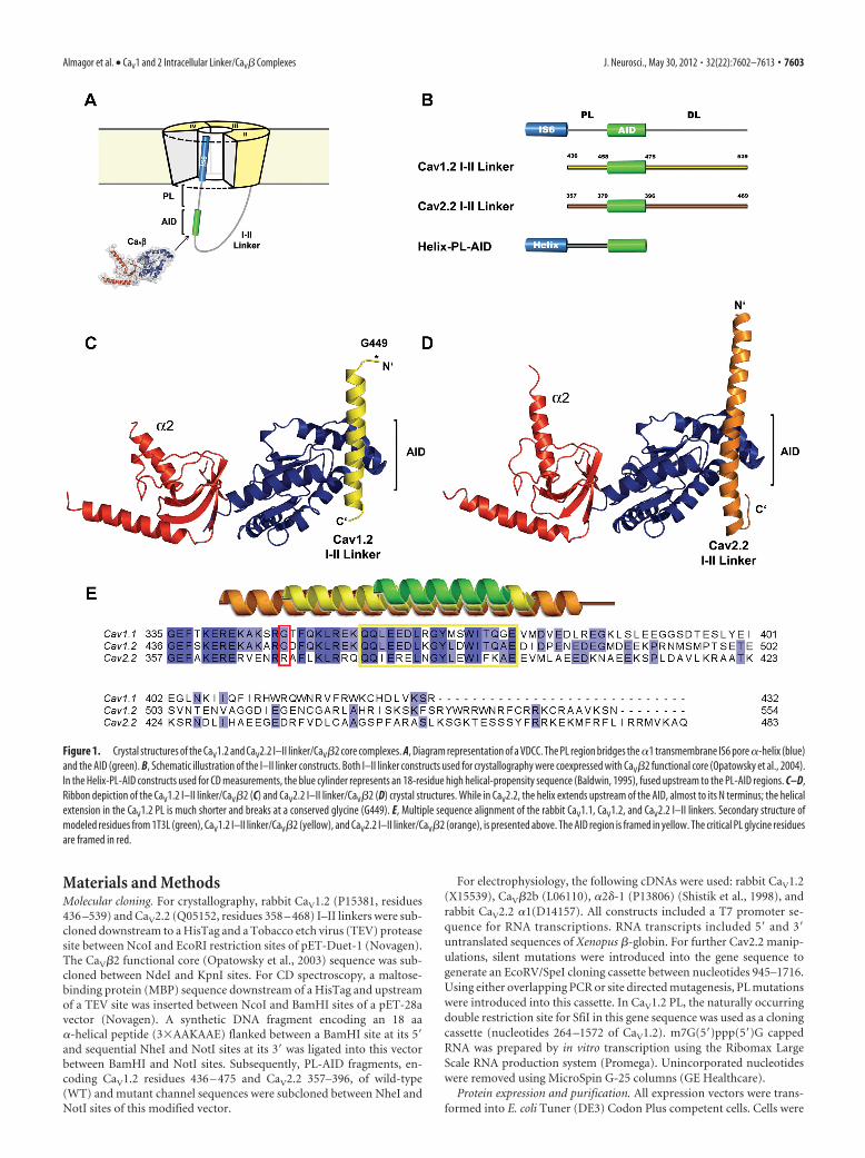
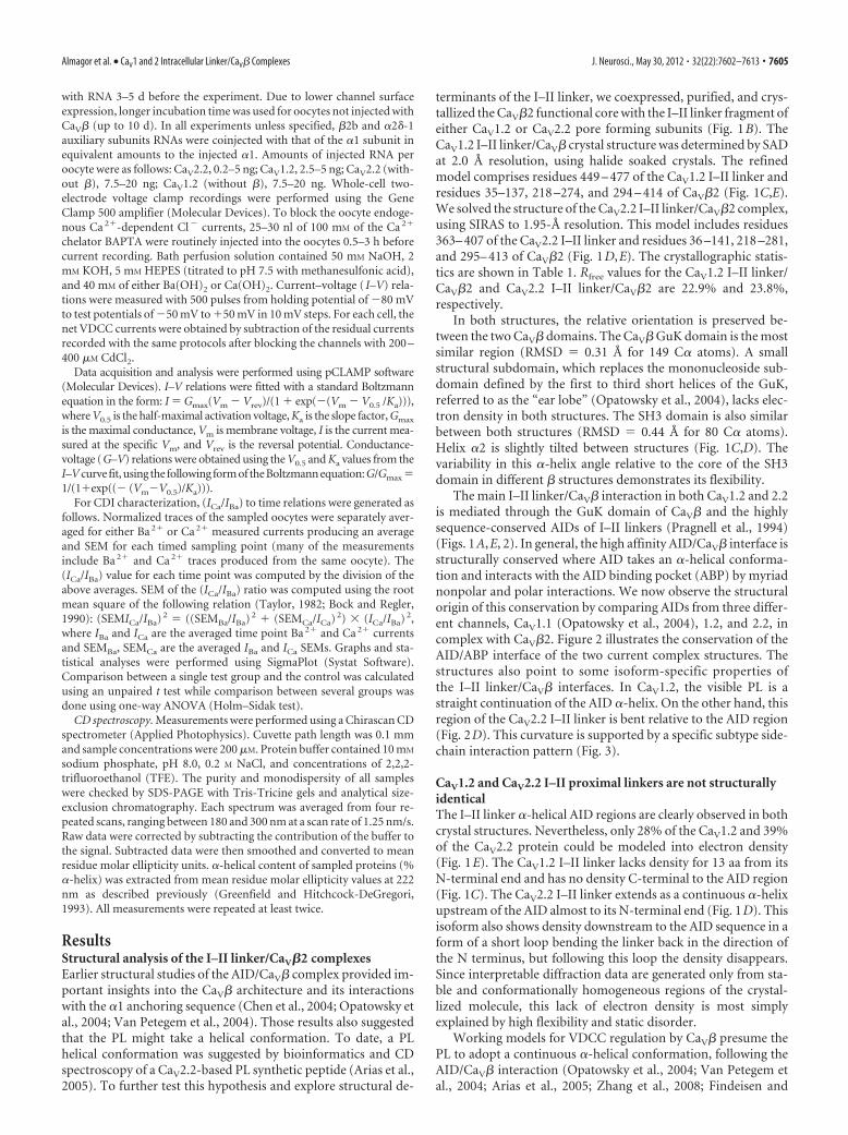
Figure 1. Crystal structures of the CaV1.2 and CaV2.2 I–II linker/CaV�2 core complexes. A, Diagram representation of a VDCC. The PL region bridges the �1 transmembrane IS6 pore �-helix (blue)and the AID (green). B, Schematic illustration of the I–II linker constructs. Both I–II linker constructs used for crystallography were coexpressed with CaV�2 functional core (Opatowsky et al., 2004).In the Helix-PL-AID constructs used for CD measurements, the blue cylinder represents an 18-residue high helical-propensity sequence (Baldwin, 1995), fused upstream to the PL-AID regions. C–D,Ribbon depiction of the CaV1.2 I–II linker/CaV�2 (C) and CaV2.2 I–II linker/CaV�2 (D) crystal structures. While in CaV2.2, the helix extends upstream of the AID, almost to its N terminus; the helicalextension in the CaV1.2 PL is much shorter and breaks at a conserved glycine (G449). E, Multiple sequence alignment of the rabbit CaV1.1, CaV1.2, and CaV2.2 I–II linkers. Secondary structure ofmodeled residues from 1T3L (green), CaV1.2 I–II linker/CaV�2 (yellow), and CaV2.2 I–II linker/CaV�2 (orange), is presented above. The AID region is framed in yellow. The critical PL glycine residuesare framed in red.
Almagor et al. • CaV1 and 2 Intracellular Linker/CaV� Complexes J. Neurosci., May 30, 2012 • 32(22):7602–7613 • 7603
grown in 2 � YT media plus antibiotics. Expression was induced withIPTG at 16°C. Cells were harvested 12–16 h after induction and stored at�80°C.
Purification of the I–II linker/�2 complexes: Cells suspended in phos-phate buffer (50 mM sodium phosphate pH 8.0, 0.3 M NaCl) plus 0.1%Triton X-100, 15 U/ml DNase, lysozyme, and 1 mM PMSF were lysed bymicrofluidizer and subjected to 1 h centrifugation at 38,700 � g. Thesoluble fraction was purified by sequential Ni 2� chelate, anion-exchange[Q-Sepharose (GE Healthcare)] and size-exclusion [Superdex-200HiPrep (GE Healthcare)] column chromatography, including removalof the HisTag by TEV proteolysis. Final buffer conditions were 20 mM
Tris-HCl, pH 8.0, 0.2 M NaCl, and 5 mM 2-mercaptoethanol.Purification of the Helix-PL-AID peptides: lysate preparation and
subsequent purification were similar as above using a phosphate buf-fer including 20% glycerol without the anion-exchange step. For size-exclusion chromatography a Superdex-75 Hi-prep (GE Healthcare)column was used, with final buffer conditions of 10 mM sodium phos-phate pH 8.0, 0.2 M NaCl.
Crystallography. CaV2.2 I–II linker/�2 complex crystallization: Plate-shaped crystals appeared using vapor diffusion with 0.1 M NaCl, 0.1 M
bicine, pH 7.6 – 8.35, 23–26% PEG 400 (Fluka) at 19°C with a 1:1 protein(13 mg/ml) to reservoir ratio. Macroseeding and microseeding (McPher-son, 1999) methods were used to improve crystal dimensions. Beforecrystal flash-freezing, an eightfold volume of a solution containing 40%(w/v) PEG 400, 5% (v/v) glycerol, 0.1 M bicine, 0.1 M NaCl was gentlyadded to the crystallization drop and air dehydrated for 0.5–2 h (Haebelet al., 2001). For the bromide-soaked datasets, after dehydration, crystalswere transferred to a drop containing 55% (w/v) PEG 400, 5% (v/v)glycerol, 0.1 M bicine, pH 8.05, 0.1 M NaCl, 1 M NaBr and soaked for30 – 60 s before flash-freezing in cryo-loops.
CaV1.2 I–II linker/�2 complex crystallization: Prism shaped crystalsappeared using vapor diffusion methods at 1.1–1.25 M potassium sodiumtartrate, 0.1 M Tris, pH 7.0 – 8.0, at 19°C at 1:1 protein (8 mg/ml) toreservoir ratio plus microseeding seedstock. To minimize crystal crack-ing in the cryoprotectant solution, a gentle chemical cross-linking
method was used in which glutaraldehyde was introduced into the crystalby vapor diffusion (Lusty, 1999). Immediately after this 1 h process, thecrystals were soaked for 30 – 60 s in 1.25 M potassium sodium tartrate, 0.1M Tris, pH 7.85, 12% (w/v) sucrose, 1 M NaBr or 1 M KI (for bromide oriodide datasets) and then flash-frozen for data collection.
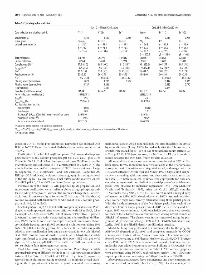
All x-ray diffraction measurements were conducted at 100° K. Forboth crystal forms, anomalous data were collected at the Br anomalousabsorption peak. Intensities were integrated, scaled, and merged with theHKL2000 software (Otwinowski and Minor, 1997). Crystal unit cell pa-rameters, crystallographic symmetry, and data statistics are summarizedin Table 1. In both cases, cell volumes were appropriate for only onecomplex per asymmetric unit. Preliminary partial phases of each of the com-plexes were obtained by molecular replacement (MR) with MOLREP(Vagin and Teplyakov, 1997), using the CaV1.1 AID/�2 complex[(Opatowsky et al., 2004), PDB:1T3L] as a search model, and rigid bodyrefinement in REFMAC5 (Murshudov et al., 1997). Anomalous differ-ence Fourier maps were directly calculated using these partial phases.With the halide substructure of the five highest peaks from each of thedifference Fourier maps, phases from SHARP (de La Fortelle and Bri-cogne, 1997) were computed. Additional halide atom sites were revealedfor each of the substructures in residual maps during several rounds ofSHARP refinement. The phases were further improved using the pro-grams DM (Cowtan and Zhang, 1999) and SOLOMON (Abrahams andLeslie, 1996) in the SHARP density modification interface.
Model building was performed first automatically by the programARP/wARP (Perrakis et al., 1999) and completed manually by COOT(Emsley and Cowtan, 2004). Atomic coordinate refinement was per-formed by alternating use of Phenix (Muller et al., 2010), CNS (Brungeret al., 1998), or REFMAC5 with rounds of manual rebuilding. Solventmolecules were added by automatic solvent building in ARP/wARP. Therefinement statistics are summarized in Table 1. All molecular graphicswere generated using PYMOL (Schrodinger, version 1.3r1). Structuresuperimposition was done using the “Align” function in PYMOL.
Electrophysiology. Xenopus laevis maintenance and oocyte preparationwere as described previously (Shistik et al., 1998). Oocytes were injected
Table 1. Crystallographic statistics
Data collection and phasing statistics
Cav1.2 I–II linker/Cav�2 core Cav2.2 I–II linker/Cav�2 core
I �(1) I �(2) Br � Native Br �(1) Br �(2)
Wavelength (Å) 1.240 1.542 0.918 0.873 0.918 0.918Space group P212121 P212121 P212121 P21 P21 P21
Unit cell parameters (Å) a � 52.7 a � 53.4 a � 53.3 a � 36.9 a � 36.5 a � 36.9b � 70.2 b � 71.0 b � 70.5 b � 67.7 b � 67.6 b � 68.2c � 134.7 c � 134.6 c � 134.3 c � 79.2 c � 77.5 c � 78.4
� � 102.5 � � 102.0 � � 102.3Total reflections 618109 703778 1104840 583596 358991 926807Unique reflections 22964 14694 37774 26222 14590 15003Completeness (%)a 97.6 (88.2) 99.1 (99.2) 91.0 (36.7) 90.1 (53.6) 90.1 (55.1) 90.1 (51.2)Rmerge (%)a,b 6.7 (26.7) 9.3 (33.4) 7.9 (38.8) 7.6 (42.1) 6.2 (23.9) 6.7 (22.7)I/�a 18.7 (3.7) 15.1 (3.3) 17.23 (1.2) 16.4 (1.7) 18.2 (2.9) 17.1 (2.8)Resolution range (Å) 30 –2.30 30 –2.70 30 –1.95 30 –2.00 30 –2.40 30 –2.40f ’/f″ �0.21/4.76 �0.58/6.83 c �8.54/3.82 �8.54/3.82 �8.54/3.82Phasing power (anomalous) 1.079 1.206 0.677 0.447 0.326Phasing power (isomorphous) 0.727 1.266 0.412 0.750Figure of merit 0.317 0.474Beamline (ESRF/Homesource) BM-14 Raxis-IV BM-14 ID-23-2 BM-14 BM-14No. of reflections (working/test) 34543/1813 22583/1235dmin (Å) 1.95 2.0Rwork /Rfree (%) 19.9/22.9 19.8/23.8Rms deviation from ideality:
Bond lengths 0.006 0.008Bond angles 1.03 1.032B factors (Å2) (Rmsd of bonded atoms—main/side chain) 2.18/3.94 2.50/4.53Averaged B factor ( Å2) 47.05 50.79No. of protein atoms/solvent 2432/175 2666/162
aValues of the highest resolution shell are given in parentheses.bRmerge � �hkl�i| Ihkl,i � �I�hkl|/�hkl�i |Ihkl,i| , where Ihkl is the intensity of a reflection and �I�hkl is the average of all observations of this reflection.cf’/f’� values were refined.
7604 • J. Neurosci., May 30, 2012 • 32(22):7602–7613 Almagor et al. • CaV1 and 2 Intracellular Linker/CaV� Complexes
with RNA 3–5 d before the experiment. Due to lower channel surfaceexpression, longer incubation time was used for oocytes not injected withCaV� (up to 10 d). In all experiments unless specified, �2b and �2�-1auxiliary subunits RNAs were coinjected with that of the �1 subunit inequivalent amounts to the injected �1. Amounts of injected RNA peroocyte were as follows: CaV2.2, 0.2–5 ng; CaV1.2, 2.5–5 ng; CaV2.2 (with-out �), 7.5–20 ng; CaV1.2 (without �), 7.5–20 ng. Whole-cell two-electrode voltage clamp recordings were performed using the GeneClamp 500 amplifier (Molecular Devices). To block the oocyte endoge-nous Ca 2�-dependent Cl � currents, 25–30 nl of 100 mM of the Ca 2�
chelator BAPTA were routinely injected into the oocytes 0.5–3 h beforecurrent recording. Bath perfusion solution contained 50 mM NaOH, 2mM KOH, 5 mM HEPES (titrated to pH 7.5 with methanesulfonic acid),and 40 mM of either Ba(OH)2 or Ca(OH)2. Current–voltage ( I–V) rela-tions were measured with 500 pulses from holding potential of �80 mVto test potentials of �50 mV to �50 mV in 10 mV steps. For each cell, thenet VDCC currents were obtained by subtraction of the residual currentsrecorded with the same protocols after blocking the channels with 200 –400 �M CdCl2.
Data acquisition and analysis were performed using pCLAMP software(Molecular Devices). I–V relations were fitted with a standard Boltzmannequation in the form: I � Gmax(Vm � Vrev)/(1 � exp(�(Vm � V0.5 /Ka))),where V0.5 is the half-maximal activation voltage, Ka is the slope factor, Gmax
is the maximal conductance, Vm is membrane voltage, I is the current mea-sured at the specific Vm, and Vrev is the reversal potential. Conductance-voltage (G–V) relations were obtained using the V0.5 and Ka values from theI–V curve fit, using the following form of the Boltzmann equation: G/Gmax �1/(1�exp((� (Vm�V0.5)/Ka))).
For CDI characterization, (ICa/IBa) to time relations were generated asfollows. Normalized traces of the sampled oocytes were separately aver-aged for either Ba 2� or Ca 2� measured currents producing an averageand SEM for each timed sampling point (many of the measurementsinclude Ba 2� and Ca 2� traces produced from the same oocyte). The(ICa/IBa) value for each time point was computed by the division of theabove averages. SEM of the (ICa/IBa) ratio was computed using the rootmean square of the following relation (Taylor, 1982; Bock and Regler,1990): (SEMICa/IBa) 2 � ((SEMBa/IBa) 2 � (SEMCa/ICa) 2) � (ICa/IBa) 2,where IBa and ICa are the averaged time point Ba 2� and Ca 2� currentsand SEMBa, SEMCa are the averaged IBa and ICa SEMs. Graphs and sta-tistical analyses were performed using SigmaPlot (Systat Software).Comparison between a single test group and the control was calculatedusing an unpaired t test while comparison between several groups wasdone using one-way ANOVA (Holm–Sidak test).
CD spectroscopy. Measurements were performed using a Chirascan CDspectrometer (Applied Photophysics). Cuvette path length was 0.1 mmand sample concentrations were 200 �M. Protein buffer contained 10 mM
sodium phosphate, pH 8.0, 0.2 M NaCl, and concentrations of 2,2,2-trifluoroethanol (TFE). The purity and monodispersity of all sampleswere checked by SDS-PAGE with Tris-Tricine gels and analytical size-exclusion chromatography. Each spectrum was averaged from four re-peated scans, ranging between 180 and 300 nm at a scan rate of 1.25 nm/s.Raw data were corrected by subtracting the contribution of the buffer tothe signal. Subtracted data were then smoothed and converted to meanresidue molar ellipticity units. �-helical content of sampled proteins (%�-helix) was extracted from mean residue molar ellipticity values at 222nm as described previously (Greenfield and Hitchcock-DeGregori,1993). All measurements were repeated at least twice.
ResultsStructural analysis of the I–II linker/CaV�2 complexesEarlier structural studies of the AID/CaV� complex provided im-portant insights into the CaV� architecture and its interactionswith the �1 anchoring sequence (Chen et al., 2004; Opatowsky etal., 2004; Van Petegem et al., 2004). Those results also suggestedthat the PL might take a helical conformation. To date, a PLhelical conformation was suggested by bioinformatics and CDspectroscopy of a CaV2.2-based PL synthetic peptide (Arias et al.,2005). To further test this hypothesis and explore structural de-
terminants of the I–II linker, we coexpressed, purified, and crys-tallized the CaV�2 functional core with the I–II linker fragment ofeither CaV1.2 or CaV2.2 pore forming subunits (Fig. 1B). TheCaV1.2 I–II linker/CaV� crystal structure was determined by SADat 2.0 Å resolution, using halide soaked crystals. The refinedmodel comprises residues 449 – 477 of the CaV1.2 I–II linker andresidues 35–137, 218 –274, and 294 – 414 of CaV�2 (Fig. 1C,E).We solved the structure of the CaV2.2 I–II linker/CaV�2 complex,using SIRAS to 1.95-Å resolution. This model includes residues363– 407 of the CaV2.2 I–II linker and residues 36 –141, 218 –281,and 295– 413 of CaV�2 (Fig. 1D,E). The crystallographic statis-tics are shown in Table 1. Rfree values for the CaV1.2 I–II linker/CaV�2 and CaV2.2 I–II linker/CaV�2 are 22.9% and 23.8%,respectively.
In both structures, the relative orientation is preserved be-tween the two CaV� domains. The CaV� GuK domain is the mostsimilar region (RMSD � 0.31 Å for 149 C� atoms). A smallstructural subdomain, which replaces the mononucleoside sub-domain defined by the first to third short helices of the GuK,referred to as the “ear lobe” (Opatowsky et al., 2004), lacks elec-tron density in both structures. The SH3 domain is also similarbetween both structures (RMSD � 0.44 Å for 80 C� atoms).Helix �2 is slightly tilted between structures (Fig. 1C,D). Thevariability in this �-helix angle relative to the core of the SH3domain in different � structures demonstrates its flexibility.
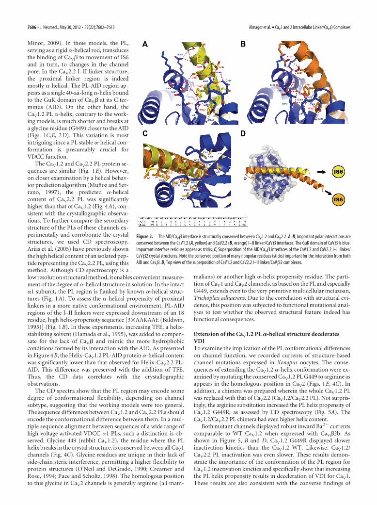
The main I–II linker/CaV� interaction in both CaV1.2 and 2.2is mediated through the GuK domain of CaV� and the highlysequence-conserved AIDs of I–II linkers (Pragnell et al., 1994)(Figs. 1A,E, 2). In general, the high affinity AID/CaV� interface isstructurally conserved where AID takes an �-helical conforma-tion and interacts with the AID binding pocket (ABP) by myriadnonpolar and polar interactions. We now observe the structuralorigin of this conservation by comparing AIDs from three differ-ent channels, CaV1.1 (Opatowsky et al., 2004), 1.2, and 2.2, incomplex with CaV�2. Figure 2 illustrates the conservation of theAID/ABP interface of the two current complex structures. Thestructures also point to some isoform-specific properties ofthe I–II linker/CaV� interfaces. In CaV1.2, the visible PL is astraight continuation of the AID �-helix. On the other hand, thisregion of the CaV2.2 I–II linker is bent relative to the AID region(Fig. 2D). This curvature is supported by a specific subtype side-chain interaction pattern (Fig. 3).
CaV1.2 and CaV2.2 I–II proximal linkers are not structurallyidenticalThe I–II linker �-helical AID regions are clearly observed in bothcrystal structures. Nevertheless, only 28% of the CaV1.2 and 39%of the CaV2.2 protein could be modeled into electron density(Fig. 1E). The CaV1.2 I–II linker lacks density for 13 aa from itsN-terminal end and has no density C-terminal to the AID region(Fig. 1C). The CaV2.2 I–II linker extends as a continuous �-helixupstream of the AID almost to its N-terminal end (Fig. 1D). Thisisoform also shows density downstream to the AID sequence in aform of a short loop bending the linker back in the direction ofthe N terminus, but following this loop the density disappears.Since interpretable diffraction data are generated only from sta-ble and conformationally homogeneous regions of the crystal-lized molecule, this lack of electron density is most simplyexplained by high flexibility and static disorder.
Working models for VDCC regulation by CaV� presume thePL to adopt a continuous �-helical conformation, following theAID/CaV� interaction (Opatowsky et al., 2004; Van Petegem etal., 2004; Arias et al., 2005; Zhang et al., 2008; Findeisen and
Almagor et al. • CaV1 and 2 Intracellular Linker/CaV� Complexes J. Neurosci., May 30, 2012 • 32(22):7602–7613 • 7605
Minor, 2009). In these models, the PL,serving as a rigid �-helical rod, transducesthe binding of CaV� to movement of IS6and in turn, to changes in the channelpore. In the CaV2.2 I–II linker structure,the proximal linker region is indeedmostly �-helical. The PL-AID region ap-pears as a single 40-aa-long �-helix boundto the GuK domain of CaV� at its C ter-minus (AID). On the other hand, theCaV1.2 PL �-helix, contrary to the work-ing models, is much shorter and breaks ata glycine residue (G449) closer to the AID(Figs. 1C,E, 2D). This variation is mostintriguing since a PL stable �-helical con-formation is presumably crucial forVDCC function.
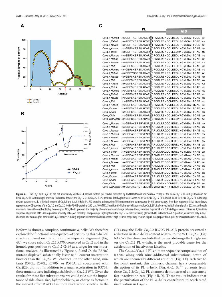
The CaV1.2 and CaV2.2 PL protein se-quences are similar (Fig. 1E). However,on closer examination by a helical behav-ior prediction algorithm (Munoz and Ser-rano, 1997), the predicted �-helicalcontent of CaV2.2 PL was significantlyhigher than that of CaV1.2 (Fig. 4A), con-sistent with the crystallographic observa-tions. To further compare the secondarystructure of the PLs of these channels ex-perimentally and corroborate the crystalstructures, we used CD spectroscopy.Arias et al. (2005) have previously shownthe high helical content of an isolated pep-tide representing the CaV2.2 PL, using thismethod. Although CD spectroscopy is alow resolution structural method, it enables convenient measure-ment of the degree of �-helical structure in solution. In the intact�1 subunit, the PL region is flanked by known �-helical struc-tures (Fig. 1A). To assess the �-helical propensity of proximallinkers in a more native conformational environment, PL-AIDregions of the I–II linkers were expressed downstream of an 18residue, high helix-propensity sequence [3�AAKAAE (Baldwin,1995)] (Fig. 1B). In these experiments, increasing TFE, a helix-stabilizing solvent (Hamada et al., 1995), was added to compen-sate for the lack of CaV� and mimic the more hydrophobicconditions formed by its interaction with the AID. As presentedin Figure 4B, the Helix-CaV1.2 PL-AID protein �-helical contentwas significantly lower than that observed for Helix-CaV2.2 PL-AID. This difference was preserved with the addition of TFE.Thus, the CD data correlates with the crystallographicobservations.
The CD spectra show that the PL region may encode somedegree of conformational flexibility, depending on channelsubtype, suggesting that the working models were too general.The sequence differences between CaV1.2 and CaV2.2 PLs shouldencode the conformational difference between them. In a mul-tiple sequence alignment between sequences of a wide range ofhigh voltage activated VDCC �1 PLs, such a distinction is ob-served. Glycine 449 (rabbit CaV1.2), the residue where the PLhelix breaks in the crystal structure, is conserved between all CaV1channels (Fig. 4C). Glycine residues are unique in their lack ofside-chain steric interference, permitting a higher flexibility toprotein structures (O’Neil and DeGrado, 1990; Creamer andRose, 1994; Pace and Scholtz, 1998). The homologous positionto this glycine in CaV2 channels is generally arginine (all mam-
malians) or another high �-helix propensity residue. The parti-tion of CaV1 and CaV2 channels, as based on the PL and especiallyG449, extends even to the very primitive multicellular metazoan,Trichoplax adhaerens. Due to the correlation with structural evi-dence, this position was subjected to functional mutational anal-yses to test whether the observed structural feature indeed hasfunctional consequences.
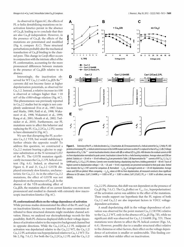
Extension of the CaV1.2 PL �-helical structure deceleratesVDITo examine the implication of the PL conformational differenceson channel function, we recorded currents of structure-basedchannel mutations expressed in Xenopus oocytes. The conse-quences of extending the CaV1.2 �-helix conformation were ex-amined by mutating the conserved CaV1.2 PL G449 to arginine asappears in the homologous position in CaV2 (Figs. 1E, 4C). Inaddition, a chimera was prepared wherein the whole CaV1.2 PLwas replaced with that of CaV2.2 (CaV1.2/CaV2.2 PL). Not surpris-ingly, the arginine substitution increased the PL helix propensity ofCaV1.2 G449R, as assessed by CD spectroscopy (Fig. 5A). TheCaV1.2/CaV2.2 PL chimera had even higher helix content.
Both mutant channels displayed robust inward Ba 2� currentscomparable to WT CaV1.2 when expressed with CaV�2b. Asshown in Figure 5, B and D, CaV1.2 G449R displayed slowerinactivation kinetics than the CaV1.2 WT. Likewise, CaV1.2/CaV2.2 PL inactivation was even slower. These results demon-strate the importance of the conformation of the PL region forCaV1.2 inactivation kinetics and specifically show that increasingthe PL helix propensity results in deceleration of VDI for CaV1.These results are also consistent with the converse findings of
Figure 2. The AID/CaV� interface is structurally conserved between CaV1.2 and CaV2.2. A, B, Important polar interactions areconserved between the CaV1.2 (A, yellow) and CaV2.2 (B, orange) I–II linker/CaV� interfaces. The GuK domain of CaV� is blue.Important interface residues appear as sticks. C, Superposition of the AID/CaV� interfaces of the CaV1.2 and CaV2.2 I–II linker/CaV�2 crystal structures. Note the conserved position of many nonpolar residues (sticks) important for the interaction from bothAID and Cav�. D, Top view of the superposition of CaV1.2 and CaV2.2 I–II linker/CaV�2 complexes.
7606 • J. Neurosci., May 30, 2012 • 32(22):7602–7613 Almagor et al. • CaV1 and 2 Intracellular Linker/CaV� Complexes
Findeisen and Minor (2009), who showed that �-helix destabi-lizing polyglycine substitution mutations in the CaV1.2 PL accel-erated VDI.
As both �1 PL and CaV� are physically coupled and impact chan-nel activity, we then asked what the PL conformational effects of ourmutations are when measured by oocytes injected with �1 and �2�
RNA alone. Xenopus oocytes are known toendogenously express small amounts ofCaV�3, capable of acting upon recombi-nantly expressed VDCCs. These endoge-nous proteins are critical for the membranetransport of expressed �1 genes in the ab-sence of exogenously added CaV� (Tareiluset al., 1997). However, VDCC currents areunaffected by these endogenous CaV�s(Zhang et al., 2008), since in the steady state,their effective concentration is too low forbinding the abundant plasma membranelocalized channels (He et al., 2007). In allour experiments, the absence of CaV� at theplasma membrane was apparent by theslower channel expression and lower cur-rent amplitudes measured. In addition, forall measured currents, the voltage depen-dence of activation was relatively depo-larized, as expected by the lack ofhyperpolarizing shift induced by CaV� (Ta-ble 2). As shown in Figure 5, B and C, theeffect of CaV1.2 G449R, as that of CaV1.2/CaV1.2 PL, on inactivation kinetics weresimilar with or without CaV�2b. Thus,CaV� is not required for the effect of PLstructure on CaV1.2 inactivation kinetics.
Extension of the CaV1.2 PL �-helicalstructure decelerates CDICaV1 channel inactivation is thought to takeplace through two separate but parallelmechanisms, CDI and VDI (Lee et al., 1985;Budde et al., 2002; Kim et al., 2004; Cens etal., 2006). Following the observation thatextension of the CaV1.2 PL �-helix deceler-ates VDI, we also investigated its effect onCDI. When Ca2� is the permeable ion as inphysiological conditions, both CDI andVDI take place. On the other hand, whenCa2� is replaced with Ba2�, only VDI oc-curs. The combined analysis of Ba2� versusCa2� currents is thus traditionally used toquantify the isolated effect of CDI (Zuhlkeand Reuter, 1998; Peterson et al., 1999, 2000;Liang et al., 2003; Mori et al., 2004). Barrettand Tsien (2008), treating VDI and CDI asprocesses with independent probabilities,suggested using the ratio of normalized (ICa/IBa) to provide a measure of CDI, especiallyin cases where changes in VDI are observed.We have used this method. As shown in Fig-ure 5E, a small but significant reduction inCDI was observed for the CaV1.2 G449Rmutant. A more significant reduction wasobserved for the CaV1.2/2.2 PL chimera.Together, these data suggest that extension
of the CaV1.2 PL �-helix decelerates both CDI and VDI.
Perturbation of CaV2.2 PL �-helical structure accelerates VDIBased on the observed effect of the PL conformation on CaV1.2inactivation, we further extended our investigation to the CaV2.2channel. As the crystal structure reveals, the PL of this channel
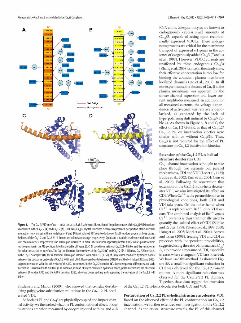
Figure 3. The CaV�/AID interface—polar contacts. A, B, A schematic illustration of the polar contacts of the CaV�/AID interfaceas observed in the CaV2.2 (A) and CaV1.2 (B) I–II linker/CaV�2 crystal structures. Schemes represent a perspective of the ABP/AIDinteraction network using the orientation of C and D (top), rotated 90° counterclockwise. CaV� residues appear as blue boxes.Residues of the CaV1.2 and CaV2.2 I–II linkers are yellow and orange, respectively. Open and closed circles denote backbone andside-chain moieties, respectively. The AID region is framed in black. The numbers appearing before AID residues point to theirrelative position in the AID positions listed in the table of Figure 2C. C, D, �-helix curvature of CaV2.2 I–II linker and the variation inthe polar contacts of its interface. Top (up) and bottom (down) views of the CaV2.2 (C) and CaV1.2 (D) I–II linker/ CaV�2 interface.In the CaV1.2 complex (D), the N-terminal AID region interacts with helix �6 (R352) of � by water-mediated hydrogen bondsbetween the backbone carbonyls of CaV1.2 K457 and L460. Hydrogen bonds between � N390 and the I–II linker E462 and D463support interaction with the other side of the AID. In contrast, in the CaV2.2 complex (C), due to sequence differences, no suchinteraction is observed with N390 of �. In addition, instead of water-mediated hydrogen bonds, polar interactions are observedbetween � residue R352 and the AID N terminus E382, allowing closer packing and supporting the curvature of the CaV2.2 I–IIlinker.
Almagor et al. • CaV1 and 2 Intracellular Linker/CaV� Complexes J. Neurosci., May 30, 2012 • 32(22):7602–7613 • 7607
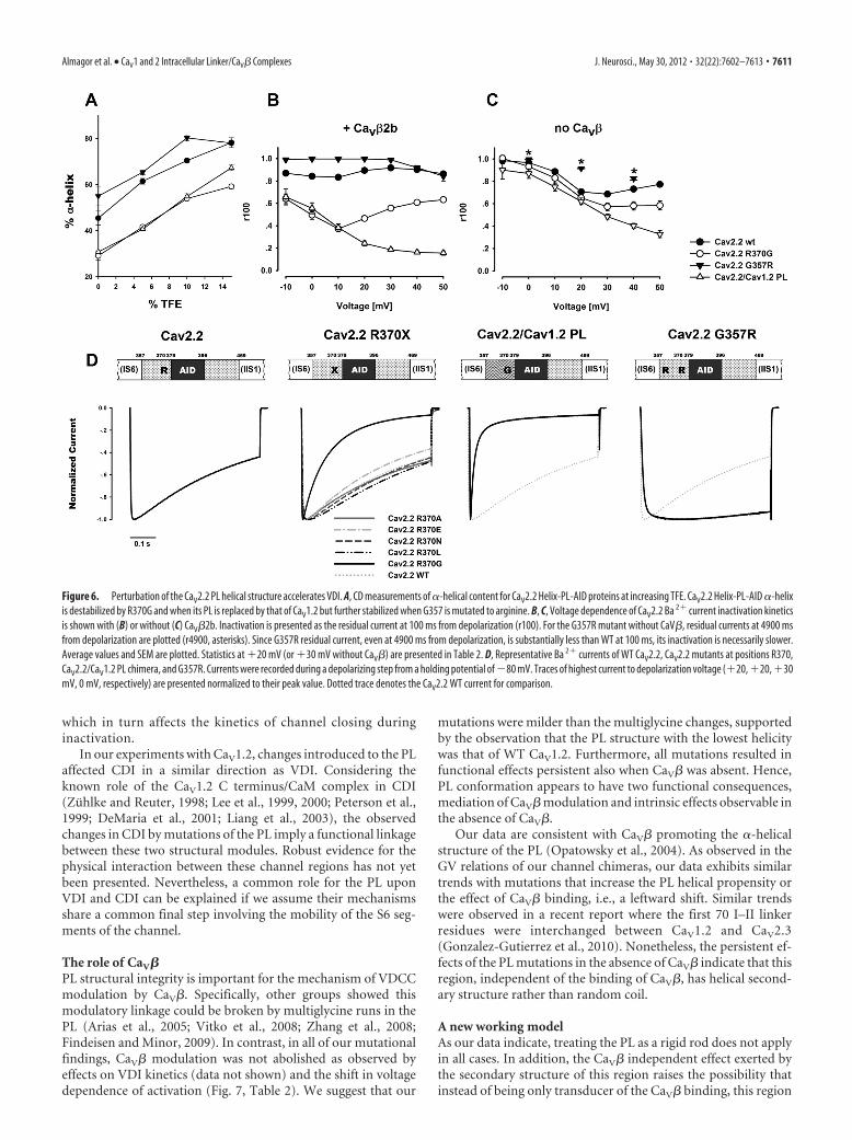
isoform is almost a complete, continuous �-helix. We thereforeexplored the functional consequences of perturbing this �-helicalstructure. Based on the PL multiple sequence alignment (Fig.4C), we chose rabbit CaV2.2 R370, conserved in CaV2 and in thehomologous position to CaV1.2 G449 as a target for our muta-tional analyses. As illustrated by Figure 6, B and D, the R370Gmutant displayed substantially faster Ba 2� current inactivationkinetics than the CaV2.2 WT channel. On the other hand, mu-tants R370E, R370L, R370N, or R370A, all coexpressed withCaV�2b, did not. In addition to a small acceleration in R370E,these mutants were indistinguishable from CaV2.2 WT. Given theresults for these five substitutions, we could rule out the impor-tance of side-chain size, hydrophobicity, or charge as factors inthe marked effect R370G has upon inactivation kinetics. In the
CD assay, the Helix-CaV2.2 R370G PL-AID protein presented areduction in its �-helix content relative to the WT CaV2.2 (Fig.6A). We therefore conclude that the destabilizing effect of glycineon the CaV2.2 PL �-helix is the most probable cause for theacceleration of inactivation kinetics.
The CaV2.2/CaV1.2 PL chimera sequence comprises that ofR370G along with nine additional substitutions, seven ofwhich are chemically different residues (Fig. 1 E). Relative tothe point mutant, this chimeric channel has a comparabledisruption of its PL �-helix (Fig. 6 A). Ba 2� currents fromthese CaV2.2/CaV1.2 PL channels demonstrated an extremelyfast inactivation rate (Fig. 6 B, D). These results indicate thatthe perturbation of the PL �-helix contributes to acceleratedinactivation in CaV2.2.
Figure 4. The CaV1 and CaV2 PLs are not structurally identical. A, Helical content per residue predicted by AGADIR (Munoz and Serrano, 1997) for the Helix-CaV1.2 PL-AID (yellow) and forHelix-CaV2.2 PL-AID (orange) proteins. Red arrow denotes the CaV1.2 G449/CaV2.2 R370 position. Total Agadir scores were 26.38 for Helix-CaV1.2 PL-AID and 40.11 for Helix-CaV2.2 PL-AID usingdefault parameters. B, �-helical content of CaV1.2 and CaV2.2 Helix-PL-AID proteins at increasing TFE concentrations as measured by CD spectroscopy. Error bars represent SEM. Inset showsrepresentative CD spectra of the CaV1.2 and CaV2.2 Helix-PL-AID proteins (200 �M, 10% TFE). Significantly higher �-helix content for CaV2.2 PL is observed by its higher signal at 222 nm. Althoughconstructs have different but highly homologous AIDs, the PL presents the majority of conformational change between them; compare Figures 5A and 6 A wild types versus chimeras. C, Multiplesequence alignment of PL-AID regions for a variety of CaV �1 orthologs and paralogs. Highlighted is the CaV1.2 �-helix-breaking glycine (G449 in Rabbit CaV1.2) position, conserved only in CaV1channels. The homologous position in CaV2 channels is mostly arginine (all mammalians) or another high �-helix propensity residue. Figure was prepared using JALVIEW (Waterhouse et al., 2009).
7608 • J. Neurosci., May 30, 2012 • 32(22):7602–7613 Almagor et al. • CaV1 and 2 Intracellular Linker/CaV� Complexes
As observed in Figure 6C, the effects ofPL �-helix destabilizing mutations on in-activation kinetics persist in the absenceof CaV�, leading us to conclude that theyare also CaV� independent. However, inthe presence of CaV�, the effects of themutations are potentiated and modified(Fig. 6, compare B,C). These structuralperturbations probably alter the mechanicaltransduction of CaV� binding to the chan-nel pore. This change in Cav� effect occursin conjunction with the intrinsic effect of thePL conformation, accounting for the morepronounced differences between mutantsin the presence of CaV�2b relative to itsabsence.
Interestingly, the inactivation ob-served for WT CaV2.2 with CaV�2b Ba 2�
currents did not become faster at higherdepolarization potentials, as observed forCaV1.2. Instead, a relative increase in r100is observed at voltages higher than �10mV of the r100/voltage relation (Fig. 6C).This phenomenon was previously reportedin CaV2.2 studies but its origin is not com-pletely understood (Fox et al., 1987; Jonesand Marks, 1989; Patil et al., 1998; Waka-mori et al., 1998; Wakamori et al., 1999;Kang et al., 2001; Meuth et al., 2002; Tsel-nicker et al., 2010). Furthermore, our PLmutations seem to affect this behavior andreplacing the PL (CaV2.2/CaV1.2 PL) seemsto have eliminated it (Fig. 6C).
We saw that disrupting the PL acceler-ates CaV2.2 VDI, but would stabilizing itfurther obtain the opposite result? Toaddress this question, we constructed aCaV2.2 mutant bearing a glycine to argi-nine substitution at the N-terminal G357of the PL. The G357R mutation signifi-cantly increases the CaV2.2 PL helical con-tent (Fig. 6A). Indeed, as observed inFigure 6, B and D, CaV2.2 G357R dis-played extremely slow kinetics, uncharac-teristic for CaV2.2. As in the other CaV2.2mutations, the effect of G357R was notdependent on the presence of CaV�. In theabsence of the VDI-accelerating effect ofCaV�2b, the mutation effect of on current kinetics was even morepronounced and resulted in channels with extremely slow macro-scopic inactivation kinetics (Fig. 6C).
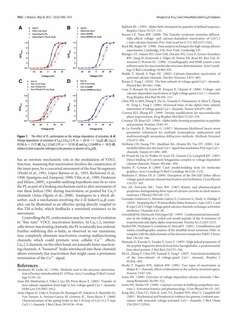
PL conformational effects on the voltage dependence of activationWhile previous studies demonstrated the effect of the PL and CaV�on inactivation kinetics, we reasoned that the same constraints at-tributed to these structural elements might modulate channel acti-vation. Hence, we analyzed our electrophysiology records for thispossibility. Both PL chimeras displayed shifts in their voltage depen-dence of activation relative to the respective background channel butin opposite directions. While the CaV2.2/CaV1.2 PL midpoint ofactivation was depolarized relative to the CaV2.2 WT, the CaV1.2/CaV2.2 PL activation was hyperpolarized relative to CaV1.2 WT (Ta-ble 2, Fig. 7A,C). For both the CaV2.2/CaV1.2 PL and the CaV1.2/
CaV2.2 PL chimeras, this shift was not dependent on the presence ofCaV� (Fig. 7A,C). The CaV� effect on V0.5 (i.e., hyperpolarization)of the activation curves was additive to the effect of the mutations.These results support our hypothesis that the PL regions of bothCaV1.2 and CaV2.2 are also important factors in VDCC voltage-dependent activation.
A small depolarizing shift in the voltage dependence of acti-vation was observed for the point mutant CaV2.2 R370G relativeto the CaV2.2 WT, only in the absence of CaV� (Fig. 7B), while nosignificant shift was observed for CaV1.2 G449R (Fig. 7D). Thesemutations were shown to affect the PL helical content (Figs. 5A,6A). However, possibly due to a smaller structural effect relativeto the chimeras or other factors, their effect on the voltage depen-dence of activation is smaller or undetectable. This finding cor-relates with their milder effect on inactivation.
Figure 5. ExtensionofthePL�-helixdeceleratesCaV1.2inactivation. A,CDmeasurementsof�-helicalcontentforCaV1.2Helix-PL-AIDproteins at increasing TFE.�-helical content increases in the G449R mutant and more so when PL is replaced to that of CaV2.2. B, C, Voltagedependence of CaV1.2 Ba 2� current inactivation kinetics shown with (B) or without (C) CaV�2b. r400 denotes the residual current at 400ms from depolarization normalized to peak current. Inactivation is slower for the �-helix favoring mutations. Average values and SEM areplotted. Statistics at �20 mV or �30 mV without CaV� are presented in Table 2. D, Representative Ba 2� currents of WT CaV1.2, CaV1.2G449R and CaV1.2 /CaV2.2 PL chimera. Currents were recorded during a depolarizing step from a holding potential of �80 mV. Traces ofhighest current to depolarization voltage (�20, �20, and �10 mV, respectively) are presented normalized to their peak value. Dottedtrace denotes the CaV1.2 WT current for comparison. E, Normalized �ICa/IBa of averaged currents at �20 mV depolarization. Averagevalues and SEM are plotted. When comparing �ICa/IBa values at 400 ms from depolarization, all measured constructs show significantdifference in CDI values. (CaV1.2 G449R: p � 0.005 vs WT, p 0.001 vs others; CaV1.2/CaV2.2 PL, p 0.001 vs all others; one-wayANOVA).
Almagor et al. • CaV1 and 2 Intracellular Linker/CaV� Complexes J. Neurosci., May 30, 2012 • 32(22):7602–7613 • 7609
DiscussionThe CaV1 and CaV2 proximal linkers are notstructurally identicalThe PL is located at the heart of a pivotal molecular junction inCaV1 and 2 channels, connected to both the � subunit and thechannel gate. To date, a rigid �-helical PL region was posited as arequirement for the direct coupling of CaV� modulation(Opatowsky et al., 2004; Van Petegem et al., 2004). Our findingshighlight the importance of PL structure for channel function.However, they indicate that CaV1 and CaV2 PLs are not structur-ally identical, with consequences for functions like activation andinactivation properties.
In consonance with this functional significance, the CaV1 andCaV2 PL sequences are extremely conserved within subtypes (Fig.4C). In fact, these protein regions have less sequence variabilitythan their respective AID sequences. Changes in only few posi-tions promote the conformational variation observed. The mostcritical variation seems to be Cav1.2 G449/Cav2.2 R370 at thefourteenth residue position of the PL. The identity of this residuecan be used to partition VDCCs into subtypes, applicable fortheir entire molecular phylogeny.
In addition to the obvious change in secondary structure, thelimited sequence variations between these two isoforms supportmultiple sequence-specific polar interactions possibly responsi-ble for the different angle between CaV� and the PL. That eachconformation is driven by multiple sequence-specific polar inter-actions argues that the observed structures represent a genuineconformational variation between the CaV1 and CaV2 subtypesrather than the result of different crystal packing environments.Notably, residues 372–389 of CaV2.2, that include most of thesequence variations directing this conformational change, areknown to be functionally important for G�� modulation (DeWaard et al., 1997). Furthermore, it was shown previously that anintact �-helical PL is required for CaV2.1 regulation by G��(Zhang et al., 2008). Thus, the low helical content of the CaV1 PLmay be one of the reasons this regulation does not occur in thischannel subtype.
The PL conformation has functional consequences in bothsubtype contextsOur mutational analysis indicates that the respective native PLconformation largely affects inactivation kinetics. Perturbationof CaV2.2 PL �-helical structure (R370G) accelerates VDI kinet-ics. The large effect of a single glycine substitution as opposed toother substitutions strongly supports the conclusion that the ob-served effect originates, in part, from change in the secondarystructure of the PL. In a complementary fashion, we show a sim-ilar effect occurs in CaV1.2, where extension of the PL �-helicalstructure of these channels clearly decelerates VDI. Previous re-sults (Findeisen and Minor, 2009) show that perturbations to theCaV1.2 (and CaV2.1) PL by substitution to multiple glycines ac-celerate VDI. Thus, despite distinct channel subtypes with dis-tinct functional characteristics, PL conformation seems to have acommon role in both CaV1.2 and CaV2.2 inactivation. While PLsecondary structure plays a major role it probably is not the ex-clusive one since the CaV2.2/CaV1.2 PL chimera exhibits evenfaster inactivation than CaV2.2 R370G despite an apparent iden-tical helix propensity.
The PL structural module plays a significant role but is notthe only factor in the inactivation mechanism. WT CaV2.2channels naturally inactivate faster than WT CaV1.2. Inclusionof the more stable �-helical CaV2.2 PL in CaV1.2 slows andalmost eliminates inactivation of these naturally slower inac-tivating channels. On the other hand, the faster inactivatingCaV2.2 inactivates even faster with the CaV1.2 PL. Swappingthis region between these channel isoforms does not exchangetheir inactivation tributes but rather influences their inactiva-tion kinetics in their respective directions, depending to alarge extent on the secondary structure of the PL introduced(Figs. 5A, 6 A). Since the PL is directly coupled to the IS6 of theinner pore one can reasonably envisage its ability to directlyaffect the mobility of the IS6. The simplest explanation forthese recorded effects is for the PL to affect inactivationthrough this TM �-helix. In some way, a change in the PLsarchitecture can be translated to a change in this mobility,
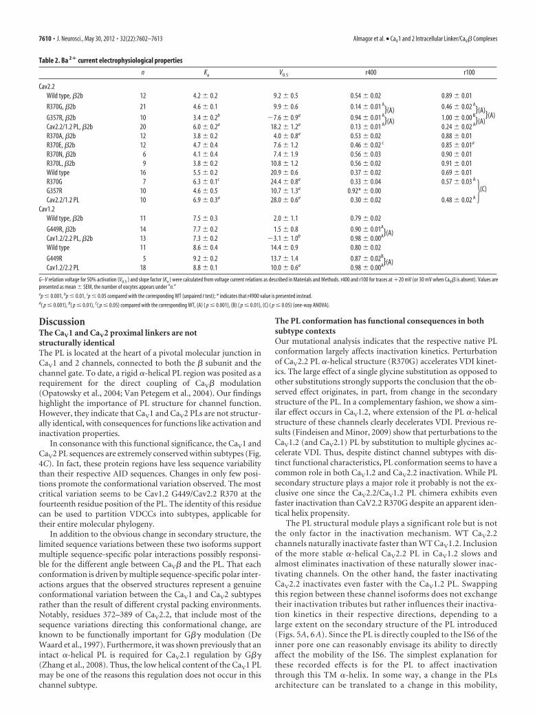
Table 2. Ba 2� current electrophysiological properties
n Ka V0.5 r400 r100
Cav2.2Wild type, �2b 12 4.2 0.2 9.2 0.5 0.54 0.02 0.89 0.01
R370G, �2b 21 4.6 0.1 9.9 0.6 0.14 0.01 A
}(A) 0.46 0.02 A
}(A)}(A)G357R, �2b 10 3.4 0.2b �7.6 0.9a 0.94 0.01 A
}(A) 1.00 0.00 B
}(A)Cav2.2/1.2 PL, �2b 20 6.0 0.2a 18.2 1.2a 0.13 0.01 A 0.24 0.02 A
R370A, �2b 12 3.8 0.2 4.0 0.8a 0.53 0.02 0.88 0.01R370E, �2b 12 4.7 0.4 7.6 1.2 0.46 0.02 c 0.85 0.01a
R370N, �2b 6 4.1 0.4 7.4 1.9 0.56 0.03 0.90 0.01R370L, �2b 9 3.8 0.2 10.8 1.2 0.56 0.02 0.91 0.01Wild type 16 5.5 0.2 20.9 0.6 0.37 0.02 0.69 0.01R370G 7 6.3 0.1c 24.4 0.8a 0.33 0.04 0.57 0.03 A
�(C)G357R 10 4.6 0.5 10.7 1.3a 0.92* 0.00Cav2.2/1.2 PL 10 6.9 0.3a 28.0 0.6a 0.30 0.02 0.48 0.02 A
Cav1.2Wild type, �2b 11 7.5 0.3 2.0 1.1 0.79 0.02
G449R, �2b 14 7.7 0.2 1.5 0.8 0.90 0.01A
}(A)Cav1.2/2.2 PL, �2b 13 7.3 0.2 �3.1 1.0b 0.98 0.00A
Wild type 11 8.6 0.4 14.4 0.9 0.80 0.02
G449R 5 9.2 0.2 13.7 1.4 0.87 0.02B
}(A)Cav1.2/2.2 PL 18 8.8 0.1 10.0 0.6a 0.98 0.00A
G–V relation voltage for 50% activation (V0.5 ) and slope factor (Ka ) were calculated from voltage current relations as described in Materials and Methods. r400 and r100 for traces at �20 mV (or 30 mV when CaV� is absent). Values arepresented as mean SEM, the number of oocytes appears under �n.�ap � 0.001, bp � 0.01, cp � 0.05 compared with the corresponding WT (unpaired t test); * indicates that r4900 value is presented instead.A( p � 0.001), B( p � 0.01), C( p � 0.05) compared with the corresponding WT, (A) ( p � 0.001), (B) ( p � 0.01), (C) ( p � 0.05) (one-way ANOVA).
7610 • J. Neurosci., May 30, 2012 • 32(22):7602–7613 Almagor et al. • CaV1 and 2 Intracellular Linker/CaV� Complexes
which in turn affects the kinetics of channel closing duringinactivation.
In our experiments with CaV1.2, changes introduced to the PLaffected CDI in a similar direction as VDI. Considering theknown role of the CaV1.2 C terminus/CaM complex in CDI(Zuhlke and Reuter, 1998; Lee et al., 1999, 2000; Peterson et al.,1999; DeMaria et al., 2001; Liang et al., 2003), the observedchanges in CDI by mutations of the PL imply a functional linkagebetween these two structural modules. Robust evidence for thephysical interaction between these channel regions has not yetbeen presented. Nevertheless, a common role for the PL uponVDI and CDI can be explained if we assume their mechanismsshare a common final step involving the mobility of the S6 seg-ments of the channel.
The role of CaV�PL structural integrity is important for the mechanism of VDCCmodulation by CaV�. Specifically, other groups showed thismodulatory linkage could be broken by multiglycine runs in thePL (Arias et al., 2005; Vitko et al., 2008; Zhang et al., 2008;Findeisen and Minor, 2009). In contrast, in all of our mutationalfindings, CaV� modulation was not abolished as observed byeffects on VDI kinetics (data not shown) and the shift in voltagedependence of activation (Fig. 7, Table 2). We suggest that our
mutations were milder than the multiglycine changes, supportedby the observation that the PL structure with the lowest helicitywas that of WT CaV1.2. Furthermore, all mutations resulted infunctional effects persistent also when CaV� was absent. Hence,PL conformation appears to have two functional consequences,mediation of CaV� modulation and intrinsic effects observable inthe absence of CaV�.
Our data are consistent with CaV� promoting the �-helicalstructure of the PL (Opatowsky et al., 2004). As observed in theGV relations of our channel chimeras, our data exhibits similartrends with mutations that increase the PL helical propensity orthe effect of CaV� binding, i.e., a leftward shift. Similar trendswere observed in a recent report where the first 70 I–II linkerresidues were interchanged between CaV1.2 and CaV2.3(Gonzalez-Gutierrez et al., 2010). Nonetheless, the persistent ef-fects of the PL mutations in the absence of CaV� indicate that thisregion, independent of the binding of CaV�, has helical second-ary structure rather than random coil.
A new working modelAs our data indicate, treating the PL as a rigid rod does not applyin all cases. In addition, the CaV� independent effect exerted bythe secondary structure of this region raises the possibility thatinstead of being only transducer of the CaV� binding, this region
Figure 6. Perturbation of the CaV2.2 PL helical structure accelerates VDI. A, CD measurements of �-helical content for CaV2.2 Helix-PL-AID proteins at increasing TFE. CaV2.2 Helix-PL-AID �-helixis destabilized by R370G and when its PL is replaced by that of CaV1.2 but further stabilized when G357 is mutated to arginine. B, C, Voltage dependence of CaV2.2 Ba 2� current inactivation kineticsis shown with (B) or without (C) CaV�2b. Inactivation is presented as the residual current at 100 ms from depolarization (r100). For the G357R mutant without CaV�, residual currents at 4900 msfrom depolarization are plotted (r4900, asterisks). Since G357R residual current, even at 4900 ms from depolarization, is substantially less than WT at 100 ms, its inactivation is necessarily slower.Average values and SEM are plotted. Statistics at �20 mV (or �30 mV without CaV�) are presented in Table 2. D, Representative Ba 2� currents of WT CaV2.2, CaV2.2 mutants at positions R370,CaV2.2/CaV1.2 PL chimera, and G357R. Currents were recorded during a depolarizing step from a holding potential of �80 mV. Traces of highest current to depolarization voltage (�20, �20, �30mV, 0 mV, respectively) are presented normalized to their peak value. Dotted trace denotes the CaV2.2 WT current for comparison.
Almagor et al. • CaV1 and 2 Intracellular Linker/CaV� Complexes J. Neurosci., May 30, 2012 • 32(22):7602–7613 • 7611
has an intrinsic mechanistic role in the modulation of VDCCfunction. Assuming that inactivation involves the constriction ofthe inner pore, by a concerted movement of the four S6 segments(Hoshi et al., 1991; Lopez-Barneo et al., 1993; Richmond et al.,1998; Spaetgens and Zamponi, 1999; Vilin et al., 1999; Findeisenand Minor, 2009), a possible unifying hypothesis may be to viewthe PL as part of a braking mechanism used to alter movement ofone these helices (IS6) during inactivation, as posited for CaV3channels (Arias-Olguín et al., 2008). Analogous to a shock ab-sorber, such a mechanism involving the I–II linker/CaV� com-plex can be illustrated as an effective spring directly coupled tothis TM �-helix, which feedbacks a restrained resistance to itsmovement.
Controlling the PL conformation may be one way of evolutionto “fine tune” VDCC inactivation kinetics. In CaV1.2, intrinsi-cally slower inactivating channels, the PL is naturally less ordered.Further stabilizing this �-helix, as observed in our mutations,may completely eliminate inactivation creating malfunctioningchannels, which could promote toxic cellular Ca 2� effects.CaV2.2 channels, on the other hand, are naturally faster inactivat-ing channels. A “loosened” brake introduced into these channelsallows extremely fast inactivation that might cause a prematuretermination of the Ca 2� signal.
ReferencesAbrahams JP, Leslie AG (1996) Methods used in the structure determina-
tion of bovine mitochondrial F1 ATPase. Acta Crystallogr D Biol Crystal-logr 52:30 – 42.
Arias JM, Murbartian J, Vitko I, Lee JH, Perez-Reyes E (2005) Transfer ofbeta subunit regulation from high to low voltage-gated Ca2� channels.FEBS Lett 579:3907–3912.
Arias-Olguín II, Vitko I, Fortuna M, Baumgart JP, Sokolova S, Shumilin IA,Van Deusen A, Soriano-García M, Gomora JC, Perez-Reyes E (2008)Characterization of the gating brake in the I–II loop of Ca(v)3.2 T-typeCa(2�) channels. J Biol Chem 283:8136 – 8144.
Baldwin RL (1995) Alpha-helix formation by peptides of defined sequence.Biophys Chem 55:127–135.
Barrett CF, Tsien RW (2008) The Timothy syndrome mutation differen-tially affects voltage- and calcium-dependent inactivation of CaV1.2L-type calcium channels. Proc Natl Acad Sci U S A 105:2157–2162.
Bock RK, Regler M (1990) Data analysis techniques for high-energy physicsexperiments. Cambridge, UK; New York: Cambridge UP.
Brunger AT, Adams PD, Clore GM, DeLano WL, Gros P, Grosse-KunstleveRW, Jiang JS, Kuszewski J, Nilges M, Pannu NS, Read RJ, Rice LM, Si-monson T, Warren GL (1998) Crystallography and NMR system: a newsoftware suite for macromolecular structure determination. Acta Crystal-logr D Biol Crystallogr 54:905–921.
Budde T, Meuth S, Pape HC (2002) Calcium-dependent inactivation ofneuronal calcium channels. Nat Rev Neurosci 3:873– 883.
Buraei Z, Yang J (2010) The beta subunit of voltage-gated Ca2� channels.Physiol Rev 90:1461–1506.
Cens T, Rousset M, Leyris JP, Fesquet P, Charnet P (2006) Voltage- andcalcium-dependent inactivation in high voltage-gated Ca(2�) channels.Prog Biophys Mol Biol 90:104 –117.
Chen YH, Li MH, Zhang Y, He LL, Yamada Y, Fitzmaurice A, Shen Y, ZhangH, Tong L, Yang J (2004) Structural basis of the alpha1-beta subunitinteraction of voltage-gated Ca2� channels. Nature 429:675– 680.
Cowtan KD, Zhang KY (1999) Density modification for macromolecularphase improvement. Prog Biophys Mol Biol 72:245–270.
Creamer TP, Rose GD (1994) Alpha-helix-forming propensities in peptidesand proteins. Proteins 19:85–97.
de La Fortelle E, Bricogne G (1997) Maximum-likelihood heavy-atomparameter refinement for multiple isomorphous replacement andmultiwavelength anomalous diffraction methods. Methods Enzymol276:472– 494.
DeMaria CD, Soong TW, Alseikhan BA, Alvania RS, Yue DT (2001) Cal-modulin bifurcates the local Ca2� signal that modulates P/Q-type Ca2�channels. Nature 411:484 – 489.
De Waard M, Liu H, Walker D, Scott VE, Gurnett CA, Campbell KP (1997)Direct binding of G-protein betagamma complex to voltage-dependentcalcium channels. Nature 385:446 – 450.
Emsley P, Cowtan K (2004) Coot: model-building tools for moleculargraphics. Acta Crystallogr D Biol Crystallogr 60:2126 –2132.
Findeisen F, Minor DL Jr (2009) Disruption of the IS6-AID linker affectsvoltage-gated calcium channel inactivation and facilitation. J Gen Physiol133:327–343.
Fox AP, Nowycky MC, Tsien RW (1987) Kinetic and pharmacologicalproperties distinguishing three types of calcium currents in chick sensoryneurones. J Physiol 394:149 –172.
Gonzalez-Gutierrez G, Miranda-Laferte E, Contreras G, Neely A, Hidalgo P(2010) Swapping the I–II intracellular linker between L-type CaV1.2 andR-type CaV2.3 high-voltage gated calcium channels exchanges activationattributes. Channels 4:42–50.
Greenfield NJ, Hitchcock-DeGregori SE (1993) Conformational intermedi-ates in the folding of a coiled-coil model peptide of the N-terminus oftropomyosin and alpha alpha-tropomyosin. Protein Sci 2:1263–1273.
Haebel PW, Wichman S, Goldstone D, Metcalf P (2001) Crystallization andinitial crystallographic analysis of the disulfide bond isomerase DsbC incomplex with the alpha domain of the electron transporter DsbD. J StructBiol 136:162–166.
Hamada D, Kuroda Y, Tanaka T, Goto Y (1995) High helical propensity ofthe peptide fragments derived from beta-lactoglobulin, a predominantlybeta-sheet protein. J Mol Biol 254:737–746.
He LL, Zhang Y, Chen YH, Yamada Y, Yang J (2007) Functional modularityof the beta-subunit of voltage-gated Ca2� channels. Biophys J93:834 – 845.
Hoshi T, Zagotta WN, Aldrich RW (1991) Two types of inactivation inShaker K� channels: effects of alterations in the carboxy-terminal region.Neuron 7:547–556.
Jones SW (1998) Overview of voltage-dependent calcium channels. J Bio-energ Biomembr 30:299 –312.
Jones SW, Marks TN (1989) Calcium currents in bullfrog sympathetic neu-rons. I. Activation kinetics and pharmacology. J Gen Physiol 94:151–167.
Kang MG, Chen CC, Felix R, Letts VA, Frankel WN, Mori Y, Campbell KP(2001) Biochemical and biophysical evidence for gamma 2 subunit asso-ciation with neuronal voltage-activated Ca2� channels. J Biol Chem276:32917–32924.
Figure 7. The effect of PL conformation on the voltage dependence of activation. A–D,Voltage dependence of activation of CaV2.2/CaV1.2 PL (n � 20/10 �/�CaV�) (A), CaV2.2R370G (n � 21/7) (B), CaV1.2/CaV2.2 PL (n � 13/10) (C) and CaV1.2 G449R (n � 14/5) (D),relative to their respective wild types in the presence or absence of CaV�2b.
7612 • J. Neurosci., May 30, 2012 • 32(22):7602–7613 Almagor et al. • CaV1 and 2 Intracellular Linker/CaV� Complexes
Kim J, Ghosh S, Nunziato DA, Pitt GS (2004) Identification of the compo-nents controlling inactivation of voltage-gated Ca2� channels. Neuron41:745–754.
Lee A, Wong ST, Gallagher D, Li B, Storm DR, Scheuer T, Catterall WA(1999) Ca2�/calmodulin binds to and modulates P/Q-type calciumchannels. Nature 399:155–159.
Lee A, Scheuer T, Catterall WA (2000) Ca2�/calmodulin-dependent facil-itation and inactivation of P/Q-type Ca2� channels. J Neurosci20:6830 – 6838.
Lee KS, Marban E, Tsien RW (1985) Inactivation of calcium channels inmammalian heart cells: joint dependence on membrane potential andintracellular calcium. J Physiol 364:395– 411.
Liang H, DeMaria CD, Erickson MG, Mori MX, Alseikhan BA, Yue DT(2003) Unified mechanisms of Ca2� regulation across the Ca2� chan-nel family. Neuron 39:951–960.
Lopez-Barneo J, Hoshi T, Heinemann SH, Aldrich RW (1993) Effects ofexternal cations and mutations in the pore region on C-type inactivationof Shaker potassium channels. Receptors Channels 1:61–71.
Lusty CJ (1999) A gentle vapor-diffusion technique for cross-linking of pro-tein crystals for cryocrystallography. J Appl Cryst 32:106 –112.
McPherson A (1999) Crystallization of biological macromolecules. ColdSpring Harbor, NY: Cold Spring Harbor Laboratory.
Meuth S, Pape HC, Budde T (2002) Modulation of Ca2� currents in ratthalamocortical relay neurons by activity and phosphorylation. EurJ Neurosci 15:1603–1614.
Mori MX, Erickson MG, Yue DT (2004) Functional stoichiometry and localenrichment of calmodulin interacting with Ca2� channels. Science304:432– 435.
Muller CS, Haupt A, Bildl W, Schindler J, Knaus HG, Meissner M, RammnerB, Striessnig J, Flockerzi V, Fakler B, Schulte U (2010) Quantitative pro-teomics of the Cav2 channel nano-environments in the mammalianbrain. Proc Natl Acad Sci U S A 107:14950 –14957.
Munoz V, Serrano L (1997) Development of the multiple sequence approx-imation within the AGADIR model of alpha-helix formation: comparisonwith Zimm-Bragg and Lifson-Roig formalisms. Biopolymers 41:495–509.
Murshudov GN, Vagin AA, Dodson EJ (1997) Refinement of macromolec-ular structures by the maximum-likelihood method. Acta Crystallogr DBiol Crystallogr 53:240 –255.
O’Neil KT, DeGrado WF (1990) A thermodynamic scale for the helix-forming tendencies of the commonly occurring amino acids. Science250:646 – 651.
Opatowsky Y, Chomsky-Hecht O, Kang MG, Campbell KP, Hirsch JA(2003) The voltage-dependent calcium channel beta subunit containstwo stable interacting domains. J Biol Chem 278:52323–52332.
Opatowsky Y, Chen CC, Campbell KP, Hirsch JA (2004) Structural analysisof the voltage-dependent calcium channel beta subunit functional coreand its complex with the alpha 1 interaction domain. Neuron 42:387–399.
Otwinowski Z, Minor W (1997) Processing of X-ray diffraction data col-lected in oscillation mode. Method Enzymol 276:307–326.
Pace CN, Scholtz JM (1998) A helix propensity scale based on experimentalstudies of peptides and proteins. Biophys J 75:422– 427.
Patil PG, Brody DL, Yue DT (1998) Preferential closed-state inactivation ofneuronal calcium channels. Neuron 20:1027–1038.
Perrakis A, Morris R, Lamzin VS (1999) Automated protein model buildingcombined with iterative structure refinement. Nat Struct Biol 6:458 – 463.
Peterson BZ, DeMaria CD, Adelman JP, Yue DT (1999) Calmodulin is theCa2� sensor for Ca 2�-dependent inactivation of L-type calcium chan-nels. Neuron 22:549 –558.
Peterson BZ, Lee JS, Mulle JG, Wang Y, de Leon M, Yue DT (2000) Criticaldeterminants of Ca(2�)-dependent inactivation within an EF-hand mo-tif of L-type Ca(2�) channels. Biophys J 78:1906 –1920.
Pragnell M, De Waard M, Mori Y, Tanabe T, Snutch TP, Campbell KP(1994) Calcium channel beta-subunit binds to a conserved motif in theI–II cytoplasmic linker of the alpha 1-subunit. Nature 368:67–70.
Richmond JE, Featherstone DE, Hartmann HA, Ruben PC (1998) Slow in-activation in human cardiac sodium channels. Biophys J 74:2945–2952.
Shistik E, Ivanina T, Blumenstein Y, Dascal N (1998) Crucial role of N ter-minus in function of cardiac L-type Ca2� channel and its modulation byprotein kinase C. J Biol Chem 273:17901–17909.
Spaetgens RL, Zamponi GW (1999) Multiple structural domains contributeto voltage-dependent inactivation of rat brain alpha(1E) calcium chan-nels. J Biol Chem 274:22428 –22436.
Tareilus E, Roux M, Qin N, Olcese R, Zhou J, Stefani E, Birnbaumer L (1997)A Xenopus oocyte beta subunit: evidence for a role in the assembly/expres-sion of voltage-gated calcium channels that is separate from its role as aregulatory subunit. Proc Natl Acad Sci U S A 94:1703–1708.
Taylor JR (1982) An introduction to error analysis: the study of uncertain-ties in physical measurements. Mill Valley, CA: University Science Books.
Tselnicker I, Tsemakhovich VA, Dessauer CW, Dascal N (2010) Stargazinmodulates neuronal voltage-dependent Ca(2�) channel Ca(v)2.2 by aGbetagamma-dependent mechanism. J Biol Chem 285:20462–20471.
Vagin A, Teplyakov A (1997) MOLREP: an automated program for molec-ular replacement. J Appl Crystallogr 30:1022–1025.
Van Petegem F, Clark KA, Chatelain FC, Minor DL Jr (2004) Structure of acomplex between a voltage-gated calcium channel beta-subunit and analpha-subunit domain. Nature 429:671– 675.
Vilin YY, Makita N, George AL Jr, Ruben PC (1999) Structural determi-nants of slow inactivation in human cardiac and skeletal muscle sodiumchannels. Biophys J 77:1384 –1393.
Vitko I, Shcheglovitov A, Baumgart JP, Arias-Olguín II, Murbartian J, AriasJM, Perez-Reyes E (2008) Orientation of the calcium channel beta rela-tive to the alpha(1)2.2 subunit is critical for its regulation of channelactivity. PLoS One 3:e3560.
Wakamori M, Strobeck M, Niidome T, Teramoto T, Imoto K, Mori Y (1998)Functional characterization of ion permeation pathway in the N-typeCa2� channel. J Neurophysiol 79:622– 634.
Wakamori M, Mikala G, Mori Y (1999) Auxiliary subunits operate as a mo-lecular switch in determining gating behaviour of the unitary N-typeCa2� channel current in Xenopus oocytes. J Physiol 517:659 – 672.
Waterhouse AM, Procter JB, Martin DM, Clamp M, Barton GJ (2009)Jalview Version 2—a multiple sequence alignment editor and analysisworkbench. Bioinformatics 25:1189 –1191.
Zhang Y, Chen YH, Bangaru SD, He L, Abele K, Tanabe S, Kozasa T, Yang J(2008) Origin of the voltage dependence of G-protein regulation of P/Q-type Ca2� channels. J Neurosci 28:14176 –14188.
Zuhlke RD, Reuter H (1998) Ca2�-sensitive inactivation of L-type Ca2�channels depends on multiple cytoplasmic amino acid sequences of thealpha1C subunit. Proc Natl Acad Sci U S A 95:3287–3294.
Almagor et al. • CaV1 and 2 Intracellular Linker/CaV� Complexes J. Neurosci., May 30, 2012 • 32(22):7602–7613 • 7613