Embed Size (px)
Citation preview

Review
s�G
ENETO
SCREEN
REVIEWS Drug Discovery Today � Volume 20, Number 1 � January 2015
Therapeutic applications of the cell-penetrating HIV-1 Tat peptideMafalda Rizzuti, Monica Nizzardo, Chiara Zanetta, Agnese Ramirez andStefania Corti
Dino Ferrari Centre, Neuroscience Section, Department of Pathophysiology and Transplantation (DEPT), University of Milan, Neurology Unit, IRCCS Foundation Ca’
Granda Ospedale Maggiore Policlinico, via Francesco Sforza 35, 20122 Milan, Italy
Over the past decades, many new therapeutic approaches have been developed for several conditions,
including neurodegenerative diseases. However, efficient biodistribution and delivery at biological
target sites are hampered by the presence of cell and tissue barriers, and a clinical therapy is prevented by
the requirement of invasive administration routes. Candidate drug conjugation to cell-penetrating
peptides, which are able to cross cellular membranes and reach biological targets even when
administered systemically, represents a promising tool to overcome this issue. Here, we review the
biology, classification and mechanisms of internalization of cell-penetrating peptides. We focus our
attention on the cell-penetrating peptide: HIV-derived Tat peptide, and discuss its efficient but
controversial use in basic, preclinical and clinical research from its discovery to the present day.
IntroductionMany human diseases, including neurodegenerative disorders, are
currently incurable. New therapeutic approaches able to correct
identified causative genetic defects or early pathogenic mecha-
nisms are strongly needed. The presence of cell and tissue barriers,
such as the blood–brain barrier (BBB) in the specific case of
neurodegenerative diseases, represents a real drawback for system-
ic drug delivery, precluding the ability of therapeutic molecules to
reach their own targets. A promising strategy to increase tissue
biodistribution of therapeutics is represented by their conjugation
with cell-penetrating peptides (CPPs) derived from proteins that
are able to cross biological membranes. CPPs would be able to bear
different therapeutic molecules, conveying them to their specific
target and increasing their concentration in difficult-to-access
tissues. Consequently, their therapeutic efficiency might also be
augmented. This approach has the potential to revolutionize the
treatment of a wide spectrum of human disorders.
One of the most promising and most studied CPPs is the HIV-1
transactivator of transcription peptide (pTat). pTat can be efficiently
linked to different potential therapeutic molecules, including small
molecules and antibodies, peptides, liposomes, nanoparticles and
Corresponding author: Nizzardo, M. ([email protected])
76 www.drugdiscoverytoday.com 1359-6446/06/$ - see front matt
nucleic acids; it represents an extremely powerful tool to increase
tissue biodistribution and the efficiency with which targets are
reached. pTat is a promising strategy for the treatment of various
human diseases, and particularly for neurodegenerative diseases. In
this review, we will first provide an analysis of CPP biology and we
will discuss CPP chemical structure and classification as well as their
mechanisms of internalization. Then, we will focus our attention on
pTat, which represents one of the first peptides identified that is
currently widely studied and used.
The biology of CPPsThe integrity of biological membranes is crucial for tissue homeo-
stasis. Some membranes, such as the BBB, are physically selective
barriers to pathogenic bacteria, viruses and large hydrophilic
molecules, whereas they allow the penetration of small or hydro-
phobic molecules [1]. By contrast, tissue barriers represent an
obstacle to the use of a systemic administration protocol; they
impair the ability of therapeutics to reach their targets when
administered in the bloodstream. Therefore, the potential for
CPP-conjugated molecules to deliver drugs to target tissues would
usher in a new era in the treatment of neurological and non-
neurological disorders, and would increase the possibility of res-
cuing the pathological phenotype of many diseases.
er � 2014 Elsevier Ltd. All rights reserved. http://dx.doi.org/10.1016/j.drudis.2014.09.017
Drug Discovery Today � Volume 20, Number 1 � January 2015 REVIEWS
Reviews�GENETO
SCREEN
CPPs as biological delivery agentsDuring the past 20 years, studies about internalization mechanisms
have identified more than 100 peptidic sequences in the range 5–40
amino acids in length that are able to conduct active molecules,
cargo and drug delivery vectors [2]. In addition, high-throughput
screening of DNA-encoded peptide libraries has resulted in the
discovery of many CPPs, for a detailed review see [3]. pTat was
the first CPP identified; pTat peptide is the basic domain of the Tat
protein, the shortest amino acid sequence that can efficiently enter
cells and promote HIV viral gene expression [4]. Another peptide
called Penetratin has also been identified; it naturally enters nerve
cells and regulates neural morphogenesis through a short sequence
with cell-penetrating properties derived from the third helix of the
Antennapedia homeodomain, a transcription factor of Drosophilia
melanogaster [5]. Many other peptides have been identified and
studied because they represent a promising tool for drug delivery,
particularly in neurological disorders, owing to their potential to
overcome biological membranes and release therapeutic biomacro-
molecules at pharmacological target sites. These peptides were
initially termed Trojan horses or protein transduction domains,
and were named CPPs after a review of their internalization path-
ways, which mainly involve endocytosis and transduction [6].
As with peptides in general, the main concern with CPPs can be
connected to their short duration of action and lack of oral
biodisponibility. Medicinal chemistry can overcome these trou-
bles through the use of artificial amino acids, conformational
stabilization of the 3D structure and the use of alternative routes
of administration to bypass poor oral bioavailability [7].
The physicochemical classification of CPPsCPPs constitute a broad heterogeneous group of peptides derived
from proteins, chimeric sequences resulting from the merger of
two natural sequences or the synthetic result of structure–activity
software prediction studies. Classification criteria are based on the
physicochemical features of the sequences [8]; the main three
classes are cationic (83%), amphipathic (44%) and hydrophobic
(15%) sequences [3].
The cationic class is the largest, and the best-known member of
this class derives from the HIV-1 protein Tat. pTat, Penetratin and
nuclear localization sequences (NLSs) also belong to this class. The
arginine-rich sequence requires at least eight arginine residues
(named octaarginine, R8) for cellular uptake [9]. The typesetting
of the sequence, particularly the relative abundance of arginines, is
crucial to peptide transduction properties. Owing to the specific
guanidine group, arginine residues can easily penetrate cells at
physiological pH. Although lysines present the same positive net
charge as arginine, they are less competent at internalization
because they lack the guanidine head group [2]. Because these
short cationic sequences develop electrostatic interactions with
negatively charged glycoproteins on the cellular surface, they
demonstrate great potential as transmembrane carriers for thera-
peutic compounds [10].
In the second CPP class, primary chimeric amphipathic peptides
such as MPG and Pep-1 are included as well as peptides derived from
natural proteins such as vascular endothelial cadherin peptide
(pVEC). Secondary amphipathic a-helical CPPs in which hydropho-
bic and hydrophilic amino acids occupy different faces of the helix,
such as model amphipathic peptides (MAP) peptide and transportan,
are also enclosed. Finally, b-sheet amphipathic CPPs, such as VT5, and
proline-rich amphipathic peptides, such as Bac7 and sweet arrow
peptide (SAP), are part of this group, for a detailed review see [3].
The third class is the hydrophobic class, which includes pep-
tides based on natural amino acids or chemically modified pep-
tides. The latter are further divided into rigid blocking peptides,
structure peptides (stapled peptides), prenylated sequences and
pepducins. Hydrophobic CPPs appear to cross the cell membrane
directly, avoiding endosomal degradation. Moreover, CPPs can be
characterized as derived from natural proteins or peptides, such as
heparin-binding proteins, DNA-binding and RNA-binding pro-
teins (e.g. Tat peptide), homeoproteins (e.g. Penetratin), signal
peptides, antimicrobial peptides and viral proteins.
Mechanisms of internalizationCPPs are translocated into cells by several mechanisms that are
independent but can occur simultaneously (Fig. 1). The protein or
peptide from which CPPs derive can often provide information
about the mechanism of internalization. Short, positively charged,
arginine-rich CPPs such as pTat increase cellular drug uptake by
interacting with the negatively charged plasma membrane and
activating permeabilization of the cell membrane through a re-
ceptor-independent pathway, which results in endocytosis of the
cargo [11]. At the beginning, the interface between CPPs and the
cell membrane involves an electrostatic interaction between basic
amino acids and negatively charged proteoglycans, mainly substi-
tuted with anionic heparan sulfate associated with arginine-rich
peptide. Nevertheless, nonspecific fluid-phase endocytosis appears
not to involve electrostatic interactions but to require only CPP
contiguity with the cell membrane for uptake [10]. Moreover, CPPs
stimulate intracellular signaling cascades that enrich the biologi-
cal pathway of the uptake process [12]. Endocytosis is a natural,
energy-dependent, cellular process that can begin with electrostat-
ic interactions with proteoglycans at the cellular surface or by
direct destabilization interactions across the lipid bilayer [8]. CPPs
and CPP–drug conjugates can penetrate cells using different en-
docytotic pathways (and in particular pinocytosis), including
macropinocytosis, clathrin-mediated endocytosis, caveolae or lip-
id-raft-mediated endocytosis, and clathrin- or caveolae-indepen-
dent endocytosis [13]. The choice between single and multiple
endocytic uptake mechanisms depends on chemical and physical
peptide sequence properties, on its cargo-molecule-conjugated
features and on cell-specific target characteristics [14]. Peptide
stability is essential to deliver the drug efficiently to the cellular
target site without being prematurely cleaved by proteases; deliv-
ery of conjugates is limited by the extracellular metabolism, which
involves peptide uptake, and intracellular degradation in endocy-
tic vesicles. Endosomal escape is essential to avoid degradation in
lysosomes and allow the cargo to reach its biological target [8].
Many studies display alternative internalization mechanisms to
endocytosis through which CPPs can cross the membranes using an
energy-independent pathway known as the direct translocation
pathway, which is based on spontaneous peptide–membrane inter-
actions. Several hypotheses have been developed suggesting that the
translocation mechanism involves direct membrane penetration,
such as the pore formation model, electroporation-like permeabiliza-
tion, entry at microdomain boundaries or shaping of inverted
micelles [8]. An additional hypothesis about the internalization of
www.drugdiscoverytoday.com 77

REVIEWS Drug Discovery Today � Volume 20, Number 1 � January 2015
Clathrin-mediatedendocytosis Caveolin-mediated
endocytosis
Clathrin-caveolin-independentendocytosis
MacropinocytosisPhagocytosis PinocytosisEndocytosis
Direct translocation
Poreformation
Carpet-likemodel
Membrane-thinningmodel
Inverted micelleformation
++
+
++
+
++
+
++
+
CPP Cargo
+
+ +
+
+ +
+
Nucleic acid
Protein Therapeutics
Imaging agents
Drug Discovery Today
FIGURE 1
Two different mechanisms for cell-penetrating peptide (CPP) internalization: direct translocation and/or endocytosis. CPPs and CPP–drug conjugates can
penetrate cells using different endocytotic pathways, in particular pinocytosis which includes macropinocytosis and clathrin-mediated, caveolae or lipid-raft-
mediated and clathrin- or caveolae-independent endocytosis or phagocytosis. Alternatively, CPPs can cross the membranes using an energy-independentpathway known as the direct translocation pathway, which is based on spontaneous peptide–membrane interactions.
Review
s�G
ENETO
SCREEN
guanidinium-rich CPPs (adaptive translocation) foresees counter
anion scavenging with charge neutralization or CPP inversion using
transmembrane potential as the driving force across the membrane
lipid bilayer [10]. However, all of the hypothesized direct transloca-
tion mechanisms can be clustered into four pathways: inverted
micelle formation, pore formation, the carpet-like model and the
membrane-thinning model [15].
Although direct translocation and endocytic processes can co-
exist at the same time, a specific CPP can use a different endocy-
tosis pathway, as has been reported for Penetratin, nonarginine
and Tat peptides [16]. Recent research reveals that, in addition to
the two main mechanisms of internalization outlined above, there
are other specific pathways of entry mediated by receptors such as
scavenger receptors [17].
HIV-1 Tat peptideSince the discovery of CPPs, many studies have been conducted
to transport a wide variety of therapeutic molecules within the
target cells. After the characterization of cellular and molecular
78 www.drugdiscoverytoday.com
mechanisms needed to support HIV infection, Tat protein was
identified for its great ability to move across cells. Tat protein is a
14 kDa, RNA-binding protein that recognizes the transactivator
response element (TAR), a specific sequence from the viral genome
(Fig. 2) [18]. Tat stimulates HIV-1 gene expression during tran-
scription initiation and elongation, enhancing the processivity of
RNA polymerase II complexes and stimulating the efficient elon-
gation of viral transcripts [19]. It contains a very strong transcrip-
tional activation domain composed of a cysteine-rich region and a
hydrophobic core motif, along with an arginine-rich RNA-bind-
ing motif (ARM) that specifies the binding of Tat. Tat is able to
increase membrane permeability through different mechanisms,
including severe vascular modifications in the expression pat-
terns of claudins, occludins and junction adhesion molecules
(JAMs) of the endothelial tight junctions [20]. Moreover, Tat
protein is able to influence tight junction morphology by regu-
lating matrix metalloproteinase (MMP)-9 [21] and exploiting the
Rho signaling pathway associated with the c-AMP response-ele-
ment-binding protein (CREB)-dependent response [22]. Tat can

Drug Discovery Today � Volume 20, Number 1 � January 2015 REVIEWS
HIV-1 Genome
HIV-1 TAT protein
HIV-1 Tat peptide
N ter Acid /proline rich Cysteine-rich/ZnF Core Basic Glutamine-rich C ter
5′ LTR 3′ LTRgag pol
tat
env
vifvpr tat rev
vputat rev
nef
EXON 1 EXON 2
RR
RR R R
QKK
Arg Arg Arg Arg Arg ArgGlnLys Lys
Drug Discovery Today
FIGURE 2
Tat peptide derivation from HIV-1 Tat protein. Tat peptide is derived from the basic domain of the Tat protein encoded by the HIV-1 genome and it is the shortest
amino acid sequence that can efficiently enter cells and promote HIV viral gene expression.
Reviews�GENETO
SCREEN
also develop a non-receptor transport-mediated mechanism to
enter the bilayer, owing to its highly cationic transduction do-
main, which is responsible for the endocytosis of high molecular
weight molecules [23].
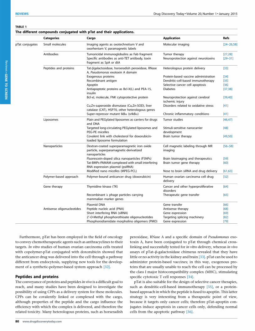
pTat has been derived from HIV-1 Tat protein. Exploiting its
ability to overcome cellular and tissue barriers, pTat has been
conjugated with molecules of a variety of sizes to facilitate their
delivery to target cells, thereby enhancing the likelihood of an
efficient pharmacological action. In particular, pTat has been used
for the transmembrane transport of small molecules, antibodies,
therapeutic peptides and proteins, and has also been conjugated to
liposomes, nanoparticles, small interfering (si)RNAs and antisense
oligonucleotides. In the next part of this review, we provide a
systematic analysis of the various compounds that could be con-
jugated with pTat (Table 1).
Small molecules, antibodies and miscellaneous deliveryagentsSmall molecules (e.g. drugs and imaging agents) have been
linked to pTat in an attempt to increase their bioavailability.
The bioavailability of these molecules has been limited by their
high degree of hydrophilicity, which impairs their ability to cross
the lipid bilayer. In particular, the imaging agents oxotechne-
tium V and oxorhenium V have been conjugated with pTat with
good results, as shown by high intracellular concentrations [24].
Paramagnetic labels complexed with pTat can be detected in
mammalian cells through magnetic resonance imaging (MRI)
[25]. In vitro assays and in vivo biodistribution studies display a
direct correlation between fluorescence intensity and scinti-
graphic and radiometric data, demonstrating that pTat is capable
of efficient cargo internalization for molecular imaging applica-
tions [26].
The use of CPPs was also considered to transport antibodies that
are unable to cross the phospholipid bilayer. For instance, pTat has
been used to convey tumoricidal immunoglobulins inside cells,
resulting in the increased uptake of antitumor antibody, such as
the Fab fragment, by tumor cells [27]. In the antitumor antibody
field, in vivo biodistribution studies of pTat–antibody conjugates
demonstrated good cellular uptake of CPP-conjugated cargo; how-
ever, at the same time, the CPP-conjugated antibodies displayed a
significant reduction in the ability to recognize targets compared
with unconjugated antibodies [28]. pTat has also been used to
deliver specific anti-tetanus toxoid (TET) antibodies required for
tetanus toxin neutralization in the nervous system, providing a
new therapeutic strategy for neuroprotection against a neurotoxin
[29]. The protein transduction domain of HIV-1 pTat has also been
used to deliver antibodies into cells through a genetically engi-
neered fusion protein with a specific staphylococcal protein do-
main named SpA [30]. Despite its success in that context, when Tat
transduction domain was fused to a specific diphtheria toxin
fragment (dtA) it was unable to deliver the enzymatically active
bound fragment to the cytosol efficiently [31].
www.drugdiscoverytoday.com 79
REVIEWS Drug Discovery Today � Volume 20, Number 1 � January 2015
TABLE 1
The different compounds conjugated with pTat and their applications.
Categories Cargo Application Refs
pTat conjugates Small molecules Imaging agents as oxotechnetium V and
oxorhenium V, paramagnetic labels
Molecular imaging [24–26,58]
Antibodies Tumoricidal immunoglobulins as Fab fragment Tumor therapy [27,28]
Specific antibodies as anti-TET antibody, toxinfragment as SpA or dtA
Neuroprotection against neurotoxins [29–31]
Peptides and proteins Tat-bgalactosidase, horseradish peroxidase, RNase
A, Pseudomonas exotoxin A domain
Heterologous protein delivery [33]
Exogenous proteins Protein-based vaccine administration [34]
Recombinant antigen Dendritic-cell-based immunotherapy [35]Apoptin Selective cancer cell apoptosis [36]
Antiapoptotic proteins as Bcl-X(L) and PEA-15,
insulin
Diabetes [37,38]
Bcl-xL molecule, FNK cytoprotective protein Neuroprotection against cerebral
ischemic injury
[39,40]
Cu,Zn-superoxide dismutase (Cu,Zn-SOD), liver
catalase (CAT), HSP70, other heterologous genes
Disorders related to oxidative stress [41]
Super-repressor mutant IkBa (srIkBa) Chronic inflammatory conditions [41]
Liposomes Plain and PEGylated liposomes as carriers for drugs
and DNA
Tumor studies [46,47]
Targeted long-circulating PEGylated liposomes andPEG-PE micelles
Stimuli-sensitive nanocarrierdevelopment
[48]
Covalent link with cholesterol for doxorubicin-
loaded liposome formulation
Brain tumor therapy [49,50]
Nanoparticles Dextran-coated superparamagnetic iron oxideparticle, superparamagnetic-derivatized
nanoparticles
Cell magnetic labeling through MRimaging
[56–58]
Fluorescein-doped silica nanoparticles (FSNPs) Brain bioimaging and therapeutics [59]
Tat-BMPs-PAMAM complexed with small interferingRNA expression plasmid (psiRNA)
Brain tumor gene therapy [60]
Modified nano micelles (MPEG-PCL) Nose to brain siRNA and drug delivery [61,63]
Polymer-based approach Polymer-bound anticancer drug (doxorubicin) Human ovarian carcinoma cell drug
delivery
[32]
Gene therapy Thymidine kinase (TK) Cancer and other hyperproliferativedisorders
[64]
Recombinant l phage particles carrying
mammalian marker genes
Therapeutic gene transfer [65]
Plasmid DNA Gene transfer [66]Antisense oligonucleotides Peptide nucleic acid (PNA) Antisense therapy [68]
Short interfering RNA (siRNA) Gene expression [69]
20-O-Methyl phosphorothioate oligonucleotides Targeting splicing machinery [82]
Phosphorodiamidate morpholino oligomers (PMO) Gene expression [79]
Review
s�G
ENETO
SCREEN
Furthermore, pTat has been employed in the field of oncology
to convey chemotherapeutic agents such as anthracyclines to their
targets. In vitro studies of human ovarian carcinoma cells treated
with copolymer-pTat conjugated with doxorubicin showed that
the anticancer drug was delivered into the cell through a pathway
different from endocytosis, supplying new tools for the develop-
ment of a synthetic-polymer-based system approach [32].
Peptides and proteinsThe conveyance of proteins and peptides in vivo is a difficult goal to
reach, and many studies have been designed to investigate the
possibility of using CPPs as a delivery system for these molecules.
CPPs can be covalently linked or complexed with the cargo,
although properties of the peptide and the cargo influence the
efficiency with which the complex is delivered, and therefore the
related toxicity. Many heterologous proteins, such as horseradish
80 www.drugdiscoverytoday.com
peroxidase, RNase A and a specific domain of Pseudomonas exo-
toxin A, have been conjugated to pTat through chemical cross-
linking and successfully tested for in vitro delivery, whereas in vivo
assays of pTat–b-galactosidase chimeras revealed that they have
little or no activity in the kidney and brain [33]. pTat can be used to
administer protein-based vaccines; in this way, exogenous pro-
teins that are usually unable to reach the cell can be processed by
the class I major histocompatibility complex (MHC), stimulating
specific cytotoxic T cell responses [34].
pTat is also suitable for the design of selective cancer therapies,
such as dendritic-cell-based immunotherapy [35], or a protein-
based approach in which the peptide is fused to apoptin. This latter
strategy is very interesting from a therapeutic point of view,
because it targets only cancer cells; therefore pTat–apoptin con-
jugates induce apoptosis in cancer cells only, defending normal
cells from the apoptotic pathway [36].
Drug Discovery Today � Volume 20, Number 1 � January 2015 REVIEWS
Reviews�GENETO
SCREEN
pTat has also been applied to enhance the survival of trans-
plantable Langherans islets in diabetic patients. The efficiency of
the system relies on the ex vivo delivery of antiapoptotic proteins
such as Bcl-x(L) and PEA-15, leading to the escape of islet cells from
the apoptosis pathway and the preservation of insulin cell secre-
tion [37]. Moreover, pTat displayed the potential to improve oral
insulin absorption through the gastrointestinal mucosa epithelial
layer; researchers observed an effective gain in hormone uptake
compared with unbound insulin [38].
Taking advantage of the ability of pTat to cross the BBB, research
studies have also been directed to deliver in vivo Bcl-xL in an
animal model of focal ischemia/reperfusion, confirming the neu-
roprotective effect of this cargo in cerebral ischemic injury [39]. For
the same application, pTat was fused to an engineered cytopro-
tective protein called FNK, derived from the antiapoptotic Bcl-x
gene, to reduce in vitro ischemic damage of hippocampal neurons
[40].
The fusion of pTat with various proteins has been applied in
many other contexts, ranging from disorders associated with
oxidative stress to inflammatory conditions, in attempts to over-
come the historical difficulty (presently mainly in vivo) of deliver-
ing proteins into cells, for a detailed review see [41].
LiposomesThe conjugation of HIV-1 pTat with liposomes represents a very
promising drug delivery system. The potential of this liposome-
based approach lies in its ability to combine the membrane-
crossing properties of CPPs with the specificity of these carrier
systems loaded with drugs [10]. Liposomes are vesicles comprising
phospholipids that can be filled with different cargos, reducing the
intrinsic drug toxicity and improving molecule biodistribution.
Because conventional liposomes are quickly degraded by the
reticuloendothelial pathway, second-generation liposomes have
been designed with a more stable lipid composition and a poly-
ethylene glycol (PEG)-coated surface that stabilizes these formula-
tions. The new generation of liposomes displays a good
pharmacokinetic profile and reduced systemic toxicity but dis-
plays a delay in liposome uptake; therefore, CPPs have been used to
enhance their poor intracellular delivery. The uptake efficiency is
related to the number of peptide molecules linked to the liposome
surface and to the specific type of target cells [42]. In fact, the
presence of a large number of peptide molecules attached to the
liposome surface allows cellular drug delivery through direct
contact between pTat and the cell surface [43] in an energy-
independent internalization process [44].
pTat conjugates at the vesicular surface can also be used as a safe
gene delivery system with an high transfection rate. pTat-modified
liposomes have been used for gene therapy in tumor cell cultures
and in vivo [45,46] for human brain tumor studies in nude mice
[47]. pTat-modified liposomes coated with PEG have also been
linked with an antibody specifically to target a cell type or an organ
affected by tumors, increasing the specific cellular uptake of the
antibody [48]. In cancer therapy, cholesterol (an electrically neu-
tral element of the liposome) has been used as a linker sequence to
bind pTat through PEG; moreover, liposomes coated with PEG are
an efficient and sturdy delivery system. The cationic charge of
pTat–liposome conjugates increases brain release in vitro and
in vivo; pTat covalently conjugated with cholesterol has been
subsequently used to prepare liposomes filled with doxorubicin
for the treatment of brain glioma. Biodistribution findings
revealed a higher efficiency in brain and heart delivery with low
cardiotoxic risk [49,50].
NanoparticlesTo overcome the BBB in a noninvasive way, different biodegradable
supramolecular nanodevices have been developed. Liposomes, nio-
somes, nanogels and cyclodextrins are some colloidal drug delivery
systems that can target and release bioactive specific molecules in
the central nervous system (CNS) [51]. In the field of brain disorders,
lipid nanoparticles have always been studied because they ensure
improved drug loading, storage stability, ease of production and
safety of formulations for pharmaceutical drug delivery [52].
Superparamagnetic nanoparticles have interesting potential in
the biomedical field. They work as specific imaging contrast
agents, useful labeling or tracking tools for cell detection and
specific purification instruments based on magnetic separation,
for reviews see [2,53]. pTat-conjugated nanoparticles revealed
their ability in cell transduction [54] and showed improvement
in vascular clearance [55]. The first biocompatible nanosystem was
linked to multiple pTat sequences to enhance cell magnetic label-
ing for in vivo cell target detection through MRI [56]. Linking a
greater number of peptide molecules to the magnetic nanoparti-
cles increased the degree of uptake and improved in vivo signaling
[57,58]. pTat-modified nanoparticles can also be used to deliver
therapeutic agents across the BBB, such as fluorescein-doped silica
nanoparticles (FSNPs) derivatized with pTat, which displayed an
ability to label cerebral blood vessels and could be used as potential
tools for bioimaging and therapeutic applications in the brain [59].
In the context of brain tumors, polyamidoamine dendrimer
(PAMAM) and pTat were conjugated to bacterial magnetic nano-
particles (BMPs) called magnetosomes to build a nanoscale mag-
netic gene delivery system for siRNA expression plasmid (psiRNA)
[60]. pTat has also been shown to enhance the delivery rate of
block copolymers to the brain in mice, when the Tat-polymer
conjugated with nano micelles was administered intranasally
through a noninvasive and effective nose-to-brain delivery system,
overcoming the BBB [61]. This approach has also been studied to
improve the delivery of siRNA to the brain as a therapy for several
neurological disorders [62].
Nucleic acidsCPP-based gene therapy has been used to overcome difficulties with
introducing genetic material into target cells, which arise because of
the low safety profile of viral vectors and the inefficiency associated
with nonviral methods, for a complete review see [63]. CPPs such as
pTat have been exploited for cancer treatment with suicide gene
therapy approaches, delivering an enzyme encoding a gene that is
able to produce a cytotoxic effect [64]. CPPs have also been used for
plasmid DNA transfection [65,66] and oligonucleotide delivery,
including peptide nucleic acid (PNA) [67,68] and siRNA [62,69].
Other synthetic CPPs with the ability to bind DNA and transport it
into cells have also been developed [70].
Antisense oligonucleotidesAntisense oligonucleotides (ASOs) are modified nucleotides that
bind specific complementary mRNA sequences. This binding can
www.drugdiscoverytoday.com 81
REVIEWS Drug Discovery Today � Volume 20, Number 1 � January 2015
Review
s�G
ENETO
SCREEN
mark a specific mRNA for degradation or linkage to specific cis-
acting splicing regulatory motifs. Different ASOs utilizing various
chemistries [71–73] have been studied in cell and animal models. A
variety of sequences have been targeted by three different ASO
chemistries: 2?-O-methyl phosphorothioate (2OMePS) oligonu-
cleotides; the more stable variant, 2?-O-methoxyethyl (MOE)
phosphorothioate oligonucleotides; and phosphorodiamidate
morpholino oligomers (MOs). Oligonucleotide studies revealed
that the sugar–phosphate structure is important for membrane
crossing. Changes to the backbone, such as the introduction of a
morpholine ring instead of deoxyribose and phosphorodiamidate
linking groups, render these modified oligos resistant to nuclease
attack [74]. Chemically modified ASOs resistant to cellular endo-
nuclease activity and insusceptible to RNase-H degradation have
already been used to develop therapeutic strategies in animal
models of neuromuscular disorders, including Duchenne muscu-
lar dystrophy and spinal muscular atrophy; they have also recently
been translated in clinical trials [72,75,76]. Although extremely
promising, ASOs (particularly MOs) do not readily cross cell
membranes. Although they can work very well in neonatal mice,
they can be less effective in adult animals, which limits their use
during the symptomatic phase of diseases. Nevertheless, the de-
livery efficiency of ASOs and MOs can be strongly increased by
their conjugation with cationic CPPs, producing a high transfec-
tion efficiency rate that could be further increased owing to the use
of peptide NLSs [66,77]. In fact, the inadequate cellular delivery
rate of MOs has been increased using cationic CPP conjugates,
which increase the volume of distribution of MOs to muscles
throughout the entire body and promote their internalization
through an active process that improves the bioavailability of
ASOs and MOs [78–81]. Although cationic peptides can interact
electrostatically with the anionic backbones of many antisense
structural types that form hairpin structures or intermolecular
aggregates, uncharged antisense oligomers such as MOs would
not have dangerous electrostatic interactions. pTat is a cationic
CPP and its ability to enhance biodistribution of ASOs, for example
phosphorothioate, 29-O-methylphosphorothiate oligonucleotide
(ODN) [82] and PNA [68], has been studied in vitro. pTat has great
potential owing to its ability to target different cell types success-
fully. Covalent conjugation of pTat to MOs significantly enhances
MO uptake, although the resulting pTat–MO conjugate is a bit
more toxic than the unconjugated peptide [79].
pTat and clinical trialsIn the past decade, pTat has proceeded to various phases of human
clinical trials, although no therapy employing pTat has been
approved by the FDA. Revance Therapeutics has completed a Phase
II clinical trial using pTat to deliver botulinum toxin type A in a
topical ointment for the removal of wrinkles (RT-001). Subcuta-
neous infusion of protein kinase Cd inhibitor conjugates with pTat
has been studied in Phase I/II clinical trials to evaluate the efficacy
of the drug in: pain caused by post-herpetic neuralgia, spinal cord
injury or post-operative pain (KAI-1678); blood flow restoration
after a heart attack (KAI-9803); and prevention of ischemic injury
(KAI-1455). A Phase I clinical trial evaluating the safety profile and
immunogenicity of a vaccination with recombinant HIV-1 Tat and
V2-deleted Env after intramuscular and intradermal injection was
recently terminated (ISS P-002). For all of these trials, no study
82 www.drugdiscoverytoday.com
results are posted on ClinicalTrials.gov. The first and the unique
successful clinical trial for the use of pTat closed in 2012 with good
results (NCT00728182). pTat was conjugated with NA-1 (Tat-
NR2B9c), a compound that disrupts pro-death signaling pathways
that involve postsynaptic density-95 protein and can prevent
damage in the brain caused by reduced blood flow. The Canadian
biotechnology company NoNO conducted a Phase II clinical trial
to assess the safety and efficacy of NA-1 in reducing small embolic
strokes in patients that underwent neurosurgery to repair aneur-
ysms. The intravenous infusion of NA-1 resulted in a neuropro-
tective effect of the drug with a reduction in the amount of brain
damage [83]. Based on these results, pTat conjugated with NA-1
can be tested in later-stage clinical trials for stroke and subarach-
noid hemorrhage.
Tat pitfallsAlthough different strategies of pTat conjugation with several
compounds have resulted in very interesting, selective delivery
to target cells, many pitfalls and critical aspects have emerged from
basic, preclinical and clinical studies. Generally speaking, the
stability of the peptide is fundamental to ensure the delivery of
the cargo molecule to the target site. In fact, CPP cleavage by
extracellular proteases influences the peptide uptake the most,
whereas intracellular metabolism in lysosomal vesicles should be
avoided by endosomal escape. Hence, even if stability depends on
the specific peptide and the associated cargo molecule, the entire
delivery system conducted by CPP and administered in vivo has
been modified over time. For instance, peptide moiety has been
modified to use a D-amino acid configuration, which is less sus-
ceptible to protease activity than the natural L-form. Peptide
stability can also be increased by using peptide mimics to increase
the ability to reach the biological target. The challenge is to
achieve a good compromise between avoiding degradation to
the biological target and obtaining a sufficient drug release rate
from the CPP complex, for a detailed review see [10]. To release the
associated drug after cellular internalization, the linker strategy
(e.g. maleimide, amide, thiolmaleimide, thioether, thiazolidine,
oximine, hydrazine and disulfide bonds) should be carefully con-
sidered, especially if it depends on a chemical approach. The
coupling is usually a covalent connection based in most cases
on disulfide binding, which is suitable for cargo release because of
the reduction of intracellular bonds [84]. Additional covalent
strategies include the employment of bifunctional crosslinker
molecules, peptide bonds or producing chimeric fusion protein
in bacteria. Noncovalent linkage takes advantage of electrostatic
interactions between the cationic peptide and the negatively
charged nucleic acid backbone or exploits streptavidin–biotin
attachment [63].
Toxicity is another issue to be considered. The nature of toxicity
is not yet well understood. The safety landscape is directly con-
nected to CPP and cargo toxicity, clearance and immunogenicity.
The potential immune response is mainly related to the genesis of
peptides, because these molecules often derive from non-human
proteins. CPP toxicity could be cell-type-related, dose-dependent
and influenced by chemical–physical features; overall, the final
toxic effect results from the perturbation of plasma membrane
dynamics that occurs at high peptide dosages [85]. Moreover,
toxicity is associated with the amino acid CPP composition and

Drug Discovery Today � Volume 20, Number 1 � January 2015 REVIEWS
Reviews�GENETO
SCREEN
the dose or frequency of administration [77]. Intratracheal or
intraperitoneal CPP administration produces pulmonary toxicity
in vivo [86] and epithelial cell damage in vitro [87]. CPP length
influences the magnitude of the neurotoxic effect, as demonstrat-
ed by pTat, the shorter isoform of which produces greater neuro-
toxic damage than the full-length protein [88]. The cysteine-rich
domain and the basic region of pTat are required for neurotoxicity.
Peptide toxicity increases if CPP is conjugated to a peptide cargo,
depending on the length of the peptide and the dose used [89]. As
described by Moulton et al. [79–81] for MOs, Tat peptide toxicity is
dose-dependent and the conjugated construct was more toxic
than either CPP or MO administered alone. Moulton’s group
found that unconjugated free pTat is toxic by itself, and they
speculated that a dose threshold might exist that should not be
exceeded to ensure safety. Overall, the observed pTat toxicity
poses a challenge for the determination of an effective and safe
dose regimen in humans.
The route of administration represents another concern. As
already mentioned above, some CPPs are associated with a specific
organ toxicity [86,87]. Moreover, oral administration can lead to
immediate degradation in the gastrointestinal system, resulting in
a shorter active plasma half-life, which can interfere with the
achievement of the final target at a meaningful pharmacological
dosage. For neurotherapies, all brain delivery should be addressed
without invasive methods (e.g. intracerebroventricular injection
or special intracranial implants). An alternate route of delivery can
be parenteral administration; however, this strategy can be a
problem for translating peptides into the clinic because of low
rates of patient compliance and adherence to therapy. Another
alternative to intravenous drug delivery into the brain is repre-
sented by intranasal administration, which exploits the wide
surface area and rich vascularity of the olfactory region [90].
The low cell, tissue and organ selectivity of CPPs is another huge
drawback for the therapeutic applications of these carriers. How-
ever, recent promising developments have been made using acti-
vatable CCPs and stimuli-responsive peptides, or by the insertion
of specific localization sequences to address the delivery toward
the proper cellular organelles. Internalization mechanisms take
advantage of tissue- and organ-specific ligands for different recep-
tors. In addition, because each organ expresses a specific set of
molecules (also called ‘zip code’ system) on their vasculature, the
insertion of one of these homing sequences into the CPP–cargo
could represent a strategy for the efficient and cell-specific CPP
delivery allowing the translocation across the cellular membrane,
increasing the potential of the first-generation CPPs [91]. Another
strategy to overcome the low CPP specificity is the recent devel-
opment of activatable CPPs (ACPPs) that become active depending
on the biochemical properties of the target sites. The first ACPP
was developed in 2009 and was a protease-activatable CPP: a
proteolytic cleavage releases the activated peptide that can lead
the cargo to target cells [92]. Moreover, pH, transmembrane po-
tential, hypoxia and the uptake pattern can represent other spe-
cific microenvironment features, detectable for example in
tumoral affected tissues, that should be investigated for exploiting
different stimuli-responsive carriers for cargos delivery [93]. The
expertise in selective intracellular transport through specific local-
ization sequences represents a great potential for the intracellular
direct transport to the nucleus by NLS or to different cell organelles
such as mitochondria, lysosomes or Golgi apparatus by specific
peptides [93].
Concluding remarksAs summarized in this review, many studies have been conducted
with pTat. Despite several promising lines of preclinical evidence
that have demonstrated its ability to overcome biological barriers to
deliver several types of drugs to target tissues, many clinical trials
involving pTat have failed and some critical issues have emerged.
However, as a CPP, pTat represents an interesting and promising
tool to improve the biodistribution of drugs and to allow their
systemic administration owing to its good cell-penetrating capacity
and the fact that it is nonimmunogenic and barely toxic. Moreover,
the development of CPPs from the second generation aiming to
overcome the low cell and tissue specificity of the first CPP genera-
tion lays the groundwork for a feasible cell-type-selective therapy or
even organelle-specific approach suitable for Tat, from a diagnostic
point of view and for therapeutic purposes.
AcknowledgmentsWe wish to thank the Associazione Amici del Centro Dino Ferrari
for its support.
References
1 Eugenin, E.A. et al. (2011) Human immunodeficiency virus infection of human
astrocytes disrupts blood–brain barrier integrity by a gap junction-dependent
mechanism. J. Neurosci. 31, 9456–9465
2 Koren, E. and Torchilin, V.P. (2012) Cell-penetrating peptides: breaking through to
the other side. Trends Mol. Med. 18, 385–393
3 Milletti, F. (2012) Cell-penetrating peptides: classes, origin, and current landscape.
Drug Discov. Today 17, 850–860
4 Frankel, A.D. and Pabo, C.O. (1988) Cellular uptake of the tat protein from human
immunodeficiency virus. Cell 55, 1189–1193
5 Joliot, A. et al. (1991) Antennapedia homeobox peptide regulates neural
morphogenesis. Proc. Natl. Acad. Sci. U. S. A. 88, 1864–1868
6 Richard, J.P. et al. (2003) Cell-penetrating peptides. A re-evaluation of the
mechanism of cellular uptake. J. Biol. Chem. 278, 585–590
7 Nestor, J.J. (2009) The medicinal chemistry of peptides. Curr. Med. Chem. 16, 4399–
4418
8 Bechara, C. and Sagan, S. (2013) Cell-penetrating peptides: 20 years later, where do
we stand? FEBS Lett. 587, 1693–1702
9 Tunnemann, G. et al. (2008) Live-cell analysis of cell penetration ability and toxicity
of oligo-arginines. J. Pept. Sci. 14, 469–476
10 Foged, C. and Nielsen, H.M. (2008) Cell-penetrating peptides for drug delivery
across membrane barriers. Expert Opin. Drug. Deliv. 5, 105–117
11 Derossi, D. et al. (1996) Cell internalization of the third helix of the Antennapedia
homeodomain is receptor-independent. J. Biol. Chem. 271, 18188–18193
12 Nakase, I. et al. (2007) Interaction of arginine-rich peptides with membrane-
associated proteoglycans is crucial for induction of actin organization and
macropinocytosis. Biochemistry 46, 492–501
13 Conner, S.D. and Schmid, S.L. (2003) Regulated portals of entry into the cell. Nature
422, 37–44
14 Tunnemann, G. et al. (2006) Cargo-dependent mode of uptake and bioavailability
of TAT-containing proteins and peptides in living cells. FASEB J. 20, 1775–1784
15 Madani, F. et al. (2011) Mechanisms of cellular uptake of cell-penetrating peptides.
J. Biophys. 2011, 414729
16 Duchardt, F. et al. (2007) A comprehensive model for the cellular uptake of cationic
cell-penetrating peptides. Traffic 8, 848–866
www.drugdiscoverytoday.com 83

REVIEWS Drug Discovery Today � Volume 20, Number 1 � January 2015
Review
s�G
ENETO
SCREEN
17 Ezzat, K. et al. (2012) Scavenger receptor-mediated uptake of cell-penetrating
peptide nanocomplexes with oligonucleotides. FASEB J. 26, 1172–1180
18 Feng, S. and Holland, E.C. (1988) HIV-1 tat trans-activation requires the loop
sequence within tar. Nature 334, 165–167
19 Ott, M. et al. (2004) Tat acetylation: a regulatory switch between early and late
phases in HIV transcription elongation. Novartis Found. Symp. 259, 182–193
20 Banks, W.A. et al. (2005) Permeability of the blood–brain barrier to HIV-1 Tat. Exp.
Neurol. 193, 218–227
21 Xu, R. et al. (2012) HIV-1 Tat protein increases the permeability of brain endothelial
cells by both inhibiting occluding expression and cleaving occludin via matrix
metalloproteinase-9. Brain Res. 1436, 13–19
22 Mediouni, S. et al. (2012) Antiretroviral therapy does not block the secretion of the
human immunodeficiency virus tat protein. Infect. Disord. Drug Targets 12, 81–86
23 Cooper, I.S.K. et al. (2012) Peptide derived from HIV-1 TAT protein, destabilizes a
monolayer of endothelial cells in an in vitro model of the blood–brain barrier, and
allows permeation of high molecular weight proteins. J. Biol. Chem. 287, 44676–
44683
24 Polyakov, V. et al. (2000) Novel TAT-peptide chelates fordirect transduction of
technetium-99m and rhenium into human cells for imaging and radiotherapy.
Bioconjug. Chem. 11, 762–771
25 Bhorade, R. et al. (2000) Macrocyclic chelators with paramagnetic cations are
internalized into mammalian cells via a HIV-tat derived membrane translocation
peptide. Bioconjug. Chem. 11, 301–305
26 Bullok, K.E. et al. (2002) Characterization of novel histidine-tagged Tat-peptide
complexes dual-labeled with (99m)Tc-tricarbonyl and fluorescein for scintigraphy
and fluorescence microscopy. Bioconjug. Chem. 13, 1226–1237
27 Anderson, D.C. et al. (1993) Tumor cell retention of antibody Fab fragments is
enhanced by an attached HIV TAT protein-derived peptide. Biochem. Biophys. Res.
Commun. 194, 876–884
28 Niesner, U. et al. (2002) Quantitation of the tumor-targeting properties of antibody
fragments conjugated to cell-permeating HIV-1 TAT peptides. Bioconjug. Chem. 13,
729–736
29 Stein, S. et al. (1999) A disulfide conjugate between anti-tetanus antibodies and HIV
(37-72)Tat neutralizes tetanus toxin inside chromaffin cells. FEBS Lett. 458, 383–386
30 Mie, M. et al. (2003) Intracellular delivery of antibodies using TAT fusion protein A.
Biochem. Biophys. Res. Commun. 310, 730–734
31 Falnes, P.O. et al. (2001) Ability of the Tat basic domain and VP22 to mediate cell
binding, but not membrane translocation of the diphtheria toxin A-fragment.
Biochemistry 40, 4349–4358
32 Nori, A. et al. (2003) Tat-conjugated synthetic macromolecules facilitate
cytoplasmic drug delivery to human ovarian carcinoma cells. Bioconjug. Chem. 14,
44–50
33 Fawell, S. et al. (1994) Tat-mediated delivery of heterologous proteins into cells. Proc.
Natl. Acad. Sci. U. S. A. 91, 664–668
34 Kim, D.T. et al. (1997) Introduction of soluble proteins into the MHC class I pathway
by conjugation to an HIV tat peptide. J. Immunol. 159, 1666–1668
35 Shibagaki, N. and Udey, M.C. (2002) Dendritic cells transduced with protein
antigens induce cytotoxic lymphocytes and elicit antitumor immunity. J. Immunol.
168, 2393–2401
36 Guelen, L. et al. (2004) TAT-apoptin is efficiently delivered and induces apoptosis in
cancer cells. Oncogene 23, 1153–1165
37 Embury, J. et al. (2001) Proteins linked to a protein transduction domain efficiently
transduce pancreatic islets. Diabetes 50, 1706–1713
38 Liang, J.F. and Yang, V.C. (2005) Insulin-cell penetrating peptide hybrids with
improved intestinal absorption efficiency. Biochem. Biophys. Res. Commun. 335,
734–738
39 Cao, G. et al. (2002) In vivo delivery of a Bcl-xL fusion protein containing the TAT
protein transduction domain protects against ischemic brain injury and neuronal
apoptosis. J. Neurosci. 22, 5423–5431
40 Asoh, S. et al. (2002) Protection against ischemic brain injury by protein
therapeutics. Proc. Natl. Acad. Sci. U. S. A. 99, 17107–17112
41 Gupta, B. et al. (2005) Intracellular delivery of large molecules and small particles by
cell-penetrating proteins and peptides. Adv. Drug Deliv. Rev. 57, 637–651
42 Tseng, Y.L. et al. (2002) Translocation of liposomes into cancer cells by cell-
penetrating peptides penetratin and tat: a kinetic and efficacy study. Mol. Pharmacol.
62, 864–872
43 Levchenko, T.S. et al. (2003) Tat peptide-mediated intracellular delivery of
liposomes. Methods Enzymol. 372, 339–349
44 Torchilin, V.P. et al. (2001) TAT peptide on the surface of liposomes affords their
efficient intracellular delivery even at low temperature and in the presence of
metabolic inhibitors. Proc. Natl. Acad. Sci. U. S. A. 98, 8786–8791
45 Torchilin, V.P. et al. (2003) Cell transfection in vitro and in vivo with nontoxic TAT
peptide-liposome-DNA complexes. Proc. Natl. Acad. Sci. U. S. A. 100, 1972–1977
84 www.drugdiscoverytoday.com
46 Torchilin, V.P. and Levchenko, T.S. (2003) TAT-liposomes: a novel intracellular
drug carrier. Curr. Protein Pept. Sci. 4, 133–140
47 Gupta, B. et al. (2007) TAT peptide-modified liposomes provide enhanced gene
delivery to intracranial human brain tumor xenografts in nude mice. Oncol. Res. 16,
351–359
48 Sawant, R.M. et al. (2006) ‘‘SMART’’ drug delivery systems: double-targeted pH-
responsive pharmaceutical nanocarriers. Bioconjug. Chem. 17, 943–949
49 Qin, Y. et al. (2011) Liposome formulated with TAT-modified cholesterol for
improving brain delivery and therapeutic efficacy on brain glioma in animals. Int. J.
Pharm. 420, 304–312
50 Qin, Y. et al. (2012) Comparison of four different peptides to enhance accumulation
of liposomes into the brain. J. Drug Target 20, 235–245
51 Paolino, D. et al. (2011) Supramolecular devices to improve the treatment of brain
diseases. Drug Discov. Today 16, 311–324
52 Bondı, M.L. et al. (2012) Lipid nanoparticles for drug targeting to the brain. Methods
Enzymol. 508, 229–251
53 Torchilin, V.P. (2007) Tatp-mediated intracellular delivery of pharmaceutical
nanocarriers. Biochem. Soc. Trans. 35, 816–820
54 Liu, J. et al. (2001) Nanostructured materials designed for cell binding and
transduction. Biomacromolecules 2, 362–368
55 Wunderbaldinger, P. et al. (2002) Tat peptide directs enhanced clearance and
hepatic permeability of magnetic nanoparticles. Bioconjug. Chem. 13, 264–268
56 Josephson, L. et al. (1999) High-efficiency intracellular magnetic labeling with novel
superparamagnetic-Tat peptide conjugates. Bioconjug. Chem. 10, 186–191
57 Zhao, M. et al. (2002) Differential conjugation of tat peptide to superparamagnetic
nanoparticles and its effect on cellular uptake. Bioconjug. Chem. 13, 840–844
58 Lewin, M. et al. (2000) Tat peptide-derivatized magnetic nanoparticles allow in vivo
tracking and recovery of progenitor cells. Nat. Biotechnol. 18, 410–414
59 Santra, S. et al. (2004) TAT conjugated, FITC doped silica nanoparticles for
bioimaging applications. Chem. Commun. 24, 2810–2811
60 Han, L. et al. (2010) Tat-BMPs-PAMAM conjugates enhance therapeutic effect of
small interference RNA on U251 glioma cells in vitro and in vivo. Hum. Gene Ther. 21,
417–426
61 Kanazawa, T. et al. (2011) Cell-penetrating peptide-modified block copolymer
micelles promote direct brain delivery via intranasal administration. Pharm. Res. 28,
2130–2139
62 Kanazawa, T. et al. (2013) Delivery of siRNA to the brain using a combination of
nose-to-brain delivery and cell-penetrating peptide-modified nano-micelles.
Biomaterials 34, 9220–9226
63 Wagstaff, K.M. and Jans, D.A. (2006) Protein transduction: cell penetrating peptides
and their therapeutic applications. Curr. Med. Chem. 13, 1371–1387
64 Tasciotti, E. et al. (2003) Transcellular transfer of active HSV-1 thymidine kinase
mediated by an 11-amino-acid peptide from HIV-1 Tat. Cancer Gene Ther. 10,
64–74
65 Eguchi, A. et al. (2001) Protein transduction domain of HIV-1 Tat protein promotes
efficient delivery of DNA into mammalian cells. J. Biol. Chem. 276, 26204–26210
66 Rudolph, C. et al. (2003) Oligomers of the arginine-rich motif of the HIV-1 TAT
protein are capable of transferring plasmid DNA into cells. J. Biol. Chem. 278, 11411–
11418
67 Jarver, P. and Langel, U. (2004) The use of cell-penetrating peptides as a tool for gene
regulation. Drug Discov. Today 9, 395–402
68 Koppelhus, U. et al. (2002) Cell-dependent differential cellular uptake of PNA,
peptides, and PNA-peptide conjugates. Antisense Nucleic Acid Drug Dev. 12, 51–63
69 Nakase, I. et al. (2013) Cell-penetrating peptides (CPPs) as a vector for the delivery of
siRNAs into cells. Mol. Biosyst. 9, 855–861
70 Morris, M.C. et al. (2000) Translocating peptides and proteins and their use for gene
delivery. Curr. Opin. Biotechnol. 11, 461–466
71 Williams, J.H. et al. (2009) Oligonucleotide-mediated survival of motor neuron
protein expression in CNS improves phenotype in a mouse model of spinal
muscular atrophy. J. Neurosci. 29, 7633–7638
72 Hua, Y. et al. (2010) Antisense correction of SMN2 splicing in the CNS rescues
necrosis in a type III SMA mouse model. Genes Dev. 24, 1634–1644
73 Passini, M.A. and Cheng, S.H. (2011) Prospects for the gene therapy of spinal
muscular atrophy. Trends Mol. Med. 17, 259–265
74 Summerton, J. and Weller, D. (1997) Morpholino antisense oligomers: design,
preparation, and properties. Antisense Nucleic Acid Drug Dev. 7, 187–195
75 Madocsai, C. et al. (2005) Correction of SMN2 pre-mRNA splicing by antisense U7
small nuclear RNAs. Mol. Ther. 12, 1013–1022
76 Muntoni, F. and Wood, M.J. (2011) Targeting RNA to treat neuromuscular disease.
Nat. Rev. Drug Discov. 10, 621–637
77 Amantana, A. et al. (2007) Pharmacokinetics, biodistribution, stability and toxicity
of a cell-penetrating peptide-morpholino oligomer conjugate. Bioconjug. Chem. 18,
1325–1331

Drug Discovery Today � Volume 20, Number 1 � January 2015 REVIEWS
Reviews�GENETO
SCREEN
78 Betts, C. et al. (2012) Pip6-PMO, a new generation of peptide-oligonucleotide
conjugates with improved cardiac exon skipping activity for DMD treatment. Mol.
Ther. Nucleic Acids 1, e38
79 Moulton, H.M. et al. (2003) HIV Tat peptide enhances cellular delivery of antisense
morpholino oligomers. Antisense Nucleic Acid Drug Dev. 13, 31–43
80 Moulton, H.M. and Moulton, J.D. (2010) Morpholinos and their peptide
conjugates: therapeutic promise and challenge for Duchenne muscular dystrophy.
Biochim. Biophys. Acta 1798, 2296–2303
81 Moulton, H.M. (2013) In vivo delivery of morpholino oligos by cell-penetrating
peptides. Curr. Pharm. Des. 19, 2963–2969
82 Astriab-Fisher, A. et al. (2002) Conjugates of antisense oligonucleotides with the Tat
and antennapedia cell penetrating peptides: effects on cellular uptake, binding to
target sequences, and biologic actions. Pharm. Res. 19, 744–754
83 Hill, M.D. et al. (2012) Safety and efficacy of NA-1 in patients with iatrogenic stroke
after endovascular aneurysm repair (ENACT): a phase 2, randomised, double-blind,
placebo-controlled trial. Lancet Neurol. 11, 942–950
84 Pooga, M. et al. (1998) Cell penetrating PNA constructs regulate galanin receptor
levels and modify pain transmission in vivo. Nat. Biotechnol. 16, 857–861
85 Saar, K. et al. (2005) Cell-penetrating peptides: a comparative membrane toxicity
study. Anal. Biochem. 345, 55–65
86 Santana, A. et al. (1993) Inflammatory responses induced by poly-L-arginine in rat
lungs in vivo. Agents Actions 39, 104–110
87 Shahana, S. et al. (2002) Effects of the cationic protein poly-L-arginine on airway
epithelial cells in vitro. Mediators Inflamm. 11, 141–148
88 Trehin, R. and Merkle, H.P. (2004) Chances and pitfalls of cell penetrating peptides
for cellular drug delivery. Eur. J. Pharm. Biopharm. 58, 209–223
89 Jones, S.W. et al. (2005) Characterisation of cell-penetrating peptide-mediated
peptide delivery. Br. J. Pharmacol. 145, 1093–1102
90 Lalatsa, A. et al. (2014) Strategies to deliver peptide drugs to the brain. Mol. Pharm.
11, 1081–1093
91 Martin, I. et al. (2010) Building cell selectivity into CPP-mediated strategies.
Pharmaceutics 3, 1456–1490
92 Olson, E.S. et al. (2009) In vivo characterization of activatable cell-penetrating
peptides for targeting protease activity in cancer. Integr. Biol. 1, 382–393
93 Reissmann, S. (2014) Cell penetration: scope and limitations by the application of
cell-penetrating peptides. J. Pept. Sci. 20, 760–784
www.drugdiscoverytoday.com 85


![Shifting from the single to the multitarget paradigm in drug …csmres.co.uk/cs.public.upd/article-downloads/1-s2.0-S1359644613000251... · mode of action [21–23]. Nonetheless,](https://img.dokumen.tips/doc/110x75/5f2c3309666eca65056a7613/shifting-from-the-single-to-the-multitarget-paradigm-in-drug-mode-of-action-21a23.jpg)


![Intestinal delivery of non-viral gene therapeutics ...csmres.co.uk/cs.public.upd/article-downloads/O'Neill_2011_Drug... · large intestine can be variable [8] and, therefore, GDV](https://img.dokumen.tips/doc/110x75/611e280a513aa518764b85fc/intestinal-delivery-of-non-viral-gene-therapeutics-neill2011drug-large.jpg)

