Embed Size (px)
Citation preview

Then University of Chicago
CCU Handbook

This handbook is dedicated to The Universityof Chicago Housestaff. May your CCU
experience continue to challenge and inspire you long after your time spent here.
1
Written and Created by:
rdiology Dina M. Sparano, M.D. hief Fellow, Section of CaCThe University of Chicago
I Atman P. Shah, M.D., FACC, FSCA
it icine
Director, Coronary Care Unssistant Professor of Medh AT e University of Chicago
DMS Publications 2012 ©

“I love those who can smile in trouble, who can gather strength from distress, and grow brave by reflection. 'Tis the business of little minds to shrink, but they whose heart is firm, and whose conscience approves their conduct, will pursue
their principles unto death.” ~Leonardo da Vinci
2

Table of Contents
I. Welcome to the Unit! 5
II. Goals & Objectives 7
nical Presen ationsIII. Delivering CCU‐Specific Cli t 8
IV. Indications for Admission 10
V. Mana emeng t of Various Conditions 11
A. ACS 11
B. Post‐Procedure Care 14
C. Heart Transplant Patients 17
Heart Failure D. Acute Decompensated 19
s E. Hypertensive Crise 22
F. Bradyarrhythmias 23
G. Tachyarrhythmias 26
. c S pportH Mechanical Hemodynami u 28
I. Invasive Hemodynamics 34
J. Therapeutic Hypothermia 40
K. TAVR 43
s VI. AHA BLS & ACLS Algorithm 44
VII. CCU‐Stocked Medications 49
3
VII. References 52
VIII. Clinical Trials by Condition 55

4
I. Welcome to the Unit!
Helpful Phone Numbers
CCU Main 5‐4601 Fax 2‐7143 Tube Station 355 Norma/CCU NM Pager 4262 Transfer Center 47782 Bed Access/Tower 49130 AOC Pager 7500 STAT ECG Pager 5130 Respiratory 50127 Anesthesia Pater 7000 Cath Lab Holding 25355 Suite A 48385 Suite B 47393 Interventional Pager 6566 Janet Karol Pager 7271 Cardiology Clinic 29461 Echo Lab Front Desk 21843 Reading Room 42808 CCU Team Fellow Pager 3228 Resident Pager 7228 Intern Pager 4228

5
Unit CleWorking with the nonphysician staff
rk ful in setting up clinic appointments for CCU patients that Help
Chargewill be discharged directly from the unit
RN Helpful in getting patients into the unit proper (i.e. D5)
Keep him/her in the loop on patients coming in/out of the unit in real time
ECGs Policies
CU ALL ECGs MUST be personally viewed by a member of the Chousestaff immediately upon acquisition
Any ECG electronically read as “ACUTE MI” will be handed nt the directly to a physician, and the technician will docume
name of this reviewer Place a STAT Page to the CCU Fellow for any concerns
CC s UStocked Med(See page 45)
S
CCU Clo ve upplies
set always/should ha
Line Kits
Swan‐Ganz Catheters Micro‐puncture kits
Ultrasound machine in the neighboring closet (between the supply closet and the pantry)

6
II. Goals & Objectives
Maintain real‐time knowledge of patients events Know when to ask for help
vant to your Locate and assimilate scientific articles rele
patients’ conditions
Identify adverse events in a timely fashion Develop teaching skills
esentations and notes that are Develop and complete case pr
both comprehensive and succinct
Hone effective listening skills
m anner Understand and incorporate principles of humanis
s in a timely m Carry out professional responsibilitie
Display sensitivity to a diverse patient population Serve as patient and family advocate
Understand rationale for diagnostic studies, pharmacologic and procedural tactics utilized
Learn to interpret findings from various cardiovascular testing hy modalities including imaging, angiography, echocardiograp
and stress test techniques ing Understand indications for various cardiovascular test
modalities Develop skills in triage of patients into and out of CCU
Take advantage of opportunities to practice bedside procedural techniques including insertion of central venous lines, arterial blood pressure monitoring lines, invasive hemodynamic monitoring catheters, temporary transvenous pacemaker wires

7
en Congested liver
Hepatojugular reflex
III. D
Basic Oelivering CCU‐Specific Clinica
l Presentations rder
One‐liner (olds) or full HPI (new admit)
ttings Events overnight
ranges) +/‐ vent se se of hourly ratio Vitals (include I/O’s – en
Physicahave sl Exam
o Neck
JVD Carotids
• • Bruits
Murmurs • Upstroke
e out CHF! o gs Lun
o CV “Clear” lungs does NOT rul
, PMI Palpati Auscult
on: Lift, heave
• ation
ks •
S1, S2, clicGallops
• Murmu, rubs
o rs
o Intensity
ystolic o
x/6 for s
o x/4 for diastolic
o c Quality
io
Holosystolic/holodiastolcendo
o aking Crescendo/decres
/late peo itched
Early/midp
o High/low Radiation
o Is it new? o Abdom

8
o Extrem
Ascites
ities Edema ool/warm C
Objecti Pulses; note symmetry ta
o – just read them off, don’t interpret yet ve Da
o Labs
o ECG
o CXR
o ECHO Cath
o Stress o
AssessmOther ent and Plan
o Always start with cardiovascular unless there is anotherorgan system that is primarily active
o Conceptually divide cardiovascular into the following 3 eas to help you organize your thoughts (it does not ed to
arne be1. Pum
stated this way) p:
o Include what we know about cardiomyopathic conditions in terms of
c/diastolic function, relevant studies systoli
2. Rhyand meds thm:
arrhythmias, device management, o Known
3. Ischrelevant studies and meds emia:
Status of coronaries/grafts, whether current or prior, relevant meds
o

9
IV. Indications for Admission
nary Syndrome Includes NSTEMI with concerning/dynamic ECG changes,
nt chest pain, hemodynamic compromise
Acute Coro
persiste
HF exacerbations
Acute, decompensated Patients requiring respiratory support
ay represent end stage, e that’s cold
Anyone warm & wet (m
vasodilatory HF) or anyonMental status changes
F/shock Evidence of MSO
Hypert ensive Emergencies
Particularly with flash pulmonary edema (these pts almost always need coronary angiogram once stabilized)
evere (particularly if new) valvular lesions including infective Sendocarditis Heart transplant recipients (low threshold)
ife threatening/unstable/hemodynamically intolerant arrhythmias L

V. Management of Various Conditions1
Acute Coronary Syndrome Coronary Anatomy
Cath Lab Activation
that a group page was sent out through x156 to: This meansCCU attg CCU fellow
6) Interventionalist‐on‐call Interventional fellow (pager 656
and tech) On‐call cath team (RN
Who sends this page attending
10
ED attending, Cards fellow or
What aOn‐hou
utomatically happens rs:
o CCU fellow, on‐call team and cath lab team go immediately to evaluate patient

11
o look up Housestaff helps get patient’s “story” and
o history in chart Prompt transport to cath lab is initiated
If cardiac arrest, patient is resuscitated and brought to lab having achieved ROSC
ocath
Off‐hours:
o tech CCU fellow and cath lab staff (interventionalist, RN,
o +/‐ cath fellow) mobilized to come in CCU housestaff go immediately to evaluate patient
o t calls CCU fellow within 10 minutes to lowing information:
CCU residenprovide him/her with the folBrief story
s/bypass grafts s to contrast agent/iodine/shellfish
Known stentPatient allergieMedicaPer ine
tions
t nt labs ‐ BUN/Cr, GFR ‐ Coagulation labs if relevant ‐ H/H, platelet count
gnificant contraindications to long‐term History of sidual antiplatelet therapy GI bleeding Any significant bleeding history Recent CVA
rgeries that cannot be Recent or known upcoming sudelayed
o End stage terminal conditions
o CCU resident should obtain serial ECGs (q10min) CCU resident works on getting D5 bed
o Must be D5 in event patient exits lab with mechanicalsupport
Notify primary team if not on CCU service or through portantly primary care attending ASAP
oED, most im
To deactivate:

12
o Must be discussed with interventionalist‐on‐call, who makes this final decision
Cath Lab Alert
On‐
Page goes out to CCU housestaff on call and CCU fellow
hours: CCU team goes immediately to evaluate patient and ECGs
CCU fellow presents case to cath lab and makes decision on appropriate management, which may include immediate
evaluation, delayed cath lab evaluation, CCU cath lab
Off‐admission, none of the above
hours: CCU housestaff evaluates patient immediately
Obtains ECG and sends promptly to CCU fellow (Fax: 708‐213‐9103)
CCU resident calls CCU fellow after ECG sent to discuss the case
STEMI
Serial ECGs (q10‐15min) until stabilize Repeat ECG every time symptoms recur
* must be administered immediately and Medications (**prior to patient going to cath lab)
** ***
ASA 325mg*Clopidogrel 600mg or Ticagrelor 180mg
Heparin gtt Beta‐blocker (carvedilol or metoprolol)
for pain relief No NSAIDS
Nitroglycerin/MorphineObtain objective data
Prior cath/CABG, ECHO Medication titration post‐procedure ASA: per Current OASIS‐72, can decrease to 81mg daily starting day after PCI. ASA 325mg is given x 1 upon arrival Clopidogrel 75mg daily, Prasugrel 10mg daily, or Ticagrelor 90mg BID

13
Beta‐blocker (carvedilol or motoprolol with transition to Toprol‐XL before discharge) ACEI/ARB for LV dysfunction Eplerenone (EPHESUS3) if ACS patient with EF <35%Follow‐up Ensure proper follow‐up arranged pertaining to EF evaluation for possible future ICD (40d post‐MI per DINAMIT
4 trial) Unstable Angina/NSTEMI
Patients with 2/3 ACS criteria (chest discomfort, ECG changes, +cardiac biomarkers better off in CCU
15min) until they Serial ECGs to assess for evolution (q10‐
very time symptoms recur stabilize
Repeat ECG eMedications ASA 325mg Clopidogrel 600mg Heparin gtt
r (carvedilol or metoprolol) itors
Beta‐blockeDiscuss GIIb/IIIa inhib
5 with CCU fellow No NSAIDS5Obtain objective data
t s go to cath lab promptly Reasons these patienRefractory chest pain Increasing enzymes Worsening ECG changes Hemodynamic instability

14
PostProcedure Care Bedrest fo ss r femoral arterial acce
Manual hold: 6 hours Closure device: 2 hours
Sheath sutured in place: bedrest until 6 hours post‐sheath pull
eaths are NB: Other than radial arterial lines, all arterial sh
pulled by fellows only Sheaths are pulled when the ACT < 180 seconds
GPIIB/IIIA antagonists are not a contraindication to sheath pull
Femostop What is it: A compression device that is secured via band placed under the patient to apply compression to the vascular access site How to use it: After the band is placed, the device is placed with the dome over the access site and the device is cinched into place The dome is inflated to 20 mmHg above the SBP for 3 minutes, then at
15 the SBP for 3 min, then 20 below SBP for 15 min, then 40 below for min. This may be adjusted pending anticoagulation and the whim of the ellow onitor distal pulses, if foot turns blue, may need to adjust pressure
FM What is bedrest?
Strict immobility of the instrumented leg No lifting head, rolling or sitting up
Pt can have head of bed elevated to 30° by RN/staff only Troubleshoot
Hematoing
o ma Immediate notification of fellow/attending
o ral pulses, listen with diaphragm of ruit
Check periphe
o stethoscope over femoral access site for bSend stat CBC
o Consider/discuss CT to rule out RP bleed

15
Suspect with hemodynamic compromise and new systolic murmur Must evaluate for papillary muscle rupture, chordal rupture, LV aneurysm or acute dilatation
Significant bleeding o ation of fellow/at
Options e: Immediate notific
o
tending for achieving hemostasis includManual pressure
o Application of Femostop device PostMI Complications
rfusion Injury ent (K>4, Mg>2)
RepeAggressive electrolyte managemECG Evaluate patient for symptoms
nt is No indication to revisit cath lab unless patie
hemodynamically unstable or has recurrent symptoms Ventricular Aneurysm/Free Wall Rupture*
k: Factors that indicate absence of collaterals and Increased rislarger infarct size
I) No prior MI
STEM >150
STEMI (vs. NMBPeak CK‐
Anterior MI Age >70 Female gender Timeframe: 5 ‐14 days post MI
ute Suspect with hemodynamic compromises/shock, ac
pericardial effusion/tamponade
Aneurysm only: persistent STE on ECG w/ Q waves
ure* Interventricular Septum RuptIncreased risk in patients with STEMI of wrap‐around LAD Timeframe: 3‐7 days post MI Suspect with hemodynamic compromise, new biventricular
systolic murmur heard best at failure, new harsh, loud, holo
left/right lower sternal borders +/‐ thrill (50% cases) Acute Mitral Regurgitation*

16
Stat ECHO
Diagnosis of any of these requires urgent surgical evaluation *
17
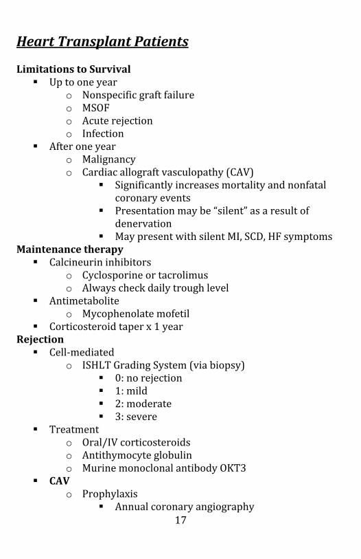
Heart Transplant Patients Limitations to
Up to o Survival
r o t failure
ne yea
o Nonspecific graf
ction MSOF
jeo Acute reo
After onInfection
o e year Maligna
o Cardiacncy allograft vasculopathy (CAV)
tal Significantly increases mortality and nonfacoronary events
Presentation may be “silent” as a result of denervation
ent with silent MI, SCD, HF symptoms May presnance
CalcineMainte therapy
urin inhibitors rine or tacrolimus
gh level o Cyclospoo
Always check daily trou
l Antimetabolite
o Mycophenolate mofeti taper x 1 year Corticosteroidon
Cell‐meRejecti
diatedo ISHLT G (via biopsy)
System
n rading
0: no rejectio
te 1: mild 2: modera
Treatm 3: severe
o ent
o Oral/IV corticosteroids Antithymocyte globulin
o clonal antibody OKT3 Murine mono
o Prophy CAV
laxis Annual coronary angiography

18
• Incidence o 2‐28% at 1 year o 40‐70% at 5 years
o CAV causes May be underestimated angiographically asntric narrowing (vs. focal)
o be considered after 5 years diffuse, conceStress t
o Medicaest maytions
ns Stati
o TreatmmTOR inhibitors
e/augment immunosuppressive tx ent
Chang
PCI CABG
nsplant Retra
o ECG Interpretation
Low voltage Bradycardia
o lock o
Any level of heart b
o If infection is suspected
o abx Culture everything Low threshold for broad‐spectrum
o Immunocompromised ID consult

19
Acute Decompensated Heart Failure Evaluation
Why di sate? d the patient decompen
o o History
Medication compliance o Caused diac conditions by other car
ACS Arrhythmia
Aortic dissection AI/MR
o Exacerb by noncardiac conditions Acute
on
ati
PNA PE
COPD/Asthma Missed dialysis sessions
o Consider inbelow)
vasive hemodynamic monitoring (see g reasons: ine filling pressures from exam
for the followinrmy
Cannot dete e therap
o DiagnoGuid
stic studies
does not rule out ADHF) ECG
CXR mes CXR (normal
iac enzy rolytes Card
Elect
ABG
BNP ECHO Cath for ACS‐induced ADHF
Goals Treatment
o Improve symptoms
o ctors o Optimize volume status
precipitating fao oral regimen
Identify and treat Establishing stable
o Patient education Mainstays of therapy

20
RB
o tals, rhythm Monitor O
o
2 sat, vio Supplemental O2
o IV access Positio
o eded n patient upright
Provide respiratory support as ne NIPPV vs. intubation
o If end‐ohemod
rgan perfusion adequate, e
ynamically stablIV vaso
IV loopdilator (NTG or Nitroprusside) diuretic
• Higher doses needed if pt taking loop diuretic chronically and/or
o If known systolic HF with severe ADHF/shock renal dysfunction
t IV inotrope +/‐ mechanical suppor(IABP)
Caution in ACS pts; inotropes may provoke ischemia by increasing
o myocardial O2 demand
/shock inotrope
If known diastolic HF with severe ADHF IV vasopressor preferred over IV
o If cardisigns/s
ac function unknown but pt has ymptoms of ADHF/shock
ro e +/‐ IV vasopressor +/‐ IV inot p
o Beta‐blmechanical support ockers7‐8
If takin
g chronically • lly Continue if hemodynamica
table
s• Decrease/hold if unstable
s If naïve
iate once pt stabilizeo Oral th table)
• Init
erapy (transition once sACEI/A
Aldoste9‐10
rone antagonist11 • If EF <30‐35%, serum potassium
>5 meq/L and GFR >/= 30

21
o Antiarrhythmics for concomitant arrhythmia (i.e. Afib), may need DCCV

22
Hypertensive Crises Hypertens y (Pt is asym e)
ive urgencptoma
Treatmtic and there is no evidence of end‐organ damag
o ral hours/days ent
veo d
Goal BP </= 160/100 over se reduction ill‐advise
o Rapid BPIf alrea
o If naïvedy on meds, reinstate ,
Initially use short‐acting tx (i.e. captopril, metoprolol) and up‐titrate
CCB (nifedipine XL 30mg QD), Beta‐blocker (metoprolol XL 50mg QD), ACEI (ramipril 10mg QD)
Hypert ergency (Eviden pt is symptomatic)
ensive emce of e
Treatmnd‐organ damage,
o ent
o Consider head CT Rapid, cautious lowering is the goal
o Malignant HTN and hypertensive encephalopathy V Nitroprussid
Ie I
V Nicardipine Labetalol IV
o ConcomEsmolol IV
itant HF exacerbation IV vasodilators (nitroprusside, NTG)
‐blockers (decrease contractility) and ork)
Avoid beta
o Aortic dhydralazine initially (may increase cardiac wissection
ce HR <60bpm, BP to ic or lowest level tolerated
IV beta‐blocker to redu100‐120 mmHg systol
STAT surgical consult

23
Bradyarrhythmias Causes
ressure Hypoxia
ial p one Increased intracran
gerated vagal t erior) Exag
Acute MI (infOSA
Medications
o o , digitalis) AVN blockers (BB, CCB
one o
Amiodar
o Methyldopa, clonidine
o valsalva
o pressure on carotid sinus (like from neck collar)
ng o to cold water (typically of face)
vomiting/coughiure
o sudden expos
o hypothyroidism
o hypothermia certain infections zold‐Jarisch reflex o Be
l Thero Atropin
Medica apy
o e 0.5‐1mg, repeat q3‐5min to a total dose of 3mg
l if Mobitz II or higher grade block o Not likely to be helpfu
o Chronosuspected
o tropic Agent Infusion
o Dopamine (2‐10mcg/kg/min) Isoproterenol (2‐10mcg/min)
o vasodilation and decrease Beta‐2 agonism can result in in MAP
o Epinephrine (2‐10mcg/min) us Pacing
This is painful and not preferred, use sedation Transcutaneo
oTemporarIndications
y Transvenous Pacing
Hemodynamically significant bradycardia
24
Asystole re)
recovery Termination of tachycardias (ra
permanent pacing or us node dysfunction Bridge to
sin k Severe
loc AV b
V Tach
AMI
VB New bifascicular block
& 1st degree A g L and R BBB New LBBB
natin AlterMobitz II CHB Infarct
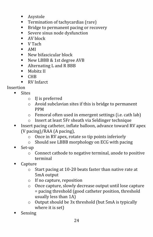
Insertio RVn
Sites o IJ is preferred o Avoid subclavian sites if this is bridge to permanent
PPM o Femoral often used in emergent settings (i.e. cath lab) o Insert at least 5Fr sheath via Seldinger technique
Insert p RV apex (V paci
acing catheter, inflate balloon, advance towardng)/RAA (A pacing), Once in RV apex, rotate so tip points inferiorly o
o Set‐up
Should see LBBB morphology on ECG with pacing
nnect cathode to negative terminal, anode to positive o Co
Captureterminal
o ts faster than native rate at Start pacing at 10‐20 bea
o 5mA output If no capture, reposition
o Once capture, slowly decrease output until lose capture = pacing threshold (good catheter position, threshold
usually less than 1A) utput should be 3x threshold (but 5mA is typically here it is set)
o Ow
Sensing

25
o asynchronous pacing is Decrease sensing setting until seen = sensing threshold
o Set at twice sensing threshold

26
Tachyarrhythmias DCCV/ Ind
Defibrillation ications
unstable arrhythmia EP lab
o Emergently –o
AntOtherwise, these are done electively in the
n o
icoagulatioPertine
o Emergent for Afib/Flutter cardioversions nt dminister therapeutic A/C just prior or immediately A
o Electiveafter DCCV
Therapeutic A/C x 3‐4 weeks prior or TEE and therapeutic A/C and DCCV if no thrombus. If thrombus, wait 3‐4 weeks with target INRs and then repeat TEE to confirm resolution
o Protocol
osterior ead ECG strip
Place pads anterior‐pn continuous 12‐l
dation o Ruo
DCCConscious se
o V
onize 00J
Synchro Charge to 2o
DefShock
o nize ibrillation
synchroo to 200J
Do notCharge
o Shock

27
Mechanical Hemodynamic Support12 General Complications
All devices carry risk of significant bleeding due to large sheath as limb ischemia size as well
ortic B Indicat
IntraA alloon Counterpulsation
o ions Hemodynamically unstable cardiogenic shock
o k Class I indication for STEMI pts with cardiogenic shoc
o if not quickly reversed with pharmacologic therapy Class I indication for STEMI and secondary acute MR
o hemia in UA/NSTEMI (Class IIa), STEMI Refractory isc
o (Class I)
CI rdiac transplantation
High risk Po Bridge to cao
ContraiVT Storm
o
ndications
o Aortic dissection
o AAA/TAA
hemia o ripheral grafts (relative)
Severe PAD, Limb iscrta/pe
o Descending aoModera
o Coagulote/Severe AI pathy
Can anticoagulate with alternative agents if heparin a problem not need anticoagulation if IABP is sheathless
Do
Hemodand on 1:1 CP
ynamico Afterlo
s ad reduction
Rapid deflation during systole allows for
o negative pressure in aorta
o Reduces afterload and improves forward flow from LV Increases CO by 20% decrease in mean PCWP
o Reduces LV wall stress from decreased filling pressures proves SV and CO, and decreased afterload im
decreased O2 demand o Augment coronary perfusion

Inflation during diastole displaces blood to proximal aorta augments diastolic pressure
augments coronary perfusion pressure elocity but
cal stenoses Will increase peak coronary flow vdoes not improve flow across criti
Does not augment collateral flow
o ion is not 1:1 Monitoring
h sheath or inflato t)
IV heparin if IABP througPt must remain supine (i.e. strict bedres
e exams firm proper placement
o Serial distal pulso R to con
TriggerDaily CXing and
o Trigger Timing
after R wave ECG – allows for appropriate delay
at dicrotic notch Pressure – if ECG tracings are poor nternal – if pt has arrested or ECG/Pressure i
o Timingtriggers are unreliable
nflation before dicrotic notch and deflates onset of next systolic waveform
Ideal ibefore
ample Waveforms S Normal
28

Early Inflation
Late Inflation
29

Early Deflation
Late Deflation
30

Percut st Device aneous Indicat
VentricularAssi
o ions Cardiogenic shock
port during high‐risk percutaneous/surgical poor cardiac function
o Backup sup
Contraiprocedures in patient with
o ndications
o AI, prosthetic aortic valve
o ular disease Aortic aneurysm/dissection
ipheral vasco thrombi
Severe aortic or perLeft ventricular/atrial
g diatheses ming sepsis
o Bleedino
CompliOverwhel
o cations
o Infection
o mechanical irritation Thromboembolism
s due to o
ArrhythmiaThrombocytopenia Hemolysis o
o Types
Effusion/tamponade
o Axial Flow Pump (Impella 2.5) Percutaneously placed into femoral artery
Inflow cannula advanced retrograde across aortic valve and seated in left ventricle
aws blood out of LV and ding aorta
Revolving pump drjects it into ascen
onpulsatile flow eN
31

o Tandom‐Heart Percutaneously placed via femoral artery
eptal Venous catheter inserted into LA via transpuncture
Arterial cannula inserted into iliac artery
venous blood via artificial ECMO
Removes CO2 from and adds O2 to
membrane
32
Pulmonary circulation is bypassed Requires systemic anticoagulation
Venovenous (severe respiratory failure)

Venoarterial (cardiac failure)
What to watch for: There is a big cannulae in the artery (13Fr, 15Fr, or 17Fr) and distal
ade sheath, need to assess limb perfusion is provided by a 5Fr antegrperfusion Groin complications should be noted If device flows drop (the ECMO oxygenates and circulates blood) transfuse and page Perfusion 3984
33

Invasive Hemodynamics
Indications
Heart Failure
Cardiogenic vs. noncardiogenic pulmonary edema/shock
e therapy nary HTN Guid
Pre‐transplant – evaluate for reversible pulmo
AMI Guide therapy in cardiogenic shock from AMI
onary edema or RV infarct refractory to Manage acute pulm
medical therapy Perioperative Use
low cardiac output (hypovolemia vs. Determine cause of
ventricular dysfunction) Pericardial Disease
n vs. Tamponade (if Constriction vs. Restrictio
echocardiography is unclear)
34
Primary Pulmonary HTN To exclude postcapillary causes (elevated PCWP)
Diagnose and establish severity of precapillary (normal PCWP) pulmonary HTN

35
fficacy of long‐term Select and establish safety and epy
nt assessment vasodilator theraPre‐lung transpla
Assess for Shunt
The Procedure
Test all
ports/balloon before inserting Choose Site
o Typically use IJ or femoral approach
and technique 7Fr) Sterile preparation
Seldinger technique to introduce sheath (typically
Insert PA catheter
the sheath f PA rupture) Inflate balloon once catheter is out ofAvoid over‐wedging (increased risk o
ule out PTX Post‐procedural CXR to rtions
CO (norCalcula
mal = 4‐8L/min)
o o 10] Fick: [Wtx3 mL/kg]/[(Ao sat – V sat) x 1.36 x Hgb x
Thermodilution method o line into RA port Performed by injecting 10cc of sa
n/m2) (balloon deflated)
rmal = 2.8‐4.2 L/mi
o CI (no
SV (normal = 40‐120 cm3/beat)
) SV = CO/HR
SVR (normal = 770‐1500 dynes*s/cm‐5
SVR = [(MAP‐CVP) x 80]/CO
PVR (normal = 20‐120 dynes*s/cm‐5) PVR = [(PAP‐PCWP) x 80]/CO
Convert to Wood Units by dividing by 80 Clinical Scenarios RA PCWP CO SVR Cardiogenic ↑ ↑ ↓ ↑ RV Infarct ↑ ↓ ↓ ↑ Y descent MR ↑ ↓ V wave PE ↑ ↓ ↓ ↑ Tamponade ↑ ↑ ↓ ↑ Sepsis ↓ ↓ ↑ ↓

36
VSD
o S
tep up in O2 sat from RA to PA TN
AP and RV
Pulmonary H
o Tampo
↑RAP, ↑RVP, ↑ PAP, normal PCWP, P
o nade D li a of pressures
o essure = PCWP iasto c equ lization
o RAP = RV diastolic pr
o ↑RAP, ↑RVP, ↑PCWP Prominent x descent
o Constrictive pericarditis

Waveform Characterist RA (normal =0‐8mmHg)
ics and Normal Values
RV (normal = 15‐30mmHg/0‐8mmHg)
37

PA (normal = 15‐30mmHg/3‐12mmHg)
PCWP (normal = 6‐12mmHg)
38

39
Therapeutic Hypothermia Background
Risk of death increases for each degree over 37° during first 48h post‐arrest
Lowering brain temp to 32‐34° during first few hrs post‐arrest reduces risk of neurological injury ions
Not following commands/lack of purposeful movements post‐Indicat
ROSC ations
Contraindic
Active, non‐compressible bleeding DNR
Not contraindicated in pregnancy, hemodynamic instability, or pts receiving thrombolytics
post‐ROSC Timing and Duration
in 6 hrs ‐arrest Achieve goal temp withNo benefit if placed mid
2 24hrs Maintain for 1 ‐s
IntravaMethod
scular13 o IV infusion of cold (4°C) isotonic saline, 30mL/kg via
pressure bag will reduce temp by >2°C/hr o 5min drops temp 1L of pressure‐infused cold saline/1
by 1°C acemo Pl
Surface
ent of endovascular catheter Can be done bedside or in cath lab
o sts, cold water
°C/hr Ice packs, cooling blankets, cooling veimmersion reduce body temp by 0.5‐1
ck o Apply ice packs to groin, axillae, ne
Sedation and Shivering
Titrate sedation to shivering suppression can delay time to achieve Shivering increases body temp and goal temp
Continuous gtt (propofol, fentanyl)

40
outcomes t in Nonshockable Rhythms
Not demonstrated in randomized trials, but still thought to incur benefit in these patients
Can use benzo gtts but may interfere with assessment of neurological function during re‐warming phase
ary Intermittent meperidine to suppress shivering, but primmetabolite is pro‐convulsive
Can use paralytics but this necessitates continuous EEG monitoring as these agents may mask seizures ring
Continuous temp monitoring via central venous, esophageal, Monito
bladder or rectal probes ming
Rewar
Rate of increase should not exceed 0.5°C/hr (0.2‐0.25°C/hr recommended)
bnormalities (hypokalemia), cerebral May induce electrolyte aedema, seizures
ctive Can be passive or ae Effec
Mild coAdvers ts
ion agulopathy
es and decreases platelet functcurs
o Slows clotting enzymo
TH should be stopped if significant bleeding oc
ction yte function, particularly >24h
Increased risk of infeo c
CardiacImpairs leuko arrhythmias
o Bradycardia ngation
o QT prolo
Hyperg “Cold d
lycemia due to insulin resistance iuresis”
o hypovolemia, hypokalemia, hypomagnesemia, hypophosphatemia may result
May interfere with metabolism of various drugs, thus prolonging their effects t in VT/VF
TH shown to decrease mortality and improve neurological Benefi
Benefi

41

42
Transcatheter Aortic Valve Replacement14‐15 Anticoagulation
ASA 81mg and clopidogrel 75mg daily x 6 months, followed by ASA 81mg daily for life site co
Access Access mplications
ilized o ess (most common)
sites potentially utal acc
o Direct femor
o Subclavian artery
o Transapical Direct aortic cannulation
o Iliac conduit (particularly for patients with severe peripheral vascular disease that precludes femoral access)
ocedural CVA
PeriprLow threshold to suspect, periprocedural risk of CVA is 10‐20%
Patients are often elderly with heavily calcified aortic arch and valve, hence significant risk of embolization/showering
TAVR issues O with any new signs of HF, SOB, hemodynamic
Other postStat ECHcompro
mise
Rupture o
Pericardial effusion/tamponade
g Heart block
o Will need temporary transvenous pacin Ventricular rupture (with transapical access)

43

2010 AHA Algorithm for BLS
44

AHA Algorithm for PEA Arrest
45

AHA Algorithm for Tachyarrhythmias
46
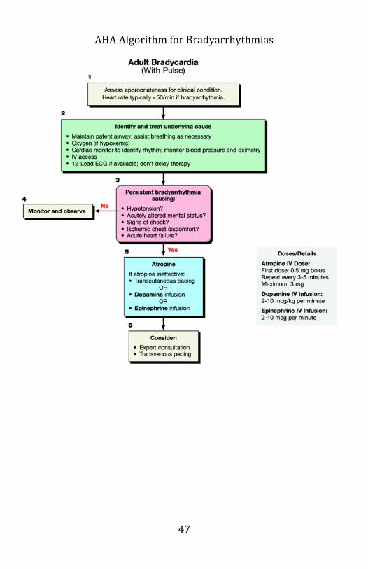
AHA Algorithm for Bradyarrhythmias
47

48
VI. CCU‐Stocked Meds
ACETAMINOPHEN (UDC) 20.3mL cup 160MG/5ML 650 MG LIQ ACETAMINOPHEN 325 MG TAB ACETAMINOPHEN/CODEINE 30MG TAB ACETAMINOPHEN/OXYCODONE 325mg/5mg TAB ADENOSINE (3MG/ML) 2 ML INJ ALBUMIN 5% 250 ML INJ ALBUTEROL NEBULIZING SOLN O.O83% 3 ML SOLN ALBUTEROL/IPRATROPIUM (DUO‐NEB) 2.5‐0.5/3 3 ML SOLN ALUMINUM‐MAGNESIUM HYDROX 1200‐1200 30 ML SUSP AMIODARONE HCL 50MG/ML 3 ML INJ ASPIRIN 325 MG TAB ASPIRIN EC 325 MG ECTA ATROPINE SULFATE (0.1MG/ML 1 MG SYRIN CADD PUMP 1 EA MISC CALCIUM CHLORIDE (100MG/ML) 10 ML SYRIN CODEINE PHOS/GUAIFENESIN 10 ML ELIX D5/0.45% NACL‐KCL 20MEQ 1000 ML INJ DEXAMETHASONE 10MG/ML 1 ML VIAL DEXTROSE 5% IN WATER 250 ML INJ DEXTROSE 5% IN WATER 5% 100 ML INJ DEXTROSE 5% IN WATER 5% 1000 ML INJ DEXTROSE 5%/0.45% NACL 1000 ML INJ DEXTROSE 50% 50 ML SYRIN DIAZEPAM 2 MG TAB DIAZEPAM 5 MG TAB DIAZEPAM 5MG/ML 10 MG SYRINGE DIGOXIN 0.25MG/ML 2 ML AMPUL DILTIAZEM HCL (5MG/ML) 50 MG VIAL DILTIAZEM HCL 30 MG TAB diphenhydrAMINE HCL 25 MG CAPSULE diphenhydrAMINE HCL 50MG/ML 50 MG INJ DOBUTAMINE/D5W 250ML 1 GM IV SOLN. DOCUSATE SODIUM 100 MG CAP DOPAMINE HCL 800 mg/D5W 250 ML IV SOLN. DRONABINOL C‐III 2.5 MG CAP

49
EPINEPHRINE 1:1000 1MG/ML 1 MG AMPUL EPINEPHRINE 1:10000 0.1 MG SYRIN ESMOLOL HCL 2.5G 250 ML PIGGYBACK FENTANYL (12MCG/HR) PATCH 12 MCG/H PATCH TD72 FENTANYL (25MCG/HR) 10 CM PTCH FENTANYL (50MCG/HR) 20 CM PTCH FENTANYL /0.9% NACL 10 MCG/ML 100 ML IV SOLN. FENTANYL /0.9% NACL 10 MCG/ML 100 ML IV SOLN. FILTER 1 EA NEEDLE FLUMAZENIL (0.1MG/ML) 5 ML INJ FUROSEMIDE 10MG/ML 40 MG INJ GLUCAGON 1 MG INJ HALOPERIDOL 5MG/ML 5 MG INJ HEPARIN 25,000U/D5W 500 ML INJ HEPARIN SODIUM 10,000U/ML 5000 UNIT SYRIN HEPARIN SODIUM 10ML VIAL, 1000 U/ML VIAL HURRICAINE 1 SPRY AERO hydrALAZINE HCL 20MG/ML 20 MG INJ HYDROCODONE/ACETAMINOPHEN 5MG/500MG 1 EA TAB HYDROCODONE/ACETAMINOPHEN 7.5MG/500MG 15 ML LIQUID HYDROMORPHONE (PCA) 1MG/ML 30 ML IV HYDROMORPHONE HCL 1MG/ML 1 MG DISP SYRIN HYDROMORPHONE HCL 2 MG TAB INSULIN ASPART 100U/ML 1 IU VIAL INSULIN GLARGINE,HUMAN (LANTUS) 100U/ML 1 UNIT VIAL INSULIN HUMAN NPH 100U/ML 1 IU INJ INSULIN HUMAN REGULAR 100U/ML 1 IU INJ INSULIN NOVOLIN 70/30 1 IU INJ IPRATROPIUM NEBULIZING SOLN 0.5MG/2.5ML 2.5 ML SOLN ISOPROTERENOL HCL 200 MCG INJ LABETALOL (5MG/ML) 100 MG INJ LACTATED RINGERS 1000 ML INJ LIDOCAINE 2% (PF) 100 MG 20MG/ML 5 ML SYRIN LIDOCAINE 2GM/D5W 250 ML INJ LIDOCAINE HCL (10MG/ML) 20 ML INJ LORAZEPAM 0.5 MG TAB LORAZEPAM 1 MG TAB
50
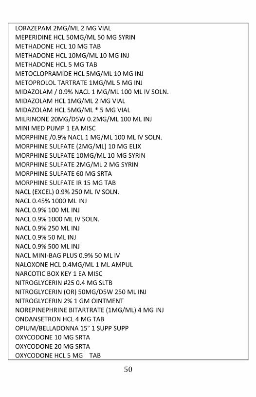
LORAZEPAM 2MG/ML 2 MG VIAL MEPERIDINE HCL 50MG/ML 50 MG SYRIN METHADONE HCL 10 MG TAB METHADONE HCL 10MG/ML 10 MG INJ METHADONE HCL 5 MG TAB METOCLOPRAMIDE HCL 5MG/ML 10 MG INJ METOPROLOL TARTRATE 1MG/ML 5 MG INJ MIDAZOLAM / 0.9% NACL 1 MG/ML 100 ML IV SOLN. MIDAZOLAM HCL 1MG/ML 2 MG VIAL MIDAZOLAM HCL 5MG/ML * 5 MG VIAL MILRINONE 20MG/D5W 0.2MG/ML 100 ML INJ MINI MED PUMP 1 EA MISC MORPHINE /0.9% NACL 1 MG/ML 100 ML IV SOLN. MORPHINE SULFATE (2MG/ML) 10 MG ELIX MORPHINE SULFATE 10MG/ML 10 MG SYRIN MORPHINE SULFATE 2MG/ML 2 MG SYRIN MORPHINE SULFATE 60 MG SRTA MORPHINE SULFATE IR 15 MG TAB NACL (EXCEL) 0.9% 250 ML IV SOLN. NACL 0.45% 1000 ML INJ NACL 0.9% 100 ML INJ NACL 0.9% 1000 ML IV SOLN. NACL 0.9% 250 ML INJ NACL 0.9% 50 ML INJ NACL 0.9% 500 ML INJ NACL MINI‐BAG PLUS 0.9% 50 ML IV NALOXONE HCL 0.4MG/ML 1 ML AMPUL NARCOTIC BOX KEY 1 EA MISC NITROGLYCERIN #25 0.4 MG SLTB NITROGLYCERIN (OR) 50MG/D5W 250 ML INJ NITROGLYCERIN 2% 1 GM OINTMENT NOREPINEPHRINE BITARTRATE (1MG/ML) 4 MG INJ ONDANSETRON HCL 4 MG TAB OPIUM/BELLADONNA 15° 1 SUPP SUPP OXYCODONE 10 MG SRTA OXYCODONE 20 MG SRTA OXYCODONE HCL 5 MG TAB

51
PHENYLEPHRINE HCL (10MG/ML) 10 MG VIAL POTASSIUM CHLORIDE (KLOR‐CON) 20 MEQ SRTA POTASSIUM CHLORIDE 20MEQ/15ML 20 MEQ LIQ POTASSIUM CHLORIDE 20MEQ/mL 100 ML IV SOLN. PREGABALIN 100 MG CAPSULE PREGABALIN 150 MG CAPSULE PREGABALIN 50 MG CAPSULE PREGABALIN 75 MG CAPSULE PROPOFOL (OR) 1% 20 ML VIAL PROPOFOL 1% 100 ML VIAL PROPOFOL 1% 100 ML VIAL PROTAMINE SULFATE (10MG/ML) 50 MG INJ SODIUM BICARBONATE (1MEQ/ML) 50 MEQ VIAL TRANSPORT 1 EA KIT VASOPRESSIN 20 UNIT VIAL ZOLPIDEM TARTRATE 5 MG TAB
VII. References
52
1. Visit the following website for links to updated ACC/AHA joint guidelines
on various cardiovascular disease processes. ttp://my.americanheart.org/professional/StatementsGuidelines/ACCAHAh‐Joint‐Guidelines_UCM_321694_Article.jsp . The CURRENT‐OASIS Investigators. Dose comparisons of clopidogrel and 2aspirin in acute coronary syndromes. NEJM 2010; 363: 930‐942 3. Pitt B, Remme W, Zannad F, et al. Eplerenone, a selective aldosterone locker, in patients with left ventricular dysfunction after myocardial binfarction. NEJM 2003; 348:1309‐1321 4. Hohnloser S, Kuck K, Dorian P, et al. Prophylactic use of an implantable ardioverter‐defibrillator after acute myocardial infarction. NEJM 2004; c351: 2481‐2488 . Stone GW, Witzenbichler B, Guagliumi G, et al. Bivalirudin during primary 5PCI in acute myocardial infarction. NEJM 2008; 358: 2218‐2230 6. Van de Werf F, Bax J, Betriu A, et al. Management of acute myocardial nfarction in patients presenting with persistent ST‐segment elevation. Eur iHeart J 2008; 29(23): 2009‐2945 7. Jondeau G, Neuder Y, Eicher J‐C, et al. B‐CONVINCED: Beta‐blocker continuation vs interruption in patients with congestive heart failure ospitalized for a decompensation episode. Eur Heart J 2009; hDOI:10.1093/eurheartj/ehp323 8. Fonarow GC, Abraham WT, Albert NM, et al. Influence of beta‐blocker ontinuation or withdrawal on outcomes in patients hospitalized with heart cfailure. Findings from the OPTIMIZE‐HF program. JACC 2008; 52: 190‐199 9. Hunt SA, Abraham WT, Chin MH, et al. 2009 focused update incorporated into the ACC/AHA 2005 Guidelines for the Diagnosis and Management of Heart Failure in Adults: a report of the American College of Cardiology Foundation/American Heart Association Task Force on Practice Guidelines: eveloped in collaboration with the International Society for Heart and dLung Transplantation. Circulation 2009; 119:e391. 10. The CONSENSUS Trial Study Group. Effects of enalapril on mortality in evere congestive heart failure. Results of the cooperative North candinavian Enalapril Survival Study (CONSENSUS). NEJM 1987; 316‐429 sS

53
Clinical Trials by Condition
11. Bertram P, Zannad F, Remme W, et al. The effect of spironolactone on orbidity and mortality in patients with severe heart failure. NEJM 1999; m
341: 709‐717 2. Delgado D, Rao V, Ross H, et al. Mechanical Circulatory Assistance. Circ 12002; 106: 2046‐2050 13. Nolan JP, Morley PT, Vanden Hoek TL, et al. Therapeutic Hypothermia after Cardiac Arrest: An advisory statement by the advanced life support ask force of the international liaison committee on resuscitation. Circ 2003; t108: 118‐121 4. Smith CR, Leon MB, Mack MJ, et al. Transcatheter versus surgical aortic‐
98 1valve replacement in high‐risk patients. NEJM 2011; 364: 2187‐21 15. Leon MB, Smith CR, Mack M, et al. Transcatheter aortic‐valve mplantation for aortic stenosis in patients who cannot undergo surgery. EJM 2010; 363: 1597‐1607
iN

54
Atrial FibAFFIRM
rillation No difference in mortality between rate and rhythm control, however, increased mortality in rhythm control in older pts, those with CAD, those without CHF; 2002
ARISTOTLE Apixaban was superior to warfarin in preventing stroke or systemic embolism, caused less
r bleeding, and resulted in lowemortality.; 2011
RELY Dabigatran is non‐inferior to warfarin in preventing stroke and systemic embolism with lower
major bleeding profile; slightincrease in GI bleeding; 2009
SPAF I, SPAF II, SPAF III Warfarin > ASA > placebo in reducing stroke events in AFib. For high risk patients with Afib, Warfarin INR 2‐3 is more effective. Low risk patients, ASA 325 has acceptable low risk of stroke < 3%. Sub‐study of SPAF III established high risk factors of the CHADS2 risk score; 1991, 1994, 1996
aker PacemAVID ICD is more effective than
antiarrythmic drugs in reducing s. arrhythmia related cardiac death
1999 MADIT Defibrillator along with BiV ICD
(CRT‐ICD therapy )is associated with improved EF and HF. Most

55
benefit in reducing HF events in ; pts with QRS > 150, EF<30%
2009 REVERSE CRT reverses remodeling in
systolic LV dysfunction, pts with asymptomatic to mild HF or wide QRS, EF < 40 ‐ significant improvement in reverse LV remodeling seen by measures of LVESV and LVEDV along with EF after 6 months in pts with CRT with further improvement overtime; there was significant decrease in morbidity and mortality; 2009
SCDHeFT Amio vs. placebo, ICD vs. placebo for CHF ‐ In pts with mild‐moderate CHF, EF <35, shock only ICD reduced risk of death (ARR 7.2% at 5 years), main effects in pts with Class II symptoms, minimal effect in Class III; Amio showed no benefit in Class II, but reduced survival in Class III; 2005
LipidsAtoZ
NSTEMI, STEMI Reduction in CV death, MI and readmission for ACS reduced in pt receiving zocor. Significant decrease in CVdeath and CHF; 2004
JUPITER Rosouvastatin reduced primary endpoint (CV death, MI, CVA, unstable angina, revascularization) in women > 60 and men > 50, LDL < 130 – low normal, elevated hsCRP > 2 by

56
44%, 2009 LIPID Pravastatin reduced mortality
from all causes and CV events in erol 155‐pts with ACS and cholest
271; 1998 PRINCESS/MIRACL Early <96hr initiation of
nd atorvastatin improved MI arevasc in AMI patients
TNT Pts with CAD (prior MI +/‐ revascularization, stable angina) ‐ Lipitor ‐ high dose has significantly lower LDL and total cholesterol levels, and reduced risk of major CV event 2005
WOSCOPS Pts hyperlipidemia and no hx of MI, pravastatin reduced CV deaths (RRR 30%) and need for revascularization (RRR 37%); 1995
tive Cardiology PreventaALLHAT Thiazide vs. CCB vs. ACEI ‐
Thiazide type diuretics (chlorthalidone) are superior at preventing 1 or more forms of CVD and should be first line of therapy; amlodipine higher 6 yr rate of HF and lisinopril had higher 6 yr rates of CHD, stroke, and HF; 2002
FRAMINGHAM High levels of LDL, Hypertension, cigarette smoking, obesity, elevated blood sugar levels, stress, lack of exercise, menopause, ECG abnormalities increase risk of coronary heart disease; 1984

57
HOPE Ramipril reduced risk of death, MI, stroke, and revascularization. Vitamin E did not lower CAD; 2000
the risk of
SHIFT Ivabradine reduction in om hospitalization or CV death fr
heart failure. 2010 UKPDS (HTN in Diabetes study) ‐ BP
control < 150/85 in pts with HTN and Diabetes with ACEI or BB, plus additional meds if needed, reduces risk of diabetic related complications and death related to diabetes (MI, PV0D, renal disease, CVA, sudden death) along with decrease in progression of neuropathy and retinopathy; 1998
WHI (Women’s Health Initiative) ‐ post menopausal women on combined hormonal therapy is associated with increased risk of CAD, PE, CVA and invasive breast cancer but decreased risk of hip fractures and colorectal cancer; absolute risk excess was 19 per 100,000 person‐years; 2002
HeartAIRE
Failure Ramiprilstarted 3–10 days after MI, benefit noticeable as early as 30 days, reduction in progression
duction in to heart failure; no rereinfarction or stroke; 1993
CAPRICORN Carvedilol decreases cardiovascular and all cause mortality in post‐ infarction pts

58
with EF < 40% ; 2001 CHARM Candesartan addition to
concurrent BB and/or ACEI therapy –significant reduction in CV death or hospital admission for CHF (16%); NNT is 23 in 1 year
CIBIS EF < 35% and NYHA class III or IV, B1 blocker bisoprolol significantly reduced all cause mortality, sudden death, and all cause hospitalizations from CHF; 1999
COPERNICUS Coreg in addition to diuretic plus ACE/ARB reduces all cause mortality and hospitalization; 2001
EPHESUS Eplerenone in addition to standard therapy reduces mortality in pts with severe CHF; 2001
MERIT‐HF Toprol CR/XL when used on top of ACE‐I reversed ventricular remodeling as shown by decreased LV‐ EDV and ESV by cardiac MRI and decreases all cause mortality when started in pts with CHF; 2000
RALES Aldactone 25 mg in addition to standard therapy reduces mortality and risk of sudden death
35, in pts with severe CHF (EF <Class 3‐4); RRR 30%; 1999
RESOLVD Enalapril plus candesartan combination was more beneficial in preventing LV dysfunction
d (reduced ESV and EDV), compareto either drug alone ; 1999
SAVE, TRACE, SOLVD ACEI reduces all cause mortality remodeling, and decreased risk of

59
worsening heart failure when started 2–10 days after MI, and in pts with CHF (EF < 35%)
VALIANT ARBs have mortality equivACEI; side note: 1999
alent to
V‐HEFT Vasodilator (enalapril vs. hydralazine/nitrate)‐Heart Failure Trial) ‐ pts with CHF on digoxin and diuretic; enalapril has greater reduction mortality 1991
Artery Disease CoronaryACUITY Enoxaparin equivalent to hepa
upstream during ACS 2004 rin
CAPRIE Plavix was slightly better than aspirin in reducing primary endpoints of MI, CVA, and vascular death; conferred benefit for CVA and PAD; no benefit in pts with previous MI; 1996
CAST Flecanide and encanide increased mortality in pts with post‐MI asymptomatic or mildly symptomatic supraventricular arrhythmias; 1989
COMMITT Plavix plus ASA reduces 30 day mortality (0.6% ARR), BB good
t after MI if pts do not have hearfailure/cardiogenic shock; 2005
CURE Plavix in addition to aspirin in patients with non‐STEMI ACS reduces risk of CV death, MI, and CVAs by 20%; 2002
GISSI‐3 Lisinopril, when given < 24 hrs in pts with acute MI, reduced

60
mortality and severe LV diastolic dysfunction (EF < 35%); 1994
GUSTO‐1 Accelerated t‐PA plus IV Heparin has lower mortality although higher bleeding than streptokinase and standard therapy. 1995
HORIZONS‐AMI Bivalirudin reduced bleeding and death compared to Heparin plus Glycoprotein IIb/IIIa inhibitor in 30 days; increased <24 hr in stent thrombosis with bivalirudin but offsetted between 24 hrs to 30 days; 2009
ISIS‐2 ASA lowers CV death, Recurrent MI, CVA when given to patients with acute MI; 1988
NORWEGIAN TIMOLOL In pts who survive acute MI, Beta blockers reduce all cause mortality, sudden death, and reinfarction; 1981
PLATO Pts with ACS, with or without ST elevation, ticagrelor reduces death from vascular, MI, and CVAs; slight increase non‐procedural related, i.e. fatal intracranial bleeding, 2009
TACTICS‐TIMI 18 GpIIb/IIIa inhibitor plus invasive strategy in pts with moderate‐high
n 001
risk UA/NSTEMI is better thaconservative management; 2
TRITON‐TIMI 38 Prasugrel reduced CV death, nonfatal MI, nonfatal stroke compared to clopidogrel 19% reduction in CV death, MI or stroke compared with clopidogrel in patients undergoing PCI for

61
ACS.2007.
ntional InterveCARP In patients with stable CAD,
coronary artery revascularization prior to elective major vascular surgery, s.a. expanding AAA, PVOD of legs, does not improve outcomes; 2004
ERACI, GABI, BARI, CABRI, RITA,EAST
PTCA and CABG have similar rates of survival and avoidance of MI and similar long term health care costs; PTCA group had increased rates of recurrent angina and revascularization; nearly ¼ of PTCA patients required CABG; At 10 year follow up some studies showed that Diabetics and pts > 65 yrs have slightly decreased mortality with CABG; Subset of CABRI trial ‐ pts with multi‐vessel or chronically occluded major vessel disease had better
, outcomes with CABG; 1994, 19941996, 1995, 1998, 1999
FRISC‐II, RITA‐3 At 5 year follow up, in moderate‐high risk pts with ACS without ST elevation, early invasive intervention strategy has improved outcomes in terms of death/MI; 2005, 2006
GUSTO‐2B PTCA has better outcomes than thrombolysis in pts with AMI. lowest 30 day mortality when D2B time < 60 minutes (1%), 60–90 minutes (4%), > 90 minutes

62
(6.5%) 1997 NORDISTEMI Early invasive strategy in pts with
STEMI has significant reductions in primary outcomes (death, stroke, reinfarction) at 30 days, but at 12 months, reductions were nonsignificant, but trended towards significance as invasive group had less incidence of death,
strokes, reinfarction at 12 months;2010
PARTNER TRIALS TAVR superior to medical therapy and equivalent to surgical therapy
of high risk patients. 2011 SHOCK In pts with cardiogenic shock due
to acute MI, early revascularization vs. medical stabilization does not improve 30 day mortality but does improve 6‐month and 12 month survival 1995
SYNTAX PCI vs. CABG in pts with severe CAD ‐ At 1 yr, CABG group had lower rates of major cardiac or cerebrovascular events (12% vs. 18%) and repeat revascularization (6% vs. 14%); however, there was increase in rate of strokes (2.2% vs. 0.6%). Conclusion ‐ CABG should be standard of care in pts with severe 3 vessel or left main disease; 2009


![WELCOME [infinitus.co.za]€¦ · certificate Will send steps ... (FSCA), announced that the Financial Services Board (FSB) had been “transformed” into the FSCA as of 1 April](https://img.dokumen.tips/doc/110x75/5fbf5772848b0b7e9575f515/welcome-certificate-will-send-steps-fsca-announced-that-the-financial.jpg)