Embed Size (px)
Citation preview
Proc. Natl. Acad. Sci. USAVol. 93, pp. 10177-10182, September 1996Biophysics
The fifth epidermal growth factor-like domain of thrombomodulindoes not have an epidermal growth factor-likedisulfide bonding pattern
[protein C/thrombin/Pichia pastoris/yeast expression/tris(2-carboxyethyl)phosphine]
CHRISTOPHER E. WHITE, MICHAEL J. HUNTER, DAVID P. MEININGER, SIV GARROD, AND ELIZABETH A. KoMIVES*Department of Chemistry and Biochemistry, University of California at San Diego, La Jolla, CA 92093-0601
Communicated by Bruno H. Zimm, University of California at San Diego, La Jolla, CA, June 28, 1996 (received for review March 28, 1996)
ABSTRACT The disulfide bonding pattern of the fourthand fifth epidermal growth factor (EGF)-like domains withinthe smallest active fragment of thrombomodulin have beendetermined. In previous work, this fragment was expressedand purified to homogeneity, and its cofactor activity, asmeasured by keat for thrombin activation of protein C, was thesame as that for full-length thrombomodulin. CNBr cleavageat the single methionine in the connecting region between thedomains and subsequent deglycosylation yielded the individ-ual EGF-like domains. The disulfide bonds were mapped bypartial reduction with tris(2-carboxyethyl)phosphine accord-ing to the method of Gray [Gray, W. R. (1993) Protein Sci. 2,1732-1748], which provides unambiguous results. The disul-fide bonding pattern of the fourth EGF-like domain was (1-3,2-4, 5-6), which is the same as that found previously in EGFand in a synthetic version of the fourth EGF-like domain.Surprisingly, the disulfide bonding pattern of the fifth domainwas (1-2, 3-4, 5-6), which is unlike that found in EGF or inany other EGF-like domain analyzed so far. This result is inline with an earlier observation that the (1-2, 3-4, 5-6) isomerbound to thrombin more tightly than the EGF-like (1-3, 2-4,5-6) isomer. The observation that not all EGF-like domainshave an EGF-like disulfide bonding pattern reveals an addi-tional element of diversity in the structure of EGF-like do-mains.
Thrombomodulin (TM) has a cysteine-rich extracellular do-main with six regions of sequence that resemble epidermalgrowth factor (EGF-like domains) (1-3). To date, more than300 sequences have been identified and classified as EGF-likedomains (4, 5). The classification is based mainly on thespacing and presence of six cysteine residues within approxi-mately 40 amino acid residues. It is commonly held that allEGF-like domains have the same disulfide bonding pattern asEGF, but we report here that the fifth EGF-like domain ofTMappears to have an anomalous disulfide bonding pattern.The structure of EGF itself is well-determined by NMR (6,
7). The molecule has a central two-stranded 3-sheet and asecond shorter sheet, or double hairpin, at the C terminus.EGF contains six cysteines that form three disulfide bondsconnecting the first cysteine to the third, the second to thefourth, and the fifth to the sixth (8). The disulfide bonds thatconnect the first cysteine to the third and the second cysteineto the fourth cross each other near the middle of the centraltwo-stranded /3-sheet. Thus, the central two-stranded /3-sheetis cemented by the crossing first-to-third and second-to-fourthdisulfide bonds, and the double hairpin is cemented by thefifth-to-sixth disulfide bond (Fig. 1). The fourth and fifthcysteines are always only one amino acid apart, so that a closeconnection exists between the two /-strand substructures.
FIG. 1. Stereoview of a ribbon diagram of EGF [3EGF from theProtein Data Bank (6)]. The disulfide bonds are shown by black lines,and the cysteines are numbered.
Structures of nine EGF-like domains have been determined,and all of the structures are consistent with an EGF-likedisulfide bonding pattern. Seven of these EGF-like domains,the first EGF from coagulation factor X (9), the first EGF fromcoagulation factor IX (10), transforming growth factor type a(11), the urokinase EGF-like domain (12), the EGF-likedomain from E-selectin (13), the heregulin EGF-like domain(14), and the EGF-like domain from tissue plasminogenactivator (15), are very similar to EGF. Two others, the secondEGF-like domain from factor X (16) and the fourth EGF-likedomain from TM (17), resemble EGF in the N-terminal halfof the molecule, but the C-terminal loops have a broadenedshape that does not superimpose well on the C-terminal loopof EGF.The disulfide bonding pattern has been unambiguously
determined by either chemical methods or by proteolyticmapping for murine EGF (8), the first EGF-like domains fromcoagulation factors IX (18) and X (19), transforming growthfactor type a (20), and a synthetic version of the fourth EGFfrom TM (17). These domains all have the same disulfidebonding pattern as EGF despite their relatively low sequencesimilarity and the large degree of variability in the numbers ofamino acids between the cysteines (Fig. 2). Although morethan 300 EGF-like domains have been identified by sequencesimilarity, there are relatively few for which the disulfidebonding pattern is unambiguous.Our previous work on a synthetic peptide corresponding to
the fifth EGF-like domain of TM showed that the domainformed several disulfide bonded isomers when refolded in aredox buffer, and that all of the isomers bound to thrombin.Surprisingly, thrombin binding affinity correlated with lack ofcrossing in the disulfide bonds, so that the uncrossed (1-2, 3-4,5-6) isomer bound to thrombin nearly an order of magnitudemore tightly than the EGF-like (1-3, 2-4, 5-6) isomer (23). At
Abbreviations: EGF, epidermal growth factor; TM, thrombomodulin;TMEGF(4-5), a fragment that is composed of the fourth and fifthEGF-like domains of TM; TCEP, tris(2-carboxyethyl)phosphine;TMEGF4, the fourth EGF-like domain of TM; TMEGF5, the fifthEGF-like domain of TM.*To whom reprint requests should be addressed. e-mail:[email protected].
10177
The publication costs of this article were defrayed in part by page chargepayment. This article must therefore be hereby marked "advertisement" inaccordance with 18 U.S.C. §1734 solely to indicate this fact.
Dow
nloa
ded
by g
uest
on
Mar
ch 5
, 202
0

Proc. Natl. Acad. Sci. USA 93 (1996)
Source Seauence % Similarity
consen:mEGFhTGFaXF. IXEG]F.XEGF:TMEGF4F.XEGF.tPAEGFuPAEGFheregEEsel-E(TMEGF5
sus
FII
r I_ I _ _XXXXC- X1-7-CXXXGXC--- Xl-13 ---CXCXXG (F/Y)Xli6GXXCX1-11
NSD-SECPLSHDGYCLHDGVCMYIEALDK---YACNCVVGYI-----GERCQYRDLKWWELRVSHFNDC-DSHTQFCFH-GTCRFLVOEDK----ACVCHSGYV-----GARCEHADLLAYVDGDQC-ESNP--CLNGGSCK--DDINS ---YECWCPFGFE-----GKNCELDVT-KDGDQC-EGHP--CLNQGHCK--DGIGD---YTCTCAEGFE-----GKNCEFSTR--PVDPCFRAN---CEYO--COPLNO-TS---YLCVCAEGFAPIPHEPHRCOMF
100%41%33%35%27%
II ---RKLCSLDNGD-C-DQ-FCH--EEQNS---VVCSCARGY-TLADNGKACIP 29%---VKSCSEPR---CFNGGTCQQALYFSD---FVCQCPEGFA-----GKSCEID 26%QVP-SNCD------CLNGGTCVSNKYFSNI--HWCNCPKKFG-----GQHCEIDK 28%
GF TSHLVKCAEKEKTFCVNGGECFMVKDLSNPSRYLCKCQPGFT-----GARCTENV 27%GF - ACTN---TSCSGHGECVETINN-----YTCKCDPGFS-----GLKCEO 29%
----MFC--NQT-AC--PADC---DPNTQAS ---CECPEGY--ILDDGFICTDIDE 28%
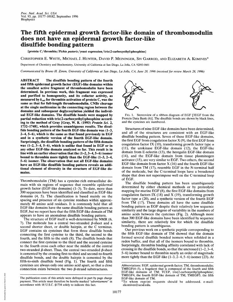
FIG. 2. Comparison of the sequences of various EGF-like domains. For the first group, the disulfide bonding pattern has been chemicallydetermined. mEGF, murine EGF; hTGFa, human transforming growth factor type a; F.IXEGFI, the EGF-like domain from coagulation factorIX closest to the N terminus; F.XEGFI, the EGF-like domain from coagulation factor X closest to the N terminus; TMEGF4, the fourth EGF-likedomain from TM. For the second group, structures that are consistent with an EGF-like disulfide bonding pattern have been determined. F.XEGFII,the EGF-like domain from coagulation factor X closest to the C terminus; tPAEGF, the EGF-like domain from tissue plasminogen activator;uPAEGF, the EGF-like domain from urokinase; heregEGF, heregulin; Esel-EGF, the EGF-like domain from E-selectin. The sequence ofTMEGF5(the fifth EGF-like domain from TM) is given last. The degree of similarity among the sequences was determined by the method of Feng andDoolittle (21) using the BLOSUM algorithm of Henzikoff and Henzikoff (22).
first, this anomalous result was thought to be an artifact ofremoving the fifth domain from the context of the rest of theTM molecule. Indeed, the first cysteine in the domain is onlythe fourth residue from the end of the polypeptide, whereas inthe full-length TM molecule, it follows the fourth domain.Misfolding of the domain could easily result from end effects.Furthermore, the connecting region between the fourth andfifth EGF-like domains in TM is critical for activity, sodisconnection at this point could easily perturb results frombiological assays. We have, therefore, determined the disulfidebonding pattern of the fifth domain within the context of afragment ofTM with full cofactor activity. Although sufficientquantities of the native protein were not available, expressionand kinetic analysis of a fragment ofTM in Pichiapastoris havenow provided sufficient quantities of a TM fragment withconvincing cofactor activity. This fragment [TMEGF(4-5)] iscomposed of the fourth and fifth EGF-like domains ofTM andextends from residue E346 to residue E426 of the human TMsequence (3). We present results that show that the fifthEGF-like domain, isolated from the active TMEGF(4-5)fragment, has an anomalous disulfide bonding pattern.
EXPERIMENTAL PROCEDURES
Reagents. Trifluoroacetic acid was from Aldrich, PNGase Fwas from Glyko (Novato, CA), and endoglycosidase H wasfrom Boehringer Mannheim. All other reagents and chemicalswere reagent grade from Fisher Scientific or Sigma.TM Fragment Characterization. TMEGF(4-5) was pro-
duced by high-density fermentation of the SMD1168 strain ofP. pastoris transformed with the gene for TMEGF(4-5) asdescribed (3). The expressed, folded protein was secreted intothe culture supernatant from which it was purified by anionexchange chromatography, ultrafiltration (PM30 membrane,Amicon) to remove high molecular weight contaminants,HiLoad Q chromatography, and finally reverse-phase HPLC(3). The purified TMEGF(4-5) was greater than 95% pure byN-terminal sequencing, amino acid analysis, and reverse-phaseHPLC. The kinetic parameters of this fragment were deter-mined previously; the Km for TM is 120 nM, which is 10-foldhigher than that for native TM and is due to the absence of thesixth domain (3). The kcat for thrombin activation of protein Cby the expressed TMEGF(4-5) 'fragment was 2 sec1, which isthe same as that for full-length human TM (24).
Proteins produced in P. pastoris have been shown to containN-linked high mannose sugars, and N-terminal analysis indi-cated the probable presence of glycosylation on two NQTsequences. Deglycosylation of TMEGF(4-5) with PNGase F(Glyko) appeared to remove all of the sugars from only theNQT site in the fifth domain, so the sugars on the fourthdomain were partially removed by treatment with endoglyco-sidase H (Boehringer Mannheim), which removes high man-nose sugars but not the core sugar residues.CNBr Cleavage of TMEGF(4-5). Glycosylated, HPLC pu-
rified TMEGF(4-5) (30 mg) was resuspended in 50 ml of 6 Mguanidine hydrochloride in 70% formic acid (Sigma). Afteraddition of 3.5 g of CNBr, the reaction proceeded for 20 hr inthe dark. The products were diluted with 500 ml of purifiedH20 (Milli-Q water purification system, Millipore) and freeze-dried. The lyophilized protein was resuspended in Milli-Q-purified H20 and chromatographed in three portions on a C18HPLC column (10 x 250 mm; Vydac, Hesperia, CA) at a flowrate of 3 ml/min. The gradient was 0.1% trifluoroacetic acidfor 10 min, then 0-10% acetonitrile over 10 min, and finally10-40% acetonitrile over 90 min at a flow rate of 3 ml/min.Detection was at 280 nm. The glycosylated fourth and fifthEGF-like domains of TM do not separate under these condi-tions.
Deglycosylation of the Mixture of the Fourth and FifthEGF-Like Domains. A dry 5-mg portion of the purified CNBrproducts containing both the glycosylated fourth and fifthEGF-like domains was resuspended in 500 ,ul of Milli-Q-purified H20 and apportioned into 20 1.5-ml Eppendorf tubescontaining 25 ,ul each. After addition of 25 ,ul of 2x reactionbuffer (100 mM sodium phosphate, pH 7.5) and 2 ,ul ofPNGase F (2.5 units/ml), incubation proceeded for 20 hr at37°C, each reaction was diluted to 100 Al with Milli-Q-purifiedH20, the pH was adjusted to 5.5 with 0.5 N HCl, and 10 ,ul ofendoglycosidase H was added (Boehringer Mannheim; 1 mil-liunit/,ul). The tubes were incubated for an additional 16 hr,and the samples were pooled and purified on a Vydac C18HPLC column (10 x 250 mm) using the same gradientdescribed above. This chromatography step afforded the sep-aration of partially deglycosylated forms of the fourth domain;however, the major partially 'deglycosylated fourth domainfraction still coeluted with the fifth domain fraction as assessedby N-terminal sequencing analysis of the single major peak.
41 1 11
10178 Biophysics: White et al.
Dow
nloa
ded
by g
uest
on
Mar
ch 5
, 202
0

Proc. Natl. Acad. Sci. USA 93 (1996) 10179
Separation of the Deglycosylated Fourth and Fifth EGF-Like Domains. The single major peak was chromatographedon a Vydac C18 HPLC column (4.6 x 250 mm) at a flow rateof 1 ml/min using the same extended gradient and detectiondescribed above. This analytical scale separation step affordedseparation of the fourth and fifth domains. N-terminal se-quencing confirmed that the leading, broader peak was thefourth domain (referred to as TMEGF4) and the sharp peakthat eluted later was the completely deglycosylated fifth do-main (referred 'to as TMEGF5).
Determination of the Disulfide Bonding Pattern of theFourth and Fifth EGF-Like Domains. Approximately 100 ,ugof TMEGF4 or TMEGF5 was partially reduced by tris(2-carboxyethyl)phosphine (TCEP) to map the disulfides (25, 26).The TCEP solution was prepared by dissolving 32.4 mg ofTCEP and 250 mg of citric acid in 5 ml of Milli-Q-purified H20and adjusting the pH to 3.0 by the dropwise addition of 1 MNaOH. The TMEGF4 or TMEGF5 was resuspended in 700 ,lIof 0.1% trifluoroacetic acid, 700 ,ll of TCEP solution wasadded, and the mixture was vortexed and incubated 60 min forTMEGF4 or 30 min for TMEGF5. The different reaction timeswere determined from previous work on the synthetic fourthand fifth domains (17, 23). Partial reduction products wereseparated on a Vydac C18 (4.6 x 250 mm) column with agradient of 0.1% trifluoroacetic acid for 10 min, then 0-10%acetonitrile over 10 min, and finally 10-40% acetonitrile over90 min at a flow rate of 1 ml/min and detection at 280 nm.Portions (500 p,l) of each HPLC peak were collected andimmediately injected into a Falcon tube containing 400 ,ul of0.5 M Tris-acetate buffer (pH 8.0), 2 mM EDTA, and either2.2 M iodoacetamide for the fourth domain or 2.2 M N-methyliodoacetamide for the fifth domain (12, 23). lodoacet-amide could not be used as the alkylating agent for the fifthdomain because the phenylthiohydantoin derivative of S-carboxamidomethylcysteine elutes at the same time as that ofglutamic acid, which is the single residue between the fourthand fifth cysteines (23). The reactions were quenched after 1min by acidification with 800 .1A of 0.5 M citric acid. Thealkylated products were purified by analytical reverse phaseHPLC using the same conditions as above, and characterizedby N-terminal sequencing.
RESULTSKinetic Characterization of TMEGF(4-5). The
TMEGF(4-5) fragment extends from amino acid E346 to
386
E426 in the human TM sequence (Fig. 3). Kinetic character-ization of this fragment revealed that it had full TM cofactoractivity as assessed by the kcat for protein C activation by thecomplex formed between the TM fragment and thrombin,which was 2 sec- 1, the same as that found for full-length humanTM (3, 24). Thus, although this fragment was derived from anexpression system, the fact that it has full cofactor activitystrongly suggests that its disulfide bonding pattern will be thesame as that found in the full-length protein.
Separation of the Fourth and Fifth Domains. The singlemethionine at position 388 in the connecting region betweenthe fourth and fifth EGF-like domains of TM offered aconvenient mechanism for separating the two domains (1).Addition of guanidine to the CNBr cleavage reaction greatlyfacilitated the cleavage, and the reaction could be carried tomore than 50% of completion before additional side reactionsoccurred. After CNBr treatment of TMEGF(4-5), the fourthand fifth domains could not be separated physically (Fig. 4A).The most likely reason for the lack of separability of the twodomains was the broad elution profile of glycosylated proteinsunder standard reverse phase HPLC conditions. Even afterdeglycosylation, a single major peak was isolated; this peak wasshown by N-terminal sequencing to contain both the fourthand fifth domains (Fig. 4B). The other peaks in the chromato-gram were also analyzed by N-terminal sequencing and wereall shown to have the N-terminal sequence of the fourthdomain. These other products most likely result from incom-plete deglycosylation of the fourth domain or incompleteCNBr cleavage. Analytical scale chromatography of the single
0.75
0.50
0.25
C.)Q
$ 0.75
o 0.50un
¢ 0.25
0.75
0.50
0.25407
20 25 30Acetonitrile (%)
FIG. 3. Schematic diagram of TMEGF(4-5). M388, at which CNBr
cleavage takes place, is striped. The cysteines (C390, C395, C399, andC407) for which the disulfide bonding pattern is questionable areshaded. The disulfide bonding pattern shown for each domain is thatdetermined in the results presented here.
35
FIG. 4. HPLC purification of the fourth and fifth EGF-like do-mains after CNBr cleavage. (A) HPLC trace from purification of aportion of the CNBr reaction products. (B) HPLC trace from purifi-cation of the deglycosylated domains. (C) HPLC trace of the separa-tion of the deglycosylated fourth and fifth domains by repurificationof the peak marked with the bracket in B.
Biophysics: White et al.
Dow
nloa
ded
by g
uest
on
Mar
ch 5
, 202
0

Proc. Natl. Acad. Sci. USA 93 (1996)
major peak obtained from reverse phase HPLC yielded theseparated domains (Fig. 4C). The earlier eluting peak, whichhad the N-terminal sequence of the fourth domain, wassomewhat broad because it still contained core sugar residues.The later eluting peak had the N-terminal sequence of the fifthdomain and was sharp because the sugars had been completelyremoved. Proof that the removal of the sugars had occurredwas obtained from the N-terminal sequencing data, whichshowed N391 had been converted to aspartic acid, which is theexpected result of PNGase F deglycosylation.
Standardization of the Partial Reduction Method for Map-ping Disulfide Bonds. In 1993, Gray published two extensivereports of a novel method for determining the disulfide bondconnectivities in highly disulfide bonded proteins (25, 26). Themethod relies on partial reduction by TCEP at low pH, reversephase HPLC purification of the partially reduced products(also at low pH), alkylation of the purified partially reducedproducts by rapid addition to a supersaturated solution ofiodoacetamide, repurification of the alkylated products, andfinally Edman sequencing to determine the location of thealkylated cysteines. Gray showed definitively that at pH valuesof 3 or below, disulfide bond exchange did not occur in any of13 various disulfide bonded proteins he mapped, some ofwhich had more closely spaced cysteines than those of EGF-like domains (25). Gray determined that disulfide bond ex-change rarely occurred during the alkylation step, resulting inmultiple products upon repurification of the alkylated, par-tially reduced peptide.
For a protein with three disulfide bonds, six partially re-duced products may be formed upon treatment with TCEP,three with a single broken disulfide bond and three with twobroken disulfide bonds. After HPLC separation, the reducedcysteines are alkylated, resulting in products that have alky-lated cysteines in pairs corresponding to disulfide bondsoriginally present in the protein. N-terminal sequencing allowsunambiguous determination of which cysteines were alkylated,and allows pairwise assignment of the disulfide bonds. For aprotein with three disulfide bonds, a minimum of two se-quences is required to unambiguously determine the disulfidebonds. One of the sequences must contain a single pair of
alkylated cysteines, and the other sequence can contain eitheranother single pair of alkylated cysteines or two pairs whereinone of the two pairs was that assigned by the first sequence.We previously used the Gray method to map the disulfide
bonds in synthetic peptides corresponding to the fourth andfifth EGF-like domain ofTM (17, 23). The synthetic TMEGF4(residues E346 to F389) folded into a single major product thathad an EGF-like (1-3, 2-4, 5-6) disulfide bonding pattern(17). The results from the synthetic TMEGF4 are comparedhere with those obtained from TMEGF4 isolated from theexpressed TMEGF(4-5) protein. After CNBr cleavage, theTMEGF4 was shorter by two amino acids at the C terminuscompared with the synthetic version, so the HPLC traces of theseparation of the partially reduced products were similar butnot identical (data not shown). For both the synthetic and theisolated TMEGF4, the major partial reduction product had thefifth and sixth cysteines alkylated, indicating a C372-C386(5-6) disulfide bond (Fig. 5A). The other products were lessabundant, but two partial sequences showed no alkylation ofthe first and third cysteines and alkylation of the secondcysteine. This information was sufficient to unambiguouslyidentify the remaining disulfides as being in a (1-3, 2-4)pattern.
In previous work from our laboratory, synthetic peptidescorresponding to three different disulfide bonded isomers ofTMEGF5 (residues Q387 to E426) had been prepared and thedisulfide bonding patterns determined by partial reduction(23). Despite their identical sequences, partial reduction ofeach disulfide bonded isomer produced a characteristic distri-bution of partially reduced products after HPLC separation(Fig. 6). Due to variations in the extent of reaction, direct prooffor a particular disulfide bonding pattern was always obtainedfrom alkylation, repurification, and Edman sequencing of thepartially reduced products. Repurification of the alkylatedproducts always showed a single major product indicating thatexchange during alkylation had not occurred. The N-terminalsequencing data used to assign the different disulfide bondedsynthetic standards are shown in Fig. SB. The sequences ofpeaks 2 and 4 allowed the unambiguous assignment of thedisulfides as being in a (1-2, 3-4, 5-6) pattern.
Source Seauence ResultA. IIM3GF4 Isolated frcan IME."F(4-5)SEQUENCE EPVDPC FRANC EYQC QPLNQTSYLCVCAEGFAPI PHEPHRC QMFpeak 2 EPVDPCFRANCEYQCQPLNQTSYLCVXAEGFAPIPHEPHRXQMF (5 - 6)peak 4 EPVDPC FRANXEYOC OPL (1 -3)B. Synthetic fifth dcmain standardsSEQUENCE QMFCNQTAC PADCDPNTQASCEC PEGYILDDGFICTDIDE(1-2,3-4,5-6)peak 2 QMFXNQTAXPADCDPNTQASCECPEGYILDDGFICT (1 - 2)peak 4 QMFXNQTAXPADXDPNTQASXECPEGYILDDGFICT (3 - 4)(1-3,2-4,5-6) *peak 4 QMFXNQTAXPADXDPNTQASXECPEGYILDDGFICT (1 - 3)(1-3,2-5,4-6)peak 1 QMFCNQTAXPADCDPNTQASCEXPEGYILDDGFICT (2 - 5)peak 3 OMFXNOTAXPADXDPNTOASCEXPEGYILDDGFICT ( 1-3 )C. IMEF5SEQUENCEpeak 2peak 4
Isolated fram 'IEF(4-5)FCNQTACPADCDPNTQASCECPEGYILDDGFICTDIDEFXDQTAXPADCDPNTQASCECPEGYILDDGFICTFXDQTAXPADXDPNTQASXECPEGYILDDGFICT
FIG. 5. Results from N-terminal sequencing of the repurified, alkylated products from partial reduction of TMEGFs. C denotes a cysteine thatremained in a disulfide bond and was registered as a blank in the N-terminal sequencer. X denotes an alkylated cysteine. (A) Sequences used todetermine the disulfide bonding pattern ofTMEGF4 isolated from TMEGF(4-5). (B) Sequences used to determine the disulfide bonding patternsof each of the synthetic standards for the fifth EGF-like domain of TM (23). *Only one analysis was carried out for the (1-3, 2-4, 5-6) disulfidebonded isomer because the 2-4 disulfide bond was chemically directed. (C) N-terminal sequences used to determine the disulfide bonding patternof TMEGF5 isolated from TMEGF(4-5). Sequences begin at F389, immediately following CNBr-sensitive M388, and end at T422 after the lastcysteine. Deglycosylation of N391 by PNGase F results in an aspartic acid at this position.
( 1-2 )( 3 - 4 )
10180 Biophysics: White et al.
Dow
nloa
ded
by g
uest
on
Mar
ch 5
, 202
0

Proc. Natl. Acad. Sci. USA 93 (1996) 10181
70 eo 90
Retention time (minutes)
FIG. 6. HPLC separation of the partial reduction products ofisolated TMEGF5 and the synthetic disulfide bond isomer standards.TMEGF5 isolated from TMEGF(4-5) is shown in the upper left panel.For the TMEGF5 trace and for the synthetic (1-2, 3-4, 5-6) isomertrace, the peaks are numbered in order of elution. Each syntheticstandard panel is marked with the disulfide bonding pattern that wasdetermined from N-terminal sequencing analysis of alkylated prod-ucts.
Disulfide Bonding Pattern of the Fifth EGF-Like Domain ofTM. The TMEGF5 isolated from CNBr digestion ofTMEGF(4-5) was subjected to partial reduction under iden-tical conditions used to analyze the synthetic fifth domainstandards. The profile of the partially reduced products ob-tained from the TMEGF5 isolated from the fully activeTMEGF(4-5) fragment most closely resembles the profile ofthe (1-2, 3-4, 5-6) synthetic standard (Fig. 6). The traces arenot identical because the TMEGF5 isolated by CNBr digestionof TMEGF(4-5) does not contain Q387 or M388, and thepartial reduction of TMEGF5 proceeded somewhat furtherthan that of the synthetic (1-2, 3-4, 5-6) standard, resulting inless of peak 1 (fully oxidized TMEGF5) and more of peak 6(fully reduced TMEGF5).
Definitive results were obtained from alkylation, repurifi-cation, and Edman sequencing of the partially reduced prod-ucts. Repurification of the alkylated products again showed asingle major product, strongly suggesting that disulfide bondexchange had not occurred (Fig. 7). Peaks 2 and 4 from theseparation of partially reduced products were analyzed byN-terminal sequencing. The peptide isolated from peak 2 wasalkylated only at the first and second cysteines, and the peptideisolated from peak 4 was alkylated on the first and second andthird and fourth cysteines (Fig. 5C). The same two peaks(albeit with two more amino acids on the N terminus) obtainedfor the synthetic fifth domain with the (1-2, 3-4, 5-6) disulfidebonding pattern gave the same alkylation patterns uponN-terminal sequence analysis. Thus, the TMEGF5 isolatedfrom TMEGF(4-5) can be unambiguously assigned the non-EGF-like (1-2, 3-4, 5-6) disulfide bonding pattern.
DISCUSSIONDuring previous studies of various disulfide bonded isomers ofa synthetic peptide corresponding to the fifth EGF-like do-main of TM, we discovered that a non-EGF-like disulfidebonded isomer bound to thrombin more tightly than theEGF-like disulfide bonded isomer (23). The synthetic singleEGF-like domains, however, only bound to thrombin; they did
0.75
" 0.500
0.25
I I I1 180 90 80 90
Retention time (minutes)FIG. 7. HPLC traces from repurification of the alkylated, partially
reduced products from TMEGF5. Peaks 2 and 4 gave sufficient Edmansequencing information to unambiguously identify the disulfide bond-ing pattern.
not possess TM cofactor activity. Also, even the isomer thatbound to thrombin most tightly still bound 100-fold less tightlythan full-length TM. It, therefore, seemed important to de-termine the disulfide bonding pattern of TMEGF5 isolatedfrom a fully active TM fragment. The work previously carriedout on the synthetic peptides provided a useful set of standardsto calibrate the partial reduction method and with which tocompare the results obtained on the native TMEGF5.TM can only be obtained in low yields from natural sources,
so we turned to an expression system to produce large amountsof active TM fragments. The TMEGF(4-5) fragment ex-pressed in P. pastoris had the same k t for protein C activationas full length TM (3). Small changes in the region of theTMEGF(4-5) fragment from residues 372 to 395 alter kcat butnot Km; for example, an H381G or M388L alteration results ina doubling of the specific activity, and oxidation of M388results in a 90% drop (27, 28). Mutation of M388 affects k1tbut not Km (3). Thus, the fact that the TMEGF(4-5) fragmenthas the same kcat as full-length TM strongly suggests that thedisulfide bonds at C390 and C395 are the same as that infull-length TM. These disulfide bonds are in the middle of theTMEGF(4-5) fragment and are not subject to end effects. Theresults presented here with TMEGF5, isolated from fullyactive TMEGF(4-5), can be taken as much stronger evidencethat TMEGF5 has an anomalous disulfide bonding pattern.
This result calls into question the dependability of sequencesimilarity algorithm results of 25-30% for indicating related-ness among sets of small, cysteine-rich polypeptides such asEGF-like domains. Indeed, sequence alignment and classifi-cation of more than 300 EGF-like domains showed thatsimilarity scores within the set were typically in the 25-35%range (5). The sequence differences are not only from aminoacid substitutions but also from a large variability in thenumber of amino acids between the six cysteines (Fig. 2). Theresults presented here suggest that caution should be takenwhen interpreting similarity scores of 25-30% as indicating asimilar disulfide bonding pattern for EGF-like domains. Theseresults also call into question the commonly held belief that allEGF-like domains have the same disulfide bonding pattern.
In general, if two proteins have similar sequences, this canbe interpreted as indicating that the proteins will have similarthree-dimensional folds. In the case of EGF and EGF-likedomains, it is not clear how or whether alteration of thedisulfide bonding pattern will alter the overall fold. To date, atleast 10 EGF-like domains have been studied, and the number
TMEGFS islated from TMEOF(4-5) (1-234,5-6)
24 - 2
CC4C9.1-C21Cr-c4~~~~~~~~~~~~C-9 Cl,3-C212
(1-3,24.5-6 (1-3,2-5,46)_ _ ~~~~~~~~~~~~~~C9-C23C4-C13, C9-C21
C4C3. C9-C23
0.75
0.50
0.25
0.Q75
0.50
0.25
70 eo esRetention time (minutes)
Biophysics: White et al.
Dow
nloa
ded
by g
uest
on
Mar
ch 5
, 202
0

Proc. Natl. Acad. Sci. USA 93 (1996)
is increasing rapidly. All of these have folds very similar to thatof EGF (Fig. 1). TMEGF5 has the same sequence similarityscore as most other EGF-like domains but a different disulfidebonding pattern. If sequence determinants other than thedisulfide bonds are important, TMEGF5 will have a foldsimilar to EGF despite its different disulfide bonding pattern.On the other hand, the disulfide bonds could provide a majordeterminant of the overall fold, in which case the structure ofTMEGF5 will be very different from other EGF-like domains.TMEGF5 (1-2, 3-4, 5-6) has three simple disulfide bonded
loops and no crossing disulfides. A likely result of the uncross-ing of the disulfides is to increase the flexibility of the domain.The C-terminal loop of the fifth domain binds to thrombin andhas been shown to be unstructured in solution and to becomea tri-stranded (-sheet upon binding to thrombin (29-32).Thus, evidence points toward a conformational change uponbinding, and perhaps an induced-fit binding mechanism.
Is this a single anomaly, or will other examples of EGF-likedomains with anomalous disulfide bonding patterns be foundin the future? The functional relatedness of TM and thelipoprotein receptors has recently been pointed out by Davis(33). If the disulfide bonding pattern really does have some-thing to do with the function, as we believe to be the case forTMEGF5, then the disulfide bonding patterns of the EGF-likedomains in the lipoprotein receptors would be a good place tobegin the search for other anomalies.
CONCLUSIONSThe disulfide bonding pattern of the fifth EGF-like domain ofTM has been shown to be (1-2, 3-4, 5-6), which is differentfrom the disulfide bonding pattern of EGF and of all otherknown EGF-like domains. This result calls into question therelatedness among domains defined by sequence similarityalgorithms as EGF-like. The fact that TMEGF5 has the samesequence similarity score as most other EGF-like domains buta different disulfide bonding pattern also raises the question ofhow important the disulfide bonding pattern is in determiningthe overall fold of EGF-like domains.
We thank Dr. Da Fae Feng for carrying out the sequence align-ments. Funding for this project was provided by National Institutes ofHealth Grant HL47463, by the Searle Scholars Fund, and by the RitaAllen Foundation. C.E.W. acknowledges support from Growth Reg-ulation Training Grant T32 CA09523 from the National CancerInstitute.
1. Stearns, D. J., Kurosawa, S. & Esmon, C. T. (1989) J. Biol. Chem.264, 3352-3356.
2. Esmon, C, T. (1995) FASEB J. 9, 946-955.3. White, C. W., Hunter, M. J., Meininger, D. P., White, L. R. &
Komives, E. A. (1995) Protein Eng. 8, 1177-1187.4. Doolittle, R. F., Feng, D. F. & Johnson, M. S. (1984) Nature
(London) 307, 558-560.5. Campbell, I. D. & Bork, P. (1993) Curr. Opin. Struct. Biol. 3,
385-392.
6. Montelione, G. T., Wuthrich, K., Burgess, A. W., Nice, E. C.,Wagner, G., Gibson, K. D. & Scheraga, H. A. (1992) Biochem-istry 31, 236-249.
7. Hommel, U., Harvey, T. S., Driscoll, P. C. & Campbell, I. D.(1992) J. Mol. Biol. 227, 271-282.
8. Savage, C. R., Hash, J. H. & Cohen, S. (1973) J. Biol. Chem. 248,7669-7672.
9. Selander-Sunnerhagen, M., Ullner, M., Persson, E., Teleman, O.,Stenflo, J. & Drakenberg, T. (1992) J. Bio. Chem. 267, 19642-19649.
10. Baron, M., Norman, D. G., Harvey, T. S., Handford, P. A.,Mayhew, M., Tse, A. G. D., Brownlee, G. G. & Campbell, I. D.(1992) Protein Sci. 1, 81-90.
11. Moy, F. J., Li, Y.-C., Rauenbuehler, P., Winkler, M. E., Scheraga,H. A. & Montelione, G. T. (1993) Biochemistry 32, 7334-7353.
12. Hansen, A. P., Petros, A. M., Meadows, R. P., Nettsheim, D. G.,Mazar, A. P., Olejniczak, E. T., Xu, R. X., Pederson, T. M.,Henkin, J. & Fesik, S. W. (1994) Biochemistry 33, 4847-4864.
13. Graves, B. J., Crowther, R. L., Chandran, C., Rumberger, J. M.,Li, S., Huang, K.-S., Presky, D. H., Familletti, P. C., Wolitsky,B. A. & Bums, D. K. (1994) Nature (London) 367, 532-538.
14. Nagata, K., Kohda, D., Hatanaka, H., Ichikawa, S., Matsuda, S.,Yamamoto, T., Suzuki, A. & Inagaki, F. (1994) EMBO J. 14,3517-3523.
15. Smith, B. O., Downing, A. K., Driscoll, P. C., Dudgeon, T. J. &Campbell, I. D. (1995) Structure 3, 823-833.
16. Padmanabhan, K., Padmanabhan, K. P., Tulinsky, A., Park,C. H., Bode, W., Huber, R., Blankenship, D. T., Cardin, A. D. &Kisiel, W. (1993) J. Mol. Biol. 232, 947-966.
17. Meininger, D. P., Hunter, M. J. & Komives, E. A. (1995) ProteinSci. 4, 1683-1695.
18. Huang, L. H., Ke, X.-H., Sweeney, W. & Tam, J. P. (1989)Biochem. Biophys. Res. Commun. 160, 133-139.
19. Hojrup, P. & Magnusson, S. (1987) Biochem. J. 245, 887-892.20. Winkler, M. E., Bringman, T. & Marks, B. J. (1986)J. Biol. Chem.
261, 13838-13843.21. Feng, D. F. & Doolittle, R. F. (1987) Methods Enzymol. 189,
375-390.22. Henikoff, S. & Henikoff, J. G. (1992) Proteins Struct. Funct.
Genet. 89, 10915-10925.23. Hunter, M. J. & Komives, E. A. (1995) Protein Sci. 4, 2129-2134.24. Parkinson, J. F., Grinnell, B. W., Moore, R. E., Hoskins, J.,
Vlahos, C. J. & Bang, N. U. (1990) J. Biol. Chem. 265, 12602-12610.
25. Gray, W. (1993) Protein Sci. 2, 1732-1748.26. Gray, W. (1993) Protein Sci. 2, 1749-1755.27. Adler, M., Seto, M. H., Nitecki, D. E., Lin, J. H., Light, D. R. &
Morser, J. (1995) J. Biol. Chem. 270, 23366-23372.28. Glaser, C. B., Morser, J., Clarke, J. H., Blasko, E., McLean, K.,
Kuhn, I., Chang, R.-J., Lin, J.-H., Vilander, L., Andrews, W. H.& Light, D. R. (1992) J. Clin. Invest. 90, 2565-2573.
29. Srinivasan, J., Hu, S., Hrabal, R., Zhu, Y., Komives, E. A. & Ni,F. (1994) Biochemistry 33, 13553-13560.
30. Hrabal, R., Komives, E. A. & Ni, F. (1996) Protein Sci. 5,195-203.31. Lougheed, J. L., Bowman, C. A., Meininger, D. P. & Komives,
E. A. (1995) Protein Sci. 4, 773-780.32. Blackmar, C., Healy, V. L., Hrabal, R., Ni, F. & Komives, E. A.
(1995) Bioorg. Chem. 23, 519-527.33. Davis, C. G. (1990) New Biol. 2, 410-419.
10182 Biophysics: White et al.
Dow
nloa
ded
by g
uest
on
Mar
ch 5
, 202
0


![Bacterial Endotoxin Isolated a Spray Air Humidification System … · were chromatographed ona glass column (6.0 ft [ca. 1.8 m] by2.0mm[inside diameter])packedwithSP-2330on100/120](https://img.dokumen.tips/doc/110x75/5c0e266509d3f20b788c88d9/bacterial-endotoxin-isolated-a-spray-air-humidification-system-were-chromatographed.jpg)