Embed Size (px)
Citation preview

J. clin. Path. (1964), 17, 520
The fibrinolytic mechanism in haemostasis: A reviewJ. L. STAFFORD
From the Haematology Department, St. George's Hospital, London
Circulating blood must remain fluid: if it does not,the culminating thrombosis is a pathological eventwith definable sequelae. It is unlikely, on teleo-logical grounds, that such a defensive mechanismshould exist only in moments of peril. Hence ageneral if tacit assumption has developed thatclotting is not an episodic but a continuous process(Macfarlane, 1945; Roos, 1957) which normally isnever allowed to progress to a physical end-pointunless vascular integrity is in jeopardy. But if themaintenance of fluidity, in circumstances whichpermit the possibility of clotting, is a physiologicalnecessity, then one must conceive a homeostaticsystem in which equilibrium is maintained by thebalance of equal yet opposing forces (Astrup, 1962).It is in this context of a continuous physiologicalrequirement, as distinct from episodic pathologicalchanges, that fibrinolysis must bejudged. Moreover,we are dealing with a biological system dependentupon protein 'activity' which can be expressed onlyas a resultant of these balanced opposing forces:the function of single protein moieties cannot bemeasured in isolation.
It is important to appreciate that haemostasis isgoverned, also, by the effects of both time andmotion: in physiology, the persistence of free flowmust directly influence the sequence of events.Study in vitro can never simulate the interplay of
forces engaged in this endless yet beneficial conflict.There is one further point to be borne in mind,intravascular thrombosis in pathology is primarilya venous phenomenon with occlusive effects on amoving stream, whereas the minor wear-and-tearthrombotic episodes acknowledged to be incidentalto everyday life occur largely in the capillary bed,parts of which, intermittently, may be shunted outof the general circulation and thus become stagnant.The physiological significance of fibrinogen
appears to depend solely on its polymerization tofibrin followed by gelation and subsequent con-densation (clot retraction). This chain reaction isgoverned, and the fluidity of blood maintained, bytwo proteolytic enzymes acting in opposition,thrombin and plasmin, each derived from a naturallycirculating plasma precursor prothrombin orplasminogen (Figure 1).
Study of the clinical effects of enhanced poly-merization is relatively simple because a clot can beseen and felt. The opposite is not so, for fibrinogeno-lysis in vivo (degradation of fibrinogen while still insolution in contrast to destruction of fibrin) canonly be assessed in terms of haemostatic failure:minimal enhancement is probably a natural eventfor which there is some evidence of diurnal variation(Fearnley, Balmforth, and Fearnley, 1957), butgrades of severity are difficult to define until absolute
* RCALU\~:~ PLATE LETS$prothrombin
...~~~~~~.:.. .... .....clo
IBRINOGEN ~~~~~~~~~~retraction
PLASMINplasminogen
FIG. 1. Intrinsic haemostasis. The chain reaction offibrinogen conversion is influenced by calcium and platelets,and governed by the opposing effects of thrombin andplasmin.
520
copyright. on F
ebruary 22, 2022 by guest. Protected by
http://jcp.bmj.com
/J C
lin Pathol: first published as 10.1136/jcp.17.5.520 on 1 S
eptember 1964. D
ownloaded from
The fibrinolytic mechanism in haemostasis: A review
reduction in circulating fibrinogen concentrationbecomes measureable. Such excessive fibrinolysiscan, however, be observed in two different patho-logical contexts: enhancement of the lytic mechanismmay lead to systemic defibrination and thence tobleeding or, in contrast, actual thrombosis may berestricted and even, in suitable circumstances,corrected.
SURVIVAL OF FIBRINOGEN
For the purpose of this review it has been necessaryto assume that the elaboration of fibrin and theprocess of its dissolution occur constantly, any suchconception implying a perpetual consumption of theprincipal substrate. The biological half-life offibrinogen seems to be shorter than that of mostplasma proteins (this may also be true of pro-thrombin, factors V, VIII, and less readily assayableclotting proteins); the fibrinogen half-life has beenestimated in man to be four to six days (Maddenand Gould, 1952; Hammond and Verel, 1959) andsomewhat less in dogs (Lewis, Ferguson, andSchoenfeld, 1961) and rabbits (Cohen, Holloway,Matthews, and McFarlane, 1956). This method ofassessment may not be valid, however, as isotopiclabelling of fibrinogen and its re-introduction intothe blood stream demonstrably leads to sequestra-tion: moreover, Lewis et al. (1961) have shown thatsystemic administration of coumarin or heparin does
not prolong the half-life although manifestlyaltering intrinsic coagulation sequences.
THE VASCULAR MILIEU
Whilst it is convenient to discuss fibrinolysis orcoagulation as apparently distinct processes, it isaxiomatic that the physiological significance of theone mechanism can be judged only in terms of theother: and any apparent pathological enhancementof either mechanism must also be viewed in thepossible context of simultaneous retardation of theother.
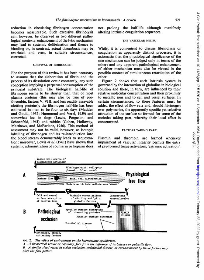
Figure 2 shows that each intrinsic system isgoverned by the interaction of globulins in biologicalsolution and these, in turn, are influenced by theirrelative molecular concentration and their proximityto metallic ions and to cell and vessel surfaces. Incertain circumstances, to these features must beadded the effect of flow rate and, should fibrinogenever polymerize, the apparently specific yet selectiveattraction of the surface so formed for some of themoieties taking part, whereby their local effect isconcentrated.
FACTORS TAKING PART
Plasmin and thrombin are formed wheneverimpairment of vascular integrity permits the entryof pre-formed tissue activators, 'extrinsic activation'.
Vessel wall source of9plasminogen activator
* Fibrinogen-rich, cell-poorplasmatic 'clear zone'.
........................................................................... ......................................................................................................................... *.A..*:*.............................*.l.*. .. Physiological, ~~~~~~........ ........ ............,Lamin&irflowI......Axial cell distribution.:............... ........... r::..::.. ....... ::::::.x......{
~~~~~~~~~c l i t i u i n ...................................:..:,
.................... ................................................... . .................................................................................. free flow......................... I Platelet-rich intermediate zone
I tCell and vesselsurface adsorptionof calcium ions
Variable concentrationsof clotting and lyticglobulin factors
-. ILipoprotein l.macromolecules
IWJmhi .......NRWX-
Specific surface adsorption IIIIIIIIIII
B Pathological of interacting proteins FI.RINPlatelet surface adherence POLY
occlusionEndothelial disease
..n 1111I Extrinsic, tissue, E m~activating factors
FIG. 2. The effect of environment on the haemostatic equilibrium.A A theoretical venule or capillary, free from the influence of turbulence or pulsatile flow.B A similar sized vessel in w,iich occlusion, endothelial disease, or encroachment by tissue factors mayalter the floiv pattern.
521
copyright. on F
ebruary 22, 2022 by guest. Protected by
http://jcp.bmj.com
/J C
lin Pathol: first published as 10.1136/jcp.17.5.520 on 1 S
eptember 1964. D
ownloaded from
522
If factors normally in equilibriurvascular tree are in any way stimul;activation', this is assumed to be inso-called contact factors (XII an
thrombin is formed, and by the releasubstances from the vessel wall when(gen is to be converted to plasmin.
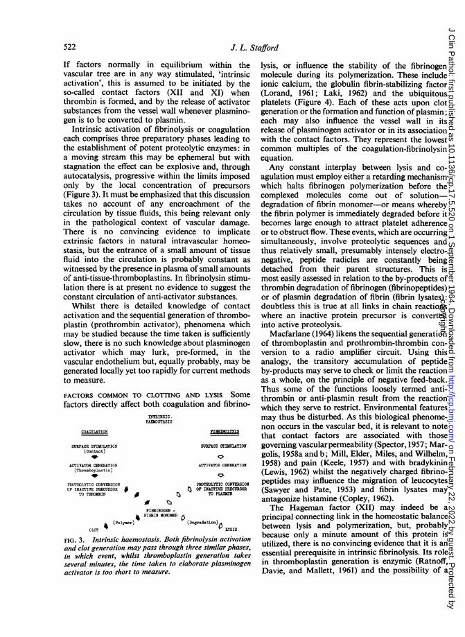
Intrinsic activation of fibrinolysiseach comprises three preparatory phathe establishment of potent proteolyta moving stream this may be ephenstagnation the effect can be explosiveautocatalysis, progressive within the I
only by the local concentration(Figure 3). It must be emphasized thattakes no account of any encroaci
circulation by tissue fluids, this beingin the pathological context of vascThere is no convincing evidenceextrinsic factors in natural intravasstasis, but the entrance of a small arrfluid into the circulation is probablwitnessed by the presence in plasma ofof anti-tissue-thromboplastins. In fibrilation there is at present no evidenceconstant circulation of anti-activator
Whilst there is detailed knowledactivation and the sequential generaticplastin (prothrombin activator), pheimay be studied because the time takeislow, there is no such knowledge abotactivator which may lurk, pre-fo:vascular endothelium but, equally prc
generated locally yet too rapidly for ctto measure.
FACTORS COMMON TO CLOTTING AND
factors directly affect both coagulaticINTRINSIC-
HADlOSTASIS
COAGULATION
SURFACE STIMULATION( Contact )
ACTIVATOR GENERATION(Thromboplastin)
PROTEOLYTIC CONVERSIONOF INACTIVE PRECURSOR
TO THROMBIN J
if t:a
FIBRINOGEN
(Polymer) FINRIN MONOMER (Degr
CLOT
FIG. 3. Intrinsic haemostasis. Both fibrinand clot generation may pass through thre,in which event, whilst thromboplastin gseveral minutes, the time taken to elaboractivator is too short to measure.
J. L. Stafford
n within the lysis, or influence the stability of the fibrinogenated, 'intrinsic molecule during its polymerization. These includeiitiated by the ionic calcium, the globulin fibrin-stabilizing factor
Id XI) when (Lorand, 1961; Laki, 1962) and the ubiquitous,se of activator platelets (Figure 4). Each of these acts upon clotever plasmino- generation or the formation and function of plasmin;
each may also influence the vessel wall in itsor coagulation release of plasminogen activator or in its associationases leading to with the contact factors. They represent the lowest
;ic enzymes: in common multiples of the coagulation-fibrinolysinneral but with equation.and, through Any constant interplay between lysis and co-
limits imposed agulation must employ either a retarding mechanismof precursors which halts fibrinogen polymerization before thethis discussion complexed molecules come out of solution-hrment of the degradation of fibrin monomer-or means wherebyrelevant only the fibrin polymer is immediately degraded before it
cular damage. becomes large enough to attract platelet adherenceto implicate or to obstruct flow. These events, which are occurring
scular homeo- simultaneously, involve proteolytic sequences andiount of tissue thus relatively small, presumably intensely electro-ly constant as negative, peptide radicles are constantly beingsmall amounts detached from their parent structures. This isinolysin stimu- most easily assessed in relation to the by-products ofto suggest the thrombin degradation of fibrinogen (fibrinopeptides)substances. or of plasmin degradation of fibrin (fibrin lysates):ge of contact doubtless this is true at all links in chain reactionsrn of thrombo- where an inactive protein precursor is convertednomena which into active proteolysis.
is sufficiently Macfarlane (1964) likens the sequential generationat plasminogen of thromboplastin and prothrombin-thrombin con-rmed, in the version to a radio amplifier circuit. Using thisibably, may be analogy, the transitory accumulation of peptideirrent methods by-products may serve to check or limit the reaction
as a whole, on the principle of negative feed-back.Thus some of the functions loosely termed anti-
)nLYSIS Some thrombin or anti-plasmin result from the reaction~nand fibrino- which they serve to restrict. Environmental features
may thus be disturbed. As this biological phenome-non occurs in the vascular bed, it is relevant to notethat contact factors are associated with those
SURFACE STIMUL&TION governing vascularpermeability (Spector, 1957; Mar-lco golis, 1958a and b; Mill, Elder, Miles, and Wilhelm,
ACTIVATOR GENERATION 1958) and pain (Keele, 1957) and with bradykinin10. (Lewis, 1962) whilst the negatively charged fibrino-
PRSOTEOLYTIC CONVERSION peptides may influence the migration of leucocytes)FTOIIVE.MCURR (Sawyer and Pate, 1953) and fibrin lysates may
antagonize histamine (Copley, 1962).The Hageman factor (XII) may indeed be a
mdation)0 principal connecting link in the homeostatic balancer LYSIS between lysis and polymerization, but, probably
'olysin activation because only a minute amount of this protein ise similar phases, utilized, there is no convincing evidence that it is an'eneration takes essential prerequisite in intrinsic fibrinolysis. Its role'ate plasminogen in thromboplastin generation is enzymic (Ratnoff,
Davie, and Mallett, 1961) and the possibility of a
O'.pt4
4
copyright. on F
ebruary 22, 2022 by guest. Protected by
http://jcp.bmj.com
/J C
lin Pathol: first published as 10.1136/jcp.17.5.520 on 1 S
eptember 1964. D
ownloaded from

The fibrinolytic mechanism in haemostasis: A review
ContactFactorsXI-XII
Calcium ionsFibrin-
stabilising AntithromboplastinsAnti-activators , -factor 4 C
* -.:::::::. :. Platelets.......... "I_
--'--Activators Thromboplastins
Plasminogen FIBRINOGEN Prothrombin
PLASMIN HROMBIN
Fibrin ........
Antiplasmins monomers Antithrombins
Fibrinogen lysates ibrinopeptides
polymerisation
gelation
SURFACE ADSORPTION
similar role in fibrinolysis has been reviewedNiewiarowski and Prou-Wartelle (1959) andlatridis and Ferguson (1961, 1962).
FIG. 4. The various factors which maintain the intrinsichaemostatic equilibrium, ionic calcium, fibrin stabilizingfactor, and platelets being common denominators.
The physiological role played by antithrombins and anti-activators is conjectural: fibrinogen lysates and fibrino-peptides may exert a retarding influence, and the surfacepresented after polymerization has a crucial effect on
subsequent events.
byby
ACTIVATORS OF PLASMINOGEN This activation is a
proteolytic reaction and the resultant plasmin has a
somewhat smaller molecule than its precursorplasminogen. The presence in plasma of a minutetrace of activator is probably normal (Fearnley,1953; Sawyer, Fletcher, Alkjaersig, and Sherry,1960), but this is so readily released from bloodvessel walls after relatively trivial stress that it isdifficult to be certain. Nor is it yet known whetherall plasminogen activation follows a similar sequencein man. Physical exercise and adrenaline injection(Biggs, Macfarlane, and Pilling, 1947), emotional or
traumatic shock (Macfarlane and Biggs, 1946), andthe administration of pyrogens or drugs, such as
nicotinic acid, which act on vessel walls, will eachrelease activator into the plasma. The principal sitefor plasma activator formation appears to be thevessel wall itself (Kwaan, Lo, and McFadzean, 1957;Todd, 1959) but other forms of plasminogenactivator are found in almost all body tissues andsecretions (Astrup and Permin, 1947; Albrechtsen,1957) with the possible exception of liver whereplasminogen itself is elaborated. Urokinase, foundin normal urine (Williams, 1951), is the form mostreadily available for study and, as it appears in
equal amounts in the bladder and the renal pelvis,there is speculation about its origin from the bloodstream or from functioning kidney tissue. The highconcentration of activator in prostatic and lungtissue can become clinically significant if these are
roughly handled during operation.
OTHER ACTIVATORS In addition to naturally occurr-
ing factors, artificial activators have been studied.Some, such as chloroform (Tagnon, 1942), achievethis effect by reducing antiplasmin activity. Thebacterial extract, streptokinase, from the haemolyticstreptococcus, was the first to be introduced (Tillettand Garner, 1933) and is of clinical importancebecause it can now be given intravenously. Ofbovine substrates, streptokinase seems to require aco-factor, pro-activator, before it can convertplasminogen: this may also be true in the human or,in this instance, plasminogen may be capable ofserving in both roles (Ablondi and Hagan, 1957;Kline, 1960).
Finally, plasmin itself can activate plasminogen(autocatalysis) as can other proteolytic enzymessuch as trypsin, a matter of consequence in acutepancreatitis.
ANTI-ACTIVATORS The function of plasminogenactivator is also subject to the retarding influence ofinhibitor proteins in vitro. This may have little or no
Vesselwall
stimulation
.+
523
copyright. on F
ebruary 22, 2022 by guest. Protected by
http://jcp.bmj.com
/J C
lin Pathol: first published as 10.1136/jcp.17.5.520 on 1 S
eptember 1964. D
ownloaded from

significance in the normal circulation but the rapiddevelopment of anti-activator activity in shed blood,after its contact with glass, can vitiate laboratoryresults. Plasminogen activator is stable in wholeblood or plasma and Flute (1960) clearly demon-strated that its apparent lability, as shown byFearnley and Lackner (1955), is due to the rise inthe function of anti-activator rather than to anyreduction in activator activity. Careful avoidanceof contact with wettable surfaces and a rigidlyapplied cold technique, during which the plasmasample is retained between 0 and 40C., reduce thedevelopment of anti-activator function to a minimum.
PLASMINOGEN This is a naturally circulating in-active beta-globulin with a molecular weight whichmay be as high as 140,000 (Shulman, Alkjaersig, andSherry, 1958) or as low as 83,800 (Davies andEnglert, 1960). Sgouris, Inman, McCall, Hyndman,and Anderson (1960) have described a techniquefor its preparation from the Cohn fraction III pasteusually discarded in the preparation of gammaglobulin from outdated blood (Oncley, Melin,Richert, Cameron, and Gross, 1949). It is stable inplasma and comes down with the 'euglobulin'fraction after dilution with acidified distilled water:the plasma euglobulin fraction or the fibrinogen-free serum euglobulin fraction can thus be utilizedto provide a convenient laboratory source (Howell,1963; Hawkey and Stafford, 1964).The plasminogen molecule is probably larger than
plasmin (Shulman et al., 1958), and there is thus adirect analogy with the relation in size of pro-thrombin to thrombin and of fibrinogen to fibrin.The conversion of plasminogen to plasmin appearsto involve the removal of a moiety so laying bare acentre of activity: as in other biological systems arelatively small alteration in configuration mayradically change protein function. Kline (1960) isconvinced that the proactivator necessary in thestreptokinase reaction is plasminogen in anotherguise, in which either two active centres are un-covered or a different surface radicle is exposed sothat two separate reactions may occur.
Plasminogen may have a specific affinity forfibrin (Back, Ambrus, and Markus, 1959) whereby itcan be adsorbed to the fibrin surface; it may be,however, that it is the activator factors which alonepossess this affinity. The fact that one or both can beconcentrated by specific attraction to fibrin surfacesleads to the ready conversion of plasmin at the sitewhere it is required yet with minimum influence fromanti-plasmins.
PLASMIN In vitro this proteolytic enzyme hassubstrate specificity for both fibrinogen and fibrin
(Ratnoff, 1953; Ambrus and Markus, 1960): it willalso digest certain protein components of thecoagulation system-factor V, factor VIII (anti-haemophilic), and prothrombin (Fletcher, Alkjaersigand Sherry, 1959; Donaldson, 1960; McNicol,Douglas, and Bayley, 1962). Its digestion of caseinpermits comparative measurement of activity, but asthis is scarcely physiological, many different methodshave been devised to assess the lysis of fibrin itselfunder controlled conditions.
Such laboratory methods are based either onexternal visible lysis of a fibrin plate (Astrup andMullertz, 1952) when the proteolytic enzyme isadded from outside, or on internal lysis promoted byincorporating the fibrinolytic agent within the clotitself. In the latter, the breakdown of 1311-fibrinogencan be measured by the iodine released (Shulmanand Tagnon, 1950; Clement and McNicol, 1959) ora standardized clot (Christensen, 1949), containingred cells (Blix, 1962; Howell, 1964a), can be formedwith standardized substrates: this approach at leastsimulates the spatial relationship between thevarious interacting components and the end-pointcan be judged as the time taken for total lysis(Hawkey and Stafford, 1964).
Plasmin apparently digests fibrinogen by hydro-lysis of peptide bonds as arginine and lysine residues:synthetic substrates containing esters of these amino-acids may act as competitive inhibitors (Troll,Sherry, and Wachman, 1954). The remarkablecapacity for attacking fibrin in vivo is inhibited bycertain plasma proteins collectively called anti-plasmins.
ANTIPLASMINS (INHIBITORS) Normal plasma orserum exhibits a considerable excess of antiplasminactivity, perhaps thirty-fold the potential quantityof plasmin (Norman, 1958; Fletcher et al., 1959);this is relevant to any discussion of a dynamicequilibrium. Such inhibitory mechanisms may alsohave a beneficial tendency to restrict plasmin to itsdigestion of fibrin. At least two antiplasmin plasmaproteins exist: an alpha 1 globulin and a morerapidly acting alpha 2 globulin (Norman and Hill,1958). Platelets also exert antiplasmin activity.The reactions with plasmin of these circulating
inhibitors are markedly affected if the proteinsubstrate, fibrin, is present. This is because fibrin ismore avid for the active site on plasmin than isanti-plasmin: conversely fibrinogen, although also a
substrate, seems to be less avid (Celander and Guest,1957). Thus a homeostatic bias promotes thedestruction of fibrin but permits the preservation offibrinogen.The three-dimensional significance of the surface
provided by formed fibrin is the basis for an attract-
J. L. Staffrd524
copyright. on F
ebruary 22, 2022 by guest. Protected by
http://jcp.bmj.com
/J C
lin Pathol: first published as 10.1136/jcp.17.5.520 on 1 S
eptember 1964. D
ownloaded from

The fibrinolytic mechanism in haemostasis: A review
ive hypothesis introduced by Sherry and hiscolleagues to explain the physiological mechanisminvolved in clot destruction or thrombolysis (Sherry,Fletcher, and Alkjaersig, 1959a; Sherry, Lindemeyer,Fletcher, and Alkjaersig, 1959b). In the intersticesof thrombi, because of the physical affinity ofplasminogen for fibrin, the local concentration ofplasminogen is relatively high whilst that of anti-plasmin is relatively low, unless there is a patho-logical excess of platelets. Thus, when circulatingplasma activator diffuses into an area of thrombosis,the intrinsic clot plasminogen is activated and theresultant plasmin, unimpeded by antiplasmin, isreadily able to digest its fibrin substrate. Anydiffusion of plasmin back into the general circulationis relatively innocuous because plasma antiplasminsserve to protect fibrinogen in solution.The difficulty of distinguishing between activation
of one factor and the inhibition of its antagonist hasled to somewhat complex explanations of what maybe a relatively simple interplay of plasma proteins.This difficulty is most apparent when proteininteraction is related to adjacent non-proteincomponents: ionic calcium classically enhancescoagulation yet Fearnley (1961) has reported it toinhibit fibrinolysis. Bruce (1964) has shown, however,that calcium probably influences plasmin function byactivating the antiplasmin inhibitors.
THE PHYSICAL CHARACTER OF FIBRIN
Whatever might be thought of the physiologicalrole played by plasmin the topic is not readilyamenable to scientific proof: most of the currentknowledge of fibrinolysis has been gleaned fromobserved pathology, when either coagulation orfibrinolysis has established hegemony over the other.
In pathological states of venous or arterialthrombosis the adsorptive capacity of the fibrinsurface is apparently decisive: furthermore, thecoincidence of endothelial abnormality or theconsequence of occlusion results in a changedflow pattern. In these circumstances the interplay ofprotein moieties is conditioned by features which areabsent from normal physiology. At this point indiscussion it is thus necessary to consider thephysical qualities of the fibrinogen molecule and ofits polymer (Scheraga, 1958).
Reference to the work of Hall and Slayter (1959)and to the many papers by Laki (1951, 1962) and byLorand (1961) permits some visualization of thefibrin surface. The fibrinogen molecule possiblycomprises a spiral chain with three separated areasof more complex folding. It is converted aftercontact with thrombin into a fibrin monomerand then polymerized. It is not known at which
points plasmin exerts its proteolytic effects but, asthis enzyme is also fibrinogenolytic, changes mayperhaps occur during the early phase if enoughplasmin exists at these sites. As polymerizationproceeds, both end-to-end and side-to-side bondingoccurs. Ultimately the fibrin band or filamentprovides a surface to which plasminogen activatorand thrombin are attracted. Whether the plasminogenmolecules are involved because they are trapped inthe mesh or also because of specific adsorption isstill not known.The surface attraction of these moieties is
depicted in relation to a broad band of fibrinmolecules polymerized by side-to-side bonding(Figure 5). Ionic calcium, platelets, and the serumglobulin 'fibrin-stabilizing factor (fsf)' seem toenjoy some special relationship denied to the othercirculating factors. The fibrin-stabilizing factor, nowknown as factor XIII, serves in the process ofconversion of the early monomeric fibrin molecule,which is urea-soluble, to the urea-insoluble polymercapable of clot retraction. Calcium may promoteadditional linkages between factor XIII and thesefibrin molecules, rendering them less susceptible toattack by plasmin (Bickford and Sokolow, 1961).
In the normal course of events, the fibrin mono-mers will condense to form fibrin polymer, assumingthat sufficient thrombin is present. Excess freeplasmin, if it is in sufficient concentration to nullifythe inhibitory power of the antiplasmins, willattack fibrinogen as well as fibrin, and the poly-peptide fragments so formed can physically interferewith any polymerization taking place in the immedi-ate vicinity. The polypeptide fragment or breakdownproduct becomes incorporated within the polymerbut, as it lacks correct configuration, the resultantclot is friable and subsequent clot retraction isdefective. Bang, Fletcher, Alkjaersig, and Sherry(1962) have studied the electron microscopicfeatures of this defective fibrin mesh. Excessiveaccumulation of these relatively large breakdownmoieties may ultimately block further polymeriz-ation, and indeed, may also provoke haemorrhagicepisodes by their effect on the vascular endothelium(Fletcher, Alkjaersig, and Sherry, 1962), a point tobe borne in mind during the phase of excessiveintravascular fibrinolysis induced by thrombolytictherapy (Flute, 1964).
THE FIELD OF ACTION
If, indeed, there is continuous destruction offibrinogen in vivo, irrespective of the actual thrombo-tic deposition of fibrin, then one must question wherein the vascular tree this event is taking place.Rheological studies must take account of the
525
copyright. on F
ebruary 22, 2022 by guest. Protected by
http://jcp.bmj.com
/J C
lin Pathol: first published as 10.1136/jcp.17.5.520 on 1 S
eptember 1964. D
ownloaded from

J. L. Stafford
calciu
.::::::::1:E::San'
fsf thrombin
f4..mia 7n a
fibrin lyates
plasminogen + PLASMIN
ACTIVATORQ_/
FIG. 5. The surfaces offered by fibriii (shaded) and comprising side-to-side bonded fibrin monomers.To this, thrombin and activator are specifically adsorbed: plasminogen and the fibrin stabilizing factor (fsf) may also
be attracted. Antiplasmins may be trapped within the clot mesh and thus inhibit plasminogen conversion; the by-products,fibrinopeptides and fibrin lysate polypeptides, may also have a retarding influence. Calcium may have a cardinal role ofenhancing thrombin and antiplasmin activity, and serving as a cationic bridge for other protein linkages.
turbulence engendered by arterial pulsation or theeddying where veins receive tributaries: only incapillaries is laminar flow likely to be constant, andin vessels of such small calibre the actual distributionof cell particles is subject to the sigma phenomenon(Scott-Blair, 1958) whereby blood alters its apparentviscosity with changing conditions of flow. Copley(1953, 1962) discusses the significance of laminarflow and its production of a relatively immobileouter, intimal, zone of plasma which at its extremeedge may present a single line offibrinogen molecules.At one time Copley suggested that fibrin itself mightbe directly opposed to the vessel wall but this hasnot attracted credence.
Possibly there are two fields of action for thedepredations of thrombin and plasmin. In thecapillary bed a protracted physiological form ofguerrilla skirmishing is dominated by plasmin: herethe permeability of the endothelial barrier toencroachment by tissue moieties, the intermittentnature of flow in alternately functioning or stagnantnetworks, and the relatively greater proportion ofplasma to cells induced by the sigma effect mayprovide an environment optimally designed to copewith the myriad but minor breaches of vascularintegrity-'wear-and-tear'. In the venous or arterialstreams, however, which should not normally bestagnant, the greater flow rate, larger proportion ofcells to plasma, and the different nature of the vesselwalls, allow only isolated forays which, as they are
induced by pathological changes, may result in alocal ascendency of thrombin over plasmin-polymerization of fibrinogen rather than its lyticdestruction. Especially might this be so if inflamm-ation of the vein wall or atherosclerotic changes inthe arterial intima provide focal points for theaggregation of platelets.One might speculate further that the essential
difference between these two intravascular fields ofaction is emphasized during the process of ageing:with senescence, capillary fragility and its attendantbleeding contrasts with the occlusive tendency inlarger vessels. This does not necessarily conflict withthe morphological concept underlying 'fibrinousvasculosis' (Lendrum, 1955, 1963) for this is basedon extravascular escape of plasma molecules.Clearly the equilibrium between fibrinolysis andcoagulation in the tissues must be governed bydifferent laws, and the fate suffered by the fibrinogenmolecule can scarcely be comparable. The exercisein semantics involving the word 'fibrinoid' (Pagel1951; Movat and More, 1957; Lendrum, Fraser,Slidders, and Henderson, 1962) is due in largemeasure to a failure to acknowledge that intra-vascular and extravascular polymerization offibrinogen need not necessarily and, indeed, canscarcely ever, be identical.
Extravascular (tissue) proteolysis, as opposed tothat evoked by intrinsic haemostatic mechanisms,may involve a sequence of different yet related
526
copyright. on F
ebruary 22, 2022 by guest. Protected by
http://jcp.bmj.com
/J C
lin Pathol: first published as 10.1136/jcp.17.5.520 on 1 S
eptember 1964. D
ownloaded from

The fibrinolytic mechanism in haemostasis: A review
enzymic reactions. Although not germane to thisreview, it is of interest to refer to work on therelease of tissue plasminogen activator (Albrechtsen,1957) and that in milk (Astrup and Sterndorff, 1953),saliva (Albrechtsen and Thaysen, 1955), and tears(Storm, 1955). Von Kaulla (1963) speculates uponthe importance of interaction between componentsof the fibrinolytic system in the process of fertiliz-ation, as seminal fluid contains plasminogenactivator (von Kaulla and Shettles, 1953). Thesignificance of lysis within the lymph system, whichcontains a substantial proportion of the bodyfibrinogen pool, has not yet been assessed, but inthe joint cavities Lack (1963) has made a closestudy of the importance of fibrinolysis in relation tojoint function and synovial integrity.
LOCAL FEATURES
The dynamic equilibrium between coagulation andfibrinolysis allows the clotting system to correctendothelial defects with a haemostatic fibrin sealwhile the fibrinolytic system prevents furtherprogress to occlusion by removing any fibrinscaffolding once the endothelium is repaired.There remain three local features which must be
brought into any consideration of the fibrinolyticmaintenance of vascular patency: the nature andfunction of endothelium (Altschul, 1954; Warren,1964), the part played by platelets (Holemans andGross, 1961), and the presence of proteins other thanthose directly involved in haemostasis, chylo-microns and low-density lipoproteins (Greig, 1956;Pappenhagen, Kopel, and Olwin, 1963; Howell,1964a and b).
THE ENDOTHELIUM Albrechtsen (1957) and Todd(1959) have shown plasminogen activator activity inthe intact vessel wall, and Warren (1963) has clearlydemonstrated this to be present in isolated endo-thelial cells themselves. In his review of endothelialultrastructure, Palade (1961) stresses the physio-logical necessity for an ample and continuousexchange across the capillary wall between plasmaand interstitial fluid. In this context this impliesmovement inwards of plasminogen activator and thereverse flow of degradation products, fibrin mono-
mer, or the partially polymerized fibrinogen referredto by Copley (1962). An earlier belief in mural pores
has been superseded by the electron microscopicdemonstration of sack-like calveolae intracellulareswhich open onto the cell surface or move across thecytoplasm. Any such carrier structure bears a
resemblance to the lysosome (de Duve, 1963) andmay thus be related to the activity of intracellularacid hydrolases. Lack (1963) has demonstratedthat certain lysosomes contain plasminogen activator,7
a finding which may be equated with the plasminogenactivator properties of disintegrated leucocytesreported by Gans (1964) and long recognized inabscess formation. Endothelial lysosomes, if theyexist, by being particularly susceptible to localhypoxia or damage, might actively contribute to theinduction of capillary permeability (Miles andWilhelm, 1960).Plasma kinins, which evoke pain, are released
into the plasma after surface contact stimulation(Armstrong, Jepson, Keele, and Stewart, 1957).One of these, kallikrein, is an active form of aninactive plasma precursor, kallikreinogen, which issimilar in many respects to plasminogen. Kallikreinmay be an enzyme which is quite distinct fromplasmin, or it might be this latter moiety after it hasbeen modified by some inhibitor or activator,consequent upon contact; its formation is thusintimately concerned with factor XII. Kallikreindiffers from plasmin as it does not attack casein orfibrin, nor is it inhibited by epsilon aminocaproicacid (Eisen, 1963). One might speculate upon therelationship between the normal endothelial surface,the contact factors, plasma kinins, and permeabilityfactors: but there is little objective physiologicalevidence upon which to base this conjecture.
PLATELETS The effect of fibrinolysis on these cellfragments depends on whether they are intact, whentheir surface adsorptive qualities may contributecalcium ions and certain protein molecules, ordisrupted. Their being intact will also influencefibrinolysis according to whether they remaindiscrete and mobile, or become sticky, undergoviscous metamorphosis and clump.
Their function should only be assessed havingregard to their simultaneous involvement in clottingand in clot retraction. Johnson and Schneider (1953)first showed platelets to contain an antiplasmin,inhibiting fibrinolysis. Bierstedt (1958) found isolatedintact platelets to possess an opposite, accelerating,effect which Holemans and Gross (1961) believe to bedue to surface adsorption of a proactivator whichthey differentiate from plasminogen activator.
Platelets do not carry or contain measurableplasminogen but during the phenomenon of clump-ing they may become intimately linked withfibrinogen--'platelet fibrinogen' (Johnson, Smathers,and Schneider, 1952). Electron microscopy ofartificially induced platelet aggregates (Poole, 1959)and platelet thrombi (Poole, French, and Cliff,1963; Rodman, Mason, and Brinkhous, 1963) doesnot necessarily show fibrin to be present betweenadjacent cell margins, and their agglomeration in theso-called 'white bodies' occasionally seen tocirculate in physiological circumstances still requires
527
copyright. on F
ebruary 22, 2022 by guest. Protected by
http://jcp.bmj.com
/J C
lin Pathol: first published as 10.1136/jcp.17.5.520 on 1 S
eptember 1964. D
ownloaded from

J. L. Stafford
explanation. Johnson and McKenna (1963) haveshown that the clotting of relatively low plateletconcentrations forms a coarse fibrin network,whilst very high platelet concentrations formsignificantly less fibrin without clot retraction.The role of platelets in the lysis of formed thrombi
is plainly different from any function they may serve
in the moving plasma stream.
LIPOPROTEINS In vitro, low-density lipoproteins,chylomicron suspensions, and phospholipid prepar-
ations may, in certain circumstances, inhibitfibrinolysis; and, in vivo, the prolongation of theclot lysis time has been observed by several workersafter alimentary hyperlipaemia has been induced bysaturated fats. Whether this is merely a function ofsurface adsorption, or whether electro-negativelipid moieties physically interfere with the contactbetween interacting protein molecules, has yet to beexplicated. The demonstration of these effects ofplasma lipids has led to speculation about therelation between inhibited physiological fibrinolysisand the incidence of atherosclerosis: among others,Merskey, Gordon, and Lackner (1960) and Howell(1964b) have shown a somewhat greater fibrinolyticpotential in the Negro as compared with Europeancontrols, and attention has thus been focused on
possible racial differences in plasma lipid componentsor their concentrations, and on the incidence ofmyocardial ischaemia. The epidemiological or
sociological implications of these findings lie more
in the province of atherosclerosis research but thesubject, as it relates to fibrinolysis, has been reviewedrecently by Howell (1964b).
CONCLUSIONS
Whatever may be true of the sequence of events inpathological thrombosis, little is known about thephysiological control of haemostasis. The dynamicinteraction of protein molecules in solution, aidedand abetted by ionic calcium, is clearly influenced bysurface adsorption. This can, perhaps, be morereadily appreciated in relation to the large surfacesof cells and the vessel lining, but all the moietiestaking part present surfaces. It remains to be seen
whether macromolecules, such as lipoproteins,fibrinogen itself, or fibrinogen breakdown products,are capable of exerting a significant physicalinfluence upon each other, but it seems very likelythat they can. Effective haemostasis thus may bedependent upon the ability of enzymic moieties tocollide with their substrates in the plasma: naturehas provided the mammalian circulation with an
excess of the latter together with a potential excess
of the former. But this plenitude is not so greatthat physical neutralization by inhibitor or non-
reacting molecules cannot occasionally upset theequilibrium.The work at St. George's Hospital referred to in thispaper was made possible by grants from the MedicalResearch Council and from the St. George's HospitalResearch Fund.
REFERENCES
Ablondi, F. B., and Hagan, J. J. (1957). Proc. Soc. exp. Biol. (N. Y.),95, 195.
Albrechtsen, 0. K. (1957). Brit. J. Haemat., 3, 284.-, and Thaysen, J. H. (1955). Acta physiol. scand., 35, 138.Alkjaersig, N., Fletcher, A. P., and Sherry, S. (1958). J. biol. Chem.,
233, 86.,Fletcher, A. P., and Sherry, S. (1962). J. clin. Invest., 41, 917.
Allwood, M. J., and Lewis, G. P. (1963). J. physiol. (Lond.), 166, 47P.Altschul, R. (1954). Endothelium. Its Development, Morphology,
Function, and Pathology. Macmillan, New York.Ambrus, C. M., and Markus, G. (1960). Amer.J. Physiol., 199, 491.Back, N., Ambrus, J. L., and Markus, G. (1959). Fed. Proc., 18, 364.Armstrong, D., Jepson, J. B., Keele, C. A., and Stewart, J.W. (1957).
J. Physiol. (Lond.), 135, 350.Astrup, T. (1962). In Proc. 8th Congr. europ. Soc. Haemat., pt. 2,
ch. 439,Vienna 1961. Karger, Basel., and Mullertz,S. (1952). Arch. Biochem., 40, 346.,
and Permin, P. M. (1947). Nature (Lond.), 159, 681., and Sterndorff, I. (1953). Proc. Soc. exp. Biol. (N.Y.), 84, 605.
Bang, N. U., Fletcher, A. P., Alkjaersig, N., and Sherry,S. (1962).J.clin. Invest., 41, 935.
Bierstedt, P. (1958). In 6th Congr. europ. Soc. Haemat. Trans., pt. 2,p. 483. Copenhagen, 1957. Karger, Basel.
Bickford, A. F.. andSokolow, M. (1 961). Thrombos. Diathes. haemorrh.(Stuttg.), 5, 480.
Biggs, R., Macfarlane, R. G., and Pilling, J. (1947). Lancet, 1, 402.Blix,S. (1962). Acta med. scand., 172, suppl. 386.Bruce, Sally (1964). J.clin. Path., 17, 282.Celander, D. R., and Guest, M. M. (1957). Arch. Biochem., 72, 176.Christensen, L. R. (1949). J.clin. Invest., 28, 163.Clement, W. E., and McNicol, G. P. (1959).J.clin. Path., 12, 544.Cohen, S., Holloway, R. C., Matthews, C., and McFarlane, A. S.
(1956). Biochem. J., 62, 143.Copley, A. L. (1953). In 19thint. Physiol. Congr. (abstracts of
communication), p. 280. Montreal.(1962). In Proc. 8th Congr. europ. soc. Haemat., pt. 2, ch. 357.Vienna 1961. Karger, Basel.
Davies, M. C., and Englert, M. E. (1960). J. Biol. Chem., 235, 1011.de Duve, C. (1963). In Lysosomes. A Ciba Foundation Symposium,
p. 1. Churchill, London.Donaldson,V. H. (1960). J. Lab.clin. Med., 56, 644.Eisen,V. (1963). J. Physiol. (Lond.), 166, 496.Fearnley, G. R. (1953). Nature (Lond.), 172, 544.
(1961). Lancet, 1, 992., Balmforth, G.V., and Fearnley, E. (1957). Clin. Sci., 16, 645.,
and Lackner, R. (1955). Brit. J. Haemat., 1, 189.Fletcher, A. P., Alkjaersig, N., and Sherry,S. (1959). J.clin. Invest.
38, 1096., - (1962). Ibid., 41, 896.Flute, P. T. (1960). In Proc. 7th Congr. europ. Soc. Haemat., pt. 2,
p. 894, London 1959. Karger, Basel.(1964). Proc. roy. Soc. Med., in the press.
Gans, H. (1964). Thrombos. Diathes. haemorrh. (Stuttg.), 10, 379.Green, J., and Thompson, W. B.(1962). Biochem. J., 84, 74P.Greig, H. B. W.(1956). Lancet, 2, 16.Hall, C. E., and Slayter, H.S. (1959). J. biophys. biochem. Cytol., 5, 11.Hammond, J. D. S., andVerel, D.(1959). Brit. J. Haemat., 5, 431.Hawkey, C. M., and Stafford, J. L. (1964). J.clin. Path., 17, 175.Holemans, R. (1963). Lancet., 2, 364.- and Gross, R. (1961). Thrombos. Diathes. haemorrh. (Stuttg.),
6, 196.Howell, M. B. (1963). J.clin. Path., 16, 289.- (I964a). In Second FibrinolysisW orkshop. J.clin. Path., 17, 310.- (1964b). Fibrinolysis. Brit. med. Bull., 20, (3), in the press.Iatridis, S. G., and Ferguson, J. H. (1961). Thrombos. Diathes.
haemorrh. (Stuttg.), 6, 411.(1962). J.clin. Invest., 41, 1277.(1963). J. appl. Physiol., 18, 337.
528
copyright. on F
ebruary 22, 2022 by guest. Protected by
http://jcp.bmj.com
/J C
lin Pathol: first published as 10.1136/jcp.17.5.520 on 1 S
eptember 1964. D
ownloaded from

The fibrinolytic mechanism in haemostasis: A review
Johnson, S. A., and McKenna, J. L. (1963). Thrombos. Diathes.haemorrh. (Stuttg.), 9, 102.and Schneider, C. L. (1953). Science, 117, 229.
, Smathers, W. M., and Schneider, C. L. (1952). Amer. J. Physiol.,170, 631.
Keele, C. A. (1957). Proc. Roy. Soc. Med., 50, 477.Kline, D. J. (1960). Amer. J. Cardiol., 6, 398.Kwaan, H. C., Lo, R., and McFadzean, A. J. S. (1957). Clin. Sci., 16,
241.Lack, C. H. (1963). Personal communication.Laki, K. (1951). Arch. Biochem., 32, 317.
(1962). Sci. Amer., 206, 60.Lendrum, A. C. (1955). J. clin. Path., 8, 180.
,Fraser, D. S., Slidders, W., and Henderson, R. (1962). Ibid., 15,401.- (1963). Canad. med. Ass. J,, 88, 442.Lewis, G. P. (1962). In British Postgraduate Medical Federation,
The Scientific Basis ofMedicine (Annual Review), p. 242.Lewis, J. H., Ferguson, E. E., and Schoenfield, C. (1961). J. Lab. clin.
Med., 58, 247.Lorand, L. (1961). In Anticoagulants and Fibrinolysins, edited by
R. L. MacMillan and J. F. Mustard, p. 333. Pitman, London.Macfarlane, R. G. (1945). Proc. roy. Soc. Med., 38, 399.
(1964). Nature, Lond. 202, 498.and Biggs, R. (1946). Lancet, 2, 862.
McNicol, G. P., Douglas, A. S., and Bayley, C. (1962). Lancet, 2, 1297.Madden, R. E., and Gould, R. G. (1952). Fed. Proc., 11, 252.Margolis, J. (1958a). Nature (Lond.), 181, 635.
((1958b). J. Physiol. (Lond.), 144, 1.Merskey, C., Gordon, H., and Lackner, H. (1960). Brit. med. J., 1, 219.Miles, A. A., and Wilhelm, D. L. (1960). In The Biochemical Response
to Injury. A Symposium edited by H. B. Stones, p. 51. Blackwell,Oxford.
Mill, P. J., Elder, J. M., Miles, A. A., and Wilhelm, D. L. (1958).Brit. J. exp. Path., 39, 343.
Movat, H. Z., and More, R. H. (1957). Amer. J. clin. Path., 28, 331.Niewiarowski, S., and Prou-Wartelle, 0. (1959). Thrombos. Diathes.
haemorrh. (Stuttg.), 3, 593.Nilsson, 1. M., and Olow, B. (1962). Ibid., 8, 297.Norman, P. S. (1958). J. exp. Med., 108, 53.
, and Hill, B. M. (1958). Ibid., 108, 639.Oncley, J. L., Melin, M., Richert, D. A., Cameron, J. W., and Gross,
P. M. Jr. (1949). J. Amer. chem. Soc., 71, 541.Pagel, W. (1951). J. cin. Path., 4, 137.Palade, G. E. (1961). Circulation, 24, 368.Pappenhagen, A. R., Kopel, J. L., and Olwin, J. H. (1963). Thrombos.
Diathes. haemorrh. (Stuttg.), 9, 164.Poole, J. C. F. (1959). Quart. J. exp. Physiol., 44, 377.
, French, J. E., and Cliff, W. J. (1963). J. clin. Path., 16, 523.Ratnoff, 0. D. (1953). J. clin. Invest., 32, 473.
and Colopy, J. E. (1955). Ibid., 34, 602.Davie, E. W., and Mallett, D. L. (1961). Ibid., 40, 803.and Rosenblum, J. M. (1958). Amer. J. Med., 25, 160.
Rodman, N. F. Jr., Mason, R. G., and Brinkhous, K. M. (1963). Fed.Proc., 22, 1356.
Roos, J. (1957). Thrombos. Diathes. haemorrh. (Stuttg.), 1, 471.Saameli, K., and Eskes, T. K. A. B. (1962). Amer. J. Physiol., 203, 261.Sawyer, P. N., Plate, J. W., and Weldon, C. S. (1953). Ibid., 175, 108.Sawyer, W. D., Fletcher, A. P., Alkjaersig, N., and Sherry, S. (1960).
J. clin. Invest., 39, 426.Scheraga, H. A. (1958). Ann. N.Y. Acad. Sci., 75, 189.Scott-Blair, G. W. (1958). Rheologica Acta, 1, 123.Sgouris, J. T., Inman, J. K., McCall, K. B., Hyndman, L. A., and
Anderson, H. D. (1960). Vox Sang. (Basel), 5, 357.Sherry, S., Fletcher, A. P., and Alkjaersig, N. (1959a). Physiol. Rev.,
39, 343., Lindemeyer, R. I., Fletcher, A. P., and Alkjaersig, N. (1959b).
J. clin. Invest., 38, 810.Shulman, N. R., and Tagnon, H. J. (1950). Ibid., 186, 69.Shulman, S., Alkjaersig, N., and Sherry, S. (1958). J. biol. Chem.,
233, 91.Sjoerdsma, A., and Nilsson, I. M. (1960). Prcc. Soc. exp. Biol. (N. Y.),
103, 533.Spector, W. G. (1957). J. Path. Bact., 74, 67.Storm, 0. (1955). Scand. J. cin. Lab. Invest., 7, 55.Tagnon, H. J. (1942). J. Lab. clin. Med., 27, 1119.Tillett, W. S., and Gamer, R. L. (1933). J. exp. Med., 58, 485.Todd, A. S. (1959). J. Path. Bact., 78, 281.Troll, W., Sherry, S., and Wachman, J. (1954). J. biol. Chem., 208, 85.von Kaulla, K. N. (1963). The Chemistry of Thrombolysis: Human
Fibrinolytic Enzymes, p. 289. Thomas, Springfield, Illinois.
von Kaulla, K. N. and Shettles, L. B. (1953). Proc. Soc. exp. Biol. (N. Y.),83,692.
Warren, B. A. (1963). Brit. J. exp. Path., 44, 365.(1964). The Structure and Function of Endothelium. Ph.D. Thesis,Oxford University.
Williams, J. R. B. (1951). Brit. J. exp. Path., 32, 530.
AppendixThis has been a review of the intrinsic mechanismsgoverning fibrinolysis; hence only a passing reference hasbeen made to extrinsic or tissue activation, and syntheticinhibitors such as epsilon-aminocaproic acid have notbeen discussed. Since thrombolytic therapy becamefeasible, however, these subjects have particular clinicalsignificance; moreover, pathologists may soon be calledupon to provide adequate laboratory surveillance,failing which this form of treatment should never beundertaken.
It would be premature to select or to recommendmethods of fibrinolysin assay: this subject is still relativelynew, and modifications or improvements have yet to beassimilated. In experienced hands, some techniques canprovide reproducible results, but probably the mostimportant single feature is the selection of reagents whichare as free as possible from contamination by otherfactors capable of influencing proteolysis; and thisselection is governed by the facility of local commercialsupply. The following recent British references, which areadditional to those in the main text, are cited by title forthose who wish to read further into the technical aspectsof this subject.
GENERAL TECHNIQUE
The investigation of plasminogen activators using aquantitative fibrinolytic method (Anderson, 1962).Determination of fibrinolytic activity of whole blood withspecial reference to the effects of exercise and fat feeding(Billimoria, Drysdale, James, and Maclagen, 1959). Therelease of thrombin from fibrin by fibrinolysis (Bloom,1962). Some physico-chemical properties of humanfibrinogen (Caspary and Kekwick, 1957). The 'fibrino-lytic potential' as a simple measure of spontaneousfibrinolysis (Chakrabarti and Feamley, 1962). Increaseof blood fibrinolytic activity by testosterone (Fearnleyand Chakrabarti, 1962). Purification of fibrinogen(Kekwick, Mackay, Nance, and Record, 1955). Anevaluation of the euglobulin method for the determin-ation of fibrinolysis (Kowalski, Kopec, and Niewiarowski,1959). Activation of endogenous plasma proteolyticenzymes with consequent production of pharmaco-logically active polypeptides (MacKay, Maycock, andCombridge, 1962). The effect of alimentary lipaemia onplasma fibrinolytic activity (Ogston and Fullerton, 1962).Estimation of plasminogen in biological fluids byagar/fibrinogen gel diffusion (Thornes, 1963).
THROMBOLYTIC THERAPY
Arterial occlusion treated with streptokinase (Clark,Hawkey, Howell, Rees, and Stubbs, 1964). Poplitealartery thrombosis treated with streptokinase (Cotton,Flute, and Tsapogas, 1962). Fibrinolytic therapy ofcoronary thrombosis (Dewar, Stephenson, Horler,
529
copyright. on F
ebruary 22, 2022 by guest. Protected by
http://jcp.bmj.com
/J C
lin Pathol: first published as 10.1136/jcp.17.5.520 on 1 S
eptember 1964. D
ownloaded from

Cassells-Smith, and Ellis, 1963). The clinical assessmentof fibrinolysis (Flute, 1963). The laboratory control ofthrombolytic therapy (Hawkey and Howell, 1964a).Intravenous streptokinase in the treatment of retinalvascular occlusion (Hawkey and Howell, 1964b).Thrombolytic therapy (McNicol, 1963). Studies in vitroand in vivo of a preparation of urokinase (McNicol,Gale, and Douglas, 1963a). Treatment of peripheralarterial occlusion by streptokinase perfusion (McNicol,Reid, Bain, and Douglas, 1963b). The effects of chlor-propamide on intermittent claudication and fibrinolysis(Tsapogas, Cotton, Flute, and Murray, 1962a). Lysis ofexperimental thrombi by streptokinase (Tsapogas,Flute, Cotton, and Milroy, 1962b). Feasibility of adequatethrombolytic therapy with streptokinase in peripheralarterial occlusions (Verstraete, Amery, and Vermylen,1963).
This list, which is by no means exhaustive, can besupplemented by reference to Human Blood Coagulation(Biggs and Macfarlane, 3rd ed., 1962), AnticoagulantTherapy (Douglas, 1962) and the symposium on fibrino-lysis in this Journal (J. clin. Path., 1964, 17, 305-370).A chapter on the fibrinolytic enzyme system (McNicoland Douglas) is to be included in the forthcomingRecent Advances in Clinical Pathology (Series IV, 1964)and a complete issue of the British Medical Bulletin(Vol. 20, No. 3, September, 1964) is devoted to thissubject.
APPENDIX REFERENCES
Anderson, A. J. (1962). Biochem. J., 83, 7P.Billimoria, J. D., Drysdale, J., James, D. C., and Maclagen, N. F.
(1959). Lancet, 2, 471.Bloom, A. L. (1962). Brit. J. Haemat., 8, 129.Caspary, E. A., and Kekwick, R. A. (1957). Biochem. J., 67, 41.Chakrabarti, R., and Fearnley, G. R. (1962). J. clin. Path., 15, 228.Clark, M. L., Hawkey, C. M., Howell, M. B., Rees, R. S. O., and
Stubbs, J. (1964). Postgrad. med. J., in the press.Cotton, L. T., Flute, P. T., and Tsapogas, M. J. C. (1962). Lancet, 2,
1081.Dewar, H. A., Stephenson, P., Horler, A. R., Cassells-Smith, A. J.,
and Ellis, P. A. (1963). Brit. med. J., 1, 915.Fearnley, G. R., and Chakrabarti, R. (1962). Lancet, 2, 128.Flute, P. T. (1963). Proc. roy. Soc. Med., 56, 411.Hawkey, C. M., and Howell, M. B. (1964a). J. clin. Path., 17, 287.
, -, (1964b). Ibid., 17, 363.Kekwick, R. A., Mackay, M. E., Nance, N. H., and Record, B. R. S.
(1955). Biochem. J., 60, 671.Kowalski, E., Kopec, M., and Niewiarowski, S. (1959). J. clin. Path.,
12, 215.MacKay, M., Maycock, W. d'A., and Combridge, B. S. (1962).
Nature, (Lond.), 195, 1206.McNicol, G. P. (1963). Proc. roy. Soc. Med., 56, 414.
Gale, S. B., and Douglas, A. S. (1963a). Brit. med. J., 1, 909.Reid, W., Bain, W. H., and Douglas, A. S. (1963b). Ibid., 1, 1508.
Ogston, D., and Fullerton, H. W. (1962). Ibid., 2, 1288.Thornes, R. D. (1963). J. clin. Path., 16, 87.Tsapogas, M. J., Cotton, L. T., Flute, P. T., and Murray, J. G.
(1962a). Lancet, 1, 1213., Flute, P. T., Cotton, L. T., and Milroy, S. D. (1962b). Brit. J.
Surg., 50, 334.Verstraete, M., Amery, A., and Vermylen, J. (1963). Brit. med. J., 1,
1499.
530 J. L. Stafford
copyright. on F
ebruary 22, 2022 by guest. Protected by
http://jcp.bmj.com
/J C
lin Pathol: first published as 10.1136/jcp.17.5.520 on 1 S
eptember 1964. D
ownloaded from