Embed Size (px)
Citation preview

THE VISCOSITY O F AMOEBA AT HIGH HYDROSTATIC PRESSURE
DUGALD E. 5. BROWN AND DOUGLAS A. MARSLAND New Pork University and Mal.ille Biological Laboratory, Woo& Hole,
MlWStlOhU8ett8
T W O FIGURES
Previous experiments, on Arbacia eggs, have indicated that high hydrostatic pressures induce marked changes in the fluidity of the protoplasm. I n the Arbacia eggs, the period of centrifuging necessary to produce a certain displacement of the pigment granules is materially reduced at high pressures (Brown, '34 a). This result might be accounted for either on the basis of an increase in the specific gravity of the displaced granules, or on the basis of a decrease in the viscosity of the medium through which they are displaced. To rule out the former possibility, as well as to establish the physical effects of pressure upon other cellular types, similar experiments have been performed on two species of Amoeba, namely Amoeba dubia and Amoeba proteus. These forms are par- ticularly well suited for the purpose because the protoplasmic granules which are subject to the centrifugal displacement appear to be quite different in the two species and because of other differences in the general structure.
The methods employed are similar to those previously de- scribed (Brown, '34 a). The Amoebae are placed in a centri- fuge-pressure chamber which is so designed that the control and pressure-treated specimens are simultaneously centri- fuged for a suitable period at a force 7000 X gravity.
I n centrifuging the Amoeba dubia, a very small quantity of isopicnotic sucrose is introduced into the experimental chamber and the cell suspension then floated on top. This
159

160 D. E. S. BROWN A N D D. A. MARSLAND
apparently prevents the specimens from being flattened when thrown by the centrifugal force against the bottom of the chamber. Immediately after centrifuging, the amoebae are fixed cytologically by immersing the centrifuge tubes in boil- ing water for 2 minutes. Otherwise the rapid onset of amoeboid movement precludes a critical examination of the centrifugal zones.
Amoeba proteus is centrifuged without the layer of sugar solution. Even without this buffer the cells assume a roughly spherical form, a condition which is essential if one is to ob- tain zones comparable in the different specimens. In the case of Amoeba proteus the slower resumption of amoeboid move- ment permits examination of the cells immediately after centrifuging without resort to cytological fixation.
When the specimens are centrifuged for a suitable period the protoplasmic granules and crystals, the nucleus, and the contractile vacuole are segregated into three well-defined zones: 1) A centripetal opaque zone of ‘oil,’ with the con- tractile vacuole on the inner border, 2) a central hyaline zone, and 3) a centrifugal granular zone with the nucleus usually lying at its centripetal border (fig. 1).
The formation of the hyaline zone requires the displace- ment of all the formed bodies originally present in this portion of the protoplasm. The greatest resistance to such displace- ment occurs in the peripheral protoplasm-namely in the plasmagel rather than in the plasmasol. At 7000 x gravity the displacement of the formed elements of the sol region is practically instantaneous (Heilbrunn, ’29). Consequently the ‘viscosity’ measurements obtained are entirely with reference to the gel portion of the protoplasm.
If the time required for the formation of a standard hyaline zone is taken as proportional to the viscosity of the plasmagel, then the viscosity-pressure relation may be determined by measuring the time required to produce the same zoning at each of a wide range of pressures. The results of extensive measurements of this sort are presented in table 1.

VISCOSITY ST HIGH PRESSURE
180
100 0.74 135 1 1.0
161
180 90 60
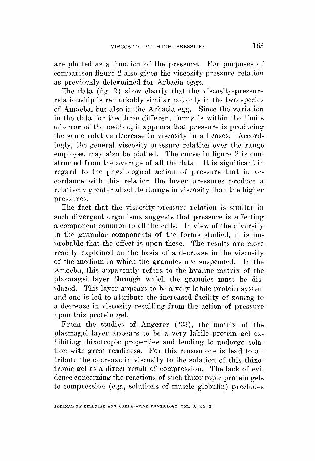
Fig. 1 The centrifugal zones formed in Amoeba dubia a t high hydrostatic pressurc. A, centrifuged 60 seconds at atmosphc=ric pressure. B, centrifuged simultaneously with A but at 136 atmospheres. C, centrifuged 90 secoiidq at 68 atmospheres. D, centrifuged 18 seconds at 400 atmosphwes. E and F centrifuged simultaiieously for 10 seconds, E a t atmospheric pressure and F a t 544 atmospheres. o, the oil zone; 11, the hyaliiie zone; g, the granular zone.
_ _ ~
PRESSURE, ATMOSPHERZ
~ ~
1 68
136 204 272 340 408 476 544 680
TBBLlI 1 .~ - - - - ~~
LRRACIA PUNCTULATA 1 AMOEBA DUBIA
1.0 0.66
0.38
0.22
0.088 0.066
AMOEBA PROTEUS -
Time
A era& 145 90 50
28
-
-7P 70
___
1.0 0.55
0.31
1 7 1 0.19
10 ‘ 0.11 6 I 0.066
7 T o
LVERAGE ’
1.0 0.65
0.36
0.21
0.11 0.081
JOUHNAL O F CELLULAR AND COMPARATIYE T’HYSIOLOOY, YOL. 8, NO. 2

162 U. E. S. BROWN A N D D. A. MARSLAND
The type of zone formation used as an end point diEers somewhat in the two kinds of Amoeba. In Amoeba proteus t hc number of protoplasmic crystals is conspicuously greater than in Amoeba dubia, and the ‘oil’ material is less in amount. This results, as might be expected, in the appearance of a wider ‘granule zone’ and a narrower ‘oil zone.’ Furthermore, at different pressures the zoning at the end point is not pre- cisely alike even when one is dealing with the same species. This is due partly to the fact that at the higher pressures the specimens are more nearly spherical. At higher pressures
Fig. 2 The decrease in viscosity mith increase in pressure. ‘I’ is the relative
.riscosity where 3. is the viscosity at 68 atmosphercs and v,, is the viscosity at any greater pressure. 0, Amoeba dubia; 0 , Aiiioeba proteus; +, Arbacia puiictulata (pigment zone formatioll).
‘ 0
also, the stratification is defined more sharply and there is a tendency toward the separation of the finer and coarser com- ponents of the heavy opaque zone.
At atmospheric pressure it is difficult to obtain a well- defined hyalirie zone even with prolonged centrifuging. For this reason, in estimating the change in viscosity with pres- sure, the viscosity q,, as indicated by the time of zone forma- tion at any pressure P, is expressed as a ratio of the viscosity, qo, at 68 atmospheres. The values f o r the viscosity expressed in this manner are given in table 1. In figure 2 these values
VISCOSITY AT H I G I I PRESSURE 163
are plotted as a function of the pressure. For purposes of comparison figure 2 also gives the viscosity-pressure relation as previously determined f o r Arbacia eggs.
The data (fig. 2) show clearly that the viscosity-pressure relationship is remarkably similar not only in the two species of Amoeba, but also in the Arbacia egg. Since the variation in the data for the three different forms is within the limits of error of the method, it appears that pressure is producing the same relative decrease in viscosity in all cases. Accord- ingly, the general viscosity-pressure relation over the range employed may also be plotted. The curve in figure 2 is coii- structed from the average of all the data. It is significant in regard to the physiological action of pressure that in ac- cordance with this relation the lower pressures produce a relatively greater absolute change in viscosity than the higher pressures.
The fact that the viscosity-pressure relation is similar in such divergent organisms suggests that pressure is affecting a component common to all the cells. In view of the diversity in the granular components of the forms studied, it is im- probable that the effect is upon these. The results are more readily explained on the basis of a decrease in the viscosity of the medium in which the granules are suspended. In the Amoeba, this apparently refers to the hyaline matrix of the plasmagel layer through which the granules must be dis- placed. This layer appears to be a very labile protein system and one is led to attribute the increased facility of zoning to a decrease in viscosity resulting from the action of pressure upon this protein gel.
From the studies of Angerer ('33), the matrix of the plasmagel layer appears to be a very labile protein gel ex- hibiting thixotropic properties and tending to undergo sola- tion with great readiness. For this reason one is lead to at- tribute the decrease in viscosity to the solation of this thixo- tropic gel as a direct result of compression. The lack of evi- dence concerning the reactions of such thixotropic protein gels to compression (e.g., solutions of muscle globulin) precludes
JOURNAL O F CELLULAR AND COMPARATIVE PHYSIOLOGY, VOL. 8 , NO. 2

164 U. E. S. BHOWN AND n. A. MABSLAND
a specific explanation of the phenomenon, but in general it may be observed tliat such gels undergo solation if subjected to a shearing force. It is conceivable therefore that a uniform compression produces a n iiitermolecular shearing force by changing the relative position of the individual molecules.
Since the present experiments demons h a t e only the viscous changes, it is of interest to mention briefly certain evidence, which shows tliat changes in rigidity and elasticity also occur. Thus, it is found that sudden compression of an Amoeba causes the immediate collapse and retraction of the psendo- podia with the formation of terminal spheres (3farslancl aiid Erowii, '36). Evidently under pressure, the rigidity of the plasmagel tube, which serves to maintain the tubular form of the pseudopodia, is lost. This data is also in agreement with results obtained 011 striated muscle which indicate that pressure decreases the elasticity of a contracting (Browxi, '34b) or a resting muscle (Edwards, unpublished). I t i p
significant that all of the assembled data agree in demonstrat- ing that pressures decrease the viscosity, rigidity o r elasticit? of certain cellular components. All of these effects would result from the solation of the cellular gels as a resnlt of compression.
SUMMABY
1. At high livdrostatic pressures the viscosity of tlic plasmagel of Amoeba dubia and Amoeba proteus is decreilsed.
2. Pressnre produces the same relative decrease in viscosit>- in Amoeba dubia, Amoeba proteus and Arbacia punctulata.
3. Evideiice is presented in support of the view that the p i m a r y action of pressure is to cause solation of protein gels ~ i t h i n the cells.

T’ISCOSITY AT H I G H PRESSURE 165
LITEBATURE CITED
-4 XGPRER, C . A . 1933
i ? e o w ~ , 1). E. S.
Tlie cffect of inechanieal stimulatioii npon the protolilasniic v.scosity of Amoeba.
The pressure voeficient of ‘viscosity’ in the cggs of Srhecia puiictulata. J. Cell. and Comp. Physiol., vol. 5, pp. 333-346.
The effect of rapid changes in hydrostatic pressure upou the contraction of skrletal muscle. J. Cell. and COIII~. Physiol., rol. 4,
TILILVIU EX, L. 1‘. 1029 The absolute \iscosity of A4~iioeba proto~ilasm. Proto-
1 \ [ 4 R S I A S D , I). A . AXD D. E. 8. BROWN 1936 Ainoeboid morement at high J. Cell. and Conip. Plipiol., rol. 8, pp. 167-1 78.
Anat. Rec., vol. 57, suppl., 11. 40. 1934 a
--_-_ 1934 IJ
~ I P . 257-281.
idasma, vol. 8, pp. 65-69.
hydrostatic pressure.