Embed Size (px)
Citation preview

ORIGINAL ARTICLE
The ‘‘vegetarian brain’’: chatting with monkeys and pigs?
Massimo Filippi • Gianna Riccitelli •
Alessandro Meani • Andrea Falini •
Giancarlo Comi • Maria A. Rocca
Received: 26 April 2012 / Accepted: 8 September 2012
� Springer-Verlag 2012
Abstract An array of brain regions in the fronto-parietal
and temporal lobes cooperates to process observation and
execution of actions performed by other individuals. Using
functional MRI, we hypothesized that vegetarians and
vegans might show brain responses to mouth actions per-
formed by humans, monkeys, and pigs different from
omnivores. We scanned 20 omnivores, 19 vegetarians, and
21 vegans while watching a series of silent videos, which
presented a single mouth action performed by a human, a
monkey, and a pig. Compared to omnivores, vegetarians
and vegans have increased functional connectivity between
regions of the fronto-parietal and temporal lobes versus the
cerebellum during observation of mouth actions performed
by humans and, to the same degree, animals. Vegans also
had increased connectivity with the supplementary motor
area. During human mouth actions, increased amygdala
activity in vegetarians and vegans was found. More criti-
cally, vegetarians recruited the right middle frontal gyrus
and insula, which are involved in social mirroring, whereas
vegans activated the left inferior frontal gyrus and middle
temporal gyrus, which are part of the mirror neuron system.
Monkey mouth actions triggered language network activity
in both groups, which might be due to the attempt to
decode monkey mouth gesture, with an additional recruit-
ment of associative temporo-occipital areas in vegans,
whereas pig mouth actions activated empathy-related
regions, including the anterior cingulum. These results
support the role of the action observation–execution
matching system in social cognition, which enables us to
interact not only with our conspecifics but also with species
in phylogenetic proximity to humans.
Keywords Action observation–execution matching
system � Animals � Humans � Mouth action � Vegetarians
Introduction
Processing and understanding actions performed with the
mouth by other individuals contribute to infer other people’s
emotional states and intentions, which is a hallmark of social
interaction. Functional imaging investigations have identi-
fied a set of brain areas, located mainly in the parietal and
frontal lobes, which are activated consistently, together with
several extrastriate occipital areas, during the observation of
mouth actions performed not only by humans but also by
other species (i.e., monkeys and dogs) (Buccino et al. 2004).
Remarkably, while actions belonging to the observer’s rep-
ertoire (e.g., speech reading) are mapped in areas of the
motor system located in the inferior frontal gyrus (IFG),
which include Broca’s area (BA44/BA45), actions which are
not part of such a repertoire (e.g., barking) are processed on
the basis of their visual properties and mapped to visual areas
(Buccino et al. 2004). Converging pieces of evidence suggest
that the neuronal network that involves the IFG and the
inferior parietal lobule (IPL), which form part of the mirror
neuron system (MNS), is also necessary for emotion
M. Filippi (&) � G. Riccitelli � A. Meani � M. A. Rocca
Neuroimaging Research Unit, Institute of Experimental
Neurology, Division of Neuroscience, San Raffaele Scientific
Institute, ‘‘Vita-Salute’’ San Raffaele University,
Via Olgettina 60, 20132 Milan, Italy
e-mail: [email protected]
M. Filippi � G. Comi � M. A. Rocca
Department of Neurology, San Raffaele Scientific Institute,
‘‘Vita-Salute’’ San Raffaele University, Milan, Italy
A. Falini
Department of Neuroradiology, San Raffaele Scientific Institute,
‘‘Vita-Salute’’ San Raffaele University, Milan, Italy
123
Brain Struct Funct
DOI 10.1007/s00429-012-0455-9
recognition and contagion, which contribute to emotional
empathy (Shamay-Tsoory 2011; Shamay-Tsoory et al.
2009). Several factors modulate empathic response, includ-
ing the subjective attitude held toward the observed indi-
vidual (Singer et al. 2006) and personal experience (Cheng
et al. 2007). A recent fMRI investigation has demonstrated
that the neuronal representation of empathy differs among
individuals with different dietary habits due to ethical rea-
sons (i.e., vegetarians and vegans vs. omnivores) (Filippi
et al. 2010). Specifically, when compared to omnivores, both
vegetarians and vegans recruit different areas of the empathy
network during observation of negative affective pictures
(showing mutilations, murdered people, human/animal
threat, torture, etc.) of human beings and, more critically,
animals. Substantial differences between vegetarians and
vegans were also found, with a prevailing activity of the
anterior cingulum in vegetarians and of the IFG in vegans,
possibly reflecting different motivational factors and beliefs.
With this background in mind, we hypothesized that the
representation of mouth actions within different brain
regions might differ among individuals with different die-
tary habits and ethical beliefs during processing of actions
performed by other humans and other species. We assumed
that, due to their propensity to identify nonconspecifics as
being like themselves, and their increased empathic
response towards animal actions, when compared to
omnivores, vegetarians and vegans would exhibit a dif-
ferent pattern of recruitment of regions involved in the
processing of mouth actions, including those located in the
fronto-parietal lobes. We also supposed that, since different
motivational factors and beliefs characterize vegetarianism
and veganism, with the first being linked to the notion of
not performing direct violence towards animals and the
second being against the rights of humans to use animals in
any way (Regan 1985), in addition to a common-shared
pattern of cortical processing of mouth actions, vegetarians
and vegans might also experience functional architecture
differences. Finally, we postulated that the recruitment of
the previous regions might differ according to the species
involved and their phylogenetic proximity to humans.
Experimental procedure
The study was approved by the Ethics Committee of Sci-
entific Institute and University Hospital San Raffaele,
Milan, Italy and written informed consent was obtained
from all subjects prior to study entry.
Subjects
We studied 60 right-handed (Oldfield 1971) human healthy
subjects (34 women and 26 men; mean age 37.7, range
18–60 years) with different dietary habits. All subjects had
normal or corrected-to-normal vision. We recruited 20
omnivores (11 women and 9 men; mean age 36.9, range
22–60 years), 19 vegetarians (11 women and 8 men; mean age
40.3, range 23–60 years), and 21 vegans (12 women and 9
men; mean age 36.3, range 18–53 years). The groups did not
differ statistically in sex, age, or level of education. A ques-
tionnaire was completed by all subjects before fMRI acqui-
sition to investigate eating habits, reasons and motivations for
eating choices, and the time elapsed from making this choice.
All vegetarians and vegans reported that they made their
dietary choice for ethical reasons. They had had stable eating
habits for a mean of 3.8 years (SD 8.7 years), and were
recruited from vegetarian societies. Omnivore subjects were
recruited by advertisement, and none of them had been veg-
etarian or vegan before the study. Eight vegans had been
vegetarian before becoming vegan. All the subjects were naı̈ve
about the nature of the study. None of the subjects had any
history of neurological, major medical, or psychiatric disor-
ders, or either alcohol or drug abuse. None of the subjects was
taking any medical treatment at the time of fMRI assessment.
Experimental design
Using a block design, all subjects were scanned while
watching a series of silent videos, which presented a single
mouth action performed by a human, a monkey, and a pig in
each block (Fig. 1). According to the experiment of Buccino
(Buccino et al. 2004), the mouth actions observed were (1)
biting, and (2) oral communicative actions (OCAs). How-
ever, our experiment differed from that of Buccino, since we
investigated pig rather than dog actions, because we thought
that dogs were likely to raise a similar degree of sympathy in
the different study groups. Furthermore, monkey scenes
were recorded in a free environment. The videos were
delivered using the Presentation software. Participants were
instructed to look at the scenes focusing their attention on
mouth movements, without providing any specific response
during the fMRI acquisition. Each block consisted of 12 s of
video, followed by a 12 s resting period. During each block,
the same action (i.e., biting or OCAs) was presented four
times. During the resting phase of a given block, the subjects
had to observe a static frame of the same action for 12 s. Each
of the six blocks of the experiment was performed four times
within a run. The order of videos in the various blocks was
counter-balanced across subjects. Two runs were performed
for each subject and averaged during the fMRI statistical
analysis.
fMRI acquisition
Using a 3.0 T scanner (Intera, Philips Medical Systems,
Best, The Netherlands) fMRI scans were acquired using a
Brain Struct Funct
123
T2*-weighted single-shot echo-planar imaging (EPI) pulse
sequence [echo time (TE) = 30 ms, flip angle = 85�,
matrix size = 128 9 128, field of view (FOV) =
240 mm 9 240 mm, repetition time (TR) = 3.0 s]. During
each functional scanning run, 192 sets of 40 axial slices,
parallel to the anterior–posterior commissural plane, with a
thickness of 3 mm, were acquired. Head movements were
minimized using foam padding.
In the same scanning session, a brain dual-echo turbo
spin echo sequence (TR = 3,500 ms, TE = 24/120 ms;
echo train length = 5; flip angle = 150�, 44 contiguous,
3-mm-thick, axial slices with a matrix size = 256 9 256
and a FOV = 240 mm 9 240 mm) was also acquired to
check for MRI lesions/abnormalities.
All subjects reported that they had not fallen asleep
during scanning, according to a questionnaire delivered
immediately after the MRI session.
fMRI analysis
fMRI data were analyzed using the statistical parametric
mapping (SPM8) software. Prior to statistical analysis, all
images were realigned to the first one of the series to
correct for subject motion, spatially normalized into the
Montreal Neurological Institute (MNI) space, and
smoothed with a 3-D 10 mm FWHM Gaussian filter.
Statistical analysis
Analysis of activations
Changes in blood oxygen level-dependent contrast associ-
ated with each condition were assessed using the general
linear model (Friston et al. 1995). In each subject, a first-
level design matrix was built, where subject motion
parameters were used as regressors of no interest. Then,
specific effects were tested by applying appropriate linear
contrasts. For each subject, activation maps were first
derived by contrasting each active condition with its own
control condition, with the following contrasts defined:
human biting [ static human biting; monkey biting [static monkey biting; pig biting [ static pig biting; human
OCAs [ static human OCAs; monkey OCAs [ static
monkey OCAs; pig OCAs [ static pig OCAs. Significant
hemodynamic changes for each contrast were assessed
using t statistical parametric maps (SPMt). Second level
random effect analyses, using 3 9 2 ANOVA models, in
which stimuli (3) and conditions (2) were entered as sep-
arate factors, were performed to assess the main effects of
the stimuli (i.e., human, monkey, and pig) and conditions
(i.e., biting and OCA) within the three groups of subjects of
the study (i.e., omnivores, vegetarians, and vegans).
Between-group differences were assessed by means of
3 9 3 or 3 9 2 ANOVA models, when comparing among
Fig. 1 Selected frames from the videos showing biting and oral communicative actions (OCAs) performed by a human, a monkey, and a pig
Brain Struct Funct
123

the three groups the effect of stimuli or conditions,
respectively (Friston et al. 1999). The following sets of
linear comparisons were performed: (1) vegetarians and
vegans, separately, versus omnivores; (2) vegetarians and
vegans, combined, versus omnivores; (3) vegetarians ver-
sus vegans, and vice versa. Common patterns of activation
between vegetarians and vegans during a given contrast as
well as regions of specific activations of each group con-
trasted to the others were identified by a conjunction
analysis (Friston et al. 2005).
We report activations below a threshold of p \ 0.05
corrected for multiple comparisons [family-wise error
(FWE)]. The identification of regions of activation was
performed using the Anatomical Automatic Labeling (aal)
atlas (Tzourio-Mazoyer et al. 2002) and the Brodmann area
(BA) atlas (Lancaster et al. 2000; Maldjian et al. 2003).
To compare the location of activations within the dif-
ferent portions of the IFG involved in mouth actions (i.e.,
pars opercularis and pars triangularis) between groups and
during the different experimental conditions, the MNI
coordinates of all peaks showing significant activations in
each single subject’s analysis were extracted using the
WFU Pickatlas software package and masks of BA44
(corresponding to pars opercularis) and BA45 (corre-
sponding to pars triangularis) (Lancaster et al. 2000;
Maldjian et al. 2003), and then compared between groups
using the SPSS software and ANOVA models (p \ 0.05).
This analysis was driven by the consideration that these
two portions of the L IFG are known to be involved in
different functions: the pars opercularis being supposed to
contain MN involved in processing mouth actions (Buccino
et al. 2001; Tettamanti et al. 2005), whereas the pars tri-
angularis has been consistently found to be activated across
semantic studies employing different design paradigms
(Xiang et al. 2010).
Analysis of functional connectivity (FC)
The realigned and normalized fMRI data entered a FC
analysis using a seed-voxel correlation approach (Biswal
et al. 1995). FC analysis estimates how strong the associ-
ations are between observed time series, without investi-
gating the causal relationships between them. For this
analysis, fMRI time series were first pre-processed to
reduce data variability unlikely to reflect neuronal activity.
These additional pre-processing steps comprised temporal
band-pass filtering (0.009 Hz \ f \ 0.08 Hz) and spatial
smoothing with a 3-D 6 mm FWHM Gaussian filter (Fox
et al. 2005). Then, the time series were processed in two
steps: first, we selected a ROI to serve as a seed region for
correlation; subsequently, we performed a regression
analysis between this seed region and all the remaining
voxels of the brain. The definition of the seed reference
time course for FC analysis relied on the maps of left
BA44/BA45 (Broca’s region) and BA21/BA22 available in
the WFU Pickatlas (Lancaster et al. 2000; Maldjian et al.
2003). Two anatomical masks for the left BA44/BA45 and
the left BA21/BA22 were created using WFU Pickatlas
(Maldjian et al. 2003) and, for each subject, the average
time series within these ROIs, separately, were extracted.
FC was then investigated using SPM8 and multiple
regression models, including as covariate of interest the
time series extracted from the reference seed region, and as
confounding covariates: (1) the six motion parameters
obtained during the rigid-body motion correction step, (2)
the whole brain signal averaged over the entire tissue, (3)
the averaged ventricular signal and, (4) the averaged white
matter signal (Fox et al. 2005). This analysis generated a
statistical FC map of the left BA44/BA45 region for each
subject. Similarly, a statistical FC map of the left BA21/
BA22 was produced. Within-group and between-group
comparisons of these FC maps were assessed using SPM8
one-sample t tests and ANOVA models, respectively
(p \ 0.05, FWE corrected).
Analysis of demographic and behavioural data
Demographic data were compared using SPSS and an
ANOVA model. Post hoc comparisons were corrected for
multiple comparisons using Bonferroni correction.
Results
Biting
Tables 1, 2 and 3 summarize the results of between-group
comparisons during the different experimental conditions.
During ‘‘human scenes’’, compared to vegetarians and
vegans, omnivores had increased activity of the left para-
hippocampal gyrus, right putamen, left insula, and left
inferior occipital gyrus (IOG) (BA19). Compared to
omnivores and vegans, vegetarians had increased activity
of the left cerebellum (crus I) and right IFG, pars opercu-
laris (BA44). Compared to omnivores and vegetarians,
vegans had increased activation of the right superior tem-
poral gyrus (STG) (BA21). Compared to vegetarians, ve-
gans also had increased activity of the left middle temporal
gyrus (MTG). During ‘‘monkey scenes’’, compared to
omnivores, vegetarians and vegans had increased activity
of the bilateral amygdala. Compared to omnivores, vege-
tarians had additional increased activity of the right su-
pramarginal gyrus (SMG), right middle occipital gyrus
(MOG), and right IFG, pars triangularis (BA45). Finally,
compared to omnivores and vegetarians, vegans had
additional increased activity of the bilateral MTG (BA21).
Brain Struct Funct
123

During ‘‘pig scenes’’, compared to omnivores and vegans,
vegetarians had increased activity of the right IFG, pars
opercularis (BA44). Compared to omnivores, they also had
increased activity of the right SMG, whereas compared to
vegans they had increased activity of the left fusiform
gyrus and left cerebellum (crus II). Compared to omni-
vores and vegetarians, vegans had increased activity of the
left MTG (BA21) and left MOG.
OCAs
No between-group differences were detected during
‘‘human scenes’’. During ‘‘monkey scenes’’, compared to
omnivores, vegetarians and vegans had increased activity
of the bilateral calcarine cortex, and left MTG (BA22,
Wernicke’s area). Compared to omnivores and vegans,
vegetarians had increased activity of the left IFG, pars
triangularis (BA45). Compared to omnivores, vegetarians
also had increased activity of another cluster in the left
MTG (BA21) and left precentral gyrus. Compared to
omnivores and vegetarians, vegans had increased activity
of the right MTG (BA21) and right MOG. Compared to
omnivores, vegans also had increased activity of the left
MOG. During ‘‘pig scenes’’, compared to omnivores and
vegans, vegetarians had increased activity of the anterior
cingulum. Compared to omnivores and vegetarians, vegans
had increased activity of the left MTG (BA 21), left IFG,
pars triangularis (BA45), and bilateral middle frontal gyrus
(MFG). Finally, compared to vegetarians, vegans also had
increased activity of the right parahippocampal gyrus.
OCAs versus biting
The results of the within-group comparisons of biting versus
OCAs and vice versa, in the three study groups, are summa-
rized in Tables 4 and 5 and are shown in Fig. 2. During
‘‘human scenes’’, vegetarians and vegans versus omnivores
had an increased activity of the right amygdala. Compared to
the other two groups, vegetarians had a selective increased
recruitment of the right MFG and the posterior portion of the
right insula (MNI coordinates: 38, -10, 8), whereas vegans
recruited selectively the left MFG, left IFG (pars opercularis)
and left MTG, posterior part (BA37) (MNI coordinates: -48,
-56, -2). During ‘‘monkey scenes’’, vegetarians and vegans
versus omnivores had increased activity of the bilateral
cuneus and left MTG (BA22) (MNI coordinates: -62, -44,
8). Vegetarians selectively activated the left MTG (BA21),
posterior portion (MNI coordinates: -52, -52, 16) and IFG
(pars opercularis), and vegans the right MTG (BA21), anterior
portion (MNI coordinates: 56, 2, -16) and MOG. During ‘‘pig
scenes’’, vegetarians showed a selective increased activity of
the anterior cingulum and vegans activated the bilateral
parahippocampal gyrus (Fig. 3).Ta
ble
1A
reas
sig
nifi
can
tly
mo
reac
tiv
ated
(act
ivat
ion
site
san
dM
on
trea
lN
euro
log
ical
Inst
itu
teco
ord
inat
es)
ino
mn
ivo
res
ver
sus
veg
etar
ian
san
dv
egan
sd
uri
ng
bit
ing
and
ora
lco
mm
un
icat
ive
acti
on
scen
es(A
NO
VA
,p\
0.0
5fa
mil
y-w
ise
erro
rco
rrec
ted
)
Co
nd
itio
nA
ctiv
atio
nsi
tes
Om
niv
ore
s
Hu
man
scen
esM
on
key
scen
esP
igsc
enes
Ver
sus
veg
etar
ian
san
d
veg
ans
Ver
sus
veg
etar
ian
s
on
ly
Ver
sus
veg
ans
on
ly
Ver
sus
veg
etar
ian
san
d
veg
ans
Ver
sus
veg
etar
ian
s
on
ly
Ver
sus
veg
ans
on
ly
Ver
sus
veg
etar
ian
san
d
veg
ans
Ver
sus
veg
etar
ian
s
on
ly
Ver
sus
veg
ans
on
ly
Bit
ing
Lp
arah
ipp
oca
mp
al
gy
rus
-2
0,
-3
8,
0–
––
––
––
–
Rp
uta
men
26
,1
6,
6–
––
––
––
–
Lin
sula
-2
4,
12
,-
10
––
––
––
––
LIO
G-
42
,-
68
,-
4
OC
As
––
––
––
––
––
Rri
gh
t,L
left
,IO
Gin
feri
or
occ
ipit
alg
yru
s,O
CA
so
ral
com
mu
nic
ativ
eac
tio
ns
Brain Struct Funct
123

Species-related recruitment
To assess whether the activity of regions associated with OCA
scenes differed in the three groups according to the species
observed (i.e., human, monkey, pig), a within-group com-
parison of species-related activations during OCAs was per-
formed. The results of this analysis are summarized in
Table 6. During human versus monkey OCAs, compared to
vegetarians and vegans, omnivores had an increased activity
of the right IOG and STG (MNI coordinate: -50, -18, -2).
Vegans had selective increased activity of the left MTG,
posterior part (MNI coordinates: -46, -56, -2), right pre-
central gyrus and left IFG (MNI coordinates: -48, 2, 14).
During human versus pig OCAs, omnivores versus vegetari-
ans and vegans had increased activation of the anterior portion
of the right insula (MNI coordinates: 36, 30, 6), and left IFG
(pars triangularis) (MNI coordinates: -36, 20, 26).
Vegetarians and vegans versus omnivores had increased
recruitment of the anterior lobe of the cerebellum (MNI
coordinates: -26, -44, -22). Vegetarians had selective
increased activity of the right amygdala, whereas vegans
activated the right precentral gyrus. During monkey versus pig
OCAs, vegetarians and vegans versus omnivores had
increased activity of the right amygdala, bilateral superior
parietal lobule (SPL) and right IOG. Vegetarians had selective
increased activity of the right globus pallidus (Fig. 4).
Location of IFG activity
A region of interest (ROI) analysis was performed to
compare the locations of activation in the different portion
of the IFG involved in mouth actions between groups and
during the different experimental conditions. In all three
study groups, activity within the left BA44 and BA45 was
Table 2 Areas significantly more activated (activation sites and Montreal Neurological Institute coordinates) in vegetarians versus omnivores
and vegans during biting and oral communicative action scenes (ANOVA, p \ 0.05 family-wise error corrected)
Condition Activation
sites
Vegetarians
Human scenes Monkey scenes Pig scenes
Versus
omnivores
and
vegans
Versus
omnivores
only
Versus
vegans
only
Versus
omnivores
and
vegans
Versus
omnivores
only
Versus
vegans
only
Versus
omnivores
and
vegans
Versus
omnivores
only
Versus
vegans
only
Biting L cerebellum -32, -74,
-36
– – – – – – – -26, -80,
-46
R IFG 46, 14, 10 – – – 40, 36, 0 – 34, 6, 34 – –
R amygdala – – – – 16, -12,
-14
– – – –
L amygdala – – – -20, -4,
-10
– – – –
R SMG – – – – 54, -34, 40 – – 56, -4,
-26
–
R MOG – – – – 32, -66, 34 – – – –
L fusiform
gyrus
– – – – – – – – -28, -24,
-26
OCAs R calcarine
cortex
– – – – 4, -78, 14 – – – –
L calcarine
cortex
– – – – -2, -80, 18 – – – –
L MTG – – – – -58, -18,
-2
– – – –
– – – – -56, 4, 14 – – – –
L IFG – – – 38, 26, 14 – – – – –
L precentral gyrus – – – – -50, 14, 32 – – – –
Anterior
cingulum
– – – – – – 6, 42, 10 – –
OCAs oral communicative actions, R right, L left, IFG inferior frontal gyrus, SMG supramarginal gyrus, MOG middle occipital gyrus, MTGmiddle temporal gyrus
Brain Struct Funct
123

Ta
ble
3A
reas
sig
nifi
can
tly
mo
reac
tiv
ated
(act
ivat
ion
site
san
dM
on
trea
lN
euro
log
ical
Inst
itu
teco
ord
inat
es)
inv
egan
sv
ersu
so
mn
ivo
res
and
veg
etar
ian
sd
uri
ng
bit
ing
and
ora
lco
mm
un
icat
ive
acti
on
scen
es(A
NO
VA
,p\
0.0
5fa
mil
y-w
ise
erro
rco
rrec
ted
)
Co
nd
itio
nA
ctiv
atio
nsi
tes
Veg
ans
Hu
man
scen
esM
on
key
scen
esP
igsc
enes
Ver
sus
om
niv
ore
s
and
veg
etar
ian
s
Ver
sus
om
niv
ore
s
on
ly
Ver
sus
veg
etar
ian
s
on
ly
Ver
sus
om
niv
ore
s
and
veg
etar
ian
s
Ver
sus
om
niv
ore
s
on
ly
Ver
sus
veg
etar
ian
s
on
ly
Ver
sus
om
niv
ore
s
and
veg
etar
ian
s
Ver
sus
om
niv
ore
s
on
ly
Ver
sus
veg
etar
ian
s
on
ly
Bit
ing
RS
TG
64
,-
4,
-4
8–
––
––
––
–
LM
TG
––
-4
8,
-7
0,
16
-5
2,
-5
6,
14
––
-5
4,
-5
2,
12
––
RM
TG
––
–4
8,
-6
0,
18
––
––
–
Ram
yg
dal
a–
––
–1
6,
-1
2,
-1
4–
––
–
Lam
yg
dal
a–
––
–-
20
,-
4,
-1
0–
––
–
LM
OG
––
––
––
-4
6,
-7
0,
16
––
OC
As
Rca
lcar
ine
cort
ex–
––
–4
,-
78
,1
4–
––
–
Lca
lcar
ine
cort
ex–
––
–-
2,
-8
0,
18
––
––
LM
TG
––
––
-5
8,
-1
8,
-2
–-
56
,-
52
,1
2–
–
RM
TG
––
–5
0,
-5
0,
14
––
––
–
LM
OG
––
––
-3
2,
-9
4,
-4
––
––
RM
OG
––
–3
6,
-9
2,
6–
––
––
LIF
G–
––
––
–-
38
,2
0,
28
LM
FG
––
––
––
32
,2
6,
40
RM
FG
––
––
––
-2
0,
38
,2
2
Rp
arah
ipp
oca
mp
al
gy
rus
––
––
––
––
34
,-
30
,-
12
OC
As
ora
lco
mm
un
icat
ive
acti
on
s,R
rig
ht,
Lle
ft,S
TG
sup
erio
rte
mp
ora
lg
yru
s,IF
Gin
feri
or
fro
nta
lg
yru
s,M
FG
mid
dle
fro
nta
lg
yru
s,M
OG
mid
dle
occ
ipit
alg
yru
s,M
TG
mid
dle
tem
po
ral
gy
rus
Brain Struct Funct
123

higher during human than ‘‘monkey’’ and ‘‘pig scenes’’.
Vegetarians had a cranial shift (z coordinate = 16)
(ANOVA model, p = 0.02) of the center of activation of
the left BA44 during the pig OCA scenes, compared to
omnivores (z coordinate = 11, p = 0.05 corrected) and
vegans (z coordinate = 11, p = 0.05 corrected), whereas
vegans had a posterior shift (ANOVA model, p = 0.04) of
the center of activation of the right BA45 during human
OCAs (y coordinate = 21) versus omnivores (y coordi-
nate = 27) (Fig. 5).
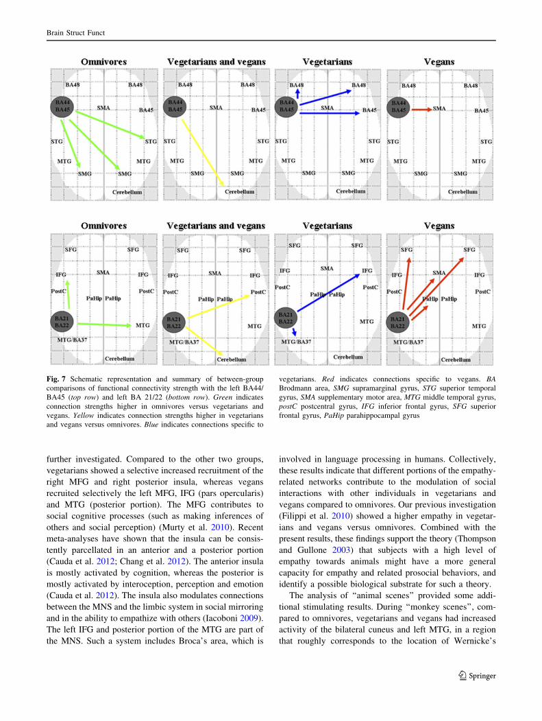
FC analysis
The results of the analysis of FC within each study group using
the BA44/BA45 and the BA21/BA22, respectively, as seed
regions are shown in Fig. 6. The analysis of left BA44/BA45
FC showed that, compared to vegetarians and vegans, omni-
vores had increased FC between the left BA44/BA45 and the
bilateral SMG and right STG. Compared to omnivores, veg-
etarians and vegans had increased FC with the right cerebel-
lum (crus I). Compared to the other two groups, vegetarians
had a selective increased FC with the right BA45 and the
bilateral BA48, whereas vegans had a selective increased FC
with the right supplementary motor area (SMA). The analysis
of left BA21/BA22 FC showed that, compared to vegetarians
and vegans, omnivores had increased FC between the left
BA21/BA22 and the right MTG and left IFG. Compared to
omnivores, vegetarians and vegans had increased FC with the
right cerebellum (crus I) and right postcentral gyrus. Com-
pared to the other two groups, vegetarians had a selective
increased FC with the right IFG (BA44/BA45) and the left
Table 4 Within-group comparisons (activation sites and Montreal Neurological Institute coordinates) of biting versus oral communicative
action scenes of human, monkey and pig in omnivores, vegetarians and vegans (paired t test in each group, p \ 0.05 family-wise error corrected)
Activation
sites
Side Human scenes Monkey scenes Pig scenes
Omnivores Vegetarians Vegans Omnivores Vegetarians Vegans Omnivores Vegetarians Vegans
MOG L -44, -74,
12
-26, -98,
16
-24, -100,
8
-36, -90,
2
-52, -78,
2
-46, -84,
-2
-44, -72,
2
-48, -72,
6
–
IOG L – – – -48, -74,
-6
-44, -82,
-4
-36, -90,
-8
-40, -80,
-2
-36, -76,
-8
–
R – – – 30, -88,
-12
– 46, -76,
-4
– 36, -76,
-4
–
ITG R 46, -66,
-2
48, -54,
-12
– 48, -70,
-4
46, -66,
-6
46, -50,
-24
52, -72,
-4
– –
MTG R – – – – 48, -62, 2 46, -60, 2 58, -70, 6 42, -66, 6 –
IPL L – – – -32, -56,
52
– – – – –
R – – – 32, -40,
50
32, -52, 52 – – – –
SPL R 32, -68,
54
30, -74, 50 – 34, -56,
60
– 38, -46, 60 – – –
Hippocampus R – – – – 20, -28,
-6
– – – –
Cerebellum L – -44, -48,
-36
-24, -88,
-30
-48, -52,
28
-42, -62,
-24
-40, -56,
-22
– -20, -38,
-22
–
R – 26, -80,
-26
48, -52,
-26
36, -46,
-26
34, -64,
-24
44, -66,
-20
– – –
Cuneus L -10,
-102, 6
-4, -102,
16
– – – – – – –
R 14, -98,
24
14, -80, 44 12, -104,
10
12, -86,
42
– – 24, -100,
12
– –
Fusiform
gyrus
L – -34, 54,
-18
– – -40, -48,
-24
-36, -68,
-12
– – –
R 38, -44,
-24
– – 40, -56,
-16
– – – 30, -48,
-14
–
Postcentral
gyrus
L – – – – – – – – -32,
-30,
52
R right, L left, MOG middle occipital gyrus, IOG inferior occipital gyrus, ITG inferior temporal gyrus, MTG middle temporal gyrus, IPL inferior
parietal lobule, SPL superior parietal lobule
Brain Struct Funct
123

Table 5 Within-group comparisons (activation sites and Montreal Neurological Institute coordinates) of oral communicative action versus
biting scenes of human, monkey and pig in omnivores, vegetarians and vegans (paired t test in each group, p \ 0.05 family-wise error corrected)
Activation
sites
Side Human scenes Monkey scenes Pig scenes
Omnivores Vegetarians Vegans Omnivores Vegetarians Vegans Omnivores Vegetarians Vegans
Postcentral
gyrus
L – -58, -12,
40
-36, -24,
40
– – -22, -32,
62
– – –
R – – – – – – 24, -32,
70
– –
Precentral
gyrus
L – – -28, -10,
52
– – -20, -14,
70
– – –
Precuneus L – – – -32, -52,
14
-16, -50,
22
-10, -44,
46
– – –
R – 18, -44, 4 – 22, -44,
12
20, -48, 14 14, -40,
44
– – –
SFG L – – -20, 36,
36
-16, 42,
18
– -28, 44,
38
– – –
R – – – 26, 14, 60 – – – – –
IFG (BA45) L – – -36, 38, 2 – -40, 24, 10 – – – -54, 26,
16
R 46, 20, 4 – – – – – – – –
IFG (BA44) L – – -48, 12,
16
– -48, 12, 16 -56, 10,
14
– – –
R – – – – 40, 14, 12 – – – –
STG L -50, -8,
0
-36, -26,
14
-44, -24,
4
– -54, 2, 2 -60, -42,
14
– – –
R 60, -14,
-2
38, -30, 4 60, -10,
10
– – – – – 64, -36,
18
Thalamus L -12, -18,
4
-10, -20,
2
-4, -20,
0
– – – – – –
Anterior
cingulum
L -2, -10,
40
– – -6, 2, 30 – – – -4, 38, 2 –
R 6, -16, 48 16, -12, 40 12, 46, 6 10, -4, 34 16, 38, 24 14, 42, 2 – – –
Caudate
nucleus
L – -6, 8, -2 -12, 18,
-2
– – – – – –
R 14, 12, -2 12, 18, 0 14, 12,
-12
– 12, 8, -8 18, 20, 0 – – –
Putamen R – 30, 12, 0 – – – – – – –
MTG L -46, -22,
-1
-56, -8,
-16
-52, -2,
-14
– -52, -52,
14
-58, -6,
-10
– – –
R 64, -2,
-16
70, -30,
-8
56, -14,
-8
– – 60, -10,
-14
– – 50, -70,
0
Amygdala L – – -18, 6,
-14
-22, -6,
-10
– – – – –
R – – 14, 6, -10 18, 0, -10 – – – – –
Insula L -34, 8, 4 -32, 12,
-10
– -28, 16,
-8
-34, 6, 4 – –
MOG L – – – – – – – – -48,
-78, 12
Cerebellum R – – – – – – – – 38, -64,
-24
SPL R – – – – – – – – 30, -76,
54
R right, L left, SFG superior frontal gyrus, IFG inferior frontal gyrus, STG superior temporal gyrus, MTG middle temporal gyrus, MOG middle
occipital gyrus, SPL superior parietal lobule
Brain Struct Funct
123

Fig. 2 Cortical activations on a rendered brain from omnivore,
vegetarian and vegan subjects during the comparison of oral
communicative actions (OCAs) versus biting by a human, a monkey,
and a pig (within-group analysis, two-sample t tests, p \ 0.05
corrected for multiple comparisons). Images are in neurological
convention
Fig. 3 Results of the between-group comparisons of oral communi-
cative actions (OCAs) versus biting during ‘‘human/monkey/pig
scenes’’ superimposed on a T1-weighted image in the Montreal
Neurological Institute space. Yellow indicates areas activated in
vegetarians and vegans versus omnivores. Blue indicates activations
specific to vegetarians. Red indicates activations specific to vegans.
Images are presented in neurological convention. MFG middle frontal
gyrus, BA Brodmann area, IFG inferior frontal gyrus, MTG middle
temporal gyrus, MOG middle occipital gyrus
Brain Struct Funct
123
Ta
ble
6W
ith
in-g
rou
pco
mp
aris
on
s(a
ctiv
atio
nsi
tes
and
Mo
ntr
eal
Neu
rolo
gic
alIn
stit
ute
coo
rdin
ates
)o
fsp
ecie
s-re
late
dre
cru
itm
ent
du
rin
go
ral
com
mu
nic
ativ
eac
tio
ns
ino
mn
ivo
res,
veg
etar
ian
san
dv
egan
s(A
NO
VA
inea
chg
rou
p,
p\
0.0
5fa
mil
y-w
ise
erro
rco
rrec
ted
)
Act
ivat
ion
site
sH
um
anv
ersu
sm
on
key
Hu
man
ver
sus
pig
Mo
nk
eyv
ersu
sh
um
an
Om
niv
ore
sV
eget
aria
ns
Veg
ans
Om
niv
ore
sV
eget
aria
ns
Veg
ans
Om
niv
ore
sV
eget
aria
ns
Veg
ans
LS
TG
BA
22
(Wer
nic
ke’
sar
ea)/
BA
41
-5
0,
-6
,-
6–
-6
2,
-2
4,
12
-6
6,
-2
0,
4,
-4
6,
-3
6,
14
––
––
–
RS
TG
(BA
22
)6
6,
-3
0,
14
58
,-
2,
-1
2–
52
,-
18
,2
––
––
–
LM
TG
-4
8,
-2
2,
0-
58
,-
18
,-
14
-6
6,
-2
2,
2-
52
,-
48
,1
6-
54
,-
30
,-
10
––
––
RM
TG
(BA
21)
–6
0,
-3
4,
-6
52
,-
32
,2
64
,-
4,
-2
07
0,
-3
8,
-6
60
,-
12
,-
12
––
–
Rte
mp
ora
lp
ole
––
––
––
––
–
LS
FG
––
––
––
-1
4,
52
,1
8–
–
RS
FG
(BA
8)
––
––
––
––
28
,2
8,
54
RM
FG
––
––
––
––
–
LIF
G(B
A4
5)
––
-4
8,
22
,1
6-
38
,2
2,
22
–-
52
,1
2,
16
–-
38
,2
4,
16
–
RIF
G(B
A4
5)
48
,3
4,
06
0,
22
,2
–5
8,
18
,8
––
––
–
Rpre
centr
algyru
s–
–60,
8,
24
––
––
––
Cin
gulu
m–
––
––
––
–-
4,
-4
4,
44
Lp
recu
neu
s(B
A7
)–
––
––
––
–0
,-
60
,4
4
LS
PL
––
––
––
––
–
RS
PL
––
––
––
––
–
Lin
sula
–-
30
,1
4,
2–
–-
26
,1
0,
8–
––
–
Rin
sula
––
52
,1
2,
03
6,
30
,6
––
––
–
Lsu
pra
mar
gin
alg
yru
s(B
A4
8)
––
-5
4,
-4
0,
30
––
––
––
Lth
alam
us
-1
6,
-1
0,
4–
–-
10
,-
12
,0
-1
0,
-1
4,
16
-2
0,
6,
-8
––
–
Rth
alam
us
–8
,-
8,
16
––
30
,6
,-
4–
––
–
Rp
alli
du
s2
6,
-2
,-
2–
––
––
––
–
LIO
G-
26
,-
10
0,
-1
2–
––
––
–-
44
,-
72
,0
–
RIO
G3
0,
-1
00
,-
8–
––
––
–4
4,
-7
4,
-4
46
,-
70
,-
2
Rcu
neu
s–
––
––
–8
,-
96
,1
48
,-
82
,2
61
2,
-8
8,
26
Rli
ng
ual
gy
rus
––
––
––
––
20
,-
82
,0
LS
OL
––
––
––
-6
,-
10
6,
4–
-1
2,
-9
6,
4
Lce
rebel
lum
-6
,-
80
,-
38
––
––
-1
2,
-8
0,
-4
2–
–-
44
,-
68
,-
50
Rce
rebel
lum
––
––
–28,
-6
4,
-4
8–
––
Ram
yg
dal
a–
––
–3
2,
-2
,-
26
––
––
Brain Struct Funct
123

Ta
ble
6co
nti
nu
ed
Act
ivat
ion
site
sP
igv
ersu
sh
um
anM
onk
eyv
ersu
sp
igP
igv
ersu
sm
on
key
Om
niv
ore
sV
eget
aria
ns
Veg
ans
Om
niv
ore
sV
eget
aria
ns
Veg
ans
Om
niv
ore
sV
eget
aria
ns
Veg
ans
LS
TG
BA
22
(Wer
nic
ke’
sar
ea)/
BA
41
––
––
––
––
–
RS
TG
(BA
22
)–
––
–4
0,
-3
0,
2–
––
–
LM
TG
––
––
-5
0,
-3
2,
-4
––
––
RM
TG
(BA
21)
––
––
–7
0,
-2
8,
-2
––
–
Rte
mp
ora
lp
ole
––
–5
0,
16
,-
38
-3
8,
8,
-3
6–
––
LS
FG
––
–-
14
,5
4,
16
––
––
–
RS
FG
(BA
8)
––
–1
8,
18
,5
8–
––
––
RM
FG
––
–3
2,
30
,3
2–
46
,2
2,
44
––
–
LIF
G(B
A4
5)
––
––
––
––
–
RIF
G(B
A4
5)
––
––
––
––
–
Rpre
centr
algyru
s–
––
––
––
––
Cin
gulu
m–
––
-1
2,
2,
34
––
––
–
Lp
recu
neu
s(B
A7
)–
––
––
––
––
LS
PL
-2
4,
-6
4,
44
––
––
–-
32
,-
66
,5
2–
–
RS
PL
18
,-
86
,4
2–
––
––
38
,-
50
,6
02
8,
-7
8,
38
–
Lin
sula
––
––
––
––
–
Rin
sula
––
––
––
––
–
Lsu
pra
mar
gin
alg
yru
s(B
A4
8)
––
––
––
––
–
Lth
alam
us
––
––
––
––
–
Rth
alam
us
––
––
––
––
–
Rp
alli
du
s–
––
–2
4,
18
,-
10
––
––
LIO
G-
50
,-
70
,-
16
-2
6,
-9
2,
12
-4
2,
-7
8,
2–
––
-5
2,
-7
0,
-1
2-
36
,-
94
,-
10
-2
4,
-9
8,
-6
RIO
G4
8,
-7
0,
-1
23
4,
-8
8,
-1
04
6,
-7
2,
-1
25
4,
-2
2,
-1
8–
–3
8,
-9
0,
-6
38
,-
88
,0
26
,-
92
,8
Rcu
neu
s–
––
––
––
––
Rli
ng
ual
gy
rus
––
––
––
––
–
LS
OL
––
–-
8,
-1
02
,1
2-
12
,-
10
2,
18
-1
0,
-1
00
,6
––
–
Lce
rebel
lum
––
––
-1
4,
-7
6,
-4
0–
––
Rce
rebel
lum
––
–28,
-7
6,
-4
6–
––
–
Ram
yg
dal
a–
––
––
–2
8,-
2,
-1
6–
–
Rri
gh
t,L
left
,B
AB
rodm
ann
area
,S
TG
sup
erio
rte
mp
ora
lg
yru
s,M
TG
mid
dle
tem
po
ral
gy
rus,
IFG
infe
rio
rfr
on
tal
gy
rus,
IOG
infe
rio
ro
ccip
ital
gy
rus,
SO
Lsu
per
ior
occ
ipit
allo
bu
le,
SF
Gsu
per
ior
fron
tal
gy
rus,
SP
Lsu
per
ior
par
ieta
llo
bu
le,
MF
Gm
idd
lefr
on
tal
gy
rus
Brain Struct Funct
123
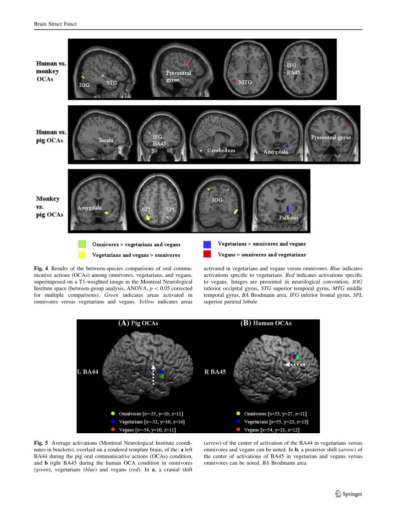
Fig. 4 Results of the between-species comparisons of oral commu-
nicative actions (OCAs) among omnivores, vegetarians, and vegans,
superimposed on a T1-weighted image in the Montreal Neurological
Institute space (between-group analysis, ANOVA, p \ 0.05 corrected
for multiple comparisons). Green indicates areas activated in
omnivores versus vegetarians and vegans. Yellow indicates areas
activated in vegetarians and vegans versus omnivores. Blue indicates
activations specific to vegetarians. Red indicates activations specific
to vegans. Images are presented in neurological convention. IOGinferior occipital gyrus, STG superior temporal gyrus, MTG middle
temporal gyrus, BA Brodmann area, IFG inferior frontal gyrus, SPLsuperior parietal lobule
Fig. 5 Average activations (Montreal Neurological Institute coordi-
nates in brackets), overlaid on a rendered template brain, of the: a left
BA44 during the pig oral communicative actions (OCAs) condition,
and b right BA45 during the human OCA condition in omnivores
(green), vegetarians (blue) and vegans (red). In a, a cranial shift
(arrow) of the center of activation of the BA44 in vegetarians versus
omnivores and vegans can be noted. In b, a posterior shift (arrow) of
the center of activations of BA45 in vegetarian and vegans versus
omnivores can be noted. BA Brodmann area
Brain Struct Funct
123

MTG, whereas vegans had a selective increased FC with the
bilateral superior frontal gyrus (SFG), SMA, and the left
parahippocampal gyrus (Fig. 7).
Discussion
In this study, we wished to map the central representation
of mouth actions performed by humans and by other spe-
cies in individuals with different dietary habits. To answer
our first question (i.e., does processing of mouth actions
performed by distinct species differ between vegetarians
and vegans vs. omnivores?), since speech processing is a
key aspect of social interactions, we compared observation
of OCAs and biting by humans, monkeys and pigs within
and between the three study groups. In contrast to OCAs,
biting was associated with an increased activation of sev-
eral regions in the temporo-occipital lobes, the parietal
lobes and the cerebellum. Conversely, in contrast to biting,
OCAs resulted in increased activity of several regions
along the middle and superior temporal cortices, and the
IFG with a different expression in the three study groups.
Several neurophysiologic and imaging studies have map-
ped mouth actions along the superior temporal sulcus
(STS) region, which includes the surface of the STG and
MTG (Allison et al. 2000). The STS is involved in several
functions, including theory of mind, audiovisual integra-
tion, motion perception, speech processing and perception
of faces (Hein and Knight 2008). A review of foci of
activations within this region reported in several fMRI
studies has led to the identification of an anterior portion,
mainly involved in speech processing, and a posterior one,
mainly recruited by cognitive demands (Hein and Knight
2008). These results argue against distinct functional sub-
regions in the STS and favor the hypothesis that the same
STS region can serve different cognitive functions as a
flexible component in networks with other brain regions. In
line with this, the foci of activations we found at the
between-group comparisons were rather heterogeneous in
terms of coordinates and laterality. The left pars opercu-
laris of the IFG contains a representation of mouth actions
performed by humans (Buccino et al. 2001). Recruitment
of this region, together with regions located in the parietal
and temporal lobes, contributes to the understanding of
actions performed by others by transcoding the observed
action into a corresponding motor plan (Rizzolatti et al.
2001), a mechanism which is critical for social interactions.
During ‘‘human scenes’’, the comparison of OCAs versus
biting showed an increased activity of the right amygdala
in vegetarians and vegans versus omnivores. The amygdala
contributes to the analysis of body movements for per-
ception of actions through its connections with the STS and
the frontal cortex (Allison et al. 2000), thus adding emo-
tional salience to sensory inputs. Therefore, its increased
activity in vegetarians and vegans suggests a different
analysis of dispositions and intentions of other people in
these individuals.
To answer our second question (i.e., do functional
architecture differences exist between vegetarians and ve-
gans?), regions of specific activations in each group were
Fig. 6 Functional connectivity analysis showing the positive corre-
lations between the left BA44/BA45 (left panel) and the left BA21/
BA22 (right panel) and all the other brain voxels in omnivores (top
row), vegetarians (middle row) and vegans (bottom row) (within-
group analysis, one-sample t test, p \ 0.05 corrected for multiple
comparisons). Images are in neurological convention
Brain Struct Funct
123
further investigated. Compared to the other two groups,
vegetarians showed a selective increased recruitment of the
right MFG and right posterior insula, whereas vegans
recruited selectively the left MFG, IFG (pars opercularis)
and MTG (posterior portion). The MFG contributes to
social cognitive processes (such as making inferences of
others and social perception) (Murty et al. 2010). Recent
meta-analyses have shown that the insula can be consis-
tently parcellated in an anterior and a posterior portion
(Cauda et al. 2012; Chang et al. 2012). The anterior insula
is mostly activated by cognition, whereas the posterior is
mostly activated by interoception, perception and emotion
(Cauda et al. 2012). The insula also modulates connections
between the MNS and the limbic system in social mirroring
and in the ability to empathize with others (Iacoboni 2009).
The left IFG and posterior portion of the MTG are part of
the MNS. Such a system includes Broca’s area, which is
involved in language processing in humans. Collectively,
these results indicate that different portions of the empathy-
related networks contribute to the modulation of social
interactions with other individuals in vegetarians and
vegans compared to omnivores. Our previous investigation
(Filippi et al. 2010) showed a higher empathy in vegetar-
ians and vegans versus omnivores. Combined with the
present results, these findings support the theory (Thompson
and Gullone 2003) that subjects with a high level of
empathy towards animals might have a more general
capacity for empathy and related prosocial behaviors, and
identify a possible biological substrate for such a theory.
The analysis of ‘‘animal scenes’’ provided some addi-
tional stimulating results. During ‘‘monkey scenes’’, com-
pared to omnivores, vegetarians and vegans had increased
activity of the bilateral cuneus and left MTG, in a region
that roughly corresponds to the location of Wernicke’s
Fig. 7 Schematic representation and summary of between-group
comparisons of functional connectivity strength with the left BA44/
BA45 (top row) and left BA 21/22 (bottom row). Green indicates
connection strengths higher in omnivores versus vegetarians and
vegans. Yellow indicates connection strengths higher in vegetarians
and vegans versus omnivores. Blue indicates connections specific to
vegetarians. Red indicates connections specific to vegans. BABrodmann area, SMG supramarginal gyrus, STG superior temporal
gyrus, SMA supplementary motor area, MTG middle temporal gyrus,
postC postcentral gyrus, IFG inferior frontal gyrus, SFG superior
frontal gyrus, PaHip parahippocampal gyrus
Brain Struct Funct
123

area. In addition, vegetarians selectively activated the left
MTG and IFG (pars opercularis), and vegans the right
MTG and MOG. All these areas are recruited consistently
in healthy individuals during lipreading, to different extents
according to the language task used (Paulesu et al. 2003).
The increased recruitment of Wernicke’s area and cuneus
in vegetarians and vegans might be secondary to their
attempt to decode monkey mouth gesture. More critically,
vegetarians had a preferential recruitment of the left pars
opercularis of the IFG, which suggests an additional pro-
cess of matching mouth action of the monkey with that of
the viewer, whereas vegans engaged associative temporo-
occipital areas in the right hemisphere, suggesting a role of
higher cognitive processes involved in sentence compre-
hension (Just et al. 1996).
The comparison of OCA versus biting in ‘‘pig scenes’’
also detected particular patterns of recruitment in vege-
tarians and vegans. Specifically, vegetarians showed a
selective increased activity of the anterior cingulum, which
likely reflects a strong empathic response (Devinsky et al.
1995; Phan et al. 2002) or simply an enhanced attention
(Singer et al. 2004), whereas vegans activated the bilateral
parahippocampal gyrus, which has a role in auditory–ver-
bal memory functions (Grasby et al. 1993) and, through its
connections with the amygdala, contributes to emotion-
driven learning (Murty et al. 2010).
To address our third question (i.e., does processing of
mouth actions differ according to the species involved
and their phylogenetic proximity to humans?), we limited
the analysis to OCAs, since they represent the basis of
intra- and inter-group relationships in humans. During
human versus monkey OCAs, compared to vegetarians
and vegans, omnivores had an increased activity of part
of the visual areas of the observation–execution matching
system. Vegans had selective increased activity of the left
MTG and the frontal portion of the system, further sup-
porting the notion of increased recruitment of regions of
the MNS in these subjects. Consistent with the role of the
anterior portion of the insula in interpersonal interactions,
this region was more active, in combination with the left
IFG, in omnivores compared to vegetarians and vegans
during human versus pig OCAs. Additional between-
group differences in processing human versus pig actions
concerned areas involved in empathy processing, includ-
ing the amygdala and the anterior lobe of the cerebellum.
This latter region plays a crucial role in motor learning
and language processing as demonstrated by a recent
meta-analysis (Jirak et al. 2010). Importantly, between-
group differences in processing monkey versus pig OCAs
revealed activity of areas that have a role in spatial
attention, such as the SPL and IOG in vegetarians and
vegans, as well as the globus pallidus in vegetarians.
Overall, these findings support the notion that, despite
vegetarians and vegans showing a particular pattern of
recruitment of regions that are part of the MNS during
processing mouth actions executed by other animals, the
activity of this system remains higher when they are
dealing with actions performed by their conspecifics,
probably as a consequence of the matching of the
observed human action with their motor repertoire, as
part of a common representational format. In addition,
species proximity with humans can modulate MNS
recruitment in these subjects, as suggested by the
between-group differences observed for monkey, but not
pig OCAs.
Complementing the analysis of activations, the analysis
of FC detected striking between-group differences in the
strengths of connections with the cerebellum (which was
highly significantly connected in vegetarians and vegans
vs. omnivores) and the SMA (which had a selective higher
connectivity in vegans). Remarkably, the connectivity of
these two areas was not influenced by the seed-regions
(IFG or STS) used to run the analysis, type of task, or
stimulus. Despite the fact that activation of these two areas
has been reported in several studies investigating the MNS,
their role, in the context of MNS and empathy theories, has
been neglected by the majority of researchers. The analysis
of single-subject data has recently led to the identification
of a series of brain areas involved in action observation and
execution (Gazzola and Keysers 2009), which extends
beyond the original MNS and also includes the cerebellum
and SMA. Within the framework of this model of action
observation and execution, such brain regions cooperate in
forward and inverse internal models to associate other
people’s actions with their own actions and sensations. In
particular, while the cerebellum, through its connections to
the premotor and cingulate cortices, provides a motor and
somatosensory representation of others’ actions and con-
tributes to predicting actions, the SMA serves as a gate-
keeper of premotor activity of the primary motor cortex,
thus determining motor behaviour (Gazzola and Keysers
2009).
Collectively, our results reveal that distinct brain
responses are evoked by mouth actions performed by dif-
ferent species in people with vegetarian and vegan eating
habits, and there are differences between vegetarians and
vegans, supporting the role of the action observation exe-
cution matching system in social cognition, enabling us to
communicate and interact with our conspecifics and also
with species in phylogenetic proximity to humans.
Acknowledgments The authors are grateful to Prof. Paul M. Mat-
thews (Department of Clinical Neurosciences, Imperial College
London, and GSK Clinical Imaging Centre, Hammersmith Hospital,
London, UK) and Dr. Mark A. Horsfield (Department of Cardiovas-
cular Sciences, University of Leicester, Leicester, UK) for their
thoughtful comments to the manuscript.
Brain Struct Funct
123

Conflict of interest The authors have no commercial interests with
regard to the study reported in the present manuscript.
References
Allison T, Puce A, McCarthy G (2000) Social perception from visual
cues: role of the STS region. Trends Cogn Sci 4:267–278
Biswal B, Yetkin FZ, Haughton VM, Hyde JS (1995) Functional
connectivity in the motor cortex of resting human brain using
echo-planar MRI. Magn Reson Med 34:537–541
Buccino G, Binkofski F, Fink GR, Fadiga L, Fogassi L, Gallese V,
Seitz RJ, Zilles K, Rizzolatti G, Freund HJ (2001) Action
observation activates premotor and parietal areas in a somato-
topic manner: an fMRI study. Eur J Neurosci 13:400–404
Buccino G, Lui F, Canessa N, Patteri I, Lagravinese G, Benuzzi F,
Porro CA, Rizzolatti G (2004) Neural circuits involved in the
recognition of actions performed by nonconspecifics: an FMRI
study. J Cogn Neurosci 16:114–126
Cauda F, Costa T, Torta DM, Sacco K, D’Agata F, Duca S, Geminiani
G, Fox PT, Vercelli A (2012) Meta-analytic clustering of the
insular cortex: characterizing the meta-analytic connectivity of
the insula when involved in active tasks. Neuroimage
62:343–355
Chang LJ, Yarkoni T, Khaw MW, Sanfey AG (2012) Decoding the
role of the insula in human cognition: functional parcellation and
large-scale reverse inference. Cereb Cortex
Cheng Y, Lin CP, Liu HL, Hsu YY, Lim KE, Hung D, Decety J
(2007) Expertise modulates the perception of pain in others. Curr
Biol 17:1708–1713
Devinsky O, Morrell MJ, Vogt BA (1995) Contributions of anterior
cingulate cortex to behaviour. Brain 118(Pt 1):279–306
Filippi M, Riccitelli G, Falini A, Di Salle F, Vuilleumier P, Comi G,
Rocca MA (2010) The brain functional networks associated to
human and animal suffering differ among omnivores, vegetar-
ians and vegans. PLoS ONE 5:e10847
Fox MD, Snyder AZ, Vincent JL, Corbetta M, Van Essen DC,
Raichle ME (2005) The human brain is intrinsically organized
into dynamic, anticorrelated functional networks. Proc Natl Acad
Sci USA 102:9673–9678
Friston KJ, Holmes AP, Poline JB, Grasby PJ, Williams SC,
Frackowiak RS, Turner R (1995) Analysis of fMRI time-series
revisited. Neuroimage 2:45–53
Friston KJ, Holmes AP, Price CJ, Buchel C, Worsley KJ (1999)
Multisubject fMRI studies and conjunction analyses. Neuroim-
age 10:385–396
Friston KJ, Penny WD, Glaser DE (2005) Conjunction revisited.
Neuroimage 25:661–667
Gazzola V, Keysers C (2009) The observation and execution of
actions share motor and somatosensory voxels in all tested
subjects: single-subject analyses of unsmoothed fMRI data.
Cereb Cortex 19:1239–1255
Grasby PM, Frith CD, Friston KJ, Bench C, Frackowiak RS, Dolan RJ
(1993) Functional mapping of brain areas implicated in
auditory–verbal memory function. Brain 116(Pt 1):1–20
Hein G, Knight RT (2008) Superior temporal sulcus—It’s my area: or
is it? J Cogn Neurosci 20:2125–2136
Iacoboni M (2009) Imitation, empathy, and mirror neurons. Annu Rev
Psychol 60:653–670
Jirak D, Menz MM, Buccino G, Borghi AM, Binkofski F (2010)
Grasping language—a short story on embodiment. Conscious
Cogn 19:711–720
Just MA, Carpenter PA, Keller TA, Eddy WF, Thulborn KR (1996)
Brain activation modulated by sentence comprehension. Science
274:114–116
Lancaster JL, Woldorff MG, Parsons LM, Liotti M, Freitas CS,
Rainey L, Kochunov PV, Nickerson D, Mikiten SA, Fox PT
(2000) Automated Talairach atlas labels for functional brain
mapping. Hum Brain Mapp 10:120–131
Maldjian JA, Laurienti PJ, Kraft RA, Burdette JH (2003) An automated
method for neuroanatomic and cytoarchitectonic atlas-based
interrogation of fMRI data sets. Neuroimage 19:1233–1239
Murty VP, Ritchey M, Adcock RA, LaBar KS (2010) fMRI studies of
successful emotional memory encoding: a quantitative meta-
analysis. Neuropsychologia 48:3459–3469
Oldfield RC (1971) The assessment and analysis of handedness: the
Edinburgh inventory. Neuropsychologia 9:97–113
Paulesu E, Perani D, Blasi V, Silani G, Borghese NA, De Giovanni U,
Sensolo S, Fazio F (2003) A functional-anatomical model for
lipreading. J Neurophysiol 90:2005–2013
Phan KL, Wager T, Taylor SF, Liberzon I (2002) Functional
neuroanatomy of emotion: a meta-analysis of emotion activation
studies in PET and fMRI. Neuroimage 16:331–348
Regan T (1985) The case for animal rights. University of California,
Berkeley 422
Rizzolatti G, Fogassi L, Gallese V (2001) Neurophysiological
mechanisms underlying the understanding and imitation of
action. Nat Rev Neurosci 2:661–670
Shamay-Tsoory SG (2011) The neural bases for empathy. Neurosci-
entist 17:18–24
Shamay-Tsoory SG, Aharon-Peretz J, Perry D (2009) Two systems
for empathy: a double dissociation between emotional and
cognitive empathy in inferior frontal gyrus versus ventromedial
prefrontal lesions. Brain 132:617–627
Singer T, Seymour B, O’Doherty J, Kaube H, Dolan RJ, Frith CD
(2004) Empathy for pain involves the affective but not sensory
components of pain. Science 303:1157–1162
Singer T, Seymour B, O’Doherty JP, Stephan KE, Dolan RJ, Frith CD
(2006) Empathic neural responses are modulated by the
perceived fairness of others. Nature 439:466–469
Tettamanti M, Buccino G, Saccuman MC, Gallese V, Danna M, Scifo
P, Fazio F, Rizzolatti G, Cappa SF, Perani D (2005) Listening to
action-related sentences activates fronto-parietal motor circuits.
J Cogn Neurosci 17:273–281
Thompson KL, Gullone E (2003) Promotion of empathy and
prosocial behaviour in children through humane education.
Australian Psychologist 38:175–182
Tzourio-Mazoyer N, Landeau B, Papathanassiou D, Crivello F, Etard
O, Delcroix N, Mazoyer B, Joliot M (2002) Automated
anatomical labeling of activations in SPM using a macroscopic
anatomical parcellation of the MNI MRI single-subject brain.
Neuroimage 15:273–289
Xiang HD, Fonteijn HM, Norris DG, Hagoort P (2010) Topographical
functional connectivity pattern in the perisylvian language
networks. Cereb Cortex 20:549–560
Brain Struct Funct
123