Embed Size (px)
Citation preview

TE
THUoeilHtiftbwbsgA
*
†
A
1
he Use of Lasers in Darker Skin Typesliot F. Battle, Jr, MD,*,† and Cylburn E. Soden, Jr, MD, MA*
The demographics of the US population continue to change at an extremely rapid pace. Asof 2008, Asians, Hispanics, and African Americans accounted for 31% of the US popula-tion, and it is estimated that by the year 2050 half of the population of America will berepresented by darker ethnic skin types. With the increase in the total number of individualsof skin of color, the demand for safe and effective laser therapy in darker skin typescontinues to increase. However, despite the increase in demand, the current literatureregarding the use of lasers in darker skin remains limited. Most of the treatment parametersdefined for laser platforms have been established primarily through extensive testing onskin phototypes I to III, and those studies that have been conducted on darker skinphototypes have been overwhelmingly conducted on Asian skin. Nevertheless, it hasbecome clear that effective cutaneous laser surgery in darker skin types can be accom-plished despite a relative overall greater risk for complications. Therefore, as the diversityof America continues to grow, the laser surgeon needs to maintain a clear understandingof the complexities associated with treating ethnic skin and remain mindful of the current,and ever-changing, therapeutic modalities available. This will allow the conscientiousphysician to maximize outcome and minimize risk when performing laser surgery on darkerskin types.Semin Cutan Med Surg 28:130-140 © 2009 Elsevier Inc. All rights reserved.
ttelmt
tsbdcsmm
acwlU1aeg
he demographics of the US population continue tochange at an extremely rapid pace. As of 2008, Asians,
ispanics, and African Americans accounted for 31% of theS population, and it is estimated that by the year 2050 halff the population of America will be represented by darkerthnic skin types.1 With the increase in the total number ofndividuals of skin of color, the demand for safe and effectiveaser therapy in darker skin types continues to increase.owever, despite the increase in demand, the current litera-
ure regarding the use of lasers in darker skin remains lim-ted. Most of the treatment parameters defined for laser plat-orms have been established primarily through extensiveesting on skin phototypes I to III, and those studies that haveeen conducted on darker skin phototypes have been over-helmingly conducted on Asian skin. Nevertheless, it hasecome clear that effective cutaneous laser surgery in darkerkin types can be accomplished despite a relative overallreater risk for complications. Therefore, as the diversity ofmerica continues to grow, the laser surgeon needs to main-
Department of Dermatology, Howard University College of Medicine,Washington, DC.
Cultura Cosmetic Dermatology & Laser Center, 5301 Wisconsin Ave.,NW, Washington, DC.
ddress reprint requests to Eliot F. Battle, Jr, MD, 5301 Wisconsin Ave.NW, Ste. 110, Washington, DC 20015–2068. E-mail: drbattle@
lculturamed.com.
30 1085-5629/09/$-see front matter © 2009 Elsevier Inc. All rights reserved.doi:10.1016/j.sder.2009.04.003
ain a clear understanding of the complexities associated withreating ethnic skin and remain mindful of the current, andver-changing, therapeutic modalities available. This will al-ow the conscientious physician to maximize outcome and
inimize risk when performing laser surgery on darker skinypes.
Most of the inherent risks involved in treating darker skinypes with cosmetic lasers relate to melanin’s very wide ab-orption spectrum (250-1200 nm). Melanin can be targetedy visible, ultraviolet (UV), and infrared light (Fig. 1). Inarker skin types, epidermal melanin competes as a signifi-ant chromophore and may lead to excessive heating of theurrounding tissue. Unintended nonspecific thermal damageay ensue, resulting in epidermal blistering, transient or per-anent dyspigmentation, textural changes, and scarring.Increased epidermal melanin absorption in skin of color
lso reduces the amount of laser light reaching the intendedhromophore. As a result, the efficacy of lasers in personsith darker skin types may be reduced when using equiva-
ent fluences. The peak absorption of melanin lies within theV range and decreases rapidly as wavelength increases (Fig.). Consequently, longer wavelengths not only reduce thebsorption of laser light by epidermal melanin but also pen-trate deeper with more selective absorption of dermal tar-eted chromophores. Thus, lasers generating longer wave-
engths that are less efficiently absorbed by endogenous
m1wieViI
tpttsuw
stdcipngdtpbmpais6
2pm
teels
HSitttcAmhhipeitlcssa
h
n spec
Use of lasers in darker skin types 131
elanin, such as the 810-nm diode and particularly the064-nm Nd:Yag, can provide a greater margin of safetyhile still allowing appropriate clinical results to be achieved
n skin types IV to VI. Nevertheless, differential absorptionven among darker skin types remains quite variable. TypeI skin may absorb as much as 40% more energy when
rradiated with visible light as compared with phototypes I orI when irradiated with the same fluence and pulse duration.2
The thermal relaxation time (TRT) is the time it takes thearget chromophore to cool down by 50% of its initial tem-erature. The TRT is directly proportional to the diameter ofhe target. The melanosome is a small structure, has a TRT inhe nanoseconds, and therefore loses heat rapidly. Melano-omes are potentially destroyed when Q-switched lasers aresed, given their nanosecond pulse durations, and sparedhen pulse durations in the milliseconds are used.Efficient cooling devices (eg, sapphire-cooled tip, cryogen
pray cooling, and forced air cooling) are essential in thereatment of darker ethnic skin types. Light absorbed by epi-ermal melanin is converted to heat and, without efficientooling, this heat creates unwanted thermal injuries, includ-ng blistering, dyspigmentation, and scarring. Cooling can beerformed by contact (cooling device touching the skin) oroncontact (eg, cooling the skin with a cooling spray, air oras). The use of excessive cooling is not without risk forarker ethnic skin types. Cryogen spray can reach tempera-ures as low as �26.2°C. Excessive cryogen spray and delayarameters can result in unwanted side effects, includinglistering and postinflammatory dyspigmentation. The exactechanism in which cold air cooling is associated with dys-igmentation after laser irradiation is unknown. However, instudy evaluating the incidence of continuous cold air cool-
ng on postinflammatory hyperpigmentation (PIH) after Q-witched Nd:Yag laser treatment of bilateral nevus of Ota,
Figure 1 Absorptio
2% of patients developed PIH on the cooled side and only p
4% developed PIH on the uncooled side. The authors hy-othesized that laser irradiated melanocytes or keratinocytesay be more reactive to stimuli, such as cold temperature.3
In short, with the continued development of novel lasershat use longer wavelengths, increased pulse durations, anxpanded range of available treatment fluences, and morefficient cooling devices, the laser surgeon can decrease theikelihood of adverse events while effectively treating darkerkin types.
airince its inception, laser-assisted hair removal has become anncreasingly popular procedure. In skin types I to III, wherehe hair tends to be straight and fine, laser hair removalypically functions in a purely cosmetic manner that reduceshe treatment time and monotony of repetitive shaving. Inontrast, in patients with darker skin types, especially Africanmericans, where the hair tends to be coarse and curly, der-atologic conditions such as pseudofolliculitis barbae andirsutism often do not respond to conventional methods ofair removal. Shaving with a straight or electric razor, pluck-
ng the hair, chemical depilatories, and electrolysis does tem-orarily reduce hair growth but does not address and canven exacerbate the initial condition, resulting in additionalnflammatory papules and postinflammatory hyperpigmen-ation. However, as laser hair removal addresses the under-ying issue of the continued hair growth, it essentially be-omes a medical necessity for the treatment of conditions,uch as pseudofolliculitis barbae and hirsutism, in darkerkin types in which there is truly no other viable therapeuticlternative.
Because melanin in the epidermis and in the hair follicleave nearly identical absorption spectra, laser hair removal
trum of melanin.
oses a real risk of epidermal injury, especially in darker skin

tcaidN(a
ialelt
IitHa
s3adcwschYmsmsa
pCttt
FbaS
132 E.F. Battle and C.E. Soden
ypes. Complications of overly aggressive treatment may in-lude blistering, dyspigmentation, and scarring. Currently,vailable laser and light sources available for photo-epilationnclude the long-pulsed ruby (694 nm), long-pulsed alexan-rite (755 nm), long pulsed diode (810 nm), long-pulsedd:Yag (1064 nm), and noncoherent intense pulsed light
IPL; 590-1200 nm).4 Among these lasers, the food and drugdministration (FDA) has approved 2 systems for photo-ep-
igure 2 (A) Baseline photograph of a patient with type IV skinefore laser-assisted hair removal of the face. (B) Photograph 2 yearsfter 9 treatments with a diode laser (Lightsheer, Lumenis, Inc.,anta Clara, CA), using 100-msecond pulse duration.
Figure 3 (A and C) Baseline photograph of a patient withneck. (B and D) Photograph of patient 4 years after 10 t
Inc., Brisbane, CA).lation in darker skin types: the long pulsed diode (810 nm)nd Nd:Yag (1064 nm).5 These 2 devices have combinedonger wavelengths, extended pulse durations, and activepidermal cooling to provide the greatest efficacy with theowest side effect profile in treating skin phototypes IVhrough VI (Figs. 2-6).
When the long-pulsed diode to treat skin phototypesV-VI is used, pulse durations of 100 milliseconds or longermprove the ability to treat darker skin more safely (Fig. 2). Inerms of treating very dark skin, phototype VI, Battle andobbs6 reported that very long pulse durations (�400 ms)
nd aggressive adjunctive skin cooling are required.Because of its longer wavelength, the Nd:Yag has been
hown to be the safest laser to treat darker skin types (Figs.-6).7 Its longer wavelength minimizes epidermal melaninbsorption and maximizes wavelength penetration to theermal hair follicular unit. Therefore, shorter pulse durationsan be more safely used with the Nd:Yag lasers as comparedith diode lasers. When the Nd:Yag is combined with aggres-
ive skin cooling and pulse durations �30 milliseconds, itan safely treat type VI skin8 (Fig. 6). With regard to overallair reduction, however, the longer wavelength of the Nd:ag is probably slightly less effective because of its reducedelanin absorption.8,9 A study conducted by Galadari8
howed a 35% reduction in hair at 12 months after 6 treat-ents with the Nd:Yag compared with a 40% reduction ob-
erved with the diode after a similar number of treatmentsnd follow-up period.
Paradoxical hypertrichosis has been consistently re-orted as a complication of laser assisted hair removal.10-13
urrently, published studies have reported this complica-ion after alexandrite (755 nm) and IPL (590-1200 nm)reatment.11,12 In the authors’ experience, this complica-ion is not dependent on a light source and may occur with
V skin before laser-assisted hair removal of the face andnts with a long pulsed Nd:Yag laser (Coolglide, Cutera,
type Ireatme

adtbrcmrb
ktLgoettmtiic
TCsvipwsttpmta
ureSsitlp
Flp
Flt(
Use of lasers in darker skin types 133
ny wavelengths used with photoepilation, including theiode (810 nm) and Nd:Yag (1064 nm). Evidence suggestshat individuals with darker skin (phototypes III-V) maye at increased risk,10,13 particularly patients of Mediter-anean or Pacific Asian descent. The current reported in-idence of paradoxical hypertrichosis after laser hair re-oval has varied between 0.6% and 5.1%.10,12 With
espect to location, hair growth may occur both within andeyond the treated sites.12
The exact mechanism of photostimulation remains un-nown, but it is speculated that low fluences can stimulatehe transformation of vellus hairs into darker terminal hairs.aser epilation may also synchronize the cycling of hairsrowing within the treatment site,10 and certain wavelengthsf energy may directly or indirectly have photo-stimulatoryffects on the hair follicle.12 In the authors’ experience, po-ential methods of limiting this complication include usinghe maximal possible safe energy, cooling outside the treat-ent area to avoid vellus hair stimulation, and minimizing
he treatment to only where the hair is located. Yet, because its not possible to predict which patients will develop laser-nduced hypertrichosis, this complication should be dis-
igure 4 (A) Baseline photograph of a patient with type V skin beforeaser-assisted hair removal of the upper lip. (B) Photograph of pa-ient 2 years after 10 treatments with a long pulsed Nd:Yag laserGentlelase, Candela Corp, Wayland, MA).
ussed during the informed consent process. (
attoososmetic tattoo removal is regularly practiced by many laser
urgeons; however, there have been few technological ad-ances during the last decade and, thus, tattoo removal stillnvolves many challenges. Tattoos often consist of multipleigments, and their removal may require the use of severalavelengths involving both the visible and near infrared
pectrum. Furthermore, tattoos can respond unpredictablyo laser irradiation not only because the components used inheir makeup are highly variable but also because they can belaced in the deep dermis. Tattoo treatment becomes evenore difficult and unpredictable in patients with skin pho-
otypes IV through VI because of the presence of significantmounts of epidermal melanin that can absorb laser energy.14
The Q-switched 694-nm ruby laser was the first laser to besed in the treatment of tattoos.15,16 It is highly effective inemoving black-and-blue tattoo pigments, and it is the mostffective laser for removing purple and violet pigments.17
tudies have demonstrated the effectiveness of the Q-witched ruby laser to remove blue and black pigmentsn skin types V and VI with excellent clearance and onlyransient hyperpigmentation.18 However, its shorter wave-ength is strongly absorbed by epidermal melanin, and itsotential for inducing long-term dyspigmentation is rela-
igure 5 (A) Baseline photograph of a patient with type V skin beforeaser-assisted hair removal of the chin and neck. (B) Photograph ofatient 1 year after 12 treatments with a long pulsed Nd:Yag laser
Coolglide, Cutera, Inc., Brisbane, CA).
tw
isrtSotHefV
lodcseQwd
psbtwQ
awoflsWY3fltbtatScl
LDttbAcltts
ceamrae
MctBviggmg
FbPN
134 E.F. Battle and C.E. Soden
ively high in patients with darker skin types when comparedith lasers that use longer wavelengths.The Q-switched alexandrite laser emits light at 755 nm, an
ntermediate wavelength between the ruby and Nd:Yag la-ers. Similar to ruby lasers, alexandrite lasers are capable ofemoving blue and black tattoo pigments and have becomehe treatment of choice for removing green tattoo pigment.tudies involving the use of alexandrite laser in the treatmentf blue and black tattoos in skin types III and IV have shownhat it is effective treatment with minimal side effects.19,20
owever, because of its relatively short wavelength, its en-rgy is also strongly absorbed by epidermal melanin. There-ore, it must also be used with extreme caution in skin types
and VI.Q-switched Nd:Yag laser are capable of emitting 2 wave-
engths of light, 532 and 1064 nm. At its shorter wavelengthf 532 nm, it is the most effective treatment for red brown,ark brown, and orange pigment.17 At the same time, be-ause of its shorter wavelength, when treating pigmented-kin, the 532-nm has a greatest risk of transient orven permanent hypopigmentation. Compared with the-switched ruby and Q-switched alexandrite, at its longeravelengths, the Q-switched Nd:Yag tends to minimize epi-
igure 6 (A) Baseline photograph of a patient with type VI skinefore laser-assisted hair removal of the chin, cheeks, and neck. (B)hotograph of patient 2 years after 12 treatments with a long pulsedd:Yag laser (Coolglide, Cutera, Inc., Brisbane, CA).
ermal melanin absorption, thereby reducing the risk of hy- t
erpigmentation and permanent hypopigmentation. As a re-ult, the Nd:Yag remains the safest laser in the treatment oflue and black tattoos in darker skin types21,22 However, inhe author’s experience, higher fluences must often be usedith the long pulsed Q-switched Nd:Yag compared with the-switched alexandrite or ruby to achieve similar efficacy.Regardless of the laser used, multiple treatments, on aver-
ge 8-12 treatments, may be required with a minimum of 6-8eeks between sessions. Side effects of laser tattoo removaln pigmented skin can be limited by starting at the minimumuence necessary to produce immediate lesional whitening,ignaling the destruction of intracellular melanosomes.
hen treating phototype VI skin with the Q-switched Nd:ag, a 3-mm spot size and starting fluences between 3.4 and.6 J/cm2 are recommended. With each successive treatment,uences are gradually increased to account for the clearing ofhe chromophore. Greater fluences resulting in pinpointleeding and tissue splattering are more likely to lead toransient hyperpigmentation, permanent hypopigmentationnd scarring. Rarely does one see 100% clearing, but mostattoos clear to the point of being cosmetically acceptable.ome brighter colors may not clear well because of a greaterhance of hypopigmentation associated with shorter wave-engths necessary to target lighter pigments.
aser for Pigmented Lesionsermatologic conditions that result in altered skin pigmen-
ation, such as Melasma, postinflammatory hyperpigmenta-ion, lentigines, and dermatosis papulosa nigra continue toe a primary concern among patients with darker skin tones.lthough chemical peeling, light eletrodessication and, inases, even cryotherapy have been used in their treatment,asers have continued to gain widespread acceptance and, atimes, may be considered a first-line treatment or, at least, aherapeutic alternative when conventional methods are notuccessful (Fig. 7).
Melasma is a commonly acquired hypermelanosis that oc-urs most frequently on the face of women and tends to bespecially prominent in women of color. Although topicalgents remain the first-line treatment for epidermal andixed type of Melasma, lasers are commonly used for more
efractory cases. Before the physician initiates laser therapy,ll patients should be placed on a topical regimen, includingxfoliating and bleaching agents and sunscreen.
IPL sources have shown promise in the treatment ofelasma for a subset of skin type IV patients. In a study
onducted by Wang et al,23 IPL was found to be effective forhe treatment of refractory dermal Melasma in Asian patients.y using 570-nm and 590- to 615-nm filters at 4-week inter-als for 4 treatments, the IPL was able to produce a 39.8%mprovement of the relative melanin index in the treatmentroups compared with 11.6% improvement in the controlroup at week 16. However, the authors noted some repig-entation at the end of the 36-week treatment session, sug-
esting that maintenance therapy may be required.A more recent investigation evaluated the treatment of
herapy resistant Melasma in 89 Chinese female subjects

(op3a6atumiltPm
hm
ppoditlmAAesgM
1t
Use of lasers in darker skin types 135
skin types III and IV) with use of a new IPL device (Lumenisne, Lumenis, Inc., Santa Clara, CA) incorporating optimalulse technology (OPT). Patients received 4 IPL treatments at-week intervals with fluences ranging from 13 to 17 J/cm2
nd 560-/590-nm filters for epidermal type and 590-/615-/40-nm filters for mixed type Melasma. Mean melasma areand severity index scores decreased from 15.2 before thereatment to 5.2 after 4 sessions and 4.5 at 3-month follow-p. Adverse events were limited with IPL treatments andainly involved transient erythema and slight edema resolv-
ng 0.5-12 hours after treatment. Temporary microcrustsasting 7-10 days were noted on the cheekbones of 72 pa-ients, and 3 patients with mixed-type Melasma had obviousIH after 1 or 2 treatment sessions. No scarring or hypopig-entation were noted during or after the treatment.24
IPL treatments may result in IPL-induced Melasma-likeyperpigmentation despite the presence of apparently nor-
al skin. Negishi et al,25 explained this concept by noting the tresence of “very subtle epidermal melasma” when using UVhotography among 63 of 223 Japanese women. From theirbservations, high fluences resulting in immediate postirra-iation erythema within areas of pigmentation previously
dentified by UV photography that lasted for several minuteso hours after irradiation were postulated to exacerbate me-anocyte activity, thus inducing postirradiation hyperpig-
entation. These findings, although observed in the skin ofsian patients, may also be extrapolated to lighter-skinnedfrican-American or Hispanic patients in whom wood’s lightxamination identifying the presence of “subtle melasma”hould prompt the physician to treat these patients less ag-ressively, reducing the possibility of exacerbating theirelasma or inducing PIH.Although not as widely used as the IPL, the Q-switched
064 Nd:Yag laser has also been shown to be effective in thereatment of refractory dermal Melasma in pigmented skin
re 7 (A) Facial hyperpigmentation in a patienth type VI skin. (B) Photograph 1 month after 4tments with microsecond Nd:Yag (Cuteraesis, Cutera, Inc., Brisbane, CA). (C) Photo-
ph of patient 6 months after 10 treatments withrosecond Nd:Yag (Cutera Genesis, Cutera,., Brisbane, CA).
FiguwittreaGengramicInc
hrough repetitive subthreshold pulsed laser treatments.

Ws1hflatc
muF(emjAtbddctgcsiccmaig
tu3ppttleTcprafcHcm
cmal
tlQapaoPaQHer
fctmtld
Fo
136 E.F. Battle and C.E. Soden
ith the use of a Medlite C6 Q-switched Nd:Yag laser (6-mmpot, 10 Hz and 3.4 J/cm2), 8-10 treatments performed at-week intervals were able to produce �80% reduction inyperpigmentation.26 It is thought that subphotothermolyticuences (�5 J/cm2) in conjunction with larger spot sizesllow melanin granules to be fragmented and dispersed intohe cytoplasm, facilitating their reabsorption without causingell damage or cell death.
Fractional photothermolysis (Reliant Technologies, Palo-ar Medical Technologies) is the newest technology to besed in the treatment of Melasma and it is currently the onlyDA-approved laser therapeutic modality for this conditionFig. 8).27,28 This modality involves the use of a 1550-nmrbium-doped fiber laser to create noncontiguous microther-al zones (MTZs), which are microcolumns of thermal in-
ury to the level of the dermis surrounded by normal tissue.lthough initial histologic evaluation of these MTZs revealed
he presence of primarily microscopic epidermal necrotic de-ris, more recent evidence suggests that in addition to epi-ermal debris, dermal contents, such as melanin and elastoticebris of solar elastosis, may also be eliminated through thesehannels of thermal injury.29 In fact, melanin content withinhese columns of damage has been found to be significantlyreater than in the surrounding normal tissue.30 This in-reased melanin content has lead to the theory of a melaninhuttle in which the damaged pigmentation of the basal layers eliminated by the rapid migration of the viable keratino-ytes present at the wound margins. This concept was re-ently corroborated by Goldberg et al,29 who noted posttreat-ent ultrastructural changes in the skin of Melasma patients
fter fractional photothermolysis, which included a decreasen the number of melanocytes and the amount of melaninranules within keratinocytes.
Early studies in which the authors studied fractional pho-othermolysis in the treatment of Melasma (skin types III-V)sed 6-12 mJ/MTZ and relatively high densities of 2000-500 MTZ/cm2 for 4-6 treatments. Approximately 60% ofatients experienced 75-100% clearing, and only 1 of 10atients experienced postinflammatory hyperpigmenta-ion.27 More recent studies by Lin et al31 involving darker skinypes showed a reduction in pain and downtime by the use ofower energy and density (125 MTZ/cm2 at energy of 8 mJ,very 2-4 weeks) without a significant reduction in efficacy.he authors concluded that greater density is not signifi-antly more effective and carries an increased risk of hyper-igmentation and rebound in darker skin types. In our expe-ience, patients should be treated with low energies (6-8 mJt 1000-2000 MTZ/cm) for 2-3 treatments. Similar to IPL,ractional photothermolysis (FP) carries a risk of PIH, espe-ially in individuals that may have hyperactive melanocytes.owever, the use of lower energy and density settings in
ombination with liberal skin cooling during and after treat-ent helps minimize these risks.Freckles and lentigines are common findings in skin of
olor, especially Asian patients. Although freckles are a nor-al aspect of development, lentigines represent a sign of
ging and photodamage. Nevertheless, lentigines and freck-
es are common reasons for consultation with laser surgeons. mQ-switched lasers produce photomechanical effects withinhe epidermis and are effective for treating both freckles andentigines. The commonly used Q-switched systems are the-switched ruby 694-nm, Q-switched alexandrite 755-nm
nd frequency-doubled Q-switched Nd:Yag 532-nm. Allroduce immediate whitening and subsequent macrocrustingt the treated sites, which typically lasts 7-14 days. Lentigines areften eradicated after 1 or 2 treatments with Q-switched lasers.IH is the most common side effect and tends to resolve withinfew months. Reported rates of PIH associated with the use-switched lasers in Asians have ranged from 4% to 25%.32,33
owever, this is significantly lower than what we have experi-nced in treating pigmented skin, with PIH more likely occur-ing in 35-50% of treated patients.
Intense pulse light sources are also capable of treating bothreckles and lentigines in a subset of skin type IV patients. Inontrast to the Q-switched systems, IPL requires multiplereatments; however, in that it produces primarily photother-al effects within the epidermis, the side effects of pigmen-
ary alteration are less common, and ensuing microcrusting isess severe.34 Wang et al35 compared the Q-switched alexan-rite laser (AlexLAZR, Candela Corp, Wayland, MA) and IPL
igure 8 (A) Melasma in a woman with type IV skin. (B) Photographf patient after 6 treatments with nonablative fractional photother-
olysis (Palomar Medical Technologies, Inc., Burlington, MA).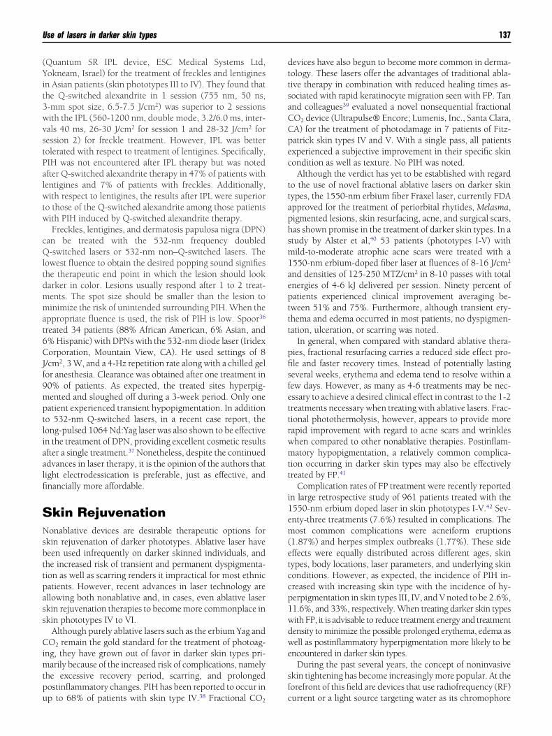
(Yit3wvstPalwtw
cQltdmmat6CJf9mptliaalfi
SNsbttpass
Cimtpu
dttsaCCpec
ttaphsm1aepttt
pfisfettrwmtt
i1em(etccp1wdwe
sf
Use of lasers in darker skin types 137
Quantum SR IPL device, ESC Medical Systems Ltd,okneam, Israel) for the treatment of freckles and lentigines
n Asian patients (skin phototypes III to IV). They found thathe Q-switched alexandrite in 1 session (755 nm, 50 ns,-mm spot size, 6.5-7.5 J/cm2) was superior to 2 sessionsith the IPL (560-1200 nm, double mode, 3.2/6.0 ms, inter-als 40 ms, 26-30 J/cm2 for session 1 and 28-32 J/cm2 foression 2) for freckle treatment. However, IPL was betterolerated with respect to treatment of lentigines. Specifically,IH was not encountered after IPL therapy but was notedfter Q-switched alexandrite therapy in 47% of patients withentigines and 7% of patients with freckles. Additionally,ith respect to lentigines, the results after IPL were superior
o those of the Q-switched alexandrite among those patientsith PIH induced by Q-switched alexandrite therapy.Freckles, lentigines, and dermatosis papulosa nigra (DPN)
an be treated with the 532-nm frequency doubled-switched lasers or 532-nm non–Q-switched lasers. The
owest fluence to obtain the desired popping sound signifieshe therapeutic end point in which the lesion should lookarker in color. Lesions usually respond after 1 to 2 treat-ents. The spot size should be smaller than the lesion toinimize the risk of unintended surrounding PIH. When the
ppropriate fluence is used, the risk of PIH is low. Spoor36
reated 34 patients (88% African American, 6% Asian, and% Hispanic) with DPNs with the 532-nm diode laser (Iridexorporation, Mountain View, CA). He used settings of 8
/cm2, 3 W, and a 4-Hz repetition rate along with a chilled gelor anesthesia. Clearance was obtained after one treatment in0% of patients. As expected, the treated sites hyperpig-ented and sloughed off during a 3-week period. Only oneatient experienced transient hypopigmentation. In additiono 532-nm Q-switched lasers, in a recent case report, theong-pulsed 1064 Nd:Yag laser was also shown to be effectiven the treatment of DPN, providing excellent cosmetic resultsfter a single treatment.37 Nonetheless, despite the continueddvances in laser therapy, it is the opinion of the authors thatight electrodessication is preferable, just as effective, andnancially more affordable.
kin Rejuvenationonablative devices are desirable therapeutic options for
kin rejuvenation of darker phototypes. Ablative laser haveeen used infrequently on darker skinned individuals, andhe increased risk of transient and permanent dyspigmenta-ion as well as scarring renders it impractical for most ethnicatients. However, recent advances in laser technology arellowing both nonablative and, in cases, even ablative laserkin rejuvenation therapies to become more commonplace inkin phototypes IV to VI.
Although purely ablative lasers such as the erbium Yag andO2 remain the gold standard for the treatment of photoag-
ng, they have grown out of favor in darker skin types pri-arily because of the increased risk of complications, namely
he excessive recovery period, scarring, and prolongedostinflammatory changes. PIH has been reported to occur in
p to 68% of patients with skin type IV.38 Fractional CO2 cevices have also begun to become more common in derma-ology. These lasers offer the advantages of traditional abla-ive therapy in combination with reduced healing times as-ociated with rapid keratinocyte migration seen with FP. Tannd colleagues39 evaluated a novel nonsequential fractionalO2 device (Ultrapulse® Encore; Lumenis, Inc., Santa Clara,A) for the treatment of photodamage in 7 patients of Fitz-atrick skin types IV and V. With a single pass, all patientsxperienced a subjective improvement in their specific skinondition as well as texture. No PIH was noted.
Although the verdict has yet to be established with regardo the use of novel fractional ablative lasers on darker skinypes, the 1550-nm erbium fiber Fraxel laser, currently FDApproved for the treatment of periorbital rhytides, Melasma,igmented lesions, skin resurfacing, acne, and surgical scars,as shown promise in the treatment of darker skin types. In atudy by Alster et al,40 53 patients (phototypes I-V) withild-to-moderate atrophic acne scars were treated with a
550-nm erbium-doped fiber laser at fluences of 8-16 J/cm2
nd densities of 125-250 MTZ/cm2 in 8-10 passes with totalnergies of 4-6 kJ delivered per session. Ninety percent ofatients experienced clinical improvement averaging be-ween 51% and 75%. Furthermore, although transient ery-hema and edema occurred in most patients, no dyspigmen-ation, ulceration, or scarring was noted.
In general, when compared with standard ablative thera-ies, fractional resurfacing carries a reduced side effect pro-le and faster recovery times. Instead of potentially lastingeveral weeks, erythema and edema tend to resolve within aew days. However, as many as 4-6 treatments may be nec-ssary to achieve a desired clinical effect in contrast to the 1-2reatments necessary when treating with ablative lasers. Frac-ional photothermolysis, however, appears to provide moreapid improvement with regard to acne scars and wrinkleshen compared to other nonablative therapies. Postinflam-atory hypopigmentation, a relatively common complica-
ion occurring in darker skin types may also be effectivelyreated by FP.41
Complication rates of FP treatment were recently reportedn large retrospective study of 961 patients treated with the550-nm erbium doped laser in skin phototypes I-V.42 Sev-nty-three treatments (7.6%) resulted in complications. Theost common complications were acneiform eruptions
1.87%) and herpes simplex outbreaks (1.77%). These sideffects were equally distributed across different ages, skinypes, body locations, laser parameters, and underlying skinonditions. However, as expected, the incidence of PIH in-reased with increasing skin type with the incidence of hy-erpigmentation in skin types III, IV, and V noted to be 2.6%,1.6%, and 33%, respectively. When treating darker skin typesith FP, it is advisable to reduce treatment energy and treatmentensity to minimize the possible prolonged erythema, edema asell as postinflammatory hyperpigmentation more likely to be
ncountered in darker skin types.During the past several years, the concept of noninvasive
kin tightening has become increasingly more popular. At theorefront of this field are devices that use radiofrequency (RF)
urrent or a light source targeting water as its chromophore
tidrotmmrfo
pdptcr
tasopsoaTalteerwtppscp
tsmisrg
eRstvbsc
lw
urtshtptr
vvibretstesetNto
F(
138 E.F. Battle and C.E. Soden
o produce dermal heating. This dermal heating results in annitial transient collagen fiber contraction followed by a well-ocumented inflammatory wound response that ultimatelyesults in dermal remodeling and neocollagenesis. Unlikether laser surgical procedures that directly or indirectly heathe epidermis while attempting to target their specific chro-ophore, RF devices accomplish their objective by deep der-al heating while keeping the epidermis relatively cool. Cur-
ent applications include rhytid reduction, tightening of laxacial and neck skin, lower eyelid rejuvenation and treatmentf atrophic facial scarring.Although the body of literature to date has been obtained
rimarily via skin types I through III, the ability of theseevices to provide deep dermal heating while concomitantlyroviding epidermal cooling should allow for the effectivereatment of Skin types IV through VI with a lessened con-ern for complications, such as dyspigmentation and scar-ing.
With regard to current radiofrequency-induced tissueightening both monopolar and bipolar RF devices are avail-ble. Thermage, a monopolar RF, has been the most widelytudied device and is approved by the FDA for the treatmentf facial rhytides. Early studies suggest that Thermage ap-ears to be both safe and effective in darker skin types. In atudy involving 85 Japanese females, Kushikata et al43 dem-nstrated improvement in nasolabial folds, marionette lines,nd jowls in skin types III-IV after a single treatment withhermage. Overall, objective improvement rates at 3 monthsfter treatment were 78.0% for jowls, 69.5% for marionetteines, and 73.8% for nasolabial folds. High patient satisfac-ion was noted at both the 3-month initial and 6-month finalvaluation time points. Interestingly, the objective physicianvaluation of facial rhytid reduction based on digital photog-aphy were more than 10% better at the 6-month period thanere noted at the initial 3-month follow-up while the subjec-
ive patient evaluation had fallen slightly by a few percentageoints in all categories at the 6-month follow-up. Minor com-lications ranging from edema, burning and blistering toecondary hyperpigmentation were noted in 7 patients. In allases, the complications were transient and resolved withoutermanent sequelae.The use of Thermage in darker skin types requires addi-
ional study, but other applications are likely amenable to allkin types. Thermage has shown effectiveness in the treat-ent moderate eyelid laxity.44 It also shows promising results
n the treatment of severe acne vulgaris and atrophic facialcarring where it may not only activate dermal remodelingesulting in scar reduction but also directly inhibit sebaceousland activity, improving acne lesions.45
Bipolar devices include the Polaris and ReFirme from Syn-ron as well the Aluma from Lumenis. Both the Polaris andeFirme incorporate bipolar radiofrequency with diode laserystems (780-910 nm for the Polaris and 700-2000 nm forhe ReFirme). The Aluma is a bipolar device with an attachedacuum. The vacuum has not only been found to reduce painut also brings the electrodes closer to the deep dermal tis-ue, reducing the energy for an effective treatment. The Ac-
ent from Alma Lasers incorporates both unipolar and bipo- tar RF. Superficial heating is accomplished with bipolar RFhile unipolar RF produces deeper dermal heating.In addition to RF devices, the other type of energy being
sed in tissue tightening is broadband infrared light in theange of 800-1800 nm. Similar to RF technology infraredechnology appears to be safe and effective in treating darkerkin tones (Fig. 9). In a recent study, an infrared nonablativeeating device (Titan, Cutera, Inc., Brisbane, CA) was showno be effective in achieving mild-to-moderate gradual im-rovement in the treatment of facial and neck laxity in type IVo V Asian skin without persistent dyspigmentation or scar-ing.46
By and large, noninvasive tissue tightening offers the ad-antages of essentially a nonexistent postoperative period, aery low risk of serious adverse effects, and promising resultsn the treatment of darker skin types. Dose selection shoulde based on each patient’s pain and tolerance levels. Mosteported complications tend to arise when nerve blocks orxcessive topical anesthetic are employed, blunting the pa-ient’s response to pain, and subsequently hindering the phy-ician’s ability to correct an excessively high energy. In addi-ion, due to a relative increase in collagen density noted inthnic skin, fewer treatments may be necessary to achieveimilar clinical results noted in skin phototypes I-III. How-ver, regardless of skin type, continued remodeling may con-inue to occur for several months after the initial treatment.evertheless, patients should be counseled regarding the po-
entially modest results despite the significant skin tighteningften observed immediately after the procedure.
igure 9 (A) Baseline skin laxity in a patient with type IV skin.B) Photograph of patient 1 year after 3 treatments with infrared skin
ightening (Titan, Cutera, Inc., Brisbane, CA).
CWwiswktvctstbin
R
1
1
1
1
1
1
1
1
1
1
2
2
2
2
2
2
2
2
2
2
3
3
3
3
3
3
3
3
3
3
4
4
4
Use of lasers in darker skin types 139
onclusionshen performing cutaneous laser surgery, one must proceedith caution regardless of skin type. However, because of the
ncreased risk of epidermal side effects when performing la-er therapy on skin of color, the laser surgeon must proceedith a greater degree of concern and possess a thoroughnowledge of the numerous and varied laser-tissue interac-ions that are possible among darker skin types. This conser-ative approach used to treat darker skin phototypes IV to VIan also be extrapolated to “tan” skin. The increased pigmen-ation seen with both immediate and delayed tanning canimilarly result in excessive energy absorption and diffusion,hus producing complications, such as pigmentary changes,listering and scarring that might otherwise not be seen in an
ndividual normally of skin type II or III in their naturalontanned state.
eferences1. US Census Bureau: Population Projections of the US by Age, Sex, Race
and Hispanic Origin: 1995-2050. Washington, DC, US Government,2002, pp 25-1130
2. Anderson RR: Laser-Tissue Interactions in Dermatology. Philadelphia,PA, Lippincott-Raven, 1997
3. Manuskiatti W, Eimpunth S, Wanitphakdeedecha R: Effect of cold aircooling on the incidence of postinflammatory hyperpigmentation afterQ-switched Nd:YAG laser treatment of acquired bilateral nevus of Otalike macules. Arch Dermatol 143:1139-1143, 2007
4. Breadon JY, Barnes CA: Comparison of adverse events of laser andlight-assisted hair removal systems in skin types IV-VI. J Drugs Derma-tol 6:40-46, 2007
5. Battle E, Suthamjariya K, Alora M, et al: Very long pulses (20-200 ms)diode laser for hair removal on all skin types (abstr). Lasers Surg Med12:21-24, 2000 (suppl)
6. Battle EF, Jr, Hobbs LM: Laser-assisted hair removal for darker skintypes. Dermatol Ther 17:177-183, 2004
7. Alster TS, Bryan H, Williams CM: Long-pulsed Nd:YAG laser-assistedhair removal in pigmented skin: A clinical and histological evaluation.Arch Dermatol 137:885-889, 2001
8. Galadari I: Comparative evaluation of different hair removal lasers inskin types IV, V, and VI. Int J Dermatol 42:68-70, 2003
9. Bouzari N, Tabatabai H, Abbasi Z, et al: Laser hair removal: Compari-son of long-pulsed Nd:YAG, long-pulsed alexandrite, and long-pulseddiode lasers. Dermatol Surg 30:498-502, 2004
0. Alajlan A, Shapiro J, Rivers JK, et al: Paradoxical hypertrichosis afterlaser epilation. J Am Acad Dermatol 53:85-88, 2005
1. Moreno-Arias GA, Castelo-Branco C, Ferrando J: Side-effects after IPLphotodepilation. Dermatol Surg 28:1131-1134, 2002
2. Radmanesh M: Paradoxical hypertrichosis and terminal hair changeafter intense pulsed light hair removal therapy. J Dermatol Treat 20:53-54, 2009
3. Hirsch RJ, Farinelli WA, Laughlin SA, et al: Hair removal induced bylaser hair removal. Lasers Surg Med 32:63, 2003 (suppl 15)
4. Tanzi EL, Alster TS: Cutaneous laser surgery in darker skin phototypes.Cutis 73:21-24, 27-30, 2004
5. Goldman L, Blaney DJ, Kindel DJ, Jr, et al: Pathology of the effect ofthe laser beam on the skin. Nature 197:912-914, 1963
6. Goldman L, Wilson RG, Hornby P, et al: Radiation from a Q-switchedruby laser. Effect of repeated impacts of power output of 10 megawattson a tattoo of Man. J Invest Dermato l44:69-71, 1965
7. Zelickson BD, Mehregan DA, Zarrin AA, et al: Clinical, histologic, andultrastructural evaluation of tattoos treated with three laser systems.Lasers Surg Med 15:364-372, 1994
8. Lapidoth M, Aharonowitz G: Tattoo removal among Ethiopian Jews in
Israel: Tradition fæces technology. J Am Acad Dermatol 51:906-909,2004
9. Bukhari IA: Removal of amateur blue-black tattoos in Arabic women ofskin type (III-IV) with Q-switched alexandrite laser. J Cosmet Dermatol4:107-110, 2005
0. Moreno-Arias GA, Casals-Andreu M, Camps-Fresneda A: Use of Q-switched alexandrite laser (755 nm, 100 nsec) for removal of traumatictattoo of different origins. Lasers Surg Med 25:445-450, 1999
1. Grevelink JM, Duke D, van Leeuwen RL, et al: Laser treatment of tattoosin darkly pigmented patients: Efficacy and side effects. J Am AcadDermatol 34:653-656, 1996
2. Jones A, Roddey P, Orengo I, et al: The Q-switched ND:YAG lasereffectively treats tattoos in darkly pigmented skin. Dermatol Surg 22:999-1001, 1996
3. Wang CC, Hui CY, Sue YM, et al: Intense pulsed light for the treatmentof refractory Melasma in Asian persons. Dermatol Surg 30:1196-1200,2004
4. Li YH, Chen JZ, Wei HC, et al: Efficacy and safety of intense pulsed lightin treatment of Melasma in Chinese patients. Dermatol Surg 34:693-700; discussion: 700-701, 2008
5. Negishi K, Kushikata N, Tezuka Y, et al: Study of the incidence andnature of “very subtle epidermal melasma” in relation to intense pulsedlight treatment. Dermatol Surg 30:881-886; discussion: 886, 2004
6. Polnikorn N: Treatment of refractory dermal Melasma with the MedLiteC6 Q-switched Nd:YAG laser: Two case reports. J Cosmet Laser Ther10:167-173, 2008
7. Rokhsar CK, Fitzpatrick RE: The treatment of Melasma with fractionalphotothermolysis: A pilot study. Dermatol Surg 31:1645-1650, 2005
8. Tannous ZS, Astner S: Utilizing fractional resurfacing in the treatmentof therapy-resistant melasma. J Cosmet Laser Ther 7:39-43, 2005
9. Goldberg DJ, Berlin AL, Phelps R: Histologic and ultrastructural anal-ysis of Melasma after fractional resurfacing. Lasers Surg Med 40:134-138, 2008
0. Laubach HJ, Tannous Z, Anderson RR, et al: Skin responses to frac-tional photothermolysis. Lasers Surg Med 38:142-149, 2006
1. Lin JY, Chan HH: Pigmentary disorders in Asian skin: Treatment withlaser and intense pulsed light sources. Skin Ther Lett 11:8-11, 2006
2. Chan HH, Leung RS, Ying SY, et al: Recurrence of nevus of Ota aftersuccessful treatment with Q-switched lasers. Arch Dermatol 136:1175-1176, 2000
3. Kono T, Nozaki M, Chan HH, et al: A retrospective study looking at thelong-term complications of Q-switched ruby laser in the treatment ofnevus of Ota. Lasers Surg Med 29:156-159, 2001
4. Kawada A, Shiraishi H, Asai M, et al: Clinical improvement of solarlentigines and ephelides with an intense pulsed light source. DermatolSurg 28:504-508, 2002
5. Wang CC, Sue YM, Yang CH, et al: A comparison of Q-switched alex-andrite laser and intense pulsed light for the treatment of freckles andlentigines in Asian persons: A randomized, physician-blinded, split-face comparative trial. J Am Acad Dermatol 54:804-810, 2006
6. Spoor T: Treatment of dermatosis papulosis nigra with the 532 nmdiode laser. Cosmet Dermatol 14:21-23, 2001
7. Schweiger ES, Kwasniak L, Aires DJ: Treatment of dermatosis papulosanigra with a 1064 nm Nd:YAG laser: Report of two cases. J CosmetLaser Ther 10:120-122, 2008
8. Sriprachya-anunt S, Marchell NL, Fitzpatrick RE, et al: Facial resurfac-ing in patients with Fitzpatrick skin type IV. Lasers Surg Med 30:86-92,2002
9. Tan KL, Kurniawati C, Gold MH: Low risk of postinflammatory hyper-pigmentation in skin types 4 and 5 after treatment with fractional CO2
laser device. J Drugs Dermatol 7:774-777, 20080. Alster TS, Tanzi EL, Lazarus M: The use of fractional laser photothermoly-
sis for the treatment of atrophic scars. Dermatol Surg 33:295-299, 20071. Glaich AS, Rahman Z, Goldberg LH, et al: Fractional resurfacing for the
treatment of hypopigmented scars: A pilot study. Dermatol Surg 33:289-294; discussion: 293-294, 2007
2. Graber EM, Tanzi EL, Alster TS: Side effects and complications offractional laser photothermolysis: Experience with 961 treatments.
Dermatol Surg 34:301-305; discussion: 305-307, 2008
4
4
4
4
140 E.F. Battle and C.E. Soden
3. Kushikata N, Negishi K, Tezuka Y, et al: Non-ablative skin tighteningwith radiofrequency in Asian skin. Lasers Surg Med 36:92-97, 2005
4. Ruiz-Esparza J: Noninvasive lower eyelid blepharoplasty: A new tech-nique using nonablative radiofrequency on periorbital skin. DermatolSurg 30:125-129, 2004
5. Ruiz-Esparza J, Gomez JB: Nonablative radiofrequency for active acne
vulgaris: The use of deep dermal heat in the treatment of moderate tosevere active acne vulgaris (thermotherapy): A report of 22 patients.Dermatol Surg 29:333-339; discussion: 339, 2003
6. Chua SH: Nonablative infrared skin tightening in type IV to V Asianskin: A prospective clinical study. Dermatol Surg 33:146-151,
2007