Embed Size (px)
Citation preview

Contact Lens and Anterior Eye, (Supplement), 21, pp. $3-$11, 1998 © 1998 British Contact Lens Association Printed in Great Britain
THE USE OF DIGITAL IMAGE CAPTURE IN CONTACT LENS PRACTICE
J o h n M e y l e r * a n d N i g e l B u r n e t t H o d d *
Abstract - - The present decade has seen the introduction of digital imaging systems to a number of medical environments, including contact lens practice. During the past year, the authors have used two anterior segment video-based digital image capture systems. These systems allow instantaneous image capture and have both diagnostic value to practitioners and educational value to patients. This paper discusses and clarifies the terminology that surrounds such systems and looks carefully at the advantages and disadvantages when compared to more traditional 35 mm photography in particular with reference to image colour, resolution and storage. The difference between video digital image capture and the more recently available digital camera image capture is also discussed. The
value of digital imaging is shown by a series of images from the authors' archives.
KEY WORDS: Analogue image, digital image, pixel, resolution, digital camera, slit lamp
Introduction
T here has been considerable debate amongst contact lens practitioners as to the benefit of
upgrading current image recording systems from photographic film to digital image capture and compu- terised storage. Traditionally, contact lens practitioners have recorded 35 mm and Polaroid ® images of the external eye with a photo slit lamp biomicroscope. With the rapid development of digital cameras, computers, software and printers, both hardware and software can become out-dated, although still functional, within months of their purchase. One of the key questions relates to the image quality compared to conventional 'silver halide' photography. Image capture, whether digital or photographic, is an excellent method of storing visual information. This is particularly important in the contact lens practice, where monitoring the subtle changes that may take place in the underlying tissue appearance is vital. Traditional methods of image documentation, such as the view through a slit lamp represented by a record card drawing or conventional photography, have inherent limitations as indicated by Cox 1 - the principal problem is that the view through the slit-lamp is only as long lasting as the clinician's memory and drawings are limited by the artistic skills of the individual clinician. Conventional photography requires a certain level of expertise to ensure the correct exposure, has high on-going development costs and cannot be viewed in real time. The introduction of both photographic and illustrative grading scale systems (e.g. CCLRU and Efron grading scales) has resulted in a more structured and objective way of measuring and monitoring tissue appearance; however, both time and skill are required for their use to be maximised.
Anterior Segment Image Capture Options Practitioners have various options for image capture of the anterior segment. These include conventional
*BSc, FCOptom, DCLP.
photography and digital imaging, whether generated by an analogue or a true digital source. The latter result in 'real time' images. These options are summarised in Figure 1. Much of the terminology surrounding the different options for digital image capture can be confusing and hence a list of 'definitions' is included in Appendix 1.
Conventional photography Traditionally, the most frequently used option for image capture of the anterior segment involves the use of a photographic slit lamp with a beam splitter attached to a 35 mm camera back. Slight modification produced the Polaroid ~ option resulting in 'instant' processing but sacrificed resolution. Silver based photographs can be scanned, using a variety of devices, and stored in digital format. This allows practitioners to view their 35 mm images on the computer monitor. Further development employed video cameras, which can be attached to the slit lamp via a beam splitter and employed in conjunction with conventional 35 mm photography. This allows anterior images to be captured by a video recorder on tape or printed using a video printer. The authors have used this option successfully for many years.
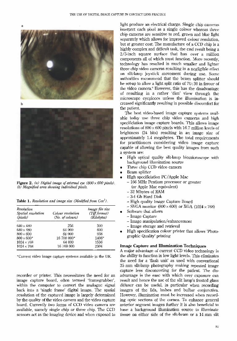
Digital Imaging Unlike an analogue signal that simulates sound or vision by variations in voltage producing corresponding varia- tions in brightness, a digital signal represents changes as a series of discrete pulses termed bits. The resultant image is formed by a rectangular grid of pixels, each of which is assigned an address and a value and can be either greyscale or colour. Figure 2a and 2b show just how a digital image is made up of thousands of pixels. Figure 2a shows a digital image whilst Figure 2b shows a small area of the same image where the individual pixels become visible following magnification. The number of bits used to represent the colour value of each pixel in the digitised image can vary from one bit, which displays two colours, to 24 bit, which shows 16.7 million
$3

JOHN MEYLER AND NIGEL BURNETr HODD
Analogue
'Silver image' Video camera . . . . . . . . . . . . . . . . . . . . . . . . . . . . . . . . . . . . . . . . . . . . . . . . . . . . . . . . . . . . . . . . . . . . . . . . . . . . . . . . . . . . . . . . . . . . . . . . . . . . . . . . . . . . . . . . . . . . . .
Scanner Image Capture Board Digital ~ ~ _ . . j ~ ~ camera
t---* COMPUTER Storage ~ 1 [ Digital ] e.g. PhotoCD
Image processing
Ana lys i s Manipulate/Enhance Pre-press
. . . . . . . . . . . . . . . . . . . . . . . . . . . . . . . . . . . . . . . . . . . . . . . . . . . . . . . . . . . . . . . . . . . . . . . . 1 - . . . . . . . . . . thermal print • film/paper Analogue litho plate or digital transmission multimedia
Figure 1. The Imaging Cycle. (Modified from Davies and Fennessy2).
colours. To create a digital image, three components are
required
• A system for recording the image • A system for converting the image data into a digital
file • A system for storage
In digital systems the number of pixels on a display surface determines resolution. The greater the number of pixels in a given area, the greater the resolution. Therefore a system that generates digital images of 800 x 600 (480 000 pixels) will have better resolution and image quality than a system that generates 640 x 480 (307 200 pixels). However, higher resolution images result in the need for more disk space when stored and therefore present a greater challenge when storing many images within easy to retrieve libraries (Table 1). An image with spatial resolution of 1024 x 768 pixels and 16.7 million possible colours should appear visually to be of 'photographic print' quality when viewed on the computer screen.
Figure 1 shows that such an image can be generated by the use of a video camera and an image capture board stored inside the PC. The former produces an
analogue signal, which is then digitised by the image capture board. The quality of the digital image captured will depend on the quality of the image capture board, video camera and optics of the slit lamp biomicroscope. More recent developments include the introduction of Digital Cameras Systems, (DCS), such as Kodak's digital system attached to a conventional Nikon ® camera. This unit is attached directly to the photo slit lamp biomicroscope and in such systems the digifising takes place in the camera and hence there is no need for an image capture board within the main computer. The quality of the image depends on the optics of both the slit-lamp and the camera as well as the resolution of the digifising system within the camera.
Video Digital Image Capture There are now a number of digital video image capture systems available to contact lens practitioners. The different options have recently received excellent re- views in the literature? -5 With all of these systems, a charged couple device (CCD) video camera achieves the initial image capture. Ironically such cameras actually digitise the image formed by the optical system and then use onboard electronics to convert the digital signal to an analogue signal which is capable of being recognised and interpreted by a conventional video
$4
THE USE OF DIGITAL IMAGE CAPTURE IN CONTACT LENS PRACTICE
Figure 2. (a) Digital image of external eye (800 × 600 pixels). (b) Magnified area showing individual pixels.
Table 1. Resolution and image size (Modified from Coxl).
Resolution Image file size Spatial resolution Colour resolution (Tiff format) (pixels) (No. of colours) (Kilobytes)
640 × 480 256 300 640 × 480 64 000 600 800 × 600 64 000 938 800 × 600* 16 700 000" 1406" 1024 × 768 64 000 1536 1024 × 768 16 700000 2304
*Current video image capture systems available in the UK.
recorder or printer. This necessitates the need for an image capture board, often termed 'framegrabber', within the computer to convert the analogue signal back into a 'single frame' digital image. The spatial resolution of the captured image is largely determined by the quality of the video camera and the video capture board. Currently two forms of CCD video camera are available, namely single chip or three chip. The CCD sensors act as the imaging device and when exposed to
light produce an electrical charge. Single chip cameras interpret each pixel as a single colour whereas three chip cameras are sensitive to red, green and blue light separately which allows for improved colour resolution, but at greater cost. The manufacture of a CCD chip is a highly complex and difficult task, the end result being a 2/3-inch square surface that has over a million components all of which must function. More recently, technology has resulted in much smaller and lighter three chip video cameras resulting in a negligible effect on slit-lamp joystick movement during use. Some authorities recommend that the beam splitter should be set-up to allow a light split ratio of 70 : 30 in favour of the video camera. 6 However, this has the disadvantage of resulting in a rather 'dim' view through the microscope eyepieces unless the illumination is in- creased significantly resulting in possible discomfort for the patient.
The best video-based image capture systems avail- able today use three chip video cameras and high specificiation image capture boards. This allows image resolutions of 800 × 600 pixels with 16.7 million levels of brightness (24 bits) resulting in an image size of approximately 1.4 megabytes. The total requirements for practitioners considering video image capture capable of allowing the best quality images from such a system are: • High optical quality slit-lamp biomicroscope with
background illumination source • Three chip CCD video camera • Beam splitter • High specificiation PC/Apple Mac
- 166 MHz Pentium processor or greater (or Apple Mac equivalen0
- 32 Mbytes of RAM - 3-4 Gb Hard Disk - High quality Image Capture Board - SVGA monitor (800 × 600) or XGA (1024 × 768)
• Software that allows - Image Capture - Image manipulation/enhancement - Image storage and retrieval
• High specification colour printer that allows 'Photo- graphic Quality' printing
I m a g e C a p t u r e a n d I l l u m i n a t i o n T e c h n i q u e s
A major advantage of current CCD video technology is the ability to function in low light levels. This eliminates the need for a 'flash unit' as used with conventional 35 mm slit-lamp photography making repeated image capture less disconcerting for the patient. The dis- advantage is the ease with which over exposure can result and hence the use of the slit lamp's frosted glass diffuser can be useful, in particular when recording images of the lids, lashes and bulbar conjunctiva. However, illumination must be increased when record- ing optic sections of the cornea. To enhance general anterior segment images further it is also beneficial to have a background illumination source to illuminate tissue on either side of the slit-beam or a 14 mm slit
$5

JOHN MEYLER AND NIGEL BURNETT HODD
height (ideally both). Reduction of the camera aperture generally results in improved colour of the captured image. A zoom facility is useful, particularly if the system is set up to allow the recording of 'moving' images stored on videotape. The most useful benefit of these systems is that what is observed on the monitor prior to capturing the image replicates the final image and there is no need to calculate film speeds and flash intensities. Images that appear under exposed on the monitor can be improved by increasing the illumination and the depth of focus can be improved by reducing the illumination aperture. Any poor quality images can be deleted with ease. There may be a need to change the spectral balance of the video camera to optimise the image quality when viewing under cobalt blue illumina- tion.
Image Storage Storage of large numbers of high-resolution digital images can prove problematic. Options include storage directly onto the computer's hard disk, which allows fast retrieval as and when required by the contact lens practitioner. Hard disk capacity is continually increasing with 3 - 4 Gigabytes hard disks now installed as standard. This allows the storage of approximately 2100-2900 1.4 Mb images, assuming the necessary software uses negligible hard disk space. Storage capability can be increased by compression that involves the 'squashing' of data to reduce file size for storage or to reduce transmission time during e-marl communication. Compression formats can either be 'lossy' or 'lossless'. The former is capable of very high levels of compression but can also degrade image quality slightly even though this may not be noticeable to the human eye. JPEG (Joint Photographic Expert Group) is an example of such a format that can compress data a great deal (up to 40 times). Tiff (Tagged Image Format File), on the other hand, is defined as 'lossless' form of file compression and is the most frequent file format used for high-resolution digital images. Other storage options include the use of external disk drives such as recordable compact disk drives, Jaz drives or SyQuest Syjet. These storage alternatives are currently used for 'backing up', hard discs and some examples are shown in Table 2.
35 mm Photography versus Video Digital Imaging Practitioners would ideally zoom in on an image in order to magnify small areas without loss of resolution and annotate captured images with clinical notes. In addi- tion, the option to print high quality prints to accompany referral letters or send images via an internal network for colleagues to view in alternative consulting rooms within the practice or for referral via the Internet would all be useful. Practitioners also expect to merge their practice management systems with clinical management systems without loss of data, speed or quality. Some current systems are close to offering the software and hardware to make video digital imaging an integral part of practice life for the busy practitioner, but concerns
are still raised about resolution of the images captured. The advantages and disadvantages of traditional 35 mm photography versus video digital imaging systems are shown in Table 3.
Image Capture Costs A slit lamp system and video digital imaging package is more expensive initially than a 35 mm camera/flash system. However, provided the practitioner is satisfied with the system purchased, there are no additional costs in the short term. The cost per digital image is significantly less than for 35 mm slides, particularly when the 'wastage factor' is considered. To obtain one suitable 35 mm picture, six slides may be required. The cost of film developing and mounting must be considered and therefore images may cost as much as £1.50 per picture. With video digital imaging, the images that are not wanted can easily be deleted. It is relatively inexpensive to save images on current data storage systems.
Table 2. Image storage options.
No. of 1. d Mb digital Storage medium Capacity (Mb) images
Internal 4 Gb hard disk 4096 2925
External ZIP disk 100 71 Recordable CD-R 650 464 1 Gb Jaz disk 1001 714 SyQuest Syjet 31/2 1500 1071
inch cartridge
Table 3. Comparison of conventional 35ram image capture versus video digital image capture (¢z = advantage).
35 mm Film Digital
Set up costs ,/ Cost per picture ,I Ease of storage ¢" Storage space ,/ Recall ,/ Patient viewing J" Image editing ,/ Image export ,/ Image duplication Image printing J Image overwriting ,/ Image resolution ¢" Image brightness ~/ Colour accuracy ,," Recording accuracy ¢" Recording versatility ,/ Reload camera ,/ Create storage medium J" Time factor ,/ Tolerance tests J" Side by side comparisons ¢" Patient education ,/ Ease of use ,/"
$6

THE USE OF DIGITAL IMAGE CAFI'URE IN CONTACT LENS PRACTICE
Storage and Recall 35 mm slide storage involves either placing images with patients' records or storing in a large, well-labelled library in filing cabinets. Projection is normally required to view the images and there is always the risk that the images may have become degraded with time. Storage can be improved by scanning 35 mm slide libraries onto CD-Rs. Systems such as Kodak ® PhotoCD allow slide images to be digitised by high resolution scanning and stored on CD. The resultant PCD file is about 5 - 6 Mb in size which allows a storage capacity of 100 slide images per CD. With digital image capture the practi- tioner has a simple patient database which lists all patients names and the dates the images were captured. The images may be recalled from the storage disk, whether internal or external, in seconds and displayed on the screen in sequence or side by side for easy comparison.
Image Editing and Exporting A captured digital image can be modified to improve sharpness, colour and brightness with the appropriate software. This facility is useful if improvement in the
J image appearance is required for use in presentations or publications. However, many current software systems that produce a captured image of the anterior eye are designed such that the original image captured will be the image filed for immediate recall for that patient. This has important legal implications, as software can be used not only to enhance the appearance of clinical signs but could also be used to remove any trace of clinical signs! Certain software also allows the addition of clinical notes and the image can also be exported to a printer or downline to other users in the same office via a central server and network.
tion of digital images captured on video-based systems as good as 35 mm resolution? The problem is the difference in terminology used when grading the quality of a digital image, measured by pixel count, when compared to a silver halide image, measured by lines per millimetre and print grain index. Therefore compar- isons are not easy but a conversion to pixels can be made to give some idea of theoretical comparisons. A 35 mm film results in 36 × 24 mm images and one line equates to two pixels. As common Kodak films vary from 100 to 200 lines per millimetre, depending on film speed, a pixel resolution as high as 14 400 x 9600 is possible assuming the camera has perfect optics. This is well above that possible from current video or 'still' digital cameras. It is generally acepted that a digital image with spatial resolution of 1024 × 768 and 16.7 million possible colours will appear to be of 'photo- graphic film' quality when viewed on an XGA computer screen. The best video digital image capture systems currently available fall short of this resolution but it is possible that this will not result in clinical implications for the contact lens practitioner.
Figures 3 and 4 show examples of images of external ocular tissue captured by 35 mm photography (Ekta- chrome 100 35 mm professional film) and a high specification video slit-lamp image capture system
Image Quality and Resolution There are two main points concerning image quality and resolution. Firstly, it is impossible to guarantee that the photograph captured on 35 mm film will be exactly as expected. There are many variables to consider in conventional photography such as film speed, flash intensity, the darkness of the eye and aperture width. There is a certain amount of 'chance' involved so usually many practitioners take a few pictures either side of the perceived best setting to increase the chance of obtaining a good picture. There is also the problem of being sure the image is in focus. Often the only good illumination comes from the flash. With video digital capture the variables include video camera aperture and the illumination of the eye which is controlled by the slit lamp intensity and background illumination. Conse- quently the learning curve is gentle and the correct settings are found by trial and error, if necessary deleting the poor quality images along the way. A series of images can be taken one after the other without any flash and discomfort for the patient. With digital imaging 'chance' is eliminated and a good result is usually guaranteed because images can be viewed directly.
The second point concerns resolution. Is the resolu- Figure 3. (a) Nasal bulbar conjunctiva (35 ram). (b) Nasal bulbar conjunctiva (digital image).
$7

JOHN MEYLER AND NIGEL BURNETI" HODD
Figure 5. Mid-peripheral sterile infiltrate.
Figure 4. (a) The cornea (35 ram). (b) The cornea (digital).
(800 × 600, 24 bit, 16.7 million colours). Both sets of images were captured through the same high specifica- tion photo slit-lamp, set-up to allow image capture by both conventional photography and video digital image capture using a 3-chip CCD camera. The ocular tissues photographed included:
1. The upper tarsal conjunctiva (white light). 2. The upper tarsal conjunctiva (blue light). 3. The inferior meibomian glands. 4. The superior limbus. 5. Nasal bulbar conjunctiva. 6. The cornea.
These structures are representative of those that would normally be recorded in routine contact lens aftercare. The 35 mm photographic images were scanned onto CD using Kodak's PhotoCD system allowing comparison, subject to the resolution at which they had been scanned, with the video captured digital images captured when viewed side by side on an XGA monitor. The same digital images were also imported into Microsoft PowerPoint, which allowed the images to be exported to 35 mm film. This allowed comparison of both sets of images when projected.
Visual comparisons confirmed that 'silver halide'
Figure 6. Early corneal vascularisation.
images provide both better resolution and colour accuracy than current digital video image capture systems. Even with illumination adjustment, digital systems are prone to 'burn out' in bright areas with loss of detail in dark areas. Initial set-up of the CCD video camera, by adjusting its colour balance, is critical to allow the best colour accuracy possible. Although exact colour replication suffers in video image capture, clinicians are more interested in tissue appearance change, particularly redness. This can still be monitored accurately as the tissue appearance for the patient is being recorded through the same system for each visit. Likewise, monitoring a resolving peripheral sterile infiltrate does not require high resolution (Figure 5). No doubt, as CCD cameras and image capture boards improve, so too will the spatial resolution of these systems which may allow the recording of the more subtle tissue changes such as striae and microcysts, challenging 35 mm photography!
Educational Value There is a time factor to consider when taking digital
$8

THE USE OF DIGITAL IMAGE CAPTURE IN CONTACF LENS PRACTICE
images. If a particular tissue appearance requires recording, it can be achieved instantaneously. However, once the image appears on the monitor, patients usually expect to discuss the relevance of the captured image. Figures 6 and 7 show typical images captured which can be used very effectively to support clinical explanations to the patient. Namely, signs of early vascularisation and the benefits of frequent replacement and disposable soft lenses. Images can also be used to show a patient how speciality lenses function such as rigid translating bifocals (Figure 8).
Digital 'Still' Camera Systems High resolution digital cameras have been available for almost a decade and have recently been introduced to photo slit-lamp units resulting in images with much higher resolution than that obtainable with current video-based image capture systems. Table 4 lists some commercially available digital cameras. One such system, which uses the Kodak DSC 420, allows slit- lamp image capture at a resolution of 1524 × 1012 (1.5 million pixels) resulting in a digital image size of approximately 8 Mb. In digital still cameras the digitis- ing occurs within the camera on the pixel array. This
allows the capture of a full frame instantly without the need for an image capture board within the computer. Images can be transferred directly to the computer's hard disk via the appropriate leads or stored in a PCMCIA card located within the camera body and transferred to a computer for viewing. Software, as with video-based digital image capture, allows the images to be viewed in thumbnail (Figure 9). Cost is currently the major limiting factor and, as with conventional 35 mm photography, a flash unit is required although the flash intensity is less than that needed for conventional 35 mm photography. With digital camera resolution increasing every year the future looks promising for this form of image capture.
The Future One of the benefits of digital imaging technology is that it continually improves. In the future, higher resolution digital video image capture systems and cheaper digital still camera systems, both offering more versatile software and higher storage capacity, will be available. Digital video cameras that can capture both moving and still images and store directly onto the internal hard disk will soon be an option for slit lamps. Practitioners may be linked via the Internet to specialists either for referral or for expert opinion following transfer of the patient's digital images. Software development may allow auto- mated clinical grading of tissue appearance as well as giving a likely diagnosis with management guidelines for individual patients. Finally the patient's contact lens prescription may include a series of dated anterior eye images all stored on a plastic card for easy access by their practitioner.
Figure 7. Soft lens deposits.
Table 4. Digital camera options.
Digital camera Spatial resolution Pixel count
Nikon E2Ns 1280 × 1000 1.3 million Minolta 1528 × 1146 1.75 million Kodak DSC 420 1524 × 1012 1.5 million Kodak DSC EOS1 3072 × 2048 6.3 million
Figure 8. Translating RGP bifocal during down gaze.
Table 5. Comparison of digital video image capture compared to digital 'still' camera systems. (J = Advantage).
Video digital Still camera digital imaging imaging
Set up costs ,/ Cost per picture J ,/ Image resolution ¢" Image brightness Colour accuracy J Image size/storage ,/ Ease of use J Patient comfort ~/ Patient education ,/ ,/
$9

JOHN MEYLER AND NIGEL BURNE~IT HODD
ment of patient education on compliance, the benefits of disposable and frequent replacement contact lenses as well as the importance of regular aftercare. In addition, there are no doubts that digital imaging can be a very useful marketing tool which, when used correctly, can only enhance professional image. Given time, patients will expect to see these systems in all contact lens practices.
Figure 9. Thumbnail view of images captured by digital 'still' camera.
Conclusions The authors' experience with traditional 35 mm photo- graphy over many years versus the new video image capture systems of today lead them to recommend image capture to be used on a daily basis even in the busiest of contact lens practices. 7 Practitioners can quickly learn to capture clinically valid images that are easy to store and recall. The image on the computer monitor is exactly as that recorded as the digital image. The instant nature of digital imaging allows improve-
Address for Correspondence John Meyler, Vistakon, The Braccans, London Road, Bracknell, Berks RG12 2AT. Nigel Burnett Hodd, Contact Lens Practice, 7 Devon- shire Street, London WIN 1FT E-mail Address 100641.2677 at COMPUSERVE.COM
REFERENCES
1 Cox, I. Digital Imaging in the Contact Lens Practice. Int. Contact Lens Clinic, 22, 62-66 (1995). Davies, A. and Fennessy, P. Electronic Imaging for Photographers (2nd edn). Focal Press, London. (1996).
a McPherson, S. The Image Makers. Optician 213 (5601), 127- 132 (1997).
4 Morgan, P., Morris, T., Newell, Z., Wood, I and Woods, C. Invasion of the Image Snatchers. Optician 213 (5588), 24-26 (1997).
'~ Hirji, N.K. Ocular Photo Documentation. Nikon's FS-3V. Optometry Today, 37 (23), 22-23 (1997).
6 Hammack, G. Clinical use of Slit Lamp Digital Imaging. Demon- stration, Ellerbrock Continuing Education Program, American Academy of Optometry, San Antonio (1997).
7 Burner Hodd, NF. Imaging Systems for Today's Practice. Optometry Today, 37 (6) 29-32 (1997).
Appendix 1
Bitmap An image made up of dots or pixels. All image capture boards or digial cameras produce bitmap images.
1-Bit Colour: Black and white images with no greys or colours. &Bit Colour: 256 Colours or greys. 24-Bit Colour: 16.7 Million colours. The maximum number of colours a computer can deal with and represents 'photorealistic' colour.
Byte Standard computer tile size measurement: contains eight bits. Other common units include Kilobyte (K; 1024 bytes), Megabyte (Mb; 1024K) and Gigabyte (Gb; 1024 Mb).
CD-ROM A non-rewritable digital storage CD often used to provide software.
CCD A Charged Couple Device (CCD) converts light into electrical current.
Colour Bit Depth The number of bits used to represent each pixel in an image. The higher the bit depth, the more colours appear in the image (see Table 1).
Compression: The 'squashing' of data to reduce file size for stoage or to reduce transmission time during transfer. Compres- sion can be 'lossy' (such as JPEG) or lossless' (such as TIFF). Greater reduction is possible with lossy com- pression than with lossless schemes.
File Word used in computers to describe a single document (i.e. a digital photograph) stored on disk. File names allow easy identification of the documents contents.
File Format The way that the information in a file is stored. In digital image capture some common file formats include JPEG, TIFF or GIF.
GIF A graphic file format developed by CompuServe for exchange of image files (only supports 8-bit colour).
$10

THE USE OF DIGITAL IMAGE CAPTURE IN CONTACI" LENS PRACTICE
Hard Disk A fast and cheap form of storage (usually refers to the computer internal hard drive).
Image Manipulation Once a digital image has been transferred into a computer, image manipulation software allows the individual pixels to be altered in many ways. Colour corrections, sharpening, photomontage and distortions are all forms of image manipulation.
Jaggies The jagged stepped effect often seen in image whose resolution is so low that individual pixels are visible.
JPEG A file compression standard established by the Joint Photographic, Expert Group that is capable of a very high level of compression, but is a 'lossy' method and so can degrade image quality slightly.
CPU (Central Processing Unit) The 'brains' of the computer; carried out all the instructions and calculations needed for a computer to w o r k .
Cropping Tool A tool found in image editing software. Allows you to trim an image as would a real photograph.
Network Two or more computers and peripherals that are connected by direct cable links or over telephone lines using modems.
PC Card (PCMCIA Card) A popular standard for removable cards that is used in both digital cameras and laptop computers. PC cards can contain hard disks, network links or modems.
Pixel (PICture Element) The smallest element of a digitised image. Also one of the tiny points of light that make-up a picture on a computer screen.
RAM (Random Access Memory) The computer memory where the CPU stores software, and other data being currently used. A large amount of RAM usually allows faster manipulation of images.
Modem A device that allows computers to communicate over distant telephone lines.
Resolution Measure of the amount of information in an image expressed in terms of the number of pixels per unit length (i.e. pixels per mm or pixels per inch). Camera resolution is usually defined as the actual number of pixels in the image (i.e. 640 x 480).
Thumbnail A small, low resolution version of a larger image that is used for quick identification and for displaying many images on a screen.
TIFF (Tagged image file format) The standard file format for high-resolution bit mapped graphics.
CD-R A recordable digital compact disk often used to store images.
$11