Embed Size (px)
Citation preview
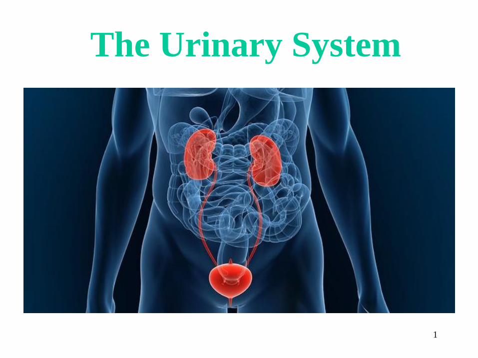
The Urinary System
1

Function
1. Remove nitrogenous wastes
2. Maintain electrolyte, acid-base,
and fluid balance of blood
3. Homeostatic organ
4. Acts as blood filter
5. Release hormones: calcitriol &
erythropoietin
2

Composition of the Urinary System
3
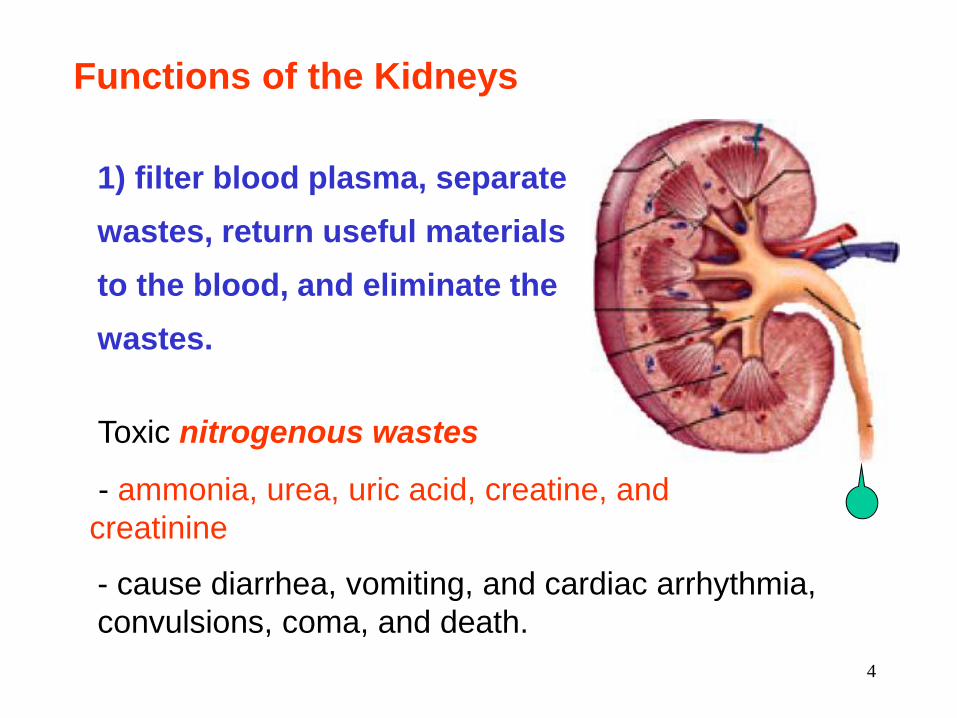
Functions of the Kidneys
1) filter blood plasma, separate
wastes, return useful materials
to the blood, and eliminate the
wastes.
Toxic nitrogenous wastes
- ammonia, urea, uric acid, creatine, and
creatinine
- cause diarrhea, vomiting, and cardiac arrhythmia,
convulsions, coma, and death.
4

Functions of the Kidneys
1) filter blood plasma,
separate wastes, return
useful materials to the
blood, and eliminate the
wastes.
2) regulate blood volume
and osmolarity.
5

3) produce hormones
1. renin
2. erythropoietin
3. calcitrol
Functions of the Kidneys
4) regulate acid-base balance
of the body fluids.
5) detoxify superoxides, free
radicals, and drugs.
6

Kidney Functions
• Filter 200 liters of blood daily, allowing
toxins, metabolic wastes, and excess ions to
leave the body in urine
• Regulate volume and chemical makeup of
the blood
• Maintain the proper balance between water
and salts, and acids and bases
7

- The kidneys lie along the posterior abdominal wall
8

- The medial surface of the kidney is concave with
a hilum carrying renal nerves and blood vessels.
The renal parenchyma is divided into an outer
cortex and inner medulla.
9
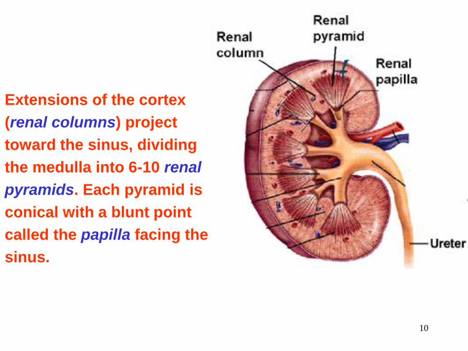
Extensions of the cortex
(renal columns) project
toward the sinus, dividing
the medulla into 6-10 renal
pyramids. Each pyramid is
conical with a blunt point
called the papilla facing the
sinus.
10
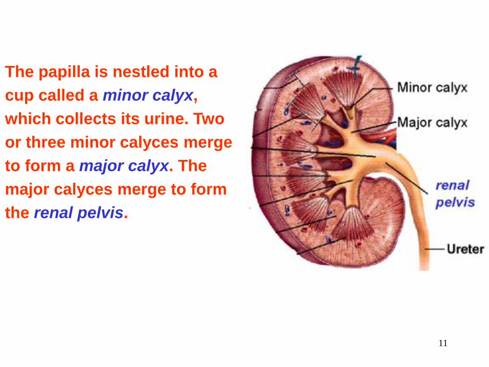
The papilla is nestled into a
cup called a minor calyx,
which collects its urine. Two
or three minor calyces merge
to form a major calyx. The
major calyces merge to form
the renal pelvis.
11

- The kidney contains 1.2 million nephrons, which
are the functional units of the kidney.
- A nephron consists of :
i. blood vessels
afferent arteriole
glomerulus
efferent arteriole
ii. renal tubules
proximal convoluted tubule
loop of Henle
distal convoluted tubule
The Nephron
12
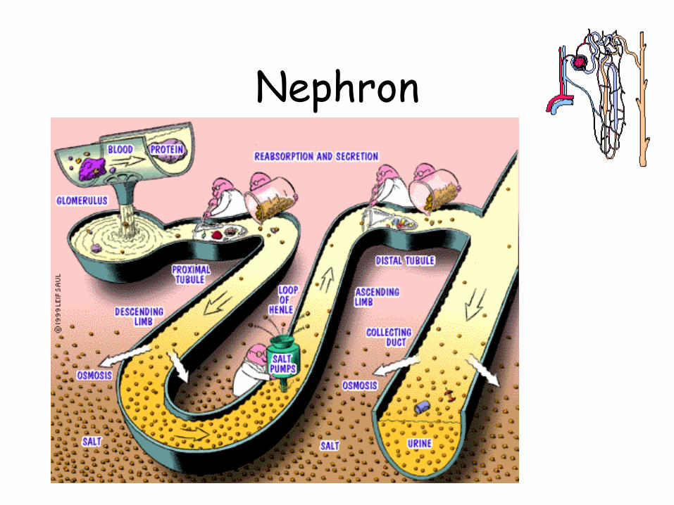
Nephron

afferent arteriole
glomerulus
efferent arteriole
proximal
convoluted
tubule
distal
convoluted
tubule
Loop of Henle
blood
blood
The Nephron
14

- Most components of
the nephron are within
the cortex.
The Nephron
15
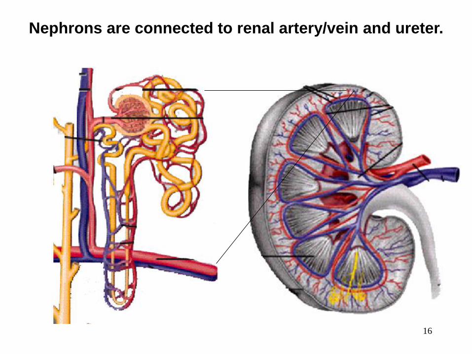
Nephrons are connected to renal artery/vein and ureter.
16
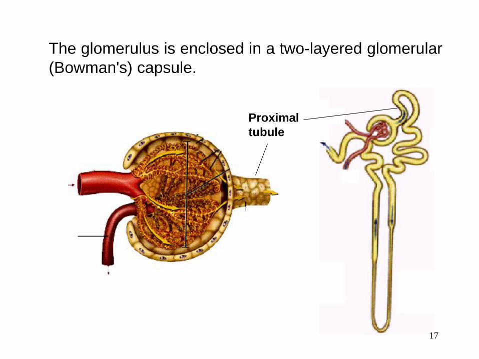
The glomerulus is enclosed in a two-layered glomerular
(Bowman's) capsule.
Proximal
tubule
17

From the original 1800 g NaCl, only 10 g appears in the urine
Urine Water- 95%
Nitrogenous waste:
• urea
• uric acid
• creatinine
Ions:
• sodium
• potassium
• sulfate
• phosphate
18
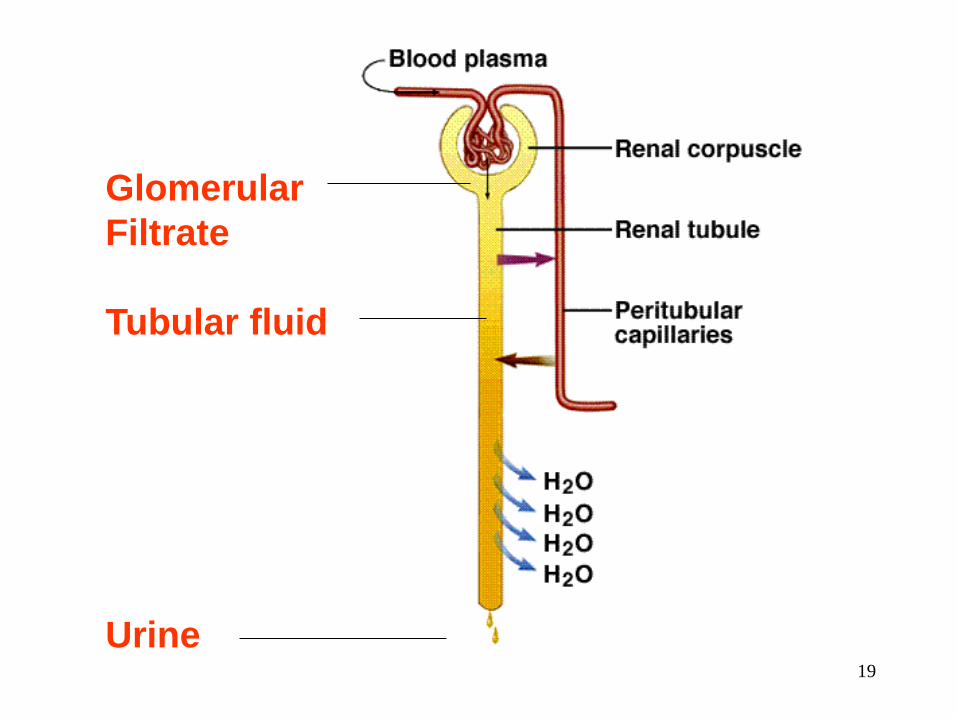
Glomerular
Filtrate
Tubular fluid
Urine 19

1) Glomerular Filtration
20
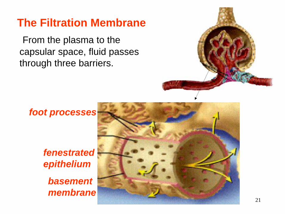
The Filtration Membrane
From the plasma to the
capsular space, fluid passes
through three barriers.
fenestrated
epithelium
basement
membrane
foot processes
21
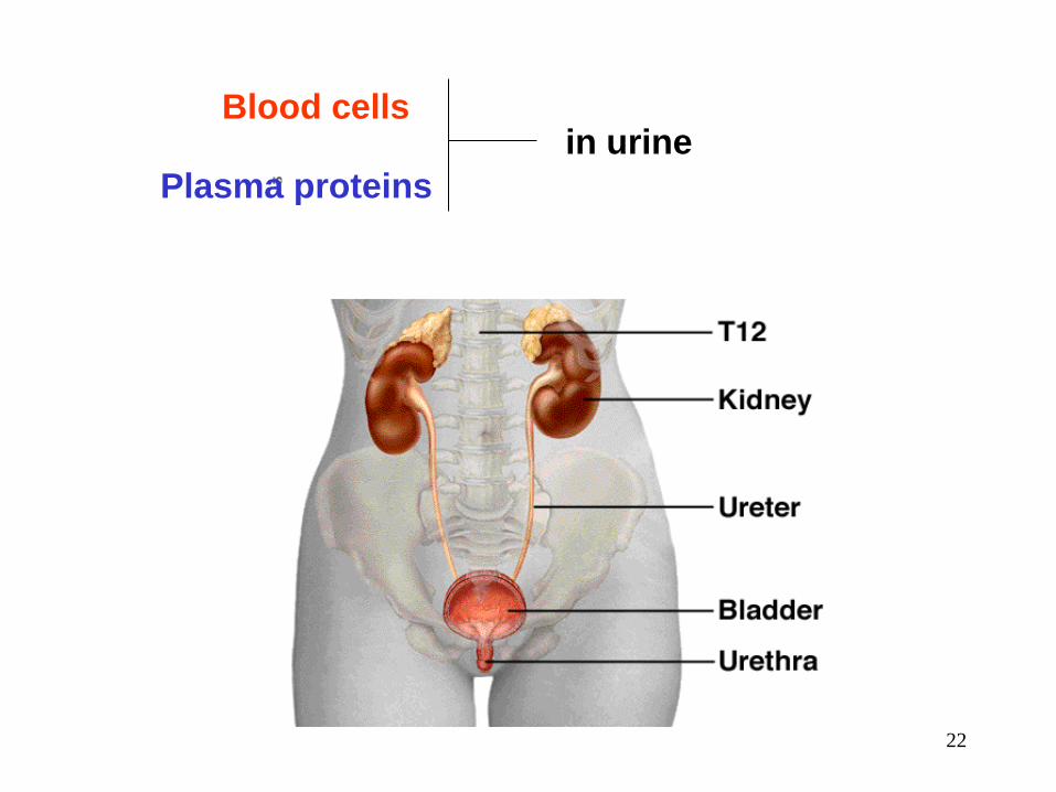
Blood cells in urine
Plasma proteins
22

About 99% of Water
and other useful small
molecules in the filtrate
are normally
reabsorbed back into
plasma by renal
tubules.
23

Reabsorption in Proximal
Convoluted Tubules
24

- The proximal convoluted
tubule (PCT) is formed by
one layer of epithelial cells
with long apical microvilli.
- PCT reabsorbs about
65% of the glomerular
filtrate and return it to the
blood.
25
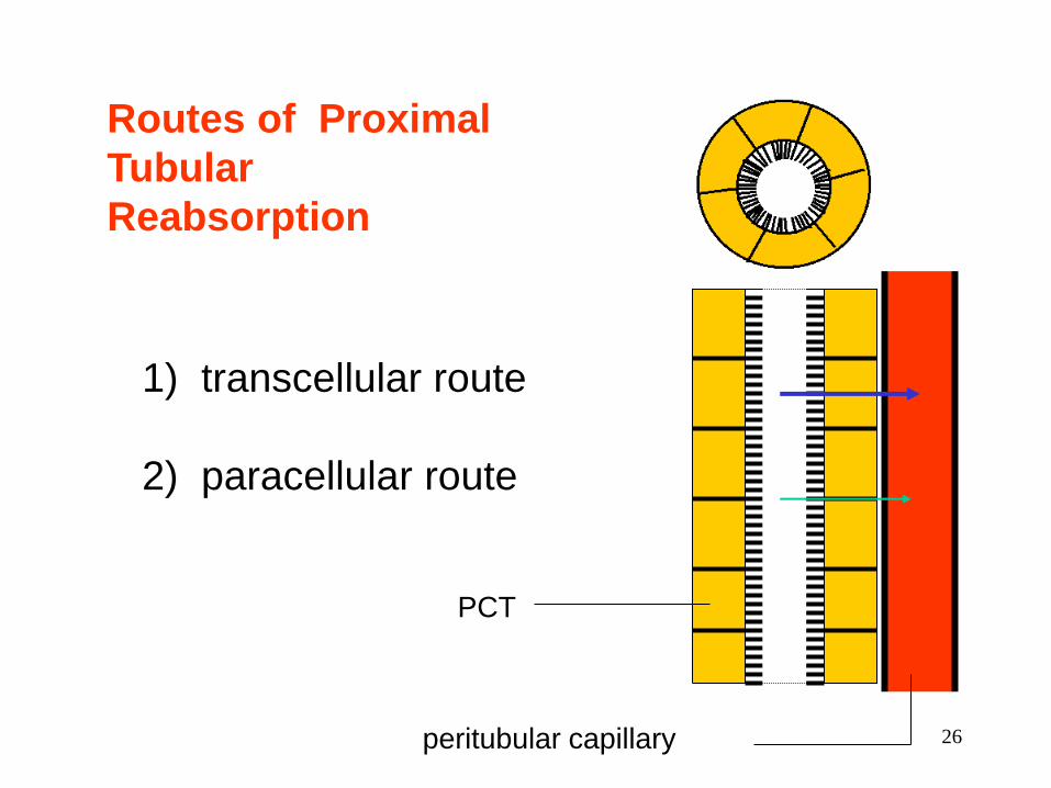
1) transcellular route
2) paracellular route
Routes of Proximal
Tubular
Reabsorption
PCT
peritubular capillary 26
Mechanisms of Proximal Tubular
Reabsorption
1) Solvent drag
2) Active transport of sodium.
3) Secondary active transport of glucose, amino
acids, and other nutrients.
4) Secondary water reabsorption via osmosis
5) Secondary ion reabsorption via electrostatic
attraction
6) Endocytosis of large solutes
27

Composition and Properties of Urine
Fresh urine is clear, containing no blood
cells and little proteins. If cloudy, it could
indicate the presence of bacteria, semen,
blood, or menstrual fluid.
Urine Properties
28

Urine Volume
An average adult produces 1-2 L of urine per
day.
a. Excessive urine output is called
polyuria.
b. Scanty urine output is oliguria. An
output of less than 400 mL/day is
insufficient to excrete toxic wastes.
29
Diabetes
- is chronic polyuria resulting from various
metabolic disorders, including Diabetes
mellitus and Diabetes insipidus
30

Ureters
Slender tubes attaching the kidney to the bladder
Continuous with the renal pelvis
Enter the posterior aspect of the bladder
Runs behind the peritoneum
Peristalsis aids gravity in urine transport
31

Urinary Bladder
Smooth, collapsible, muscular sac
Temporarily stores urine
32
Urinary Bladder
Slide 15.21b
Copyright © 2003 Pearson Education, Inc. publishing as Benjamin Cummings
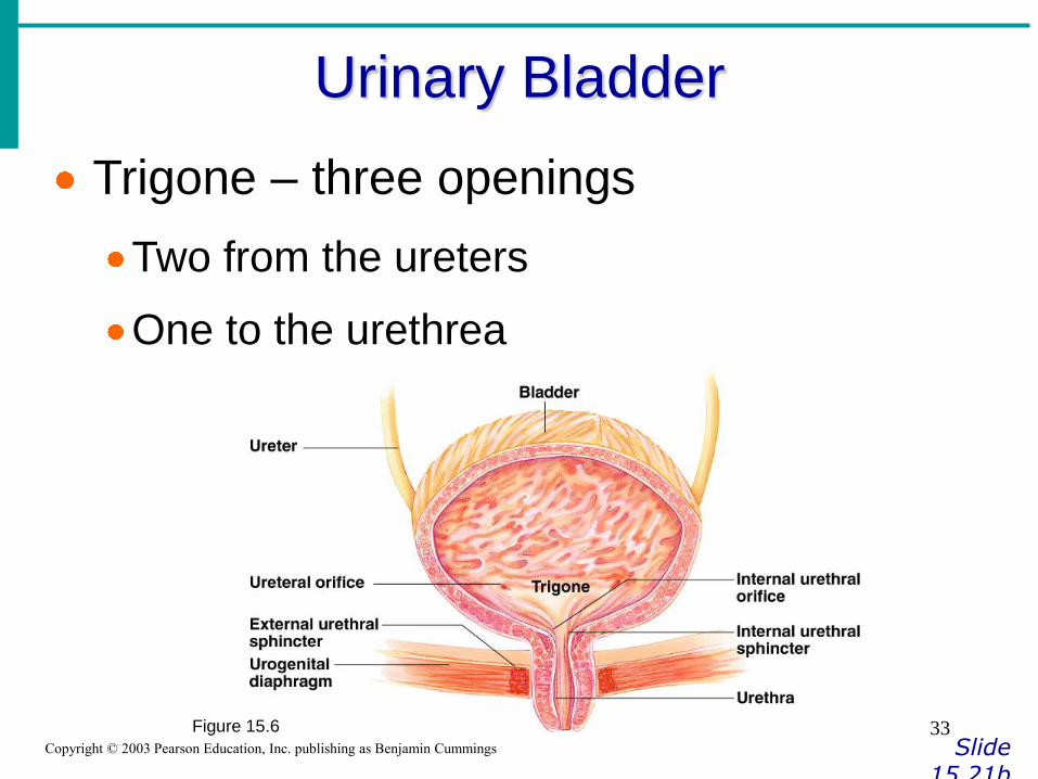
Trigone – three openings
Two from the ureters
One to the urethrea
Figure 15.6 33
Urinary Bladder Wall
Three layers of smooth muscle (detrusor muscle)
Mucosa made of transitional epithelium
Walls are thick and folded in an empty bladder
Bladder can expand significantly without increasing internal pressure
34
Urethra
Thin-walled tube that carries urine from the bladder to the outside of the body by peristalsis
Release of urine is controlled by two sphincters
Internal urethral sphincter (involuntary)
External urethral sphincter (voluntary)
35
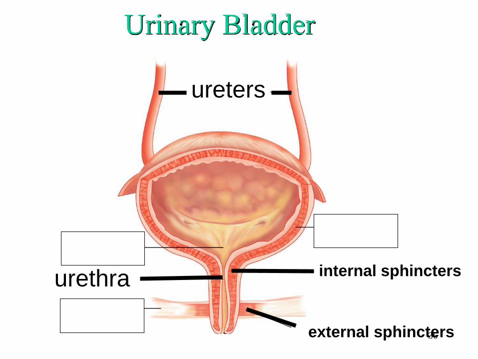
Urinary Bladder
ureters
internal sphincters
external sphincters
urethra
36
Urethra thin walled tube – conveys urine from bladder to
external environment. Similar structure to ureter
Sphincters: internal urethral [involuntary] & external urethral [ voluntary]
• Male Urethra
• 20 cm long [7-8 in]
• Transports both urine & semen
Eternal urethral orifice opens at tip of penis
• Female Urethra
• 3-4 cm long [1.5 in]
• External urethral orifice opens just anterior to the vaginal opening
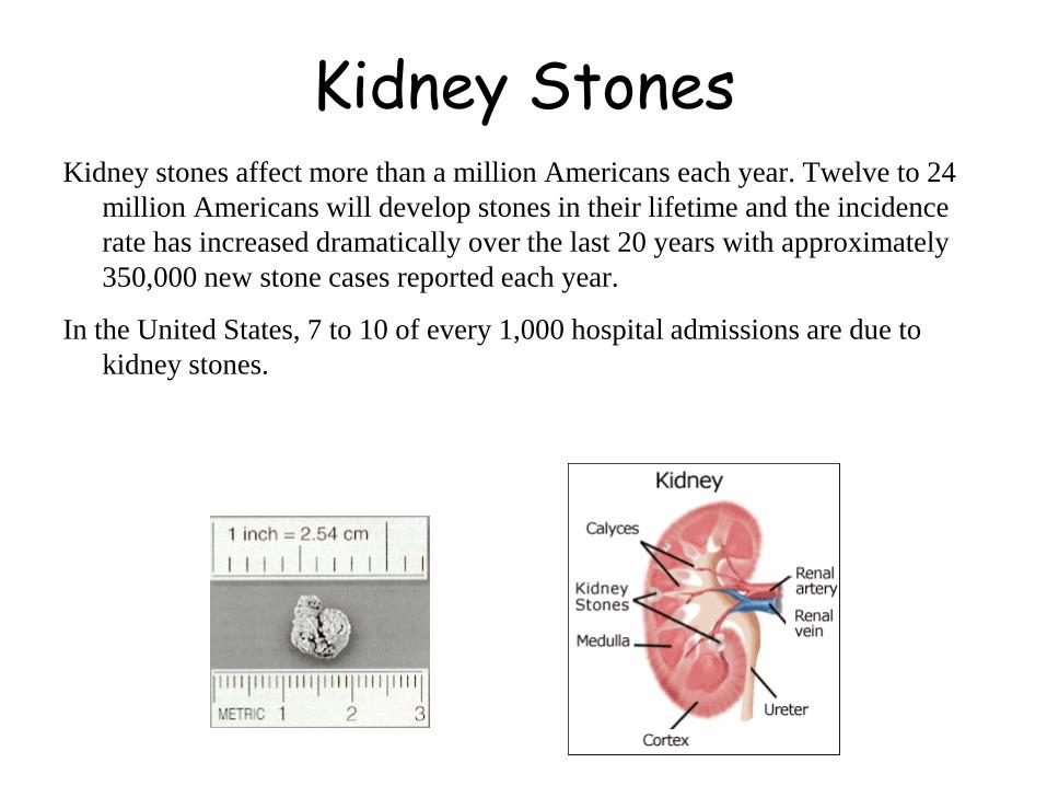
Kidney Stones Kidney stones affect more than a million Americans each year. Twelve to 24
million Americans will develop stones in their lifetime and the incidence
rate has increased dramatically over the last 20 years with approximately
350,000 new stone cases reported each year.
In the United States, 7 to 10 of every 1,000 hospital admissions are due to
kidney stones.

Kidney stones are solid masses of mineral salt deposits that are normally filtered through the kidney and voided in urine. Urine naturally contains substances that dissolve the waste materials that form these solids or calculi. However, when the amounts of these salts are excessive, the urine may be unable to dissolve them all, leaving crystals that accumulate in the kidney and gradually increase in size.
The stones can be as small as a grain of sand or as large as a golf ball. The size, shape, and location of the stone can cause many different symptoms.

Most renal calculi are so small they are passed through the urinary tract without any symptoms. Larger calculi can obstruct the renal ducts, or become lodged in the ureters. These larger obstructions cause sharp, severe pain in the sides and back as they move through the urinary tract. In medical parlance, this condition is called renal colic.
Well, I guess you
don’t have kidney
stones after all.
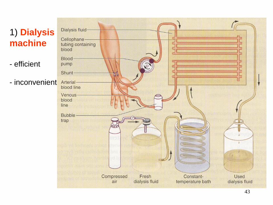
1) Dialysis
machine
- efficient
- inconvenient
43
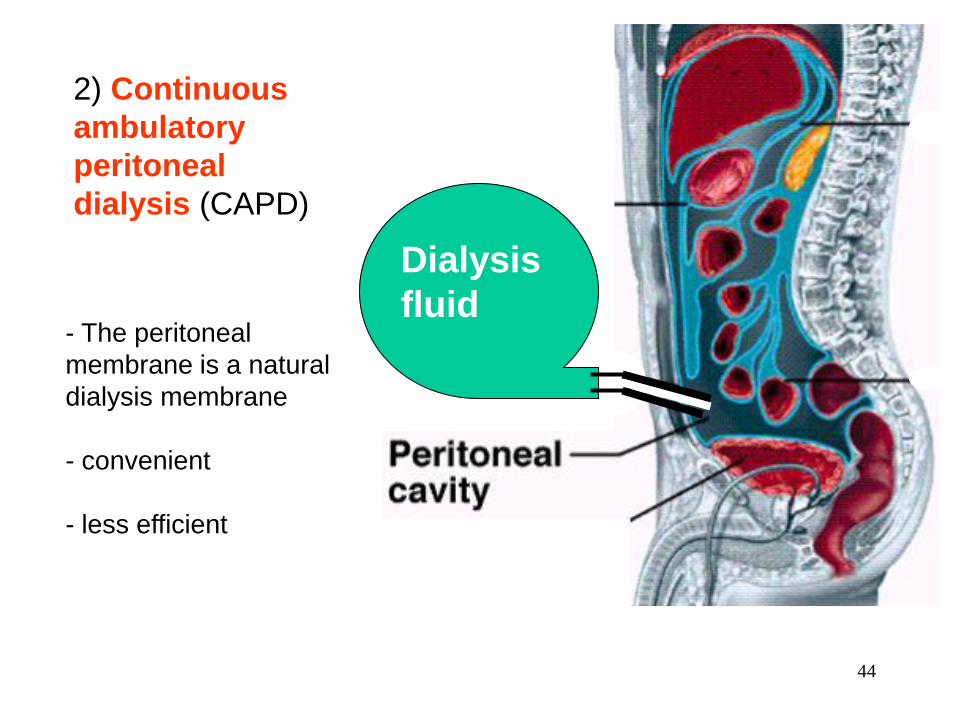
2) Continuous
ambulatory
peritoneal
dialysis (CAPD)
Dialysis
fluid - The peritoneal
membrane is a natural
dialysis membrane
- convenient
- less efficient
44

The Ureters
The ureters are muscular tubes leading from the
renal pelvis to the lower bladder.
45
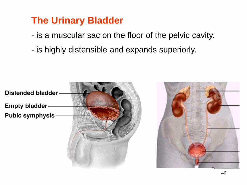
The Urinary Bladder
- is a muscular sac on the floor of the pelvic cavity.
- is highly distensible and expands superiorly.
46
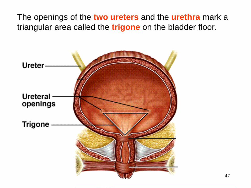
The openings of the two ureters and the urethra mark a
triangular area called the trigone on the bladder floor.
47

The Urethra
- conveys urine from the urinary bladder to the
outside of the body.
Females male
3-4 cm ~18 cm
greater risk of
urinary tract
infections
48

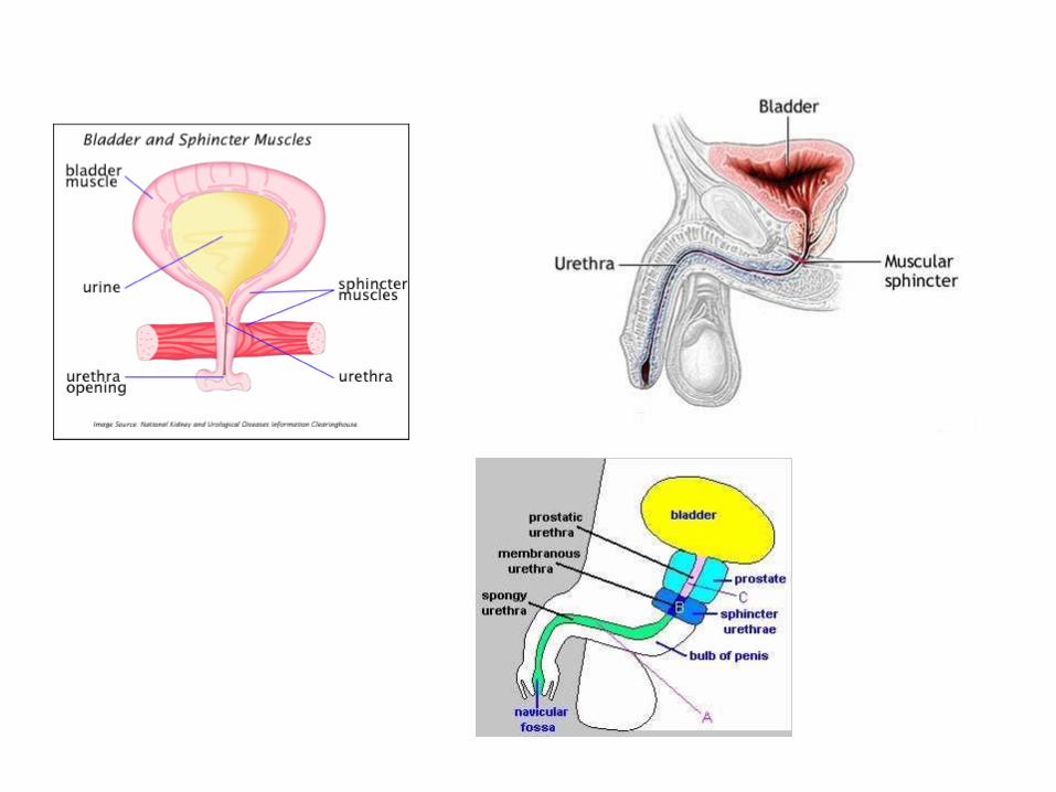
The male urethra has three
regions:
1) prostatic urethra
2) membranous urethra
3) penile urethra.
Difficulty in voiding urine
with enlarged prostate 49

In both sexes:
- internal urethral sphincter- under involuntary control.
- external urethral sphincter - under voluntary control
internal urethral sphincter
external urethral sphincter
50

Spinal
cord
Voiding Urine in infants
micturition reflex When the bladder contains about 200 ml of urine, stretch receptors in the wall send
impulses to the spinal cord. Parasympathetic signals return to stimulate contraction
of the bladder and relaxation of the internal urethral sphincter.
51
2. Once voluntary control has developed, emptying of the bladder
is controlled predominantly by a micturition center in the pons. This
center receives signals from stretch receptors and integrates this
information with cortical input concerning the appropriateness of
urinating at the moment. It sends back impulses to stimulate
relaxation of the external sphincter.
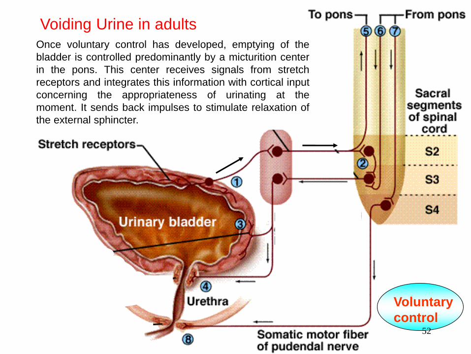
Once voluntary control has developed, emptying of the
bladder is controlled predominantly by a micturition center
in the pons. This center receives signals from stretch
receptors and integrates this information with cortical input
concerning the appropriateness of urinating at the
moment. It sends back impulses to stimulate relaxation of
the external sphincter.
Voiding Urine in adults
Voluntary
control 52

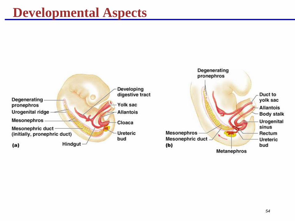
Developmental Aspects
Three sets of embryonic kidneys develop, with only
the last set persisting
The pronephros never functions but its pronephric
duct persists and connects to the cloaca
The mesonephros claims this duct and it becomes
the mesonephric duct
The final metanephros develop by the fifth week and
develop into adult kidneys
53
Developmental Aspects
54
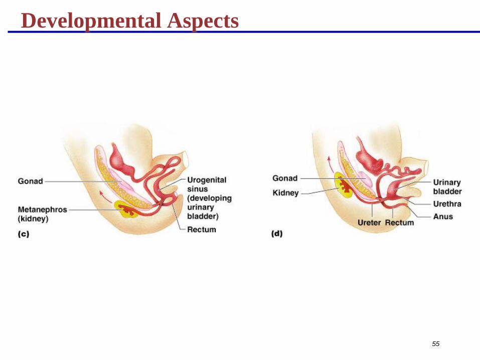
Developmental Aspects
55
![7 Catheter-associated Urinary Tract Infection (CAUTI) · UTI Urinary Tract Infection (Catheter-Associated Urinary Tract Infection [CAUTI] and Non-Catheter-Associated Urinary Tract](https://img.dokumen.tips/doc/110x75/5c40b88393f3c338af353b7f/7-catheter-associated-urinary-tract-infection-cauti-uti-urinary-tract-infection.jpg)