Embed Size (px)
Citation preview

The Urinary System
Part 1- Rat Dissection

Pre-lab continued
• Prepare the dissection tray and equipment.• Put on your gloves.• Retrieve your rat from the tray in your cabinet. • Lay the rat in the dissection tray ventral side up.• If you have not removed the digestive organs from
the abdominal cavity do so at this time.• Carefully remove any fat in the lower pelvic region.

Pre-lab
• Open the shared Google Presentation- “Urinary System Dissection”
• Make a copy and name it: Urinary system / Hour ? / Names
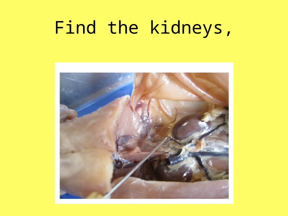
Find the kidneys,

adrenal gland,

blood vessels,
9
10
This rat was injected with colored latex to help students view the structures of the circulatory system.
Recall that red symbolizes oxygenated blood and blue symbolizes deoxygenated blood.

non-injected blood vessels
1211

Ureters,
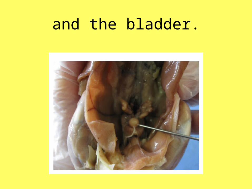
and the bladder.

Cut bladder Take a picture(s).
Insert the picture(s) into the Google presentation.
Label each structure and state the function.
Consider animating your slides.

Spray your rat with preservative and put it back in the bag, squeezing out the air.

Part 2
Sheep Kidney
• Obtain a preserved sheep kidney and rinse it with water to remove as much of the preserving fluid as possible.

Observe the Renal Capsule,
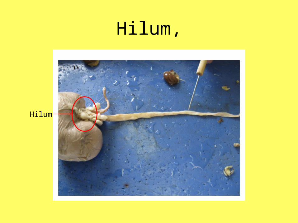
Hilum,
Hilum

Ureter,

Renal Blood Vessel,

• Take a picture of the external features of the kidney.
• Download the picture into the Google Presentation.
• Label each structure and answer any questions requested.
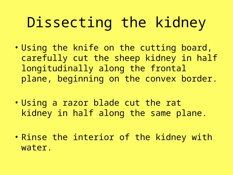
Dissecting the kidney
• Using the knife on the cutting board, carefully cut the sheep kidney in half longitudinally along the frontal plane, beginning on the convex border.
• Using a razor blade cut the rat kidney in half along the same plane.
• Rinse the interior of the kidney with water.

Observe the Renal Cortex and Renal Medulla,
Renal cortex
Renal medulla

Renal Pyramid,

• Stop and take a picture of the internal features of the kidney.

• To see the next three structures, remove the renal medulla from the kidney.

Find the Minor Calyx,

Major Calyx,

And Renal Pelvis.

• Stop and take another picture of the internal structures.
• Insert the needed pictures into the Google Presentation, labeling structures and answering questions.
• Watch the rest of this presentation.

This rat has prostate cancer!

Throw away the sheep kidney.
Clean and dry any instruments you used.
Share your presentation with me as soon as you are done but no later than 7pm tonight.