Embed Size (px)
Citation preview
The Urinary System

Objectives: Describe location of kidneys in body
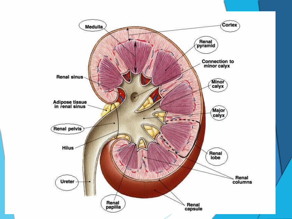
Identify the following regions of a kidney: hilum, cortex, medulla, medullary pyramids, calyces, pelvis, and renal columns
Recognize a nephron and describe its anatomy
Describe process of urine formation, identify areas of the nephron that are responsible for filtration, reabsorption and secretion
Describe the function of the kidneys in excretion of nitrogen-containing wastes
Define polyuria, anuria, oliguria, and diuresis
Describe the composition of normal urine
List abnormal urinary components

Helpful Word Parts:
-Continence …… to hold
Neph- …… kidney
Pyel …… renal pelvis
Ren- …… kidney
-ur- …… urine

Location & Structure
Soap
Extend from T12 to L3
Small, dark red organs
Kidney-bean shape
Lie against dorsal body wall

Basic Function
Maintain the purity and constancy
Bear the major responsibility of excreting nitrogenous wastes, toxins and drugs from the body
Regulate the blood’s volume and chemical makeup
Produces enzymes renin and erythropoietin
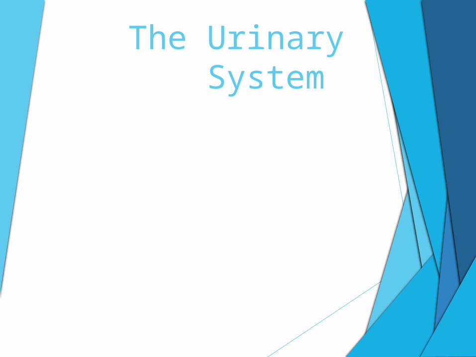
Simplified Structure: Medial indentation - renal hilum
Ureters, renal blood vessels and nerves
Adrenal gland
A transparent fibrous capsule
The perirenal fat capsule surrounds each kidney
The renal fascia (outermost capsule) anchors the kidney
Homeostatic Imbalance
The fat surrounding kidneys is vital
If the fat diminish kidneys may drop to a lower position called ptosis
Ptosis becomes a problem if the ureters, -which do what again?- become kinked
Once kinked, the urine can no longer pass through, causing back up and pressure on the kidney tissue called hydronephrosis
More On Structure
Renal cortex (cortex – bark)
Renal medulla: has many triangular regions with a striped appearance
Renal or medullary pyramids: triangular regions with striped appearance
Separated by renal columns
Renal pelvis


Structure Continued…
Calyces: extensions of the pelvis,
Collect urine which constantly drains from the pyramids
Drain it into the renal pelvis.
Renal pelvis to the ureter
Bladder for temporary storage

Blood Supply
25% each minute
The arterial supply = renal artery
Divides into segmental arteries
Interlobar arteries
Interlobar arteries give off the arcuate arteries
The arcuate arteries then branch off into cortical radiate arteries
Venous blood draining from the kidney flowes through veins that trace the pathway, but run in an opposite direction: cortical radiate veins to arcuate veins to interlobar viens to the renal vein
Nephrons & Urine Formation
Over a million nephrons
Nephrons are the structural and functional units of kidneys
Two main structures: a glomerulus and a renal tubule
A glomerulus is a knot of capillaries
Has a closed end which is enlarged and cup-shaped to completely surround the glomerulus
Called the glomerular (glom = little ball)
The inner layer of the tubule is made of highly modified octopus-like cells called podocytes.


Podocytes
Long branching processes….foot processes
Intertwine and cling to the glomerulus
“Filtration slits” between extensions
Podocytes form a porous membrane around the glomerulus
As the tubule extends, it coils and twists before forming a hairpin loop


More on Podocytes…
Coils and twists again
Collecting tubule called the collecting duct
These regions of the tubule have specific names:
• Proximal convoluted tubule (PCT)
• Loop of Henle
• Distal convoluted tubule
- The lumen surfaces (exposed to filtrate) of the tubule cells in the proximal convoluted tubules are covered with dense microvilli, which increase surface area

Nephrons:
Cortical nephrons are located almost entire in the cortex
Few times nephrons are called juxtamedullary nephrons which are located close to the cortex-medulla junction
- Their loops of Henle dip deep into the medullary pyramids
- This is what gives the pyramids their striped appearance
- Deliver the final urine product into the calyces and renal pelvis

More On Nephrons
Associated with two capillary beds:
The glomerular. Fed and drained by arterioles.
The afferent arteriole is the feeder vessel
The efferent arteriole receives blood
The glomerular is specifically different from other capillary beds

How?
Both fed and drained by arterioles
Diameter of afferent arteriole > diameter of efferent arteriole
Blood pressure is higher in the glomerular capillaries
High pressure forces fluid and solutes out of the blood
99% of this filtrate is reclaimed by the renal tubule cells and returned to the blood

Capillary Beds
Peritubular capillaries arise from the efferent arteriole that drains the glomerulus
Capillaries are low-pressure, porous vessels that are adapted for absorption
Cling closely to the whole length of the renal tubule
Receive solutes and water from the tubule cells
These substances are reabsorbed from the filtrate
The peritubular capillaries drain into interlobular veins leaving the cortex

Urine Formation (1 of 3) Glomerular filtration
a) Nonselective, passive process - fluid passes from blood into glomerular capsule
b) The fluid is then filtrated
c) Both proteins & blood cells = to large
d) Systemic blood pressure is normal, filtrate will be formed
e) If arterial blood pressure drops too low, glomerular pressure will also
Urine Formation (2 of 3)Tubular Reabsorption
a) Begins as soon as filtrate enters the proximal convoluted tubule
b) Tubule cells act as transporters
c) Absorbed into the capillary blood by active transport processes
d) Some reabsorption is done passively (Water – osmosis)
e) Active transport involves membrane carriers which are very selective
f) Abundance of carriers for substances that will be reused
g)Needed substances will be entirely removed from the filtrate
h)Nitrogenous waste products are poorly reabsorbed
i) Urea which is formed by the liver and an end product of protein breakdown
j) Uric acid is released when nucleic acids are metabolized
k) Creatinine associated with creatinine metabolism in muscle tissue
l) These tend to be found in high concentrations in urine
m) Most reabsorption occurs in the proximal convoluted tubules

Urine Formation (3 of 3)
Tubular secretion
a) The opposite of tubular reabsorption
b) Some substances move from the blood of the peritubular capillaries through the tubule cells
c) This process is important to rid the body of substances not previously in the filtrate

Homeostatic Imbalance
Oliguria – abnormally low urinary output
Between 100 and 400 ml per day
Anuria – extremely low urinary output
Less than 100 ml per day
Usually indicate glomerular blood pressure is too low to cause filtration
Anuria a can also be a result from transfusion reactions and acute inflammation of the kidney
Characteristics of Urine: -Filtrate contains everything that blood plasma does
except proteins
Urine remains, containing nitrogenous wastes and unneeded substances
Urochrome is a pigment
More solutes in urine call for a deeper yellow color
Urine may be strange colors at times
Sterile
pH is slightly acidic
Specific gravity is the term used to compare how much heavier urine is than distilled water
Chronic renal failure is a condition in which the kidney loses its ability to concentrate urine
Kidney inflammation is pyelonephritis

Homework:
Read pages 517-527
“Did you get it?” Questions 1-7 … In class
Self Test 15.1

Citations:
Marieb, E. N. (2009). Essentials of human anatomy & physiology. San Francisco, CA: Pearson Education, Inc.