Embed Size (px)
Citation preview

R E V I E W
The unfolding drama of flower development: recent results from genetic and molecular analyses H o n g M a
Cold Spring Harbor Laboratory, Cold Spring Harbor, New York 11724-2212 USA
There has been an explosion of information about flower development recently, largely because of genetic and molecular studies in Arabidopsis thaliana and Antirrhi- hum majus. A number of homeotic genes have been identified that regulate flower development, and models have been proposed for the specification of meristem and floral organ identities. Molecular cloning of many of these genes has allowed the testing of specific predic- tions of the models but also has led to modifications of a floral organ identity model. Furthermore, several of the floral genes contain a conserved region, the MADS box, which encodes a domain with striking sequence similar- ity with known transcription factors from human and yeast. Additional MADS box genes have been isolated from several plants; these genes are likely to play impor- tant regulatory roles during flower development. The ge- netic and molecular studies have uncovered many of the components of a complex network of regulatory proteins that directs flower development. Further characteriza- tion of these and other yet to be discovered components promises to contribute a great deal to our understanding of the mechanisms controlling flower development.
Flowering plants, like other land plants, have vegeta- tive organs such as roots, stems, and leaves, which ab- sorb nutrients and water from the soil, transport them to other parts of the plant, and synthesize organic com- pounds using the sun's energy. In addition, flowering plants produce elaborate reproductive structures, the flowers, which, following fertilization, become fruits and bear seeds. From the seasoned gardener to the casual observer, from the naturalist to the florist shopper, peo- ple have always been fascinated by the enormous variety of flowers, ranging from 2 mm to > 10 cm in length, covering the whole visual spectrum with their colors, and differing in the arrangement of flowers and the sym- metry within a flower. How do flowers develop? What genes regulate this complex process? In recent years, rapid advances have been made in addressing these ques- tions, largely as a result of genetic and molecular studies in two distantly related flowering plants, Arabidopsis thaliana, a relative of cauliflower and cabbage, and the snapdragon, Antirrhinum majus.
Postembryonic plant development is repetitive, be-
cause of the reiterative nature of the morphogenesis ini- tiated from the meristems, which are groups of undiffer- entiated progenitor cells. The meristem at the apex of the plant [or the tip of a steml is called the apical mer- istem. While the cells at the very summit of the dome- shaped apical meristem divide and maintain the mer- istem, the cells at the periphery of the apical meristem divide to give rise to additional meristems (e.g., of branches) or organ primordia, which are groups of cells with specified fates and which develop into different or- gans, such as leaves. The cells just below the apical mer- istem divide and differentiate to form the stem itself. Many plants, such as trees, have a recognizable domi- nant stem [trunk), with the primary apical meristem, and additional branches, each with a meristem at the tip. Other plants lack an obvious dominant stem and a pri- mary meristem, and have a bushy morphology. In Ara- bidopsis, the vegetative apical meristem produces leaves arranged in a spiral arrangement with very short dis- tances between successive leaves, forming a rosette (Fig.la). In contrast, the Antirrhinum vegetative apical meristem produces pairs of leaves opposite tO each other, and each pair is at right angle with the previous pair (Fig. lb; Coen and Meyerowitz 1991).
Flower development requires several steps (Meyerow- itz et al. 1991}. The first step is floral induction, which establishes a reproductive meristem(s). The reproductive meristem is often called an inflorescence meristem be- cause it gives rise to a series of flowers, called an inflo- rescence. The number of flowers in an inflorescence var- ies between species and ranges from several (determinate inflorescence) to indefinite (indeterminate inflores- cence). Both Arabidopsis and Antirrhinum have indeter- minate inflorescences (Fig. la, b). The second step is the formation of a floral meristem (Fig. lc), which is homol- ogous to a branch meristem, for a flower can be regarded as a very short stem with specialized organs. In some plants, floral induction results in the formation of a flo- ral meristem directly, and a single flower develops at the end of a stem. In Arabidopsis, the primary inflorescence meristem gives rise to, in a spiral, first a small number of cauline leaf primordia (Fig. la), each with an adjacent secondary inflorescence meristem, and then a large (in-
GENES & DEVELOPMENT 8:745-756 © 1994 by Cold Spring Harbor Laboratory Press ISSN 0890-9369/94 $5.00 745
Cold Spring Harbor Laboratory Press on January 5, 2019 - Published by genesdev.cshlp.orgDownloaded from
Ma
a
C
G
O
G PI
N _Fs.
Ip o
o
o
b e
TF
f g
B
c d h i 2 ~ I 1 SP pest Ca Ca ~ - ~ T F St ~a
C a - - s e • Br St ~ ~ ~ S F r
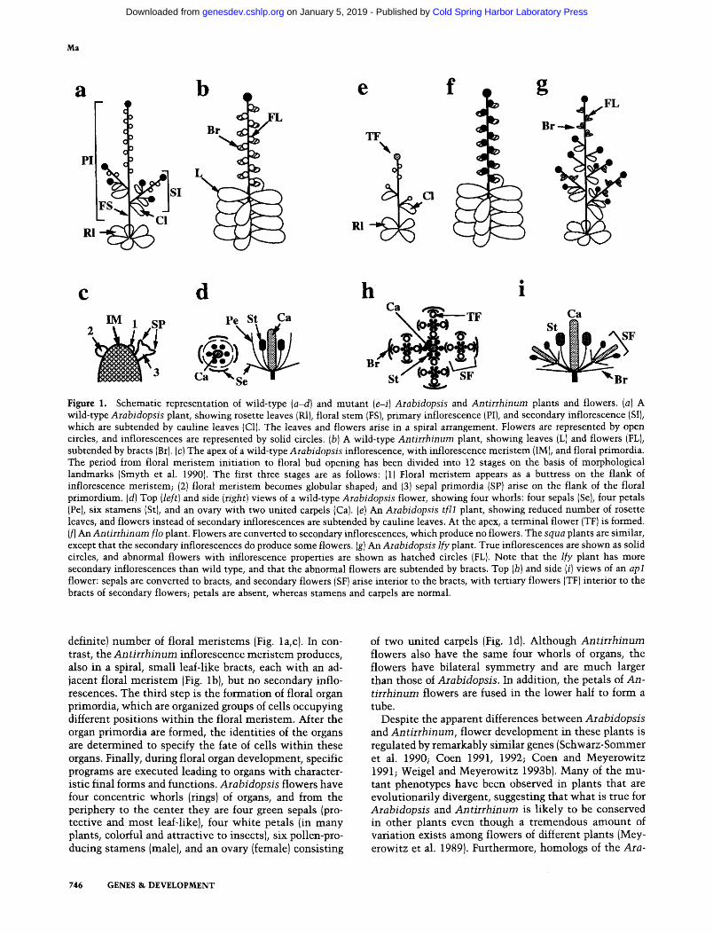
Figure 1. Schematic representation of wild-type (a-d} and mutant (e-i) Arabidopsis and Antirrhinum plants and flowers. (a) A wild-type Arabidopsis plant, showing rosette leaves (R1), floral stem (FS), primary inflorescence {PI), and secondary inflorescence (SI), which are subtended by cauline leaves (C1). The leaves and flowers arise in a spiral arrangement. Flowers are represented by open circles, and inflorescences are represented by solid circles. (b) A wild-type Antirrhinurn plant, showing leaves (L) and flowers (FL), subtended by bracts (Br). (c) The apex of a wild-type Arabidopsis inflorescence, with inflorescence meristem (IM}, and floral primordia. The period from floral meristem initiation to floral bud opening has been divided into 12 stages on the basis of morphological landmarks ISmyth et al. 1990). The first three stages are as follows: (1) Floral meristem appears as a buttress on the flank of inflorescence meristem; (2) floral meristem becomes globular shaped; and (3) sepal primordia (SP) arise on the flank of the floral primordium. (d) Top (left) and side (right) views of a wild-type Arabidopsis flower, showing four whorls: four sepals (Se), four petals (Pc), six stamens (St), and an ovary with two united carpels (Ca). (e) An Arabidopsis fill plant, showing reduced number of rosette leaves, and flowers instead of secondary inflorescences are subtended by cauline leaves. At the apex, a terminal flower (TF) is formed. (f) An Antirrhinum flo plant. Flowers are converted to secondary inflorescences, which produce no flowers. The squa plants are similar, except that the secondary inflorescences do produce some flowers. (g) An Arabidopsis lfy plant. True inflorescences are shown as solid circles, and abnormal flowers with inflorescence properties are shown as hatched circles (FL}. Note that the ify plant has more secondary inflorescences than wild type, and that the abnormal flowers are subtended by bracts. Top (h) and side (i) views of an apl flower: sepals are converted to bracts, and secondary flowers (SF) arise interior to the bracts, with tertiary flowers (TF) interior to the bracts of secondary flowers; petals are absent, whereas stamens and carpels are normal.
definite) number of floral mer is tems (Fig. la, c). In con- trast, the Antirrhinum inflorescence mer i s tem produces, also in a spiral, smal l leaf-like bracts, each wi th an ad- jacent floral mer i s tem (Fig. lb), but no secondary inflo- rescences. The third step is the formation of floral organ primordia, which are organized groups of cells occupying different positions wi th in the floral meristem. After the organ primordia are formed, the identities of the organs are determined to specify the fate of cells wi th in these organs. Finally, during floral organ development, specific programs are executed leading to organs wi th character- istic final forms and functions. Arabidopsis flowers have four concentric whorls (rings) of organs, and from the periphery to the center they are four green sepals (pro- tective and most leaf-like), four whi te petals (in many plants, colorful and attractive to insects), six pollen-pro- ducing s tamens (male), and an ovary (female) consisting
of two united carpels (Fig. ld). Although Antirrhinum flowers also have the same four whorls of organs, the flowers have bilateral symmet ry and are m u c h larger than those of Arabidopsis. In addition, the petals of An- tirrhinum flowers are fused in the lower half to form a tube.
Despite the apparent differences between Arabidopsis and Antirrhinum, flower development in these plants is regulated by remarkably similar genes (Schwarz-Sommer et al. 1990; Coen 1991, 1992; Coen and Meyerowitz 1991; Weigel and Meyerowitz 1993b). Many of the mu- tant phenotypes have been observed in plants that are evolutionarily divergent, suggesting that what is true for Arabidopsis and Antirrhinum is l ikely to be conserved in other plants even though a t remendous amount of variation exists among flowers of different plants (Mey- erowitz et al. 1989). Furthermore, homologs of the Ara-
746 GENES & DEVELOPMENT
Cold Spring Harbor Laboratory Press on January 5, 2019 - Published by genesdev.cshlp.orgDownloaded from

Genes controlling flower development
bidopsis and Antirrhinum floral genes have been identi- fied in many other plants, including the monocot maize (e.g., Pnueli et al. 1991; Mandel et al. 1992a; Kempin et al. 1993; Schmidt et al. 1993; van der Krol et al. 1993). This review summarizes recent genetic studies in Ara- bidopsis and Antirrhinum, and emphasizes recent re- sults from molecular analyses in these two plants, as well as some studies from a few other plants. These re- sults indicate that a complex network of regulators con- trols the formation of the inflorescence meristem and its transition to the floral meristem, the initiation of floral organ primordia, and the floral organ identities. The first three sections present brief descriptions of mutant phe- notypes. These are followed by discussions of genetic interactions based on double and triple mutant studies and on molecular analyses using cloned genes. Finally, the biochemical nature of many of the gene products and possible regulatory mechanisms is discussed. Because of limited space, morphological and physiological studies in other plants are not discussed here, nor are there de- tailed accounts of earlier genetic studies, for which the reader is referred to several excellent reviews (Meyerow- itz et al. 1989, 1991; Schwarz-Sommer et al. 1990; Coen 1991; Coen and Meyerowitz 1991). Other recent reviews offer different perspectives and emphasis (Coen 1992; Coen and Carpenter 1992; Jack et al. 1993; Weigel and Meyerowitz 1993b).
Prelude: floral i n d u c t i o n
Floral induction is regulated by developmental and en- vironmental factors (Bernier et al. 1993). In Arabidopsis, it seems that reproductive development is a default path- way and that normal vegetative growth occurs when the reproductive program is delayed as a result of inhibition. This inhibition is removed only when developmental and environmental signals are present, as indicated by genetic studies. The recessive mutant emf produces a single flower upon seed germination, without any vege- tative growth (Sung et al. 1992), suggesting that EMF is required for the inhibition of reproductive growth. Mu- tations in another gene, TFL1, cause a reduction of the number of vegetative (rosette) leaves, and an earlier than normal appearance of inflorescence (Shannon and Meeks-Wagner 1991; Schultz and Haughn 1993). This suggests that the TFL1 gene may enhance the inhibition of reproductive growth before the appropriate develop- mental stage, although this could be related to a negative role TFLI has in floral meristem initiation {Fig. le; also see below in Act 1).
Flower induction in Arabidopsis is influenced by the length of daylight and temperature (Bernier et al. 1993). Long days (14--16 hr light/8-10 hr dark) accelerate flow- ering as compared with short days (8-10 hr light/14--16 hr dark); furthermore, cold treatment promotes early flowering (Bemier et al. 1993). A group of mutants, known as late-flowering mutants, have been found to change the regulation of flowering by environmental conditions (Koomneef et al. 1991; Araki and Komeda 1993; Lee et al. 1993). Both long-day conditions and cold
reduce the delay of flowering in some, but not all, of the late flowering mutants. Biochemical studies (Burn et al. 1993) indicate that cold treatment causes demethylation of DNA and that the DNA demethylation may be in- volved in reducing the delay of flowering. It will be pos- sible in the near future to learn the molecular mecha- nisms of the late flowering gene functions, for efforts have begun in earnest to isolate and characterize the late flowering genes at the molecular level (e.g., Putterill et al. 1993).
Act 1: f loral m e r i s t e m i n i t i a t i o n
The characterization of Arabidopsis and Antirrhinum mutants have identified several genes that control floral meristem initiation (Table 1). The phenotypes of several mutants (leafy and apetalat in Arabidopsis, and flori- caula and squamosa in Antirrhinum; see below) suggest that the corresponding wild-type genes promote the tran- sition from inflorescence to floral meristems (Coen et al. 1990; Irish and Sussex 1990; Schultz and Haughn 1991; Huala and Sussex 1992; Huijser et al. 1992; Mandel et al. 1992b; Weigel et al. 1992; Bowman et al. 1993). In the Antirrhinum floricaula (rio) mutants (Fig. If), floral mer- istems fail to form; secondary inflorescence meristems form instead. Similarly, in the Arabidopsis leafy (lfy) mutants (Fig. lg), early flowers are replaced by secondary inflorescences. Although late Ify flowers do form, they are abnormal and subtended by bracts (not present in the wild type). These flowers resemble secondary inflores- cences in several ways: (1) The outer organs resemble cauline leaves, with occasional secondary flowers; (2) the distance between whorls is extended; and (3) the organ arrangement is partially spiral. The Arabidopsis AP- ETALA1 (AP1) and the Antirrhinum SQUAMOSA (SQUA) genes also act positively in the inflorescence-to- flower transition. The flowers of the apl-1 mutant (Fig. lh, i) contain secondary flowers interior to the first whorl organs and tertiary flowers interior to the first whorl organs of the secondary flowers, making each primary flower a miniature inflorescence (although the arrange- ment is whorled instead of spiral). Likewise, squa mu- tants produce inflorescence-like shoots instead of flow- ers, indicating that the mutants are deficient in floral meristem initiation; however, these secondary shoots do eventually produce some flowers.
The Antirrhinum FLO and SQ UA, and the Arabidop- sis LFY and API genes, have been isolated (Coen et al. 1990; Huijser et al. 1992; Mandel et al. 1992b; Weigel et al. 1992). FLO and LFY encode homologous proteins with proline-rich and acidic regions, both characteristics of transcription factors. The idea that LFY is a transcrip- tional regulator is further supported by its localization to the nucleus (Weigel and Meyerowitz 1993a). The AP1 and the SQUA genes were isolated independently and found homologous to each other; their gene products are members of a family with similarity to transcription fac- tors (see below in The Players). All four genes are ex- pressed in very early floral meristems, consistent with their roles in determining meristem identity (Coen et al.
GENES & DEVELOPMENT 747
Cold Spring Harbor Laboratory Press on January 5, 2019 - Published by genesdev.cshlp.orgDownloaded from
Ma
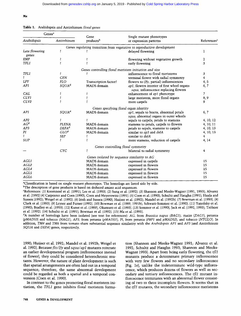
Table 1. Arabidopsis and Antirrhinum floral genes
Genes a Gene Single mutant phenotypes
Arabidopsis Antirrhinum products b or expression patterns References c
Genes regulating transition from vegetative to reproductive development Late flowering ~ ~ delayed flowering 1
genes EMF ? ? flowering without vegetative growth 2 TFL1 ? z early flowering 3
Genes controlling floral meristem initiation and size z inflorescence to floral meristems 3
terminal flower with radial symmetry 4 Transcription factor? flowers to (If)', partial) inflorescences 4, 5 MADS domain apl, flowers interior of first whorl organs 6, 7
squa, inflorescence replacing flowers enhancement of apl phenotype 7
z large meristem, more floral organs 8, 9 i more carpels 9
Genes specifying floral organ identity MADS domain apl, sepals to bracts, abnormal petals 6, 7
squa, abnormal organs in outer whorls z ~ sepals to carpels, petals to stamens 4, 10, 12 PLENA MADS domain stamens to petals, carpels to flowers 4, 10, 11 DEFA a MADS domain petals to sepals, stamens to carpels 4, 10, 13 GLO d MADS domain similar to ap3 and defA 4, 10, 13 SEP ~ similar to defA 4
~ more stamens, reduction of carpels 4, 14
Genes controlling floral symmetry bilateral to radial symmetry 4
Genes isolated by sequence similarity to AG MADS domain expressed in carpels 15 MADS domain expressed in flowers 15 MADS domain expressed in flowers 15 MADS domain expressed in flowers 15 MADS domain expressed in flowers 15
TFL1 ? CEN
LFY FLO AP1 S Q U A d
CAL 2 CLV1 z CL V2
AP1
AP2 A G d AP3 PI ?
SUP
AGL1 AGL2 AGL4 AGL5 AGL6
SQUA d
CYC
aClassification is based on single mutant phenotypes. The homologs are listed side by side. bThe description of gene products is based on deduced amino acid sequences. CReferences: {1) Koornneef et al. {1991); Lee et al. {1993). {2} Sung et al. (1992). {3) Shannon and Meeks-Wagner (1991, 1993); Alvarez et al. (1992) (4) Carpenter and Coen {1990); Coen and Meyerowitz (1991). (S) Coen et al. (1990); Schultz and Haughn (1991); Huala and Sussex (1992); Weigel et al. (1992). (6) Irish and Sussex (1990); Huijser et al. (1992)~ Mandel et al. (1992b). (7) Bowman et al. [1993). {8) Clark et al. {1993). (9) Leyser and Furner {1992). (10) Bowman et al. {1989, 199 l b); S chwarz-Sommer et al. (1990). (11) Yanofsky et al. (1990}; Bradley et al. (1993). (12) Kunst et al. (1989); Okamuro et al. (1993). {13) Sommer et al. (1990); Jack et al. (1992, 1993); Tr6bner et al. (1992). (14) Schultz et al. (1991); Bowman et al. (1992). (15) Ma et al. (1991). dA number of homologs have been isolated (see text for references): AG, from Brassica napus (BAG1), maize (ZAG1), petunia (pMADS3) and tobacco (NAG1); AP3, from petunia (pMADS1); PI, from petunia (FBP1 and pMADS2), and tobacco (NTGLO). In addition, TM4 and TM6 from tomato share substantial sequence similarity with the Arabidopsis AP1 and AP3 {and Antirrhinum SQUA and DEFA) genes, respectively.
1990; Huijser et al. 1992; Mande l et al. 1992b; Weigel et al. 1992). Because f l o / l f y and s q u a / a p l m u t a n t s rei terate an earlier deve lopmenta l p rogram (inflorescence instead of flower), they could be considered he terochronic mu- tants. However , the na tu re of plant deve lopment is such tha t spatial a r r angement s are of ten laid out in a tempora l sequence; therefore, the same abnormal deve lopment could be regarded as both a spatial and a tempora l con- vers ion (Coen et al. 1990).
In contras t to the genes p romot ing floral m e r i s t e m ini- t iat ion, the TFL1 gene inhibi ts floral m e r i s t e m forma-
t ion (Shannon and Meeks-Wagner 1991; Alvarez et al. 1992; Schultz and H a u g h n 1993; Shannon and Meeks- Wagner 1993). Apart f rom being early flowering, the f i l l m u t a n t s produce a de te rmina te p r imary inf lorescence wi th very few flowers and no secondary inf lorescence (Fig. le), unl ike the inde te rmina te wi ld- type inflores- cence, wh ich produces dozens of f lowers as wel l as sec- ondary and ter t iary inflorescences. The t f l l m u t a n t in- f lorescence t e rmina tes wi th an abnorma l f lower consist- ing of two to three incomple te flowers. It seems tha t in the f i l l mutan t s , the secondary inf lorescence m e r i s t e m s
748 GENES & DEVELOPMENT
Cold Spring Harbor Laboratory Press on January 5, 2019 - Published by genesdev.cshlp.orgDownloaded from

Genes controlling flower development
are converted to floral meristems and that the primary inflorescence meristem is converted to two or three closely spaced floral meristems.
Act 2: organ primordia initiation and identity
Several Arabidopsis homeotic genes have been identified that affect the identity of floral organs, including AP1, APETALA2 (AP2), APETALA3 (AP3), PISTILLATA (PI), and AGAMOUS (AG)(Table 1). Each of these genes con- trois the identity of two adjacent whorls of floral organs. Both the apl and ap2 mutations affect the organ identity in the outer two whorls, with severity of the phenotype dependent on the particular apl or ap2 mutant alleles. The strong apl-1 mutant has a sepal-to-bract conversion in the first whorl, whereas weaker apl mutants have abnormal petals. In plants with one of the most severe ap2 alleles, the first and second whorl organs, if present, are converted from sepals and petals to carpels and sta- mens, respectively. Weaker alleles of ap2 lead to conver- sion of sepals to leaves and petals to petaloid stamens. The AP3 and PI genes are both required for correct sec- ond and third whorl organ types; ap3 and pi mutants have conversion of petals to sepals and stamens to car- pels. The AG gene is required for determining the iden- tities of stamens and the carpels. In ag mutant flowers, the six stamens are converted to petals and the ovary is replaced by a second ag flower, such that the pattern of sepals, petals, and petals is repeated more than five times. The ag mutant phenotype indicates that AG func- tion is also required for the maintenance of a determi- nate floral meristem. That the number of floral organs is altered in ap2 and ag mutants indicates that these genes also function in regulating organ primordia initiation.
Mutations affecting organ identity have also been iso- lated in other plants {Meyerowitz et al. 19891, particu- larly in A. majus (Table 1). Many of the Antirrhinum mutations cause homeotic conversions similar to those found in Arabidopsis. The deficiens (defA) and globosa (glo) mutants have similar phenotypes to the Arabidop- sis ap3 and pi mutants, and so do mutants of another Antirrhmum gene, SEPALOIDEA (SEP). The Antirrhi- num plena mutant is similar to the Arabidopsis ag mu- tant, with conversion of stamens to petals, and carpels to additional floral organs. In petunia, the blind mutant produces flowers with sepals exhibiting carpel features and conversion of petals (corolla) to staminoid organs (Tsuchimoto et al. 1993), similar to the flowers of weak Arabidopsis ap2 mutants. Another petunia floral mu- tant, green petal (gp), has flowers with the petals con- verted to sepals (van der Krol et al. 1993).
Other mutants have flowers with altered numbers of organs. For example, the Arabidopsis superman (sup) mutant flowers are normal in the three outer whorls but produce more whorl(s) of stamens at the expense of the ovary (Schultz et al. 1991; Bowman et al. 1992). Studies of sup ap3 or sup pi double mutants suggest that SUP negatively regulates AP3 and PI function in the fourth whorl. The Arabidopsis clavatal (clvl) and clavata2 (clv2) mutant plants have ovaries consisting of more
than the normal two carpels (Koornneef 1987). Recent studies indicate that clvl mutants have a larger apical meristem and increased numbers of floral organs in all four whorls (Leyser and Furner 1992; Clark et al. 1993). Finally, mutations in the TOUSLED (TSL) gene cause a reduction in the number of floral organs in all whorls, as well as abnormal leaf morphology (Roe et al. 1993).
The dialogues: genetic interactions
Interactions controlling floral meristem formation
Double mutant analyses indicate that the meristem identity genes of Arabidopsis, LFY and AP1, interact with each other to control floral meristem formation {Fig. 2). The lfy apl double mutant has a more complete conversion of floral to inflorescence meristem than ei- ther single mutant, suggesting that these genes function in parallel pathways to promote floral meristem forma- tion and that these pathways are partially redundant. Similar studies indicate that the Antirrhinum FLO and SQUA genes also act in parallel pathways (Coen 1992). This functional parallel is further supported by the ob- servations that LFY (FLO) is expressed in the apl (squa) mutant and that SQUA gene is expressed in the flo mu- tant (Huijser et al. 1992; Weigel et al. 1992). In addition, other genes contribute to the process of floral meristem initiation (Fig. 2). A variant allele of the Arabidopsis gene, CAULIFLOWER (CAL), was found to enhance the apl mutant phenotype, although the cal-1 allele alone has no phenotype, nor does the cal-1 allele enhance the
Inflorescence Meristem
T ; Inflorescence ~ Floral Meristem I Meristem
Figure 2. Control of transition from inflorescence to floral meristem in Arabidopsis. The meristem identity genes LFY and AP1 are two main players promoting floral meristem initiation, whereas AP2 and CAL have little or no effect on meristem identity in the wild-type LFY AP1 background. However, cal enhances the effect of apl, and ap2 enhances those of both lfy and apl, on meristem identity. In addition, AP1 and CAL to- gether positively affect the expression of LFY and AP1. The box around these four genes indicates the positive genetic interac- tions and/or functional redundancy among these genes. CLV1 also promotes the transition to floral meristem. TFL1 promotes inflorescence meristem and antagonizes LFY, AP1, and AP2. Based on information from the following references, Alvarez et aI. {1992); Huala and Sussex (1992); Weigel et al. (1992}; Bow- man et al. (19931; Clark et al. (1993}; Okamuro et al. (1993); Schultz and Haughn {1993); Shannon and Meeks-Wagner (1993); and Weigel and Meyerowitz (1993c). (--*) Positive interaction and {'qJ negative interaction.
GENES & DEVELOPMENT 74.9
Cold Spring Harbor Laboratory Press on January 5, 2019 - Published by genesdev.cshlp.orgDownloaded from

Ma
phenotype of a strong lfy mutant (Bowman et al. 1993). In the apl mutant background, CAL has been shown to positively affect the expression of both the Arabidopsis LFY and AP1 genes. It is possible that in the wild type, the combination of both LFY and AP1 activities is in excess of the minimal requirement (threshold) for floral meristem formation, and CAL is not necessary. In apl mutants, the total activity for floral meristem is reduced to near the threshold, and further reduction due to a defect in CAL function manifests as a more severe phe- notype than those of the apl single mutants. Double mutant analyses also revealed that the organ identity gene AP2 contributes to meristem identity. Although ap2 single mutants show no defect in floral meristem formation, ap2 mutations enhance the apl or lfy mutant phenotypes, leading to a more complete conversion of floral to inflorescence meristems. One explanation is that AP2 is required for floral meristem in the absence of LFY or AP1 activities; this is consistent with the finding that AP2 is also expressed at stages before floral organ formation. Finally, clvl mutations also enhance the phe- notypes of lfy and lfy apl mutants, indicating that CLV1 is also involved in determining meristem identity. These double and triple mutant analyses indicate that although LFY and AP1 are the major functions that promote floral meristem formation, CAL, AP2, and CLV1 also contrib- ute to this process.
Contrary to these positively acting genes, the Arabi- dopsis TFL1 gene inhibits floral meristem development. Moreover, on the basis of double mutant studies it was proposed that TFL1 antagonizes LFY, AP1, and AP2 (Fig. 2). This is supported by the observation that the expres- sion of LFY and AP1 expands in a tfll mutant to include the meristems occupying the positions of the wild-type inflorescence meristems (Weigel et al. 1992; Bowman et al. 1993; Gustafson-Brown et al. 1994). Although apl mutations do not, lfy mutations partially suppress till mutant phenotypes, especially under short-day condi- tions (Alvarez et al. 1992; Bowman et al. 1993; Schultz and Haughn 1993; Shannon and Meeks-Wagner 1993). Furthermore, combinations of lfy and apl, or lfy and ap2, mutations clearly suppress tfll meristem phenotypes {Schultz and Haughn 1993).
At least part of the function of the Arabidopsis mer- istem identity genes LFY and AP1 is to regulate the ex- pression of floral organ identity genes (Weigel and Mey- erowitz 1993a), bridging these two stages of flower de- velopment. Analyses of the expression patterns of AP3, PI, or AG in lfy and apl single or double mutants indi- cate that LFY and AP1 synergistically activate the ex- pression of AP3 and PI, and regulate AG expression pat- tern (Weigel and Meyerowitz 1993a). These results are consistent with the finding that the Arabidopsis LFY and AP1 genes are expressed in developing flowers during floral organ primordia formation, in addition to being expressed in earlier stages of floral meristems (Mandel et al. 1992b; Weigel et al. 1992). Likewise, the Antirrhinum FLO and SQUA genes are also expressed in early floral primordia at the time of organ primordia initiation (Coen et al. 1990; Huijser et al. 1992). On the basis of the tran-
sient expression of FLO in whorls 1, 2, and 4, it was proposed that FLO activates some of the organ identity genes sequentially (Coen et al. 1990). It seems that the meristem identity genes function to set up the stage on which the floral homeotic genes play their roles in con- trolling the organ identities (see Fig. 3 and discussion below).
Models for the specification of floral organ identity
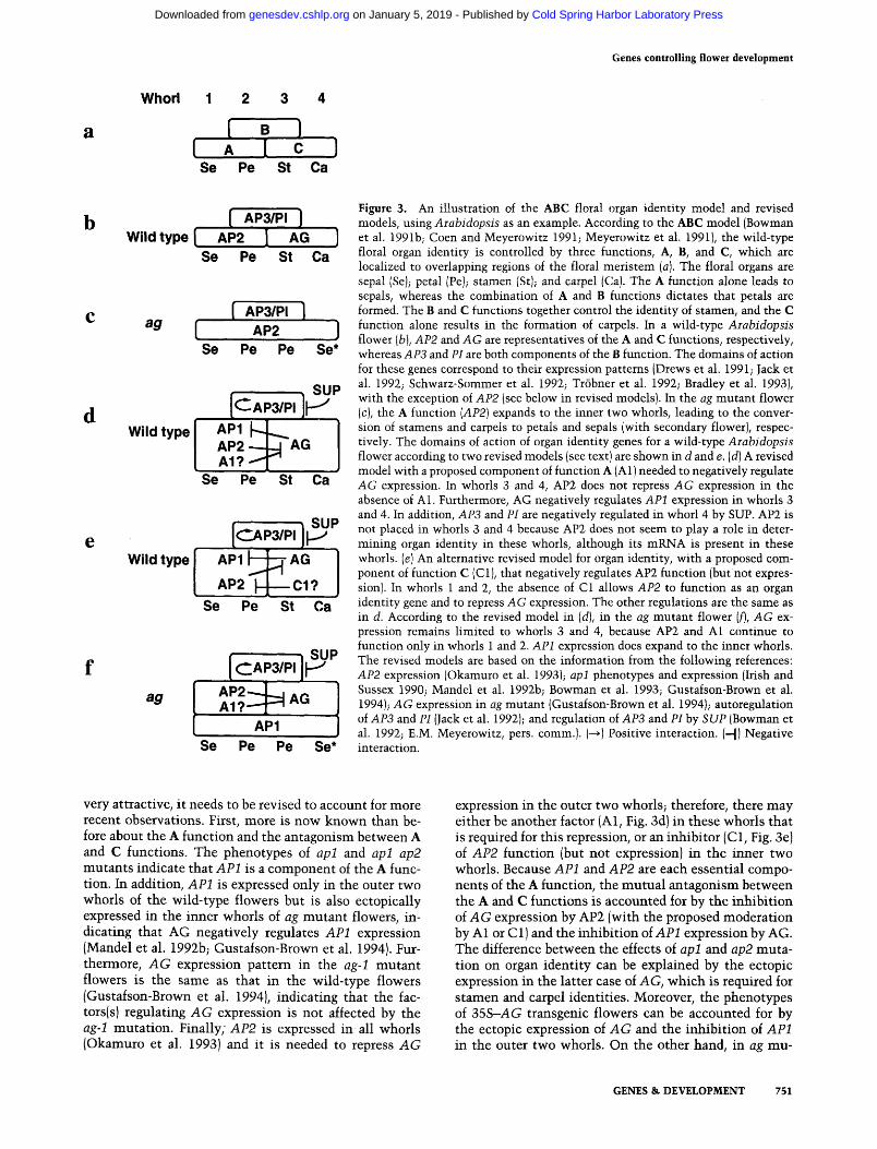
A model {the ABC model, Fig. 3a) for the determination of floral organ identity was proposed on the basis of ge- netic analyses in Arabidopsis and Antirrhinum. The Ar- abidopsis and Antirrhinum mutant phenotypes indicate that AP2 is essential for the A function, that the Arabi- dopsis AP3 and H genes (DEFA and GLO from Antirrhi- num) are components of the B function, and that the Arabidopsis AG gene {PLENA from Antirrhinum) is a necessary part of the C function, as shown in Fig. 3b. The proposed domains of gene action are supported by the expression patterns of several homeotic genes, including the Arabidopsis AG and AP3, and the Antirrhinum DEFA, GLO, and PLENA genes. An important aspect of this model is that the A and C functions antagonize each other, such that A inhibits C in whorls 1 and 2, and C inhibits A in whorls 3 and 4. This antagonism is sup- ported by the fact that the expression of AG and its pe- tunia homolog (pMADS3) expands outward to all floral whorls in the Arabidopsis ap2 and the petunia blind mutant flowers, respectively. Moreover, ectopic expres- sion of AG or the AG homologs from Brassica, petunia, and tobacco in the outer whorls of transgenic flowers results in ap2-1ike flowers, and a similar ectopic expres- sion of the PLENA gene in Antirrhinum leads to the ovulata {ap2-1ike} mutant phenotype (Mandel et al. 1992a; Mizukami and Ma 1992; Bradley et al. 1993; Kempin et al. 1993; Tsuchimoto et al. 1993). The A-C antagonism also predicts that in the ag mutant, the A function would expand to whorls 3 and 4 and inhibit AG expression (Fig. 3b).
The ABC model is similar to an earlier model based on mutant analysis in Antirrhinum, which proposed that there are two functions (equivalent to B and C of the ABC model) but no equivalent to the A function (Schwarz-Sommer et al. 1990}. This model can be recon- ciled with the ABC model because A and C are mutually exclusive and A is equivalent of the absence of C. Be- cause the expression of the Antirrhinum C function gene PLENA has now been shown to be negatively regulated in whorls 1 and 2 (Bradley et al. 1993), the yet to be identified negative regulator may be an A function com- ponent in Antirrhinum. Other models for organ identity were also proposed in which the identity of an inner (later) whorl of organs was dependent on the organ iden- tity of adjacent outer whorl{s) (Heslop-Harrison 1963; Green 1988). Because many mutants exist that show nor- mal organs in some whorls and abnormal organs in the next whorl or vice versa, these sequential models are not consistent with the genetic results.
Although the simple ABC model for organ identity is
750 GENES & DEVELOPMENT
Cold Spring Harbor Laboratory Press on January 5, 2019 - Published by genesdev.cshlp.orgDownloaded from
Genes controlling flower development
Whorl 1 2 3 4
[ . ! [ A I c
Se Pe St Ca
b
d
e
Wild type I
ag [
Wild type
Wild type
ag
[ AP3/PI ] AP2 I AG ]
So Pe St Ca
Se
[ AP3/PI ] AP2 ]
Pe Pe Se*
AP1 ~ AP2 AG AI?
Se Pe St Ca
• SUP C.JpaIPa
AP1 ~/____AG
AP2 C1? Se Pe St Ca
, SU ,P
A I ? - . - - ~ AG
AP1 Se Pe Pe Se*
Figure 3. An illustration of the ABC floral organ identity model and revised models, using Arabidopsis as an example. According to the ABC model (Bowman et al. 1991b; Coen and Meyerowitz 1991; Meyerowitz et al. 1991), the wild-type floral organ identity is controlled by three functions, A, B, and C, which are localized to overlapping regions of the floral meristem (a). The floral organs are sepal (Se); petal (Pe); stamen (St); and carpel (Ca). The A function alone leads to sepals, whereas the combination of A and B functions dictates that petals are formed. The B and C functions together control the identity of stamen, and the C function alone results in the formation of carpels. In a wild-type Arabidopsis flower (b), AP2 and AG are representatives of the A and C functions, respectively, whereas AP3 and PI are both components of the B function. The domains of action for these genes correspond to their expression patterns (Drews et al. 1991; Jack et al. 1992; Schwarz-Sommer et al. 1992; Tr6bner et al. 1992; Bradley et al. 1993), with the exception of AP2 (see below in revised models). In the ag mutant flower (c), the A function (AP2) expands to the inner two whorls, leading to the conver- sion of stamens and carpels to petals and sepals (with secondary flower), respec- tively. The domains of action of organ identity genes for a wild-type Arabidopsis flower according to two revised models (see text) are shown in d and e. (d) A revised model with a proposed component of function A (A1) needed to negatively regulate AG expression. In whorls 3 and 4, AP2 does not repress AG expression in the absence of A1. Furthermore, AG negatively regulates AP1 expression in whorls 3 and 4. In addition, AP3 and PI are negatively regulated in whorl 4 by SUP. AP2 is not placed in whorls 3 and 4 because AP2 does not seem to play a role in deter- mining organ identity in these whorls, although its mRNA is present in these whorls. (e) An alternative revised model for organ identity, with a proposed com- ponent of function C (C1), that negatively regulates AP2 function {but not expres- sion). In whorls 1 and 2, the absence of C1 allows AP2 to function as an organ identity gene and to repress AG expression. The other regulations are the same as in d. According to the revised model in (d), in the ag mutant flower (f), AG ex- pression remains limited to whorls 3 and 4, because AP2 and A1 continue to function only in whorls 1 and 2. AP1 expression does expand to the inner whorls. The revised models are based on the information from the following references: AP2 expression (Okamuro et al. 1993); apl phenotypes and expression (Irish and Sussex 1990; Mandel et al. 1992b; Bowman et al. 1993; Gustafson-Brown et al. 1994); AG expression in ag mutant (Gustafson-Brown et al. 1994); autoregulation of AP3 and PI (Jack et al. 1992); and regulation of AP3 and PI by SUP (Bowman et al. 1992; E.M. Meyerowitz, pers. comm.). (--~) Positive interaction. (-]) Negative interaction.
very attractive, it needs to be revised to account for more recent observations. First, more is now known than be- fore about the A function and the antagonism between A and C functions. The phenotypes of apI and apI ap2 mutants indicate that AP1 is a component of the A func- tion. In addition, AP1 is expressed only in the outer two whorls of the wild-type flowers but is also ectopically expressed in the inner whorls of ag mutan t flowers, in- dicating that AG negatively regulates AP1 expression (Mandel et al. 1992b; Gustafson-Brown et al. 1994). Fur- thermore, A G expression pattern in the ag-1 mutan t flowers is the same as that in the wild-type flowers (Gustafson-Brown et al. 1994), indicating that the fac- tors(s) regulating A G expression is not affected by the ag-1 mutat ion. Finally; AP2 is expressed in all whorls (Okamuro et al. 1993) and it is needed to repress A G
expression in the outer two whorls; therefore, there m a y either be another factor (A1, Fig. 3d} in these whorls that is required for this repression, or an inhibitor [C1, Fig. 3e) of AP2 function (but not expression} in the inner two whorls. Because AP1 and AP2 are each essential compo- nents of the A function, the mutua l antagonism between the A and C functions is accounted for by the inhibit ion of A G expression by AP2 {with the proposed moderat ion by A1 or C 1) and the inhibition of API expression by AG. The difference between the effects of apl and ap2 muta- tion on organ identity can be explained by the ectopic expression in the latter case of AG, which is required for s tamen and carpel identities. Moreover, the phenotypes of 35S-AG transgenic flowers can be accounted for by the ectopic expression of A G and the inhibit ion of AP1 in the outer two whorls. On the other hand, in ag mu-
GENES & DEVELOPMENT 751
Cold Spring Harbor Laboratory Press on January 5, 2019 - Published by genesdev.cshlp.orgDownloaded from

Ma
tants, the ectopic expression of AP1 and the lack of AG function (but not AG expression) in the inner whorls result in the replacement of stamens and carpels by pet- als and sepals (Fig. 3f). A second aspect of the revisions involves the regulation of the B function genes. The analyses of expression of the Arabidopsis AP3 gene in the ap3 or pi mutant, and that of the Antirrhinum DEFA and GLO genes in defA or glo mutants, indicate that each gene pair regulates its own expression. AP3 and PI are both required for persistent, although not the initial, expression of AP3, whereas DEFA and GLO are both needed for the elevated, but not low, levels of expression for each gene. In addition to the autoregulation, the Ar- abidopsis AP3 and PI genes are regulated by AP2 and SUP. The wild-type AP2 function is required for a nor- mal level of AP3 expression at early stages and for any expression at later stages. The negative regulation of AP3 and PI by SUP at the center of the flower (Figure 3d and e), as suggested from genetic studies, has been confirmed by the finding that expression of both AP3 and PI ex- pands toward the center in the sup mutant flower. Al- though the revised ABC models are probably still sim- plifications of a more complex reality, they contain a number of testable hypotheses and provide a framework for understanding the mechanisms that specify organ identities.
Other roles: possible late functions of the floral genes
Molecular analysis of floral genes not only supports pre- dictions from genetic studies but also suggests functions of these genes other than specifying meristem or organ identity. The expression of the floral meristem identity genes, FLO from Antirrhinum and LFY from Aral~dop- sis, was observed in developing floral organs (Coen et al. 1990; Weigel et al. 1992), suggesting that FLO and LFY may have other functions in addition to specifying mer- istem identity. However, the nature of the potential late functions is not known. The floral organ identity genes, AG, AP1, and AP3 from Arabidopsis, and PLENA, SQUA, DEFA, and GLO from Antirrhmum, are also ex- pressed during later stages of floral organ development, after the organ types have been determined. In particular, AG is expressed in specific cell types in late stamens and gynoecium, as are the AG homologs from Antirrhinum (PLENA) and maize (ZAG1)(Bowman et al. 1991a; Bra- dley et al. 1993; Schmidt et al. 1993). Furthermore, ec- topic expression of AG or its Brassica homolog (BAG1) leads to sterility in transgenic plants (Mandel et al. 1992a; Mizukami and Ma 1992). These results suggest that AG and its homologs are required for proper differ- entiation of specific cell types during late floral organ differentiation. Expression of DEFA is also found throughout organ development. Moreover, small sectors of as few as four petal cells have been observed in the second whorl sepals (converted from petals) of plants car- lying a transposon insertion in DEFA (Coen and Mey- erowitz 1991). These petal cells most likely result from the restoration of wild-type DEFA function after the ex- cision of the transposon. The expression and somatic
reversion results of the DEFA gene suggest that it func- tions throughout organ development. Extended gene ac- tion for the Arabidopsis AP3 gene has also been sug- gested by the analysis of a temperature-sensitive ap3 mutant {Bowman et al. 1989}.
The players: members of a conserved gene family
The sequences of many of the floral genes reveal that they are members of a conserved gene family. The amino-terminal region of the predicted Arabidopsis AG protein (Yanofsky et al. 1990} shares striking sequence similarity with the DNA-binding domains of the human serum response factor {SRF) and a regulator of yeast cell- type specific genes, MCM1 (Norman et al. 1988; Pass- more et al. 1988), as well as a region of a yeast regulator of arginine metabolic genes, ARG80 {Dubois et al. 19871. The Antirrhinum DEFA gene also encodes a protein with a similarly conserved motif ISommer et al. 1990}. The conserved region found in these genes has been desig- nated the MADS box {for MCM1, AG and ARG80, DEFA, and _SRF; Schwarz-Sommer et al. 1990}. The fact that A G and DEFA genes function in different pathways controlling flower development and still share a con- served MADS box, and the fact that the MADS box is an ancient motif, led to the proposal that other floral regu- latory genes may also contain a MADS box (Ma et al. 1991). Several Arabidopsis and Antirrhinum floral ho- meotic genes have recently been shown to contain a MADS box {see Table 1). The petunia DEFA (AP3) ho- molog, pMADS1, was found to correspond to the floral homeotic gene GP (van der Krol et al. 1993}. The gp mu- tant {a large deletion} has a petal-to-sepal conversion but nearly normal stamens, whereas AP3 and DEFA are needed for petal and stamen formation. It is possible that in petunia, a gene functionally overlapping with pMADS1 acts in the stamens; this gene may even be a MADS box gene similar to pMADS1, as petunia seems to have two genes very similar to the Antirrhinum GLO [see below}. The phenotypic difference between gp and defA {or ap3) indicates that in different plants the B func- tion genes may be functionally distinct Ivan der Krol et al. 1993).
The complexity of flower development suggests that other regulatory proteins are probably involved in addi- tion to those identified by genetic analysis {Table 1}. Us- ing AG sequences as probes, six additional MADS box genes were isolated from Arabidopsis and designated AGL1-AGL6, for AG-Like (Ma et al. 19911. Five of these genes are flower specific, suggesting that, like AG, they encode regulators of flower development. In particular, AGL1 is expressed in carpels. Five MADS box genes were isolated from tomato and designated TM3, TM4, TM5, TM6, and TM8 {Pnueli et al. 1991}. All but TM3 are pref- erentially expressed in flowers. Among these, TM6 and TM4 are most similar in their sequences to DEFA and SQUA from Antirrhinum, respectively, and TM5 may be a homolog of AGL6 from Arabidopsis. In addition to pMADS1 and pMADS3, other MADS box genes from pe- tunia include fbpl, fbp2, pMADS2, and pMADS4 (Ange-
752 GENES & DEVELOPMENT
Cold Spring Harbor Laboratory Press on January 5, 2019 - Published by genesdev.cshlp.orgDownloaded from

Genes controlling flower development
nent et al. 1992; Tsuchimoto et al. 1993; van der Krol et al. 1993). All except pMADS4 are expressed specifically in flowers. Furthermore, the expression of both fbpl and pMADS2 is limited to the second and third whorls and is altered in the gp mutant, and the fbpl and pMADS2 se- quences are most similar to that of the Antirrhinum GLO gene (Angenent et al. 1992, 1993; van der Krol et al. 1993). MADS box genes have also been isolated from the monocot maize (Schmidt et al. 1993). In addition to the homolog of the Arabidopsis AG gene (ZAGI), there is another maize MADS box gene (ZAG2), which is most similar to the Arabidopsis AGL5 gene in sequence and expression pattern. It is intriguing that ZAG1 and ZAG2 map very close to two loci known to affect flower devel- opment, Polytypic ear (Pt) and Tassel seed4 (Ts4), respec- tively. The fact that several of the genetically defined floral homeotic genes in both Arabidopsis and Antirrhi- num are MADS box genes, and that additional MADS box-containing genes from several plants are also flower specific, suggests that a family of MADS box genes func- tion as regulators during flower development.
In addition to the MADS domain, the plant MADS domain proteins share a second conserved region. Al- though the sequences in this region are not as highly conserved as the MADS domain sequences, they show substantial similarity to a portion of the intermediate filament protein keratin; therefore, the region was des- ignated the K domain and may potentially form amphip- athic (coiled-coil) helices and possibly mediate protein- protein interaction {Ma et al. 1991; Pnueli et al. 1991). This is consistent with the finding that temperature-sen- sitive mutations in both the Arabidopsis AP3 gene and the Antirrhinum DEFA gene are localized to this region and affect a lysine residue near either the caroboxy-ter- minal (ap3-1) or the amino-terminal {defA-lO1) ends of the K domain (Jack et al. 1992; Schwarz-Sommer et al. 1992). Both of these lysines are highly conserved among the K domains, suggesting that they may play a struc- tural or functional role shared by these proteins.
Backstage: molecular m e c h a n i s m s
The extensive in vivo and in vitro studies on SRF and MCM1 can provide useful hints about the mechanism of plant MADS box gene functions. SRF and MCM1 pro- teins have overlapping binding sequence specificities, with consensus target sequences containing a CArG box (CC(A/TI6GG; Pollock and Treisman 1990; Wynne and Treisman 1992). Containing the same type of conserved motif, the plant MADS domain proteins may also bind to similar sequences. In vitro experiments indicate that AG binds to sequences containing a CArG box (Mueller and Nordheim 1991; Huang et al. 1993; Shiraishi et al. 1993). Studies with the Antirrhinum DEFA and GLO proteins indicate that neither can bind DNA alone; however, to- gether they bind as heterodimers to sequences with a CAxG box (Schwarz-Sommer et al. 1992; Tr6bner et al. 1992). Similarly, the Arabidopsis AP3 and PI proteins interact in vitro (E.M. Meyerowitz, pers. comm.). These results suggest that plant MADS domain proteins are
probably sequence-specific DNA-binding proteins, as are SRF and MCM1. Furthermore, the regulatory functions of the floral homeotic genes indicate that the mutant phenotypes are probably attributable to the alteration in the transcription of genes normally controlled by the MADS domain proteins.
Many of the floral homeotic genes are expressed in more than one type of floral organs. For example, AG is expressed in stamens and carpels (Bowman et al. 1991a; Drews et al. 1991), whereas AP3 is expressed in petals and stamens (Jack et al. 1992). Some of the morphologi- cal differences of the organs may be attributable to the activities of different sets of genes regulated in different organs by the same MADS domain regulator, perhaps through interactions with other factors, as is the case for MCM1. MCM1 is expressed in both the MATa and MATa haploid cells. However, different genes are ex- pressed in these cell types because of the combined func- tions of MCM1 and other factors (Herskowitz 1990). In the MATa cells, MCM1 alone activates the a-specific genes. In the MATcx cells, MCM1 binds cooperatively with MATal to the promoters of a-specific genes to ac- tivate them while the MCM1-MATa2 complex binds the upstream region of a-specific genes to repress them. It is possible that plant MADS domain proteins interact with other factors through the K domain. The same MEADS domain protein may interact with different ac- cessory factors in different cells. For example, AG may regulate stamen and carpel development by cooperating with stamen- and carpel-specific factors.
Conclus ion
From the extensive genetic and molecular studies, it is clear that many regulatory proteins function in a com- plex network to control flower development. In this net- work, multiple genes/proteins interact with each other; the interaction could be either positive or negative, and at any level from transcription to protein-protein inter- action. The parts of this network that operate at different developmental stages are connected in at least two ways. First, many of the genes, such as TFL1 and AP1, are in- volved in more than one stage of flower development, suggesting that a single regulatory protein may interact with different factors, and/or control different genes, at different stages of development. Second, genes required at one stage can regulate genes that function at the next stage. This was clearly shown for the Arabidopsis floral mefistem genes LFY and AP1, which regulate the expres- sion of the organ identity genes AP3, PI, and AG. In addition, many of the regulatory genes identified thus far are members of the MADS box gene family, which in- cludes several additional flower-specific MADS box genes. The fact that MADS domain proteins from mam- mals and yeast are known to interact with different pro- teins to differentially regulate transcription suggests a mechanism for the multifaceted functions of many of the floral genes. Although most of the regulatory genes isolated thus far seem to encode transcription factors, other types of proteins are likely to participate in the
GENES & DEVELOPMENT 753
Cold Spring Harbor Laboratory Press on January 5, 2019 - Published by genesdev.cshlp.orgDownloaded from
Ma
network. Furthermore, some of the genes controlling flower development may also function during vegetative development. For instance, the Arabidopsis TSL gene is expressed in both floral and vegetative organs and en- codes a protein wi th a domain similar to protein kinases; in addition, the t s / m u t a n t s have abnormal leaves and flowers, indicating that the wild-type gene function is required for normal leaf and floral development (Roe et al. 1993). The Arabidopsis AP2 gene may be another ex- ample because it is also expressed in vegetative organs (Okamuro et al. 1993).
These studies also provide a large amount of new in- formation supporting the idea advanced previously by Coen and Meyerowitz {1991) that s imilar genes operate in the two distantly related plants Arabidopsis and An- tirrhinum. Several pairs of homologs have now been iso- lated that funct ion in similar ways in these two plants. Furthermore, homologs of floral genes discovered in these two plants have now been isolated in many plants, including the monocot maize. These results and the ob- servation that mutants s imilar to those in Arabidopsis and Antirrhinum have been found in many other plants indicate that much of the regulatory network is con- served in flowering plants.
How, then, can a conserved network of genes control the development of such diverse array of flowers? One possibility is that the homologs do not function in ex- actly the same way in each plant. This is the case for all of the pairs of homologs between Arabidopsis and An- tirrhinum. For example, FLO and LFY are slightly differ- ent in that flo mutants show a complete conversion of flower to inflorescence, whereas lfy mutants show only partial conversions. Another example is the petunia gp mutant , which has a conversion of petals to sepals, but nearly normal stamens, while homologous mutat ions in Arabidopsis and Antirrhinum affect both petals and sta- mens. There are many possible mechanisms that could account for the divergence; for example, the homologs may interact wi th other factors or bind to target sites differently. In the case of the gp mutant , petunia may have a redundant gene function in s tamens but not pet- als. Another way diversity can be achieved is by more dramatic changes: addition or e l iminat ion of a gene. The Arabidopsis TFL1 and Antirrhinum CEN genes, which when el iminated lead to the formation of a terminal flower, may be examples of such changes. It is possible that in plants wi th terminal flowers, the equivalent of TFL1 or CEN function is absent (Carpenter and Coen 1990). Another example is the CYC gene of Antirrhi- hum, which is required for the bilateral symmetry of the flower (Carpenter and Coen 1990). Therefore, it is l ikely that wi th variations on a conserved network of regula- tors, nature has created such an enormous amount of diversity in floral forms.
The results obtained thus far also raise many more questions and suggest possible ways to address these questions. How do the developmental and environmen- tal signals regulate floral induction and the establish- ment of an inflorescence meristem? The cloning of the EMF and TFL1 genes, as well as the late flowering genes,
wil l go a long way to uncover some of the mechanisms. What controls the floral mer i s tem genes? Analysis of expression of the floral mer i s tem genes in mutan ts af- fecting floral induction may provide some answers. In addition, molecular studies that identify factors that in- teract wi th the promoters of these genes can also be very powerful. How do the mer i s tem genes and others regu- late the floral homeotic genes? AP1 and SQUA (MADS box) are l ikely to be transcription factors, and FLO and LFY may also be; it wil l be particularly interest ing to learn whether they interact wi th other factors, and what are the direct target genes. What are the potential factors with which the homeotic gene products interact? And what target genes do the homeotic genes regulate? These questions go to the heart of homeot ic gene functions and may be solved with a combinat ion of genetic, molecular, and biochemical approaches.
In short, the fascinating story of flower development that we have learned so far is only the early part of an exciting drama. As it unfolds, new players will come onto the scene, and new secrets will be unlocked to pro- vide us with further intriguing insights into the control of flower development.
A c k n o w l e d g m e n t s
I apologize for not citing some of the earlier references because of space constraints. I thank J. Bowman, N.-H. Chua, C. Gustaf- son-Brown, S. Kempin, E. Meyerowitz, J. Okamuro, J. Roe, R. Schmidt, D. Smyth, S. Tsuchimoto, A. van der Krol, D. Weigel, and M. Yanofsky for communicating results prior to publica- tion, and for discussion on the revised models for floral organ identity, and N. Comfort, C. Flanagan, D. Helfman, D. Kostic, E. Meyerowitz, P. Rubinelli, P. Springer, Z. Tao, M. Tudor, T. Volpe, D. Weigel, C. Weiss, M. Yanofsky and the anonymous reviewers for helpful comments on this manuscript. The re- search in my laboratory was supported by the Cold Spring Har- bor Laboratory Robertson Fund and by grants from the National Science Foundation (DCB91-05260) and the U.S. Department of Agriculture (92-015511-
R e f e r e n c e s
Alvarez, J., C.L. Guli, X.-H. Yu, and D.R. Smyth. 1992. terminal flower: A gene affecting inflorescence development in Ara- bidopsis thaliana. Plant J. 2: 103-116.
Angenent, G.C., M. Busscher, J. Franken, J.N.M. Mol, and A.J. van Tunen. 1992. Differential expression of two MADS box genes in wild-type and mutant petunia flowers. Plant Cell 4: 983-993.
Angenent, G.C., J. Franken, M. Busscher, L. Colombo, and A.J. van Tunen. 1993. Petal and stamen formation in petunia is regulated by the homeotic gene fbpl. Plant J. 4: 101-112.
Araki, T. and Y. Komeda. 1993. Analysis of the role of the late- flowering locus, GL in the flowering of Arabidopsis thaliana. Plant J. 3" 231-239.
Bernier, G., A. Havelange, C. Houssa, A. Petitjean, and P. Leje- une. 1993. Physiological signals that induce flowering. Plant Cell 5" 1147-1155.
Bowman, J.L., D.R. Smyth, and E.M. Meyerowitz. 1989. Genes directing flower development in Arabidopsis. Plant Cell 1: 37-52.
Bowman, J.L., G.N. Drews, and E.M. Meyerowitz. 1991a. Ex-
754 GENES & DEVELOPMENT
Cold Spring Harbor Laboratory Press on January 5, 2019 - Published by genesdev.cshlp.orgDownloaded from

Genes controlling flower development
pression of the Arabidopsis floral homeotic gene AGA- MO US is restricted to specific cell types late in flower de- velopment. Plant Cell 3: 749-758.
Bowman, J.L., D.R. Smyth, and E.M. Meyerowitz. 1991b. Ge- netic interactions among floral homeotic genes of Arabidop- sis. Development 112: 1-20.
Bowman, J.L., H. Sakai, T. Jack, D. Weigel, U. Mayer, and E.M. Meyerowitz. 1992. SUPERMAN, a regulator of floral ho- meotic genes in Arabidopsis. Development 114: 599-615.
Bowman, J.L., J. Alvarez, D. Weigel, E.M. Meyerowitz, and D.R. Smyth. 1993. Control of flower development in Arabidopsis thaliana by APETALA1 and interacting genes. Development 119: 721-743.
Bradley, D., R. Carpenter, H. Sommer, N. Hartley, and E.S. Coen. 1993. Complementary floral homeotic phenotypes re- sult from opposite orientations of a transposon at the plena locus of Antirrhinum. Cell 72: 85-95.
Burn, J.E., D.J. Bagnall, J.D. Metzger, E.S. Dennis, and W.J. Pea- cock. 1993. DNA methylation, vernalization, and the inhi- bition of flowering. Proc. Natl. Acad. Sci. 90: 287-291.
Carpenter, R. and E.S. Coen. 1990. Floral homeotic mutations produced by transposon-mutagenesis in Antirrhinum majus. Genes & Dev. 4: 1483-1493.
Clark, S.E., M.P. Running, and E.M. Meyerowitz. 1993. CLAV- ATA1, a regulator of meristem and floral development in Arabidopsis. Development 119: 397-418.
Coen, E.S. 1991. The role of homeotic genes in flower develop- ment and evolution. Annu. Rev. Plant Physiol. Plant Mol. Biol. 42: 241-279.
1992. Flower development. Curr. Opin. Cell Biol. 4: 929-933.
Coen, E.S. and R. Carpenter. 1992. The power behind the flower. New Sci. 134: 24-27.
Coen, E.S. and E.M. Meyerowitz. 1991. The war of the whorls: Genetic interactions controlling flower development. Na- ture 353: 31-37.
Coen, E.S., J.M. Romero, S. Doyle, R. Elliot, G. Murphy, and R. Carpenter. 1990. floricaula: A homeotic gene required for flower development in Antirrhinum majus. Cell 63: 1311- 1322.
Drews, G.N., J.L. Bowman, and E.M. Meyerowitz. 1991. Nega- tive regulation of the Arabidopsis homeotic gene AGA- MOUS by the APETALA2 product. Cell 65: 991-1002.
Dubois, E., J. Bercy, and F. Messenguy. 1987. Characterization of two genes, ARGRI and ARGRIII, required for specific reg- ulation of the arginine metabolism in yeast. Mol. Gen. Genet. 207: 142-148.
Green, P.B. 1988. A theory for inflorescence development and flower formation based on morphological and biophysical analysis in Echeveria. Planta 175: 153-169.
Gustafson-Brown, C., B. Savidge, and M.F. Yanofsky. 1994. Reg- ulation of the Arabidopsis floral homeotic gene APETALA1. Cell 76: 131-143.
Herskowitz, I. 1990. A regulatory hierarchy for cell specializa- tion in yeast. Nature 342: 749-757.
Heslop-Harrison, J. 1963. Sex expression in flowering plants. Brookhaven Symp. Biol. 16: 109-125.
Huala, E. and I. M. Sussex. 1992. LEAFY interacts with floral homeotic genes to regulate Arabidopsis floral development. Plant Cell 4: 901-913.
Huang, H., Y. Mizukami, Y. Hu, and H. Ma. 1993. Isolation and characterization of the binding sequences for the product of the Arabidopsis floral homeotic gene AGAMOUS. Nucleic Acid Res. 21: 4769-4776.
Huijser, P., J. Klein, W.-E. L6nnig, H. Meijer, H. Saedler, and H. Sommer. 1992. Bracteomania, an inflorescence anomaly, is
caused by the loss of function of the MADS-box gene squamosa in Antirrhinum majus. EMBO J. 11: 1239-1249.
Irish, V.F. and I.M. Sussex. 1990. Function of the apetala-1 gene during Arabidopsis floral development. Plant Cell 2: 741- 753.
Jack, T., L.L. Brockman, and E.M. Meyerowitz. 1992. The ho- meotic gene APETALA3 of Arabidopsis thaliana encodes a MADS box and is expressed in petals and stamens. Cell 68: 683-697.
Jack, T., L.E. Sieburth, and E.M. Meyerowitz. 1993. Genes that control flower development in Arabidopsis. Sem. Dev. Biol. 4: 51-63.
Kempin, S.A., M.A. Mandel, and M.F. Yanofsky. 1993. Conver- sion of perianth into reproductive organs by ectopic expres- sion of the tobacco floral homeotic gene NAG1. Plant Phys- iol. 103: 1041-1046.
Koomneef, M. 1987. Linkage map of Arabidopsis thaliana (2n = 10). In Genetic maps 1987: A compilation of linkage and restriction maps of genetically studied organisms (ed. S.J. O'Brien), pp. 742-745. Cold Spring Harbor Laboratory, Cold Spring Harbor, New York.
Koomneef, M., C.J. Hanhart, and J.H. van der Veen. 1991. A genetic and physiological analysis of late flowering mutants in Arabidopsis thaliana. Mol. Gen. Genet. 229: 57-66.
Kunst, L., J.E. Klenz, J. Martinez-Zapater, and G.W. Haughn. 1989. AP2 gene determines the identity of perianth organs in flowers of Arabidopsis thaliana. Plant Cell 1: 1131-1135.
Lee, I., A. Bleecker, and R. Amasino. 1993. Analysis of naturally occuring late flowering in Arabidopsis thaliana. Mol. Gen. Genet. 237: 171-176.
Leyser, H.M.O. and I.J. Fumer. 1992. Characterization of three shoot apical meristem mutants of Arabidopsis thaliana. De- velopment 116: 397-403.
Ma, H., M.F. Yanofsky, and E.M. Meyerowitz. 1991. AGL1- AGL6, an Arabidopsis gene family with similarity to floral homeotic and transcription factor genes. Genes & Dev. 5: 484-495.
Mandel, M.A., J.L. Bowman, S.A. Kempin, H. Ma, E.M. Mey- erowitz, and M.F. Yanofsky. 1992a. Manipulation of flower structure in transgenic tobacco. Cell 71: 133-143.
Mandel, M.A., C. Gustafson-Brown, B. Savidge, and M.F. Yanof- sky. 1992b. Molecular characterization of the Arabidopsis floral homeotic gene APETALA1. Nature 360: 273-277.
Meyerowitz, E.M., D.R. Smyth, and J.L. Bowman. 1989. Abnor- mal flowers and pattern formation in floral development. Development 106: 209-217.
Meyerowitz, E.M., J.L. Bowman, L.L. Brockman, G.N. Drews, T. Jack, L.E. Sieburth, and D. Weigel. 1991. A genetic and mo- lecular model for flower development in Arabidopsis thaliana. Development (Suppl.) 1: 157-167.
Mizukami, Y. and H. Ma. 1992. Ectopic expression of the floral homeotic gene AGAMO US in transgenic Arabidopsis plants alters floral organ identity. Cell 71:119-131.
Mueller, C.G. and A. Nordheim. 1991. A protein domain con- served between yeast MCM1 and human SRF directs ternary complex formation. EMBO J. 10: 4219-4229.
Norman, C., M. Runswick, R. Pollock, and R. Treisman. 1988. Isolation and properties of cDNA clones encoding SRF, a transcription factor that binds to the c-fos serum response element. Cell 55: 989-1003.
Okamuro, J.K., B.G.W. den Boer, and K.D. Jofuku. 1993. Regu- lation of Arabidopsis flower development. Plant Cell 5: 1183-1193.
Passmore, S., G.T. Maine, R. Elble, C. Christ, and B.K. Tye. 1988. A Saccharomyces cerevisiae protein involved in plas- mid maintenance is necessary for mating of MATs cells. J.
GENES & DEVELOPMENT 755
Cold Spring Harbor Laboratory Press on January 5, 2019 - Published by genesdev.cshlp.orgDownloaded from

Ma
Mol. Biol. 204: 593-606. Pnueli, L., M. Abu-Abeid, D. Zamir, W. Nacken, Z. Schwarz-
Sommer, and E. Lifschitz. 1991. The MADS box gene family in tomato: Temporal expression during floral development, conserved secondary structures and homology with ho- meotic genes from Antirrhinum and Arabidopsis. Plant I. 1: 255-266.
Pollock, R. and R. Treisman. 1990. A sensitive method for the determination of protein-DNA binding specificities. Nucleic Acids Res. 18: 6197-6204.
Putterill, J., F. Robson, K. Lee, and G. Coupland. 1993. Chro- mosome walking with YAC clones in Arabidopsis: Isolation of 1700 kb of contiguous DNA on chromosome 5, including a 300 kb region containing the flowering-time gene CO. Mol. Gen. Genet. 239: 145-157.
Roe, ].L., C.J. Rivin, R.A. Sessions, K.A. Feldmann, and P.C. Zambryski. 1993. The Tousled gene in A. thaliana encodes a protein kinase homolog that is required for leaf and flower development. Cell 75: 939-950.
Schmidt, R.J., B. Veit, M.A. Mandel, M. Mena, S. Hake, and M.F. Yanofsky. 1993. Identification and molecular characteriza- tion of ZAG1, the maize homologue of the Arabidopsis floral homeotic gene, AGAMOUS. Plant Cell 5: 729-737.
Schultz, E.A. and G.W. Haughn. 1991. LEAFY, a homeotic gene that regulates inflorescence development in Arabidopsis. Plant Cell 3: 771-781.
~ . 1993. Genetic analysis of the floral initiation process [FLIP) in Arabidopsis. Development 119: 745-765.
Schultz, E.A., F.B. Pickett, and G.W. Haughn. 1991. The FL010 gene product regulates the expression domain of homeotic genes AP3 and PI in Arabidopsis flowers. Plant Cell 3: 1221- 1237.
Schwarz-Sommer, Z., P. Huijser, W. Nacken, H. Saedler, and H. Sommer. 1990. Genetic control of flower development: Ho- meotic genes in Antirrhinum majus. Science 250: 931-936.
Schwarz-Sommer, Z., I. Hue, P. Huijser, P.J. Flor, R. Hansen, F. Tetens, W.-E. L6nnig, H. Saedler, and H. Sommer. 1992. Characterization of the Antirrhinum homeotic MADS-box gene deficiens: Evidence for DNA binding and autoregula- tion of its persistent expression throughout flower develop- ment. EMBO 1. 11: 251-263.
Shannon, S. and D.R. Meeks-Wagner. 1991. A mutation in the Arabidopsis TFL1 gene affects inflorescence meristem devel- opment. Plant Cell 3: 877-892.
• 1993. Genetic interactions that regulate inflorescence development in Arabidopsis. Plant Cell 5: 639-655.
Shiraishi, H., K. Okada, and Y. Shimura. 1993. Nucleotide se- quences recognized by the AGAMOUS MADS domain of Arabidopsis thaliana in vitro. Plant I. 4: 385-398.
Smyth, D.R., J.L. Bowman, and E.M. Meyerowitz. 1990. Early flower development in Arabidopsis. Plant Ce//2: 755-767.
Sommer, H., J.-P. Beltran, P. Huijser, H. Pape, W.-E. Lonnig, H. Saedler, and Z. Schwarz-Sommer. 1990. Deficiens, a ho- meotic gene involved in the control of flower morphogenesis in Antirrhinum majus: The protein shows homology to tran- scription factors. EMBO J. 9: 605-613.
Sung, Z.R., A. Belachew, B. Shunong, and R. Bertrand-Garcia. 1992. EMF, an Arabidopsis gene required for vegetative shoot development. Science 258: 1645-1647.
Tr6bner, W., L. Ramirez, P. Motte, I. Hue, P. Huijser, W.-E. L6nnig, H. Saedler, H. Sommer, and Z. Schwarz-Sommer. 1992. GLOBOSA: A homeotic gene which interacts with DEFICIENS in the control of Antirrhinum floral organogen- esis. EMBO J. 11: 4693-4704.
Tsuchimoto, S., A.R. van der Krol, and N.-H. Chua. 1993. Ec- topic expression of pMADS3 in transgenic petunia pheno-
copies the petunia blind mutant. Plant Cell 5: 843-853. van der Krol, A.R., A. Brunelle, S. Tsuchimoto, and N.-H. Chua.
1993. Functional analysis of petunia floral homeotic MADS box gene pMADS1. Genes & Dev. 7: 1214-1228.
Weigel, D., J. Alvarez, D.R. Smyth, M.F. Yanofsky, and E.M. Meyerowitz• 1992. LEAFY controls floral meristem identity in Arabidopsis. Cell 69: 843-59.
Weigel, D. and E.M. Meyerowitz. 1993a. Activation of floral homeotic genes in Arabidopsis. Science 261: 1723-1726.
1993b. Genetic hierarchy controlling flower develop- ment. In Molecular basis of morphogenesis (ed. M. Bem- field), pp. 91-105• Wiley-Liss, New York.
~ . 1993c. LEAFY controls meristem identity in Arabidop- sis. In Cellular communication in plant (ed. R. Amasino), pp. 115-122. Plenum, New York.
Wynne, J. and R. Treisman. 1992. SRF and MCM1 have related but distinct DNA binding specificities. Nucleic Acids Res. 20: 3297-3303.
Yanofsky, M.F., H. Ma, J.L. Bowman, G.N. Drews, K.A. Feld- mann, and E.M. Meyerowitz. 1990. The protein encoded by the Arabidopsis homeotic gene agamous resembles tran- scription factors. Nature 346: 35-39.
756 GENES & DEVELOPMENT
Cold Spring Harbor Laboratory Press on January 5, 2019 - Published by genesdev.cshlp.orgDownloaded from

10.1101/gad.8.7.745Access the most recent version at doi: 8:1994, Genes Dev.
H Ma genetic and molecular analyses.The unfolding drama of flower development: recent results from
References
http://genesdev.cshlp.org/content/8/7/745.full.html#ref-list-1
This article cites 68 articles, 30 of which can be accessed free at:
License
ServiceEmail Alerting
click here.right corner of the article or
Receive free email alerts when new articles cite this article - sign up in the box at the top
Copyright © Cold Spring Harbor Laboratory Press
Cold Spring Harbor Laboratory Press on January 5, 2019 - Published by genesdev.cshlp.orgDownloaded from