Embed Size (px)
Citation preview
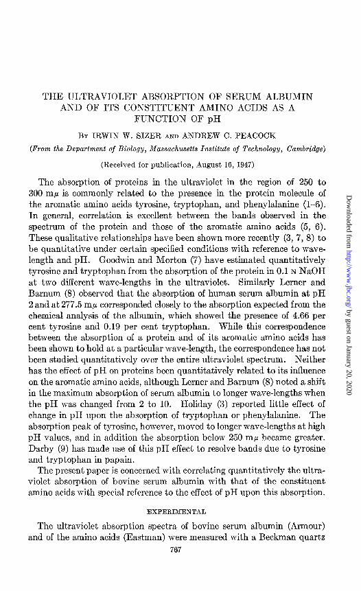
THE ULTRAVIOLET ABSORPTION OF SERUM ALBUMIN AND OF ITS CONSTITUENT AMINO ACIDS AS A
FUNCTION OF pH
BY IRWIN W. SIZER AND ANDREW C. PEACOCK
(From the Department of Biology, Massachusetts Institute of Technology, Cambridge)
(Received for publication, August 16, 1947)
The absorption of proteins in the ultraviolet in the region of 250 to 300 rnp is commonly related to the presence in the protein molecule of the aromatic amino acids tyrosine, tryptophan, and phenylalanine (l-6). In general, correlation is excellent between the bands observed in the spectrum of the protein and those of the aromatic amino acids (5, 6). These qualitative relationships have been shown more recently (3, 7, 8) to be quantitative under certain specified conditions with reference to wave- length and pH. Goodwin and Morton (7) have estimated quantitatively tyrosine and tryptophan from the absorption of the protein in 0.1 N NaOH at two different wave-lengths in the ultraviolet. Similarly Lerner and Barnum (8) observed that the absorption of human serum albumin at pH 2 and at 277.5 rnp corresponded closely to the absorption expected from the chemical analysis of the albumin, which showed the presence of 4.66 per cent tyrosine and 0.19 per cent tryptophan. While this correspondence between the absorption of a protein and of its aromatic amino acids has been shown to hold at a particular wave-length, the correspondence has not been studied quantitatively over the entire ultraviolet spectrum. Neither has the effect of pH on proteins been quantitatively related to its influence on the aromatic amino acids, although Lerner and Barnum (8) noted a shift in the maximum absorption of serum albumin to longer wave-lengths when the pH was changed from 2 to 10. Holiday (3) reported little effect of change in pH upon the absorption of tryptophan or phenylalanine. The absorption peak of tyrosine, however, moved to longer wave-lengths at high pH values, and in addition the absorption below 250 rnp became greater. Darby (9) has made use of this pH effect to resolve bands due to tyrosine and tryptophan in papain.
The present paper is concerned with correlating quantitatively the ultra- violet absorption of bovine serum albumin with that of the constituent amino acids with special reference to the effect of pH upon this absorption.
EXPERIMENTAL
The ultraviolet absorption spectra of bovine serum albumin (Armour) and of the amino acids (Eastman) were measured with a Beckman quartz
767
by guest on January 20, 2020http://w
ww
.jbc.org/D
ownloaded from
768 ABSORPTION SPECTRUM OF SERUM ALBUMIN
spectrophotometer. Readings were made at 2 rnp intervals except in the region of a maximum or a minimum, when they were made at intervals of 1 mp. The data are recorded as extinction coefficients’ at each wave- length. Since there was no significant shift in absorption of either the albumin or the amino acids in the acid region as determined in preliminary experiments, subsequent work in acid solution wz+s done only at pH 4.3. The alkaline region is represented by studies made at both pH 10.5 and 12.0.
SERUM ALBUMIN
240 260 280 WAVE-LENGTH.mu
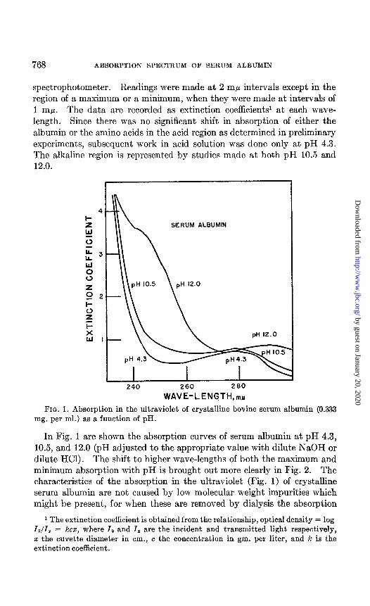
FIG. 1. Absorption in the ultraviolet of crystalline bovine serum albumin (0.333 mg. per ml.) as a function of pH.
In Fig. 1 are shown the absorption curves of serum albumin at pH 4.3, 10.5, and 12.0 (pH adjusted to the appropriate value with dilute NaOH or dilute HCl). The shift to higher wave-lengths of both the maximum and minimum absorption with pH is brought out more clearly in Fig. 2. The characteristics of the absorption in the ultraviolet (Fig. 1) of crystalline serum albumin are not caused by low molecular weight impurities which might be present, for when these are removed by dialysis the absorption
1 The extinction coefficient is obtained from the relationship, optical density = log lo/Z, = kcz, where IO and Z, are the incident and transmitted light respectively, x the cuvette diameter in cm., c the concentration in gm. per liter, and k is the extinction coefficient.
by guest on January 20, 2020http://w
ww
.jbc.org/D
ownloaded from

I. W. SIZER AND A. C. PEACOCK 769
curve is unaltered. Lerner and Barnum (8) observed a similar shift when they changed the pH of human serum albumin from 2 to 10. More striking than the effect of pH upon the wave-length of maximum and minimum ab- sorption, however, is the plateau in the curve for pH 12, which appears at about 240 rnp (Fig. 1). This effect is not evident in the work of Lerner and Barnum (8), since they did not explore the region above pH 10.
An attempt to interpret the data on the ultraviolet absorption of serum albumin was made by studying the ultraviolet absorption of the constituent amino acids of serum albumin. A synthetic mixture was prepared which
ABSORPTION MAXIMA
z (3
5 270
h >
ABSORPTION MINIMA
250
6 8 IO 12
PH
FIG. 2. Ultraviolet absorption maximum and minimum as a function of the pH of the medium. 0, bovine serum albumin (data from Fig. 1); A, amino acid mixture (data from Fig. 3) ; 0, L-tyrosine (data from Fig. 4) ; X, peptic digest of bovine serum albumin (data from Fig. 5).
contained the amino acids in the same proportions as they exist in the protein (10). The ultraviolet absorption as a function of pH is presented in Figs. 2 and 3. The wave-lengths of the maximum and minimum and their shift with pH are strikingly similar to those for serum albumin. Although at pH 4.3 the absorption in the ultraviolet of the amino acid mix- ture is practically identical in every respect with that of serum albumin (compare Figs. 1, 2, and 3), marked differences occur in the region of 240 rnp at pH 10.5 and 12.0. A plateau characterizes the amino acid mixture, but not the albumin, at pH 10.5, while at pH 12.0 t.here is a sharp peak in ab- sorption for the mixture, but only a plateau in the albumin curve at 240 rnp.
by guest on January 20, 2020http://w
ww
.jbc.org/D
ownloaded from

770 ABSORPTION SPECTRUM OF SERUM ALBUMIN
In attempts at interpretation of the ultraviolet curves of the amino acid mixture, detailed studies were made of the absorption in the ultraviolet of the aromatic amino acids, phenylalanine, tryptophan, and tyrosine. The results with the first two are summarized in Table I, from which it is ap- parent that only a slight shift in absorption spectrum occurs as a function of pH. With neither phenylalanine nor tryptophan is there any evidence in strongly alkaline solutions of a plateau or peak in the absorption curve at 240 mp. For tyrosine, however, the situation is very different (Fig. 4);
4
I- AMINO ACID MIXTURE
12.0
240 260 280
WAVE- LE NGTH,mp FIG. 3. Absorption in the ultraviolet as a function of pH of amino acids (0.333
mg. per ml.) mixed in the proportions in which they exist in serum albumin.
both the usual maximum and minimum shift to higher wave-lengths as the pH is increased in the alkaline region. This shift, as shown in Fig. 2, is closely analogous to that for serum albumin. The wave-length of the maxi- mum for serum albumin as well as the shift in maximum with pH is ac- counted for to a high degree of precision by the tyrosine present in the protein. The minimum and its shift with pH are not quite the same for tyrosine and serum albumin except in strongly alkaline solutions. At lower pH values it is clear that other amino acids contribute in determining the wave-length of minimum absorption of serum albumin.
The plateau in the absorption curve at 240 rnp of serum albumin at pH
by guest on January 20, 2020http://w
ww
.jbc.org/D
ownloaded from

I. W. SIZER AND A. C. PEACOCK 771
12.0 can be explained on the basis of the presence in the protein of tyrosine, which shows at 240 m/l a plateau at pH 10.5 and a sharp peak at pH 12.0. The shift of the 245 rnp minimum and of the 275 mp maximum of tyrosine to longer wave-lengths with increase in alkalinity has been observed by
TABLE I
Ahsorption Maxima and Minima in Ultraviolet of DL-Phenylalanine and of DL-Tryptophan As Function of pH
PH
4.3 10.5 12.0
Phenylalanine Tryptophan
Minimum Maximum Minimum M&IllUll
m tnlr ?w nrlr
230 258 243 278 234 259 244 281 236 259 245 281
L - TYROSINE
240 260 280
WAVE-LENGTH,mfl
FIG. 4. Absorption in the ultraviolet of L-tyrosine (1.0 mg. per ml.) as & function of pH.
others (3, 8) who ascribed this shift to ionization in alkali of the phenolic hydroxyl group of tyrosine. The maximum at 240 rnp in strongly alkaline solutions reported here has not been observed before, but the suggestion is made that it is also related to the formation of the phenoxide group. Greenberg and Barnum (11) have recently studied the dissociation of the
by guest on January 20, 2020http://w
ww
.jbc.org/D
ownloaded from

772 ABSORPTION SPECTRUM OF SERUM ALBUMIN
phenolic hydroxide group of tyrosine and report a pK of approximately 10. Since the absorption of the dissociated and undissociated groups is mutually independent (12), the absorption of an equal mixture of the phenoxide ion and of tyrosine with the undissociated phenolic group will represent the sum of the absorption of the two. At pH 12.0 the ionization should be complete and hence the peak at 240 rnp characteristic of t,he phenoxide ion becomes apparent. The extinction coefficient of this maximum at pH 12.0 is 53 (see Fig. 4). At pH 7.3, however, the phenolic group is almost com- pletely undissociated, and the corresponding extinction coefficient is 2.0. At pH 10.5 the concentration of the phenoxide ion and of the tyrosine with an undissociated hydroxyl group should be about equal, and the extinction coefficient at 240 rnE.c should therefore be the average of the two, or 27.5. The good agreement of the observed coefficient for pH 10.5 of 24.6 at 240 rnp with the theoretical value is evidence in favor of the concept that ionization of the phenolic group accounts for the change in absorption of tyrosine as a function of pH.
It appears that in serum albumin as in tyrosine the shift in absorption with pH is related to ionization of the phenolic group. Failure of the serum albumin to show the pronounced absorption at pH 10.5 at 240 rnp, typical of tyrosine, must mean that in serum albumin the phenolic groups are bound in some way that prevents ionization. This binding must be of a chemical type and not due to a mutual interaction of the constituent amino acids, since in the amino acid mixture (Fig. 3) the phenolic groups are free to ionize. Studies made with the amino acid mixture (Merck) from which the tyrosine had been omitted clearly indicate that almost all of the characteristic absorption in the ultraviolet of serum albumin and of its constituent amino acids can be accounted for by tyrosine.
Information was sought on the binding of phenolic groups in serum albumin by studying the effects of hydrolysis upon the absorption in the ultraviolet, since it seemed likely that hydrolysis might sufficiently alter the configuration of the protein molecule so as to permit the ionization of phenolic groups. For enzymatic hydrolysis, pepsin is one of the most interesting enzymes to use in view of its ability to cleave peptide bonds involving tyrosine or phenylalanine (13, 14). 100 mg. of serum albumin were digested for 2 hours at pH 2.0, at 37” with 1.0 mg. of crystalline pepsin (Lehn and Fink). Hydrolysis by pepsin2 was extensive, as is indi- cated by the failure of any precipitate to form when trichloroacetic acid was added to the digest, and by the fact that after dialysis of the digest the residue contained no tyrosine as measured by ultraviolet absorption or by chemical analysis (15). The effect of pH on the ultraviolet absorption of the peptic digest of serum albumin is shown in Fig. 5. As might be ex-
2 Suitable controls showed that hydrolysis was not caused by the acid present.
by guest on January 20, 2020http://w
ww
.jbc.org/D
ownloaded from
I. W. SIZER AND A. C. PEACOCK 773
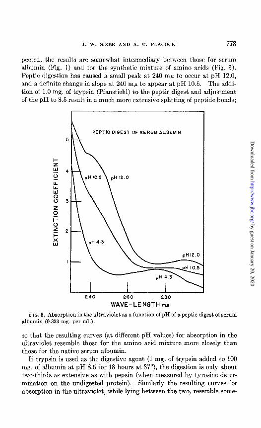
petted, the results are somewhat intermediary between those for serum albumin (Fig. 1) and for the synthetic mixture of amino acids (Fig. 3). Peptic digestion has caused a small peak at 240 rnp to occur at pH 12.0, and a definite change in slope at 240 rnp to appear at pH 10.5. The addi- tion of 1.0 mg. of trypsin (Pfanstiehl) to the peptic digest and adjustment of the pH to 8.5 result in a much more extensive splitting of peptide bonds;
n PEPTIC DIGEST OF SERUM ALBUMIN
5
I
240 260 280
WAVE-tENGTH,ma
FIG. 5. Absorption in the ultraviolet as a function of pH of a peptic digest of serum albumin (0.333 mg. per ml.).
so that the resulting curves (at different pH values) for absorption in the ultraviolet resemble those for the amino acid mixture more closely than those for the native serum albumin.
If trypsin is used as the digestive agent (1 mg. of trypsin added to 100 mg. of albumin at pH 8.5 for 18 hours at 37”), the digestion is only about two-thirds as extensive as with pepsin (when measured by tyrosine deter- mination on the undigested protein). Similarly the resulting curves for absorption in the ultraviolet, while lying between the two, resemble some-
by guest on January 20, 2020http://w
ww
.jbc.org/D
ownloaded from

774 ABSORPTION SPECTRUM OF SERUM ALBUMIN
what more closely the original protein than they do the mixture of amino acids. These results are noteworthy in view of the fact that, unlike pepsin, trypsin requires the presence of basic amino acids rather than tyrosine or phenylalanine in order to attack a peptide bond (13).
Since alkaline hydrolysis is frequently used as a preliminary step in the analysis of many amino acids, it was also employed in this study to deter- mine its effect on the absorption in the ultraviolet. Alkaline hydrolysis was performed in the usual manner (16) by boiling the serum albumin for 18 hours in 6 N NaOH. The absorption curves of the hydrolyzed protein
ALKALINE HYDROLYSATE
OF SERUM ALBUMIN
240 260 260
WAVE-LENGTH,mu
FIG. 6. Absorption in the ultraviolet as a function of pH of an alkaline hydrolysate of serum albumin (0.333 mg. per ml.).
(Fig. 6) clearly show, from the absorption at 240 rnp, the presence of the phenoxide ion at pH 10.5 and 12.0, but its presence is not nearly as evident as in the amino acid mixture. This is surprising, since it is generally ac- cepted that digestion with boiling alkali brings about complete hydrolysis. Other effects of treatment with alkali appear when the maxima and minima are examined. At pH 4.3 the usual minimum at 255 rnp has been almost eliminated and the maximum at 270 rnp has only a slightly higher extinction coefficient than the minimum. A similar situation prevails at pH 10.5, and, to a much lesser extent, at pH 12.0. The failure of alkaline treatment to bring out the expected peak in absorption at 240 rnp at pH 12.0, together
by guest on January 20, 2020http://w
ww
.jbc.org/D
ownloaded from
I. W. SIZER AND A. C. PEACOCK 775
with the effects of alkali on the relative extinction coefficients at 255 and 270 rnp, indicates that hot alkali has modified the absorption in the ultra- violet of tyrosine and probably of other amino acids.
DISCUSSION
The absorption in the ultraviolet of serum albumin can be interpreted in terms of the absorption of the tyrosine present in the molecule. The cor- relation is excellent at the maximum of 278 rnp at all pH values studied, but for the minimum at about 260 rnp this correlation is good only in strongly alkaline solutions (Fig. 2). In less alkaline solutions the amino acids tryptophan and phenylalanine as well as tyrosine contribute toward determining the extinction coefficient of the minimum. The change in shape of the curve at 240 rnp of the serum albumin at pH 12.0 seems to be determined by the dissociation of the phenolic group of tyrosine. Failure to find evidence of the phenoxide ion at pH 10.5 from the absorption of serum albumin at 240 rnp indicates that in the albumin molecule this group must be combined. The bond can hardly be a very stable one, however, for at pH 12.0 it is partly broken, allowing some of the phenolic groups to ionize. A hydrogen bond seems to be the most plausible type of linkage to be involved. A hydrogen bond might form between the amino group of lysine, which extends 7.54 A from the backbone of the polypeptide chain, and the phenolic group of tyrosine, which is also about 7.3 A from the back- bone (calculated from Pauling (17)). Basic amino acids other than lysine, however, might also be involved in forming this type of bond.
Since the plateau in the curve at 240 rnp of serum albumin at pH 12.0 is ascribed to the ionization of phenolic groups, it might be expected that a similar plateau (or even a peak) would occur in the curve for other proteins. Crystalline insulin (Armour) (final concentration 0.0667 mg. per ml.) at pH 12.0 shows a definite peak in absorption at 241 rnp in addition to the usual maximum at 293 rnp. In their study of insulin, Crammer and Neuberger (18) attributed the shift with pH of the absorption maximum at 280 rnp to ionization of the phenolic group of tyrosine. It is interesting to note that insulin contains a much higher tyrosine content than does serum albumin and that its tyrosyl groups are more reactive with tyrosinase than are those of serum albumin (19). Experiments are in progress to relate the absorption at 240 rnp at pH i2.0 of other proteins to their tyrosine content and reactivity of the tyrosyl groups.
The results of enzymatic hydrolysis in general confirm earlier data (see (8)) that proteases have practically no effect on the absorption of proteins in the ultraviolet. As a result of cleavage of peptide bonds by the enzyme, some of the bonds (hydrogen ?) attached to phenolic groups become broken, so that after digestion the phenoxide ions (as indicated by absorption at
by guest on January 20, 2020http://w
ww
.jbc.org/D
ownloaded from
776 ABSORPTION SPECTRUM OF SERUM ALBUMIN
240 mp) are more apparent in strongly alkaline solutions. On the other hand, hydrolysis by boiling in alkali has much more drastic effects in alter- ing the absorption of serum albumin in the ultraviolet. Since phenolic groups are also released by alkali, the absorption at 240 mE.c in strongly alkaline solutions is also increased. In view of the effects of pH and of alkali upon the absorption in the ultraviolet, any quantitative methods for determination of the aromatic amino acids based upon their absorption in the ultraviolet will require careful attention to experimental detail and caution in interpretation.
The authors wish to acknowledge the assistance in this work rendered by Miss Janette Robinson and by Mrs. Gloria Peacock.
SUMMARY
The maximum and minimum absorption of tyrosine moves progressively to longer wave-lengths in the ultraviolet as the pH is increased above pH 7.0. In solutions of pH 12.0 a second sharp absorption maximum appears at 240 rnp, which is ascribed to the ionization of the phenolic group of tyrosine. The ultraviolet spectra of tryptophan shifted only slightly with pH, while those of phenylalanine are relatively independent of pH.
The ultraviolet absorption spectrum, and the effect of pH thereon, of bovine serum albumin can be interpreted almost quantitatively on the basis solely of the tyrosine present in the protein. The major difference lies at 240 rnp at pH 12.0, where serum albumin shows only a plateau in the ab- sorption curve as compared with the peak for tyrosine. The difference is interpreted as indicating that in the protein most of the phenolic groups are bound, probably by hydrogen bonds, and are not free to ionize. When the protein is hydrolyzed by pepsin, trypsin, or alkali, the phenolic groups are set free to ionize, as is indicated by an increase in absorption caused by the phenoxide ion at 240 rnp in strong alkali. Although the action of proteases has little additional effect on the over-all absorption, alkaline hydrolysis modifies the shape of the absorption curve, making it in general unsuitable for quantitative analysis of the constituent amino acids.
An absorption maximum at 241 mp at pH 12.0 is also apparent in the absorption curve of insulin as well as of serum albumin and can probably be related to the concentration of phenoxide ions in most proteins.
BIBLIOGRAPHY
1. Anslow, G. A., and Nassau, S. C., J. Optical Xoc. America, 31, 114 (1941). 2. Coulter, C. B., Stone, F. M., and Kabat, E. A., J. Gen. Physiol., 19, 739 (1936). 3. Holiday, E. R., Biochem. J., 30, 1795 (1935). 4. Holiday, E. R., J. Scient. Instruments, 14, 166 (1937). 5. Lavin, G. I., and Northrop, J. H., J. Am. Chem. Sot., 67, 874 (1935).
by guest on January 20, 2020http://w
ww
.jbc.org/D
ownloaded from

I. W. SIZER AND A. C. PEACOCK 777
6. Smith, E. L., and Coy, N. H., J. Biol. Chem., 164, 367 (1946). 7. Goodwin, T. W., and Morton, R. A., Biochem. J., 40, 628 (1946). 8. Lerner, A. B., and Barnum, C. P., Arch. Biochem., 10, 417 (1946). 9. Darby, H. H., J. Biol. Chem., 139, 721 (1941).
10. Calvery, H. O., in Schmidt, C. L. A., The chemistry of the amino acids and pro- teins, Springfield (1945).
11. Greenberg, G. R., and Barnum, C. P., Federation Proc., 6, 256 (1947). 12. Feraud, K., Dunn, M. S., and Kaplan, J., J. Biol. Chem., 112, 323 (1935-36). 13. Bergmann, M., and Fruton, J. S., in Nord, F. F., and Werkman, C. H., Advances
in enzymology and related subjects, New York, 1, 63 (1941). 14. Bergmann, M., in Nord, F. F., and Werkman, C. H., Advances in enzymology and
related subjects, New York, 2,49 (1942). 15. Bernhart, F. W., Proc. Am. Sot. Biol. Chem., J. Biol. Chem., 123, p. x (1938). 16. Block, R. J., and Bolling, D., The amino acid composition of proteins and foods,
Springfield (1945). 17. Pauling, L., The nature of the chemical bond, Ithaca, 2nd edition (1945). 18. Crammer, J. L., and Neuberger, A., Biochem. J., 37,302 (1943). 19. Sizer, I. W., J. Biol. Chem., 163, 145 (1946); 169, 303 (1947).
by guest on January 20, 2020http://w
ww
.jbc.org/D
ownloaded from
Irwin W. Sizer and Andrew C. PeacockFUNCTION OF pH
CONSTITUENT AMINO ACIDS AS ASERUM ALBUMIN AND OF ITS
THE ULTRAVIOLET ABSORPTION OF
1947, 171:767-777.J. Biol. Chem.
http://www.jbc.org/content/171/2/767.citation
Access the most updated version of this article at
Alerts:
When a correction for this article is posted•
When this article is cited•
alerts to choose from all of JBC's e-mailClick here
tml#ref-list-1
http://www.jbc.org/content/171/2/767.citation.full.haccessed free atThis article cites 0 references, 0 of which can be by guest on January 20, 2020
http://ww
w.jbc.org/
Dow
nloaded from