Embed Size (px)
Citation preview

The Ubiquitin-Specific Protease Family from Arabidopsis.AtUBP1 and 2 Are Required for the Resistance to theAmino Acid Analog Canavanine1
Ning Yan2, Jed H. Doelling, Tanya G. Falbel3, Adam M. Durski, and Richard D. Vierstra*
Cellular and Molecular Biology Program and the Department of Horticulture, University of Wisconsin,Madison, Wisconsin 53706
Ubiquitin-specific proteases (UBPs) are a family of unique hydrolases that specifically remove polypeptides covalentlylinked via peptide or isopeptide bonds to the C-terminal glycine of ubiquitin. UBPs help regulate the ubiquitin/26Sproteolytic pathway by generating free ubiquitin monomers from their initial translational products, recycling ubiquitinsduring the breakdown of ubiquitin-protein conjugates, and/or by removing ubiquitin from specific targets and thuspresumably preventing target degradation. Here, we describe a family of 27 UBP genes from Arabidopsis that contain boththe conserved cysteine (Cys) and histidine boxes essential for catalysis. They can be clustered into 14 subfamilies based onsequence similarity, genomic organization, and alignments with their closest relatives from other organisms, with sevensubfamilies having two or more members. Recombinant AtUBP2 functions as a bona fide UBP: It can release polypeptidesattached to ubiquitins via either a- or e-amino linkages by an activity that requires the predicted active-site Cys within theCys box. From the analysis of T-DNA insertion mutants, we demonstrate that the AtUBP1 and 2 subfamily helps conferresistance to the arginine analog canavanine. This phenotype suggests that the AtUBP1 and 2 enzymes are needed forabnormal protein turnover in Arabidopsis.
The ubiquitin/26S proteasome proteolytic path-way plays an important role in eukaryotic cellgrowth, development, stress responses, and environ-mental adaptation by degrading short-lived and ab-normal proteins (Hershko and Ciechanover, 1998;Vierstra, 1996; Callis and Vierstra, 2000). In this path-way, ubiquitin functions as a reusable tag to targetspecific proteins for breakdown. Via an ATP-dependent reaction cascade involving the sequentialaction of three classes of enzymes, E1s, E2s, and/orE3s, chains of ubiquitins become attached to proteo-lytic substrates through an isopeptide bond betweenthe C-terminal Gly-76 of ubiquitin and a Lys residuein the target. These chains then serve as degradationsignals for the 26S proteasome, a 2-MDa multisub-unit protease that breaks down the protein into smallpeptides and amino acids but releases the ubiquitinsintact.
Both the characterization of ubiquitin/26S protea-some pathway mutants and the analysis of individ-ual substrates indicate that the pathway degrades awide variety of short-lived proteins (Hershko andCiechanover, 1998; Callis and Vierstra, 2000). To de-fine how these substrates are chosen, most studieshave focused on the E2/E3 enzymes that direct ubi-quitin attachment. However, recent data indicate thatthe steps that release ubiquitins from targets andgenerate free monomers can also affect the selectivityof the pathway and the half-life of a substrate(Wilkinson, 1997; D’Andrea and Pellman, 1998;Chung and Baek, 1999). These steps are performed bya unique group of deubiquitinating enzymes (DUBs);they are thiol proteases that specifically cleave thepeptide bond between the C-terminal Gly of ubi-quitin and covalently attached polypeptides. Yeastsand animals contain a number of DUBs that varysubstantially in sequence, suggesting that they rec-ognize distinct substrates and/or have discrete func-tions (Wilkinson, 1997; D’Andrea and Pellman, 1998).Mutant analyses have implicated specific DUBs innumerous cellular processes, including cell growth(Papa and Hochstrasser, 1993; Zhu et al., 1996; Nav-iglio et al., 1998), cell differentiation (Chung et al.,1998; Lindsey et al., 1998; Liu et al., 1999), eye devel-opment (Huang et al., 1995; Taya et al., 1998; Taya etal., 1999), neural function (Wilkinson et al., 1989;Hegde et al., 1997; Leroy et al., 1998), coordinatedDNA replication (Singer et al., 1996), gene silencing(Moazed and Johnson, 1996; Kahana and Gottschling,1999), endocytosis (Galan and Haguenauer-Tsapis,
1 This work was supported by the U.S. Department ofAgriculture-National Research Initiative Competitive Grants Pro-gram (grant nos. 97–35301– 4218 and 00 –35301–9040) and the Re-search Division of the University of Wisconsin, College of Agri-culture and Life Sciences (grant no. Hatch 142–N936), a NationalInstitutes of Health Postdoctoral Fellowship (to J.H.D.), and a U.S.Department of Agriculture Postdoctoral Grant (to T.G.F.).
2 Present address: Department of Biochemistry, University ofWashington, Seattle, WA 98195.
3 Present address: Department of Biochemistry, University ofWisconsin, Madison, WI 53706.
* Corresponding author; e-mail [email protected]; fax608 –262– 4743.
1828 Plant Physiology, December 2000, Vol. 124, pp. 1828–1843, www.plantphysiol.org © 2000 American Society of Plant Physiologists www.plantphysiol.orgon October 29, 2018 - Published by Downloaded from Copyright © 2000 American Society of Plant Biologists. All rights reserved.

1997), oncogenesis (Nakamura et al., 1992; Papa andHochstrasser, 1993; Gray et al., 1995), heat shock(Baxter and Craig, 1998), and the breakdown of ab-normal proteins (Papa and Hochstrasser, 1993;Amerik et al., 1997).
Enzymatic analyses indicate that DUBs have threegeneral roles in the ubiquitin/26S proteasome path-way, each of which can profoundly influence theoverall activity and/or specificity of the pathway(Fig. 1). One role is to generate ubiquitin monomersfrom the initial translation products of ubiquitingenes (Callis and Vierstra, 1989; Eytan et al., 1993;Baek et al., 1998). Ubiquitin is unusual in that it isnaturally synthesized as a translational fusion. Thesefusions contain either a single ubiquitin fused to theN terminus of an unrelated protein (ubiquitin exten-sion) or tandem repeats of ubiquitin linked head-to-tail and capped at the C terminus with one or moreadditional amino acids (polyubiquitin). DUBs are es-sential for releasing ubiquitin monomers from thesefusions by cleaving the a-amino peptide bond aftereach ubiquitin moiety. The second role of DUBs is toregenerate free ubiquitin monomers during thebreakdown of ubiquitin-protein conjugates by the26S proteosome. In this case, DUBs remove peptide
fragments that remain attached following hydrolysisof the target and disassemble the multiubiquitinchain (Hadari et al., 1992; Papa and Hochstrasser,1993; Amerik et al., 1997). The third role is to de-ubiquitinate conjugates; in this case both the targetand the attached ubiquitins are released intact(Chung et al., 1998; Taya et al., 1998; Taya et al.,1999). The last two roles are accomplished by cleav-ing isopeptide bonds in which the C-terminal Gly ofubiquitin is attached to lysyl e-amino groups.
Substrate recognition by all DUBs is highly depen-dent on the ubiquitin moiety, especially theC-terminal Gly (Wilkinson, 1997). However, UBPsare remarkably non-specific with respect to the ap-pended polypeptide. For those cleaving isopeptidebonds, all attached polypeptides appear to be accept-able. For those cleaving peptide bonds, all transla-tional fusions can be tolerated with the exceptions ofappended sequences beginning with Pro, which areoften resistant to cleavage. DUBs are divided intotwo general groups based on their amino acid se-quence and substrate specificity (Wilkinson, 1997).One group called ubiquitin C-terminal hydrolases(UCHs) is composed of relatively small proteins(20–30 kD) that are structurally defined by the pres-ence of a catalytic triad consisting of a positionallyconserved Cys, His, and Asp (Johnston et al., 1997).In vitro, UCHs can remove small molecules (e.g. esteradducts and lysines) and peptides (Wilkinson, 1997),some of which attach non-specifically by reactingwith activated ubiquitin during its conjugation cas-cade. UCHs can also process short multiubiquitinchains (Lam et al., 1997) and ubiquitin precursors(Pickart and Rose, 1985), suggesting a role in theproduction of free ubiquitin monomers.
The second group of DUBs, called ubiquitin-specific proteases (UBPs), cleaves ubiquitins linkedto larger proteins by either peptide or isopeptidebonds. Enzymes in this group vary greatly in size butcan be easily identified by the presence of two con-served catalytic motifs; one contains an essential Cys(Cys box) and the other contains two essential histi-dines (His box; Huang et al., 1995; Wilkinson et al.,1995; Wilkinson, 1997). Outside of these domains,their amino acid sequences diverge, possibly reflect-ing specific targets and/or cellular functions. Largefamilies of UBPs have been identified in a variety oforganisms. For example, whereas yeast (Saccharomy-ces cerevisae) has a single UCH, it contains 16 differentUBPs (Wilkinson, 1997).
Despite their potential importance, little is knownabout UBPs in plants. Prior to this study, only threehave been described, AtUBP3, 4, and 5 from Arabi-dopsis (Chandler et al., 1997; Rao-Naik et al., 2000),and no UBP mutants had been reported. Using theconserved Cys and His boxes as queries, we haveidentified 24 additional UBP genes in the near fin-ished Arabidopsis genome. Protein sequence com-parisons cluster the complete gene collection into 14
Figure 1. Possible functions of DUBs in the ubiquitin/26S protea-some proteolytic pathway. A, Generating free ubiquitin monomersfrom the translation products of polyubiquitin and ubiquitin-extension genes by cleaving the a-amino peptide bonds followingeach ubiquitin moiety. XY represent additional amino acids ap-pended to the C-terminal Gly of the last ubiquitin repeat. B, Cleavingubiquitin linked either to itself or to other proteins through e-aminoisopeptide bonds. Two routes are possible. DUBs could removemultiubiquitin chains bound to proteins, releasing both intact, andthen the free ubiquitin chains would be disassembled into freeubiquitin monomers. During or following degradation of the target,free multiubiquitin chains could be released from peptide fragmentsof the target and then would be disassembled into free ubiquitinmonomers. White arrowheads indicate the various bonds cleaved byDUBs.
Ubiquitin-Specific Proteases from Arabidopsis
Plant Physiol. Vol. 124, 2000 1829 www.plantphysiol.orgon October 29, 2018 - Published by Downloaded from Copyright © 2000 American Society of Plant Biologists. All rights reserved.
possible AtUBP subfamilies, with seven having twoor more members. Substantial diversity among thesubfamilies suggests that each may have unique rolesin Arabidopsis physiology, growth, and develop-ment. Analysis of a subfamily encoded by AtUBP1and 2 indicates that these proteins are bona fide UBPenzymes, capable of cleaving ubiquitin linked viapeptide or isopeptide bonds. T-DNA insertion mu-tants of AtUBP1 and 2 are phenotypically normalunder standard growth conditions. However, the sin-gle and double homozygous plants are hypersensi-tive to the amino acid analog canavanine (CAN),supporting a role for these UBPs in particular, andthe ubiquitin/26S proteasome pathway in general inaberrant protein turnover in plants.
RESULTS
Identification of UBPs in Arabidopsis
Sullivan et al. (1990) first reported that plants haveUBP-like activities capable of cleaving ubiquitin at-tached to other proteins via peptide or isopeptidelinkages. To identify the responsible enzymes, weused the sequence of yeast UBP4 (Papa and Hoch-strasser, 1993) as the query to search the Arabidopsisexpressed sequence tag (EST) database for relatedproteins. Various yeast and Arabidopsis UBP se-quences subsequently were used to examine the Ara-bidopsis bacteria artificial chromosome (BAC) andEST databases for additional candidate genes. Thisextensive search (last completed on September 26,2000) ultimately identified 27 distinct genes that en-code proteins with both the Cys- and His-box signa-ture motifs (Wilkinson, 1997). Three of these Arabi-dopsis genes (AtUBP3-5) have been describedrecently (Chandler et al., 1997; Rao-Naik et al., 2000).Partial or complete cDNAs have been identified for21 of the additional 24 genes (the exceptions beingAtUBP11, 18, and 19), indicating that most AtUBPfamily members (at least 24 of the 27) are activelyexpressed. By comparing the genomic sequenceswith their corresponding cDNAs, or by deducingintron/exon boundaries using alignments with pos-sible paralogs, the complete coding regions were pre-dicted for all 27 (Chandler et al., 1997; Rao-Naik et al.,2000; data not shown). In many cases, these codingsequences disagreed with those annotated in the AGIdatabase.
Figure 2 shows the organization of the AtUBP pro-tein family and the relationship of some members topossible orthologs from other species. As found inother organisms, Arabidopsis UBPs vary consider-ably in size with lengths ranging from 365 to 1,116amino acids. As expected, each contains the Cys andHis boxes essential for catalysis. Within these boxesare the positionally conserved Cys and His residuesthat comprise parts of the active-site (Fig. 3). The Cysboxes of the AtUBP family show high conservationboth in sequence (60%–100% similarity) and length
(all are 18 residues). In contrast, the His boxes aremore diverse in sequence (40%–100% similarity) andshow substantial differences in length (55–100 resi-dues) as a result of several insertions/extensions(Fig. 3). In addition, the collection of AtUBPs containsseveral less conserved motifs common among UBPs(Wilkinson, 1999); these include the Q, G, L, and Fboxes, defined by the presence of one or more ofthese amino acids in their respective domains (Fig. 2).The function(s) of these domains are unknown atpresent.
Using overall amino acid sequence homology, thepositions of the Cys, His, Q, G, L, and F boxes, thepresence of additional protein sequence motifs, andthe positions of known or predicted introns/exons,the family of AtUBP genes was tentatively clusteredinto 14 subfamilies. In all cases, these four criteriawere in agreement, supporting our subfamily classi-fication (Figs. 2 and 3; data not shown). Percentamino acid sequence similarity among members ofthe predicted subfamilies ranged from 95% (for theAtUBP3 and 4 subfamily) to 55% (for the AtUBP20and 21 subfamily) but dropped to ,40% when non-family members were compared. As can be seen inFigure 2, discriminating features of each subfamilyinclude the presence of N- and/or C-terminal exten-sions beyond the Cys and His boxes and insertionsthat often separate the G and L motifs. Seven of thesubfamilies have two or more members, includingthe AtUBP5, 8-11 subfamily, and the AtUBP15-19subfamily that contain five members each. The re-maining seven subfamilies contain only a single gene(AtUBP14 and 22-27). Outside of the six conservedregions, the AtUBP proteins display little similarityoutside of their subfamilies, suggesting that most ifnot all subfamilies have unique functions and/orrecognize distinct substrates.
A number of the UBP subfamilies are predictedto contain additional sequence motifs, includingzinc fingers, MATH, ubiquitin-like, and ubiquitin-associated (UBA) domains that may insinuate func-tion (Fig. 2). Potential zinc fingers were found nearthe N terminus of the AtUBP1-2, AtUBP14, and theAtUBP15-19 subfamilies. These fingers often mediatedirect protein-protein interactions following chela-tion of a zinc ion by positionally conserved Cys andHis residues (Freemont, 1993; Takatsuji, 1998; Tyersand Jorgensen, 2000). The AtUBP12-13 subfamilycontains a MATH domain common among a numberof proteins, including TRAF-related proteins and themeprin family of metalloproteases, and may be in-volved in dimerization or protein-protein interac-tions (Uren and Vaux, 1996). Ubiquitin-like domainswere detected in AtUBP6-7 and AtUBP26. Similar totheir yeast ortholog ScUBP6 (Wyndham et al., 1999),this domain is near the N terminus of AtUBP6 and 7.However for AtUBP26, the ubiquitin-like domain isnear its C terminus. The ubiquitin-like domain couldhelp these UBPs bind to ubiquitin-interacting pro-
Yan et al.
1830 Plant Physiol. Vol. 124, 2000 www.plantphysiol.orgon October 29, 2018 - Published by Downloaded from Copyright © 2000 American Society of Plant Biologists. All rights reserved.

teins; for ScUBP6, it is dispensable for catalytic activ-ity (Wyndham et al., 1999). Two consensus UBAdomains are located near the C terminus of AtUBP14.UBA domains have been found in a number of pro-teins related to ubiquitin metabolism, including E2s,
E3s, and other UBPs (Amerik et al., 1997; Bates andVierstra, 1999; Hofmann and Pickart, 1999). It hasbeen proposed that this motif binds ubiquitin non-covalently but its function is unknown (Hofmannand Pickart, 1999).
Figure 2. Structure of the members of the Arabidopsis UBP family. Locations of the Cys, Q, G, L, F, and His boxes areindicated. AtUBP proteins with similar structures are grouped by brackets. Predicted amino acid lengths are shown on theright. Potential orthologs in yeast and animals are indicated if available. Amino acid sequence alignments of the Cys and Hisboxes are shown in Figure 3. AtUBP3, 4, and 5 were recently described by Chandler and Callis (1997) and Rao-Naik et al.(2000). Accession number for the other AtUBP sequences are: AtUBP1 (AF302658), AtUBP2 (AF302659), AtUBP6(AF302660), AtUBP7 (AF302661), AtUBP8 (AF302662), AtUBP12 (AF302663), AtUBP14 (AF302664), AtUBP15(AF302665), AtUBP16 (AF302666), AtUBP17 (AF302667), AtUBP20 (AF302668), AtUBP21 (AF302669), AtUBP22(AF302670), AtUBP23 (AF302671), AtUBP24 (AF302672), AtUBP25 (AF302673), AtUBP26 (AF302674), and AtUBP27(AF302675). The remaining AtUBP proteins can be located in various BAC clones annotated in the AGI database: AtUBP9(AF118222), AtUBP10 (AF118222), AtUBP11 (AC006424), AtUBP13 (AC0016795), AtUBP18 (AL031004), and AtUBP19(AC006954). l, Indicates the presence of potential zinc finger; F, indicates the ubiquitin-like domains; E, indicates theMATH domains; ‚, indicates the UBA domains.
Ubiquitin-Specific Proteases from Arabidopsis
Plant Physiol. Vol. 124, 2000 1831 www.plantphysiol.orgon October 29, 2018 - Published by Downloaded from Copyright © 2000 American Society of Plant Biologists. All rights reserved.

AtUBP1 and 2 Subfamily
To further define the functions of the ArabidopsisUBPs, we continued an in-depth characterization ofthe AtUBP 1 and 2 subfamily. To confirm that thesetwo genes represent the entire subfamily, genomicDNA from the ecotype Wassilewskija (WS) of Arabi-dopsis was subjected to DNA gel-blot analysis usingeither AtUBP1 or 2 as the probe. As can be seen inFigure 4, only AtUBP1- and 2-derived fragmentscould be detected following either high- or low-stringency washes after digestion of the genomicDNA with three different restriction enzymes. Theseresults indicate that AtUBP1 and 2 are the only mem-bers in this subfamily. By sequence analysis ofgenomic and cDNA clones, the partial organizationfor AtUBP1 and the complete organization ofAtUBP2 was determined (Fig. 5A). Each contains apositionally conserved intron between the sequences
for the F and His boxes, whereas AtUBP1 is predictedto contain a second intron following the sequence forthe G box. A 531-bp intron was detected upstream ofthe Met start codon in AtUBP2; a similarly positionedintron may be present in AtUBP1 but could not beidentified without an available cDNA sequence inthat region.
The encoded AtUBP1 and 2 proteins are 120 and106 kD, respectively, and contain all six of the con-served UBP motifs (Fig. 5A). Sequence comparisonsrevealed that AtUBP1 and AtUBP2 are more relatedto each other than to any of the other UBPs in Ara-bidopsis, sharing 62% amino acid sequence similar-ity. Dotplot comparisons show that this homology isevident even outside the six conserved domains,where most other Arabidopsis UBPs show little re-latedness (Fig. 5B). No orthologs have been detectedthus far in any other plant species. Their closest
Figure 3. Alignment of the Cys and His boxes from the members of the Arabidopsis UBP family. Black and white arrowheadsindicate the positions of the essential Cys and His residues, respectively. Reverse type and gray boxes denote identical andsimilar amino acids, respectively. Dots indicate gaps. Comparisons were made with the University of Wisconsin-GeneticsComputer Group program Pileup and displayed by MacBoxshade 2.7.
Yan et al.
1832 Plant Physiol. Vol. 124, 2000 www.plantphysiol.orgon October 29, 2018 - Published by Downloaded from Copyright © 2000 American Society of Plant Biologists. All rights reserved.

homologs outside of plants are human UBP-M (Cai etal., 1999), UBPY (Naviglio et al., 1998), and the onco-protein tre-2 (Papa and Hochstrasser, 1993) (49%,49%, and 45% similar to AtUBP1, respectively). How-ever, Dotplot comparisons of UBPM, UBPY, and tre-2versus AtUBP1 or 2 showed that this similarity isrestricted to the six conserved motifs, suggesting thatthese human UBPs are not functional orthologs (Fig.5C; data not shown). The absence of possible or-thologs suggests that AtUBP1 and 2 are unique toplants.
AtUBP2 Is Active in Vivo and in Vitro
UBPs are best defined by their ability to cleaveubiquitin attached via peptide (a-amino) and/orisopeptide (e-amino) bonds to other proteins. To con-firm this activity for AtUBP1 and 2 and to identify thenature of their preferred linkages, the recombinantAtUBP2 protein was assayed against a variety ofsubstrates both in vitro and in vivo. For ubiquitinlinked via a peptide bond, three translational fusionsof varying sizes were tested: the hexameric polyubi-quitin protein AtUBQ10 (Callis et al., 1995), theAtUBQ1 ubiquitin-extension protein bearing the 52-amino acid ribosomal protein appended to a single
ubiquitin moiety (Callis and Vierstra, 1989), and afusion of ubiquitin and b-galactosidase (Ub-bgal)(Varshavsky, 1997). For ubiquitin linked via anisopeptide bond, a population of multiubiquitinchains linked through Lys-48 was the substrate (vanNocker and Vierstra, 1993). The wild-type (WT)AtUBP2 was tested along with two mutant forms inwhich the active-site Cys at position 240 (Fig. 3) wassubstituted for either Ser (AtUBP2C240S) or Ala(AtUBP2C240A). All three proteins could be expressedto high levels as soluble proteins in Escherichia coli.
As can be seen in Figure 6, A and B, AtUBP2effectively cleaved ubiquitin attached via pep-tide linkages in vivo. When the recombinant pro-tein was co-expressed with a hexameric polyubi-quitin (AtUBQ10) or a ubiquitin-extension protein(AtUBQ1), free ubiquitin of the correct mobility wasgenerated (Fig. 6, A and B). For the polyubiquitinreactions, the cleavage products were released asdoublets. The species of higher mass in each doubletrepresented ubiquitin polymers containing an 11amino acids N-terminal extension, which was addedduring the construction of the AtUBQ10 vector forexpression in E. coli (Fig. 6A). The activity of AtUBP2was similar to that of yeast ScUBP1, which has beenpreviously shown to cleave ubiquitin attached via
Figure 4. DNA gel-blot analysis of AtUBP1 and 2 from WT Arabidopsis (WT) and ubp1-1/ubp2-1 mutant plants. Arabidopsisgenomic DNA was isolated from the ecotype WS and the double homozygous ubp1-1/ubp2-1 line, digested with BglII (B),EcoRI (E), or EcoRV (V) and then probed with either an AtUBP1 or 2 gene-specific probe. A, Analysis of WT Arabidopsisgenomic DNA following washes at either low stringency (LS) or high stringency (HS). Each band marked by a whitearrowhead represents a genomic fragment that corresponds to the gene-specific probe used in that blot. B, Analysis ofgenomic DNA from WT or the ubp1-1/ubp2-1 (1-1/2-1) double mutant. Blots were washed at high stringency. L, Indicatesfragments only detected in DNA from WT and not the mutant plants. l, Denotes fragments present in DNA from the mutantand not WT plants.
Ubiquitin-Specific Proteases from Arabidopsis
Plant Physiol. Vol. 124, 2000 1833 www.plantphysiol.orgon October 29, 2018 - Published by Downloaded from Copyright © 2000 American Society of Plant Biologists. All rights reserved.

a-amino peptide linkages (Tobias and Varshavsky,1991). As expected, the activity of AtUBP2 was de-pendent on the active-site Cys; both the AtUBP2C240Sand AtUBP2C240A mutants were inactive.
AtUBP2 was also co-expressed with Ub-X-b-galactosidase in which either a Met (Ub-M-bgal) or aLeu (Ub-L-bgal) residue immediately followed theubiquitin moiety. As shown by Papa et al. (1993), thiscombination of substrates helps confirm that cleav-age occurred at the correct site, i.e. immediately fol-lowing the C-terminal Gly of the ubiquitin moiety.Correct cleavage of Ub-M-bgal releases M-bgal,which is stable and accumulates to high levels in E.coli. In contrast, correct cleavage of Ub-L-bgal gener-ates L-bgal, which is rapidly degraded by the N-endrule pathway, and thus accumulates to substantiallylower levels (Varshavsky, 1997). For example, whenyeast ScUBP1 is used, loss of Ub-M-bgal and Ub-L-bgal was evident. However, whereas detectable lev-els of the Ub-M-bgal digestion product could be seen,the expected cleavage product of Ub-L-bgal was un-detectable (Fig. 6C; Papa and Hochstrasser, 1993). Asimilar outcome was observed for AtUBP2; whereasthe M-bgal accumulated, the L-bgal product did not(Fig. 6C). Like the results obtained with the poly-ubiquitin and ubiquitin-extension protein substrates,
the activity of AtUBP2 on Ub-X-b-gal substrates wasdependent on the active-site Cys.
Recombinant AtUBP2 could also cleave in vitroubiquitin attached via isopeptide (e-amino) linkages.Similar to yeast UBP14 (Amerik et al., 1997), AtUBP2digested Lys-48-linked multiubiquitin chains andgenerated free ubiquitin monomers in a reaction thatalso required Cys-240 (data not shown).
Analysis of T-DNA Insertion Mutants of AtUBP1 and 2
To investigate the biological function(s) of theAtUBP1 and 2 subfamily, we screened availableT-DNA-transformed populations of Arabidopsis(Krysan et al., 1996; Krysan et al., 1999) for disrup-tional insertion(s) in the corresponding genes. Inser-tion mutants ubp1-1 and ubp2-1 were identified thatcontain a T-DNA insertion in the coding region, 703-and 2,539-bp downstream of the respective transla-tion start site, with the T-DNA either upstream of theCys box (ubp1-1) or between the F and His boxes(ubp2-1) (Fig. 5A). Both insertions were predicted togenerate a truncated protein missing one or moredomains essential for catalysis and hence should rep-resent loss-of-function alleles. To eliminate potentialsecond-site mutations, three back crosses of themutants to the WT ecotype WS were performed be-fore the homozygous ubp1-1 and ubp2-1 lines werecrossed and a double homozygote was isolated.
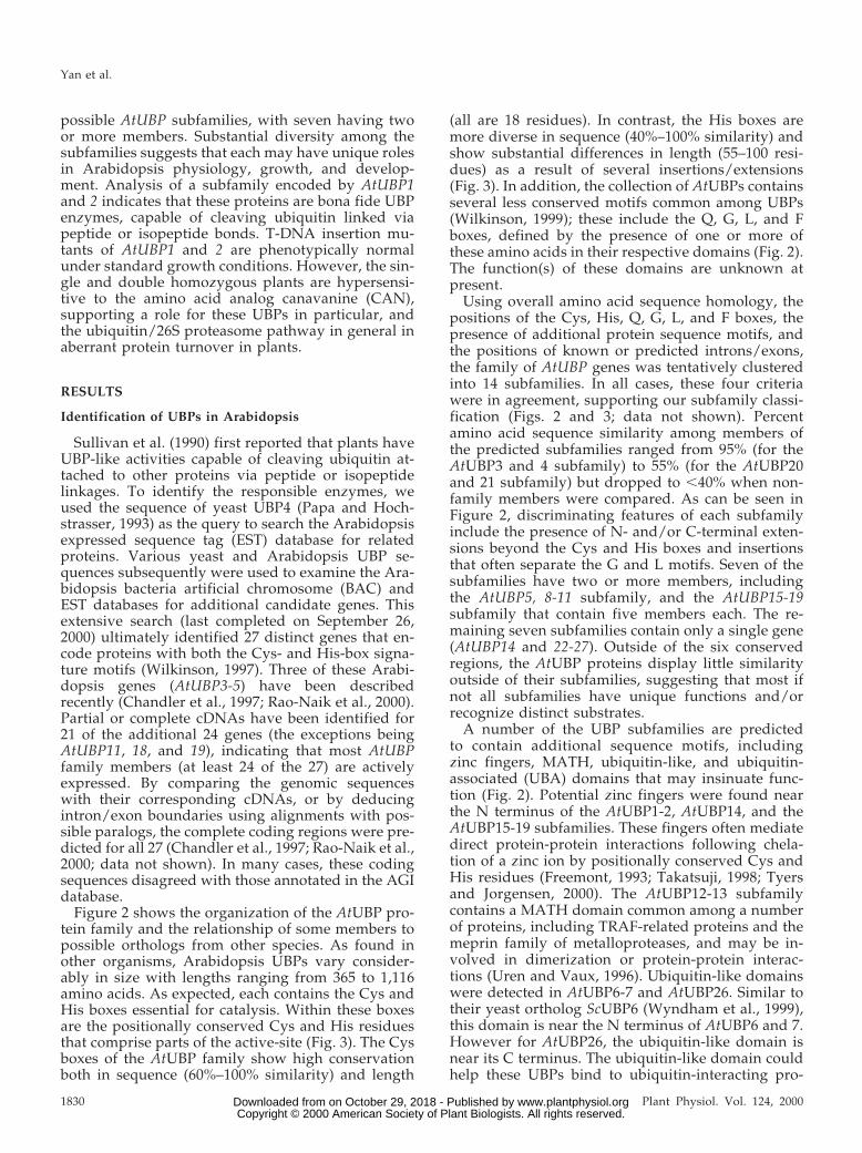
To confirm that the AtUBP1 and 2 genes wereaffected in the ubp1-1, ubp2-1, and ubp1-1/ubp2-1lines, genomic DNA was isolated from the homozy-gotes and analyzed by DNA gel-blot analysis. In eachcase, the banding patterns of the mutant differed aspredicted from that of WT at the respective loci (Fig.4B; data not shown). By using RT-PCR, we found thatthe T-DNA insertion also affected expression of theAtUBP1 and 2 genes. Whereas, the AtUBP1 and 2mRNAs could be easily detected by RT-PCR, using asa template RNA isolated from WT plants treated withor without CAN, none could be detected using RNAfrom the corresponding mutants treated similarly(Fig. 7). As a result, we consider it likely that ubp1-1and ubp2-1 represent null alleles.
To assess the phenotypic functions of the AtUBP1and 2 subfamily, the ubp1-1 and ubp2-1 mutant plantswere examined under a variety of growth conditions.Under normal conditions, either on minimal agarmedia or in soil, the homozygous ubp1-1, ubp2-1,and ubp1-1/ubp2-1 plants were phenotypically in-distinguishable from WT plants, including time ofgermination, growth rate, flowering time, and overalldevelopment. To potentially reveal more subtle phe-notypes, we also grew the plants under a variety ofadverse conditions, including media that containedamino acid analogs, heavy metals, high concentra-tions of salts or hormones, and various environmen-tal stresses, e.g. heat, cold, high, and low light (J.C.Young, personal communication). Several of these
Figure 5. Structure and derived amino acid sequence alignments ofthe AtUBP1 and 2 genes. A, Structure of AtUBP1 and 2 genes. Linesindicate introns and boxes indicate exons; white boxes, untranslatedregions; gray/black boxes, translated regions. The Cys, Q, G, L, F,and His boxes are indicated in black. The T-DNA insertion sites forthe ubp1-1 and ubp2-1 mutants are indicated by the triangles. B,Dotplot comparison of the deduced amino acid sequence of AtUBP1with that of AtUBP2 (left) or human UBP-M (AF12636; Cai et al.,1999) (right). The positions of the conserved Cys, Q, G, L F, and Hisdomains are labeled. Axes denote amino acid position.
Yan et al.
1834 Plant Physiol. Vol. 124, 2000 www.plantphysiol.orgon October 29, 2018 - Published by Downloaded from Copyright © 2000 American Society of Plant Biologists. All rights reserved.

conditions were chosen based on the reported in-volvement of the ubiquitin/26S proteasome in theresponse of plants to hormones (Ruegger et al., 1998;Xie et al., 1998; Girod et al., 1999), light (Jabben et al.,1989), drought (Kiyosue et al., 1994), and exposure toamino acid analogs (Bachmair et al., 1990; Girod etal., 1999). For almost all conditions, the mutant plantsresponded similar to WT.
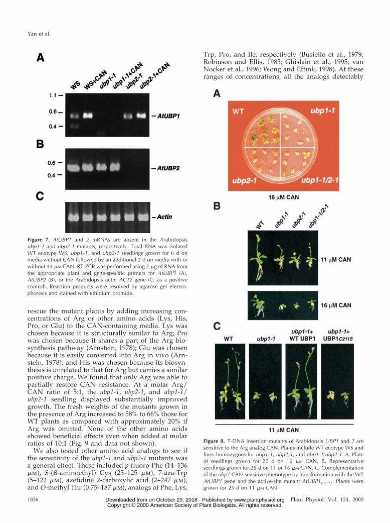
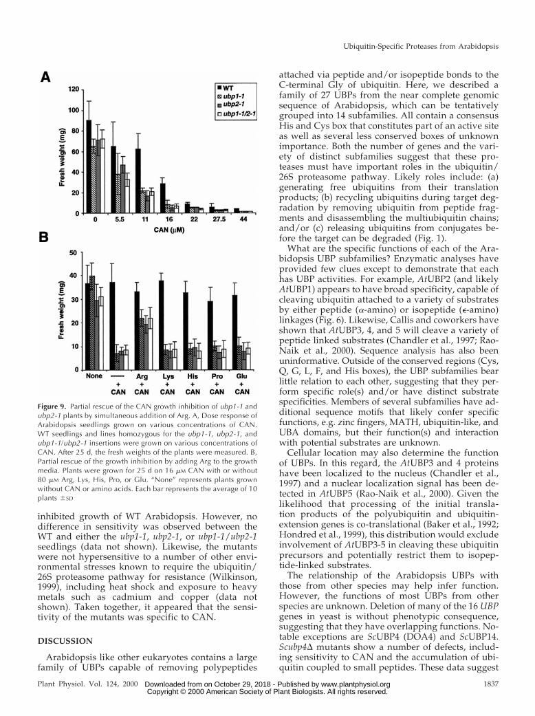
The only exception was growth of the mutants onmedia containing the Arg analog, CAN. Whereas WTplants were mildly affected by concentrations ofCAN greater than 5.5 mm, the ubp1-1, ubp2-1, andubp1-1/ubp2-1 homozygous plants were severelystunted and had shorter roots and chlorotic leaves(Fig. 8). Comparisons of seedling fresh weight indi-cated that the greatest differences occurred whenCAN concentrations were between 11 and 16 mm; atthese levels, the fresh weights of mutant plants were19% to 23% of those for WT plants (Fig. 9A). That thetwo single mutants and the double mutant showedsimilar CAN-sensitivity indicates that both AtUBP1and 2 are necessary for optimal resistance to thisamino acid analog (Figs. 8 and 9A).
To confirm that the sensitivity to CAN was a directresult of the T-DNA disruption of either the AtUBP1or 2 genes, we attempted to rescue the mutant phe-notype of ubp1-1 by complementation with the WTAtUBP1 gene and the active-site Cys mutantAtUBP1C211S (Fig. 5C). These two genes were intro-duced into a homozygous ubp1-1 line using anAgrobacterium-based pCAMBIA3300 vector and T0transformed seedlings were selected by BASTAherbicide resistance. T1 plants (heterozygous for
the transgene), containing either the AtUBP1 orAtUBP1C211S transgene, were then self-pollinated.The progenies (T2) from each independently trans-formed T1 plant were grown on 11 mm CAN togetherwith WT ecotype WS and the homozygous ubp1-1mutant. Those plants containing the AtUBP1 trans-gene showed a 3:1 co-segregation of the CAN resis-tance with that of BASTA resistance, a marker for theAtUBP1 transgene, whereas all those harboringAtUBP1C211S remained CAN-sensitive like the ho-mozygous ubp1-1 mutant (Fig. 8C; data not shown).The results collectively demonstrated that AtUBP1 isrequired for optimal CAN resistance and that anactive enzyme is required. We also tried to comple-ment the homozygous ubp2-1 line with AtUBP1.However, none of the BASTA-resistant plants re-gained resistance to CAN (data not shown), suggest-ing that the sensitivity is not simply caused by alower dosage of the AtUBP1/2 activity in the ubp1-1or ubp2-1 plants.
CAN is naturally produced by certain legumes asan anti-herbivore compound. Its toxicity is a result ofits ability to substitute for Arg during translation.Once incorporated into a protein, CAN can pro-foundly alter both protein charge and structure, oftenleading to the production of an abnormal protein(Rosenthal and Dahlman, 1991; Pazlarova et al.,1993). In plants and animals, such CAN-containingproteins are rapidly purged by the ubiquitin/26Sproteasome system (Bachmair et al., 1990; Seufertand Jentsch, 1990; Girod et al., 1999). To help provethat the CAN toxicity was a direct result of its abilityto substitute for Arg, we attempted to phenotypically
Figure 6. AtUBP2 encodes a functional UBP protein capable of cleaving polypeptides linked by a-amino peptide bonds tothe C terminus of ubiquitin. The substrates UBQ10 (hexameric polyubiquitin) (A), UBQ1 (ubiquitin-extension protein) (B),and Ub-X-b-galactosidase (C) (X 5 Met or Leu) were co-expressed in E. coli NovaBlue (DE3) strain (Novagen) with eithera control vector, a vector expressing yeast ScUBP1, or a vector expressing Arabidopsis AtUBP2, AtUBP2C240S, orAtUBP2C240A. The intact polyubiquitin hexamer (A) and the ubiquitin-extension protein (B) and their cleavage products weredetected by immunoblot analyses with anti-ubiquitin antibodies. Ub-X-b-gal and X-b-gal (C) were detected using anti-b-galantibodies. The positions of the relevant products are indicated, Ub1–6 5 polyubiquitins of the indicated lengths. The asteriskin C denotes the position of the v fragment of b-gal expressed constitutively in the NovaBlue (DE3) strain.
Ubiquitin-Specific Proteases from Arabidopsis
Plant Physiol. Vol. 124, 2000 1835 www.plantphysiol.orgon October 29, 2018 - Published by Downloaded from Copyright © 2000 American Society of Plant Biologists. All rights reserved.
rescue the mutant plants by adding increasing con-centrations of Arg or other amino acids (Lys, His,Pro, or Glu) to the CAN-containing media. Lys waschosen because it is structurally similar to Arg; Prowas chosen because it shares a part of the Arg bio-synthesis pathway (Arnstein, 1978); Glu was chosenbecause it is easily converted into Arg in vivo (Arn-stein, 1978); and His was chosen because its biosyn-thesis is unrelated to that for Arg but carries a similarpositive charge. We found that only Arg was able topartially restore CAN resistance. At a molar Arg/CAN ratio of 5:1, the ubp1-1, ubp2-1, and ubp1-1/ubp2-1 seedling displayed substantially improvedgrowth. The fresh weights of the mutants grown inthe presence of Arg increased to 58% to 66% those forWT plants as compared with approximately 20% ifArg was omitted. None of the other amino acidsshowed beneficial effects even when added at molarratios of 10:1 (Fig. 9 and data not shown).
We also tested other amino acid analogs to see ifthe sensitivity of the ubp1-1 and ubp2-1 mutants wasa general effect. These included p-fluoro-Phe (14–136mm), S-(b-aminoethyl) Cys (25–125 mm), 7-aza-Trp(5–122 mm), azetidine 2-carboxylic acid (2–247 mm),and O-methyl Thr (0.75–187 mm), analogs of Phe, Lys,
Trp, Pro, and Ile, respectively (Busiello et al., 1979;Robinson and Ellis, 1985; Ghislain et al., 1995; vanNocker et al., 1996; Wong and Eftink, 1998). At theseranges of concentrations, all the analogs detectably
Figure 7. AtUBP1 and 2 mRNAs are absent in the Arabidopsisubp1-1 and ubp2-1 mutants, respectively. Total RNA was isolatedWT ecotype WS, ubp1-1, and ubp2-1 seedlings grown for 6 d onmedia without CAN followed by an additional 2 d on media with orwithout 44 mM CAN. RT-PCR was performed using 2 mg of RNA fromthe appropriate plant and gene-specific primers for AtUBP1 (A),AtUBP2 (B), or the Arabidopsis actin ACT2 gene (C; as a positivecontrol). Reaction products were resolved by agarose gel electro-phoresis and stained with ethidium bromide.
Figure 8. T-DNA insertion mutants of Arabidopsis UBP1 and 2 aresensitive to the Arg analog CAN. Plants include WT ecotype WS andlines homozygous for ubp1-1, ubp2-1, and ubp1-1/ubp2-1. A, Plateof seedlings grown for 20 d on 16 mM CAN. B, Representativeseedlings grown for 25 d on 11 or 16 mM CAN. C, Complementationof the ubp1 CAN-sensitive phenotype by transformation with the WTAtUBP1 gene and the active-site mutant AtUBP1C211S. Plants weregrown for 25 d on 11 mM CAN.
Yan et al.
1836 Plant Physiol. Vol. 124, 2000 www.plantphysiol.orgon October 29, 2018 - Published by Downloaded from Copyright © 2000 American Society of Plant Biologists. All rights reserved.
inhibited growth of WT Arabidopsis. However, nodifference in sensitivity was observed between theWT and either the ubp1-1, ubp2-1, or ubp1-1/ubp2-1seedlings (data not shown). Likewise, the mutantswere not hypersensitive to a number of other envi-ronmental stresses known to require the ubiquitin/26S proteasome pathway for resistance (Wilkinson,1999), including heat shock and exposure to heavymetals such as cadmium and copper (data notshown). Taken together, it appeared that the sensi-tivity of the mutants was specific to CAN.
DISCUSSION
Arabidopsis like other eukaryotes contains a largefamily of UBPs capable of removing polypeptides
attached via peptide and/or isopeptide bonds to theC-terminal Gly of ubiquitin. Here, we described afamily of 27 UBPs from the near complete genomicsequence of Arabidopsis, which can be tentativelygrouped into 14 subfamilies. All contain a consensusHis and Cys box that constitutes part of an active siteas well as several less conserved boxes of unknownimportance. Both the number of genes and the vari-ety of distinct subfamilies suggest that these pro-teases must have important roles in the ubiquitin/26S proteasome pathway. Likely roles include: (a)generating free ubiquitins from their translationproducts; (b) recycling ubiquitins during target deg-radation by removing ubiquitin from peptide frag-ments and disassembling the multiubiquitin chains;and/or (c) releasing ubiquitins from conjugates be-fore the target can be degraded (Fig. 1).
What are the specific functions of each of the Ara-bidopsis UBP subfamilies? Enzymatic analyses haveprovided few clues except to demonstrate that eachhas UBP activities. For example, AtUBP2 (and likelyAtUBP1) appears to have broad specificity, capable ofcleaving ubiquitin attached to a variety of substratesby either peptide (a-amino) or isopeptide (e-amino)linkages (Fig. 6). Likewise, Callis and coworkers haveshown that AtUBP3, 4, and 5 will cleave a variety ofpeptide linked substrates (Chandler et al., 1997; Rao-Naik et al., 2000). Sequence analysis has also beenuninformative. Outside of the conserved regions (Cys,Q, G, L, F, and His boxes), the UBP subfamilies bearlittle relation to each other, suggesting that they per-form specific role(s) and/or have distinct substratespecificities. Members of several subfamilies have ad-ditional sequence motifs that likely confer specificfunctions, e.g. zinc fingers, MATH, ubiquitin-like, andUBA domains, but their function(s) and interactionwith potential substrates are unknown.
Cellular location may also determine the functionof UBPs. In this regard, the AtUBP3 and 4 proteinshave been localized to the nucleus (Chandler et al.,1997) and a nuclear localization signal has been de-tected in AtUBP5 (Rao-Naik et al., 2000). Given thelikelihood that processing of the initial transla-tion products of the polyubiquitin and ubiquitin-extension genes is co-translational (Baker et al., 1992;Hondred et al., 1999), this distribution would excludeinvolvement of AtUBP3-5 in cleaving these ubiquitinprecursors and potentially restrict them to isopep-tide-linked substrates.
The relationship of the Arabidopsis UBPs withthose from other species may help infer function.However, the functions of most UBPs from otherspecies are unknown. Deletion of many of the 16 UBPgenes in yeast is without phenotypic consequence,suggesting that they have overlapping functions. No-table exceptions are ScUBP4 (DOA4) and ScUBP14.Scubp4D mutants show a number of defects, includ-ing sensitivity to CAN and the accumulation of ubi-quitin coupled to small peptides. These data suggest
Figure 9. Partial rescue of the CAN growth inhibition of ubp1-1 andubp2-1 plants by simultaneous addition of Arg. A, Dose response ofArabidopsis seedlings grown on various concentrations of CAN.WT seedlings and lines homozygous for the ubp1-1, ubp2-1, andubp1-1/ubp2-1 insertions were grown on various concentrations ofCAN. After 25 d, the fresh weights of the plants were measured. B,Partial rescue of the growth inhibition by adding Arg to the growthmedia. Plants were grown for 25 d on 16 mM CAN with or without80 mM Arg, Lys, His, Pro, or Glu. “None” represents plants grownwithout CAN or amino acids. Each bar represents the average of 10plants 6SD
Ubiquitin-Specific Proteases from Arabidopsis
Plant Physiol. Vol. 124, 2000 1837 www.plantphysiol.orgon October 29, 2018 - Published by Downloaded from Copyright © 2000 American Society of Plant Biologists. All rights reserved.

that ScUBP4 is required for the regeneration of ubi-quitins during or following target degradation byremoving peptide fragments that remain bound toubiquitin during hydrolysis of the target by the 26Sproteasome. ScUBP14 (and its human ortholog IsoT)appears responsible for disassembling free multi-ubiquitin chains, thereby replenishing the supply ofubiquitin monomers following target degradation(Amerik et al., 1997). Scubp14D strains are hypersen-sitive to CAN and exhibit a strong sporulation defect,a common feature among many ubiquitin pathwaymutants in yeast (Hochstrasser, 1996; van Nocker etal., 1996). They also accumulate higher levels of freemultiubiquitin chains. Arabidopsis UBP14 could bean ortholog of ScUBP14 (Fig. 2). It is interesting thatdisruptions of AtUBP14 cause embryonic lethality,suggesting that multiubiquitin chain disassembly isvital during embryogenesis (unpublished data).
With regard to AtUBP1 and 2, we show here thatthis subfamily is not essential in Arabidopsis. How-ever, it is needed for optimal resistance to CAN, theArg analog that can increase the production of ab-normal proteins by substituting for Arg during trans-lation. The fact that disruption of either AtUBP1 or 2individually generates a similar CAN sensitivity in-dicates that both members have non-overlappingroles in removing abnormal proteins. The failure ofAtUBP1 to complement the ubp2-1 mutant wouldpreclude the possibility that gene dosage is an im-portant consideration. Whether the need for bothenzymes reflects unique expression patterns, differ-ent cellular locations, and/or different substrates isunknown. Sequence comparisons of AtUBP1 with 2identified two patches of unrelated sequences (posi-tioned at 393–414 and 869–929 in AtUBP1) that couldimpart distinct functions. Using RT-PCR under semi-quantitative conditions, we did not observe anychanges in AtUBP1 or 2 mRNA levels when WTseedlings were exposed to CAN (data not shown),indicating that the expression of neither gene is en-hanced by the analog.
It was surprising that we did not find that theubp1-1 and ubp2-1 mutants had increased sensitivityto other toxic amino acid analogs or other stressfulconditions, which suggests that the correspondingUBP proteins do not contribute to a general stressresponse pathway. However, it is possible that theother amino acid analogs are detrimental to growthfor reasons other than the increased production ofabnormal proteins (e.g. inhibition of amino acid bio-synthesis or transport).
How do AtUBP1 and 2 help confer CAN resis-tance? The lack of an obvious ortholog in yeast oranimals suggests a novel function. If these UBPs arerequired for generating ubiquitin monomers fromtheir translation products or regenerating free ubiq-uitin by disassembling multiubiquitin chains (Fig. 1),ubp1-1 and ubp2-1 mutants should have substantiallyreduced levels of free ubiquitin, which in turn could
impair overall protein turnover by the ubiquitin/26Sproteasome pathway. Like the CAN-sensitive ubp14Dmutant of yeast (Amerik et al., 1997), this effect canbe observed by a reduction in the pool of free ubi-quitin and by an increase in the pool of free multi-ubiquitin chains and ubiquitin-protein conjugates.However, when crude extracts from WT, ubp1-1,ubp2-1, and ubp1-1/ubp2-1 plants grown with orwithout CAN were subjected to immunoblot analysiswith anti-ubiquitin antibodies, indistinguishable pro-files of ubiquitin, free multiubiquitin chains, and ubi-quitinated proteins were observed (data not shown).This similarity implies that the levels and overalldistribution of ubiquitin are unaffected by eithermutation.
It is conversely possible that AtUBP1 and 2 areinvolved in a more subtle change in ubiquitinationpatterns. One scenario is that they are required toregulate the ubiquitination levels of one or morespecific proteins essential for CAN resistance. Deubi-quitination of these factors by AtUBP1 and 2 couldsave them from degradation by the 26S proteasome,leading to increased levels and a concomitant in-crease in CAN resistance. In a similar fashion, it hasbeen proposed that the Drosophila UBP fat facets par-ticipates in eye development by deubiquitinating andthus stabilizing a negative regulator of facet devel-opment (Huang et al., 1995). Likewise, yeast UBP3has been proposed to participate in the heat shockresponse by deubiquitinating a specific target, ratherthan affecting overall ubiquitination (Baxter andCraig, 1998). Certainly the nature of these specificsubstrates will help confirm this possibility. How-ever, at present only three physiological substratesfor UBPs have been identified, the ubiquitinatedform of MEK kinase for Dictyostelium UbpB and ubi-quitinated forms of AF-6 and b-catenin for the mouseUBP Fam (Chung et al., 1998; Taya et al., 1998; Tayaet al., 1999). With the discovery of more naturalsubstrates, the regulatory roles of UBPs will be betterunderstood.
MATERIALS AND METHODS
Identification of Arabidopsis UBP Genes
The Arabidopsis ecotype Columbia genomic and ESTdatabases (http://genome-www.stanford.edu/Arabidop-sis/) were searched by BLAST (Altschul et al., 1990) forpotential UBP sequences using the consensus Cys and Hisboxes of yeast UBPs as queries. cDNA sequence informa-tion was obtained for 18 of the UBP genes by variousstrategies. Full-length cDNAs (AtUBP6, 15, and 22) andpartial cDNAs (AtUBP8, 12, 16, and 25) were provided bythe Arabidopsis Biological Resource Center (Ohio StateUniversity, Columbus, OH). Full-length cDNA clones forAtUBP2 and 14 were identified in size-selected cDNA li-braries (Kieber et al., 1993). RT-PCR was performed toamplify all or part of the predicted coding region forAtUBP1, 7, 12, 16, 17, 20, 21, 23, 24, 25, and 27. RACE was
Yan et al.
1838 Plant Physiol. Vol. 124, 2000 www.plantphysiol.orgon October 29, 2018 - Published by Downloaded from Copyright © 2000 American Society of Plant Biologists. All rights reserved.

used to obtain the 59 and/or 39 ends for the AtUBP7, 16, 20,21, and 26 cDNAs. The nucleotide sequence for each cDNAwas determined by the PCR-based dideoxy method(Perkin-Elmer Applied Biosystems, Foster City, CA). In-tron/exon boundaries were identified by comparing thegenomic DNA and the corresponding cDNA sequences.For several genes (AtUBP1, 8–13, 18, 19, 25, and 26), all orpart of the coding regions were predicted using the Net-PlantGene program (The Arabidopsis Information Re-source database [TAIR]) and by matching the genomicsequence with probable Arabidopsis paralogs. DNA anddeduced amino acid sequences were analyzed by using theUniversity of Wisconsin-Genetics Computer Group soft-ware packages (Madison, WI). Clustering of the proteinsinto subfamilies was performed by the programs Pileupand ClustalX. Amino acid sequence alignments were cre-ated using Pileup and displayed using MacBoxshade 2.7(Institute for Animal Health, Pirbright Surrey, UK), andDotplot analyses were performed using Dotplot with thewindow set at 30 and the stringency set at 30. Homologs ofthe AtUBP family from other species were identified byBLAST (Altschul et al., 1990) using all available DNA se-quences in the GenBank database; possible orthologs weredefined using a cut off of e-100.
The genomic sequences of AtUBP1 and 2 were identifiedin the BAC clones F24L7 and F13M7, respectively. By com-parison to its genomic sequence, the AtUBP2 cDNA wasdiscovered to contain a single nucleotide deletion at posi-tion 1251, thus causing a frame shift at codon 84. Thismutation was corrected by PCR amplifying a 1.4-kb frag-ment (from nucleotide 11 to 11,462) of AtUBP2 fromArabidopsis genomic DNA (ecotype Columbia) using Pfupolymerase and a 59 primer that introduced a BamHI siteat the native start codon. This PCR product was digestedwith BamHI and XhoI to generate an approximately 1.2-kbfragment (from nucleotide 11 to 11,196), which was thenused to replace the region containing the error in theAtUBP2 cDNA harbored in pET32a (Novagen, Madison,WI). The AtUBP2 active-site mutants (pAtUBP2C240S andpAtUBP2C240A) were generated using the QuickChangesite-directed mutagenesis method (Stratagene, La Jolla,CA). The primer pairs were CCTTGGGAACACATCGT-TCTTTAATTCGATAATGCAG and CTGCATTATCGAA-TTAAAGAACGATGTGTTCCCAAGG for the Cys3Sermutant, and CCTTGGGAACACAGCTTTCTTTAATTC-GATAATGCAG and CTGCATTATCGAATTAAGAA-AGCTGTGTTCCCAAGG for the Cys3Ala mutant (themutated nucleotides are underlined).
Genomic DNA Gel-Blot Analysis and RACE
Total genomic DNA was isolated from WT and mutantArabidopsis (ecotype WS) (Cone et al., 1989), digestedwith various restriction enzymes, and subjected to DNAgel-blot analysis as described (Fu et al., 1998). 32P-labeledAtUBP1 or 2 DNA probes were hybridized to the mem-brane-bound DNA at 65°C in 0.5 m sodium phosphate (pH7.2), 7% (v/v) SDS, 1 mm Na4EDTA. High-stringency washconditions were 65°C in 0.53 SSC and 0.1% (v/v) SDS (203
SSC 5 3 m NaCl and 0.3 m Na3 citrate). Low-stringencywash conditions were 65°C in 33 SSC and 0.1% (v/v) SDS.Following the washes, the blots were subjected toautoradiography.
Total RNA was extracted from 2-week-old Arabidopsis(ecotype Columbia) seedlings grown on Gamborg B-5 agarmedium (GIBCO-BRL, Gaithersburg, MD) and purified byLiCl precipitation (Rapp et al., 1992). Residual DNA wasdigested with DNAse RQ1 (Promega, Madison, WI). 59 and39 RACE was performed according to the manufacturer’sinstructions (CLONTECH, Palo Alto, CA). For RT-PCR,first-strand cDNA was generated using 1 mg of RNA, 80units Moloney murine leukemia virus reverse transcrip-tase, and 6 pmol of a 39-gene-specific primer in a 25-mLreaction at 37°C for 1 h. One microliter of this reaction wasthen used as template DNA in a 25-mL PCR containing 6pmol each of 59- and 39-gene-specific primers and 1 unitEx-Taq polymerase (PanVera, Madison, WI).
UBP Activity Assays
The ability of UBPs to cleave ubiquitin linked viaa-amino linkages was determined in vivo using the sub-strates polyubiquitin AtUBQ10 (modified from p8190; Rao-Naik et al., 2000), ubiquitin-extension protein AtUBQ1(p8185; Chandler et al., 1997), and Ub-X-b-galactosidase(X 5 Met or Leu; Papa and Hochstrasser, 1993). To atten-uate the expression of AtUBQ10, the first three nucleotidesof the transcription start site were changed to unfavorablebases (Milligan and Uhlenbeck, 1989) by the QuickChangemethod (Stratagene) using the two degenerate oligonucle-otides AATACGACTCACTATAC[A/C][A/C/G]AGACCACAACGGTTTC and GAAACCGTTGTGGTCT[C/G/T][G/T]GTATAGTGAGTCGTATT (substitutions are underlined;degenerate nucleotides are bracketed). A low-expressingclone (pAtUBQ10-LE) of AtUBQ10 was identified by immu-noblot analysis of individual colonies using anti-ubiquitinantibodies (van Nocker and Vierstra, 1993). All substrateconstructs were pACYC184-based plasmids.
Each of the three a-amino substrates was co-expressedwith WT or mutant versions of AtUBP2 in pET32a (seeabove) in the Escherichia coli strain NovaBlue (DE3) using thestandard conditions (Novagen). Lysates were subjected toSDS-PAGE, transferred to nitrocellulose (Millipore, Bedford,MA) for AtUBQ10 or AtUBQ1 substrates or Immobilon-Ppolyvinylidene difluoride (Millipore) for Ub-X-b-gal sub-strates, and probed with anti-ubiquitin antibodies or anti-b-gal antibodies (Promega). Alkaline phosphatase-conjugatedgoat anti-rabbit immunoglobulins (Kirkegaard and PerryLaboratories, Gaithersburg, MD), in conjunction with thesubstrates nitroblue tetrazolium and 5-bromo-4-chloro-3-indolyl phosphate, were used for detection. Yeast UBP1,expressed from plasmid RB293 (Tobias and Varshavsky,1991; RT Baker, unpublished data), was used as a positivecontrol.
The in vitro cleavage assay for ubiquitin attached via ane-amino isopeptide bond used Lys-48-linked multiubiquitinchains as substrates. These chains were synthesized in vitrousing the wheat E2 TaUBC7 as described (van Nocker and
Ubiquitin-Specific Proteases from Arabidopsis
Plant Physiol. Vol. 124, 2000 1839 www.plantphysiol.orgon October 29, 2018 - Published by Downloaded from Copyright © 2000 American Society of Plant Biologists. All rights reserved.

Vierstra, 1993). Cell extracts containing recombinantAtUBP2 or yeast UBP14 (Amerik et al., 1997) were obtainedby sonicating packed cells expressing the correspondingproteins resuspended in 1/20 of the original culture vol-umes with 50 mm Tris-HCl, pH 8.0, 5% (v/v) glycerol, 1 mmdithiothreitol, and 1 mm Na4 EDTA. Lysates (37.5 mL) wereincubated for 2 h at 37°C with 2.5 mL of multiubiquitinchains (50 ng). The reactions were quenched by adding 53SDS-PAGE sample buffer and heating for 5 min. Reactionproducts were subjected to SDS-PAGE and immunoblotanalysis with anti-ubiquitin antibodies.
Isolation of T-DNA Insertion Mutants in AtUBP1 and 2
Using the PCR-based method of Krysan et al. (1999),Arabidopsis lines containing a T-DNA insertion withineither AtUBP1 or 2 were identified. For the initial screens,DNA pools prepared from approximately 1,200 indi-vidually transformed plants were PCR amplified with a39-gene-specific primer of either AtUBP1 (AAGATATCAAGCTTCCGTGTTCTCAGATTC) or AtUBP2 (ACCTCCTCTAACATACGCCACATAATGACC) in combinationwith either a left border or right border (RB) T-DNA-specific primer (Krysan et al., 1996). The PCR productswere subjected to DNA gel-blot analysis using AtUBP1 orAtUBP2 gene-specific probes. The candidate PCR productswere sequenced to confirm disruption of the correct geneand locate the exact insertion site of the T-DNA. DNAsubpools from pools that tested positive were screenedindividually by PCR using the appropriate primer combi-nations. Individual mutant plants were identified by PCRand DNA gel blotting using the corresponding gene as theprobe. ubp1-1 and ubp2-1 were found within the T-DNAinsertion lines generated by Dr. K. Feldmann (obtainedfrom the Arabidopsis Biological Resource Center and fromE.I. du Pont de Nemours & Company, Wilmington, DE).Homozygous lines for each mutant were isolated followingthree backcrosses of the heterozygous mutants to WTecotype WS and then one round of self pollination. Thepresence of the T-DNA (which carries the NPTII gene) wasidentified by both PCR and kanamycin resistance. Theubp1-1/ubp2-1 double mutant was generated by crossinghomozygous ubp1-1 with homozygous ubp2-1 plants; indi-viduals homozygous for both insertions were identified inthe F2 generation.
To verify that AtUBP1 and 2 are not expressed in theArabidopsis ubp1-1 and ubp2-1 mutants, respectively, RNAwas from each line was subjected to RT-PCR. Total RNAwas isolated from the mutant and WT ecotype WS seed-lings (see above; Rapp et al., 1992) grown for 6 d onminimal media and an additional 2 d with or without 44mm CAN. RT-PCR was performed using 2 mg RNA fromthe appropriate plants and gene-specific primers forAtUBP1 (GGCTTTTGATGAGTGTAGAGAC and CATT-GCCCCTAAATGTTCC), AtUBP2 (ATCAAGCAACAC-CAGCAAC and GCCACATAATGACCTCCTC), or theArabidopsis actin ACT2 gene (GGTTTTGCTGGTGAT-GATG and ACCATAAGGTTCTAAAGAG). The conditionsfor RT-PCR were as described above.
Phenotypic Analysis of AtUBP1 and 2 T-DNAInsertion Mutants
Sterilized seeds of WT (ecotype WS) and mutant plantswere stratified for 4 d at 4°C and then spotted on agarplates containing 0.53 Murashige and Skoog media(GIBCO BRL) with or without supplements. Under mostconditions, seedlings were grown at 21°C with a 19-h-light/5-h-dark photoperiod. The effects of the various sup-plements were assayed after 25 d of growth by measuringseedling fresh weight. For immunoblot analysis, leaveswere collected and homogenized in extraction buffer (3mL/g fresh weight) containing 50 mm Tris, pH 8.0, 1 mmNa4EDTA, and 10 mm Na2S2O5. Following clarification at14,000g for 5 min, an appropriate volume of 53 SDS-PAGEsample buffer was added to each supernatant, and the totalmixture was boiled for 5 min. Samples were assayed bySDS-PAGE and immunoblot analysis using anti-ubiquitinantibodies as described above.
Complementation of ubp1-1
For complementation of the ubp1-1 mutation, a WTAtUBP1 gene was reconstructed from a 5-kb XbaI/XhoIfragment from BAC F24L7 containing the 59 region ofAtUBP1 and a 400-bp XhoI/EcoRI fragment from theAtUBP1 cDNA containing the 39 region. The resulting5.4-kb sequence included the entire coding region ofAtUBP1 and 1.8 kb upstream of the predicted start codon.The Cys3Ser active-site mutant was prepared by convert-ing the Cys211 codon in the XbaI/XhoI 59 fragment to thatfor Ser by the QuickChange method. The WT and mutatedgenes were cloned into the binary vector pCAMBIA3300(CAMBIA, Canberra, Australia). The vectors were intro-duced into the Agrobacterium strain GV3101, which thenwas used to infect the Arabidopsis ubp1-1 mutant by thefloral dip method (Clough and Bent, 1998). Transgenicplants harboring the BAR selection marker were identifiedby spraying T1 seedlings from the original transformantswith 200 mg/L of the herbicide BASTA (Casas et al., 1993).
ACKNOWLEDGMENTS
We thank Judy Callis, Alex Varshavsky, Rohan Baker,and Mark Hochstrasser for providing several of the UBPsubstrates and two yeast UBP enzymes. We are also grate-ful for the technical assistance from Joe Walker, RichClough, Jeff Young, and Peggy Hatfield.
Received September 11, 2000; accepted September 26, 2000.
LITERATURE CITED
Altschul SF, Gish W, Miller W, Myers EW, Lipman DJ(1990) Basic local alignment search tool. J Mol Biol 215:403–410
Amerik A, Swaminathan S, Krantz BA, Wilkinson KD,Hochstrasser M (1997) In vivo disassembly of free poly-
Yan et al.
1840 Plant Physiol. Vol. 124, 2000 www.plantphysiol.orgon October 29, 2018 - Published by Downloaded from Copyright © 2000 American Society of Plant Biologists. All rights reserved.

ubiquitin chains by yeast Ubp14 modulates rates of pro-tein degradation by the proteasome. EMBO J 16:4826–4838
Arnstein HRV, ed. (1978) Amino Acid and Protein Biosyn-thesis. University Park Press
Bachmair A, Becker F, Masterson RV, Schell J (1990)Perturbation of the ubiquitin system causes leaf curling,vascular tissue alterations and necrotic lesions in ahigher plant. EMBO J 9: 4543–4549
Baek SH, Park KC, Lee JI, Kim KI, Yoo YJ, Tanaka K,Baker RT, Chung CH (1998) A novel family of ubiquitin-specific proteases in chick skeletal muscle with distinctN- and C-terminal extensions. Biochem J 334: 677–684
Baker RT, Tobias JW, Varshavsky A (1992) Ubiquitin-specific proteases of Sacchraromyces cerevisiae: cloning ofUBP2 and UBP3, and functional analysis of the UBP genefamily. J Biol Chem 267: 23364–23375
Bates PW, Vierstra RD (1999) UPL1 and 2, two 405 kDaubiquitin-protein ligases from Arabidopsis thaliana relatedto the HECT-domain protein family. Plant J 20: 183–195
Baxter BK, Craig EA (1998) Isolation of UBP3, encoding ade-ubiquitinating enzyme, as a multicopy suppressor ofa heat-shock mutant strain of S. cerevisiae. Curr Genet 33:412–419
Busiello V, Di Girolamo M, De Marco C (1979) Thiaiso-leucine and protein synthesis. Biochim Biophys Acta 561:206–214
Cai SY, Babbitt RW, Marchesi VT (1999) A mutant de-ubiquitinating enzyme (Ubp-M) associates with mitoticchromosomes and blocks cell division. Proc Natl AcadSci USA 96: 2828–2833
Callis J, Vierstra RD (1989) Ubiquitin and ubiquitin genesin higher plants. Oxford Surv Plant Mol Cell Biol 6: 1–30
Callis J, Vierstra RD (2000) Protein degradation in signal-ing. Curr Opin Biol 3: 381–386
Callis JA, Carpenter TB, Sun CW, Vierstra RD (1995)Structure and evolution of genes encoding polyubiquitinand ubiquitin-like proteins in Arabidopsis thaliana ecotypeColumbia. Genetics 139: 921–939
Casas AM, Kononowicz AK, Zehr UB, Tomes DT, AxtellJD, Butler LG, Bressan RA, Hasegawa PM (1993) Trans-genic sorghum plants via microprojectile bombardment.Proc Natl Acad Sci USA 90: 11212–11216
Chandler JS, McArdle B, Callis J (1997) AtUBP3 andAtUBP4 are two closely related Arabidopsis thalianaubiquitin-specific proteases present in the nucleus. MolGen Genet 255: 302–310
Chung CH, Baek SH (1999) Deubiquitinating enzymes:their diversity and emerging roles. Biochem Biophys ResCommun 266: 633–640
Chung CY, Reddy TB, Zhou K, Firtel RA (1998) A novel,putative MEK kinase controls developmental timing andspatial patterning in Dictyostelium and is regulated byubiquitin-mediated protein degradation. Genes Dev 12:3564–3578
Clough SJ, Bent AF (1998) Floral dip: a simplified methodfor Agrobacterium-mediated transformation of Arabidopsisthaliana. Plant J 16: 735–743
Cone KC, Frisch EB, Phillips TE (1989) dek1 interferes withaleurone differentiation. Maize Genet Coop Newsl 63:67–68
D’Andrea A, Pellman D (1998) Deubiquitinating enzymes:a new class of biological regulators. Crit Rev BiochemMol Biol 33: 337–352
Eytan E, Armon T, Heller H, Beck S, Hershko A (1993)Ubiquitin C-terminal hydrolase activity associated withthe 26S protease complex. J Biol Chem 268: 4668–4674
Freemont PS (1993) The RING finger: a novel protein se-quence motif related to the zinc finger. Ann NY Acad Sci684: 174–192
Fu H, Doelling JH, Arendt CS, Hochstrasser M, VierstraRD (1998) Molecular organization of the 20S proteasomegene family from Arabidopsis thaliana. Genetics 149:677–692
Galan J, Haguenauer-Tsapis R (1997) Ubiquitin lys63 isinvolved in ubiquitination of a yeast plasma membraneprotein. EMBO J 16: 5847–5854
Ghislain M, Frankard V, Jacobs M (1995) A dinucleotidemutation in dihydrodipicolinate synthase of Nicotianasylvestris leads to lysine overproduction. Plant J 8:733–743
Girod PA, Fu H, Zryd JP, Vierstra RD (1999) Multiubi-quitin chain binding subunit MCB1 (RPN10) of the 26Sproteasome is essential for developmental progression inPhyscomitrella patens. Plant Cell 11: 1457–1472
Gray DA, Inazawa J, Gupta K, Wong A, Ueda R, Taka-hashi T (1995) Elevated expression of Unph, a proto-oncogene at 3p21.3, in human lung tumors. Oncogene 10:2179–2183
Hadari T, Warms JV, Rose IA, Hershko A (1992) A ubi-quitin C-terminal isopeptidase that acts on polyubiquitinchains: role in protein degradation. J Biol Chem 267:719–727
Hegde AN, Inokuchi K, Pei W, Casadio A, Ghirardi M,Chain DG, Martin KC, Kandel ER, Schwartz JH (1997)Ubiquitin C-terminal hydrolase is an immediate-earlygene essential for long-term facilitation in aplysia. Cell89: 115–126
Hershko A, Ciechanover A (1998) The ubiquitin system.Annu Rev Biochem 67: 425–479
Hochstrasser M (1996) Ubiquitin-dependent protein deg-radation. Annu Rev Genet 30: 405–439
Hofmann RM, Pickart CM (1999) Noncanonical MMS2-encoded ubiquitin-conjugating enzyme functions in as-sembly of novel polyubiquitin chains for DNA repair.Cell 96: 645–653
Hondred D, Walker JM, Mathews DE, Vierstra RD (1999)Use of ubiquitin fusions to augment protein expressionin transgenic plants. Plant Physiol 119: 713–724
Huang Y, Baker RT, Fischer-Vize JA (1995) Control of cellfate by a deubiquitinating enzyme encoded by the fatfacets gene. Science 270: 1828–1831
Jabben M, Shanklin J, Vierstra RD (1989) Ubiquitin-phytochrome conjugates: pool dynamics during in vivophytochrome degradation. J Biol Chem 264: 4998–5005
Johnston SC, Larsen CN, Cook WJ, Wilkinson KD, HillCP (1997) Crystal structure of a deubiquitinating enzyme
Ubiquitin-Specific Proteases from Arabidopsis
Plant Physiol. Vol. 124, 2000 1841 www.plantphysiol.orgon October 29, 2018 - Published by Downloaded from Copyright © 2000 American Society of Plant Biologists. All rights reserved.
(human UCH-L3) at 1.8 Å resolution. EMBO J 16:3787–3796
Kahana A, Gottschling DE (1999) DOT4 links silencingand cell growth in Saccharomyces cerevisiae. Mol Cell Biol19: 6608–6620
Kieber JJ, Rothenberg M, Roman G, Feldmann KA, EckerJR (1993) CTR1, a negative regulator of the ethyleneresponse pathway in Arabidopsis, encodes a member ofthe raf family of protein kinases. Cell 72: 427–441
Kiyosue T, Yamaguchi-Shinozaki K, Shinozaki K (1994)ERD15, a cDNA for a dehydration-induced gene fromArabidopsis thaliana. Plant Physiol 106: 1707
Krysan PJ, Young JC, Sussman MR (1999) T-DNA asan insertional mutagen in Arabidopsis. Plant Cell 11:2283–2290
Krysan PJ, Young JC, Tax F, Sussman MR (1996) Identifi-cation of transferred DNA insertions within Arabidopsisgenes involved in signal transduction and ion transport.Proc Natl Acad Sci USA 93: 8145–8150
Lam YA, Xu W, DeMartino GN, Cohen RE (1997) Editingof ubiquitin conjugates by an isopeptidase in the 26Sproteasome. Nature 385: 737–740
Leroy E, Boyer R, Auburger G, Leube B, Ulm G, Mezey E,Harta G, Brownstein MJ, Jonnalagada S, Chernova T,Dehejia A, Lavedan C, Gasser T, Steinbach PJ, Wilkin-son KD, Polymeropoulos MH (1998) The ubiquitin path-way in Parkinson’s disease. Nature 395: 451–452
Lindsey DF, Amerik A, Deery WJ, Bishop JD, Hoch-strasser M, Gomer RH (1998) A deubiquitinating en-zyme that disassembles free polyubiquitin chains is re-quired for development but not growth in Dictyostelium.J Biol Chem 273: 29178–29187
Liu LQ, Ilaria R Jr, Kingsley PD, Iwama A, van Etten RA,Palis J, Zhang DE (1999) A novel ubiquitin-specific pro-tease, UBP43, cloned from leukemia fusion proteinAML1-ETO-expressing mice, functions in hematopoieticcell differentiation. Mol Cell Biol 19: 3029–3038
Milligan JF, Uhlenbeck OC (1989) Synthesis of smallRNAs using T7 RNA polymerase. Methods Enzymol 180:51–62
Moazed D, Johnson D (1996) A deubiquitinating enzymeinteracts with SIR4 and regulates silencing in S. cerevisiae.Cell 86: 667–677
Nakamura T, Hillova J, Mariage-Samson R, Onno M,Huebner K, Cannizzaro LA, Boghosian-Sell L, CroceCM, Hill M (1992) A novel transcriptional unit of the treoncogene widely expressed in human cancer cells. On-cogene 7: 733–741
Naviglio S, Mattecucci C, Matoskova B, Nagase T, No-mura N, Di Fiore PP, Draetta GF (1998) UBPY: a growth-regulated human ubiquitin isopeptidase. EMBO J 17:3241–3250
Papa FR, Hochstrasser M (1993) The yeast DOA4 geneencodes a deubiquitinating enzyme related to a productof the human tre-2 oncogene. Nature 366: 313–319
Pazlarova J, Dvorakova M, Chaloupka J (1993) Turnoverof canavanine-containing proteins in Saccharomyces cer-evisiae. Folia Microbiol 38: 225–228
Pickart CM, Rose IA (1985) Ubiquitin carboxyl-terminalhydrolase acts on ubiquitin carboxyl-terminal amides.J Biol Chem 260: 7903–7910
Rao-Naik C, Chandler JS, McArdle B, Callis J (2000)Ubiquitin-specific proteases from Arabidopsis thaliana:cloning of AtUBP5 and analysis of substrate specificity ofAtUBP3, AtUBP4, and AtUBP5 using Escherichia coli invivo and in vitro assays. Arch Biochem Biophys 379:198–208
Rapp JC, Baumgartner BJ, Mullet J (1992) Quantitativeanalysis of transcription and RNA levels of 15 barleychloroplast genes: transcription rates and mRNA levelsvary over 300-fold, predicted mRNA stabilities vary 30-fold. J Biol Chem 267: 21404–21411
Robinson C, Ellis RJ (1985) Transport of proteins intochloroplasts: the effect of incorporation of amino acidanalogues on the import and processing of chloroplastpolypeptides. Eur J Biochem 152: 67–73
Rosenthal GA, Dahlman DL (1991) Studies ofl-canavanine incorporation into insectan lysozyme.J Biol Chem 266: 15684–15687
Ruegger M, Dewey E, Gray WM, Hobbie L, Turner J,Estelle M (1998) The TIR1 protein of Arabidopsis func-tions in auxin response and is related to human SKP2and yeast grr1p. Genes Dev 12: 198–207
Seufert W, Jentsch S (1990) Ubiquitin-conjugating en-zymes UBC4 and UBC5 mediate selective degradation ofshort-lived and abnormal proteins. EMBO J 9: 543–550
Singer JD, Manning BM, Formosa T (1996) CoordinatingDNA replication to produce one copy of the genomerequires genes that act in ubiquitin metabolism. Mol CellBiol 16: 1356–1366
Sullivan ML, Callis J, Vierstra RD (1990) High perfor-mance liquid chromatography resolution of ubiquitinpathway enzymes from wheat germ. Plant Physiol 94:710–716
Takatsuji H (1998) Zinc-finger transcription factors inplants. Cell Mol Life Sci 54: 582–596
Taya S, Yamamoto T, Kanai-Azuma M, Wood SA, Kaibu-chi K (1999) The deubiquitinating enzyme Fam interactswith and stabilizes beta-catenin. Genes Cells 4: 757–767
Taya S, Yamamoto T, Kano K, Kawano Y, Iwamatsu A,Tsuchiya T, Tanaka K, Kanai-Azuma M, Wood SA,Mattick JS, Kaibuchi K (1998) The Ras target AF-6 is asubstrate of the Fam deubiquitinating enzyme. J Cell Biol142: 1053–1062
Tobias JW, Varshavsky A (1991) Cloning and functionalanalysis of the ubiquitin-specific protease gene UBP1 ofSaccharomyces cerevisiae. J Biol Chem 266: 12021–12028
Tyers M, Jorgensen P (2000) Proteolysis and the cell cycle:with this RING I do thee destroy. Curr Opin Genet Dev10: 54–64
Uren AG, Vaux DL (1996) TRAF proteins and meprinsshare a conserved domain. Trends Biochem Sci 21:244–245
van Nocker S, Sadis S, Rubin DM, Glickman M, Fu H,Coux O, Wefes I, Finley D, Vierstra RD (1996) Themultiubiquitin-chain-binding protein Mcb1 is a compo-
Yan et al.
1842 Plant Physiol. Vol. 124, 2000 www.plantphysiol.orgon October 29, 2018 - Published by Downloaded from Copyright © 2000 American Society of Plant Biologists. All rights reserved.

nent of the 26S proteasome in Saccharomyces cerevisiaeand plays a nonessential, substrate-specific role in pro-tein turnover. Mol Cel Biol 16: 6020–6028
van Nocker S, Vierstra RD (1993) Multiubiquitin chainslinked through lysine-48 are abundant in vivo and com-petent intermediates in the ubiquitin-dependent proteo-lytic pathway. J Biol Chem 268: 24766–24773
Varshavsky A (1997) The N-end rule pathway of proteindegradation. Genes Cells 2: 13–28
Vierstra RD (1996) Proteolysis in plants: mechanisms andfunctions. Plant Mol Biol 32: 275–302
Wilkinson KD (1997) Regulation of ubiquitin-dependentprocesses by deubiquitinating enzymes. FASEB J 11:1245–1256
Wilkinson KD (1999) Ubiquitin-dependent signaling: therole of ubiquitination in the response of cells to theirenvironment. J Nutr 129: 1933–1936
Wilkinson KD, Lee KM, Deshpande S, Duerksen-HughesP, Boss JM, Pohl J (1989) The neuron-specific protein
PGP 9.5 is a ubiquitin carboxyl-terminal hydrolase. Sci-ence 246: 670–673
Wilkinson KD, Tashayev VL, O’Connor LB, Larsen CN,Kasperek E, Pickart CM (1995) Metabolism of the poly-ubiquitin degradation signal: structure, mechanism, androle of isopeptidase T. Biochemistry 34: 14535–14546
Wong CY, Eftink MR (1998) Incorporation of tryptophananalogues into Staphylococcal nuclease: stability towardthermal and guanidine-HCl induced unfolding. Bio-chemistry 37: 8947–8953
Wyndham AM, Baker RT, Chelvanayagam G (1999) TheUbp6 family of deubiquitinating enzymes contains aubiquitin-like domain: SUb. Protein Sci 8: 1268–1275
Xie DX, Feys BF, James S, Nieto-Rostro M, Turner JG(1998) COI1: an Arabidopsis gene required for jasmonate-regulated defense and fertility. Science 280: 1091–1094
Zhu Y, Carroll M, Papa FR, Hochstrasser M, D’AndreaAD (1996) DUB-1, a deubiquitinating enzyme withgrowth-suppressing activity. Proc Natl Acad Sci USA 93:3275–3279
Ubiquitin-Specific Proteases from Arabidopsis
Plant Physiol. Vol. 124, 2000 1843 www.plantphysiol.orgon October 29, 2018 - Published by Downloaded from Copyright © 2000 American Society of Plant Biologists. All rights reserved.


![SIZ1 Small Ubiquitin-Like Modifier E3 Ligase …...SIZ1 Small Ubiquitin-Like Modifier E3 Ligase Facilitates Basal Thermotolerance in Arabidopsis Independent of Salicylic Acid1[W][OA]](https://img.dokumen.tips/doc/110x75/5f808b34f08f5c13890b6672/siz1-small-ubiquitin-like-modiier-e3-ligase-siz1-small-ubiquitin-like-modiier.jpg)