Embed Size (px)
Citation preview

The TNM classification of lung tumours
Controversies in cancer staging and registration
in Belgium
1
Dr. Mia Slabbaert
Thoracic Oncology Workshop 20-21 October 2016

OVERVIEW
| 2
Introduction: purpose of TNM-classification
Usefulness of TNM – data
Which problems are detected by BCR and how to avoid them ?
To ameliorate reporting TNM
(completeness / accuracy)

3
Introduction

INTRODUCTION – Classification systems
4
Classification of tumours can be done According to localisation of primary tumour tumourtype (histology) specific characteristics such as hormonal status, mutations, …. presence/absence/duration of symptoms sex of the patient age of the patient clinical assessment of the tumour (cTNM) histopathological assessment of the tumour (pTNM) …..
All those factors have an influence on the prognosis of the patient

INTRODUCTION – Purpose of TNM
5
Based on the description of the ANATOMICAL EXTENT OF THE DISEASE to facilitate the choice of treatment to give an indication of the prognosis to make it possible to compare treatment results of different
hospitals/countries to facilitate cancer research
to sustain control activities (eg evaluation of quality of care : feedback
reports on process and outcome indicators)

INTRODUCTION : is registration of TNM obligatory?
| 6
21 MARCH 2003. — Royal Decree concerning standards to be met by oncological care programs to be recognised
Art. 11. § 1. Every care program (…) has to participate in cancer registration This cancer registration contains minimally following parameters : 1) Unique patient identification (…) 2) Diagnosis according to International Classification and incidence date 3) Tumorstage (cTNM) 4) Conclusion of the pathological report (including pTNM); 5) Treatment with reference to guidelines or justification of divergence 6) Follow-up plan 7) Side effects 8) Survival 9) Date of death
Yes
INTRODUCTION : general principles
| 7
T : extent of the primary Tumour N : presence/absence of regional lymph Nodes metastasis M : presence/absence of distant Metastasis
With the 3 variables, groups are created with comparable prognosis or treatment modalities so called TNM-stages - cStage - pStage / ypStage, - BCR : combined TNM-stage (compilation of pTNM en cTNM. If both are present, pStage
prevails over cStage except when clinical stage is IV)

TNM STAGES calculated with T, N, M
8
7th edition Lung tumours

9
USEFULNESS of TNM
variables/stages ILLUSTRATION OF : - Selection of treatment - Composition of patient population - Survival analysis according to T, N, M or stage - Evaluation of Quality of Care

10
2013

11

12
Different patient-population in different hospitals
THE USE-FULLNESS OF TNM-VARIABLES
HOSPITAL 1 HOSPITAL 2

13
THE USE-FULLNESS OF TNM-VARIABLES O
vera
ll su
rviv
al
Number of years
TNM 7th edition
tumour size
Cases from 45 sources in 20 countries 1990-2000
cT3 > 7 cm

14
THE USE-FULLNESS OF TNM-VARIABLES O
vera
ll su
rviv
al
Number of years
TNM 7th edition
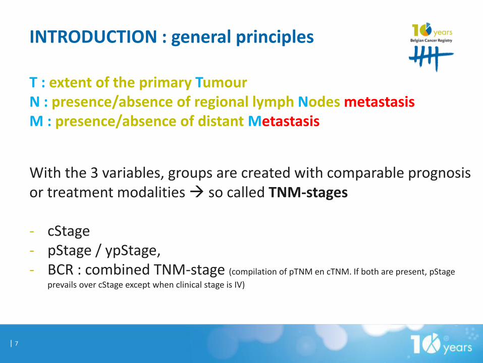
Cases from 45 sources in 20 countries 1990-2000
15
III
IV
THE USE-FULLNESS OF TNM-VARIABLES O
vera
ll su
rviv
al
Number of years
TNM 7th edition
Cases from 45 sources in 20 countries 1990-2000

16
THE USE-FULLNESS OF TNM-VARIABLES

17
THE USE-FULLNESS OF TNM-VARIABLES

18
Distribution of clinical stage (incidence 2010-2011)
Part of figure 23

GOOD NEWS : Clinical stage availability
19

20
By adding “stage by source” to ‘stage calculated by BCR’
GOOD NEWS : availability of stage information
topo histo /3 inc year cT cN cM pT pN pM cStage by BCR
pStage by BCR
COMBstage by BCR
Stage by source
349 8000 2014 NS NS NS NS NS NS X X X IV
349 8041 2014 NS NS NS NS NS NS X X X IV
341 8041 2014 NS NS NS NS NS NS X X X IV
343 8140 2014 x 0 x NS NS NS X X X IV
349 8041 2014 NS NS NS NS NS NS X X X IV
343 8481 2014 NS NS NS NS NS NS X X X IV
341 8140 2014 NS NS NS NS NS NS X X X IIIB
343 8041 2014 NS NS NS NS NS NS X X X IV
Field for ‘other classifications’ or ‘remark’

GOOD NEWS : availability of stage information
21
Stage IV any T, any N, M1 Stage IIA T2b N0 M0 or T1a/b N1 M0 or T2a N1 M0 Clinical stage or pathological stage ?

22
Inc year 2014 : LUNG TUMOURS (stageable)
CIB2014 with stages calculated by BCR
Missing comb stage 860 (10 %)
% really missing stages after adding TNM stage by source
790 (9,3 %)
Added stages
Stage I 5
Stage III 9
Stage IV 56
20 % not stage IV

23
RECURRENT PROBLEMS
RECURRENT PROBLEMS
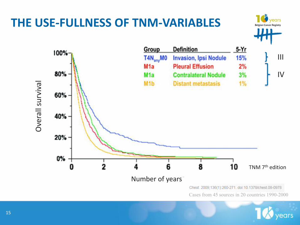
| 24
1) No TNM variables

RECURRENT PROBLEMS
| 25
1) No TNM variables
Problems : - How to calculate QI (treatment following guidelines based on stage)? - How to know your patient population ? - How to interpret survival results ? Solution : - wait until staging examinations are done - wait until surgery is executed and AP-report is available - in case of referral to other centre : please mention ! - ask a question in case of difficulties to assign a TNM

RECURRENT PROBLEMS
| 26
2) No cTNM when pTNM is present

RECURRENT PROBLEMS
| 27
2) No cTNM when pTNM is present Both are important ! - cTNM will help do decide if surgery is indicated ( Quality Indicators) - pTNM will help to decide if adjuvant treatment is necessary and gives more accurate prognostic information - cTNM maybe different from pTNM preop understaging preop overstaging … if cTNM = pTNM … ‘rather suspicious’ for
RECURRENT PROBLEMS
| 28
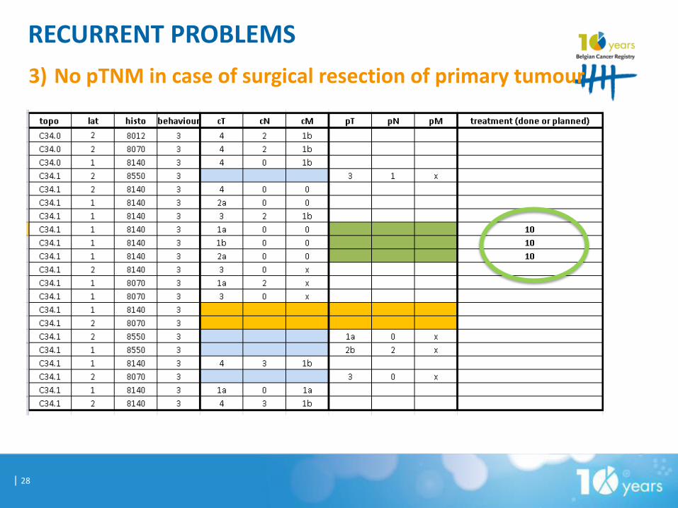
3) No pTNM in case of surgical resection of primary tumour

RECURRENT PROBLEMS
| 29
3) No pTNM in case of surgical resection of primary tumour Solution : - wait until surgery is executed and AP-report is available - in case of referral to other centre : please mention ! - use “10” in correct manner : only for surgery of the primary tumour, not for staging surgical procedures (mediastinoscopy, thoracoscopy, lymph node removal,…) : surgical staging is part of clinical staging !

RECURRENT PROBLEMS
| 30
4) Presence of pTNM without evidence of surgical procedure is (rarely) possible
pT = only possible after resection of the primary tumour OR a biopsy allowing to evaluate the highest T-category eg : CT-scan : lung tumour possibly invading oesophagus Biopsy of nodule in oesophagus = ingrowth of lung tumour cT4 Even when no surgery pT4 can be registered because of microscopic proof of the highest pT category

RECURRENT PROBLEMS
| 31
5) pN without pT Information about lymph nodes obtained by - physical examination - imaging - endoscopy (EBUS/EUS) - mediastinoscopy, mediastinotomy, thoracoscopy, surgical exploration,…
And no further surgical intervention on primary tumour
7th edition of TNM, page 8: An excisional biopsy of a lymph node without pathological assessment of the primary is insufficient to fully evaluate the pN category and is a clinical classification, in other words: no pN without pT. Thus, in this cases cN should be used.
APD
pN or cN ?

RECURRENT PROBLEMS
| 32
6)Copy-paste of cTNM pTNM

RECURRENT PROBLEMS
| 33
7)Wrong TNM-variables • Not existing values (eg T3a, T4b,…) • Wrong choice of value
RECURRENT PROBLEMS
| 34
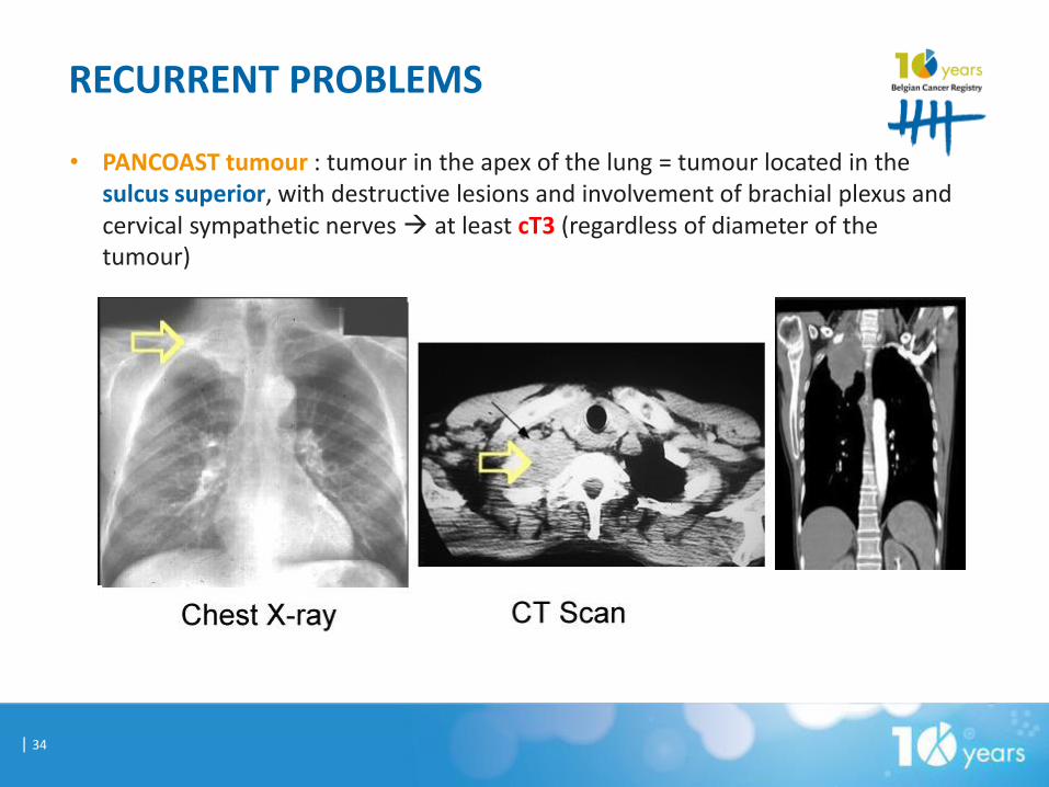
• PANCOAST tumour : tumour in the apex of the lung = tumour located in the sulcus superior, with destructive lesions and involvement of brachial plexus and cervical sympathetic nerves at least cT3 (regardless of diameter of the tumour)

RECURRENT PROBLEMS
| 35
• Do not forget ATELECTASIS OR OBSTRUCTIVE PNEUMONITIS !
Regardless diameter of lung tumour : In 7th edition :
- atelectasis/pneumonitis extending to the hilus but not involving entire lung at least cT2/pT2 - atelectasis/pneumonitis involving entire lung at least cT3/pT3 (changed in 8th edition also T2)

RECURRENT PROBLEMS
| 36
• Be careful with SYNCHRONOUS BILATERAL LESIONS !
1) Bilateral lesions with PROVEN same histology 1 tumour in a metastatic setting at least cM1a AND pM1a
2) Bilateral lesions but histology of one or both lesions unknown
considered to be the same histology 1 tumour in a metastatic setting at least cM1a
3) Bilateral lesions but PROVEN different histology (histological FAMILY) 2 primary tumours each with own TNM stage
VERY IMPORTANT IMPLICATIONS FOR TREATMENT / PROGNOSIS

RECURRENT PROBLEMS
| 37
• Make a clear difference between NEW LESIONS AND RECURRENT LESIONS !
1) New lesion in lung after previous one and PROVEN same histology (regardless laterality) 1 tumour with recurrent lesion (no New Diagnosis) maybe rTNM (not asked by BCR)
2) New lesion in lung after previous one and PROVEN ≠ histology (regardless laterality) New Diagnosis
3) New lesion in lung after previous one and no histology available (regardless laterality) histology considered to be the same 1 tumour with recurrent lesion (no New Diagnosis) maybe rTNM (not asked by BCR)
• Make a clear difference between METASTASIS IN LYMPH NODES (REGIONAL N) AND AT A DISTANCE (non-regional LN included M) Eg. metastatic ipsilat hilar LN = N1, not M1

TNM : a fascinating but never ending story…..
38

CASE 1 :
39
CT-scan : - Tumour of 2,5 cm in upper lobe of right lung + nodule of 1 cm in
lower lobe of right lung - Enlarged mediastinal lymph nodes - No other lesions observed Bronchoscopy + biopsy of nodule in upper lobe EUS : punction of ipsilateral mediastinal lymph nodes APO lung biopsy : adenocarcinoma APO EUS : compatible with metastasized adenoca of lung
How to stage this tumour ? 1) cT1N2M1 2) cT4NxM0 + pT_N2M_ 3) cT4N2M0 4) cT4N2M1 + pT_N_M1

ANSWER TO CASE 1 :
40
CT-scan : - Tumour of 2,5 cm in upper lobe of right lung + nodule of 1 cm in lower lobe of right
lung cT4 (no pT4 since no microscopic proof of second nodule) - Enlarged mediastinal lymph nodes - No other lesions observed cM0 Bronchoscopy + biopsy of nodule in upper lobe EUS : punction of ipsilateral mediastinal lymph nodes APO lung biopsy : adenocarcinoma APO EUS : compatible with metastasized adenoca of lung cN2 (regional node metastasis)
How to stage this tumour ? 1) cT1N2M1 (wrong : no metastasis at a distance) 2) cT4NxM0 + pT_N2M_ (wrong : EUS provides information for
clinical staging) 3) cT4N2M0 = correct 4) cT4N2M1 + pT_N_M1 (wrong = no proof of distant metastasis)

CASE 2 :
41
CT-scan : - Pneumonitis of left upper lobe due to obstruction by tumour of 2,5 cm - Enlarged mediastinal lymph nodes - No other lesions observed Bronchoscopy + biopsy of tumour : APO : spinocellular carcinoma Mediastinoscopy : biopsy of multiple LN APO : 1 node paratracheal right positive for meta of spinocellular ca
How to stage this tumour ? 1) cT1N0M1 2) cT2N2M0 3) cT1N3M0 4) cT2N3M0

Answer to CASE 2 :
42
CT-scan : - Pneumonitis of left upper lobe due to obstruction by tumour of 2,5 cm - Enlarged mediastinal lymph nodes at least cT2, regardless 2.5 cm - No other lesions observed M0 Bronchoscopy + biopsy of tumour : APO : spinocellular carcinoma Mediastinoscopy : biopsy of multiple LN APO : 1 node paratracheal right positive for meta of spinocellular ca
How to stage this tumour ? 1) cT1N0M1 (wrong : pneumonitis overrules size ; no distant
metastasis) 2) cT2N2M0 (N3 because of contralateral mediastinal) 3) cT1N3M0 (wrong : pneumonitis overrules size) 4) cT2N3M0 (in the TNM 8th edition cT2aN3M0)
= contralateral mediastinal