Embed Size (px)
Citation preview
36 BIOCIfflMICA ET BIOPHYSICA ACTA
T H E T E R M I N A L R E S I D U E S OF W H E A T G ERM R I B O N U C L E A T E S
]3. G, L A N E * AND F R A N K W O R T H I N G T O N A L L E N
Department o] Biochemistry, University o/ Cali/ornia School o/Medicine, San Francisco, Gali[. (U.S.A.)
(Received J u n e 12, 196o)
S U M M A R Y
I. Ribonucleates have been extracted from wheat germ and separated into high and low molecular weight fractions by selective precipitation from aqueous sodium chloride solution.
2. The high molecular weight ribonucleates yielded one nucleoside residue for about twelve hundred nucleotide residues after hydrolysis by alkali while the low molecular weight ribonucleates yielded one nucleoside residue for about ninety nucleotide residues.
3. The high molecular weight ribonucleates yielded roughly equal amounts of the four major ribonucleosides when hydrolyzed by alkali whereas the amount of adenosine derived from the low molecular weight ribonucleates was greater than the combined amount of the other nucleosides.
4 -Both high and low molecular weight ribonucleates yielded nucleoside di- phosphates when hydrolyzed in alkali. The amount of guanosine diphosphate exceeded the combined amount of all other nucleoside diphosphates. The quanti ty of nucleoside diphosphates derived from the low molecular weight ribonucleates was much larger than the quanti ty from the high molecular weight ribonucleates as would be expected if they were derived from terminal positions.
INTRODUCTION
I t is generally assumed that the ribonucleates are terminated at one end by a nucleo- side or phosphonucleoside residue to be termed the T-terminal residue which is linked through its 3' position via a phosphodiester bridge to the 5' position of the adjacent residue, and at the other end by a nucleoside or phosphonucleoside residue to be termed the F-terminal residue which is joined through its 5' position to the 3' position of the adjacent nucleoside residue. This situation is illustrated in Fig. x.
Since the early work of MARKHAM AND SMITH 1, it has been demonstrated repeatedly that hydrolysis of ribonucleates in alkali gives rise to a small amount of nucleosides and nucleoside diphosphates which are considered to arise from the F-terminal and T-terminal residues, respectively 2-7. Indeed the absence of nucleosides and nucleoside diphosphates in the alkaline hydrolysates of the ribonucleates of
* P re sen t address : Rockefel ler In s t i t u t e , New York 21, N.Y. , U.S.A.
Biochim. Biophys. Acta, 47 (1961) 36 46

TERMINAL R E S I D U E S O1 v W H E A T GERM RIBONUCLEATES .37
Tobacco Mosaic Virus led REDDI AND KNIGHT = and MATTHEWS AND SMITH a to con- clude that there were no F-terminal nucleoside or T-terminal phosphonucleoside residues in these ribonucleates within the limits of detection.
"T" TE RMINU~,"
B[ B B
3' (P) P P
II"F" TERMINU~
B B ] B
. . . . . . . . . . . . . p P (P) - , i " 4 , -, i 51
Fig. 1,
Degradation of the ribonucleates of yeast with pancreatic ribonuclease has been found to give rise to nucleosides and dinucleoside monophosphates s. I t is believed that the nucleosides arise from F-terminal nucleoside residues linked to subterminal pyrimidine nucleoside residues and that the dinucleoside phosphates arise from the combination of an F-terminal nucleoside residue with a subterminal purine nucleoside which is in turn linked to a pyrimidine nucleoside residue. There is evidence that the same ribonucleates from yeast have F-terminal residues esterified at the 2' or 3' positions with phosphomonoester groups in view of the findings of CRESTFIELD AND ALLEN 9 that the diphosphonucleosides of adenine, guanine, cytosine and uracil are present in digests of the ribonucleates with snake venom. The presence of adenosine and guanosine 2' ,3'-phosphates in digests of yeast ribonucleates with pancreatic ribo- nuclease led MARKHAM AND SMITH to conclude z that some F-terminal residues may bear a cyclic phosphodiester group between the 2' and 3' positions.
When the method of LASKOV et al. 1° was applied to wheat germ, a preparation of ribonucleates was obtained which could be conveniently fractionated by selective precipitation of the high molecular weight ribonucleates from an aqueous 3 M sodium chloride solution in which the relatively short chain ribonucleates remained soluble. The high molecular weight ribonucleates insoluble in aqueous 3 M sodium chloride solution (to be termed the insoluble ribonucleates) are thought to be derived primarily from the ribosomes and the low molecular weight ribonucleates (to be termed the soluble ribonucleates) correspond to the ribonucleates of the soluble fraction of the cell (sRNA). The intention of the present investigation was to study the nucleo- side residues in the F-terminal position and the phosphonucleoside residues in the T-terminal position by employing the technique of hydrolysis in alkali. The emphasis of this s tudy was placed on the insoluble ribonucleates since there have been relatively few reports in the recent literature on the end group studies of such high molecular weight preparations n, 12.
In accordance with numerous reports by other workers on preparations of sRNA6,7,13-15, it was found that the predominant F-terminal end group of the soluble ribonucleates of wheat germ was adenosine, the predominant T-terminal residue was guanosine-5'-phosphate7,16 and the amount of nucleosides was consistent with a chain length of about 9 ° residues.
The insoluble ribonucleates from wheat germ had a roughly equimolar distribu- tion of adenosine, guanosine, cytidine and uridine in the F-terminal position, and
Biochim. Biophys. Acta, 47 (19¢~li 36 46
~8 B . G . LANE, F. W. ALLEN
guanosine-5'-phosphate was the pr imary T-terminal residue. The quantitat ive estimates indicated that there was one nucleoside residue for about I~OO nucleotide residues in the hydrolysates of the insoluble ribonucleates.
Incidental to this investigation, it was found that evidence could be adduced, on the basis of the quantitat ive data for terminal residues and for 5-ribosyluracil phosphate, that 5-ribosyluracil is probably a covalently linked component of the high molecular weight ribonucleates is.
MATERIALS AND METHODS
Preparation of ribonucleates from wheat germ
Fresh wheat germ can be used for the isolation of ribonucleates but it was found that the yields were improved when the wheat germ was first de-fatted. The procedure used to de-fat the wheat germ consisted of extracting IOO g of fresh wheat germ with 500 ml of a solution containing equal volumes of ethanol and ether. The suspen- sion was allowed to stand for one hour at room temperature with occasional stirring and was then filtered. The wheat germ from the first extraction was extracted once again in the same manner and then dried in air.
The procedure employed for extracting the ribonueleates was based upon the published procedure of LASKOV et al. 1° for isolating the ribonucleates of rat liver. 5 ° g of de-fatted wheat germ were suspended in 300 ml of IO 4 M ethylenediamine- tetraacetate, pH 8, mixed immediately with 300 ml of 9 ° ~:/o phenol and homogenized in a Waring blendor for 5 rain. The phenol employed in this procedure was white, crystalline, reagent grade material (Baker and Adamson Co.) and was not re-distilled. The suspension was stirred for 60 rain at room temperature and then centrifuged for 3 rain at IO,OOO ~; g at 4 °. The upper aqueous phase (pH 6-6.5) was siphoned off and the lower phenol phase and cell debris were extracted with 200 ml of IO ~ M ethylenediaminetetraacetate, pH 8 and the suspension was centrifuged for 3 min at IO,OOO × g at 4 °. The remainder of the procedure was conducted at 4 °. The aqueous supernatant phases from the two centrifugations were pooled and centrifuged for 20 rain at IO,OOO x g. The supernatant solution from this centrifugation was made 3 M by the addition of solid sodium chloride and centrifuged immediately at 6,000 x g. The supernatant solution was withdrawn and allowed to stand for IO rain before removing phenol crystals by filtration. The filtrate was allowed to stand at 0-2 ° for 3 days during which time a precipitate formed which was recovered by centrifugation for 5 rain at IO,OOO >~ g. The pellet from this centrifugation was washed by stirring in 40 ml of 67 ~o ethanol and the suspension was centrifuged at ~,ooo rev./min in a clinical type centrifuge. This procedure was repeated once more with 67 °/o ethanol, twice with 95 ~o ethanol, twice with absolute ethanol and twice with ether before drying under a stream of nitrogen. The white powder which was obtained is termed the insoluble ribonueleates in this paper. The washing procedure removed salt, phenol and certain flavone-like compounds which became yellow in alkaline solution and were not removed by extensive dialysis. The dry white powder became gelatinous when suspended in water and dissolved to give a clear, colorless solution. When the insoluble ribonucleates were hydrolyzed in I M alkali for 24 h
o/ solution increased from at room temperature, the extinction coefficient of a I /o 216 to 286 (u.v.-absorption was measured in 0. 5 M phosphate buffer, pH 7 at 260 m/~).
Biochi~2. Biophys. ~ eta, 47 {190 ~ ) 3 ° -40
T E R M I N A L R E S I D U E S OF W H E A T GERM R I B O N U C L E A T E S .)~)
The soluble ribonucleates remaining in the 3 M sodium chloride solution were precipitated with 2 volumes of 95 % ethanol and were washed and dried by the pro- cedure described for the insoluble ribonucleates. A I °:o solution of the white powder containing the soluble ribonucleates had an extinction coefficient of lO-2O at 26o m~ in o.5 M phosphate buffer, pH 7- The soluble ribonucleates, therefore, comprised only about io °/o of the dry weight of this powder, and no removal of non-dialyzable, non-nucleate material was effected prior to hydrolysis in alkali.
The preparation of insoluble ribonucleates was essentially all nucleate material (extinction coefficient of a I O/0 solution being 216 at 26o m/z) and the yield of 400 mg from 50 g of wheat germ accounted for roughly 3o o; of the wheat germ ribonucleates. The soluble ribonucleates accounted for roughly IO (~'o of the wheat germ ribonucleates.
This investigation was undertaken as an exploratory a t tempt to obtain quantita- tive and qualitative data on the terminal residues of a preparation of high molecular weight ribonucleates insoluble in aqueous 3 M sodium chloride, and as such, the extraction of wheat germ with aqueous phenol served as a relatively simple device for obtaining quantities of material suitable for analysis. SII~ATANI and co-workers have very recently shown that aqueous phenol extraction of mammalian tissues results in a discrete fractionation of tissue ribonucleates with widely differing rates of metabolic renewaW -19. I t is tentatively assumed by analogy with the work of SIBATANI et al. that the high molecular weight ribonucleates studied in this work correspond to the ribonucleates with a low rate of metabolic renewal. An investigation is currently under way to ascertain if the aqueous phenol extraction of wheat germ does effect an analogous fractionation since the singularly large quanti ty of nuclear ribonucleates in wheat embryo (50 %0 of total) 2°,21, and the large fraction of ribo- nucleates not extracted by aqueous phenol (60 (!~, of total) indicate that this embryo may be particularly useful for isolating large amounts of the ribonucleates exhibiting a rapid rate of metabolic renewal, particularly" in view of the report that nuclei and associated nucleolar ribonucleates can resist extraction into the aqueous phase during the extraction with aqueous phenol ~2.
Analysis for pyrimidine 1,ucleotides and purine bases
The insoluble ribonucleates were analyzed according to the procedures of MARKHAM AND SMITH ~a. The samples of soluble ribonucleates were hydrolyzed to the constituent mononucleotides by alkali and the bulk of the non-nucleate material was precipitated with acid as described below. The solution of acid soluble nucleotides was then analyzed by the methods of Markham and Smith.
Hydrolysis of ribonucleates in alkali
Two hundred milligrams of the insoluble ribonucleates were dissolved in 20 ml of distilled water before the addition of 2.65 ml of 8.6 M potassium hydroxide. The solution was allowed to stand for 24 h (or longer) at 22 ° and then the solution was adjusted to pH 2 by the addition of concentrated perehloric acid solution at 2 ~'. The solution was centrifuged for 5 rain at z,ooo rev./min using a clinical type centrifuge and the supernatant solution was drawn off. The sediment was washed twice bv stirring with Io-ml portions of water and the supernatant solutions from the washinga were combined with the supernatant solution from the first centrifugation. The com- bined supernatant solutions were adjusted to pH 7 by the addition of potassium
Hi.ck im. Biopkvs. .tcta, 47 (I~¢~1) 3~ -4b

4o B . G . LANE, F. W. ALLEN
hydroxide solution and the neutralized hydrolysate was frozen. The frozen solution was melted and centrifuged to remove the additional potassium perchlorate which precipitated when the temperature was lowered.
Analysis for trace nucleotide components
The trace components were estimated using the two-dimensional paper chro- matographic procedure employed by DAVIS, CARLUCCI AND ROUBEIN for this purpose 2~. The hydrolysis products from 2.5 mg of ribonucleates were applied to Whatman No. I paper for chromatographic resolution.
Analysis for the nucleosides present in alkaline hydrolysates of ribonucleates
Direct paper chromatography of the neutralized hydrolysates of soluble ribo- nucleates was usually satisfactory; however, the amount of material which had to be used in order to detect the nucleosides present in neutralized hydrolysates of the insoluble ribonucleates was very large and led to difficulties of solvent penetration at the origin of paper chromatograms and diffuseness in the nucleoside bands. For these reasons a preliminary separation of nucleosides from nucleotides was carried out using DEAE-cellulose anion exchange columns. DEAE-cellulose was preferable to polystyrene anion exchange resins for this purpose since the interaction of guanosine with the polystyrene backbone of such resins impedes elution.
The nucleoside fraction from the DEAE-cellulose column was concentrated and de-salted by adsorption on and elution from charcoal followed by paper chromato- graphic resolution of the individual nucleosides.
(i) Adsorption on DEAE-cellulose. A portion of the neutralized hydrolysate (which had been frozen and melted as indicated earlier) of the insoluble ribonucleates containing 4oo/~g of nucleotide in about 4 ° m l was charged onto a 2.5 cm x 2o-cm column of DEAE-cellulose pre-equilibrated with o.o25 M ammonium acetate, pH 7. The DEAE-cellulose (selectacel, purchased from Brown Co.) column was prepared according to a published procedurO 6. Elution was continued with o.o5 M ammonium acetate, pH 7 until 4oo ml of effluent, containing all of the nucleosides present in the hydrolysate, had been collected.
(ii) Adsorption on charcoal. An aqueous suspension of coarse celite (No. 545) was poured into a chromatographic tube 2.5 cm in diameter, and a pressure of 5 lbs/in~' was applied to force the water out of the column and form a pad of moist celite about 3 cm deep on the sintered glass disc of the chromatographic tube. An aqueous suspension of 50 mg of charcoal in IO ml of water was carefully drained from a pipette onto the surface of the celite pad and water was forced from the column in order to form a disc of moist charcoal completely covering the celite pad. Two discs of filter paper cut to the diameter of the chromatographic tube were placed on the surface of the charcoal pad to prevent any slurrying of the charcoal when solutions were added to the column. Darco G-6o charcoal was employed and was washed according to a published procedurO ~. Care was taken in preparing the column to maintain a uniform contact of the circumference of the celite and charcoal discs with the inside of the chromatographic tube.
The effluent containing the nucleosides (about o.35/~g) from the DEAE-cellulose column was passed through the disc of charcoal at a flowrate of about IOO ml/min under a pressure of 5 lbs/inc. The effluent from this first filtration was passed through
t3iochim. Biophys. .4 c/a, 47 11961 ) 36-46

T E R M I N A L R E S I D U E S OV W H E A T GERM R I B O N U ( ' L E A T E S 41
the charcoal and then 15 ml of water were passed through the charcoal disc to remow~ residual salt. The nucleosides were eluted with IOO ml of a solution made by mixing 45 ml of water, 45 ml of ethanol and IO ml of pyridine. The effluent containing the nucleosides was evaporated in a flash evaporator at 4 o°. The dry residue after evapora- tion was dissolved in a total volume of 2 ml of water and streaked as a 5-cm band on Wha tman No. I MM paper for resolution of nucleosides by paper chromatography.
(iii) Resolution of nucleosides by paper chromatography. Descending chromatog- raphy on paper employing a solvent prepared by mixing 80 volumes of saturated aqueous ammonium sulphate, 18 volumes of aqueous I M sodium acetate and 2 volumes of isopropanol 2s separated the nucleosides into three bands. In order of increasing RF value, the first band contained adenosine, the second band guanosine and the third contained cytidine and uridine. The spectral ratios of the adenosine and guanosine bands were in agreement with published values and the values for the third band were what would have been expected for a mixture of roughly equiva- lent amounts of eytidine and uridine. The pyrimidine nucleosides were resolved by paper chromatographic development with either an aqueous HCl-tert . butanol solvent 2a or with a butanol-water solvent 26. Alternatively the pyrimidine nucleosides were separated by electrophoresis on paper using o.05 M potassium borate buffer, pH 9.2 (see ref. 27).
Each step of the fractionation procedure (fractionation on DEAE-cellulos(~, charcoal adsorption and elution, elution from paper chromatograms) has been shown to be quantitative. The soluble ribonucleates were dialyzed prior to hydrolysis in order to ensure adequate removal of salt which, if present, interfered with adsorption of nucleotides on DEAE-cellulose.
Analysis for nucleoside diphosphates present in alkaline hydrolysates of ribonucleates
The nucleoside diphosphates present in the neutralized hydrolysates of the soluble ribonucleates were separated directly by electrophoresis of the unfractionated hydrolysates on paper, using 0.05 M ammonium formate buffer, pH 9.2.
The diphosphates present in the neutralized hydrolysates of the insohtble ribonucleates were detected following all initial fractionation of the hydrolysates on DEAE-cellulose. The nucleosides were eluted from DEAE-eellulose with 0.025 M ammonium acetate, pH 7 and nucleotides were eluted in two peaks by 0.075 M ammonium acetate, pH 7; the pyrimidine mononucleotides appeared in the first peak and the purine mononucleotides were present in the second peak. The material remaining on the DEAE-cellulose following elution of the mononueleotides (the postmononueleotide fraction) was eluted by an aqueous solution containing 0.6 M sodium sulphate and I M acetic acid. The postmononucleotide fraction was desalted by adsorption on charcoal, the eluate from the charcoal was evaporated and the dry residue was dissolved in water for electrophoretic separation on paper using 0.05 M ammonium formate buffer, pH 9.2.
Sedimentation data
The sedimentation coefficient was estimated by means of a technique applying; ultraviolet light optical methods to the sedimentation of ribonueleates at low con-- eentrations 2s.
Biochim. Biophys, Acta, 47 (TO61) 36 46
4 2 B . G . L A N E , F. W. A L L E N
R E S U L T S AND DISCUSSION
Quantitative estimates of F-terminal nucleoside residues and of chain length
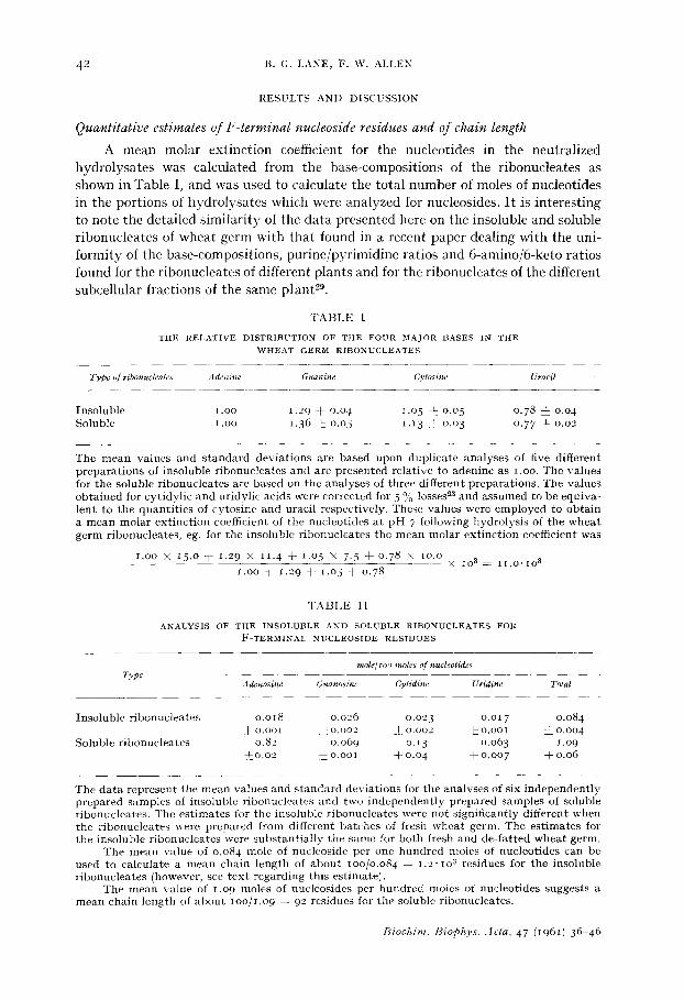
A mean molar extinction coefficient for the nucleotides in the neutralized hydrolysates was calculated from the base-compositions of the ribonucleates as shown in Table I, and was used to calculate the total number of moles of nucleotides in the portions of hydrolysates which were analyzed for nucleosides. It is interesting to note the detailed similarity of the data presented here on the insoluble and soluble ribonucleates of wheat germ with that found in a recent paper dealing with the uni- formity of the base-compositions, purine/pyrimidine ratios and 6-amino/6-keto ratios found for the ribonucleates of different plants and for the ribonucleates of the different subcellular fractions of the same p lan t 29.
T A B L E I
THE RELATIVE DISTRIBUTION OF THE FOUR MAJOR BASES IN THE ~rHEAT GERM RIBONUCLEATES
Type of ribonuch'ates Adenine Guanine Cytosine Urazil
I n s o l u b l e l . o o 1 .29 :k 0 .o4 1 .o5 ~: o .o5 o . 7 8 j : o .o 4 S o l u b l e l . o o 1 .36 ~: o .o 5 1.13 2 o .o 3 o .77 ± o .o2
T h e m e a n v a l u e s a n d s t a n d a r d d e v i a t i o n s a r e b a s e d u p o n d u p l i c a t e a n a l y s e s of f ive d i f f e r e n t p r e p a r a t i o n s of i n s o l u b l e r i b o n u c l e a t e s a n d a r e p r e s e n t e d r e l a t i v e t o a d e n i n e a s I.OO. T h e v a l u e s f o r t h e s o l u b l e r i b o n u c l e a t e s a r e b a s e d o n t h e a n a l y s e s of t h r e e d i f f e r e n t p r e p a r a t i o n s . T h e v a l u e s o b t a i n e d fo r c y t i d y l i c a n d u r i d y l i c a c i d s w e r e c o r r e c t e d fo r 5 o~ losses~3 a n d a s s u m e d t o b e e q u i v a - l e n t t o t h e q u a n t i t i e s of c y t o s i n e a n d u r a c i l r e s p e c t i v e l y . T h e s e v a l u e s w e r e e m p l o y e d t o o b t a i n a m e a n m o l a r e x t i n c t i o n coeffÉcient of t h e n u c l e o t i d e s a t p H 7 f o l l o w i n g h y d r o l y s i s of t h e w h e a t g e r m r i b o n u c l e a t e s , eg. f o r t h e i n s o l u b l e r i b o n u c l e a t e s t h e m e a n m o l a r e x t i n c t i o n c o e f f i c i e n t w a s
I.OO ;K 15 .o @ 1 .29 x 11. 4 @ 1.o 5 x 7.5 @ o .78 >~ lO.O × 1° 3 = i i . o . i o 3
1 .oo + 1 .29 + 1.o5 + 0 .78
T A B L E I I
ANALYSIS OF THE INSOLUBLE AND SOLUBLE RIBONUCLEATES FOR F-TERMINAL NUCLEOSIDE RESIDUES
Type mole/zoo moles of nucleotides
Adenosine G,canosine Cytidine Uridine Total
I n s o l u b l e r i b o n u c l e a t e s o . o i 8 0 . 0 2 6 0 . 0 2 3 o .o 17 0 . 0 8 4 ~: o . o o l ± 0 . 0 0 2 k 0 . 0 0 2 ~ o . o o i ~_ 0 . 0 0 4
S o l u b l e r i b o n u c l e a t e s 0 .82 0 . 0 6 9 o. 13 0 . o 6 3 t . o 9 ~ o . o 2 + o . o o l ± 0 .04 ~ 0 . 0 0 7 ± 0 .06
T h e d a t a r e p r e s e n t t h e m e a n v a l u e s a n d s t a n d a r d d e v i a t i o n s f o r t h e a n a l y s e s o f s ix i n d e p e n d e n t l y p r e p a r e d s a m p l e s of i n s o l u b l e r i b o n u c l e a t e s a n d t w o i n d e p e n d e n t l y p r e p a r e d s a m p l e s of s o l u b l e r i b o n u c l e a t e s . T h e e s t i m a t e s f o r t h e i n s o l u b l e r i b o n u c l e a t e s w e r e n o t s i g n i f i c a n t l y d i f f e r e n t w h e n t h e r i b o n u c l e a t e s w e r e p r e p a r e d f r o m d i f f e r e n t b a t c h e s o f f r e s h w h e a t g e r m . T h e e s t i m a t e s f o r t h e i n s o l u b l e r i b o n u c l e a t e s w e r e s u b s t a n t i a l l y t h e s a m e f o r b o t h f r e s h a n d d e - f a t t e d w h e a t g e r m .
T h e m e a n v a l u e of o . o 8 4 m o l e o f n u c l e o s i d e p e r o n e h u n d r e d m o l e s of n u c l e o t i d e s c a n b e u s e d t o c a l c u l a t e a m e a n c h a i n l e n g t h of a b o u t lOO/O.O84 = 1 . 2 - 1 o s r e s i d u e s f o r t h e i n s o l u b l e r i b o n u c l e a t e s ( h o w e v e r , see t e x t r e g a r d i n g t h i s e s t i m a t e ) .
T h e m e a n v a l u e of 1 .o9 m o l e s of n u c l e o s i d e s p e r h u n d r e d m o l e s of n u c l e o t i d e s s u g g e s t s a m e a n c h a i n l e n g t h of a b o u t 1 o o / i . o 9 = 92 r e s i d u e s f o r t h e s o l u b l e r i b o n u c l e a t e s .
B i o c h i m . B i o p h y s . . 4 c l a , 4 7 (1961) 3 6 - 4 6

TERMINAL RESIDUES OF WHEAT GERM RIBONU('LEATES 43
The data for the F-terminal nucleoside residues for two preparations of soluble ribonucleates are summarized in Table I I and are in general agreement with the results in the literature which show a large preponderance of adenosine residues. The chain length estimate of about 9 ° residues is based on the assumptions that there are no significant amounts of F-terminal phosphonucleoside residues and that the nucleosides are derived from chains which are representative of the mean chain length for the preparation.
The data for the F-terminal nucleoside residues of several preparations of the insoluble ribonucleates are summarized in Table II. I t is clear that there are roughly equivalent amounts of the four nucleosides in the F-terminal position. There is also an indication that the frequency of occurrence of the different nucleosides in the F-terminal position tends to parallel the relative frequency of occurrence of the corresponding purines and pyrimidines in the ribonucleate chains.
Owing to the trace nature of the nucleosides in the hydrolysates of the insoluble ribonucleates, it was considered advisable to demonstrate that they did not result from either trace dephosphorylation of nucleotides or from an unusual cleavage of the internucleotide phosphate linkages that yielded a nucleoside and a nucleoside diphosphate, It was established that no detectable dephosphorylation occurred in synthetic mixtures of nucleotides under the conditions employed for hydrolysis and further that there was no significant increase in the amount of nucleosides present in 48-h hydrolysates as compared with 24-h hydrolysates of the same preparation of insoluble ribonucleates. The data of REDDI AND KNIGHT 2 support the foregoing observations since no nucleosides or nucleoside diphosphates were found in hvdro- lysates of the ribonucleates of Tobacco Mosaic Virus under conditions of hydrolysis similar to those employed in this work. Additionally, the data of REDDI and KNmHT provide evidence that nucleosides and nucleoside diphosphates do not arise from an unusual splitting of the phosphodiester linkages of the ribonucleates.
When the insoluble ribonucleates were re-dissolved in water and then re- precipitated by making the solution 3 M with respect to sodium chloride, the values for adenosine decreased by about 25 O/:o while the values for the other nucleosides remained unaltered. Further dissolution and precipitation in this manner caused no further changes in the quantities of the nucleosides. The values quoted in Table II have been corrected for this small contamination of the insoluble ribonucleates with soluble ribonucleates.
An estimate of a mean chain length of about 1.2. lO 3 residues for the insoluble ribonucleates could be made on the basis of the assumptions stated earlier for the mean chain length estimate for the soluble ribonucleates. This would indicate a mean molecular weight of about 4.1o ~ for the insoluble ribonucleates of wheat germ. Although no detailed physical-chemical study was made, a single ultracentrifugal experiment indicated a mean sedimentation coefficient of 8 S for the insoluble ribonucleates. It is noteworthy in this regard that ribonucleates with a mean sedi- mentation coefficient of 8 S and a mean molecular weight of 1.2.1o 5 have been proposed as the basic structural unit of liver ribosomal ribonucleates by HALL AND DOTY 30.
I t has not been demonstrated that the F-terminal nucleoside residues do not arise from dephosphorylation of phosphonucleoside residues during preparation of the ribonucleates; however, the reasonably good quantitative consistency of the
Biochim. Biophys. Acta, 47 (1061) 3(' 4~

44 B.G. LANE, F. W. ALLEN
analyses for different preparations supports tile suggestion that the native ribo- nucleates do possess F-terminal nueleoside residues such as would be anticipated for the F-terminus of ribonucleates which would be produced by the currently known ribonucleotide polymerases and by the enzymes effecting incorporation of ribonucleotides into an F-terminus ~,31,32. The quantitat ive consistency of the data suggests that ribonucleates with a molecular weight of about 4"Io5 may represent synthetic units which are subsequently cleaved into smaller units having a mean sedimentation coefficient of about 8S (the F-terminal residues resulting from such cleavages being phosphonucleosides*). Although no strong claim for this interpreta- tion of the data can be made it is noted that the major component, of the ribonucleates isolated by LITTAUER AND EISENBERG 33 from Escherichia coli had a mean sedimenta- tion coefficient of 14.2 S and were estimated by physical-chemical procedures to have a molecular weight of 3.5 to 4.5" lOS.
Estimates of T-terminal phosphonucleoside residues
The analyses for three independently prepared samples of soluble ribonucleates indicated that there was 0.58 dz 0.02 mole of nucleoside diphosphate per IOO moles of nucleotides after hydrolysis by alkali. The nucleoside diphosphates had a greater mobility than any of the other products derived from the hydrolysis of ribonucleates in alkali and moved as an unresolved group during electrophoresis at pH 9.2. When the compounds of the diphosphate area were subjected to paper chromatographic separation using the saturated aqueous ammonium sulphate-aqueous molar sodium acetate-isopropanol solvent, two major spots were detected, one of which had an RF value of about 0.64 and the other had an Rv value of about 0.55. Both compounds had ultraviolet absorption spectra almost identical with those of guanylic acid at pH values of 2, 7 and 12. Both compounds were completely dephosphorylated by prostatic phosphomonoesterase at a much slower rate than the nucleoside mono- phosphates to yield guanosine. By analogy with the RE values of the guanosine 2'- and 3'-phosphates for the solvent system mentioned above, it was concluded that the compound with the higher RE value was guanosine 2',5' diphosphate and the slower moving compound was guanosine 3',5'-diphosphate. Paper chromatography using the same solvent system also indicated the presence of much smaller amounts of compounds with the RE values which would be expected for the corresponding diphosphates of adenosine, cytidine and uridine.
The analyses for the nucleoside diphosphates present in hydrolysates of the insoluble ribonucleates were conducted on the post mononucleotide fraction obtained when the neutralized hydrolysates were fractionated on DEAE-eellulose columns. The postmononucleotide fraction has been shown 16 to consist of at least three basic types of ultraviolet absorbing compounds: (a) alkali-labile dinucleotides, (b) alkali- stable dinucleotides and (c) the nucleoside diphosphate of guanine. Data on the post- mononucleotide fraction after different periods of hydrolysis are presented in Table I I I . The post mononucleotide fraction of the insoluble ribonucleates comprised about 7-5 % of the total ultraviolet absorption (260 m/~) of the neutralized 24-h hydrolysates
* At this early stage of investigation, it cannot be assumed that all of the terminal residues of the ribonucleates fall into the arbitrary classification outlined earlier in the paper, and the assumption that the F-terminal residues which do not appear as nucleosides after hydrolysis by alkali are necessarily simple phosphonue]eosides may be invalid.
Biochim. Hiophys. Acta, 47 (I96I) 36-46

T E R M I N A L R E S I D U E S OF W H E A T ( ;ERM R I B ( ) N U C I . E A T E S 4 5
T A B L E I I I
1)ATA ON T I l E P O S T M O N O N U C I . E O T I D E F R A C T I O N S OF T H E W H E A T G E R M R I B O N U C L E A T E S
T y p e of T i m e of hyd~olvsls In Yie ld A & ' n i n e Gu,~nine Cytosine Uracil
rihonuch'ah's 1 .cJ M K O H , 2 5
I n s o l u b l e 24 h o u r s 7 .7 % ~ .oo o.3~ o . 2 6 o . I 7 I n s o l u b l e 48 h o u r s 4 .4 % 1 .oo o . 7 o 0 .65 o .65 S o l u b l e 9 ° h o u r s 4 .o °/o I .oo 2 .44 o ,68 o .57
T h e r e l a t i v e q u a n t i t i e s of b a s e s a r e e x p r e s s e d in t e r m s of a d e n i n e a s ~ .oo.
and about 4.5 ')"o of the 48-h hydrolysates.The decreased yield of post mononucleotide fraction which is observed for 48-h hydrolysates of wheat germ ribonucleates was established to be due primarily to the hydrolysis of diadenylate present in 24-h hydrolysates.
The analysis of five independent preparations of insoluble ribonucleates indicated that there was o.o25 i O.OLO mole of nucleoside diphosphate per IOO moles of nucleotides after hydrolysis by alkali. A sample of the insoluble ribonucleates which had been precipitated from 3 M sodium chloride solution three times was found to contain o.o23 mole of nucleoside diphosphate per ioo moles of nucleotide residues after hydrolysis in alkali. Paper chromatography of the compounds of the nucleoside diphosphate area using the saturated aqueous ammonium sulphate-aqueous molar sodium acetate-isopropanol solvent showed two major spots with the same RF values and spectral properties as the compounds which have been concluded earlier in this paper to be guanosine 2',5'-diphosphate and guanosine 3',5'-diphosphate.
Other constituents present in small quantities in hydrolysates of wheat germ ribonucleates
Analysis for trace components by the procedure of DAVIS, CARLUCCI AND ROUBEIN 24 indicated the presence of the thymine, i-methyl guanine and N6-methyl adenine nucleotides, each accounting for o.5-o.8 % of the total absorption (26o m~) of the neutralized hydrolysates of the soluble ribonucleates. The unidentified nucleo- tide with the spectral properties of guanine-type compounds was present and accounted for about o.5 % of the total absorption of the hydrolysates. The foregoing compounds were not detected in the corresponding analyses of the insoluble ribo- nucleates. It is therefore concluded that the methylated base derivatives, reported originally by DUN~ et al. to be present in wheat germ ribonucleates 34,35, are confined largely to the ribonucleates from the soluble fraction of wheat germ cells, as has been shown to be the case with liver tissuO ~.
Soluble ribonucleates were found to contain about lO-I2 ~/o of their uracil in the form of 5-ribosyluracil phosphate whereas about 3-4 % of the uracil of the in- soluble ribonucleates was found in the form of 5-ribosyluracil phosphate after hydrolysis by alkali. The amount of 5-ribosyluracil phosphate was not detectably changed after the insoluble ribonucleates were re-precipitated from aqueous 3 M sodium chloride solution twice. The quanti ty of 5-ribosyluracil phosphate derived from the insoluble ribonucleates was far in excess of the amount that could have been contributed by contamination with soluble ribonucleates containing a pre-- ponderance of F-terminal adenosine residues.
Biochim. 13iophys. :lcta, 47 (1o(il) 3 ~ - 4 t~

46 B . G . LANE, F. W. ALLEN
ACKNOWLEDGEMENTS
This work was supported in part by Grant-in-Aid RG2496, U.S. Public Health Service, Grant G897I, National Science Foundation and Cancer Research Funds of the University of California.
We are indebted to Professor H. K. SCHACHMAN for the estimate of the sedimenta- tion coefficient of the high molecular weight ribonucleates.
We also wish to express our thanks to Mr. JAMES BOUDOURIS for capable technical assistance in this work.
R E F E R E N C E S
1 R. MARKHAM AND J. D. SMITH, Biochem. J., 52 (1952) 565. 2 K. K. REDDI AND C. A. KNIGHT, Nature, 18o (1957) 37,1. 3 R. E. F. MATTHEWS AND J. O. SMITH, Nature, 18o (I957) 375. 4 R. MARKHAM, R. E. V. MATTHEWS AND J. D. SMITH, Nature, 173 (I954) 537. 5 L. A. HEPPEL, J. D. SMITH, P. J. ORTIZ AND S. OCHOA. Federation Proc., 15 (1956) 273. 5 L. I. HECHT, P. C. ZAMECNIK, M. L, STEPHENSON AND J. F. SCOTT. J. Biol. Chem., 233 (1958) 954. 7 M. F. SINGER AND G. L. CANTONI, Biochim. Biophys. Acta, 39 (196o) 182. 8 A. M. CRESTFIELD AND F. W. ALLEN, Arch. Biochem. Biophys., 78 (I958) 334. 9 A. M. CRESTFIELD AND F. W. ALLEN, J. Biol. Chem., 219 (1956) lO3.
10 R. LASKOV, E. MARGOLIASH, U. Z. LITTAUER AND H. EISENBERG, Biochim. Biophys. Ac/a, 33 (I959) 247.
11 R. DULBECCO AND J. D. SMITH, Biochim. Biophys. Acta, 39 (196o) 358. 12 M . P . G O R D O N , B . SINGER AND H . FRAENKEL-CONRAT, J. Biol. Chem., 235 (196o) lOl 4. 13 H. G. ZACHAU, G. Acs AND V. LIPMANN, Proc. Natl. Acad. Sci. U.S., 44 (1958) 885. 14 j . STARR AND D. GOLDTHWAIT, Federation Proc., I9 (196o) 317 . 15 D. 13. DUNN, Biochim. Biophys. Acta, 34 (1959) 286. 16 B. G. LANE AND G. C. BUTLER, Can. J. Biochem. Physiol., 37 (1959) 1329. 17 A. SIBATANI, K. YAMANA, K. KIMURA AND H. OKAGAKI, Biochim. Biophys. Acla, 33 (1959) 590. 18 A . SIBATANI, I{. YAMANA, K. KIMURA AND T. TAKAHASHI, Nature, 186 (196o) 215. 19 K. YAMANA AND A. SIBATANI, Biochim. Biophys. Acta, 41 (I96o) 295. 20 H. STERN AND A. E. MIRSKY, J. Gen. Physiol., 36 (1952-53) i81. 21 N. M. SISAK~rAN, N. A. VASILYEVA AND G. I. SPIRIDONOVA, Biochemistry, 22 (1957) 761. 23 G. P. GEORIEV AND V. L. MANT'EVA, Biochemistry, 25 (196o) to 3 23 R. MARKHAM AND J. D. SMITH, Biochem. J., 46 (19501 509. "°4 F. F. DAVIS, A. F. CARLUCCI AND I. F. ROUBEIN, J. Biol. Chem., 234 (1959) 1525. 25 R. MARKHAM AND J. D. SMITH, Biochem. J., 52 (I952) 552. 28 R. MARKHAM AND J. D. SMITH, Biochem. J., 45 (1949) 294. 2~ A. M. CRESTFIELD AND F. ~¢V. ALLEN, Anal. Chem., 27 (1955) 424 • 28 V. N. SCHUMAKER AND H. K. SCHACHMAN, Biochim. Biophys. Acta, 23 (1957) 628. 29 N. M. SISAKYAN, M. S. ODINTSOVA AND N. A. CHERKASHINA, Biochemistry, 25 (196o) 115. s0 B. D. HALL AND P. DOT'C, J. Mol. Biol., i (1959) i i t . 31 M. GRUNBERG-MANAGO, P. J. ORTIZ AND S. OCHOA, Science, 122 (1955) 907. 32 C. W. CHUNG, Iq. R. MAHLER AND M. ENRIONE, J. Biol. Chem., 235 (I96o) 1448. 3s U. Z. LITTAUER AND ]7[. EISENBERG, Biochim. Biophys. Acta, 32 (1959) 320. 34 j . W'. LITTLEFIELD AND D. B. DUNN, Biochem. J . , 70 (1958) 642. 35 j . D. SMITH AND D. B. DUNN, Biochem. J , 72 (I959) 294.
Biochim. Biophys. Acta, 47 (1961) 36-46