Embed Size (px)
Citation preview

T H E S T R U C T U R E OF OPAL AND T H E ORIGIN O F I T S I R I D E S C E N C E
BY SIR C. V. RAMAN AND A. JAYARAMAN (From the Raman Research Institute, Bangalore)
Received August 5, 1953
1. INTRODUCTION
MmTJALOGISTS designate as opal a whole group of naturally occurring substances which have silica as their principal chemical constituent, this being usually accompanied by a small but variable percentage of water; they distinguish opal from chalcedony by its lower density. It will be obvious that not all the substances thus designated are necessarily identical in their ultimate physical constitution. The designation of opal is here used in a restricted sense; we are concerned with the species of opal which exhibit certain characteristic optical effects, the origin of which forms the subject of the present paper. Even as thus circumscribed, the category includes substances showing a diversity of optical behaviour. At one extreme, we have the so-called precious opals displaying their characteristic brilliantly coloured internal reflections, and at the other extreme, we have the material known as hyalite which looks very much like ordinary glass. Intermediately, we have other varieties exhibiting distinctive features of their own.
The present investigation was undertaken with the aim of elucidating the origin of the characteristic iridescence of precious opal. Almost inevita- bly, however, we were led to extend our studies to hyalite and other forms of opal. Our observations showed that the optical behaviour of these materials exhibits certain relationships with each other, indicating certain general structural similarities amongst them. Our X-ray studies have also enabled us to throw some light on the nature of these structures and to put forward an explanation of the iridescence of opal. It may be remarked here that in spite of the great volume of literature which has grown up around the subject, the origin of this well-known phenomenon has so far remained a mystery.
2. HYALITE EXHIBITING DIFFRACTION HALOES
The material at our disposal included a piece of transparent hyalite (I). This had been shaped by the lapidary into the form of a thick double-convex
A1 101

102 C.V. RAMAN AND A. JAYARAMAN
lens with a sharp edge of division between its two surfaces. The diameter of the lens was 11 millimetres and its thickness 6 millimetres. The density of the material was 2.04 and its refractive index as determined by the usual methods was 1-442. Photographs of the specimen (marked as I) are re- produced in Plate IX, Figs. 5 and 6, the illumination being respectively transverse and parallel to the direction of observation.
Seen edgewise in diffuse light, the hyalite exhibited a bluish-white opalescence, but this was not very noticeable when it was viewed normally. If, however, a small bright source of light was viewed through the specimen, the image of the source formed by the lens-shaped body was seen to be sorrounded by a diffraction halo exhibiting vivid colours. To elucidate this effect, the specimen was immersed in a fiat-sided glass cell containing a mixture of dichloro-ethylene and xylene which was adjusted to have the same refractive index as the hyalite. A bright narrow pencil of light tra- versed the specimen and after emergence from the cell was received on a viewing screen. Surrounding the central bright spot due to the primary beam appeared a bluish white corona, and beyond this again a brightly coloured diffraction halo. The angular diameter of the halo for red light was 30 °. Indications of a second halo appeared further out and clearly separated from the first.
A photograph of the effects described above (marked I) is reproduced in Fig. 1, Plate VI. The area illuminated by the primary beam is heavily overexposed, while per contra the exposure was inadequate to record the second outer halo. The first halo is however clearly seen. It is not quite circular but has a hexagonal shape. The photograph was taken with the beam of light passing normally through the lens-shaped hyalite. The general nature of the phenomenon was however not notably influenced by the setting of the piece within the cell.
3. HYALITE EXHIBITING DIFFRACTION SPECTRA
The second specimen (II) which we shall now consider is a material of the same nature as that described in the preceding section, its density being 2.01 and the refractive index 1.441. It had been shaped by the lapidary into a double-convex lens of ellipsoidal form 21 millimetres long, 14 millimetres wide and 9 millimetres thick, a well-defined edge separating its two sides. Under diffuse illumination, the hyalite exhibited a faint milki- ness with only a hint of iridescence in its interior. The image of a bright narrow source of light viewed through the piece did not exhibit the halo described above for our specimen I. However, in directions inclined at angles ranging from 20 ° upwards to the direction of the source, brilliant

The Structure of Opal and the Origin of Its Iridescence 103
patches of colour were seen inside the specimen. These became more numerous and vivid as the direction of observation was made more oblique. They reached their maximum brilliancy in directions nearly transverse to the incident light. On the other hand, only vague indications of colour were noticeable in directions lying backwards towards the source.
The significance of the phenomena described above became clear when the specimen was immersed in a fiat-sided cell containing liquid, and a bril- liant narrow pencil of light sent through it was received on a viewing screen. A whole series of elongated diffraction spectra pointing radially outwards from the direction of the central spot were then seen on the screen, a region surrounding the centre however remaining clear. The spectra appeared over a field of angular radius of 90 °. It was, therefore, not possible to photograph the entire group on a single flat plate. The spectra nearest to the direction of the incident beam could, however, be recorded. A photo- graph thus obtained (marked II) is reproduced in Fig. 1, Plate VI.
The enormous difference in the appearance of the specimen (marked II) when illuminated in directions transverse and parallel to the direction of observation will be evident on a comparison of Figs. 5 and 6 in Plate IX In the former, the whole specimen appears filled with brilliantly coloured patches of fight of characteristic shape, while in the latter it appears quite dark.
4. HYALITE EXHIBITING MONOCHROMATIC REFLECTIONS The third specimen (III) in our collection of which we shall now de-
scribe the optical behaviour had a density of 2.03 and a refractive index of 1.442. It had been shaped by the lapidary so that one surface was a highly convex ellipsoid and the other a very flat one, the two being separated by a well-defined edge. The specimen exhibits very beautiful effects when it is illuminated by the light from a brilliant source of small angular extension and is viewed by the observer with the source of light behind him and the specimen held with the highly convex surface facing him. The whole speci- men then appears filled inside with brilliant sparkling spangles of colour whose position alters as the specimen is turned round. On the other hand, when a light source is viewed through the specimen, nothing whatever is seen except the transmitted light which is pale yellow in colour. Under diffuse illumination the specimen exhibits a distinct brownish-yellow body- colour which is overlaid by a great number of variously coloured patches of light seen within its volume.
On immersing the specimen in a fiat-sided cell containing liquid and sending a narrow pencil of light through it, nothing is seen on the screen

104 C. V. RAMAN AND A. JAYARAMAN
in the forward directions. If, however, a white screen with a hole cut to allow the incident pencil of light to pass through it is placed between the source of light and the cell, the internal reflections by the specimen appear as monochromatic spots of light on the screen. It is not possible to photo- graph all of them on a flat film owing to the large range of angles involved, and only those appearing in directions nearly opposite to that of the incident beam are recorded. Such a photograph (marked l i d is reproduced in Fig. 1, Plate VI. Two photographs of the specimens (marked III)are re- produced in Figs. 5 and 6 of Plate IX. The internal reflections are much more evident in Fig. 5 than in Fig. 6.
A fourth specimen of hyalite (IV) in our collection may also be men- tioned here, since we have made use of it in our X-ray studies. It is a small facetted piece shaped by the lapidary into the form of a brilliant with a sharp edge encircling it. The specimen exhibits a very pretty orange-red body colour. It could, however, easily be mistaken for a piece of coloured glass. When immersed in liquid and traversed by a powerful beam of light, one could glimpse patches of internal iridescence. The density of the piece was 2.03 and its refractive index 1.441.
It may be remarked that all the four specimens described above exhibit no noticeable luminescence under ultra-violet illumination.
5. THE X-RAY DIFFRACTION PATTERNS
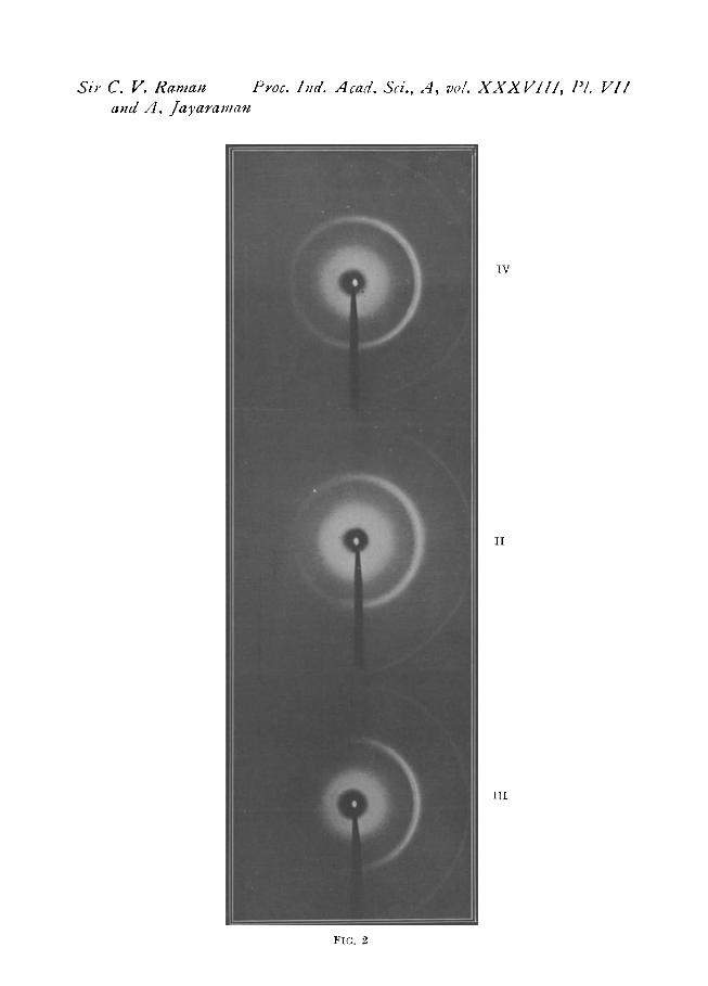
As already remarked, all the four specimens included in the present study had been shaped into forms having a sharp edge girdling them. It was, therefore, possible to record their X-ray diffraction patterns merely by allowing a fine pencil of X-rays to graze any chosen point on this edge and receiving the diffracted X-rays on a photographic film. The width of the X-ray pencil and its position relative to the edge were adjusted so as to record the pattern with satisfactory definition. The radiation employed was that from a copper target filtered through a nickel foil. The technique employed was tested by using edges of agate; the diffraction rings of crypto' crystalline quartz were then found recorded as sharply defined lines in the correct positions.
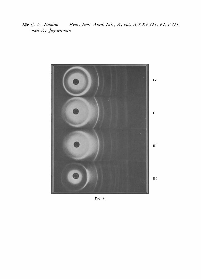
The records made with our hyalite specimens using a flat camera are reproduced in Fig. 2, Plate VII, the distance from edge to plate being 4 cm. The patterns recorded with a cylindrical camera of 6 cm. radius are re- produced in Fig. 3, Plate VIII. The numbers entered against each photo- graph indicate the particular specimen used.

The Structure of Opal and the Origin of Its Iridescence 105
Various authors ~, ~, 3 have claimed that the X-ray pattern of opal is identical with that of high cristobalite. Apart from the difficulty of under- standing why the high-temperature form should remain unchanged at the low temperatures, the result reported by those authors leaves the origin of the optical effects displayed by opal unexplained. It seemed to us there- fore necessary that a critical comparison should be made of the X-ray patterns of our specimens with those of high and low cristobalite respectively. With this aim and for purposes of comparison with our specimens, we have made use of a porcelain-like material found associated with some specimens of agate collected by one of us near Indore in Central India. The density of this material ranges between 2" 30 and 2.35 and its X-ray patterns show it to be low cristobalite mixed with a little tridymite. On heating the material to 300 ° C., the cristobalite changes reversibly to the high- temperature form, as is shown by its altered X-ray pattern. In Fig. 4, Plate IX the pattern of our hyalite (No. IV) is reproduced in juxtaposition with the patterns of the comparison material recorded at the high and low temperatures respectively.
From a scrutiny of the photographs reproduced in Plates VII, VIII and IX, it is evident that though the X-ray patterns of our specimens exhibit certain similarities with high cristobalite, there are some significant differ- ences demanding attention.
A. The principal ring of hyalite exhibits two components distinctly resolved from each other. This effect is particularly clear in the photo- graphs reproduced in Plate VII. In strongly exposed pictures, the ring exhi- bits an unsymmetrical broadening.
B. The rings other than the first appearing in the patterns also exhibit large variations in sharpness which are uncorrelated either with the angle of diffraction or with the intensity of the line.
C. Several diffuse bands are present in the patterns of the hyalites which are not seen in the comparison photographs.
6. INTERPRETATION OF THE RESULTS
The experimental facts set forth above preclude us from describing opal as being merely a cryptocrystalline form of high cristobalite. The doubling of the principal ring indicates, on the other hand, that the material is composite and consists of two distinct phases distributed throughout its volume. A reasonable explanation of the observed features is forthcoming if these two phases are identified respectively with high and low cristobalite. In particular, the doubling of the principal ring appears as an immediate

106 C .V. RAMAN AND A. JAYARAMAN
consequence of the fact that the spacings for high and low cristobalite are different, being 4.11 A and 4.04/~ respectively.
The lower symmetry of low cristobalite results in its X-ray pattern exhibiting many more spacings than that of high cristobalite. 4, 5, e Indeed, while the latter shows only 12 lines with spacings greater than 1 A, the former shows 43 lines in the same region. Many of the latter lie very close to each other and their intensities are also small. Diffuseness of the rings resulting from the smallness of the crystallites in the material would therefore influence the pattern of low cristobalite with its numerous closely-packed faint lines to a much greater extent than in the case of high cristobalite which has relatively fewer and brighter lines. Indeed, they would in such cir- cumstances, appear in the former case as diffuse bands instead of discrete lines as in the latter case. The presence of low cristobalite in the material would, if the particle sizes are small, thus be readily capable of being over- looked.
7. THE OPTICAL EVIDENCE
The conclusion deduced above from the X-ray data that opal includes two distinct crystalline species of silica in its physical make-up may be also inferred from its optical behaviour.
The optics of stratified media is sufficiently well understood to enable us from a study of the spectral character of the light reflected and transmitted by such a medium, to infer the number and nature of its stratifications. For a high degree of perfection in the monochromatism of the reflections to be possible, it is necessary that the stratifications should be regular in their spacing as well as in their optical character, and further that great numbers of the stratifications should be present. It is also necessary that the reflect- ing power of an individual stratification is small. For, if the reflecting power of an individual stratification were large, the first few alone of the stratifica- tions would function, and the reflections would necessarily cease to be monochromatic.
It is well-known that the coloured reflections given by precious opal are highly monochromatic and that the light transmitted by them also exhibits monochromatic extinctions. Two further observations that we have made are significant in this connection. In the first place, we have noticed that the internal reflections of opal are verfectly polarised when observed in a direction making an angle of 90 ° with the incident beam. We have also noticed that a reflection which is highly monochromatic when observed at or near normal incidence retains this character even when it is shifted towards shorter wavelengths by making the incidence oblique,
The Structure of Opal and the Origin of Its Iridescence 107
The optical behaviour of opal thus compels us to recognise the existence of a remarkably perfect periodicity in its structure. It also emerges from the observations that the difference in the refractive indices of the alternate layers is small and yet precisely the same throughout the stratifications. The only reasonable hypothesis that could be put forward to account for these facts is that the alternate layers consist of two distinct and specific modifications of silica.
The conclusions set forth above are supported by the observations described earlier on the optical behaviour of the specimens of hyalite in our collection. Though the individual specimens exhibit distinctive features, it is clear that the phenomena observed have a common origin, namely, the existence of regular optical stratifications within the material; the differences observed arise from the spacing of the stratifications and the dimensions of the individual domains in which they appear not being the same for all the specimens. Thus, hyalite in spite of its apparent dissimilarity in appear- ance belongs optically to the same category as precious opal.
It may be remarked in conclusion that the density of low cristobalite is 2" 32 and its refractive index 1.484, whereas the density of high cristobalite is 2.27 and the refractive index calculated therefrom is 1-468. The differ- ence between the two refractive indices is sufficiently small to be reconcilable with the observed characters of the internal reflections. That the density of our hyalite specimens is distinctly lower than that which might be antici- pated on the basis of their structure may be ascribed to the presence in them of ultra-microscopic cavities partly or wholly filled with water. Their existence makes itself evident in a distinct turbidity of the material, a pheno- menon which is quite distinct from the optical effects having their origin in the regularly stratified structure of the silica.
8. SUMMARY
A study of the optical behaviour of iridescent opal indicates very clearly that the silica present in the material has a regularly stratified structure in which the alternate layers differ in refractive index, such difference being small but the same throughout the stratifications. A critical examination of the X-ray diffraction patterns of cryptocrystalline hyalites exhibiting optical phenomena identical with or analogous to those of precious opal confirms this finding and enables the two species of silica present in associa- tion with each other and giving rise to these phenomena to be identified respectively with high and low cristobalite. The different optical effects ~xhibited by a stratified medium when the spacing of the stratifications and

108 C .V. ~ N AND A. J ^ Y ~ U X N
the extent of the domains in which they are present are varied have been observed with different specimens of hyalite and are illustrated in the paper by a series of photographs.
9. REFERENCES
1. I. Levin and Emil Ott .. 2. Dwyer, E.P. and Mellor, D. P . . .
3. M.J. Buerger
4. R . W . G . Wycoff 5. Nieuwenkamp 6. A . H . Jay
Z . Krist . , 1933, 85A, 305. Jour. Roy . Soc . , New South Wales, 1934,
68, 47. . . Phase Transformation in Solids, John Wiley
& Sons, 1951, pages 206--09. . . Z . Kris t . , 1925, 62, 189. . . Ibid., 1935, 92A, 82. . . Min. Mag. , 1944, 27, 54.

Sir C. U. Ra~na~z Proc. Ind. Acact. Sci., A, vol. X X X V l l I , Pl. III altd A. JayaraJnalz
TI
l i t
FIG. 1
Sir C. V. RelnaJz Proc. I~lct. Xcad. Sci., A , vo/. X X X U I I ] , P/. U I / and A. /ayara~Jlan
IV
I I
I I I
FIG, 2
Sir C. V. Ifan~au Proc. Ind. Mcad. SoL, A, z,ol. X X X V I I I , PL V I I I and A. ]ayaramall
I V
1!
I I I
FIG, 3

Sir C. I1. Rama,z Proc. ]Jzd. Acad. Sd., A , vol. X . V X U I I I , I'L [A" alzd ,4. ]a;,afama,z
P (hot)
IV
p (co!d)
FI(;. 4
1]I
I I
I I I
I[
I:IG. 5 FIG. 6