Embed Size (px)
Citation preview

The Structure of CollagenAuthor(s): Maurice L. HugginsSource: Proceedings of the National Academy of Sciences of the United States of America,Vol. 43, No. 2 (Feb. 15, 1957), pp. 209-213Published by: National Academy of SciencesStable URL: http://www.jstor.org/stable/89812 .
Accessed: 05/05/2014 23:13
Your use of the JSTOR archive indicates your acceptance of the Terms & Conditions of Use, available at .http://www.jstor.org/page/info/about/policies/terms.jsp
.JSTOR is a not-for-profit service that helps scholars, researchers, and students discover, use, and build upon a wide range ofcontent in a trusted digital archive. We use information technology and tools to increase productivity and facilitate new formsof scholarship. For more information about JSTOR, please contact [email protected].
.
National Academy of Sciences is collaborating with JSTOR to digitize, preserve and extend access toProceedings of the National Academy of Sciences of the United States of America.
http://www.jstor.org
This content downloaded from 195.78.109.27 on Mon, 5 May 2014 23:13:08 PMAll use subject to JSTOR Terms and Conditions

:Communication No. 1864 from the Kodak Research Laboratories. W. T. Astbury and A. Street, Phil. Trans. Roy. Soc. London, A, 230, 75, 1931.
2 I. MacArthur, Nature, 152, 38, 1943. 3 L. Pauling and R. B. Corey, these PROCEEDINGS, 37, 261, 1951. 4 L. Pauling, R. B. Corey, and H. R. Branson, these PROCEEDINGS, 37, 205, 1951. 5 L. Pauling and R. B. Corey, these PROCEEDINGS, 37, 235, 1951. 6 M. F. Perutz, Nature, 168, 653, 1951. 7 G. R. Tristram, in The Proteins, ed. H. Neurath and K. Bailey (New York: Academic Press, 3). 8 V. H. Ward and H. P. Lundgren, Advances in Protein Chemnistry, 9, 243, 1954. 9 R. S. Bear, J. Am. Chem. Soc., 65, 1784, 1943. 0 R. S. Bear and H. J. Rugo, Ann. N.Y. Acad. Sci., 53, 627, 1951. 1 F. H. C. Crick, Nature, 171, 59, 1952. 2 F. H. C. Crick, Acta cryst., 6, 689, 1953. 3A. R. Lang, Acta cryst., 9, 436, 446, 1956. 4L. Pauling and R. B. Corey, Nature, 171, 59, 1953. 5 A. Elliott, Textile Research J., 22, 783, 1952.
H. L. Yakel, Jr. and E. W. Hughes, Acta cryst., 7, 291, 1954. 7 M. L. Huggins, J. Am. Chemn. Soc., 74, 3963, 1952.
THE STRUCTURE OF COLLAGEN*
BY MAURICE L. HUGGINS
RESEARCH LABORATORIES, EASTMAN KODAK COMPANY, ROCHESTER, NEW YORK
Commtunicated by W. A. Noyes, Jr., November 23, 1956
Many models have been proposed for the structure of collagen. The older ones, vever, have all been shown to be in definite contradiction with experimental ts.
Recently, Rich and Crick' and Ramachandran2 have made new proposals, in-
ving 3-chain coiled-coils, which avoid some of the difficulties of their earlier dels. In the writer's opinion, there is little chance that these new proposals a correct or even approximately so. Since only half the NH groups in each main Lin are hydrogen-bonded (unless to unspecified side chains), these structures con- m neither to the principle that hydrogen bonds are formed wherever possible nor the principle that like groups tend to be surrounded by close neighbors in like nner.3 Also, since pulling out the chains into an extended zigzag arrangement uld lengthen the structure by only 25 per cent, there seems to be no way of ex-
ining the large extensions (at least 7- or 8-fold) of the band pattern occasionally served in the electron microscope pictures.4 [he writer5 has proposed a structure which agrees with the primary requirements the X-ray data,6 gives maximum hydrogen bonding, permits about 31/2-fold ension without breaking of primary bonds, and has various other advantages. It one serious disadvantage, however: half the CO -NH groups have the cis orien-
ion. This seems to be in contradiction to the infrared evidence.7 k new structure, recently discovered, has only trans orientations of the CO -NH ups and seems satisfactory from other points of view, judging from a model de approximately to scale. (Precise co-ordinates have not yet been deduced.)
This content downloaded from 195.78.109.27 on Mon, 5 May 2014 23:13:08 PMAll use subject to JSTOR Terms and Conditions
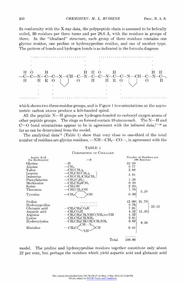
conformity with the X-ray data, the polypeptide chain is assumed to be helically iled, 30 residues per three turns and per 28.6 A, with the residues in groups of
tee. In the "idealized" structure, each group of three residues contains one rcine residue, one proline or hydroxyproline residue, and one of another type. ie pattern of bonds and hydrogen bonds is as indicated in the formula diagram
HO H HH O H H H C-C-N-C-C-N-CH-C-N-C-C-N- C -C-N -CH- C-N-C-, H HRO O0 H H RO | 0 H
. . . . . . . . . . . . . . . . . . . . . . . . . . . . . . . . . . . . . . . . . . . . . . . . . . . . . . . . . .
Lich shows two three-residue groups, and in Figure 1 levo orientations at the asym- atric carbon atoms produce a left-handed spiral. All the peptide N-H groups are hydrogen-bonded to carbonyl oxygen atoms of
ler peptide groups. The rings so formed contain 10 atoms each. The N-H and =0 bond orientations appear to be in agreement with the infrared data,7-l0 as a as can be determined from the model. The analytical data11 (Table 1) show that very close to one-third of the total mber of residues are glycine residues, -NH-CH2---CO-, in agreement with the
TABLE 1
COMPOSITION OF COLLAGEN
Amino Acid Number of Residues per (by Hydrolysis) -R 100 Residues
Glycine -H 33.19 Alanine -CH3 9.77 Valine -CH(CH3)2 2.66 Leucine -CH2CH(CH3)2 3.91 Isoleucine -CH(CH3)CH2CH3 - Phenylalanine --CH2( CHs) 1.39 Methionine -CH2CH2SCH3 0.49 Serine -CHO2H 2.93 Threonine -CH(CH3)OH 1.76 5 Tyrosine -CH2 -OH.50
Proline . ....... 12.00 21.791 Hydroxyproline .......... 9.78 33 .15 Glutamic acid -CH2CH2CO2H 7.04 Aspartic acid -CH2CO2H 4.32 11.36 Arginine -CH2CH2CH2NHC(NH2)=-NH 4.52 Lysine -CH2CH2CH2NH2 2.81 Hydroxylysine -CH2CH2CH(OH)CH2NH2 0.62 8.38
CH--N Histidine -CHC CH 0.44 --
NH/H
Total 100.00
)del. The proline and hydroxyproline residues together constitute only about
per cent, but perhaps the residues which yield aspartic acid and glutamic acid
This content downloaded from 195.78.109.27 on Mon, 5 May 2014 23:13:08 PMAll use subject to JSTOR Terms and Conditions

hydrolysis,12 when in the native collagen, contain rings which act structurally ich like proline and hydroxyproline rings. Such rings might be produced by drogen bonding or by condensation reactions:
NH2 OH I I
C C / % / H,2
H2C O H2C 0 C
H H2C H H2C C--O I _ I I I
-CO-CH-N- - CO-CH-N- -CO-CH--N-
is perhaps significant that the sum of the numbers of residues of these four kinds
very near to one-third of the total (Table 1).
',, / N '- R/ H \H /--- "
N.CCH '/"H
N-CH cHH OH H HHHH
FIG. 1. -Diagrammatic representation of ahe pattern of bonds and hydrogen bonds in the proposed structure for collagen.
Without producing much disruption of the structure, the -NH-CHR-CO- idue just to the left of a proline residue (see Fig. 1) can also be made a proline or a iroxyproline residue. This would account for the proline-hydroxyproline se- mnces which have been found in partial hydrolyzates.13 lVith the model described, the peptide groups can be strictly planar and the -H - a .-0 hydrogen bonds straight. Some other models which have been tried m reasonably satisfactory otherwise but fail in this regard. t has already been noted that the band pattern in the electron microscope is asionally stretched, up to seven or eight times its normal length, i.e., to at least ce the length obtainable by pulling out the proposed helix into an extended zig- r. This extra stretching can be attributed to regions in which the polypeptide ins are irregularly arranged or are arranged in a more extendable pattern, or i reversal of the directions of some chains or portions of chains, as they are pulled
N,
H / "\,C H
HCH FIG. l.--Diagrammatic representation of t-he pattern of bonds
and hydrogen bonds in the proposed structure for collagen.
Without producing much disruption of the structure, the--NH--CHR--CO-- idue just to the left of a proline residue (see Fig. 1) can also be made a proline or a Jroxyproline residue. This would account for the proline-hydroxyproline se- races which have been found in partial hydrolyzates.l3 Wlith the model described, the peptide groups can be strictly planar and the -H...O hydrogen bonds straight. Some other models which have been tried m reasonably satisfactory otherwise but fail in this regard. Zt has already been noted that the band pattern in the electron microscope is asionally stretched, up to seven or eight times its normal length, i.e., to at least ce the length obtainable by pulling out the proposed helix into an extended zig-
This extra stretching can be attributed to regions in which the polypeptide ,ins are irregularly arranged or are arranged in a more extendable pattern, or t reversal of the directions of some chains or portions of chains, as they are pulled
This content downloaded from 195.78.109.27 on Mon, 5 May 2014 23:13:08 PMAll use subject to JSTOR Terms and Conditions

t. This could result, for example, if the molecules included two or more helices, indicated diagrammatically in Figure 2. The interhelix connections might be
stine or other crosslinks or bends in the main polypeptide chain, such as might cur where two proline residues (or a proline and a hydroxyproline residue) come gether.
FIG. 2.-Diagram illustrating a possible explanation for the very large band pattern extensions observed in the electron microscope.
The model proposed here of course requires further checking and elaboration. It
presented tentatively as the model which, in the opinion of the writer, is most
ely to approximate the true structure.
f Communication No. 1866 from the Kodak Research Laboratories. A. Rich and F. H. C. Crick, Nature, 176, 915, 1955.
2 G. N. Ramachandran, Nature, 177, 710, 1956. 3 M. L. Huggins, Chem. Revs., 32, 195, 1943. 4F. O. Schmitt, C. E. Hall, and M. A. Jakus, J. Cellular Comp. Physiol., 20, 11, 1942. 5 M. L. Huggins, J. Am. Chem. Soc., 76, 4045, 1954.
This content downloaded from 195.78.109.27 on Mon, 5 May 2014 23:13:08 PMAll use subject to JSTOR Terms and Conditions

6 R. S. Bear, Advances in Protein Chemistry, 7, 69, 1952. 7 R. M. Badger and A. D. E. Pullin, J. Chem. Phys., 22, 1142, 1954. 8 E. J. Ambrose and A. Elliott, Proc. Roy. Soc. London, A, 206, 206, 1951. 9 G. B. B. M. Sutherland, K. N. Tanner, and D. L. Wood, J. Chem. Phys., 22, 1621, 1954. 10 W. E. Seeds, in The Nature and Structure of Collagen, ed. J. T. Randall ("Butterworth's entific Publications") (London: Butterworth & Co., Ltd.; New York: Academic Press, Inc., 53), p. 250. a1 G. R. Tristram, in The Proteins, ed. H. Neurath and K. Bailey (New York: Academic Press, a., 1953), Vol. 1, Part A. .2 Analyses show that the amount of amide nitrogen is practically equivalent to the amount of )artic acid obtained, suggesting that in the native material the aspartic residues are present amide form, or as an amide condensation product, with the glutamic residues in an acidic form as an acid condensation product. 3 T. D. Kroner, W. Tabroff, and J. J. McGarr, J. Am. Chem. Soc., 77, 3356, 1955.
THE CONTRIBUTION OF THE a-HELICAL CONFIGURATION TO THE OPTICAL ROTATION OF POLYPEPTIDES
AND PROTEINS*
BY PAUL DOTY AND R. D. LUNDBERGI
DEPARTMENT OF CHEMISTRY, HARVARD UNIVERSITY, CAMBRIDGE, MASSACHUSETTS
Communicated by G. B. Kistiakowsky, December 24, 1956
Shortly after it was established that polypeptides could exist in solution as rigid, Ilike a-helices as well as randomly coiled chains,l it became evident that the opti-
rotation of the helical form was substantially more positive than that of the
idomly coiled form. For a while it seemed possible to explain this effect either a result of the change in the environment of the optically active residues in the o configurations or as arising from the existence of a single screw sense in the heli-
configuration which could then make a contribution to the optical rotation inde- ident of the contribution made by the optically active residues. Within the t year three different experiments have demonstrated that the stable helical
ifiguration is exclusively of one screw sense, that is, right-handed or left-handed. one experiment it was shown that the rotatory dispersion was normal for the
Idomly coiled configuration and abnormal for the helical form.2 This unusual ect could not arise simply from the changed environment of the optically active iters. In another experiment it was shown that the racemic polypeptide formed
using L-polypeptide to initiate the polymerization of a D,L N-carboxyanhydride s itself optically active.3 This showed clearly the preservation of at least some the screw sense characteristic of the L-polypeptide in the grafted racemic poly- ptide and the association of optical activity with the helical configuration. In a rd experiment the optical rotation of a series of copolymers of D- and L-residues s shown to increase linearly as the concentration of D-residues increased from zero 10-30 per cent.4- 5 Extrapolation from this linear region to 50 per cent D-resi- es yielded a positive value which was an estimate of the optical rotation of a helix
nposed of racemic residues. As a consequence of these experiments, there re- ins no doubt that the difference in rotation between the helical and randomly
This content downloaded from 195.78.109.27 on Mon, 5 May 2014 23:13:08 PMAll use subject to JSTOR Terms and Conditions