Embed Size (px)
Citation preview
RESEARCH ARTICLE Open Access
The structure of a prophenoloxidase (PPO)from Anopheles gambiae provides newinsights into the mechanism of PPOactivationYingxia Hu1, Yang Wang2, Junpeng Deng1*† and Haobo Jiang2*†
Abstract
Background: Phenoloxidase (PO)-catalyzed melanization is a universal defense mechanism of insects againstpathogenic and parasitic infections. In mosquitos such as Anopheles gambiae, melanotic encapsulation is a resistancemechanism against certain parasites that cause malaria and filariasis. PO is initially synthesized by hemocytes andreleased into hemolymph as inactive prophenoloxidase (PPO), which is activated by a serine protease cascade uponrecognition of foreign invaders. The mechanisms of PPO activation and PO catalysis have been elusive.
Results: Herein, we report the crystal structure of PPO8 from A. gambiae at 2.6 Å resolution. PPO8 forms a homodimerwith each subunit displaying a classical type III di-copper active center. Our molecular docking and mutagenesisstudies revealed a new substrate-binding site with Glu364 as the catalytic residue responsible for the deprotonation ofmono- and di-phenolic substrates. Mutation of Glu364 severely impaired both the monophenol hydroxylase anddiphenoloxidase activities of AgPPO8. Our data suggested that the newly identified substrate-binding pocket is theactual site for catalysis, and PPO activation could be achieved without withdrawing the conserved phenylalanineresidue that was previously deemed as the substrate ‘placeholder’.
Conclusions: We present the structural and functional data from a mosquito PPO. Our results revealed a novelsubstrate-binding site with Glu364 identified as the key catalytic residue for PO enzymatic activities. Our data offered anew model for PPO activation at the molecular level, which differs from the canonical mechanism that demandswithdrawing a blocking phenylalanine residue from the previously deemed substrate-binding site. This study providesnew insights into the mechanisms of PPO activation and enzymatic catalysis of PO.
Keywords: Innate immunity, Melanization, Mosquito, Type III copper proteins, Zymogen activation
BackgroundPhenoloxidase (PO) is a critical enzyme involved in mul-tiple physiological processes including innate immunityof insects and crustaceans. As the close homolog ofarthropod hemocyanins, PO is produced in hemocytesas a zymogen, the prophenoloxidase (PPO) [1]. Uponpathogenic infections or physical injuries, a serine proteasecascade is triggered to activate PPO as a local response.
The last step of PPO activation involves a trypsin-likeenzyme, named PPO-activating proteinase (PAP), whichcleaves PPO at a conserved proteolytic cleavage site nearthe N-terminus to generate active PO [2, 3]. In vitro, PPOcan also be activated without proteolytic cleavage by certainchemicals such as ethanol or detergents (e.g. cetylpyridi-nium chloride, CPC) [4–6].In general, the active PO possesses o-hydroxylase (EC
1.14.18.1) and o-diphenoloxidase (EC 1.10.3.1) activitiesthat convert a variety of monophenolic and o-diphenolicsubstrates to o-quinones [6, 7]. Quinones may act ascross-linkers for wound healing, and they alsopolymerize to form melanin capsules around parasites
* Correspondence: [email protected]; [email protected]†Equal contributors1Department of Biochemistry and Molecular Biology, Oklahoma StateUniversity, Stillwater, OK 74078, USA2Department of Entomology and Plant Pathology, Oklahoma State University,Stillwater, OK 74078, USA
© 2016 Hu et al. Open Access This article is distributed under the terms of the Creative Commons Attribution 4.0International License (http://creativecommons.org/licenses/by/4.0/), which permits unrestricted use, distribution, andreproduction in any medium, provided you give appropriate credit to the original author(s) and the source, provide a link tothe Creative Commons license, and indicate if changes were made. The Creative Commons Public Domain Dedication waiver(http://creativecommons.org/publicdomain/zero/1.0/) applies to the data made available in this article, unless otherwise stated.
Hu et al. BMC Biology (2016) 14:2 DOI 10.1186/s12915-015-0225-2

and parasitoids [7, 8]. Quinones and other reactive inter-mediates (e.g. 5,6-dihydroxyindole) directly kill microbialpathogens and parasitoids [9].POs, together with hemocyanins, tyrosinases, and cat-
echol oxidases, belong to the type III di-copper family ofproteins, which share an antiferromagnetically coupled di-copper center [10, 11]. This group of proteins are widelydistributed in different organisms: POs in arthropods,tyrosinases in microbes, plants and mammals, catecholoxidases in plants and fungi, and hemocyanins in arthro-pods and molluscs [12–14]. Tyrosinases and catechol oxi-dases are responsible for browning of fruits and plants,and they may also play a role in defense mechanism inplants and fungi [15]. Mammalian tyrosinase is a majorenzyme required for coloring of hair, skin and eyes, andits deficiency and excessive expression could lead to albin-ism and skin cancer, respectively [7, 16, 17]. Hemocyaninswere initially recognized as oxygen carriers in hemolymph[18], but their PO activities and roles in antimicrobialdefense were discovered later [6, 19]. In spite of having asimilar active site, vital structural and functional differ-ences do exist in these proteins as has been demonstratedin the past decades. Tyrosinases and POs catalyze boththe o-hydroxylation and oxidation reactions, but catecholoxidases only possess the oxidase activity [20, 21]. Basedon their similarities in the primary and tertiary structures,POs were considered to be evolutionarily more related toarthropod hemocyanins, whereas tyrosinases and catecholoxidases are closer to molluscan hemocyanins [19, 22, 23].In the catalytic cycle, type III di-copper center goes
through three redox states: the reduced deoxy state[Cu(I)-Cu(I)], the oxy state [Cu(II)-O2
2−-Cu(II)] inwhich a peroxide molecule binds to the two Cu ions ina μ-η2:η2 side-on bridging fashion, and the met state[Cu(II)-OH−-Cu(II)] in which the two Cu ions areligated to a hydroxide ion [10]. The oxy state enzyme iscapable of catalyzing both the hydroxylation of mono-phenols and the oxidation of o-diphenols to o-qui-nones, while the met state only undertakes the latterdiphenol oxidase reaction [10].PO catalyzed-melanogenesis is indispensable in the
immune system of invertebrates [24, 25] and two PPOstructures from Manduca sexta and Marsupenaeus japo-nicus have been investigated [26, 27]. Although thesestudies on PPO provided important structural and func-tional insights, the detailed enzymatic mechanism of POis still undetermined and the fundamentals of PPO acti-vation remain elusive. A. gambiae is a major vector ofhuman malaria parasites in Africa, whose innate im-mune system protects the mosquito from infection byincompatible malaria parasites [28]. It contains ninePPO genes which are expressed at different tissues andlife stages [29–32]. Herein, we report the crystal struc-ture of AgPPO8 at 2.6 Å resolution, representing the
first structure from a recombinant PPO and the firstfrom a mosquito species. Our structural and functionalstudies on AgPPO8 revealed a novel substrate-bindingpocket that differs from the previously deemed ‘place-holder’ position occupied by a phenylalanine residue,which is conserved in PPOs, but not in molluscanhemocyanins, tyrosinases, or catechol oxidases. Weidentified E364 as a catalytic residue key to the PO activ-ities. Our data provide new insights into the mechanismof PO catalysis, which could be applicable to other typeIII di-copper proteins, and suggest a new model for PPOactivation at the molecular level.
ResultsOverall structure of AgPPO8The crystal structure of AgPPO8 contains two identicalsubunits, each comprising 700 residues, which are asso-ciated tightly to form a homodimer in the asymmetricunit. The overall structure of the AgPPO8 displays abutterfly shape of 147 × 70 × 60 Å in size (Fig. 1a). Mostresidues are well defined, except for 40 amino acids thatlack electron densities. These include residues 59–65,580–587, 629–632, and 698–700 in chain A and residues1, 2, 65–68, 579–587, and 698–700 in chain B. AgPPO8adopts a similar architecture as that of M. sexta PPO(MsPPO) [26], with the three conserved domains(Fig. 1b). The pro-region (17–81) contains the conservedproteolytic cleavage site (R51*F52). Domain I (1–16,82–196) is dominant in α-helices. Domain II (197–435)contains the di-copper active site buried in an α-helixbundle. Domain III (436–697) is mainly a seven-stranded β barrel, with an additional β-hairpin extendedto domains I and II. There are two disulfide bonds(C592-C636 and C594-C643) within domain III of eachsubunit, which are highly conserved among hemocya-nins and PPOs. C592-C636 is situated on the surfaceand exposed to the solvent, while C594-C643 is locatedat the interface of domains II and III, which may con-tribute to the structural stability. The overall structureof AgPPO8 homodimer closely resembles that ofMsPPO heterodimer (Fig. 1c), with a 1.14 Å root-mean-square deviation over 1,129 aligned Cα atoms.
Dimerization of AgPPO8AgPPO8 was found to stay mainly as a homodimer insolution by size exclusion chromatography and dynamiclight scattering (Additional file 1). Two types ofdimerization patterns exist in the crystal structure, a tighthomodimer and a loose one. The tight dimer associates(chain A and chain B) via a two-fold non-crystallographicsymmetry axis in the asymmetric unit. The loose dimerinvolves chain A molecule and a crystallographicsymmetry-related chain B molecule in the crystal lattice.The tight dimer interface is mainly stabilized through
Hu et al. BMC Biology (2016) 14:2 Page 2 of 13

extensive hydrophobic and charge-charge interactionspredominantly from domains I and II, burying a large(4,624 Å solvent accessible surface area which is com-parable to that of MsPPO heterodimer [26]. In contrast,the loose dimer buries a much smaller surface area (934 Å2) at the interface. The tight homo-dimeric asso-ciation of AgPPO8 was also confirmed with an analysisperformed with the PISA server (http://www.ebi.ac.uk/msd-srv/prot_int/pistart.html), suggesting it is the bio-logically functional dimer.
Active site of AgPPO8During refinement, we observed a large positive differenceelectron density at the supposed bi-metal center (Add-itional file 2), although no additional metal ions were sup-plied during the protein expression. We believe the boundmetal ions came from the trace amount of metal in LBmedium. The identities of the metal consequently couldnot be unambiguously determined in this study, sinceeither Zn or Cu atoms could be positioned at the samelocations and refine well. This observation is consistentwith a previous report that heterogeneous incorporation
of zinc and copper ions was found at the active site of Ba-cillus megaterium tyrosinase [33]. In this study, we inter-preted the metals as Cu ions and finalized that in thedeposited structure. Each metal ion is coordinated withthe NE2 atoms of three histidine residues, which arehighly conserved in this protein family (Fig. 1d): CuA isassociated with H223, H227 and H252, while CuB is asso-ciated with H379, H383 and H419. All six histidine resi-dues are located in the α-helix bundle from domain II,and stabilized by three Phe residues (F99, F248 and F415)through hydrophobic interactions: F99 with H379 andH383, F248 with H227, H252 and H379, and F415 withH223, H252, H383 and H419 (Additional file 3). The dis-tance between the two copper atoms is 4.5 Å, which isthe typical distance for the reduced form of type III di-copper proteins. The UV/Vis absorption spectrum didnot exhibit any apparent characteristic feature from 250to 700 nm (Additional file 4), supporting that theAgPPO8 structure we obtained was in the deoxy state.The di-copper center observed in AgPPO8 is similar
to those in other type III copper enzymes. However, thecatalytic residue has been elusive due to the amino acid
Fig. 1 Crystal structure of A. gambiae PPO8. (a) Overall structure of the homodimer with each subunit shown in cyan and green, respectively.Cu atoms are shown as brown spheres. (b) The domain structure of AgPPO8: pro-region, magenta; domain I, blue; domain II, yellow; domain III, green.The Cu atoms are shown as brown spheres and the disulfide bonds are shown as red sticks. The side chain of Arg51 preceding the proteolyticcleavage site is shown as a black stick. (c) Superposition of AgPPO8 (cyan) with M. sexta PPO heterodimer (PPO-1 in magenta and 2 in yellow, PDB ID3HHS). AgPPO8 is 41.1 % identical in a.a. sequence to MsPPO1 and 38.8 % to MsPPO2. (d) The di-copper active site of A. gambiae PPO8. The Cu atomsare shown as brown spheres. Six conserved His residues (magenta) coordinate CuA and CuB, and are stabilized by three Phe residues (F248, F415 andthe 'placeholder' F99, shown as red sticks). V406 (orange) aligns to a putative catalytic residue E395 in MsPPO2. Glu364 (green) forms a salt bridge (blackdash) with R244 (yellow), and is required for the enzymatic activities of AgPPO8. The secondary structures of AgPPO8 are shown and colored in cyan
Hu et al. BMC Biology (2016) 14:2 Page 3 of 13

variations near the dicopper center (Fig. 2). In MsPPO2,one unique acidic residue (E395) was proposed to be acatalytic residue responsible for the hydroxylase activityof the enzyme since its carboxylic oxygen in the sidechain may deprotonate the hydroxyl group of the mono-phenol substrate (Fig. 2c) [26, 34]. Remarkably, althoughAgPPO8 was able to convert monophenol to diphenol(see below), it contains a valine residue (V406) at theequivalent position, which lacks an acidic side chain
(Fig. 1d, 2a). Therefore, the deprotonation of the phen-olic substrate during AgPPO8 enzymatic catalysis mayinvolve a different residue.
Substrate binding by AgPPO8As seen in other arthropod PPOs and hemocyanins,AgPPO8 also contains a putative ‘placeholder’ for phenolicsubstrates, which is F99 in each subunit (Fig. 1d, Additionalfile 3). This conserved residue resides in a loop of domain I
Fig. 2 Comparison of the di-copper centers from different type III copper proteins. (a) A. gambiae PPO8 (AgPPO8), PDB ID 4YZW; (b) M. sextaPPO1 (MsPPO1), 3HHS-B; (c) M. sexta PPO2 (MsPPO2), 3HHS-A; (d) Marsupenaeus japonicus (Mj) PPOβ, 3WKY; (e) Limulus polyphemus hemocyanindeoxy state (LpHc), 1LLA; (f) Octopus hemocyanin functional unit Odg, 1JS8; (g) Ipomoea batatas catechol oxidase (IbCO) in complex with1-phenyl-2-thiourea (PTU), 1BUG; (h) Vitis vinifera catechol oxidase (VvCO), 2P3X; (i) Streptomyces castaneoglobisporus tyrosinase (ScTyr),2ZMX; (j) Agaricus bisporus tyrosinase (AbTyr), 2Y9W; (k) Aspergillus oryzae pro-tyrosinase (AoProTyr), 3W6W; (l) Superposition of BmTyr structures incomplex with bound tyrosine (4P6R) and L-DOPA (4P6S) (BmTyr:T/D); (m) Superposition of the active sites from AgPPO8, ScTyr, AoProTyr, LpHc andBmTyr:D. Space overlapping between the placeholders and phenolic substrate is indicated by a red dashed cycle. The Cu or Zn atoms are shown aslarge brown or gray spheres, the Cu-coordinated or hydrogen-bonded water molecules are shown as small red spheres. The hydrogen bonds are indi-cated as black dashes
Hu et al. BMC Biology (2016) 14:2 Page 4 of 13

with its aromatic side chain protruding to the di-coppercenter and stacking onto the imidazole ring of H383 at theCuB site. It was assumed in the prior studies that the place-holder aromatic residue would be withdrawn from the di-copper center during PPO activation through major con-formational changes (see discussion), allowing its positionbe replaced by a natural phenolic substrate during enzym-atic catalysis. Interestingly, the analysis of the AgPPO8structure revealed a prominent cavity in the di-copperreaction center, which is close (~1.5 Å) to the solvent-accessible surface (Fig. 3a). This cavity is surrounded bythe six Cu-coordinating His residues, F99, F415, E364,N380, V406, along with other residues from domain I andII (Fig. 3a). The volume of the cavity (451 Å3, calculatedby program CASTp [35]) appears large enough to ac-commodate monophenolic or diphenolic substrates suchas tyramine and dopamine (approximately 190–200 Å3).Therefore, we named this pocket as the second substrate-
binding site (Site II) with respect to the placeholder pos-ition (Site I). To test if this pocket could hold substrates,we carried out docking experiments using the Autodockprogram. After selecting certain amino acids at the activesite as the flexible residues to accommodate potential pro-tein dynamics (see Methods), we individually docked tyr-amine and dopamine into AgPPO8 (Additional file 5).Although a larger grid box covering the entire di-coppercenter and the empty cavity was set as the search space,both substrates were successfully docked into the putativesubstrate-binding Site II in similar orientations (Fig. 3b)without significant reconfiguration of the active site(Fig. 3c,d). This observation suggested substrate bindingto PPO could be achieved without displacing the place-holder. Interestingly, in the docked protein structures, wefound residue E364 located very close to the docked sub-strates (Fig. 3c,d). Albeit distant from the di-copper center(6.7 and 7.6 Å from CuA and CuB in apo-AgPPO8), the
Fig. 3 A new and unique substrate-binding pocket (Site II) of AgPPO8. (a) AgPPO8 contains a cavity approximate to surface (Site II, side view).The cavity and solvent exposed surface areas are represented in cyan and gray, respectively. The key residues surrounding the cavity are shownas violet sticks. (b) The side view of Site II pocket with tyramine (green) and dopamine (yellow) docked inside. The distances (Å) from the phenolicoxygens of the substrates to CuA and CuB are indicated. (c and d) Superposition of the active sites before and after docking. The active site residues ofthe apo-AgPPO8 are shown as magenta sticks, while the complex structures with docked substrates bound are colored in cyan (tyramine) and green(dopamine), respectively. The hydrogen bonding between the substrate and E364 is highlighted with black dash. The distances (Å) from E364 to CuA,CuB, the putative placeholder F99, and the phenolic oxygens of docked substrates (yellow sticks) are indicated. The distance of the salt bridge betweenresidues E364 and R244 is also labeled (before docking, magenta; after docking, tyramine, cyan and dopamine, green). CuA and CuB are shown asbrown spheres
Hu et al. BMC Biology (2016) 14:2 Page 5 of 13

OE2 atom of E364 is 3.0 and 3.1 Å to the phenolic oxygensof tyramine and dopamine, respectively. This observationsuggested that E364 could play an important role in thedeprotonation of the phenolic substrates, which was shownto be essential for binding of the phenolic oxygen to one ofthe copper atoms [34, 36, 37]. E364 also forms a salt bridgewith R244 in a stable conformation, which may assist incorrectly positioning the carboxylic side chain during thedeprotonation process (Figs. 1d and 3c,d). We found thisGlu residue highly conserved among other type III di-copper proteins (Fig. 2). These observations indicated thatthis Glu residue could be a common catalytic residue forPO enzymatic activities, and may serve in an alternativemechanism for PPO activation (see below).
E364 is key to hydroxylation and oxidationTo test our hypothesis that E364 is essential for the en-zymatic activities of AgPPO8 by deprotonating phenolicsubstrates, we mutated it to a neutral Gln residue. Therecombinant E364Q mutant was expressed as a solubleprotein in E. coli cells and folded properly with an ap-proaching melting point as the wild type as shown fromthe Differential Scanning Fluorimetry analysis (Add-itional file 6). The enzymatic activities of both wild typeand mutant AgPPO8 were assayed in vitro. When usingdopamine as the substrate, the E364Q mutant lost nearly89 % of its oxidase activity compared to the wild type(Fig. 4a), indicating E364 is the base responsible for thedeprotonation of diphenolic substrate. When using tyr-amine as the substrate, E364Q mutant impaired 93 % ofits hydroxylase activity compared to the wild type(Fig. 4b), by detecting the dopamine formation at
280 nm. This demonstrated that E364 is also necessaryfor deprotonating the monophenolic substrate. Takentogether, we conclude that E364 is essential for boththe hydroxylase and oxidase activities of AgPPO8 withits carboxylate group acting as a general base for mono-phenol and diphenol deprotonation.
DiscussionIn this study, we determined the crystal structure ofAgPPO8, which is the first structure of recombinant PPOand the first from a mosquito species. The structural andfunctional studies on AgPPO8 revealed a novel substrate-binding pocket and identified E364 as a catalytic residuekey to the PO activities. Our data also provide newinsights into the catalytic mechanism of PO and suggest anew model for PPO activation at the molecular level.
Catalytic residues for phenol deprotonationEnzymatic activities of type III copper protein requiredeprotonation of the phenolic substrates, which is essen-tial for coordinating the phenolic oxygen to one of thecopper atoms and subsequent catalytic reactions [34, 36,37]. So far, at least five different residues have been pro-posed as the potential deprotonating base. In Streptomycescastaneoglobisporus tyrosinase (ScTyr), a flexible, CuA-coordinating H54 was supposed to serve as the base [38].This is not applicable to PPOs due to inflexibility of theirHis residues in the active site [26]. In mouse tyrosinase, afree His preceding the sixth coordinating His is vital forthe oxidation of diphenols [39], yet this residue is absentin arthropod hemocyanins and PPOs. In MsPPO2, thereare two acidic Glu residues in the vicinity of its binuclear
Fig. 4 E364 is key to PO enzymatic catalysis. (a) E364Q mutation severely impairs diphenol oxidase activity of AgPPO8. Dopamine was used asthe substrate and specific PO activity was shown as ΔmOD470/min/μg or U/μg; P <0.001. (b) E364Q mutation severely impairs the monophenolhydroxylase activity of AgPPO8. Tyramine was used as the substrate and the rate of dopamine formation catalyzed by 1 μg enzyme wasestimated as ΔmOD280/min/μg or U/μg; P <0.001. All data were presented as mean ± SEM (n = 3)
Hu et al. BMC Biology (2016) 14:2 Page 6 of 13

center, E353 (equivalent to E364 of AgPPO8) and E395[26]. Since E395 is located much closer to CuA, thepresumed site for hydroxylase activity [40, 41], andwithin 3 Å distance from residue F88, the ‘placeholder’for phenolic substrates, it was postulated to be respon-sible for tyrosine deprotonation in the monooxygenasereaction. However, this Glu residue is not present inmost type III di-copper proteins that exhibit the hy-droxylase activity (Fig. 2). Due to technical difficultiesin expressing recombinant MsPPOs, the putative cata-lytic function of E395 in MsPPO2 has not been verifiedexperimentally through mutagenesis analysis. Recentstudies on B. megaterium tyrosinase provided complexstructures in met form with bound tyrosine or L-DOPA(BmTyr:T/D), revealing that the CuA site is solely re-sponsible for its monooxygenase and diphenoloxidaseactivities [42]. A conserved water molecule activated byE195 was proposed to be the intermediate base fordeprotonating the entering substrates, since it is closerto the di-copper center than the candidate residue E195(Fig. 2l). Nevertheless, the water molecule was notobserved to have direct contact with the bound sub-strates in the met form structures. In AgPPO8, E364 isthe equivalent residue to BmTyr E195. The proposedcatalytic water molecule also exists in the current struc-ture, which is situated at a 6 Å distance from CuA andstabilized by E364 and N380 (Fig. 2a). It may be pos-sible that AgPPO8 also adopts a similar water-mediateddeprotonation mechanism in its catalytic reaction.However, the role of this water molecule has not beenconfirmed since it is not found in the crystal structureof Vitis vinifera catechol oxidase (VvCO) (Fig. 2h),which contains the conserved E235 and N240 [43]. Inthe structure of Ipomoea batatas catechol oxidase(IbCO) (Fig. 2g), E236 was suggested to be responsiblefor the deprotonation of diphenolic substrates [44].This Glu also aligns to E364 of AgPPO8, which is infact highly conserved among type III di-copper proteins(Fig. 2). Our functional study confirmed the importanceof E364 in both hydroxylase and oxidase activities ofAgPPO8, representing the crucial mutagenesis data forPPO for the first time in the field. Collectively, theselines of evidence suggest that the Glu residue at thisposition is essential to type III copper enzyme activitiesby playing a critical role in the deprotonation of phen-olic substrates. E364 of AgPPO8 is 7 Å away from theputative ‘placeholder’ F99 (Fig. 3d), making it unlikelyto deprotonate the phenolic substrate at Site I. In con-trast, E364 is in proximity to the docked substrates inthe newly identified substrate-binding pocket Site II,implying that Site II might be the actual substrate-binding site for PO activities, at least for the initial sub-strate binding, which is independent of the canonical‘placeholder’ position.
The activation mechanism of PPOs: a loop gated entrancefor substratePPO is synthesized in hemocytes and released to plasma,while partially transported to cuticles [45]. The activa-tion of MsPPO requires the presence of PAP and SPHssimultaneously [46]. Without the SPHs, PAP can cleavePPO at the correct position but the product does notdisplay PO activities. It was hypothesized that PPO acti-vation is carried out by a PAP on the surface of a largecomplex of the SPHs [47]. In vitro, it was shown thatPPO could be alternatively activated by treatment ofdetergents without proteolytic cleavage [4]. However, themechanism of PPO activation remains elusive at themolecular level.The di-copper active centers of all known type-3 cop-
per protein structures could be well superimposed(Fig. 2). It was supposed that, in these structures, theentrances to the di-copper center are blocked by hydro-phobic residues that need to be dislodged for activation.The blocker is a highly conserved Phe residue in allknown arthropod hemocyanins, and a less conserved ali-phatic amino acid in mollusc hemocyanins [11, 19]. InScTyr, a caddie protein ORF378 acts as a shieldingdomain with its Y98 inserted into the substrate-bindingpocket of tyrosinase [38]. This Tyr residue is kept awayfrom the active site by the caddie protein at a sufficientdistance to avoid reaction. The structure of Aspergillusoryzae pro-tyrosinase (AoProTyr) also confirms thatF513 of C-terminal domain extends into the binuclearactive site, protecting the enzyme from early reaction[48]. In the structure of IbCO, an inhibitor (1-phenyl-2-thiourea) is located at the equivalent position [44].Because the aromatic rings of these blocking residuescould be well superimposed to each other, they weresuggested as the placeholders for incoming substratesand are stabilized by stacking interactions with a Hisresidue at the CuB site [40]. By overlaying the activesites of these known type III protein structures onto theBmTyr:T/D complex [42], space clashes between theplaceholders and phenolic substrates can be easily de-tected (Fig. 2m). These data implied that the placeholdermust be removed to accommodate the substrate binding.In arthropod PPOs, a conserved Phe residue from theN-terminal domain I was considered as the substrateplaceholder [26, 27]. Therefore, dislocation of the place-holder to make place for substrate access was assumedas a necessary step for PPO activation.Hemocyanins are generally functional as oxygen car-
riers, although they were shown to be able to get acti-vated in vitro by certain detergents and chemicals in thesame way as PPOs [6, 19]. The conformational changesof the placeholder Phe residue in certain hemocyaninswere observed. For example, the structural comparisonof L. polyphemus hemocyanin subunit II of oxygenated
Hu et al. BMC Biology (2016) 14:2 Page 7 of 13

state with deoxygenated Panulirus interruptushemocyanin revealed an 8° rotation of domain I uponoxygen binding, pulling the placeholder F49 away fromthe active site [49, 50]. SDS activation of P. imperatorhemocyanin oligomer also twisted domain I away fromdomains II and III, consequently removing F49 about3.5 Å away from its original position [51].The structure of AgPPO8 revealed an alternative
substrate-binding Site II. In our docking analysis ofAgPPO8, substrates could be accommodated in theputative binding pocket Site II without displacing theplaceholder F99. This observation led to a reevaluationof the activation mechanism of type III di-copper pro-teins. There is a profound structural difference betweenthe proteolytic activation of PPOs and other precursorsof type III di-copper proteins. In pro-tyrosinases andhemocyanins, the proteolysis removes the shieldingdomain including the placeholder, exposing the catalyticdi-copper center for substrate binding [48, 52]. A similaractivation mechanism was also suggested for pro-catecholoxidases [53], while in PPOs multiple cleavage sites havebeen identified among different species [13, 46, 54–59]. Inmost cases, the proteolytic cleavage of the N-terminalfragment does not remove the placeholder, which remainsin the core of the enzyme downstream to the proteolyticcleavage site [46, 57–59], and a subsequent conform-ational change is required to induce the enzyme activitysince the active site is still buried. Indeed, the conservedplaceholder Phe residue of PPOs is involved in extensivenon-polar interactions with the Cu-coordinating His resi-dues, stabilizing the active center. Dislodging the place-holder in PPO would require major conformationalchanges involving disruption of multiple domain–domaininteractions. Should this happen in vivo, it could possiblybe assisted by other protein complexes, which stabilize thelabile intermediate conformation. However, a smaller con-formational change could be sufficient for substrate toenter the active center via Site II (Fig. 5). The putativesubstrate-binding pocket in AgPPO8 is close to the solv-ent with the entrance gated by a flexible loop YPASGP(230–235) from domain II and the α-helix P101-D116from domain I at the molecular surface (Fig. 5a). The loopcontacts the α-helix merely via loose van der Waals inter-actions, suggesting that reorientation of the loop bymodulating PPO surface plasticity is possible. The N-terminal Tyr-Pro and C-terminal Gly-Pro of the loop,which are highly conserved among different PPOs (Fig. 5a,Inset), have a higher frequency of occurrences in cis-transconformational switching [60, 61], and may thereforeserve as the hinge facilitating the loop to flip over andopen up the gate towards Site II, allowing its access by thesubstrates (Fig. 5b). This model apparently does not needthe withdrawal of the placeholder F99, which is buried inthe core of the protein. Notably, when we manually
removed the placeholder F99 from the structure ofAgPPO8 in the docking experiment, the phenolic sub-strate still docked to Site II (Additional file 7), further sup-porting its preferential binding of substrate. Taking intoaccount that the phenolic oxygen of the docked substratesis 6.5–7.3 Å away from the di-nuclear coppers (Fig. 3b),the orientation of docked substrates could represent theinitial substrate binding. Further shifting of the substratetowards the di-copper center is required in order for thecatalysis to occur. In fact, there is extra vacant spacewithin the Site II pocket immediately above the di-coppercenter (Fig. 3b). Therefore, subsequent fine-tuning of theactive site geometry could be sufficient for coordinatingthe bound substrate to the di-copper atoms within Site IIpocket.Our data here suggested an alternative model for PPO
activation without displacing the canonical ‘placeholder’Phe residue. This model implies that Site II could be theactual substrate-binding site for PO activities. The PPOactivation process involves a movement of a flexible loopat the entrance to the substrate binding Site II. This con-formation change could be induced by the interaction ofPPO with other molecules. In vitro, the binding of ionicdetergents to the charged surface of PPO could induceallosteric conformational changes at the loop Y230-P235.In vivo, the mechanism of PPO activation is not yet wellunderstood, but our data presented here suggest that asimilar activation mechanism could also be adopted. Theopening of the gate at Site II could be modulated by ex-quisite protein–protein interactions between PPO and theactivating protein complexes, which have been extensivelyinvestigated in the prior studies [47, 62–65]. It is possiblethat the proteolytic cleavage of PPO in the pro-regioncould simply create surface complementarities betweenthese proteins for optimal binding and recruitment [26].The ‘placeholder’ Phe residue therefore may not be dis-lodged from its original stable position during this PPOactivation process.
ConclusionsIn this work, we determined the crystal structure of arecombinant PPO8 from a mosquito species, Anophelesgambiae, which forms a homodimer with each subunitcontaining a conserved type III di-copper active site. Weidentified E364 as the catalytic residue key to the POactivities through mutagenesis and functional analysis,which elucidated a conserved catalytic mechanismapplicable to other type III di-copper enzymes. Ourresults also revealed the actual substrate-binding siteand offered a novel model for PPO activation withoutwithdrawing the previously proposed ‘placeholder’ phenyl-alanine residue. The data we presented here provides newinsights into the mechanisms of PPO activation and
Hu et al. BMC Biology (2016) 14:2 Page 8 of 13

enzymatic catalysis, and should provide an importantfocus for future investigations.
MethodsPlasmid construction, protein expression, purification andcrystallizationThe DNA of the 5’ and 3’ AgPPO8 cDNA fragmentswere amplified from a cDNA pool of second instar A.gambiae larvae. The PCR products were separatelyligated with pGEM-T DNA (Promega) for transformingJM109 (Promega). After sequence verification, the 5’ and3’ fragments were retrieved and ligated with a modifiedpET vector as a SUMO fusion with an N-terminal6xHis-tag. There are two additional FLAG and c-Myctags next to the N-terminal and C-terminal of AgPPO8sequence, respectively, which are used for functionalstudies other than this work. Protein expression was car-ried out in E. coli BL21 gold (DE3) cells (Stratagene).The colony carrying the recombinant plasmid wasgrown in LB medium and protein expression wasinduced by 0.5 mM isopropyl-β-D-thiogalactopyranosideat 16 °C for 16 h. The point mutation of AgPPO8 wasconstructed following the protocol of QuikChange IISite-Directed Mutagenesis Kit (Agilent Technologies,Inc.). The AgPPO8 mutant was expressed in the sameway as the wild type protein. The individual proteinswere purified using a similar double Ni-nitrilotriaceticacid procedure as previously described [66]. The purified
AgPPO8 was concentrated to 7.2 mg/mL. For optimalreproducibility of crystallization, all purified proteins wereflash frozen and stored at −80 °C until usage [67].AgPPO8 crystallized in sitting drops at room temperaturewith a reservoir solution containing 0.2 M lithium citratetribasic tetrahydrate, 20 % PEG 3,350, at pH 8.4. Crystalswere cryoprotected by soaking in mother crystallizationsolution containing 20 % glycerol.
Data collection and structural determinationA set of data was collected at 100 K to 2.60 Å resolutionat the Advanced Photon Source, beam-line 19-ID,Argonne National Laboratory (Argonne, IL) and proc-essed by the HKL3000 program [68]. The initial phasingwas obtained by molecular replacement with the Phaserprogram of CCP4 suit, in which chain B of the crystalstructure of MsPPOs (PDB code 3HHS) was used as thesearching model. Subsequent model building was carriedout by Autobuild program of Phenix [69] coupled withmanual modeling using WinCoot [70]. The structurewas further refined using Phenix.refine and the finalmodel was analyzed by the Molprobity server [71]. Thecurrent model is of excellent geometry and refinementstatistics (Table 1), and validated by wwpdb validationservers [72]. The structure factors and atomic coordinatesfor AgPPO8 have been deposited in the protein data bankwith accession code 4YZW. All structural figures weregenerated using PyMol [73].
Fig. 5 A loop gating model for AgPPO8 activation. (a) The top view of Site II pocket with docked tyramine (green) and dopamine (yellow). Thesecondary structures of AgPPO8 are shown in gray. The flexible loop Y230-P235 (red) forms loose van der Waals contacts with the α-helix P101-D116 (red),controlling the entrance to the substrate binding Site II (cyan). The putative placeholder F99 and the catalytic residue E364 are shown as violet sticks. Insetshows the sequence alignment of the loop among different PPOs from insects and crustaceans. Ms, Manduca sexta; Dm, Drosophila melanogaster; Ag,Anopheles gambiae; Aa, Aedes aegypti; Tm, Tenebrio molitor; Bm, Bombyx mori; Mr, Macrobrachium rosenbergii; Pt, Portunus trituberculatus; Mj, Marsupenaeusjaponicas. The distances from E364 to the putative placeholder F99 (magenta) and the docked tyramine (green)/dopamine (yellow) are indicated.(b) A cartoon illustration of the loop gated AgPPO8 activation. The side view of the Site II pocket is shown with the loop-helix controlled entrance locatedat the top. Left, closed state. Right, open state. Conformational changes at the molecular surface disrupt the loose contact between the loop Y230-P235and the helix P101-D116, opening the gate and allowing the substrates to access the active site
Hu et al. BMC Biology (2016) 14:2 Page 9 of 13
UV/vis absorption spectrophotometryBeckman DU520 General Purpose UV/vis Spectropho-tometer was used for recording the UV/vis absorptionspectrum of AgPPO8. A quartz micro cell cuvette of1 cm path length was used. AgPPO8 protein was dilutedto 0.5 mg/mL, with a buffer of 20 mM Tris–HCl and500 mM NaCl, at pH 7.8. The absorption spectrum scanwas recorded from 250–700 nm.
Dynamic light scattering of AgPPO8The protein sample (7.2 mg/mL) in 20 mM Tris–HCland 500 mM NaCl, at pH 7.8 was added into theZMV1002 quartz batch cuvette and particle size wasmeasured by using dynamic light scattering in the dualcapability Zetasizer μV (Malvern, Inc.).
Docking analysisThe docking of tyrosine or dopamine into the AgPPO8active site was performed using AutoDock (version4.2.6) [74]. The structures of tyramine and dopaminewere obtained from Protein Database Bank. Based onthe analysis from the previously published structures,
H223, 227, 252, 379, 383, 419, L98, F99, F248, F415,E364, N380, and V406 were selected as flexible residues,and all of the bonds between CuA and CuB, except forthat of V406, were inactivated, to allow slight proteindynamics upon substrate binding. Lamarckian geneticalgorithm with 2,500,000 evaluations per run was chosenas the searching method. Default settings were used forall other docking parameters. The copper parameterswere set as r (van der Waal’s radii) = 3.50, ε (vdW welldepth) = 0.005 kcal/mol, and a charge of +2.0e. Thedocked conformation with the lowest docked energy andcorrect orientation (with the phenol groups of the sub-strate pointing towards the dicopper site) was selectedfor binding analysis.
Enzyme activity assayEnzymatic activities were measured by a microplateassay as previously described [57]. For determination ofdiphenol oxidase activity, 0.5 μg PPO, 50 μM CuCl2 and0.002 % (w/v) CPC were mixed in the buffer of 20 mMTris–HCl at pH 7.5, bringing a final volume to 15 μL.The mixture was incubated at room temperature for10 min, followed by addition of 150 μL of 2 mM dopa-mine dissolved in 50 mM MOPS buffer at pH 6.5. Theabsorbance at 470 nm was monitored on a plate reader(Molecular Device VersaMax). One unit of PO activitywas defined as the amount of activated PPO causing theincrease of 0.001 absorbance unit per min. For hydroxy-lase activity measurement, 5 μg AgPPO8 was incubatedwith 50 μM CuCl2 and 0.02 % (w/v) CPC in the sameway and 150 μL of 2 mM tyramine was used as the sub-strate. Dopamine formation was detected as the increaseof absorbance at 280 nm [75]. Note that the absorbanceincrease at 280 nm includes a small contribution from thefurther dopamine–dopamine quinone conversion cata-lyzed by CPC-activated PPO. For each reaction, three rep-licates were performed and specific activity (U/μg) waspresented as mean ± SEM.
Differential scanning fluorimetryAgPPO8 wild type and E364Q mutant proteins werepurified in the buffer containing 100 mM HEPES and150 mM NaCl, at pH 7.5; 40 μL of the protein at0.3 mg/mL concentration were mixed with 0.8 μL of100× SYPRO-Orange fluorescence dye (Invitrogen) tobring to a final 2× concentration. Thermal denaturationcurves were monitored on Bio-Rad CFX Connect Real-time PCR Detection System, with a thermal gradient of0.5 °C increment per 30 seconds from 24–95 °C (excita-tion wavelength at 515–535 nm, emission wavelength at560–580 nm). For each reaction, three replicates wereperformed, and the Tm value was calculated using theCFX manager software v3.1 (Bio-Rad Laboratories, Inc.).
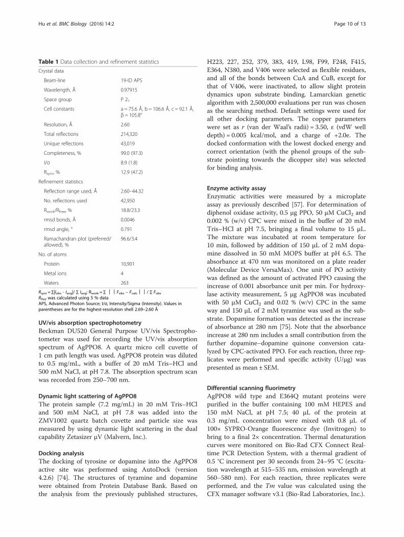
Table 1 Data collection and refinement statistics
Crystal data
Beam-line 19-ID APS
Wavelength, Å 0.97915
Space group P 21
Cell constants a = 75.6 Å, b = 106.6 Å, c = 92.1 Å,β = 105.8°
Resolution, Å 2.60
Total reflections 214,320
Unique reflections 43,019
Completeness, % 99.0 (97.3)
I/σ 8.9 (1.8)
Rsym, % 12.9 (47.2)
Refinement statistics
Reflection range used, Å 2.60–44.32
No. reflections used 42,950
Rwork/Rfree, % 18.8/23.3
rmsd bonds, Å 0.0046
rmsd angle, ° 0.791
Ramachandran plot (preferred/allowed), %
96.6/3.4
No. of atoms
Protein 10,901
Metal ions 4
Waters 263
Rsym = ∑|Iobs - Iavg|/ ∑ Iavg; Rwork = ∑ ││Fobs – Fcalc││/ ∑ FobsRfree was calculated using 5 % dataAPS, Advanced Photon Source; I/σ, Intensity/Sigma (Intensity). Values inparentheses are for the highest-resolution shell 2.69–2.60 Å
Hu et al. BMC Biology (2016) 14:2 Page 10 of 13

Availability of supporting dataThe atomic coordinates and structure factors of AgPPO8have been deposited in the Protein Data Bank, www.rcsb.org (PDB accession code 4YZW).
Additional files
Additional file 1: AgPPO8 exists as a homodimer in solution.(A) AgPPO8 appears as dimeric on size exclusion chromatography (SEC).The theoretical molecular weight (MW) of AgPPO8 monomer is 81 kDa,calculated by ExPASy server (http://web.expasy.org/compute_pi/). Theretention volumes are aligned between the standard proteins (Upperpanel) and AgPPO8 (Lower panel). Standards: thyroglobulin (bovine),670 kDa; γ-globulin (bovine), 158 kDa; ovalbumin (chicken), 44 kDa;myoglobin (horse), 17 kDa; vitamin B12, 1.35 kDa. AgPPO8 was eluted outat a similar retention volume as that of γ-globulin (bovine). The calculatedMW of AgPPO8 from the chromatograph is 142 kDa, indicating a dimer.(B) AgPPO8 displays as dimeric from dynamic light scattering. AgPPO8 ata concentration of 1.5 mg/mL was used in the experiment. AgPPO8 displaysas monodisperse in solution with a MW of 165.9 ± 30.5 kDa, correspondingto a dimer and consistent with the result from SEC analysis. (TIF 1376 kb)
Additional file 2: The electron density map at the di-nuclear activesite of AgPPO8. The electron densities of 2Fo-Fc map and Fo-Fc differencemap are contoured at the sigma level of 1.0 and 3.0, and shown as grayand green mesh, respectively. (TIF 3813 kb)
Additional file 3: The six Cu-coordinating His residues are stabilizedby three Phe residues at the active site of AgPPO8 via hydrophobicinteractions. The six His and three Phe residues are shown as magentaand red sticks, respectively. The shortest carbon-carbon distances (Å) areindicated as black (F99), yellow (F248), and green (F415) dashes, respectively.CuA and CuB are shown as brown spheres. (TIF 3116 kb)
Additional file 4: UV/Vis absorption spectrum of AgPPO8. The purifiedAgPPO8 (0.5 mg/mL) was used for absorbance scanning from 250 to700 nm. A sharp peak at 280 nm was observed for the protein and noobvious absorbance peak was detected at other wavelength. (TIF 270 kb)
Additional file 5: Docking of substrates to the Site II pocket ofAgPPO8. (A) The binding energy for each individually dockedconformation of tyramine/dopamine is ranked and listed in the table. (B)Superposition of all docked conformations with the correct orientationsof tyramine (left) and dopamine (right). The best models of the boundsubstrates with the lowest binding energies are shown as yellow sticks,with all the other docking modes shown in lines. (TIF 799 kb)
Additional file 6: Characterization of the purified AgPPO8 wild type(WT) and E364Q mutant proteins. (A) SDS-PAGE analysis of the purifiedAgPPO8 WT and E364Q mutant under reducing and non-reducing conditions.(B) Thermal denaturation (Tm) values (°C) of AgPPO8 WT and E364Q mutant.All data were presented as mean ± SD (n = 3). (TIF 623 kb)
Additional file 7: Docking analysis of phenolic substrates into theF99-deleted AgPPO8 active site. The docked active site structures, with(magenta) or without (silver) the putative placeholder F99, are superimposed.(A) tyramine, (B) dopamine. The docked substrates in the active site are shownas yellow in F99-containing and green in the absence of F99, respectively. Theenvelope of Site II pocket is delineated in cyan, while CuA and CuB are shownas brown spheres. All the docking parameters were the same with the onlydifference being the presence or absence of F99. The best models with thelowest binding energy and correct orientation were selected for analysis.(TIF 1513 kb)
Competing interestsThe authors declare no competing interests.
Authors’ contributionsYH and YW cloned, expressed and purified AgPPO8. YH crystallized thematerials and determined the crystal structure. YH, JD and HJ designed theresearch project and wrote the manuscript. All authors read and approvedthe final manuscript.
AcknowledgementsWe thank the staff of beamline 19-ID at the Advanced Photon Source fortheir generous support. This work was supported by NIH grants AI113539(JD), GM58634, AI112662 (to HJ), and by Oklahoma Agricultural ExperimentStation at Oklahoma State University under Projects OKL02848 (JD) andOKL02450 (HJ). The authors declare no conflicts of interest.
Received: 24 July 2015 Accepted: 23 December 2015
References1. Ashida M, Brey PT. Recent advances on the research of the insect
prophenoloxidase cascade. In: Brey PT, Hultmark D, editors. MolecularMechanisms of Immune Response in Insects. London: Chapman & Hall;1997. p. 135–72.
2. Cerenius L, Soderhall K. The prophenoloxidase-activating system ininvertebrates. Immunol Rev. 2004;198:116–26.
3. Jiang H. The biochemical basis of antimicrobial responses in Manduca sexta.Insect Sci. 2008;15(1):53–66. doi:10.1111/j.1744-7917.2008.00187.x.
4. Hall M, Scott T, Sugumaran M, Soderhall K, Law JH. Proenzyme of Manducasexta phenol oxidase: purification, activation, substrate specificity of theactive enzyme, and molecular cloning. Proc Natl Acad Sci U S A.1995;92(17):7764–8.
5. Asada N, Fukumitsu T, Fujimoto K, Masuda K. Activation ofprophenoloxidase with 2-propanol and other organic compounds inDrosophila melanogaster. Insect Biochem Mol Biol. 1993;23(4):515–20.
6. Coates CJ, Nairn J. Diverse immune functions of hemocyanins. Dev CompImmunol. 2014;45(1):43–55. doi:10.1016/j.dci.2014.01.021.
7. Sugumaran M. Comparative biochemistry of eumelanogenesis and theprotective roles of phenoloxidase and melanin in insects. Pigment Cell Res.2002;15(1):2–9.
8. Cerenius L, Lee BL, Soderhall K. The proPO-system: pros and cons for itsrole in invertebrate immunity. Trends Immunol. 2008;29(6):263–71.doi:10.1016/j.it.2008.02.009.
9. Zhao P, Li J, Wang Y, Jiang H. Broad-spectrum antimicrobial activity of thereactive compounds generated in vitro by Manduca sexta phenoloxidase.Insect Biochem Mol Biol. 2007;37(9):952–9. doi:10.1016/j.ibmb.2007.05.001.
10. Solomon EI, Sundaram UM, Machonkin TE. Multicopper oxidases andoxygenases. Chem Rev. 1996;96(7):2563–606.
11. Decker H, Schweikardt T, Nillius D, Salzbrunn U, Jaenicke E, Tuczek F.Similar enzyme activation and catalysis in hemocyanins and tyrosinases.Gene. 2007;398(1–2):183–91. doi:10.1016/j.gene.2007.02.051.
12. Kaintz C, Mauracher SG, Rompel A. Type-3 copper proteins: recentadvances on polyphenol oxidases. Adv Protein Chem Struct Biol.2014;97:1–35. doi:10.1016/bs.apcsb.2014.07.001.
13. Lu A, Zhang Q, Zhang J, Yang B, Wu K, Xie W, et al. Insectprophenoloxidase: the view beyond immunity. Front Physiol.2014;5:252. doi:10.3389/fphys.2014.00252.
14. Boeckx T, Winters AL, Webb KJ, Kingston-Smith AH. Polyphenol oxidase inleaves: is there any significance to the chloroplastic localization? J Exp Botany.2015;66(12):3571–9. doi:10.1093/jxb/erv141.
15. Mayer AM. Polyphenol oxidases in plants and fungi: going places? A review.Phytochemistry. 2006;67(21):2318–31. doi:10.1016/j.phytochem.2006.08.006.
16. Oetting WS, King RA. Molecular basis of albinism: mutations and polymorphismsof pigmentation genes associated with albinism. Hum Mutat. 1999;13(2):99–115.doi:10.1002/(SICI)1098-1004(1999)13:2<99::AID-HUMU2>3.0.CO;2-C.
17. Simonova M, Wall A, Weissleder R, Bogdanov Jr A. Tyrosinase mutants arecapable of prodrug activation in transfected nonmelanotic cells. Cancer Res.2000;60(23):6656–62.
18. Mangum CP. Oxygen transport in invertebrates. Am J Physiol.1985;248(5 Pt 2):R505–14.
19. Decker H, Jaenicke E. Recent findings on phenoloxidase activity andantimicrobial activity of hemocyanins. Dev Comp Immunol. 2004;28(7–8):673–87. doi:10.1016/j.dci.2003.11.007.
20. Sanchez-Ferrer A, Rodriguez-Lopez JN, Garcia-Canovas F, Garcia-CarmonaF. Tyrosinase: a comprehensive review of its mechanism. Biochim BiophysActa. 1995;1247(1):1–11.
21. Gerdemann C, Eicken C, Krebs B. The crystal structure of catechol oxidase:new insight into the function of type-3 copper proteins. Acc Chem Res.2002;35(3):183–91.
Hu et al. BMC Biology (2016) 14:2 Page 11 of 13

22. Burmester T. Molecular evolution of the arthropod hemocyaninsuperfamily. Mol Biol Evol. 2001;18(2):184–95.
23. van Holde KE, Miller KI, Decker H. Hemocyanins and invertebrate evolution.J Biol Chem. 2001;276(19):15563–6. doi:10.1074/jbc.R100010200.
24. Liu H, Jiravanichpaisal P, Cerenius L, Lee BL, Soderhall I, Soderhall K.Phenoloxidase is an important component of the defense againstAeromonas hydrophila infection in a crustacean, Pacifastacus leniusculus.J Biol Chem. 2007;282(46):33593–8. doi:10.1074/jbc.M706113200.
25. Binggeli O, Neyen C, Poidevin M, Lemaitre B. Prophenoloxidase activation isrequired for survival to microbial infections in Drosophila. PLoS Pathog.2014;10(5):e1004067. doi:10.1371/journal.ppat.1004067.
26. Li Y, Wang Y, Jiang H, Deng J. Crystal structure of Manduca sextaprophenoloxidase provides insights into the mechanism of type 3 copperenzymes. Proc Natl Acad Sci U S A. 2009;106(40):17002–6.doi:10.1073/pnas.0906095106.
27. Masuda T, Momoji K, Hirata T, Mikami B. The crystal structure of acrustacean prophenoloxidase provides a clue to understanding thefunctionality of the type 3 copper proteins. FEBS J. 2014;281(11):2659–73.doi:10.1111/febs.12812.
28. Yassine H, Osta MA. Anopheles gambiae innate immunity. Cell Microbiol.2010;12(1):1–9. doi:10.1111/j.1462-5822.2009.01388.x.
29. Jiang H, Wang Y, Korochkina SE, Benes H, Kanost MR. Molecular cloning ofcDNAs for two pro-phenol oxidase subunits from the malaria vector,Anopheles gambiae. Insect Biochem Mol Biol. 1997;27(7):693–9.
30. Lee WJ, Ahmed A, Della Torre A, Kobayashi A, Ashida M, Brey PT. Molecularcloning and chromosomal localization of a prophenoloxidase cDNA fromthe malaria vector Anopheles gambiae. Insect Mol Biol. 1998;7(1):41–50.
31. Müller HM, Dimopoulos G, Blass C, Kafatos FC. A hemocyte-like cell lineestablished from the malaria vector Anopheles gambiae expresses sixprophenoloxidase genes. J Biol Chem. 1999;274(17):11727–35.
32. Müller HM. The prophenoloxidases of Anopheles gambiae. EMBL/GenBank/DDBJ databases 2002.
33. Sendovski M, Kanteev M, Ben-Yosef VS, Adir N, Fishman A. Firststructures of an active bacterial tyrosinase reveal copper plasticity. JMol Biol. 2011;405(1):227–37. doi:10.1016/j.jmb.2010.10.048.
34. Rolff M, Schottenheim J, Decker H, Tuczek F. Copper-O2 reactivity oftyrosinase models towards external monophenolic substrates: molecularmechanism and comparison with the enzyme. Chem Soc Rev.2011;40(7):4077–98. doi:10.1039/c0cs00202j.
35. Dundas J, Ouyang Z, Tseng J, Binkowski A, Turpaz Y, Liang J. CASTp:computed atlas of surface topography of proteins with structural andtopographical mapping of functionally annotated residues. Nucleic AcidsRes. 2006;34(Web Server issue):W116–8. doi:10.1093/nar/gkl282.
36. Itoh S, Fukuzumi S. Monooxygenase activity of type 3 copper proteins.Acc Chem Res. 2007;40(7):592–600. doi:10.1021/ar6000395.
37. Itoh S, Kumei H, Taki M, Nagatomo S, Kitagawa T, Fukuzumi S. Oxygenationof phenols to catechols by a (mu-eta 2:eta 2-peroxo)dicopper(II) complex:mechanistic insight into the phenolase activity of tyrosinase. J Am ChemSoc. 2001;123(27):6708–9.
38. Matoba Y, Kumagai T, Yamamoto A, Yoshitsu H, Sugiyama M.Crystallographic evidence that the dinuclear copper center of tyrosinaseis flexible during catalysis. J Biol Chem. 2006;281(13):8981–90.doi:10.1074/jbc.M509785200.
39. Olivares C, Garcia-Borron JC, Solano F. Identification of active site residuesinvolved in metal cofactor binding and stereospecific substrate recognition inmammalian tyrosinase. Implications to the catalytic cycle. Biochemistry.2002;41(2):679–86.
40. Decker H, Schweikardt T, Tuczek F. The first crystal structure of tyrosinase: allquestions answered? Angew Chem Int Ed Engl. 2006;45(28):4546–50.doi:10.1002/anie.200601255.
41. Olivares C, Solano F. New insights into the active site structure and catalyticmechanism of tyrosinase and its related proteins. Pigment Cell MelanomaRes. 2009;22(6):750–60. doi:10.1111/j.1755-148X.2009.00636.x.
42. Goldfeder M, Kanteev M, Isaschar-Ovdat S, Adir N, Fishman A. Determinationof tyrosinase substrate-binding modes reveals mechanistic differencesbetween type-3 copper proteins. Nat Commun. 2014;5:4505. doi:10.1038/ncomms5505.
43. Virador VM, Reyes Grajeda JP, Blanco-Labra A, Mendiola-Olaya E, Smith GM,Moreno A, et al. Cloning, sequencing, purification, and crystal structure ofGrenache (Vitis vinifera) polyphenol oxidase. J Agric Food Chem.2010;58(2):1189–201. doi:10.1021/jf902939q.
44. Klabunde T, Eicken C, Sacchettini JC, Krebs B. Crystal structure of aplant catechol oxidase containing a dicopper center. Nat Struct Biol.1998;5(12):1084–90. doi:10.1038/4193.
45. Asano T, Ashida M. Cuticular pro-phenoloxidase of the silkworm,Bombyx mori. Purification and demonstration of its transport fromhemolymph. J Biol Chem. 2001;276(14):11100–12. doi:10.1074/jbc.M008426200.
46. Gupta S, Wang Y, Jiang H. Manduca sexta prophenoloxidase (proPO)activation requires proPO-activating proteinase (PAP) and serineproteinase homologs (SPHs) simultaneously. Insect Biochem Mol Biol.2005;35(3):241–8. doi:10.1016/j.ibmb.2004.12.003.
47. Wang Y, Jiang H. Prophenoloxidase (proPO) activation in Manduca sexta: ananalysis of molecular interactions among proPO, proPO-activatingproteinase-3, and a cofactor. Insect Biochem Mol Biol. 2004;34(8):731–42.doi:10.1016/j.ibmb.2004.03.008.
48. Fujieda N, Yabuta S, Ikeda T, Oyama T, Muraki N, Kurisu G, et al.Crystal structures of copper-depleted and copper-bound fungalpro-tyrosinase: insights into endogenous cysteine-dependent copperincorporation. J Biol Chem. 2013;288(30):22128–40. doi:10.1074/jbc.M113.477612.
49. Volbeda A, Hol WG. Crystal structure of hexameric haemocyanin from Panulirusinterruptus refined at 3.2 A resolution. J Mol Biol. 1989;209(2):249–79.
50. Magnus KA, Hazes B, Ton-That H, Bonaventura C, Bonaventura J, HolWG. Crystallographic analysis of oxygenated and deoxygenated statesof arthropod hemocyanin shows unusual differences. Proteins.1994;19(4):302–9. doi:10.1002/prot.340190405.
51. Cong Y, Zhang Q, Woolford D, Schweikardt T, Khant H, Dougherty M,et al. Structural mechanism of SDS-induced enzyme activity of scorpionhemocyanin revealed by electron cryomicroscopy. Structure.2009;17(5):749–58. doi:10.1016/j.str.2009.03.005.
52. Decker H, Rimke T. Tarantula hemocyanin shows phenoloxidase activity.J Biol Chem. 1998;273(40):25889–92. doi:10.1074/jbc.273.40.25889.
53. Gerdemann C, Eicken C, Galla HJ, Krebs B. Comparative modeling of thelatent form of a plant catechol oxidase using a molluskan hemocyaninstructure. J Inorg Biochem. 2002;89(1–2):155–8.
54. Aspan A, Huang TS, Cerenius L, Soderhall K. cDNA cloning ofprophenoloxidase from the freshwater crayfish Pacifastacus leniusculus andits activation. Proc Natl Acad Sci U S A. 1995;92(4):939–43.
55. Kawabata T, Yasuhara Y, Ochiai M, Matsuura S, Ashida M. Molecular cloningof insect pro-phenol oxidase: a copper-containing protein homologous toarthropod hemocyanin. Proc Natl Acad Sci U S A. 1995;92(17):7774–8.
56. Lee SY, Kwon TH, Hyun JH, Choi JS, Kawabata SI, Iwanaga S, et al. In vitroactivation of pro-phenol-oxidase by two kinds of pro-phenol-oxidase-activating factors isolated from hemolymph of coleopteran, Holotrichiadiomphalia larvae. Eur J Biochem. 1998;254(1):50–7.
57. Jiang H, Wang Y, Yu XQ, Kanost MR. Prophenoloxidase-activatingproteinase-2 from hemolymph of Manduca sexta. A bacteria-inducibleserine proteinase containing two clip domains. J Biol Chem.2003;278(6):3552–61. doi:10.1074/jbc.M205743200.
58. Jiang H, Wang Y, Yu XQ, Zhu Y, Kanost MR. Prophenoloxidase-activatingproteinase-3 (PAP-3) from Manduca sexta hemolymph: a clip-domain serineproteinase regulated by serpin-1 J and serine proteinase homologs. InsectBiochem Mol Biol. 2003;33(10):1049–60.
59. Chosa N, Fukumitsu T, Fujimoto K, Ohnishi E. Activation of prophenoloxidaseA1 by an activating enzyme in Drosophila melanogaster. Insect Biochem MolBiol. 1997;27(1):61–8.
60. Pal D, Chakrabarti P. Cis peptide bonds in proteins: residues involved, theirconformations, interactions and locations. J Mol Biol. 1999;294(1):271–88.doi:10.1006/jmbi.1999.3217.
61. Lorenzen S, Peters B, Goede A, Preissner R, Frommel C. Conservation of cisprolyl bonds in proteins during evolution. Proteins. 2005;58(3):589–95.doi:10.1002/prot.20342.
62. Yu XQ, Jiang H, Wang Y, Kanost MR. Nonproteolytic serine proteinasehomologs are involved in prophenoloxidase activation in the tobaccohornworm, Manduca sexta. Insect Biochem Mol Biol. 2003;33(2):197–208.
63. Piao S, Song YL, Kim JH, Park SY, Park JW, Lee BL, et al. Crystal structure of aclip-domain serine protease and functional roles of the clip domains. EMBOJ. 2005;24(24):4404–14. doi:10.1038/sj.emboj.7600891.
64. Kan H, Kim CH, Kwon HM, Park JW, Roh KB, Lee H, et al. Molecular controlof phenoloxidase-induced melanin synthesis in an insect. J Biol Chem.2008;283(37):25316–23. doi:10.1074/jbc.M804364200.
Hu et al. BMC Biology (2016) 14:2 Page 12 of 13

65. Clark KD, Strand MR. Hemolymph melanization in the silkmoth Bombyx moriinvolves formation of a high molecular mass complex that metabolizestyrosine. J Biol Chem. 2013;288(20):14476–87. doi:10.1074/jbc.M113.459222.
66. Krumm B, Meng X, Wang Z, Xiang Y, Deng J. A unique bivalent binding andinhibition mechanism by the yatapoxvirus interleukin 18 binding protein.PLoS Pathog. 2012;8(8):e1002876. doi:10.1371/journal.ppat.1002876.
67. Deng J, Davies DR, Wisedchaisri G, Wu M, Hol WG, Mehlin C. An improvedprotocol for rapid freezing of protein samples for long-term storage. ActaCrystallogr D Biol Crystallogr. 2004;60(Pt 1):203–4.
68. Minor W, Cymborowski M, Otwinowski Z, Chruszcz M. HKL-3000: theintegration of data reduction and structure solution–from diffraction images toan initial model in minutes. Acta Crystallogr D Biol Crystallogr. 2006;62(Pt 8):859–66. doi:10.1107/S0907444906019949.
69. Adams PD, Afonine PV, Bunkoczi G, Chen VB, Davis IW, Echols N, et al.PHENIX: a comprehensive Python-based system for macromolecularstructure solution. Acta Crystallogr D Biol Crystallogr. 2010;66(Pt 2):213–21.doi:10.1107/S0907444909052925.
70. Emsley P, Lohkamp B, Scott WG, Cowtan K. Features and development ofCoot. Acta Crystallogr D Biol Crystallogr. 2010;66(Pt 4):486–501.doi:10.1107/S0907444910007493.
71. Chen VB, Arendall 3rd WB, Headd JJ, Keedy DA, Immormino RM, Kapral GJ,et al. MolProbity: all-atom structure validation for macromolecularcrystallography. Acta Crystallogr D Biol Crystallogr. 2010;66(Pt 1):12–21.doi:10.1107/S0907444909042073.
72. Berman H, Henrick K, Nakamura H. Announcing the worldwide Protein DataBank. Nat Struct Biol. 2003;10(12):980. doi:10.1038/nsb1203-980.
73. DeLano W. The PyMOL molecular graphics system. Palo Alto, CA, USA:DeLano Scientific; 2002.
74. Morris GM, Huey R, Lindstrom W, Sanner MF, Belew RK, Goodsell DS, et al.AutoDock4 and AutoDockTools4: Automated docking with selectivereceptor flexibility. J Comput Chem. 2009;30(16):2785–91.doi:10.1002/jcc.21256.
75. Duckworth HW, Coleman JE. Physicochemical and kinetic properties ofmushroom tyrosinase. J Biol Chem. 1970;245(7):1613–25.
• We accept pre-submission inquiries
• Our selector tool helps you to find the most relevant journal
• We provide round the clock customer support
• Convenient online submission
• Thorough peer review
• Inclusion in PubMed and all major indexing services
• Maximum visibility for your research
Submit your manuscript atwww.biomedcentral.com/submit
Submit your next manuscript to BioMed Central and we will help you at every step:
Hu et al. BMC Biology (2016) 14:2 Page 13 of 13