Embed Size (px)
Citation preview

THE STRUCTURE AND FUNCTION OF THE sIHF PROTEIN
CHARACTERIZING THE STRUCTURE AND FUNCTION OF A NOVEL NUCLEOID-ASSOCIATED
PROTEIN sIHF
By TAMIZA NANJI, B. Sc. (Hons.)
A Thesis Submitted to the School of Graduate Studies in Partial Fulfillment of the Requirements
for the Degree Master of Science
McMaster University
© Copyright by Tamiza Nanji, July 2014

ii
MASTER OF SCIENCE (2014) McMaster University
(Biochemistry and Biomedical Sciences) Hamilton, Ontario
TITLE: Characterizing the structure and function of a novel nucleoid-associated
protein sIHF
AUTHOR: Tamiza Nanji, B. Sc (Hons.) (McMaster University)
SUPERVISOR: Dr. Alba Guarné
NUMBER OF PAGES: xi, 68
iii
Abstract
All living organisms must organize their genome so that it not only fits within the cell, but
remains accessible for cellular processes. In bacteria, an arsenal of nucleoid-associated proteins
contributes to chromosome condensation. A novel nucleoid-associated protein was recently
discovered in actinobacteria, and is essential in Mycobacterium. It was classified as an
integration host factor protein (IHF); however, it does not share sequence or structural
homology with the well characterized Escherichia coli IHF. In this study, we characterize the
structure and function of Streptomyces coelicolor IHF (sIHF). We have used a combination of
biochemistry and structural biology to characterize the role of sIHF in DNA binding and DNA
topology. We have solved crystal structures of sIHF bound to various double-stranded DNA
substrates, and show that sIHF is able to contact DNA at multiple surfaces. Furthermore, sIHF
inhibits the activity of TopA, impacting DNA topology in vitro. Our work demonstrates that sIHF
is a novel nucleoid-associated protein with key roles in condensing DNA. We believe that sIHF
performs its function by differentially using multiple nucleic-acid binding surfaces. Further
characterization is required to confirm this hypothesis in vivo. Given that the Mycobacterium
homolog of sIHF (mIHF) is essential, our studies lay the foundation to explore novel drug targets
for Mycobacterium tuberculosis and Mycobacterium leprae.

iv
Acknowledgements
I would like to thank my supervisor, Dr. Alba Guarné, for her guidance and support. She has
gone above and beyond the role of a supervisor to ensure that my journey through graduate
school was successful. I would also like to thank her for all the hard work she has put into this
project and for teaching me techniques and skills that I will carry with me for the rest of my
scientific career. I would like to thank my committee members; Dr. Marie Elliot for fostering my
learning of Streptomyces and for making me feel welcome at her lab meetings, and Dr. Yingfu Li
for his valuable input and thought provoking discussions.
I would like to thank the past and present members of the Guarné and Elliot labs that
have made my experience in the lab more engaging and enjoyable. I owe much of my success to
senior members who have not only helped me with laboratory techniques, but have encouraged
my growth through stimulating scientific conversations. To Dr. Marie Elliot, Dr. Julia Swiercz,
Melanie Gloyd, Sabrina Lue Tam, and Dr. Emma Sherwood, thank you for all your help and
contribution to the project.
I would also like to thank my family and friends for their support. To my parents,
Shamim and Moazali Nanji, thank you for taking care of me and instilling the values of hard work
and discipline which have been instrumental in completing this degree. To my brother, Jamil
Nanji, thank you for always making sure I have fun in my life. To Safir Kassam, thank you for
your support and encouragement.
v
Table of Contents
Abstract……………………………………………………………………………………………………………………… iii Acknowledgements……………………………………………………………………………………………………. iv List of Figures…………………………………………………………………………..................................... vii List of Tables………………………………………………………………………………………………………………. viii List of Abbreviations and Symbols……………………………………………………………………………… ix Declaration of Academic Achievement………………………………………………………………………. xi Chapter 1-Introduction……………………………………………………………………………………………… 1 1.1 The bacterial nucleoid…………………………………………………………………………….. 1 1.2 Nucleoid-associated proteins in E. coli..................................................…. 3 1.2.1 IHF……………………………………………………………………………………………. 4 1.2.2 HU……………………………………………………………………………………………. 4 1.2.3 Fis…………………………………………………………………………………………….. 5 1.2.4 H-NS…………………………………………………………………………………………. 6 1.2.5 MukB……………………………………………………………………………………….. 8 1.2.6 Lrp……………………………………………………………………………………………. 8 1.2.7 Dps…………………………………………………………………………………………… 9 1.3 Streptomyces coelicolor as a model organism to study novel
nucleoid-associated proteins in actinobacteria……………………………………… 10 1.4 Thesis objective……………………………………………………………………………………….. 14 Chapter 2-Methods and Materials……………………………………………………………………………. 15 2.1 Overexpression of sIHF…………………………………………………………………………….. 15 2.2 Purification of sIHF…………………………………………………………………………………… 16 2.3 Forming the sIHF-DNA complex………………………………………………………………… 18 2.4 Crystallization of sIHF bound to DNA………………………………………………………… 19 2.5 Data collection and structure determination……………………………………………. 20 2.6 Cloning of sIHF mutants……………………………………………………………………………. 20 2.7 Dynamic light scattering (DLS)………………………………………………………………….. 23 2.8 Electrophoretic mobility shift assays (EMSAs)…………………………………………... 24 2.9 Topoisomerase assays………………………………………………………………………………. 24 2.10 Data collection and processing for small-angle X-ray scattering (SAXS)….. 25 Chapter 3-Results……………………………………………………………………………………………………….. 26 3.1 Purification of sIHF……………………………………………………………………………………. 26
3.2 Crystallization, data collection and structure of sIHF bound to a 19-bp dupex DNA substrate……………………………………………………………………. 28
3.3 Crystallization, data collection and structure of sIHF bound to an 8-bp DNA substrate………………………………………………………………………………… 34
3.4 sIHF contacts DNA at three interfaces in the crystal structure of sIHF bound to a hairpin DNA substrate…………………………………………………………… 39
3.5 Three interfaces on sIHF contribute to DNA binding in vitro……………………. 43 3.6 sIHF does not bridge DNA…………………………………………………………………………. 47
vi
3.7 sIHF affects TopA activity independently of DNA binding ………………………… 50 Chapter 4-Discussion…………………………………………………………………………………………………… 54 Chapter 5-Conclusion………………………………………………………………………………………………….. 62 References…………………………………………………………………………………………………………………… 63

vii
List of Figures
Figure 1.1 Factors contributing to forming a condensed bacterial nucleoid………………… 2 Figure 1.2 NAPs that bend DNA…………………………………………………………………………………… 6 Figure 1.3 Streptomyces life cycle………………………………………………………………………………… 12 Figure 3.1 Purification scheme of untagged sIHF…………………………………………………………. 27 Figure 3.2 Crystals of sIHF bound to a 19-bp duplex DNA substrate……………………………. 29 Figure 3.3 Structure of sIHF ………………………………………………………………………………………… 32 Figure 3.4 Structure of sIHF bound to TN08-BP1………………………………………………………….. 38 Figure 3.5 Structure of sIHF bound to TN23-HP9…………………………………………………………. 42 Figure 3.6 EMSAs of sIHF mutants with a radiolabeled 23-bp duplex DNA substrate…… 45 Figure 3.7 Electrostatics of sIHF bound to DNA……………………………………………………………. 47 Figure 3.8 sIHF affects the activity of TopA…………………………………………………………………… 51 Figure 3.9 Gel filtration assay of sIHF with TopA…………………………………………………………… 52 Figure 3.10 Topoisomerase assays of sIHF mutants ……………………………………………………… 53 Figure 4.1 Ribbon structure of sIHF bound to TN08-BP1 and possible
interactions with the N-terminal helix………………………………………………………… 56 Figure 4.2 Proposed mechanism of action of sIHF………………………………………………………… 59

viii
List of Tables
Table 1.1 Well characterized nucleoid-associated proteins…………………………………………. 10 Table 2.1 Expression plasmids……………………………………………………………………………………… 16 Table 2.2 DNA substrates used for crystallization………………………………………………………… 18 Table 2.3 Primers used to generate sIHF mutants………………………………………………………… 22 Table 2.4 Cloning plasmids…………………………………………………………………………………………… 23 Table 3.1 X-ray data collection and refinement…………………………………………………………… 31 Table 3.2 Base-pair parameters of duplex DNA alone and in complex with sIHF ………… 36 Table 3.3 Small-angle X-ray scattering data collection and analysis……………………………… 49

ix
List of Abbreviations and Symbols
α alpha Å angstrom aIHF actinobacterial integration host factor protein BNL Brookhaven National Laboratory bp base pair BSA bovine serum albumin C Celsius COOT Crystallographic Object-Oriented Toolkit ° degree DPS DNA protection during starvation protein DLS dynamic light scattering DTT dithiothreitol DNA deoxyribonucleic acid E. coli Escherichia coli EDTA ethylenediaminetetraacetic acid EMSA electrophoretic mobility shift assay Fis factor for inversion stimulation Ƴ Gamma GE General Electric H2TH helix-two turns-helix HU histone-like protein from E. coli strain U93 IDT Integrated DNA Technologies IHF integration host factor IPTG isopropyl-beta-D-thiogalactopyranoside LB Luria-Bertani Lrp leucine responsive regulatory protein mIHF mycobacterial integration host factor μg microgram μL microliter μM micromolar mL milliliter mM millimolar MOBIX McMaster Institute for Molecular Biology and Biotechnology MR molecular replacement MWCO molecular weight cut-off NMR nuclear magnetic resonance NAP nucleoid-associated protein NSLS National Synchrotron Light Source OD600 optical density measured at 600 nm PAGE polyacrylamide gel electrophoresis PDB Protein Data Bank PCR polymerase chain reaction

x
PEG polyethylene glycol PHENIX Python-based Hierarchical Environment for Integrated Xtallography PMSF phenylmethanesulfonyl fluoride RMSD root-mean-squared deviation SAD single-wavelength anomalous dispersion SAXS small angle X-ray scattering S. coelicolor Streptomyces coelicolor SDS sodium dodecyl sulphate SELEX systematic evolution of ligands by exponential enrichment Sel-Met selenomethionine sIHF Streptomyces integration host factor SMC structural maintenance of chromosome complex SOC super optimal broth with catabolite repression TAE Tris base, acetic acid and EDTA UV ultraviolet v/v volume per volume w/v weight per volume WT wild type

xi
Declaration of Academic Achievement
I devised the protocol for large-scale sIHF over-expression and purification. I conducted initial
electrophoretic mobility shift assays and determined the optimal DNA substrates for
crystallization of the sIHF-DNA complexes. I grew all crystals that resulted in the structures
presented in this work. Data was processed and refined by Dr. Alba Guarné and myself. I
prepared the samples for small-angle X-ray scattering studies and Dr. Alba Guarné conducted
the analysis. All sIHF mutants were designed by Dr. Alba Guarné and myself; aside from sIHF Δ1-
36 and sIHF 35-37Gly, which were made with the help of Sabrina Lue Tam, I constructed the sIHF
variants. Electrophoretic mobility shift assays and topoisomerase activity assays were
conducted by Dr. Emma Sherwood and Melanie Gloyd respectively using sIHF protein that I
purified. Gel filtration assays of sIHF with TopA were performed by Melanie Gloyd.

M.Sc Thesis – T. Nanji; McMaster University – Biochemistry and Biomedical Sciences
1
Chapter 1
Introduction
1.1 The bacterial nucleoid
DNA stores essential information required for replication, and for the synthesis of RNA and
protein molecules. It is contained within the bacterial genome, which unconstrained exceeds
the size of the cell by 1 000- 10 000 times (Gitai et al., 2005). Therefore, chromosome
compaction is essential. Even more impressive is that the condensed chromosome remains
dynamic and accessible for cellular processes, such as DNA replication, transcription, and
segregation, throughout the cell cycle (Thanbichler et al., 2005a; Thanbichler et al., 2005b).
Bacteria lack features of eukaryotic cells that aid in chromosome compaction, such as a
membrane bound nucleus and histone proteins (Zhu and Wani, 2010). Instead, bacteria
compact their DNA into a condensed, yet dynamic, structure called the bacterial nucleoid.
Nucleoid organization is mediated through molecular crowding and using multiple proteins.
These proteins include topoisomerases that affect the superhelicity of the bacterial
chromosome, RNA polymerases and effector proteins that mediate their activity, as well as
abundant nucleoid associated proteins (NAPs) (Travers and Muskhelishvili, 2005; Luijsterburg et
al., 2008) (Figure 1.1).
In crowded environments, characteristic of the bacterial cell, entropic forces impact the
interaction between macromolecules to a greater extent than in diluted environments
(Luijsterburg et al., 2008). This causes larger macromolecules to come together allowing smaller
molecules to move more freely. This phenomenon is known as attraction and depletion forces

M.Sc Thesis – T. Nanji; McMaster University – Biochemistry and Biomedical Sciences
2
(Marenduzzo et al., 2006; Hancock, 2004) and causes the nucleoid to self-condense (Figure 1.1;
blue stars represent crowding from the environment).
Figure 1.1. Factors contributing to forming a condensed bacterial nucleoid. Unconstrained, bacterial DNA is diffuse, unstructured, and exceeds the size of the bacterial cell. Black lines indicate duplex DNA. Molecular crowding, indicated by the blue stars, acts as a force which condenses bacterial DNA. DNA supercoiling allows the bacterial chromosome to intertwine in an orderly manner to condense the nucleoid, but also allows for DNA dynamics. Nucleoid-associated proteins (NAPs), depicted as green circles and red squares, are a diverse group of bacterial proteins that bind DNA and aid in DNA compaction.
DNA supercoiling allows DNA to pack closer together to form a condensed structure.
Supercoils are introduced and removed by topoisomerase enzymes creating a dynamic nucleoid
(Champoux, 2001). Once regions of the nucleoid are relaxed, transcription factors and
polymerase enzymes can access parts of the genome that were occluded. This modifies levels of
gene expression which is necessary for cell cycle progression (Myers et al., 2013; Azam et al.,
1999). The topological organization of the bacterial chromosome is not random. It has been
shown to be organized into small topological domains (~10 kb) where diffusion of supercoils is

M.Sc Thesis – T. Nanji; McMaster University – Biochemistry and Biomedical Sciences
3
restricted (Postow et al., 2004; Sinden and Pettijohn, 1981). The torsional tension of supercoiled
DNA within these domains drives cellular processes such as transcription (Lim et al., 2003),
replication (Funnell et al., 1986), and recombination (Nash, 1990). Furthermore, the
organization of DNA into smaller domains ensures that a break in one region of the chromosome
only affects that topological unit, and does not cause changes to the overall superhelicity of the
chromosome which can lead to cell death (Wang, 1996).
Eukaryotes mediate DNA organization using histone proteins that wrap DNA in an
orderly fashion, to tightly package the chromosome. Histones can unravel DNA to alter the
accessibility of various regions of the genome (Luger, 2006). Bacteria use an arsenal of nucleoid-
associated proteins (NAPs) (Figure 1.1; depicted by red squares and green circles) to compact
the genome; however, they are not structurally similar to histone proteins. We will look into
these proteins in greater detail in the next sections.
1.2 Nucleoid-associated proteins in E. coli
NAPs are an abundant, diverse group of proteins that are expressed at various levels throughout
the cell cycle (Azam et al., 1999). Most NAPs bind DNA promiscuously to play a significant part
in compacting the bacterial nucleoid. Many of these proteins bind DNA with little sequence
specificity, but prefer binding DNA sequences that are AT-rich. Promoter sequences are AT-rich;
therefore, NAPs have also been characterized as transcription factors as they modulate gene
expression (Myers et al., 2013; Dillon and Dorman, 2010; Dorman, 2013). NAPs have numerous
roles in the bacterial cell and have pleiotropic effects. They are diverse in structure, DNA binding
ability, and their effects on DNA upon binding. They are generally small, basic proteins and have
M.Sc Thesis – T. Nanji; McMaster University – Biochemistry and Biomedical Sciences
4
been shown to compact the bacterial chromosome through bridging, bending, and/or wrapping
DNA (Dillon and Dorman, 2010; Rimsky and Travers, 2011). Although many NAPs have been well
characterized in E. coli, there is less known in other classes of bacteria. We will first explore well
characterized NAPs in E. coli.
1.2.1 IHF
The E. coli integration host factor protein (IHF) was first identified for its role in recombination of
bacteriophage lambda (Miller and Friedman, 1980) and was later classified as a NAP (Rice et al.,
1996). IHF binds DNA and induces sharp bends which alters the trajectory of the DNA duplex,
contributing to a more condensed nucleoid. IHF functions as a heterodimer and is composed of
two subunits; an alpha- and beta-subunit. The alpha-subunit is 11 kDa and the beta-subunit is
9.5 kDa (Rice et al., 1996, Luijsterburg et al., 2006). Both subunits are structurally homologous;
they consist of a body composed of alpha-helices flanked by flexible beta-hairpins (Figure 1.2A).
A conserved proline residue within each beta-hairpin intercalates into the minor groove of DNA
to induce a sharp bend (Swinger et al., 2003; Swinger and Rice, 2004). IHF binds double-
stranded DNA with a footprint of ~30 base-pairs at a consensus sequence rich in adenines and
thymine nucleobases (Goodrich et al., 1990) (Table 1.1). It is maximally expressed during early
stationary phase (Azam et al., 1999).
1.2.2 HU
In E. coli and other enterobacteriaceae, the histone-like protein first identified in E. coli strain
U93 (HU) (Oberto et al., 1994) has a role in chromosome condensation. Similar to IHF, it induces
bends in the DNA (Figure 1.2B). It also functions as a heterodimer and is composed of an alpha-

M.Sc Thesis – T. Nanji; McMaster University – Biochemistry and Biomedical Sciences
5
and beta-subunit (Dame and Goosen, 2002) that are 9.5 kDa each and share 70% homology
(Table 1.1) (Swinger et al., 2003). HU binds DNA with a footprint of ~9 base-pairs and prefers
sequences rich in adenine and thymine nucleobases which are inherently distorted.
Furthermore, HU prefers binding to curved or distorted regions of DNA (Swinger et al., 2003).
HU is structurally homologous to IHF; it consists of a body composed of alpha helices capped by
two beta-hairpin arms that intercalate into the minor groove of DNA using a conserved proline
residue (Figure 1.2B). The bend induced by HU is less pronounced compared to that of IHF, and
HU’s minimal DNA binding site (~9 bp) is smaller than that of IHF (~30 bp) illustrating the
diversity that exists within NAPs, even when they are structurally homologous (Swinger and Rice,
2004). Moreover, HU induces negative supercoiling directly by bending DNA (Rouviѐre-Yaniv et
al., 1979), and indirectly through stimulating the activity of DNA gyrase (Swinger and Rice, 2004).
HU is maximally expressed during logarithmic phase to alter DNA dynamics required during
development in this stage of the cell cycle (Azam et al., 1999).
1.2.3 Fis
The factor for inversion stimulation (Fis) protein is a NAP that condenses the chromosome by
bending DNA, as well as by aiding in loop formation (Luijsterburg et al., 2006; Schneider et al.,
1999). It functions as a 22 kDa homodimer and is composed of four alpha-helices that are
connected by beta-turns and two beta-hairpins (Table 1.1). It binds DNA through a common
DNA binding fold; a helix-turn-helix motif (Figure 1.2C). Fis prefers binding DNA at consensus
nucleotide sequence, but also binds DNA at other sequences with high affinity (Table 1.1). It is
the most abundant NAP during early exponential growth; however, it is completely absent
during stationary phase (Luijsterburg et al., 2006). Fis has been suggested to mediate DNA

M.Sc Thesis – T. Nanji; McMaster University – Biochemistry and Biomedical Sciences
6
supercoiling by inhibiting the expression of DNA gyrase in a manner dependant on DNA topology
(Schneider et al., 1999; Schneider et al., 2001). Fis is a multifaceted NAP that affects
chromosome organization via many avenues. This shows that NAPs are complex and can affect
multiple cellular processes.
Figure 1.2. NAPs that bend DNA. (A) IHF induces sharp bends into duplex DNA by intercalating into the minor groove of DNA using conserved proline residues within the β-hairpins. The protein is indicated in red while duplex DNA is in blue in all panels. (B) HU induces bends by intercalating into the minor groove of DNA using conserved proline residues within the β-hairpin arms. (C) Fis compacts the bacterial chromosome by bending duplex substrates. The crystal structure was solved without DNA; duplex DNA is modeled in with a blue line illustrating where the protein is thought to contact DNA.
1.2.4 H-NS
The histone-like nucleoid structuring protein (H-NS) plays a key role in compacting and
organizing the bacterial nucleoid, and has a role in controlling gene expression. H-NS is a 15.4
kDa protein that functions as a dimer (Table 1.1) (Luijsterburg et al., 2006) and is maximally

M.Sc Thesis – T. Nanji; McMaster University – Biochemistry and Biomedical Sciences
7
expressed during the exponential phase of the cell cycle (Azam et al., 1999). Unlike HU and IHF,
H-NS bridges adjacent DNA molecules (Dame et al., 2005; Dame et al., 2000). H-NS binds DNA
through its C-terminal DNA binding domain, and forms a homodimer with another H-NS
molecule bound to DNA, through its N-terminal dimerization domain. In this way, H-NS is able
to bridge distant DNA molecules together to aid in chromosome condensation. The dimerization
domain consists of a long alpha-helix and two smaller alpha-helices (Bloch et al., 2003), whereas
the DNA binding domain consists of one alpha-helix and two anti-parallel beta-strands (Shindo et
al., 1995). H-NS is suggested to bind DNA at the major groove through a positively charged face
formed by residues Arg80 to Lys96 on the beta-strand closest to the N-terminus, and residues
Thr110 to Ala117 located between the other beta-strand and alpha-helix (Shindo et al., 1995;
Shindo et al., 1999).
H-NS does not bind DNA at a specific sequence; however, it prefers AT-rich sequences as
they are intrinsically curved (Table 1.1) (Dame et al., 2001). Furthermore, H-NS affects the
expression of many genes, mainly through negatively affecting transcription (Atlung and Ingmer,
1997). This is potentially due to its preference for binding AT-rich DNA sequences. AT-rich DNA
sequences are normally found at promoters (Newton-Foot and Gey van Pittius, 2013); hence, H-
NS represses transcription of a wide variety of genes. In addition, overexpression of H-NS results
in more condensed nucleoids compared to wildtype (Spuiro et al., 1992), illustrating its function
in DNA compaction.
M.Sc Thesis – T. Nanji; McMaster University – Biochemistry and Biomedical Sciences
8
1.2.5 MukB
MukB in E. coli and its structural homologue, the structural maintenance of chromosome
complex (SMC) in Bacillus subtilis, belong to a large class of proteins that have been
characterized for their roles in chromosome condensation and chromosome segregation. This
group of proteins is conserved from bacteria to humans (Losada and Hirano, 2005). They are
larger than other NAPs at 150-200 kDa (Table 1.1) (Luijsterburg et al., 2006). They function as a
V-shaped homodimer to gather distant DNA molecules and bring them together; this function is
dependent on their ATPase activity (Chen et al., 2008). It is suggested that these proteins form
larger oligomers mediated by auxiliary cofactor proteins (MukE and MukF in E. coli and ScpA and
ScpB in Bacillus subtilis) to gather DNA and separate the genetic information into the two poles
of replicating cells (Gloyd et al., 2011; Kleine-Borgmann et al., 2013). Hence, these large
complexes aid in chromosome condensation, DNA organization and are thought to help separate
newly replicated chromosomes (Kleine-Borgmann et al., 2013).
1.2.6 Lrp
The leucine-responsive regulatory protein (Lrp) aids in nucleoid compaction by bridging and
wrapping distant DNA molecules. Lrp is a 15 kDa protein that functions as a dimer to bridge
DNA, but can also form an octamer to effectively wrap DNA molecules. The protein is composed
of an N-terminal DNA binding domain and a C-terminal dimerization domain. The DNA binding
domain consists of 3 alpha-helices that adopt a helix-turn-helix motif, which is a common DNA
binding fold (Cui et al., 1995). Lrp binds DNA at a consensus sequence but can also bind at
multiple suboptimal sequences to modulate gene expression and chromosome organization

M.Sc Thesis – T. Nanji; McMaster University – Biochemistry and Biomedical Sciences
9
(Table 1.1). Lrp is suggested to affect the expression of about 10% of all genes (Luijsterburg et
al., 2006; Cui et al., 1995). This NAP functions via multiple mechanisms, illustrating the
variability that exists within this group of proteins.
1.2.7 Dps
The DNA protection during starvation protein (Dps) is an important NAP as it protects cells when
nutrients are limited and aids in the transition from growth to stationary phase. Unlike most
NAPs that affect chromosome structure at the local level, Dps acts globally and affects most of
the nucleoid. This 19 kDa protein (Table 1.1) binds DNA non-specifically (based on DNase I
footprinting). The mechanism of DNA binding is not well known, although DNA becomes DNase
resistant upon binding (Almirón et al., 1992). Electron microscopy has shown that Dps alone
forms 6 membered rings, but in complex with DNA it forms a honeycomb like sheet with
interconnected rings (Almirón et al., 1992). It also binds DNA to form sheets of condensed
dodecamers that significantly compact the nucleoid (Frenkiel-Krispin et al., 2004). This suggests
that Dps forms a hexameric structure that is multi-layered. It is thought that Dps interacts with
DNA through a positively charged surface composed of three lysine residues (Grant et al., 1998).
As Dps is expressed maximally during stationary phase (Azam et al., 1999), it has a significant
role in protecting, organizing, and compacting DNA consistent with stationary phase
requirements. Furthermore, it alters the expression of many genes which are required during
the transition between exponential to stationary phase which manifests in physiological and
morphological changes (Almirón et al., 1992). Based on the X-ray crystal structure of Dps, the
protein consists of a fold similar to that of ferritin. As such, it is hypothesized that Dps protects
cells by sequestering iron ions (Grant et al., 1998).

M.Sc Thesis – T. Nanji; McMaster University – Biochemistry and Biomedical Sciences
10
Table 1.1. Well characterized nucleoid-associated proteins
NAP Oligomerization
state Molecular
mass Bridge/Bend/
Wrap DNA binding preference
IHF Heterodimer alpha: 11 kDa
Bend (A/T)ATCAANNNNTT(A/G)* beta: 9.5 kDa
HU Heterodimer alpha: 9.5 kDa
Bend distorted DNA substrates beta: 9.5 kDa
Fis Homodimer 22 kDa Bend (G/T)NN(C/T)(A/G)NN(A/T)NN(C/T)(
A/G)NN (C/A)*
H-NS Homodimer 15.4 kDa Bridge AT rich sequences
Lsr2 Homodimer 12 kDa Bridge AT rich sequences
SMC Homodimer 150-200 kDa Alternate Non-specific
Lrp Octamer 15 kDa Wrap/bridge AGAATTTTATTCT
Dps Hexamer/
Dodecamer 19 kDa Alternate Non-specific
*N is any nucleotide
1.3 Streptomyces coelicolor as a model organism to study novel nucleoid-
associated proteins in actinobacteria
E. coli has a relatively small genome compared to Streptomyces coelicolor. E. coli contains one
circular chromosome that is 4.6 Mb in size and has a GC content of approximately 50% (Postow
et al., 2004); whereas S. coelicolor has a linear chromosome of 8.7 Mb (Bentley et al., 2002) and
additional plasmids, including SCP1 and SCP2 which are 365 Kb and 31 Kb respectively (Bibb et
al., 1981). Streptomyces belong to a group of Gram-positive bacteria within the phylum of
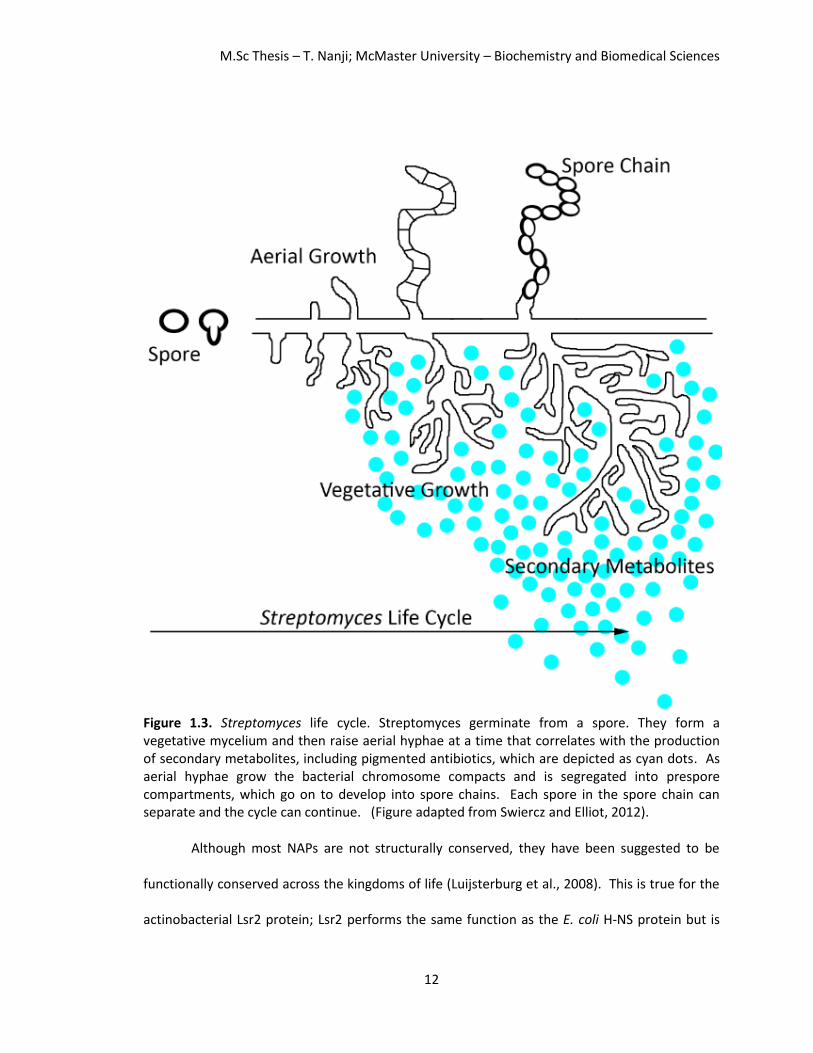
actinobacteria. Streptomyces have a characteristic, complex life cycle (Figure 1.3); they begin as
a spore that germinates and develop into vegetative mycelium that forms branches. They then
develop aerial hyphae which coincide with the production of secondary metabolites (Figure 1.3,
depicted as cyan dots) which include many antibiotic compounds such as actinorhodin and

M.Sc Thesis – T. Nanji; McMaster University – Biochemistry and Biomedical Sciences
11
undecylprodigiosin. These compounds are blue and red pigmented, respectively. Streptomyces
may require a large genome and accessory plasmids to maintain their complex life cycle and
mediate the production of antibiotics. The Streptomyces chromosomes remain uncondensed
until the reproductive stages of their life cycle, at which point the chromosomes segregate into
compartments and the pre-spore chain is formed. This spore-chain goes on to develop into a
spore chain. Each spore in the spore chain can separate and the cycle can re-occur. To
understand how DNA organization is coordinated with this complex life cycle, their NAPs can be
studied.
As previously discussed, NAPs can bind DNA and act as transcription factors to cause
changes in gene expression (Dillon and Dorman, 2010). Changes in gene expression can be
easily studied in S. coelicolor as they produce many secondary metabolites. S. coelicolor are a
good model organism to study NAPs in actinobacteria, as they allow us to monitor changes in
the production of pigmented antibiotics. Furthermore, aerial hyphae have a distinct appearance
which allows us to monitor changes in cell cycle progression. Moreover, in a laboratory setting,
chromosome compaction and cell division are not essential allowing us to monitor chromosome
condensation.
M.Sc Thesis – T. Nanji; McMaster University – Biochemistry and Biomedical Sciences
12
Figure 1.3. Streptomyces life cycle. Streptomyces germinate from a spore. They form a vegetative mycelium and then raise aerial hyphae at a time that correlates with the production of secondary metabolites, including pigmented antibiotics, which are depicted as cyan dots. As aerial hyphae grow the bacterial chromosome compacts and is segregated into prespore compartments, which go on to develop into spore chains. Each spore in the spore chain can separate and the cycle can continue. (Figure adapted from Swiercz and Elliot, 2012).
Although most NAPs are not structurally conserved, they have been suggested to be
functionally conserved across the kingdoms of life (Luijsterburg et al., 2008). This is true for the
actinobacterial Lsr2 protein; Lsr2 performs the same function as the E. coli H-NS protein but is

M.Sc Thesis – T. Nanji; McMaster University – Biochemistry and Biomedical Sciences
13
not structurally homologous (Chen et al., 2008). Like H-NS, Lsr2 prefers binding AT-rich DNA
sequences and is able to bridge disparate DNA molecules together (Gordon et al., 2011; Chen et
al., 2008, Qu et al., 2013). Not only do Lsr2 and H-NS have similar properties, Lsr2 has been
shown to be functionally analogous to H-NS as per complementation assays and DNA binding
assays. lsr2 is able to complement hns null mutants and Lsr2 specifically binds to genes
regulated by H-NS (Gordon et al., 2008). This illustrates that multiple NAPs may have
synonymous roles across bacterial species even though they are not structurally similar.
A new actinobacterial specific NAP has recently been discovered; the actinobacterial
integration host factor (aIHF) protein (Yang et al., 2012). These proteins are conserved among
actinobacteria, but share low amino acid similarity to the E. coli IHF protein. Synteny suggests
that these proteins are homologous as they are found within the same gene cluster (Yang et al.,
2012). The aIHF family of proteins are classified as IHF proteins since they were first identified as
having a role in phage integration; the protein is required to form the recombinogenic intasome
complex. Furthermore, the aIHF in Mycobacterium (mIHF) does not bind specifically to the
sequence specifying the site of integration (Goosen and van de Putte, 1995), and ΔmIHF strains
cannot be complemented with E. coli IHF or HU to restore recombination (Pedulla and Hatful,
1998). The aIHF proteins are of particular importance as they are essential in many
actinobacterial species, including Mycobacterium tuberculosis and M. leprae (Pedulla and Hatful,
1998; Sassetti et al., 2003), the bacterial infections resulting in tuberculosis and leprosy, making
them potential drug targets. Furthermore, as multidrug resistant bacterial strains are becoming
more prevalent, novel drugs and novel drug targets are important research topics.

M.Sc Thesis – T. Nanji; McMaster University – Biochemistry and Biomedical Sciences
14
As the mIHF protein is essential in Mycobacterium, we decided to study the aIHF protein
in the model organism Streptomyces coelicolor. Streptomyces is a good model organism to study
this protein as it shares a genetic core with Mycobacterium, which includes many of the same
housekeeping and essential genes (Cole et al., 1998; Bentley et al., 2002). The aIHF protein is
also well conserved and shares 65% identity and 95% similarity between these two organisms. It
is advantageous to study the aIHF protein in Streptomyces (sIHF) as we have been successful in
generating a viable ΔsIHF strain (Swiercz et al., 2013). The viability of this strain is thought to be
due to the complex life cycle of Streptomyces described above, whereas Mycobacterium divides
by binary fission. Using the S. coelicolor ΔsIHF strain, we have shown that sIHF associates with
the nucleoid, plays a significant role in chromosome compaction, and affects gene expression; S.
coelicolor lacking sIHF displayed decondensed nucleoids, elongated cells and aberrant
production of pigmented antibiotics (Swiercz et al., 2013).
1.4 Thesis objective
My thesis objective was to understand how sIHF interacts with DNA and modulates the function
of other proteins important in DNA topology. To this end, I completed three specific aims during
my MSc:
1) Solve the crystal structure of sIHF bound to duplex DNA,
2) Determine the regions of sIHF that are important for DNA binding,
3) Explore how sIHF affects the activity of the topoisomerase TopA.

M.Sc Thesis – T. Nanji; McMaster University – Biochemistry and Biomedical Sciences
15
Chapter 2
Methods and Materials
2.1 Overexpression of sIHF
S. coelicolor sIHF was cloned into the pET-15b vector (Novagen), which contains a removable N-
terminal poly-histidine tag, between restriction sites NdeI and BamHI (pAG 8380, see Table 2.1).
This plasmid was incubated with calcium chloride competent E. coli BL21(DE3) Rosetta cells
(Invitrogen Life Technologies) at 4°C for 1 hour prior to heat shock at 42°C for 45 seconds and
subsequent incubation at 4°C for 5 minutes. Super Optimal broth with Catabolite repression
(SOC) medium was added to the cells, incubated for 1 hour at 37 °C and plated onto Luria-
Bertani (LB) agar plates with 100 μg/mL ampicillin and 25 μg/mL chloramphenicol. Mixed
colonies were picked and grown in LB medium at 37˚C until an OD600 of ~0.7. Protein expression
was induced with 1.0 mM isopropyl-beta-D-thiogalactopyranoside (IPTG) and allowed to
proceed for 3 hours at 37˚C. Cells were harvested by centrifugation for 15 minutes at 3,315 x g.
Cell pellets were washed with 1x phosphate buffered saline (BioShop) and spun at 4°C for 10
minutes at 2,930 x g, and stored at -80°C for later use.
Selenomethionine labeled sIHF was produced by transforming pAG 8380 into calcium
chloride competent E. coli B843 Rosetta cells and grown at 37°C in minimal medium (Sigma)
supplemented with 2.0 x 10⁻⁴ mM selenomethionine (Sigma), 100 μg/mL ampicillin and 25 μg/mL
chloramphenicol to an OD600 of ~ 1.0. Cultures were induced, harvested and stored as described
above. All sIHF mutants were overexpressed in the same manner as wildtype, using their
respective plasmid listed in Table 2.1.

M.Sc Thesis – T. Nanji; McMaster University – Biochemistry and Biomedical Sciences
16
Table 2.1. Expression plasmids
Name Construct Vector Cloning restriction sites Source
pAG 8380 sIHF full length pET-15b NdeI, BamHI Gift from Elliot M. A
pAG 8775 sIHF RR85AS pET-15b NdeI, NheI*, BamHI This work
pAG 8779 sIHF Δ1-13 pET-15b NdeI, BamHI This work
pAG 8780 sIHF NQ93AS pET-15b NdeI, NheI*, BamHI This work
pAG 8845 sIHF S(19) pET-15b NdeI, BamHI This work
pAG 8846 sIHF RR85AS+NQ93AS pET-15b NdeI, NheI*, BamHI This work
pAG 8851 sIHF Δ1-36 pET-15b NdeI, BamHI This work
pAG 8860 sIHF S(19)+RR pET-15b NdeI, BamHI This work
pAG 8866 sIHF G66+ pET-15b NdeI, BamHI This work
pAG 8867 sIHF 35-37 Gly pET-15b NdeI, BamHI This work
*This enzyme was included to screen for the presence of the point mutation.
2.2 Purification of sIHF
E. coli BL21(DE3) Rosetta cells overexpressing His-sIHF, were resuspended in 20 mL of buffer A
(20 mM Tris pH 8.0, 300 mM NaCl, 1.4 mM beta-mercaptoethanol, 5% (v/v) glycerol) and lysed
at 4°C by sonication. Lysis was complete after two 60 second pulses. Protease inhibitors (5
g/ml leupeptin, 1 mM phenylmethanesulfonyl fluoride (PMSF), 0.7 g/ml pepstatin A, and 1
mM benzamidine) were added before and immediately after cell lysis to reduce proteolysis and
maintain protein integrity. Cellular debris was removed by centrifugation at 39,191 x g for 40
minutes at 4°C.
The supernatant containing the His-sIHF protein was loaded onto a 5 mL HiTrap
Chelating HP column (GE Healthcare) using immobilized nickel. The column was washed with 20
column volumes of buffer A to remove unbound protein, 10 column volumes of buffer A
supplemented with 7.5 mM imidazole, and 5 column volumes of buffer A with 20 mM imidazole
to remove proteins that interact with the column unspecifically. sIHF was eluted with buffer A
supplemented with 150 mM imidazole. Fractions containing sIHF were pooled and diluted using

M.Sc Thesis – T. Nanji; McMaster University – Biochemistry and Biomedical Sciences
17
buffer B (20 mM Tris pH 8.0, 1.4 mM beta-mercaptoethanol, 5% (v/v) glycerol) to dilute the
imidazole and reduce the salt concentration to 100 mM. The diluted protein was loaded onto a
Mono S 10/100 GL column (GE Healthcare). His-sIHF was eluted using a linear gradient from 100
mM to 1000 mM NaCl. His-sIHF eluted off the cation exchange column at 420 mM NaCl. Pooled
fractions of the protein, typically at ~170 μM, were diluted in buffer C (20 mM Tris pH 8, 150 mM
NaCl, 5 mM CaCl2, 5 mM DTT, 5% (v/v) glycerol) to 40 μM with a final concentration of 5 mM
CaCl2 to ensure optimal digestion of the histidine tag using the thrombin protease.
A small scale thrombin (Sigma) digestion reaction (10 L) was conducted from 0.01-
0.125 units/μL to determine the concentration of enzyme required to fully digest the poly-
histidine tag. Nine μL of diluted sIHF at 40 μM was incubated with 1 μL of each thrombin
concentration for 1 hour at room temperature. Digestion products were resolved on an 18%
sodium dodecyl sulphate-polyacrylamide gel (SDS-PAG) stained with coomassie containing 20%
(v/v) acetic acid. The remainder of His-sIHF at 40 μM was incubated with the optimal
concentration of thrombin for 1 hour at room temperature. The reaction was quenched with 1
mM benzamidine and tagless sIHF was purified using a linear gradient from 100 mM to 1000 mM
NaCl on a Mono S 5/50 GL column (GE Healthcare). Purified sIHF eluted off the cation exchange
column at 420 mM NaCl. Pooled fractions were concentrated in a Vivaspin 2 5,000 molecular
weight cut-off (MWCO) centrifugal concentrator (GE Healthcare) and the buffer was exchanged
to buffer D (40 mM Tris pH 8.0, 300 mM NaCl, 20 mM MgCl2, 2.8 mM beta-mercaptoethanol,
10% (v/v) glycerol). Protein concentrations were determined using the Bradford assay
(Bradford, 1976).

M.Sc Thesis – T. Nanji; McMaster University – Biochemistry and Biomedical Sciences
18
2.3 Forming the sIHF-DNA complex
Oligonucleotides were purchased from IDT and suspended in filtered autoclaved water.
Complementary oligonucleotides were mixed at equal concentrations and annealed by boiling
for 3 minutes prior to cooling overnight. Oligonucleotides containing longer than an 8-bp duplex
were cooled to room temperature. Substrates containing 7 or 8 pairing bases were cooled to
4°C, as their melting temperature was approximately ~24°C. See Table 2.2 for oligonucleotide
sequences. Equal volumes of sIHF (diluted in buffer D) and dsDNA (diluted in autoclaved water)
were mixed. sIHF-DNA complexes were formed in 20 mM Tris pH 8.0, 150 mM NaCl, 10 mM
MgCl2, 1.4 mM beta-mercaptoethanol, and 5% (v/v) glycerol. Protein-DNA complexes of duplex
substrates larger than 8-bp were incubated for 10 minutes at room temperature, followed by 30
minutes on ice, while protein-DNA complexes of duplex substrates that are 8-bp were incubated
on ice at 4°C overnight.
Table 2.2. DNA substrates used for crystallization
TN22-Top 5'
GGG AGT GCG TGG GTC TGA AGC C3'
TN22-Bottom 5'
GGC TTC AGA CCC ACG CAC TCC C3'
TN20O-Top 5’
GAG GGA GTG CGT GGG TCT GA3’
TN20O-Bottom 5’
CTC AGA CCC ACG CAC TCC CT3’
TN19O2-Top 5’
GAG GGA GTG CGT GGG TCT G3’
TN19O2-Bottom 5’
TCC AGA CCC ACG CAC TCC C3’
TN15-Top 5'
GGG AGT GCG TGG GTC3'
TN15-Bottom 5'
GAC CCA CGC ACT CCC3'
TN08-BP1 5’
CAT GCA TG3’
TN08O-Top 5’
GGG CGC GG3’
TN08O-Bottom 5’
CCC GCG CC3’
TN23-HP9 5’
GTGCGTGGATTTTTTCCACGCAC3’

M.Sc Thesis – T. Nanji; McMaster University – Biochemistry and Biomedical Sciences
19
2.4 Crystallization of sIHF bound to DNA
Crystallization trials of sIHF bound to duplex DNA substrates (Table 2.2) were conducted using
sparse matrix screens set by the Phoenix Liquid Handling System (Art Robbins Instruments).
Initial crystal hits of sIHF bound to TN20O (Table 2.2) were obtained using the sitting drop
method in condition 14 (0.2 M KSCN, 20% (w/v) PEG 3350, pH 7) of the PEG/Ion Screen
(Hampton Research). Crystals were optimized by the hanging drop method with the addition of
HEPES at pH 7.6 and ethylene glycol. Optimal crystals at 1.0 mM complex grew at a 1:1 ratio in
0.1 M HEPES pH 7.6, 0.21 M KSCN, 19% (w/v) PEG 3350, and 5% (v/v) ethylene glycol. Once
crystals reached maximum size, drops were dehydrated against increasing concentrations of KCl
(1.0, 1.25, 1.5, and 1.75 M KCl) for 8-12 hours prior to flash freezing in liquid nitrogen.
Crystals of Sel-Met labeled sIHF bound to TN20O were obtained at a 1.4:1 ratio (1.05
mM:0.75 mM) in 0.1 M HEPES pH 7.6, 0.21 M KSCN, 18% (w/v) PEG 3350, and 5% (v/v) ethylene
glycol. Crystals of sIHF bound to TN08O (Table 2.2), at 1.0 mM at a 1:1 ratio, grew in 0.1 M
HEPES pH 7.6, 0.1 M KSCN, 28% (w/v) PEG 3350, and 5% (v/v) ethylene glycol. Crystals of sIHF
bound to TN08-BP1 (Table 2.2) at 1.5 mM at a 1:1 ratio were obtained in 0.1 M HEPES pH 7.6,
0.2 M KSCN, 19% (w/v) PEG 3350, and 5% (v/v) ethylene glycol. Crystals of sIHF bound to TN23-
HP9 (Table 2.2) were grown using the streak-seeding method. Crushed crystals obtained at a
1:1.2 ratio (1.0 mM:1.2 mM) in 0.1 M MES pH 5.6, 0.12 M MgCl2, 16% (w/v) PEG 3350, and 5%
(v/v) ethylene glycol were seeded into crystallization drops of sIHF bound to TN23-HP9 at a ratio
of 1:1.2 (0.63 mM:0.75 mM) in 0.1 M MES pH 5.6, 0.1 M MgCl2, 14% (w/v) PEG 3350, and 5%
(v/v) ethylene glycol after 1 day using a cat whisker. Crystals were dehydrated and frozen as
above.
M.Sc Thesis – T. Nanji; McMaster University – Biochemistry and Biomedical Sciences
20
2.5 Data collection and structure determination
Data were collected using beamline X25 at the National Synchrotron Light Source (NSLS),
Brookhaven National Laboratory (BNL) (Upton, NY). Crystal TN055 (sIHF bound to TN20O)
diffracted to 2.85 Å, crystal TN051 (sIHF Sel-Met bound to TN20O) diffracted to 2.6 Å, crystal
TN100 (sIHF bound to TN08O) diffracted to 3.00 Å, crystal TN103 (sIHF bound to TN08-BP1)
diffracted to 1.66 Å, and crystal TN131 (sIHF bound to TN23-HP9) diffracted to 2.9 Å. Data were
indexed, processed, and merged using HKL2000 (Otwinowski and Minor, 1997). Refer to Table
3.1 for full data collection and refinement statistics.
Crystals of Sel-Met sIHF:TN20O were phased by single-wavelength anomalous dispersion
(SAD) using SOLVE (Terwilliger, T. C. and Berendzen, J., 1999). The initial model was manually
built using COOT and refined using iterative cycles of model building in COOT and refinement in
phenix.refine (Afonine et al., 2012; Emsley and Cowtan, 2004). Crystals of sIHF bound to TN08O,
TN08-BP1 and TN23-HP9 were phased by molecular replacement (MR) using Phaser-MR in
Phenix and subsequent model building and refinement was done using standard protocols in
phenix.refine and COOT (Afonine et al., 2012; Emsley and Cowtan, 2004).
2.6 Cloning of sIHF mutants
sIHF mutants were produced by either site-directed mutagenesis or overlap polymerase chain
reaction (PCR). sIHF RR85AS (pAG 8775) (Table 2.1) was generated by site-directed mutagenesis
using the QuikChange II kit (Agilent Technologies) from template pAG 8380 (Table 2.1) with
primers, purchased from IDT, AG 1794, and AG 1795 (Table 2.3). These primers were designed
to have a NheI restriction site to discriminate between template and product DNA. The site-

M.Sc Thesis – T. Nanji; McMaster University – Biochemistry and Biomedical Sciences
21
directed mutagenesis reaction was incubated with DpnI, for 1 hour in a 37°C water bath to
digest template DNA, and transformed into Top10 electrocompetent cells (Life Technologies) by
electroporation. Plasmid DNA was isolated using the GeneJET Plasmid Miniprep Kit (Thermo
Scientific) and the presence of the desired mutation was assessed by analytical restriction
digestion with NheI. Presence of the mutation was confirmed by DNA sequencing (MOBIX Lab).
sIHF NQ93AS (pAG 8780), was also generated by site-directed mutagenesis using pAG
8380 (Table 2.1) as the template and primers AG 1825 and AG 1826 (Table 2.3) as described
above. Lastly, we combined these mutations to make a quadruple sIHF mutant, sIHF
RR85AS+NQ93AS (pAG 8846), using site-directed mutagenesis from pAG 8775 as a template and
primers AG 1825, and AG 1826. The reaction was digested by DpnI and transformed, isolated,
and sequenced as described above.
N-terminal truncations lacking the first 13 and 36 residues were generated by PCR using
primers AG 1796 and AG 1797 as forward primers respectively and AG 1759 as the reverse
primer (Table 2.3). Both forward primers contained an NdeI restriction site to be incorporated
at the beginning of the gene, and the reverse primer contained a BamHI site, to be incorporated
at the end of the gene for cloning purposes. Each PCR reaction product was ligated into the
blunt cloning vector pJET1.2 (Thermo Scientific) to yield plasmids pAG 8771 and pAG 8777 (Table
2.4). These plasmids along with a plasmid containing the pET-15b vector (pAG 8160) (Table 2.4)
were digested by NdeI for 15 minutes and BamHI for 5 minutes at 37°C, and run on an agarose
gel. DNA fragments were extracted using the QIAEX II Gel Extraction Kit (Qiagen) and each sIHF
truncation plasmid was ligated with the digested pET-15b vector overnight at 16°C with T4 DNA
M.Sc Thesis – T. Nanji; McMaster University – Biochemistry and Biomedical Sciences
22
Ligase (New England Biolabs) to yield plasmids pAG 8779 and pAG 8851 (Table 2.1). (pAG 8851
was generated with the help of Sabrina Lue Tam).
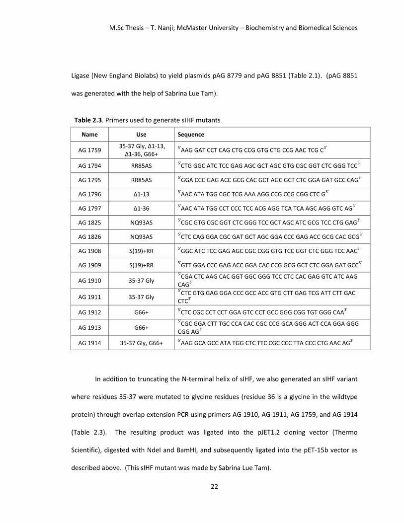
Table 2.3. Primers used to generate sIHF mutants
Name Use Sequence
AG 1759 35-37 Gly, Δ1-13,
Δ1-36, G66+ 5'
AAG GAT CCT CAG CTG CCG GTG CTG CCG AAC TCG C3'
AG 1794 RR85AS 5'
CTG GGC ATC TCC GAG AGC GCT AGC GTG CGC GGT CTC GGG TCC3'
AG 1795 RR85AS 5'
GGA CCC GAG ACC GCG CAC GCT AGC GCT CTC GGA GAT GCC CAG3'
AG 1796 Δ1-13 5'
AAC ATA TGG CGC TCG AAA AGG CCG CCG CGG CTC G3'
AG 1797 Δ1-36 5'
AAC ATA TGG CCT CCC TCC ACG AGG TCA TCA AGC AGG GTC AG3'
AG 1825 NQ93AS 5'
CGC GTG CGC GGT CTC GGG TCC GCT AGC ATC GCG TCC CTG GAG3'
AG 1826 NQ93AS 5'
CTC CAG GGA CGC GAT GCT AGC GGA CCC GAG ACC GCG CAC GCG3'
AG 1908 S(19)+RR 5'
GGC ATC TCC GAG AGC CGC CGG GTG TCC GGT CTC GGG TCC AAC3'
AG 1909 S(19)+RR 5'
GTT GGA CCC GAG ACC GGA CAC CCG GCG GCT CTC GGA GAT GCC3'
AG 1910 35-37 Gly 5'
CGA CTC AAG CAC GGT GGC GGG TCC CTC CAC GAG GTC ATC AAG CAG
3'
AG 1911 35-37 Gly 5'
CTC GTG GAG GGA CCC GCC ACC GTG CTT GAG TCG ATT CTT GAC CTC
3'
AG 1912 G66+ 5'
CTC CGC CCT CCT GGA GTC CCT GCC GGG CGG TGT GGG CAA3'
AG 1913 G66+ 5'
CGC GGA CTT TGC CCA CAC CGC CCG GCA GGG ACT CCA GGA GGG CGG AG
3'
AG 1914 35-37 Gly, G66+ 5'
AAG GCA GCC ATA TGG CTC TTC CGC CCC TTA CCC CTG AAC AG3'
In addition to truncating the N-terminal helix of sIHF, we also generated an sIHF variant
where residues 35-37 were mutated to glycine residues (residue 36 is a glycine in the wildtype
protein) through overlap extension PCR using primers AG 1910, AG 1911, AG 1759, and AG 1914
(Table 2.3). The resulting product was ligated into the pJET1.2 cloning vector (Thermo
Scientific), digested with NdeI and BamHI, and subsequently ligated into the pET-15b vector as
described above. (This sIHF mutant was made by Sabrina Lue Tam).
M.Sc Thesis – T. Nanji; McMaster University – Biochemistry and Biomedical Sciences
23
A sIHF variant containing a glycine residue insertion after position 66 was generated
(sIHF G66+). The overexpression plasmid for sIHF G66+ (pAG 8866) was generated using overlap
extension PCR using primers AG 1759, AG 1912, AG 1913, and AG 1910, (Table 2.3). The
resulting product was ligated into the pJET1.2 cloning vector (Thermo Scientific), digested with
NdeI and BamHI and subsequently ligated into the pET-15b vector as described above.
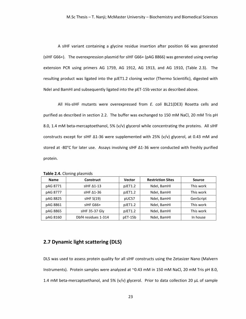
All His-sIHF mutants were overexpressed from E. coli BL21(DE3) Rosetta cells and
purified as described in section 2.2. The buffer was exchanged to 150 mM NaCl, 20 mM Tris pH
8.0, 1.4 mM beta-mercaptoethanol, 5% (v/v) glycerol while concentrating the proteins. All sIHF
constructs except for sIHF Δ1-36 were supplemented with 25% (v/v) glycerol, at 0.43 mM and
stored at -80°C for later use. Assays involving sIHF Δ1-36 were conducted with freshly purified
protein.
Table 2.4. Cloning plasmids
Name Construct Vector Restriction Sites Source
pAG 8771 sIHF Δ1-13 pJET1.2 NdeI, BamHI This work
pAG 8777 sIHF Δ1-36 pJET1.2 NdeI, BamHI This work
pAG 8825 sIHF S(19) pUC57 NdeI, BamHI GenScript
pAG 8861 sIHF G66+ pJET1.2 NdeI, BamHI This work
pAG 8865 sIHF 35-37 Gly pJET1.2 NdeI, BamHI This work
pAG 8160 Dbf4 residues 1-314 pET-15b NdeI, BamHI In house
2.7 Dynamic light scattering (DLS)
DLS was used to assess protein quality for all sIHF constructs using the Zetasizer Nano (Malvern
Instruments). Protein samples were analyzed at ~0.43 mM in 150 mM NaCl, 20 mM Tris pH 8.0,
1.4 mM beta-mercaptoethanol, and 5% (v/v) glycerol. Prior to data collection 20 μL of sample

M.Sc Thesis – T. Nanji; McMaster University – Biochemistry and Biomedical Sciences
24
was centrifuged for 10 minutes at 4°C at 15,700 x g and 15 μL was loaded into a 12 μL quartz
cuvette.
2.8 Electrophoretic mobility shift assays (EMSAs)
sIHF variants at increasing concentrations of protein (0-100 μM) were added to 0.02 μM of *ƴ-
32P+dATP 5’end-labelled duplex DNA (Motif 1: top strand 5’TCGAAAAATCGGAATCTGGTGCA;
bottom strand 5’TGCACCAGATTCCGATTTTTCGA) with 1 mg/mL bovine serum albumin (BSA) and
binding buffer (10 mM Tris pH 7.8, 5 mM MgCl2, 60 mM KCl and 10% (v/v) glycerol). The
reaction was incubated at room temperature for 10 minutes, followed by 30 minutes on ice. A
glycerol-based loading dye was added and the samples were separated on a 15% native
polyacrylamide gel at 100 V for 40 minutes. Gels were first exposed to a phosphor plate for ~30
minutes and visualised using a phosphorimager (Amersham Biosciences Ltd.), then subsequently
exposed to Kodak Biomax XAR film for ~1 hour and developed. Assays were conducted in
triplicate. (Assays were conducted by Dr. Emma Sherwood).
2.9 Topoisomerase assays
sIHF variants (5 μL at 540 nM and 2160 nM) were incubated with 5 μL pUC19 (64 nM) for 10
minutes at room temperature followed by 30 minutes on ice. One μL of TopA (7730 nM), 0.6 μL
BSA (1 mg/mL) and 8.5 μL of reaction buffer (50 mM Tris pH 7.5, 50 mM KCl, 10 mM MgCl2, 0.1
mM EDTA, 0.5 mM DTT, 0.06 mg/mL BSA) were added. Reactions were incubated at room
temperature for 30 minutes and stopped with 5 μL of stop buffer (6% SDS, 30% (v/v) glycerol, 10
mM EDTA and 0.25% bromophenol blue). Samples were separated on a 1% TAE-agarose gel at

M.Sc Thesis – T. Nanji; McMaster University – Biochemistry and Biomedical Sciences
25
45 V for ~16 hours, stained with ethidium bromide and visualized using UV light. Assays were
conducted in triplicate. (Assays were conducted by Melanie Gloyd).
2.10 Data collection and processing for small-angle X-ray scattering (SAXS)
sIHF was purified as described in section 2.2 and subsequently resolved using a Superdex-75 (GE
Healthcare) size exclusion chromatography column in 40 mM Tris pH 8.0, 200 mM NaCl, 20 mM
MgCl2, 2.8 mM beta-mercaptoethanol, and 10% (v/v) glycerol. Sample homogeneity was
assessed by dynamic light scattering (see Section 2.7). The protein-DNA complex was formed as
described in section 2.3. Scattering data for His-sIHF bound to duplex DNA (Motif 1: top strand
5’TCGAAAAATCGGAATCTGGTGCA; bottom strand 5’TGCACCAGATTCCGATTTTTCGA) at ratios of
1:1, 2:1, and 1:2 (sIHF:DNA) over a range of protein concentrations (0.868-0.109 mM) was
collected on a BioSAXS-1000 mounted on a MicroMax-007HF X-ray generator. Data was
collected for 120 minutes at each concentration with images refreshing every 20 minutes.
Sample scatter curves were generated using Rigaku SAXSLab 3.0.0r1 by subtracting buffer
scatter from sample scatter. Data quality was assessed for aggregation using Guinier plots
(Guinier and Fournet, 1955), and Kratky plots (Glatter and Kratky, 1982) were used to compare
protein concentrations and exposure times. Refer to Table 3.3 for SAXS data collection and
analysis.

M.Sc Thesis – T. Nanji; McMaster University – Biochemistry and Biomedical Sciences
26
Chapter 3
Results
3.1 Purification of sIHF
The sIHF gene was inserted into the pET-15b overexpression vector, downstream of a 6xhistidine
tag and thrombin cleavage site. Cell lysates overexpressing His-sIHF were loaded onto an
immobilized metal affinity chromatography column. His-sIHF binds the nickel resin as the
histidine tag at the N-terminus of the protein is exposed. Unbound contaminants were washed
off the column with buffer A (see Methods), and weakly bound contaminants were eluted using
a step gradient of buffer A supplemented with 7.5 and 20 mM imidazole. The protein was
eluted from the column using buffer A supplemented with 150 mM imidazole (Figure 3.1A). A
step gradient of imidazole was chosen over a linear gradient to enhance protein purity. Before
the histidine tag was removed, eluted fractions from the nickel column were further purified
using a cation exchange chromatography column to obtain pure His-sIHF (Figure 3.1B).
Eluted fractions containing His-sIHF (indicated by the single asterisks in Figure 3.1B)
were pooled and diluted to 40 μM. A small scale thrombin digestion reaction was conducted
using a fraction of the pooled protein at a range of thrombin (Sigma) concentrations (0-0.125
units/μL) to determine the optimal concentration of thrombin needed for complete, but not
over digestion. The optimal concentration of thrombin was selected based on the lowest
concentration necessary to yield full digestion as visualized on an 18% SDS-PAG (Figure 3.1C).
This concentration of thrombin was used to remove the histidine tag from the remainder of the
pooled protein.

M.Sc Thesis – T. Nanji; McMaster University – Biochemistry and Biomedical Sciences
27
Figure 3.1. Purification scheme of untagged sIHF. 18% SDS-PAGs of fractions collected (left panels) and chromatograms (right panels) of the 4-step purification for the sIHF protein. sIHF bands are indicated by the arrows. (A) Cell lysate was loaded onto a HiTrap chelating affinity column and eluted using imidazole. (B) Fractions containing the protein were pooled and loaded onto an ion exchange column. Eluted fractions are indicated by the asterisks. (C) The 6xHis tag was removed using the thrombin protease. The optimal concentration of thrombin to be used was determined by a small scale thrombin digestion where 40 μM of sIHF was digested with 0.01-0.125 units/μL of thrombin. (D) The untagged sIHF protein was further purified over an ion exchange column. Eluted fractions indicated by the double asterisks were pooled and concentrated.
M.Sc Thesis – T. Nanji; McMaster University – Biochemistry and Biomedical Sciences
28
Untagged sIHF was further purified by cation exchange chromatography. The protein
was eluted using a linear salt gradient (Figure 3.1D). Eluted fractions containing pure sIHF
(indicated by the double asterisks in Figure 3.1D) were concentrated.
3.2 Crystallization, data collection and structure of sIHF bound to a 19-bp
duplex DNA substrate
We determined that sIHF binds DNA in a sequence independent manner with a strong
preference for double stranded over single stranded substrates (Swiercz et al., 2013). To
understand how sIHF interacts with DNA we wanted to solve the crystal structure of the protein
bound to DNA. We attempted to crystallize sIHF with various G-C rich duplex DNA substrates, as
this protein is from an organism with a high G-C content. DNA lengths ranging from 15- and 22-
bp, that were blunt ended as well as contained 1- or 2-bp complementary overhangs (Table 2.2)
were tested. We designed the oligonucleotides to have a unique pattern of purines and
pyrimidines so that if we were successful in obtaining crystals, and subsequently obtaining an
experimental electron density map, we could identify the DNA sequence from the pattern of
large and small densities for each base. Furthermore, we tested DNA substrates with
complementary overhangs as these overhangs could potentially aid in crystal packing; the
oligonucleotides may stack end to end allowing complementary overhangs to base-pair forming
a pseudo-continuous helix across the crystal.
We were successful in obtaining crystals of sIHF bound to a 19-bp duplex with a one
base-pair overhang (TN20O, see Table 2.2), at a 1:1 ratio of protein to DNA, using the PEG/Ion
screen (Hampton Research) in the following condition: 20% PEG 3350 (w/v), 0.2 M KSCN, pH 7.

M.Sc Thesis – T. Nanji; McMaster University – Biochemistry and Biomedical Sciences
29
These crystals were optimized using the hanging drop method to favour the growth of large
single crystals. We first determined the optimal pH for crystal growth using a pH screen
(Hampton Research). We also varied the ratio of protein to DNA and found that a 1:1 ratio
yielded the largest single crystals (Figure 3.2).
To minimize crystal damage by X-ray radiation during data collection, crystals were
cryoprotected and data were collected at 100 K. Conventional cryoprotection of crystals is
achieved by transferring crystals to a solution with the crystallization condition supplemented
with a cryoprotecting agent such as ethylene glycol, polyethylene glycol or glycerol. These
methods damaged our crystals. In order to surmount this problem and still achieve good
cryoprotection, ethylene glycol was included in the crystallization solution and the crystals were
dehydrated over increasing concentrations of potassium chloride (see Methods). A complete
data set of the native sIHF-TN200 crystal (TN055, see Table 3.1) was collected at the NSLS
(Brookhaven National Laboratory, NY, USA). This crystal diffracted to 2.85 Å.
The structure of this complex could not be determined by molecular replacement
because a good structural homologue for sIHF does not exist. Therefore, to solve the phase
problem, crystals of selenomethionine substituted protein bound to TN20O were obtained (see
Methods). The best crystal (TN051, see Table 3.1) diffracted to 2.6 Å and a complete data set
Figure 3.2. Crystals of sIHF bound to a 19-bp duplex DNA substrate. Crystals of sIHF bound to TN20O grown in 18% (w/v) PEG 3350, 0.21 M KSCN, 0.1 M HEPES pH 7.6, and 5% (v/v) ethylene glycol.

M.Sc Thesis – T. Nanji; McMaster University – Biochemistry and Biomedical Sciences
30
was collected using the X25 beam line at the NSLS (Brookhaven National Laboratory, NY, USA) at
a wavelength of 0.979. This wavelength was chosen as it is the X-ray absorption edge of
selenium, allowing for anomalous signal from the selenium atoms to be collected and used to
phase the crystal. As this crystal diffracted to a higher resolution than the native crystal, we
decided to refine the structure of sIHF bound to TN20O using the Sel-Met data set. Data for this
crystal was indexed, processed and merged in the space group C2221, C centered orthorhombic,
using HKL2000 (Otwinowski and Minor, 1997). The single-wavelength anomalous dispersion
(SAD) method was used to phase the crystals using SOLVE (Terwilliger, T. C. and Berendzen, J.,
1999). sIHF contains three methionine residues in the 107 residue protein. This provides
enough anomalous signal to phase the structure by SAD. The initial model was built manually in
COOT by modeling in the protein sequence into the experimental electron density map using
signal from the selenium atoms as landmarks. Residues 14-103 were readily identified and a
final model was refined to a resolution of 2.70 Å. Complete statistics for data collection and
refinement can be found in Table 3.1.
The refined structure of sIHF bound to TN20O contained 98.84% of residues in the
preferred region of the Ramachandran plot, 1.16% in the allowed region and none in the
disallowed region. The structure revealed that sIHF is composed of a long protruding N-terminal
α-helix (α1) followed by four shorter α-helices that make up the core of the protein (Figure
3.3A). The sIHF protein contacts DNA at two distinct interfaces. One interface uses the loop
region between helices 4 and 5 which spans residues Gly80 to Gly91 and has been termed the lid
region (Figure 3.3 A, B in pink). This lid serves two functions; it conceals the hydrophobic core of

M.Sc Thesis – T. Nanji; McMaster University – Biochemistry and Biomedical Sciences
31
the protein as well as provides a positively charged flat surface that is free to interact with the
negatively charged phosphate backbone of one strand of the DNA duplex.
Table 3.1. X-ray data collection and refinement
Crystal TN055 (native)
TN051 (Sel-Met)
TN100 (native)
TN103 (native)
TN131 (native)
DNA TN20O TN20O TN08O TN08-BP1 TN23-HP9
Data Collection
Space Group C222 C2221 C2221 C2221 P21
Unit Cell (Å) a=41.8, b=72.7, c=102.3
a=43.4, b=71.8, c=102.9
a=45.7, b=71.9, c=100.8
a=42.3, b=72.1, c=103.3
a=67.3, b=40.4, c=89.9; α=90, β=108.6, ƴ=90
Wavelength (Å) 1.1 0.979 0.979 0.979 1.1
Resolution (Å)* 50-2.85 (2.90-2.85)
50-2.60 (2.64-2.60)
35-3.00 (3.11-3.00)
50-1.66 (1.69-1.66)
35-2.90 (2.95-2.90)
Completeness (%)* 99.7 (98.9) 100 (99.5) 99.8 (99.7) 98.7 (99.8) 94.1 (81.3)
Redundancy* 4.8 (5.8) 6.1 (4.6) 3.4 (3.3) 6.1 (5.9) 3.2 (2.9)
I/σ* 19.2 (1.9) 31.2 (1.4) 23.1 (1.3) 21.1 (3.7) 24.1 (2.7)
Data Refinement
Resolution (Å)* 29.4-2.7 (3.1-2.7)
39.11-1.60 (1.64-1.60)
34.13-3.00 (3.11-3.00)
Reflections (work)* 4613 (1349)
42000 (2632)
8132 (1014)
Reflections (test)* 447 (142) 1998 (131) 914 (120)
Atoms refined 1006 2485 2261
Solvent atoms 6 229 6
Rfree (%) 31.7 29.5 32.68
Rwork (%) 26.1 26.9 28.6
Rmsd in bonds (Å) 0.003 0.009 0.012
Rmsd in angles (°) 0.789 1.4 2.1
*Data in the highest resolution shell shown in parenthesis.

M.Sc Thesis – T. Nanji; McMaster University – Biochemistry and Biomedical Sciences
32
Figure 3.3. Structure of sIHF. (A) Ribbon diagram of sIHF with the N-terminal helix shown in yellow, the H2TH motif shown in blue and the lid region shown in pink. The image is rotated 90° in the second panel. (B) Secondary structure of sIHF highlighting the N-terminal helix, the H2TH, and the lid regions in yellow, blue, and pink respectively. (C-E) Ribbon diagrams of topoisomerase VI (residues Lys230-Phe306, PDB 1MU5), endonuclease VIII (residues Pro132-Gln214, PDB 1K3W) and ribosomal protein S13 (residues Ala1-Phe62, PDB 2GY9). The H2TH motif is displayed in blue, and the lid region in pink.
The second interface of sIHF that contacts DNA uses helices 3 and 4, spans residues
Lys56 to Leu79 (Figure 3.3 A, B in blue) and forms a fold classified as a helix-two turns-helix
(H2TH) motif. This motif is found in a few other DNA binding proteins including topoisomerase

M.Sc Thesis – T. Nanji; McMaster University – Biochemistry and Biomedical Sciences
33
VI (a type IIb topoisomerase), the endonuclease VIII family of base excision repair enzymes, and
the ribosomal protein S13 (Corbett and Berger, 2003; Zharkov et al., 2002; Brodersen et al.,
2002) (Figure 3.3 C-E respectively, in blue). The function of this motif is unknown, although it is
thought to aid in peripheral nucleic acid binding. Endonuclease VIII and ribosomal protein S13
contact DNA and RNA respectively, through this interface in their crystal structures; however,
this motif is embedded within much larger proteins that form extensive contacts with nucleic
acids (Zharkov et al., 2002; Brodersen et al., 2002). Interestingly, the lid region of sIHF is also
found in the three proteins mentioned above and adopts a similar conformation to that found in
sIHF (Figure 3.3 in pink). In both the monomer and dimer structures of topoisomerase VI the lid
region is more exposed than the H2TH motif; however, as this structure has not been solved in
complex with DNA, we cannot infer which interface(s) contact DNA (Corbett and Berger, 2003).
The DNA substrate used to form the sIHF-DNA crystal was 20-bp long; however, we only
identified 8-bp within the asymmetric unit in the experimental electron density map. The
electron density, which represents an average of each unit cell within the crystal, was well
defined for the DNA phosphate backbone, but not for the nucleobases. Therefore, we modeled
in the nucleobases as guanine or cytosine based on the size of electron density, as the substrate
used was G-C rich. We believe that the nucleobases were disordered because the crystals
packed to form a pseudo-continuous duplex allowing sIHF to bind several different sequences
along the duplex within the crystal. This reaffirms our observation that sIHF does not bind DNA
at a specific sequence (Swiercz et al., 2013). We next wanted to obtain a crystal structure of
sIHF bound to DNA where the nucleobases could be identified. As 8-bp were identified in the
asymmetric unit, we decided to pursue the structure of sIHF bound to an 8-bp DNA substrate.
M.Sc Thesis – T. Nanji; McMaster University – Biochemistry and Biomedical Sciences
34
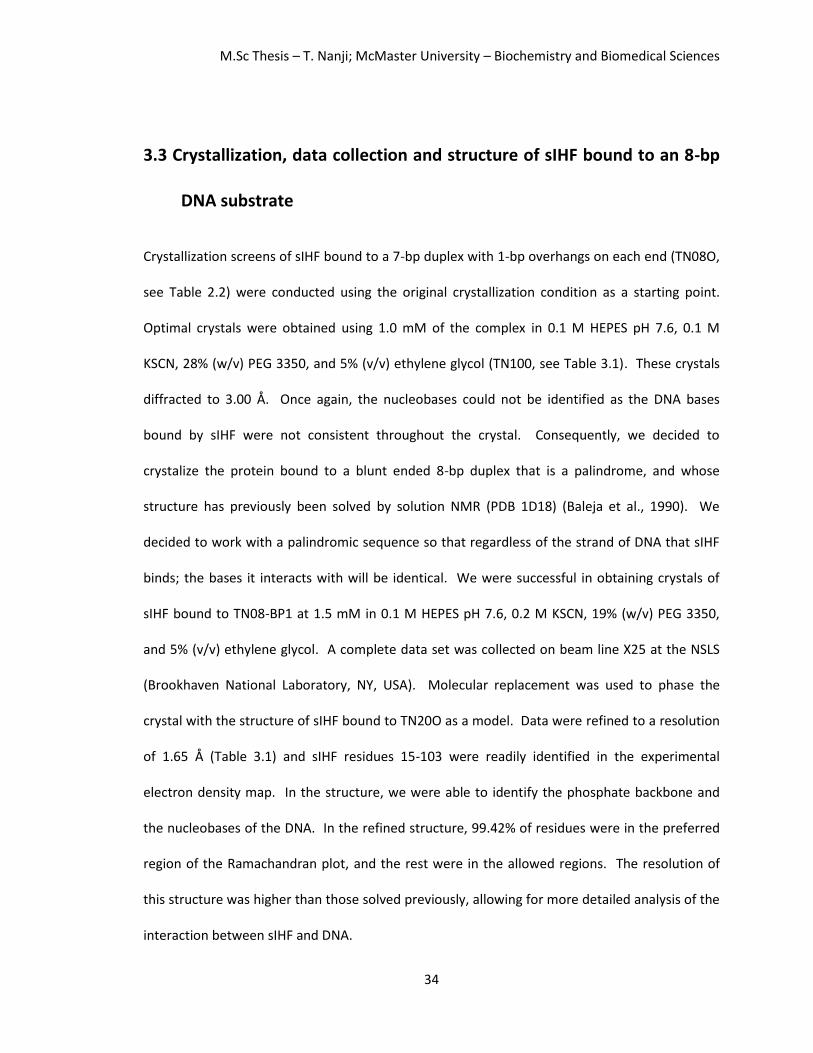
3.3 Crystallization, data collection and structure of sIHF bound to an 8-bp
DNA substrate
Crystallization screens of sIHF bound to a 7-bp duplex with 1-bp overhangs on each end (TN08O,
see Table 2.2) were conducted using the original crystallization condition as a starting point.
Optimal crystals were obtained using 1.0 mM of the complex in 0.1 M HEPES pH 7.6, 0.1 M
KSCN, 28% (w/v) PEG 3350, and 5% (v/v) ethylene glycol (TN100, see Table 3.1). These crystals
diffracted to 3.00 Å. Once again, the nucleobases could not be identified as the DNA bases
bound by sIHF were not consistent throughout the crystal. Consequently, we decided to
crystalize the protein bound to a blunt ended 8-bp duplex that is a palindrome, and whose
structure has previously been solved by solution NMR (PDB 1D18) (Baleja et al., 1990). We
decided to work with a palindromic sequence so that regardless of the strand of DNA that sIHF
binds; the bases it interacts with will be identical. We were successful in obtaining crystals of
sIHF bound to TN08-BP1 at 1.5 mM in 0.1 M HEPES pH 7.6, 0.2 M KSCN, 19% (w/v) PEG 3350,
and 5% (v/v) ethylene glycol. A complete data set was collected on beam line X25 at the NSLS
(Brookhaven National Laboratory, NY, USA). Molecular replacement was used to phase the
crystal with the structure of sIHF bound to TN20O as a model. Data were refined to a resolution
of 1.65 Å (Table 3.1) and sIHF residues 15-103 were readily identified in the experimental
electron density map. In the structure, we were able to identify the phosphate backbone and
the nucleobases of the DNA. In the refined structure, 99.42% of residues were in the preferred
region of the Ramachandran plot, and the rest were in the allowed regions. The resolution of
this structure was higher than those solved previously, allowing for more detailed analysis of the
interaction between sIHF and DNA.

M.Sc Thesis – T. Nanji; McMaster University – Biochemistry and Biomedical Sciences
35
These crystals packed in the same space group as crystal TN051 (Table 3.1) and interact
with DNA at the same interfaces. The resolution obtained in this structure is high enough to
assess if sIHF alters DNA upon binding. We analyzed differences in the structure of the DNA
alone (PDB 1D18) and the DNA from the crystal structure. The Web 3DNA software for three-
dimensional analysis of nucleic-acid structures (Zheng et al., 2009) was used to compare
complementary base-pair and base-pair step parameters. These results are summarized in Table
3.2. We observed that the stagger (vertical displacement between complementary base-pairs)
of the DNA alone is greater than that of the DNA in complex with sIHF, with greater differences
at the center of the substrate. Differences are also seen in the buckle (when both
complementary base-pairs fold upwards and are not planar) of the base-pairs with more
significant differences at the ends of the substrate. A difference in the angle of propeller twist
(when complementary base-pairs twist in opposite directions) is also observed and can be
visualized in the crystal structure in the central base-pair.
There is also a difference in the opening (the space between complementary base-pairs)
of the base-pairs. The opening between base-pairs for the 8-bp duplex previously solved deviate
from standard B-DNA (reported in Table 3.2) more than our structure. This could be because the
structure of PDB 1D18 was solved in solution by NMR, whereas the structures of DNA solved in
this work as well as that stated for standard B-DNA are from crystal structures that are in a
locked conformation. Differences in the tilt and roll DNA structural parameters also vary
between DNA alone and in complex with sIHF. These parameters have been affiliated with
mediating the widths of the major and minor grooves (Oguey et al., 2010).

M.Sc Thesis – T. Nanji; McMaster University – Biochemistry and Biomedical Sciences
36
Table 3.2. Base-pair parameters of duplex DNA alone and in complex with sIHF
PBD 1D18 Standard B-DNA*
Parameter C-G A-T T-A G-C C-G A-T T-A G-C Buckle (°) 8.97 10.61 9.55 3.44 -3.78 -8.84 -10.60 -7.45 0.50 Propeller (°) -7.56 -12.48 -8.91 -17.22 -17.23 -7.88 -12.80 -6.34 -11.40 Opening (°) -4.43 -6.81 -7.20 -4.87 -4.12 -6.74 -6.65 -4.78 0.60 Shear (Å) 0.22 -0.44 0.25 -0.40 0.46 -0.25 0.43 -0.18 0.00 Stretch (Å) -0.18 -0.21 -0.17 -0.25 -0.24 -0.18 -0.21 -0.19 -0.15 Stagger (Å) 0.20 0.29 0.42 0.37 0.34 0.47 0.24 0.20 0.09 Tilt (°) -0.73 0.15 2.70 0.29 -3.01 0.05 0.76 -0.10 Roll (°) 8.03 3.72 3.50 -6.07 3.44 4.47 8.74 0.60 Twist (°) 29.01 32.79 34.71 44.02 33.91 32.60 29.37 36.00 Shift (Å) -0.75 0.02 0.44 0.04 -0.45 -0.01 0.70 0.02 Slide (Å) -0.18 -0.08 -0.42 -0.37 -0.45 -0.02 -0.09 0.23 Rise (Å) 3.17 3.41 3.34 3.47 3.30 3.45 3.12 3.32 DNA in crystal TN103
Parameter C-G A-T T-A G-C C-G A-T T-A G-C Buckle (°) -6.40 -6.49 -2.62 4.27 -2.66 2.16 5.81 10.72 Propeller (°) -10.97 10.91 -7.86 -13.86 -8.46 -9.78 -5.90 -12.47 Opening (°) 0.00 -0.49 0.19 0.52 -0.35 -0.56 0.74 -0.46 Shear (Å) 0.03 0.06 -0.06 -0.05 0.18 0.11 0.07 -0.45 Stretch (Å) -0.07 -0.15 -0.13 -0.15 -0.21 -0.12 -0.13 -0.15 Stagger (Å) 0.10 0.02 -0.10 0.27 0.20 -0.13 -0.11 0.21 Tilt (°) 0.00 -2.11 0.94 -4.27 0.00 1.40 -0.55 -0.36 Roll (°) 0.00 7.33 -0.57 9.89 0.18 7.34 0.60 13.17 Twist (°) 0.00 34.95 28.35 37.45 30.24 32.65 30.98 33.76 Shift (Å) 0.00 -0.49 0.19 0.52 -0.35 -0.56 0.74 -0.46 Slide (Å) 0.00 0.41 -0.41 -0.19 -0.52 0.24 -0.62 -0.29 Rise (Å) 0.00 3.36 3.20 3.19 3.53 3.27 3.24 3.19 DNA in crystal TN131
Parameter G-C T-A G-C C-G G-C T-A G-C G-C A-T T-T
Buckle (°) 3.31 -11.19 -10.98 0.11 -7.60 11.63 1.88 10.86 5.28 -5.63 Propeller (°) -8.72 -23.67 -2.55 -7.77 -12.68 -10.96 -12.11 -17.50 -34.91 -25.55 Opening (°) -11.26 4.10 -6.65 6.80 -2.06 -4.49 6.68 12.75 21.09 3.61 Shear (Å) -0.04 -0.47 -0.83 -0.29 1.04 -0.90 -0.80 -0.58 0.98 1.77 Stretch (Å) -0.53 0.36 0.05 0.64 -0.18 -0.12 -0.11 0.19 0.05 -2.01 Stagger (Å) -0.53 0.41 -0.86 -0.19 0.14 0.11 0.56 -0.15 0.15 -0.68 Tilt (°) -3.22 5.35 -4.13 0.16 1.55 0.74 5.23 1.03 -0.12 Roll (°) 1.88 18.50 1.95 6.60 3.25 -5.25 9.16 -7.64 -0.38 Twist (°) 32.80 28.05 38.08 40.95 21.16 42.37 34.92 42.42 32.50 Shift (Å) 0.21 -0.20 0.17 -0.48 0.17 0.82 -0.12 1.01 0.57 Slide (Å) -1.06 -0.60 0.23 0.64 -1.03 1.90 0.32 0.71 -1.02 Rise (Å) 3.74 3.26 3.06 3.61 2.77 3.66 3.13 3.69 3.80
*Average values from crystal structures solved at 2 Å resolution or higher (Olson et al 2001, Berman et al., 1992).

M.Sc Thesis – T. Nanji; McMaster University – Biochemistry and Biomedical Sciences
37
This suggests that sIHF induces structural changes to DNA upon binding. In addition, the
width of the minor groove of the DNA alone (8.3 Å) is much smaller than the minor groove of the
DNA in the crystal structure (13.5 Å). sIHF binds DNA within the minor groove of DNA and may
widen the minor groove upon binding to cause changes in complementary base-pair and base-
pair step parameters.
The residues of sIHF that interact with DNA could also be analyzed in more detail in this
high resolution structure. sIHF contacts DNA at two interfaces (Figure 3.4A). One surface, which
from now on will be referred to as Interface I, is mediated by the lid region at four key residues;
Arg85, Arg86, Asn93, and Gln94 (Figure 3.4B and C). Both positively charged arginine residues
pack well against the negatively charged DNA phosphate backbone. Furthermore, these
residues form extensive contacts with the DNA sugar-phosphate backbone. The guanidinium
group in the arginine side chain at position 85 contacts the DNA ribose sugar at the oxygen, as
well as the carbon atoms at the 1’, 2’ and 4’ positions, and the oxygen atoms forming the
phosphodiester linkage. Arg86 does not form direct contacts with DNA; however this residue is
positively charged and could aid in DNA binding by neutralizing the negatively charged DNA
phosphate backbone, allowing for a stronger interaction between sIHF and DNA. Further
contacts were made between the amino group of Asn93 and the 3’ carbon of the DNA ribose
sugar, while the oxygen atom of Asn93 contacts an ordered water molecule. The amino group of
Gln94 makes extensive contacts with the phosphate group of the DNA backbone.

M.Sc Thesis – T. Nanji; McMaster University – Biochemistry and Biomedical Sciences
38
Figure 3.4. Structure of sIHF
bound to TN08-BP1. (A)
Cartoon of sIHF bound to
TN08-BP1 with the same color
scheme used in Figure 3.3.
The image is rotated 90° in the
second panel illustrating the
interfaces of sIHF that contact
DNA. (B) 2Fo-Fc electron
density map, obtained from
crystal TN103, and modeled
structure indicating positions
of residues R85 and R86 with
respect to the DNA phosphate
backbone. (C) 2Fo-Fc electron
density map, obtained from
crystal TN103, and modeled
structure illustrating hydrogen
bonds observed between sIHF
residues N93 and Q94 with the
DNA phosphate backbone. (D)
2Fo-Fc electron density map,
obtained from crystal TN103,
and modeled structure with
focus on interface II. In panels
B-D carbon atoms are coloured
in green, nitrogen atoms in
blue, oxygen atoms in red and
phosphorous atoms in orange.

M.Sc Thesis – T. Nanji; McMaster University – Biochemistry and Biomedical Sciences
39
The second interface of sIHF that contacts DNA, which will from now on be referred to
as Interface II, is mediated by the H2TH motif, using main chain contacts between the protein
peptide backbone and the DNA phosphate backbone (Figure 3.4D). In the two crystal structures
mentioned, sIHF contacts DNA at the same two interfaces. In an attempt to determine if these
two interfaces are real, or if they are an artifact of crystal packing, we decided to solve the
crystal structure of sIHF bound to DNA in a different crystal form, as interaction surfaces caused
by crystal contacts, are less likely to be conserved amongst crystals grown in different space
groups.
3.4 sIHF contacts DNA at three interfaces in the crystal structure of sIHF
bound to a hairpin DNA substrate
To obtain crystals of sIHF bound to DNA in a different space group than the crystals previously
obtained, we decided to conduct crystallization screens using a hairpin DNA substrate (TN23-
HP9, see Table 2.2). We picked a hairpin substrate as the loop at the end of the hairpin would
disrupt the continuity of the pseudo-continuous helix found in crystals TN051 and TN103, forcing
the crystals to pack differently. Crystals of sIHF bound to TN23-HP9 were difficult to produce;
however, they were successfully grown using the streak seeding method. The best crystal
(TN131, see Table 3.1) diffracted to 2.9 Å and was obtained in 0.1 M MES pH 5.6, 0.1 M MgCl2,
14% (w/v) PEG 3350, and 5% (v/v) ethylene glycol (see Methods).
Data for the best crystal were collected on beamline X25 at the NSLS (Brookhaven
National Laboratory, NY, USA). The crystal diffracted weakly and anisotropically, resulting in a
data set that was only 94.1% complete (only 81.3% complete in the highest resolution shell) with
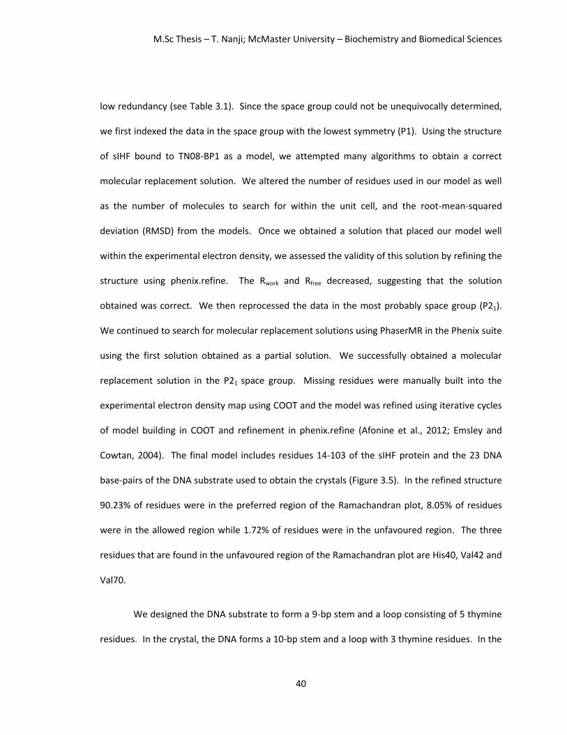
M.Sc Thesis – T. Nanji; McMaster University – Biochemistry and Biomedical Sciences
40
low redundancy (see Table 3.1). Since the space group could not be unequivocally determined,
we first indexed the data in the space group with the lowest symmetry (P1). Using the structure
of sIHF bound to TN08-BP1 as a model, we attempted many algorithms to obtain a correct
molecular replacement solution. We altered the number of residues used in our model as well
as the number of molecules to search for within the unit cell, and the root-mean-squared
deviation (RMSD) from the models. Once we obtained a solution that placed our model well
within the experimental electron density, we assessed the validity of this solution by refining the
structure using phenix.refine. The Rwork and Rfree decreased, suggesting that the solution
obtained was correct. We then reprocessed the data in the most probably space group (P21).
We continued to search for molecular replacement solutions using PhaserMR in the Phenix suite
using the first solution obtained as a partial solution. We successfully obtained a molecular
replacement solution in the P21 space group. Missing residues were manually built into the
experimental electron density map using COOT and the model was refined using iterative cycles
of model building in COOT and refinement in phenix.refine (Afonine et al., 2012; Emsley and
Cowtan, 2004). The final model includes residues 14-103 of the sIHF protein and the 23 DNA
base-pairs of the DNA substrate used to obtain the crystals (Figure 3.5). In the refined structure
90.23% of residues were in the preferred region of the Ramachandran plot, 8.05% of residues
were in the allowed region while 1.72% of residues were in the unfavoured region. The three
residues that are found in the unfavoured region of the Ramachandran plot are His40, Val42 and
Val70.
We designed the DNA substrate to form a 9-bp stem and a loop consisting of 5 thymine
residues. In the crystal, the DNA forms a 10-bp stem and a loop with 3 thymine residues. In the
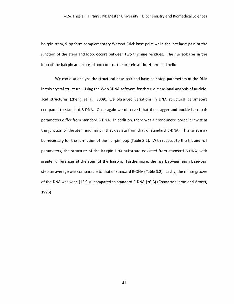
M.Sc Thesis – T. Nanji; McMaster University – Biochemistry and Biomedical Sciences
41
hairpin stem, 9-bp form complementary Watson-Crick base pairs while the last base pair, at the
junction of the stem and loop, occurs between two thymine residues. The nucleobases in the
loop of the hairpin are exposed and contact the protein at the N-terminal helix.
We can also analyze the structural base-pair and base-pair step parameters of the DNA
in this crystal structure. Using the Web 3DNA software for three-dimensional analysis of nucleic-
acid structures (Zheng et al., 2009), we observed variations in DNA structural parameters
compared to standard B-DNA. Once again we observed that the stagger and buckle base pair
parameters differ from standard B-DNA. In addition, there was a pronounced propeller twist at
the junction of the stem and hairpin that deviate from that of standard B-DNA. This twist may
be necessary for the formation of the hairpin loop (Table 3.2). With respect to the tilt and roll
parameters, the structure of the hairpin DNA substrate deviated from standard B-DNA, with
greater differences at the stem of the hairpin. Furthermore, the rise between each base-pair
step on average was comparable to that of standard B-DNA (Table 3.2). Lastly, the minor groove
of the DNA was wide (12.9 Å) compared to standard B-DNA (~6 Å) (Chandrasekaran and Arnott,
1996).

M.Sc Thesis – T. Nanji; McMaster University – Biochemistry and Biomedical Sciences
42
Figure 3.5. Structure of sIHF bound to TN23-HP9. (A) Ribbon diagram of sIHF bound to TN23-HP9 highlighting the 4 duplex substrates that contact sIHF. Image is rotated 90° in the second panel. (B-D) 2Fo-Fc electron density maps and model of sIHF bound to TN23-HP9 determined from crystal TN131 highlighting the interactions with sIHF at (B) Interface I, (C) Interface II, (D) and the N-terminal helix and DNA.

M.Sc Thesis – T. Nanji; McMaster University – Biochemistry and Biomedical Sciences
43
In this crystal structure, the DNA contacts the protein at three interfaces (Figure 3.5);
Interface I and II, previously discussed, and the N-terminal helix. sIHF makes contacts with DNA
through several residues. At Interface I, sIHF contacts DNA using residues Arg85, Asn93, and
Gln94 which contact the DNA phosphate backbone (Figure 3.5B). sIHF contacts DNA at Interface
II through the peptide backbone (Figure 3.5C). The N-terminal helix of sIHF interacts with DNA
at residues Arg22, Asn30, and His34. Arg22 contacts an oxygen atom in the phosphate group of
the thymine residue located at the intersection of the stem and loop of duplex 3, and the sugar
at the loop of the hairpin at C atoms in the 1’ and 2’ positions (Figure 3.5C). Asn30 contacts the
methyl group of the exposed thymine base within the loop of the hairpin in duplex 3 while His34
forms extensive contacts with the exposed thymine base of duplex 4 (Figure 3.5C). In order to
assess if these three interfaces are important for DNA binding in solution, the sIHF protein was
mutated to disrupt these interfaces and DNA binding was probed.
3.5 Three interfaces on sIHF contribute to DNA binding in vitro
The X-ray crystal structures of sIHF bound to DNA substrates TN20O and TN08-BP1 show sIHF-
DNA interactions at two interfaces: Interface I which is composed of the lid region (Figure 3.4A in
pink) of the protein and is mediated by residues Arg85, Arg86, Asn93 and Gln94 (Figure 3.4B and
C); and Interface II which is facilitated by the peptide backbone of the H2TH motif (Figure 3.4A in
blue). The N-terminal helix was also identified to contact DNA in the crystal structure of sIHF
bound to DNA substrate TN23-HP9 (Figure 3.5D). To validate that these surfaces are important
for DNA binding in solution in vitro, electrophoretic mobility shift assays (EMSAs) of sIHF
mutants were conducted with radiolabeled duplex DNA. sIHF was shown to bind the sequence

M.Sc Thesis – T. Nanji; McMaster University – Biochemistry and Biomedical Sciences
44
of duplex DNA chosen for this assay preferentially through systematic evolution of ligands by
exponential enrichment (SELEX) (Swiercz, 2013).
To test if sIHF Interface I contributes to DNA binding, sIHF mutants were constructed
where residues arginine 85 and arginine 86 were mutated to alanine and serine residues (sIHF-
RR85AS); asparagine 93 and glutamine 94 were mutated to alanine and serine residues (sIHF-
NQ93AS); and where all four key residues on Interface I were mutated (sIHF-RR85AS+NQ93AS).
EMSAs using these sIHF variants were conducted and the results were compared with those
done using wildtype sIHF to determine if DNA binding was reduced for these mutants, indicating
that the mutated residue is important for DNA binding.
Duplex DNA that was not bound by protein migrated further on a native acrylamide gel
(Figure 3.6A indicated by the asterisks) than the sIHF-DNA complex (Figure 3.6A indicated by the
arrow). Wildtype sIHF was used as a control to compare DNA binding affinity of the sIHF
mutants. Wildtype sIHF bound duplex DNA (0.02 μM) readily at the lowest concentration of
protein assayed (3 μM) as visualized by a shift of the radiolabeled DNA to form the sIHF-DNA
complex. sIHF-RR85AS displayed a reduction in DNA binding; even at the highest concentration
of protein tested (100 μM) sIHF-RR85AS did not fully shift the DNA (migration of the band was
smeared). The sIHF-NQ93AS mutant behaved similar to wildtype (Figure 3.5A). The quadruple
mutant, sIHF-RR85AS+NQ9AS, displayed a similar phenotype to the sIHF-RR85AS mutant (Figure
3.6B), suggesting that residues Arg85 and/or Arg86 were important for DNA binding, whereas
residues Asn93 and Gln94 were dispensable in vitro.

M.Sc Thesis – T. Nanji; McMaster University – Biochemistry and Biomedical Sciences
45
Figure 3.6. EMSAs of sIHF mutants with a radiolabeled 23-bp duplex DNA substrate. (A, B) EMSAs of sIHF variants to test Interface I. Wildtype sIHF, sIHF RR85AS (sIHF RR85) and sIHF NQ93AS (sIHF NQ93) with radiolabeled duplex DNA (0.02 μM) at sIHF concentrations of 0, 3, 10 and 100 μM. (C) EMSA of wildtype sIHF and an sIHF variant to test Interface II. sIHF G66+ with radiolabeled duplex DNA (0.02 μM) at sIHF concentrations of 0, 3, 10 and 100 μM. (D) EMSA of wildtype sIHF and sIHF variants to test the N-terminal helix. sIHF 35-37Gly (sIHF 35-37G) and sIHF Δ1-36 with radiolabeled duplex DNA (0.02 μM) at sIHF concentrations of 0, 3, 10 and 100 μM. Asterisks indicate the migration of duplex DNA alone while the arrows indicate the migration of the sIHF-DNA complex. sIHF interacts with DNA through a second interface defined by the loop region of the
H2TH motif, solely through the peptide backbone (Figure 3.4D). In order to alter the length and
flexibility of the loop at this motif, we decided to insert an extra glycine residue after position 66
(sIHF-G66+). sIHF-G66+ displayed reduced binding compared to wildtype (Figure 3.6C),
suggesting that Interface II is also important for DNA binding.
The N-terminal helix of sIHF contacts DNA in the crystal structure of sIHF bound to a
hairpin DNA substrate (Figure 3.5D). The N-terminal helix might interact with DNA at this region
as it contains many positively charged residues including Arg22, Arg25, Lys29 and Lys33. These
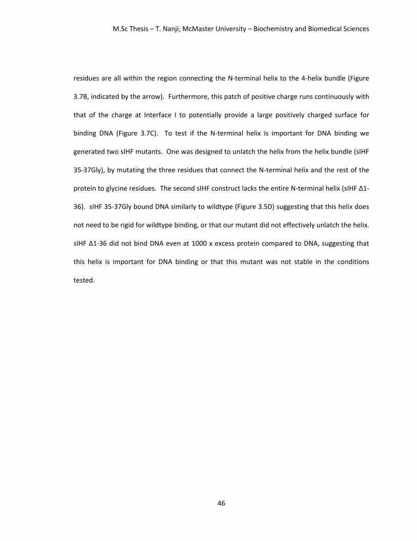
M.Sc Thesis – T. Nanji; McMaster University – Biochemistry and Biomedical Sciences
46
residues are all within the region connecting the N-terminal helix to the 4-helix bundle (Figure
3.7B, indicated by the arrow). Furthermore, this patch of positive charge runs continuously with
that of the charge at Interface I to potentially provide a large positively charged surface for
binding DNA (Figure 3.7C). To test if the N-terminal helix is important for DNA binding we
generated two sIHF mutants. One was designed to unlatch the helix from the helix bundle (sIHF
35-37Gly), by mutating the three residues that connect the N-terminal helix and the rest of the
protein to glycine residues. The second sIHF construct lacks the entire N-terminal helix (sIHF Δ1-
36). sIHF 35-37Gly bound DNA similarly to wildtype (Figure 3.5D) suggesting that this helix does
not need to be rigid for wildtype binding, or that our mutant did not effectively unlatch the helix.
sIHF Δ1-36 did not bind DNA even at 1000 x excess protein compared to DNA, suggesting that
this helix is important for DNA binding or that this mutant was not stable in the conditions
tested.
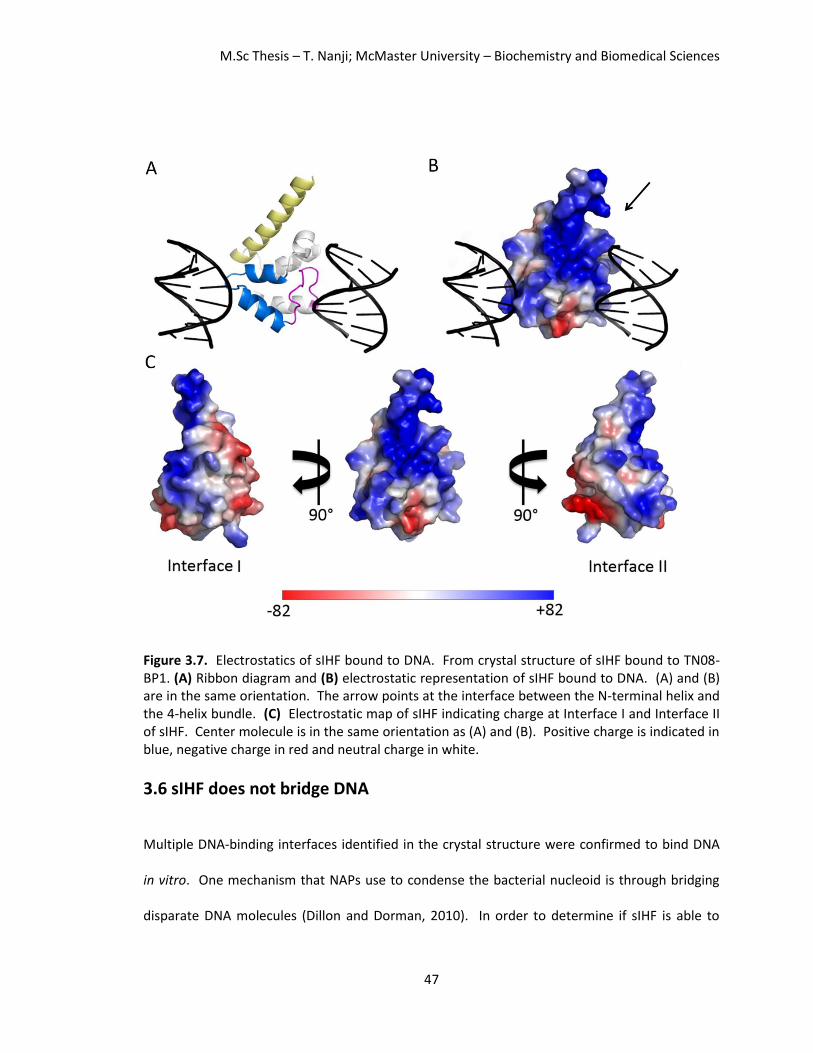
M.Sc Thesis – T. Nanji; McMaster University – Biochemistry and Biomedical Sciences
47
Figure 3.7. Electrostatics of sIHF bound to DNA. From crystal structure of sIHF bound to TN08-BP1. (A) Ribbon diagram and (B) electrostatic representation of sIHF bound to DNA. (A) and (B) are in the same orientation. The arrow points at the interface between the N-terminal helix and the 4-helix bundle. (C) Electrostatic map of sIHF indicating charge at Interface I and Interface II of sIHF. Center molecule is in the same orientation as (A) and (B). Positive charge is indicated in blue, negative charge in red and neutral charge in white.
3.6 sIHF does not bridge DNA
Multiple DNA-binding interfaces identified in the crystal structure were confirmed to bind DNA
in vitro. One mechanism that NAPs use to condense the bacterial nucleoid is through bridging
disparate DNA molecules (Dillon and Dorman, 2010). In order to determine if sIHF is able to

M.Sc Thesis – T. Nanji; McMaster University – Biochemistry and Biomedical Sciences
48
bring together two DNA molecules, we employed small-angle X-ray scattering (SAXS). SAXS
provides structural information at low resolution of samples in solution, thereby bypassing the
need to grow crystals. Although, crystal structures allow for high resolution structural data, they
are obtained in an environment where the molecules of interest are highly concentrated, and
the conditions are not physiological. This often leads to crystallographic artifacts. Furthermore,
the crystal is in a locked conformation prohibiting molecules from moving. SAXS allows us to
monitor complexes in dynamic equilibrium, in optimal experimental conditions; samples do not
need to be labeled or altered, and data can be collected at low temperatures (Tuukkanen and
Svergun, 2014). Collecting data at a low temperature is important to ensure that our DNA
duplex remains double stranded.
In our SAXS experiment, we incubated the protein with a 23-bp duplex DNA substrate
(with the same sequence used for our DNA binding assays) at different ratios to assess if sIHF
could simultaneously bind two DNA molecules. We first determined the maximum particle
diameter (Dmax) and experimental molecular weights of the sIHF protein and the DNA duplex
individually. The molecular weights determined experimentally are very close to the calculated
molecular weights of the protein and DNA alone (Table 3.3).
We first incubated the protein and DNA at a 1:1 ration (sIHF:DNA) to ensure that the
protein forms a complex with DNA in solution in the experimental conditions used. The Dmax for
the complex (94.5 Å) is larger than that observed for the protein (66 Å) and DNA (71.5 Å) alone,
suggesting that the complex is able to form. Furthermore, the molecular weight determined
from the experiment (26.9 kDa) is in good agreement with the estimated molecular weight of

M.Sc Thesis – T. Nanji; McMaster University – Biochemistry and Biomedical Sciences
49
the complex (27.8 kDa), confirming that the complex forms at a 1:1 ratio in this condition (Table
3.3).
Based on the crystal structures determined in this work, we predicted that more than
one sIHF molecule could bind the 23-bp DNA substrate. We tested our prediction using SAXS by
incubating the protein at a 2:1 ratio (sIHF:DNA) with duplex DNA prior to data collection. The
Dmax as well as the molecular weights determined increased (103 Å and 46 kDa), indicating that
two protein monomers are bound to the 23-bp duplex substrate (Table 3.3).
Table 3.3. Small angle X-ray scattering data collection and analysis sIHF DNA (sIHF:DNA) 1:1 (sIHF:DNA) 2:1 (sIHF:DNA) 1:2
Data collection
Instrument BioSAXS-1000 BioSAXS-1000 BioSAXS-1000 BioSAXS-1000 BioSAXS-1000
X-ray generator MicroMax-007HF
MicroMax-007HF
MicroMax-007HF
MicroMax-007HF
MicroMax-007HF
Beam geometry (mm) 0.5 0.5 0.5 0.5 0.5
Wavelength (Å) 1.5418 1.5418 1.5418 1.5418 1.5418
Exposure time (min) 20 180 20 20 20
Concentration (μM) 434 109 109 109 217
Structural parameters
Io 0.24 0.2 0.6 0.6 0.6
Io/conc 0.5 1.8 1.4 5.6 1.4
Rg (Å) 21.3 21.9 25.7 29.6 24.7
Dmax (Å) 66 71.5 94.5 103 84
MWExperimental
(kDa) 16.4 10.6 26.9 46 20
MWCalculated
(kDa) 13.7 14 27.8 41.4 41.7
In order to determine if sIHF could bridge DNA, we then incubated the protein at a 1:2
ratio (sIHF:DNA). The experimental molecular weight for this sample is smaller than expected
(20 kDa compared to 41.7 kDa). Furthermore, the Dmax, at 84 Å is between that of the DNA alone
and the sIHF:DNA complex at a 1:1 ratio. This is also true for the experimental molecular weight;

M.Sc Thesis – T. Nanji; McMaster University – Biochemistry and Biomedical Sciences
50
the experimental molecular weight determined is between that of the DNA alone and that of the
1:1 protein-DNA complex (Table 3.2). This suggests that sIHF cannot bridge two DNA molecules
together, and instead the sample collected is a mixture of sIHF:DNA complex at a 1:1 ratio and
free DNA (Table 3.3).
3.7 sIHF affects TopA activity independently of DNA binding
Since sIHF does not bend nor bridge DNA; we next explored if it compacts the nucleoid indirectly
by modulating the activity of other enzymes that affect chromosome compaction. The H2TH
fold has been found in topoisomerase VI and, thus, we thought that sIHF could potentially
modulate the activity of one of the topoisomerases found in S. coelicolor. We decided to test if
sIHF alters the activity of the sole type I topoisomerase expressed in Streptomyces; TopA.
We cloned and purified TopA and compared its ability to relax supercoiled plasmids in
the absence or presence of sIHF. We found that sIHF inhibits TopA activity in vitro as the
formation of relaxed plasmids by TopA is reduced in the presence of sIHF (Swiercz et al., 2013).
As a control, we performed the same experiment replacing sIHF with a different protein of
similar molecular weight (lysozyme) and found that only sIHF was able to inhibit the activity of
TopA (Figure 3.8). We later found that this effect is not due to a direct interaction between sIHF
and TopA as the two proteins do not interact using gel filtration chromatography (Figure 3.9). If
sIHF were to form a complex with TopA we would expect sIHF to co-elute with TopA; however,
even when we incubate sIHF with a 10 x excess over TopA, sIHF does not form a complex with
TopA (Figure 3.9C). Interestingly, high sIHF concentrations alter the migration of TopA (Figure
3.9B). Since sIHF does not directly interact with TopA, we reasoned that binding of sIHF to DNA

M.Sc Thesis – T. Nanji; McMaster University – Biochemistry and Biomedical Sciences
51
either renders DNA inaccessible to TopA, or causes a conformational change that inhibits TopA
activity.
Figure 3.8. sIHF affects the activity of TopA. Agarose gel of sIHF or lysozyme incubated with supercoilied pUC19 plasmid DNA and the Streptomyces coelicolor TopA. Protein concentrations tested range from 135-540 molar excess over DNA. The migration of supercoiled plasmid DNA is indicated by the asterisk while bands that migrate slower indicate plasmids relaxed by TopA (Figure adapted from Swiercz et al., 2013). In order to determine if sIHF is simply coating DNA, we assessed the ability of TopA to
relax DNA when incubated with different sIHF variants. We found that sIHF-RR85AS and sIHF-
RR85AS+NQ93AS no longer inhibit TopA activity, whereas sIHF-NQ93AS is still able to inhibit
TopA activity; albeit, to a lesser extent than wildtype (Figure 3.10A). Therefore, the integrity of
Interface I and, in particular residues Arg85 and Arg86 are important for this role of sIHF.
Conversely, the sIHF-G66+ variant inhibits TopA activity similar to wildtype sIHF (Figure 3.10B)
even though its affinity for DNA binding is reduced compared to wildtype (Figure 3.6C). This
illustrates that Interface I is important for sIHF’s role in inhibiting TopA activity and DNA binding,
whereas Interface II is only implicated in binding DNA.

M.Sc Thesis – T. Nanji; McMaster University – Biochemistry and Biomedical Sciences
52
Figure 3.9. Gel filtration assay of sIHF with TopA. SDS-PAG and chromatogram of fractions eluted from the gel filtration column of sIHF incubated with TopA. 0.1 mL fractions were collected and loaded onto a 4-15% precast gel (Bio-Rad). Migration of TopA and sIHF are as indicated. sIHF and TopA were incubated at various ratios. Panel (A) displays fractions collected with TopA:sIHF at a 1:1 ratio, while panel (B) displays fractions collected from TopA:sIHF at a 1:10 ratio.

M.Sc Thesis – T. Nanji; McMaster University – Biochemistry and Biomedical Sciences
53
Figure 3.10. Topoisomerase assays of sIHF mutants. Topoisomerase assay, using TopA (7.73
μM) from S. coelicolor, in the presence of sIHF variants to test (A) Interface I; wildtype sIHF, sIHF RR85AS (sIHF RR85), sIHF NQ93AS (sIHF NQ93) and sIHF RR85AS+NQ93AS (sIHF RR+NQ) (B) Interface II; wildtype sIHF and sIHF G66+ (C) the N-terminal helix; wildtype sIHF, sIHF 35-37Gly (sIHF 35-37G) and sIHF Δ1-36 with plasmid DNA (pUC19) at sIHF concentrations of 0, 0.54, and 2.16 μM. The asterisks indicate supercoiled DNA, while DNA bands migrating slower than the supercoiled plasmid indicate relaxed topoisomers.
Next we tested whether the N-terminal helix of sIHF is important to modulate the
activity of TopA. We observed that a variant of sIHF lacking the N-terminal helix (sIHF Δ1-36)
was no longer able to inhibit the activity of TopA whereas sIHF 35-37Gly inhibited TopA activity
to a similar extent to wildtype (Figure 3.8C). These results reinforce the idea that the 35-37Gly
variant of sIHF does not unlatch the N-terminal helix from the H2TH domain, and that the N-
terminal helix is relevant for the function of sIHF.

M.Sc Thesis – T. Nanji; McMaster University – Biochemistry and Biomedical Sciences
54
Chapter 4
Discussion
In this work, we have refined three crystal structures of sIHF bound to various duplex DNA
substrates, and have identified three distinct surfaces on the protein that contact DNA. These
three surfaces have been confirmed to bind DNA in solution in vitro (Figure 3.6). The first
interface, Interface I, uses the lid region of the protein (Figures 3.3, 3.4A, and 3.5A in pink) and
contacts the DNA phosphate backbone through residues Arg85, Asn93, and Gln94 (Figure 3.4C,
3.5B). Arg86 is in close proximity to the DNA duplex, and could potentially aid in DNA binding
(Figure 3.4B). Based on in vitro DNA binding data, residues Arg85 and/or Arg86 are important
for the interaction between sIHF and DNA (Figure 3.6A).
Interface I interacts with DNA within the asymmetric unit of the structures refined from
crystals TN051 and TN103; however, sIHF also contacts DNA at a second interface, Interface II,
through symmetry. Interface II, contacts DNA solely through the peptide backbone using the
H2TH domain (Figures 3.4 and 3.5). This domain is also found in other proteins that interact
with nucleic acids (Figure 3.3 C-E in blue) and is thought to aid in peripheral DNA binding. The lid
region of Interface I is also found amongst these proteins (Figure 3.3 C-E in pink) and is more
exposed than the H2TH domain in their crystal structures (Corbett and Berger, 2003; Zharkov et
al., 2002; Brodersen et al., 2002). Therefore, the contribution of these two regions for DNA
binding must be explored further.
A third interface of sIHF, the N-terminal helix, has also been shown to contribute to DNA
binding (Figures 3.4A and 3.5A in yellow). Residues involved in this interaction, identified from

M.Sc Thesis – T. Nanji; McMaster University – Biochemistry and Biomedical Sciences
55
the crystal structure of sIHF bound to a hairpin DNA substrate (crystal TN131), include Arg22,
Asn30, and His34 (Figure 3.5D). Crystals TN051 and TN103 failed to capture the interaction
between this interface and DNA. This could be because the DNA in these crystals adopts a
pseudo-continuous helix, and favours contacts at Interface I and Interface II. The N-terminal
helix is approximately one third of the protein as it contains 34 of 107 residues of the protein.
The crystal structures of sIHF bound to TN20O (from crystal TN051) and sIHF bound to TN08-BP1
(from crystal TN103) successfully identified residues 13-103 and 15-103, respectively, in their
experimental electron density maps. However, the N-terminal residues are missing from our
model. It is common for the ends of proteins and flexible regions to be unidentified in electron
density maps as they do not adopt the same conformation in each repeating unit across the
crystal. In the crystal, perhaps the N-terminal residues are flexible and interact with duplex DNA
on either side, causing these residues to be unidentified in the crystal structure (Figure 4.1). This
would explain why these residues are disordered even if they interact with DNA.
To test the relevance of the interaction between the N-terminal helix of sIHF and DNA,
we removed the entire N-terminal helix, which may have caused the protein to become
unstable. Assessed by dynamic light scattering (DLS), the Δ1-36 sIHF protein is stable at the
concentration tested (see Methods), but the protein may have become unstable upon dilution.
Therefore we should repeat our DNA binding and topoisomerase activity assays with a less
severe mutation. Instead of removing the entire helix, we could remove the first 13 residues
(sIHF Δ1-13) which are disordered in the crystal structures, or mutate the residues believed to
facilitate the interaction between DNA and sIHF at this interface (Arg22, Asn30 and His34).

M.Sc Thesis – T. Nanji; McMaster University – Biochemistry and Biomedical Sciences
56
Once we verified that multiple surfaces of sIHF contact DNA, we were curious to see if
sIHF could simultaneously bind DNA at these interfaces to bring together two disparate DNA
molecules. Common NAPs that bridge DNA, such as H-NS and Lsr2, are composed of a DNA
binding domain and an oligomerization domain. They function as a homodimer and bring
together distant DNA molecules by binding DNA through their DNA binding domain, and
subsequently forming a homodimer with another H-NS/Lsr2 molecule bound to DNA (Dillon and
Dorman, 2010). Conversely, sIHF functions as a monomer and would need to simultaneously
bind multiple DNA substrates, with one sIHF molecule, to bridge DNA. Based on SAXS data
collected of sIHF incubated with DNA at various ratios, we did not observe bridging of DNA by
sIHF (Table 3.3). However, sIHF may still use multiple interfaces to compact DNA and neutralize
the charge repulsion between DNA molecules in vivo.
Figure 4.1. Ribbon structure of sIHF bound to TN08-BP1 and possible interactions with the N-terminal helix. sIHF residues 15-103 with TN08-BP1. The 14 residues missing from this model may interact with DNA, but are not identified as they are flexible and may interact with either duplex forming the pseudo-continuous helix (indicated by the arrows).

M.Sc Thesis – T. Nanji; McMaster University – Biochemistry and Biomedical Sciences
57
To further investigate sIHF’s role in chromosome organization, we wanted to explore if
sIHF affected the structure of DNA at the local level. We decided to crystallize sIHF in complex
with a DNA palindrome of known structure to analyze the conformational changes induced by
sIHF upon binding. We found differences in DNA base-pair, and base-pair step parameters
between duplex DNA with and without sIHF (Table 3.2). Collectively, this caused the minor
groove of the DNA to widen from 8.3 Å to 13.5 Å, determined by comparing the structures of
identical DNA sequences alone (by solution NMR) and in complex with sIHF (by X-ray
crystallography). Furthermore, when we analyzed the structure of the DNA in the crystal
structure of sIHF bound to the hairpin DNA substrate, we observed base-pair and base-pair step
parameters that deviated from standard B-DNA (Table 3.2). In addition, the minor groove has a
similar width to that in crystal TN103 (sIHF bound to TN08-BP1) at 12.9 Å, illustrating that sIHF
may widen the minor groove of DNA upon binding.
The crystal structures presented in this work, show that sIHF interacts with DNA through
the minor groove (Figure 3.4 and 3.5). sIHF is a small positively charged protein, and may prefer
binding DNA through the minor groove as the negative electrostatic potential is increased due to
the relative concentration of negative charge from the phosphate backbone (Rohs et al., 2009).
It is also worth noting that SELEX assays conducted by Julia Swiercz (Swiercz, 2013) identified
three motifs that sIHF had preferred binding, all of these had under 50% GC content and at least
three A-T consecutive base-pairs. sIHF may prefer binding AT-rich sites as they contain narrow
minor grooves (Gavathiotis et al., 2000, Oguey et al., 2010) where the negative electrostatic
potential is enhanced even more (Rohs et al., 2009). This suggests that perhaps sIHF prefers
binding to DNA substrates with narrow minor grooves, and upon binding widens the groove.

M.Sc Thesis – T. Nanji; McMaster University – Biochemistry and Biomedical Sciences
58
Thus, sIHF may prefer binding certain DNA structures (narrow minor grooves), rather than
particular sequences.
In addition to altering the conformation of DNA at the local level, sIHF may affect
chromosome organization in vivo through mediating the activity of other enzymes that alter
DNA topology, such as topoisomerases (Dean and Cozarelli, 1985). We show that sIHF inhibits
the activity of the sole type I topoisomerase in S. coelicolor, ScTopA in vitro (Figures 3.8) (Swiercz
et al., 2013); where ScTopA has been shown to function as a processive enzyme (Szafran et al.,
2014). We have found a separation of function sIHF mutant, whereby Interface II (tested using
sIHF-G66+) is important for DNA binding but does not affect sIHF’s ability to mediate TopA
activity. sIHF variants where Interface I is disrupted have reduced DNA binding and have lost
their ability to affect TopA activity (Figures 3.6 and 3.10). Interface I, in conjunction with the N-
terminal helix, form a continuous positively charged patch. This network of residues (Arg22,
Asn30, Arg85, Arg86, Asn93 and Gln94) would allow the DNA duplex to form extensive contacts
with the DNA phosphate backbone. In this way, Interface I and the N-terminal helix may bind
DNA over a larger surface area stalling TopA from relaxing supercoiled DNA. Interface II covers
less surface area, and sIHF may be displaced from the DNA in the presence of TopA.
Since sIHF can bind DNA through multiple interfaces there is the possibility that each
interface binds DNA with a different affinity and stability. We propose that sIHF may bind more
stably at Interface I and effectively stall the processive TopA enzyme (Figure 4.2A), while the
interaction between DNA at Interface II may be less stable allowing for TopA to progress (Figure
4.2B). The difference in stability for various interfaces of sIHF is consistent with having a
dynamic nucleoid. sIHF is expressed throughout the cell cycle (Swiercz et al., 2013); if it were to

M.Sc Thesis – T. Nanji; McMaster University – Biochemistry and Biomedical Sciences
59
bind DNA with high affinity at multiple surfaces, the nucleoid would be constrained and not
dynamic. The interaction between sIHF Interface II and DNA being less stable would allow for
the nucleoid to remain dynamic. Interface I in combination with the positive patch along the N-
terminal helix may form a more stable complex than Interface II, allowing for sIHF complexes
that bind DNA through Interface I to effectively stall TopA progression, while sIHF Interface II
and DNA may form a more transient complex which will allow TopA to continue to relax
supercoiled DNA. In this way, sIHF may use multiple interfaces to compact the bacterial nucleoid
and affect gene expression; while only Interface I can stall TopA and inhibit chromosome
relaxation.
Figure 4.2. Proposed mechanism of action of sIHF. (A) sIHF (blue) bound to DNA using Interface I and the N-terminal helix binds strongly to DNA inhibiting the progression of the TopA (red) enzyme from further relaxing duplex DNA. (B) sIHF bound to DNA at Interface II is weak; the interaction between sIHF and DNA cannot be maintained in the presence of TopA which is able to progress along the duplex.
M.Sc Thesis – T. Nanji; McMaster University – Biochemistry and Biomedical Sciences
60
In order to confirm our model, follow up studies must be conducted. First, to determine
if Interface I of sIHF with the positive patch along the N-terminus forms a more stable complex
with DNA than Interface II, we should compare the stability of the complexes of sIHF, sIHF
RR85AS and sIHF G66+ with duplex DNA. This can be done using gel filtration chromatography,
isothermal titration calorimetry and surface plasmon resonance. If our model is correct, sIHF
G66+ should form a more stable complex with DNA than sIHF RR85AS as Interface I is still intact
in the former mutant.
Our model is based on the idea that the N-terminal helix and Interface I both
simultaneously contribute to DNA binding, allowing for a strong complex to form as the
interaction surface area increases when these two surfaces are combined. If this is true, the
positively charged patch between the N-terminal helix and 4-helix bundle composed of residues
Arg22, Arg25, Lys29 and Lys33 should contribute to DNA binding. In order to test this, we will
mutate these residues to serine residues and assess this variant (sIHF-R22, R25, K29, K33(S)) for
DNA binding. This sIHF variant can also be assessed for its role on TopA activity.
The assays discussed above have been conducted in vitro. In order to assess if the
interfaces identified in this study are important for wildtype function in vivo, with the help of our
collaborators, we would like to complement ΔsIHF strains with sIHF-RR85AS, sIHF-G66+, and
sIHF-R22, R25, K29, K33(S) to observe their phenotype. This will allow us to assess the various
roles of each region of the protein on chromosome condensation, gene expression and
sporulation.
Lastly, we have obtained several crystal structures of sIHF bound to DNA; however, we
believe it would be beneficial to determine the crystal structure of sIHF bound to the DNA

M.Sc Thesis – T. Nanji; McMaster University – Biochemistry and Biomedical Sciences
61
sequence determined using SELEX, at a 2:1 ratio (sIHF:DNA). This structure would allow us to
investigate whether sIHF coats DNA and, if the A-tract and associated narrow minor groove aid
in sIHF binding. By conducting this experiment at progressively higher protein:DNA ratios, we
will be able to assess if sIHF binds DNA cooperatively.
M.Sc Thesis – T. Nanji; McMaster University – Biochemistry and Biomedical Sciences
62
Chapter 5
Conclusion
We have shown that sIHF is a novel NAP that is expressed throughout the cell cycle, binds DNA
in a sequence independent manner, associates with the nucleoid, plays a role in chromosome
condensation, when absent leads to aberrant levels of gene expression, and affects the activity
of TopA (Swiercz et al., 2013). Following this work, we have solved crystal structures of sIHF
bound to DNA and confirmed that sIHF binds DNA at multiple interfaces, yet does not bridge
DNA molecules together. We have also discovered an interface of sIHF that contributes to DNA
binding, but does not affect the activity of TopA. With these findings, we propose a model for
the mechanism of action of sIHF. sIHF contributes to chromosome organization at two levels.
The first uses all DNA binding surfaces of the positively charged sIHF protein, to neutralize the
charge repulsion from the negatively charged DNA phosphate backbone. This aids in
chromosome condensation. The second uses Interface I and the positively charged patch along
the N-terminal helix to stall ScTopA from relaxing DNA supercoils. Further studies must be
conducted to confirm this model.

M.Sc Thesis – T. Nanji; McMaster University – Biochemistry and Biomedical Sciences
63
References
Afonine, P. V., Grosse-Kunstleve, R. W., Echols, N., Headd, J. J., Moriarty, N. W., Mustyakimov, M., Terwilliger, T. C., Urzhumtsev, A., Zwart, P. H., and Adams, P. D. (2012). Towards automated crystallographic structure refinement with phenix.refine. Acta Crystallographica D Biological Crystallography 68:352-367. Almirón, M., Link, A. J., Furlong, D., and Kolter, R. (1992). A novel DNA-binding protein with regulatory and protective roles in starved Escherichia coli. Genes and Development 6:2646-2654. Atlung, T., and Ingmer, H. (1997). H-NS: a modulator of environmentally regulated gene expression. Molecular Microbiology 24:7-17. Azam, T. A., Iwata, A., Nishimura, A., Ueda, S., and Ishihama, A. (1999). Growth phase-dependent variation in protein composition of the Escherichia coli nucleoid. Journal of Bacteriology 181:6361-6370. Baleja J, D., Germann, M.W., van de Sande, J. H., and Sykes, B. D. (1990). Solution conformation of purine-pyrimidine DNA octamers using nuclear magnetic resonance, restrained molecular dynamics and NOE-based refinement. Journal of Molecular Biology 215:411-428. Bentley, S. D., Chater, K. F., Cerdeno‐Tarraga, A. M., Challis, G. L., Thomson, N. R., James, K. D., Harris, D. E., Quail, M. A., Kieser, H., Harper, D., Bateman, A., Brown, S., Chandra, G., Chen, C. W., Collins, M., Cronin, A. Fraser, A. Goble, A., Hidalgo, J., Hornsby, T., Howarth, S., Huang, C. H., Kieser, T., Larke, L., Murphy, L., Oliver, K., O'Neil, S., Rabbinowitsch, E., Rajandream, M. A., Rutherford, K., Rutter, S., Seeger, K., Saunders, D., Sharp, S., Squares, R., Squares, S., Taylor, K., Warren, T., Wietzorrek, A., Woodward, J., Barrell, B. G., Parkhill, J., and Hopwood, D. A (2002). Complete genome sequence of the model actinomycete Streptomyces Coelicolor A3(2). Nature 417:141–147. Berman, H. M., Olson, W. K., Beveridge, D. L., Westbrook, J., Gelbin, A., Demeny, T., Hsieh, S., Srinivasan, R., and Schneider, B. (1992). A comprehensive relational database of three-dimensional structures of nucleic acids. Biophysical Journal 63:751-759. Bibb, M. J., Ward, J. M., Kieser, T., Choen, S. N., and Hopwood, D. A. (1981). Excision of chromosomal DNA sequences from Streptomyces coelicolor forms a novel family of plasmids detectable in Streptomyces lividans. Molecular Genetics and Genomics 184:230-240. Bloch, V., Yang, Y., Margeat, E., Chavanieu, A., Augé, M. T., Robert, B., Arnold, S., Rimsky, S., and Kochoyan, M. (2003). The H-NS dimerization domain defines a new fold contributing to DNA recognition. Nature Structural Biology 10:212-218. Bradford, M.M. (1976). A rapid and sensitive method for the quantitation of microgram quantities of protein utilizing the principle of protein-dye binding. Analytical Biochemistry 72:248-254.
M.Sc Thesis – T. Nanji; McMaster University – Biochemistry and Biomedical Sciences
64
Brodersen, D. E., Clemons, W. M. Jr., Carter, A. P., Wimberly, B. T., and Ramakrishnan, V. (2002). Crystal structure of the 30 S ribosomal subunit from Thermus thermophilus: structure of the proteins and their interactions with 16 S RNA. Journal of Molecular Biology 316:725-768. Champoux, J.J. (2001). DNA topoisomearses: structure, function and mechanism. Annual Review of Biochemistry. 70:369-413. Chandrasekaran, R., and Arnott, S. (1996). The structure of B-DNA in oriented fibers. Journal of Biomolecular Structure and Dynamics 13:1015-1027. Chen, J. M., Ren, H., Shaw, J. E., Wang, Y. J., Li, M., Leung, A. S., Tran, V., Berbenetz, N. M., Kocíncová, D., Yip, C. M., Reyrat, J., and Liu, J. (2008). Lsr2 of Mycobacterium tuberculosis is a DNA-bridging protein. Nucleic Acids Research 36:2123-2135. Chen, N., Zinchenko, A. A., Yoshikawa, Y., Araki, S., Adachi, S., Yamazoe, M., Hiraga, S., and Yoshikawa, K. (2008). ATP-induced shrinkage of DNA with MukB protein and the MukBEF complex of Escherichia coli. Journal of Bacteriology 190:731-3737. Corbette, K. D., and Berger, J. M. (2003). Structure of the topoisomerase VI-B subunit: implications for the type II topoisomerase mechanism and evolution. The EMBO Journal 22:151-163. Cui, Y., Wang, Q., Stormo, G. D., and Calvo, J.M. (1995). A consensus sequence for binding of Lrp to DNA. Journal of Bacteriology 177:4872-4880. Dame, R. T., Wyman, C., and Goosen. N. (2000). H-NS mediated compaction of DNA visualised by atomic force microscopy. Nucleic Acids Research 28:3504-3510. Dame, R. T., Wyman, C., and Goosen, N. (2001). Structural basis for preferential binding of H-NS to curved DNA. Biochimie 83:231-234. Dame, R. T., and Goosen, N. (2002). HU: promoting or countering DNA compaction? FEBS Letters 529:151-156. Dame, R. T., Luijsterburg, M. S., Krin, E., Bertin, P. N., Wagner, R., and Wuite, G. J. L. (2005). DNA bridging: a property shared among H-NS-like proteins. Journal of Bacteriology 187:1845-1848. Dean, F. B., and Cozzarelli, N. R. (1985). Mechanism of strand passage by Escherichia coli topoisomerase I. The role of the required nick in catenation and knotting of duplex DNA. Journal of Biological Chemistry 260:4984–4994. Dillon, S. C., and Dorman, C. J. (2010). Bacterial nucleoid-associated proteins, nucleoid structure and gene expression. Nature Reviews Microbiology 8:185-195.
M.Sc Thesis – T. Nanji; McMaster University – Biochemistry and Biomedical Sciences
65
Dorman, C. J. (2013). Co-operative roles for DNA supercoiling and nucleoid-associated proteins in the regulation of bacterial transcription. Biochemical Society Transactions 41:542-547. Emsley, P., and Cowtan, K. (2004). Coot: model-building tools for molecular graphics. Acta Crystallographica D Biological Crystallography 60:2126-2132. Frenkiel-Krispin, D., Ben-Avraham, I., Englander, J., Shimoni, E., Wolf, S. G., and Minsky, A. (2004). Nucleoid restructuring in stationary-state bacteria. Molecular Microbiology 51:395-405. Funnell, B. E., Baker, T. A., and Kornberg, A. (1986). Complete enzymatic replication of plasmids containing the origin of the Escherichia coli chromosome. Journal of Biological Chemistry 261:5616-5624. Gavathiotis, E., Sharman, G. J., and Searle, M. S. (2000). Sequence-dependent variation in DNA minor groove width dictates orientational preference of Hoechst 33258 in A-tract recognition: solution NMR structure of the 2:1 complex with d(CTTTTGCAAAG)(2). Nucleic Acids Research 28:728-735. Gitai, Z., Thanbichler, M., and Shapiro, L. (2005) The choreographed dynamics of bacterial chromosomes. Trends in Microbiology. 13:221-228. Glatter, O., and Kratky., O. (1982) Small-angle X-ray Scattering (London, Academic Press). Gloyd, M., Ghirlando, R. and Guarné, A. (2011). The role of MukE in assembling a functional MukBEF Complex. Journal of Molecular Biology 412:578-590. Goodrich, J. A., Schwartz, M. L., McClure, W. R. (1990). Searching for and predicting the activity of sites for DNA binding proteins: compilation and analysis of the binding sites for Escherichia coli integration host factor (IHF). Nucleic Acids Research 18:4993-5000. Grant, R. A., Filman, D. J., Finkel, S. E., Kolter, R., and Hogle, J. M. (1998). The crystal structure of DNA, a ferritin homologue that binds and protects DNA. Nature Structure 5:294-303. Guinier, A., and Fournet, G. (1995). Small-angle Scattering of X-rays (New York: John Wiley & Sons, Inc). Hancock, R. (2004). A role for macromolecular crowding effects in the assembly and function of compartents in the nucleus. Journal of Structural Biology 146:281-290. Kleine Borgmann, L. A., Ries, J., Ewers, H., Ulbrich, M. H., and Graumann, P. L. (2013). The bacterial SMC complex displays two distinct modes of interaction with the chromosome. Cell Reports 3:1483-1492. Lim, H. M., Lewis, D. E., Lee, H. J., Liu, M., and Adhya, S. (2003). Effect of varying the supercoiling of DNA on transcription and its regulation. Biochemistry 42:10718-10725.
M.Sc Thesis – T. Nanji; McMaster University – Biochemistry and Biomedical Sciences
66
Losada, A., and Hirano, T. (2005). Dynamic molecular linkers of the genome: the first decade of SMC proteins. Genes and Development. 19:1269-1287. Luger, K. (2006). Dynamic nucleosomes. Chromosome Research 14:5-16. Luijsterburg, M. S., Noon, M. C., Wuite, G. J. L., and Dame, R. T. (2006). The architectural role of nucleoid-associated proteins in the organization of bacterial chromatin: A molecular perspective. Journal of Structural Biology 156:262-272. Luijsterburg, M. S., White, M. F., van Driel, R., and Dame, R. T. (2008). The major architects of chromatin: architectural proteins in bacteria, archaea and eukaryotes. Critical Reviews in Biochemistry and Molecular Biology 43:393-418. Marenduzzo, D., Finan, K., and Cook, P. R. (2006). The depletion attraction: an underappreciated force driving cellular organization. Journal of Cell Biology 175:681-686. Myers, K. S., Yan, H., Ong, I. M., Chung, D., Liang, K., Tran, F., Keleȿ, S., Landick, R., and Kiley, P. J. (2013). Genome-scale analysis of Escherichia coli FNR reveals complex features of transcription factor binding. PLoS Genetics. 9:e1003565. Nash, H. A. (1990). Bending and supercoiling of DNA at the attachment site of bacteriophage lambda. Trends in Biochemical Sciences 15:222-227. Newton-Foot, M., and Gey van Pittius, N. C. (2013). The complex architecture of mycobacterial promoters. Tuberculosis 93:60-74. Oberto, J., Drlica, K., and Rouviѐre-Yaniv, J. (1994). Histones, HMG, HU, IHF: Même combat. Biochimie 76:901-908. Oguey, C., Foloppe, N., and Hartmann, B. (2010). Understanding the sequence-dependence of DNA groove dimensions: implications for DNA interactions. PLoS One 29:e15931. Olson, W. K., Bansal, M., Burley, S. K., Dickerson, R. E., Gerstein, M., Harvey. S. C., Heinemann, U., Lu, X., Neidle, S., Shakked, Z., Sklenar, H., Suzuki, M., Tung, C., Westhof, E., Wolberger, C., and Berman, H. M. (2001). A standard reference frame for the description of nucleic acid base-pair geometry. Journal of Molecular Biology 313:229-237. Otwinowski, Z., and Minor, W. (1997). Processing of X-ray diffraction data collected in oscillation mode. Methods in Enzymology 276:307-326. Postow, L., Hardy, C. D., Arsuaga, J., and Cozzarelli, N. R. (2004). Topological domain structure of the Escherichia coli chromosome. Genes and Development 18:1766-1779.

M.Sc Thesis – T. Nanji; McMaster University – Biochemistry and Biomedical Sciences
67
Rimsky, S., and Travers, A. (2011). Pervasive regulation of nucleoid structure and function by nucleoid-associated proteins. Current Opinion in Microbiology 12:136-141. Rohs, R., West, S. M., Sosinsky, A., Liu, P., Mann, R. S., and Honig, B. (2009) The role of DNA shape in protein-DNA recognition. Nature 461: 1248–1253. Rouviѐre-Yaniv, J., Yaniv, M., and Germond, J. (1979). E. coli DNA binding protein HU forms nucleosome-like structure with circular double-stranded DNA. Cell 17:265-274. Schneider, R., Lurz, R., Lüder, G., Tolksdorf, C., Travers, A., and Muskhelishvili, G. (2001). An architectural role o the Escherichia coli chromatin protein FIS in organizing DNA. Nucleic Acids Research 29:5107-5114. Schneider, R., Travers, A., Kutateladze, T., and Muskhelishvili, G. (1999). A DNA architectural protein couples cellular physiology and DNA topology in Escerichia coli. Molecular Microbiology 34:953-964. Shindo, H., Iwaki, T., Ieda, R., Kurumizaka, H., Ueguchi, C., Mizuno, T., Morikawa, S., Nakamura, H., and Kuboniwa, H. (1995). Solution structure of the DNA binding domain of a nucleoid-associated protein, H-NS from Escherchia coli. FEBS Letters 360:125-131. Shindo, H., Ohnuki, A., Ginba, h., Katoh, E., Ueguchi, C. Mizuno, T., and Yamazaki, T. (1999). Identification of the DNA binding surface of H-NS protein from Escherichia coli by heteronuclear NMR spectroscopy. FEBS Letters 455:63-69. Sinden, R. R., and Pettijohn, D. E. (1981). Chromosomes in living Escherichia coli cells are segregated into domains of supercoiling. Proceedings of the National Academy of Sciences 78:224-228. Spuiro, R., Dürrenberger, M., Falconi, M., La Teana, A., Pon, C. L., and Gualerzi, C. O. (1992). Lethal overproduction of the Escherichia coli nucleoid protein H-NS: ultramicroscopic and molecular autopsy. Molecular Genetics and Genetics 231:201-211. Swiercz, J. P. (2013). sIHF IS A NOVEL NUCLEOID-ASSOCIATED PROTEIN SPECIFIC TO THE ACTINOBACTERIA. Open Access Dissertations and Theses. Paper 8293. Swiercz, J. P., and Elliot, M. A. (2012) Bacterial Spores: Current Research and Applications Edited by: Ernesto Abel-Satos pp 39-50. Swiercz, J. P., Nanji, T., Gloyd, M., Guarné, A., and Elliot, M. A. (2013). A novel nucleoid-associated protein specific to the actinobacteria. Nucleic Acids Research 41:4171-4184. Swinger, K. K., and Rice, P. A. (2004). IHF and HU: flexible architects of bent DNA. Current Opinion in Structural Biology 14:28-35.
M.Sc Thesis – T. Nanji; McMaster University – Biochemistry and Biomedical Sciences
68
Swinger, K. K., Lemberg, K. M., Zhang, Y., and Rice, P. A. (2003). Flexible DNA bending in HU-DNA cocrystal structures. The EMBO Journal 22:3749-3760. Szafran, M.J., Strick, T., Strzałka, A., Zakrzewska-Czerwioska, J., and Jakimowicz, D. (2014). A highly processive topoisomerase I: studies at the single-molecule level. Nucleic Acids Research 42:7935-7946. Terwilliger, T. C., and Berendzen, J. (1999). Automated MAD and MIR structure solution. Acta Crystallographica D55:849-861. Thanbichler, M., Viollier, P. H., and Shapiro, L. (2005a). The structure and function of the bacterial chromosome. Current Opinion in Genetics and Development 15:153-162. Thanbichler, M., Wang, S. C., and Shapiro, L. (2005b). The bacterial nucleoid: a highly organized and dynamic structure. Journal of Cellular Biochemistry 96:506-521. Travers, A., and Muskhelishvili, G. (2005). Bacterial chromatin. Current Opinion in Genetics and Development 15:507-514. Tuukkanen, A. T., and Svergun, D. I. (2014). Weak protein-ligand interactions studied by small-angle X-ray scattering. FEBS Journal 281:1974-1987. Wang, J. C. (1996). DNA topoisomerases. Annual Review in Biochemistry 65:635-692. Zharkov, D. O., Golan, G., Gilboa, R., Fernandes, A. S., Gerchman, S. E., Kycia, J. H., Rieger, R. A., Grollman, A. P., and Shoham, G. (2002). Structural analysis of Escherichia coli endonuclease VIII covalent reaction intermediate. The EMBO Journal 21:789-800. Zheng, G., Lu, X. J., and Olsen, W. K. (2009). Web 3DNA—a web server for the analysis, reconstruction, and visualization of three dimensional nucleic-acid structures. Nucleic Acids Research 37:W240-6. Zhu, Q. and Wani, A. A. (2010). Histone modifications: crucial elements for damage response and chromatin restoration. Journal of Cellular Physiology 223:283-288.