Embed Size (px)
Citation preview

The Structural Basis for mRNARecognition and Cleavage by theRibosome-Dependent Endonuclease RelECajetan Neubauer,1,4 Yong-Gui Gao,1,4 Kasper R. Andersen,2,4 Christine M. Dunham,1,5 Ann C. Kelley,1
Jendrik Hentschel,1 Kenn Gerdes,3 V. Ramakrishnan,1,* and Ditlev E. Brodersen2,*1MRC Laboratory of Molecular Biology, Cambridge CB2 0QH, UK2Department of Molecular Biology, Aarhus University, DK-8000 Aarhus C, Denmark3Institute for Cell and Molecular Biosciences, The Medical School, University of Newcastle, Newcastle NE2 4HH, UK4These authors contributed equally to the work5Present address: Department of Biochemistry, Emory University School of Medicine, Atlanta, GA 30322, USA*Correspondence: [email protected] (V.R.), [email protected] (D.E.B.)DOI 10.1016/j.cell.2009.11.015
SUMMARY
Translational control is widely used to adjust geneexpression levels. During the stringent response inbacteria, mRNA is degraded on the ribosome by theribosome-dependent endonuclease, RelE. Themolecular basis for recognition of the ribosome andmRNA by RelE and the mechanism of cleavage areunknown. Here, we present crystal structures ofE. coli RelE in isolation (2.5 A) and bound to pro-grammed Thermus thermophilus 70S ribosomesbefore (3.3 A) and after (3.6 A) cleavage. RelEoccupies the A site and causes cleavage of mRNAafter the second nucleotide of the codon by reorient-ing and activating the mRNA for 20-OH-inducedhydrolysis. Stacking of A site codon bases withconserved residues in RelE and 16S rRNA explainsthe requirement for the ribosome in catalysis andthe subtle sequence specificity of the reaction. Thesestructures provide detailed insight into the transla-tional regulation on the bacterial ribosome by mRNAcleavage.
INTRODUCTION
Rapid adaptation to environmental stress is vital for free-livingbacteria. During deprivation of nutrients, uncharged transferRNAs (tRNAs) bind to the ribosome and activate the stringentfactor, RelA, which recognizes stalled ribosomes and catalyzessynthesis of the signal nucleotide, (p)ppGpp. This alarmoneregulates the stringent response, a far-reaching adaptation ofthe bacterial metabolism to the changing growth conditions(Potrykus and Cashel, 2008). The stringent response also leadsto activation of RelE, an effective inhibitor of protein synthesis.Under normal physiological conditions, RelE forms a tightcomplex with another protein, RelB, and is inactive (Christensenand Gerdes, 2003; Christensen et al., 2001; Galvani et al., 2001;
Gotfredsen and Gerdes, 1998; Li et al., 2009; Overgaard et al.,2008; Pedersen et al., 2003). After the controlled degradationof RelB by Lon protease, RelE is able to bind the ribosome andspecifically cleave messenger RNA (mRNA) in the A site (Chris-tensen and Gerdes, 2003; Christensen et al., 2001; Galvaniet al., 2001; Li et al., 2009; Pedersen et al., 2003).Such toxin-antitoxin pairs are very common in bacteria, and
the RelE superfamily also encompasses HigB, YoeB, YafQ,and YhaV, associated with a variety of cellular response mecha-nisms (Christensen-Dalsgaard and Gerdes, 2006; Grady andHayes, 2003; Prysak et al., 2009; Schmidt et al., 2007). Thecrystal structure of YoeB showed that its fold and catalyticmechanism resembles compact bacterial endonucleases likeRNase T1 (Kamada and Hanaoka, 2005). RelE is structurallysimilar to YoeB (Li et al., 2009; Takagi et al., 2005) but lacksthe conserved catalytic histidine and glutamate residuespromoting the idea that it might work by activating an intrinsicnuclease activity of the ribosome (Garza-Sanchez et al., 2008;Hayes and Sauer, 2003; Kamada and Hanaoka, 2005; Li et al.,2009; Sunohara et al., 2004; Takagi et al., 2005). Although thiscould explain how pausing ribosomes are recovered for rescueby tmRNA in the absence of stringent response factors (Hayesand Sauer, 2003; Kamada and Hanaoka, 2005; Sunoharaet al., 2004), recent data suggest that cleavage is most likelya result of the combined action of RelE-like endonucleasesand exonucleases like RNase II (Garza-Sanchez et al., 2009).RelE-induced cleavage most commonly occurs after the secondnucleotide of the A site codon, although it is occasionally alsoseen after the third nucleotide and, upon peptide release, evenin the E site (Pedersen et al., 2003). The mRNA sequenceappears to have a significant effect on the cleavage efficiency,with the UAG and UGA stop codons and sense codons likeUCG and CAG, among the most efficiently cleaved (Pedersenet al., 2003). Together, the data suggest a subtle preference forpyrimidines in the first position of the codon, and purines in posi-tions two and three (Pedersen et al., 2003).To investigate the mechanism of ribosome-dependent mRNA
recognition and cleavage by RelE, we have determined the iso-lated crystal structure of Escherichia coli RelE at 2.5 A resolution
1084 Cell 139, 1084–1095, December 11, 2009 ª2009 Elsevier Inc.

and structures of RelE bound to Thermus thermophilus 70Sribosomes in complex with mRNA and tRNAfMet in the pre-and postcleavage states at 3.3 and 3.6 A resolution, respec-tively. The structures reveal that RelE only undergoes slightconformational changes upon binding the ribosome, where itoccupies the A site on the 30S subunit. Binding of RelE sig-nificantly reorganizes the mRNA of the A site, leading to20-OH-induced hydrolysis between codon positions two andthree. The structure suggests a model in which RelE usesconserved basic residues to promote activation of the RNA20-OH, stabilize of the transition state, and protonate of the50 leaving group of the reaction, and provides a firm basisfor understanding both the cleavage mechanism and sequencespecificity of RelE and other related ribosome-dependentendonucleases.
RESULTS AND DISCUSSION
Structure DeterminationWild-type RelE (RelEwt) and an inactive mutant, RelER45A/R81A,were expressed in complex with RelB and isolated by denatur-ation followed by renaturation (Pedersen et al., 2002). IntactT. thermophilus 70S ribosomes were purified as previously
described (Selmer et al., 2006) and incubated with RelE, E. colitRNAfMet, and mRNA, either unmodified or 20-O-methylated atall three nucleotides of the A site codon, prior to crystallization.Under these conditions, RelEwt cleaves the unmodified mRNAinside the ribosome, and complexes representing the uncleaved(20-O-methyl mRNA + RelER45A/R81A) and cleaved (unmodifiedmRNA + RelEwt) states were subsequently crystallized in spacegroup P212121 with two ribosomes per asymmetric unit (Selmeret al., 2006). In this crystal form, the ribosomes contain a noncog-nate tRNAfMet bound to the AAA codon in the E site, tRNAfMet
bound to the AUG codon in the P site, and UAG in the A site, astop codon which has previously been shown to elicit efficientcleavage by RelE on the E. coli ribosome in vitro (Pedersenet al., 2003).Difference electron density maps calculated after initial refine-
ment of diffraction data extending to 3.3 A (20-O-methyl mRNA +RelER45A/R81A) and 3.6 A (unmodified mRNA + RelEwt) usinga model containing only ribosomal RNA (rRNA) and ribosomalproteins allowed modeling of mRNA and RelE in the two states(Table 1, and Figure S1 available online). The final RelE modelsare complete for residues 2–95 (Figures 1A and 1B). All threenucleotides of the A site codon (UAG) and two downstreamresidues (AA) could be traced in the structure determined with
Table 1. Summary of Crystallographic Data and Refinement
70S-RelE Complex
Precleavage State
70S-RelE Complex
Postcleavage State Isolated RelE
Data Collection Statistics
Space group P212121 P212121 P1211
Unit cell dimensions
a, b, c, A a = 211.6, b = 452.0, c = 622.4 a = 211.2, b = 451.4, c = 623.3 a = 40.35, b = 60.79, c = 70.32
a, b, g a = b = g = 90 a = b = g = 90 a = g = 90, b = 104.53
Resolution, A 50–3.3 (3.4–3.3) 50–3.6 (3.7–3.6) 19.5–2.5 (3.0–2.5)
Rsym, % 16.3 (119.3) 22.9 (114.1) 9.6 (47.4)
I / sI 9.80 (1.95) 8.39 (2.11) 11.8 (3.6)
Completeness, % 99.8 (99.6) 99.8 (99.7) 89.4 (81.7)
Redundancy 8.46 (7.43) 8.47 (7.26) 3.0 (2.4)
Refinement Statistics
Resolution, A 50.0–3.3 50.0–3.6 19.5–2.5
Number of unique reflections 883,810 681,993 11,259
Rwork / Rfree, % 21.9 / 24.7 21.5 / 24.8 22.6 / 26.8
Molecules per a.s.u. 2 2 3
Number of atoms
RNA 99,764 (per molecule) 99,751 (per molecule) !Protein 47,875 (per molecule) 47,889 (per molecule) 2,333
Ions 536 (per molecule) 536 (per molecule) !Solvent ! ! 18
Average B factor
RNA 113 106 !Protein 123 117 67.9
Rmsd from ideality
Bond lengths (A) 0.007 0.007 0.009
Bond angles (") 1.17 1.19 1.36
X-ray data and refinement statistics. Numbers in parentheses indicate the values for the outermost resolution shell.
Cell 139, 1084–1095, December 11, 2009 ª2009 Elsevier Inc. 1085

20-O-methyl mRNA and RelER45A/R81A, representing a precleav-age state (Figure 1C). In the structure determined using RelEwt
and unmodified mRNA, cleavage has occurred prior to crystalli-zation, and there is weak but consistent density for just positions1 and 2 of the A site codon, thus representing a postcleavagestate (Figure 1D). Final refinement of the structures includingmRNA and RelE gave crystallographic R/Rfree = 21.9% / 24.7%(precleavage) and R/Rfree = 21.5% / 24.8% (postcleavage).
To analyze possible conformational changes in RelE uponbinding the ribosome, we also determined the crystal structureof E. coli RelE in its isolated form. Untagged RelER81A was ex-pressed in complex with His-RelB, separated from His-RelB,and purified to homogeneity. The untagged form of RelE crystal-
lized in the monoclinic space group P1211 and diffraction datawere collected to 2.5 A. The structure was determined bymolec-ular replacement using the nuclear magnetic resonance (NMR)structure of RelE (Li et al., 2009) and has three molecules in thecrystallographic asymmetric unit. Refinement was performedusing individual B factors with final crystallographic R/Rfree =22.6% / 26.8%.
Overall Structures of Isolatedand Ribosome-Bound RelEE. coli RelE is an 11.2 kDa protein consisting of a compact,four-stranded antiparallel b sheet flanked by three a helices(Figure S1B). The crystal structure of isolated RelE is very similar
A B
50S
30S
E sitetRNA
P sitetRNA
A siteRelE
16S helix 31
16S helix 18P site tRNA
E site tRNA
RelE
mRNA
C
2’-3’ cP
D
mRNA
RelE
P site tRNA
precleavage
P siteG21 A20 U19 G18 U17 A16
mRNA
RelE
P site tRNA
postcleavage
A siteG18 U17 A16A20 U19
mRNA
A site P site
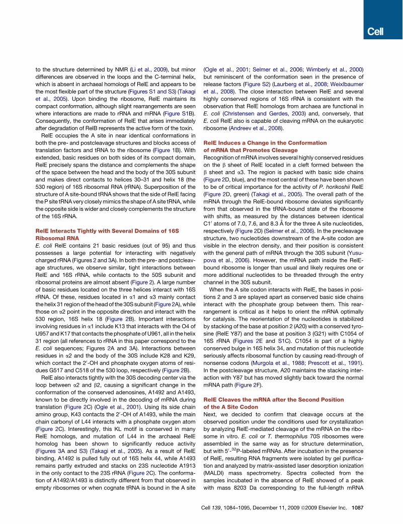
Figure 1. Overview of the RelE-Bound 70S Ribosome(A) Top view of the 70S ribosome with the 50S (blue) and 30S (wheat) subunits surrounding RelE (A site, blue), tRNAfMet (P site, green), a noncognate tRNAfMet
(E site, red), and mRNA (magenta). (A)–(C) are based on the precleavage structure.
(B) Close-up of the A and P sites of the 30S subunit viewed from the interface to the 50S. RelE (blue cartoon) spans the 16S rRNA from the head (helix 31 region,
green) to the body (helix 18, pink). The mRNA is shown in purple sticks, and the P and E site tRNAs colored as in (A).
(C) Close-up view of the A and P sites showing RelE (blue Ca trace), mRNA (purple sticks), and P site tRNA (green cartoon) alongwith the DFo-mFc electron density
of the precleavage structure contoured at 1.5 s. The mRNA sequence is indicated.
(D) The postcleavage structure showing the position of the 20-30 cyclic phosphate generated upon cleavage (20-30 cP). The map is contoured at 1.2 s.
See also Figure S1.
1086 Cell 139, 1084–1095, December 11, 2009 ª2009 Elsevier Inc.
to the structure determined by NMR (Li et al., 2009), but minordifferences are observed in the loops and the C-terminal helix,which is absent in archaeal homologs of RelE and appears to bethe most flexible part of the structure (Figures S1 and S3) (Takagiet al., 2005). Upon binding the ribosome, RelE maintains itscompact conformation, although slight rearrangements are seenwhere interactions are made to rRNA and mRNA (Figure S1B).Consequently, the conformation of RelE that arises immediatelyafter degradation of RelB represents the active form of the toxin.RelE occupies the A site in near identical conformations in
both the pre- and postcleavage structures and blocks access oftranslation factors and tRNA to the ribosome (Figure 1B). Withextended, basic residues on both sides of its compact domain,RelE precisely spans the distance and complements the shapeof the space between the head and the body of the 30S subunitand makes direct contacts to helices 30–31 and helix 18 (the530 region) of 16S ribosomal RNA (rRNA). Superposition of thestructure of A site-bound tRNA shows that the side of RelE facingtheP site tRNAvery closelymimics the shapeof A site tRNA,whilethe opposite side is wider and closely complements the structureof the 16S rRNA.
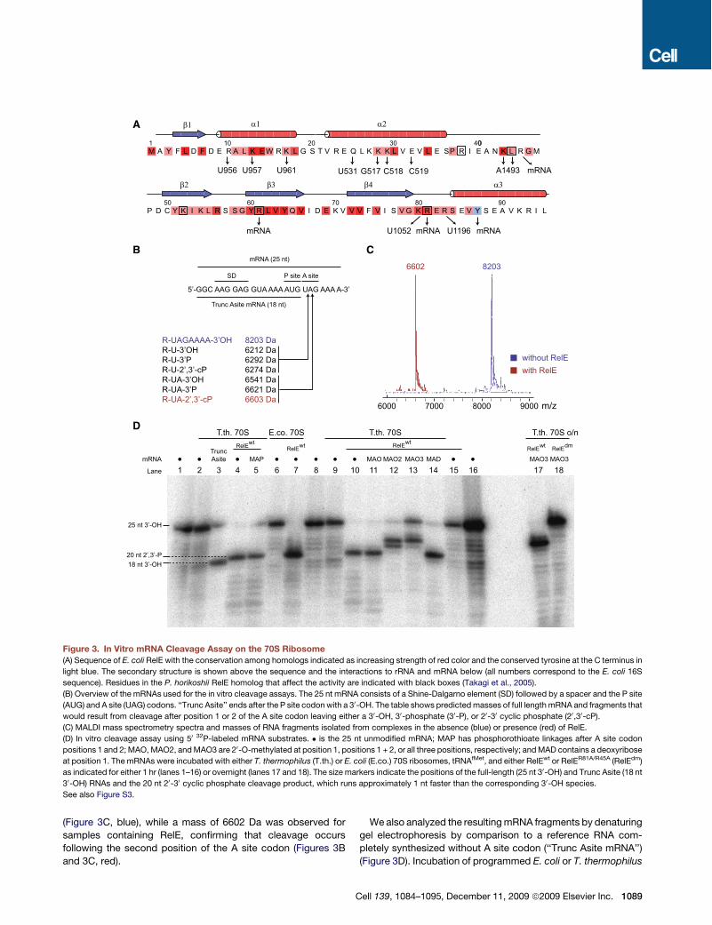
RelE Interacts Tightly with Several Domains of 16SRibosomal RNAE. coli RelE contains 21 basic residues (out of 95) and thuspossesses a large potential for interacting with negativelycharged rRNA (Figures 2 and 3A). In both the pre- and postcleav-age structures, we observe similar, tight interactions betweenRelE and 16S rRNA, while contacts to the 50S subunit andribosomal proteins are almost absent (Figure 2). A large numberof basic residues located on the three helices interact with 16SrRNA. Of these, residues located in a1 and a3 mainly contactthehelix 31 regionof theheadof the 30Ssubunit (Figure 2A),whilethose on a2 point in the opposite direction and interact with the530 region, 16S helix 18 (Figure 2B). Important interactionsinvolving residues in a1 include K13 that interacts with the O4 ofU957andK17 that contacts thephosphate ofU961, all in the helix31 region (all references to rRNA in this paper correspond to theE. coli sequences; Figures 2A and 3A). Interactions betweenresidues in a2 and the body of the 30S include K28 and K29,which contact the 20-OH and phosphate oxygen atoms of resi-dues G517 and C518 of the 530 loop, respectively (Figure 2B).RelE also interacts tightly with the 30S decoding center via the
loop between a2 and b2, causing a significant change in theconformation of the conserved adenosines, A1492 and A1493,known to be directly involved in the decoding of mRNA duringtranslation (Figure 2C) (Ogle et al., 2001). Using its side chainamino group, K43 contacts the 20-OH of A1493, while the mainchain carbonyl of L44 interacts with a phosphate oxygen atom(Figure 2C). Interestingly, this KL motif is conserved in manyRelE homologs, and mutation of L44 in the archaeal RelEhomolog has been shown to significantly reduce activity(Figures 3A and S3) (Takagi et al., 2005). As a result of RelEbinding, A1492 is pulled fully out of 16S helix 44, while A1493remains partly extruded and stacks on 23S nucleotide A1913in the only contact to the 23S rRNA (Figure 2C). The conforma-tion of A1492/A1493 is distinctly different from that observed inempty ribosomes or when cognate tRNA is bound in the A site
(Ogle et al., 2001; Selmer et al., 2006; Wimberly et al., 2000)but reminiscent of the conformation seen in the presence ofrelease factors (Figure S2) (Laurberg et al., 2008; Weixlbaumeret al., 2008). The close interaction between RelE and severalhighly conserved regions of 16S rRNA is consistent with theobservation that RelE homologs from archaea are functional inE. coli (Christensen and Gerdes, 2003) and, conversely, thatE. coli RelE also is capable of cleaving mRNA on the eukaryoticribosome (Andreev et al., 2008).
RelE Induces a Change in the Conformationof mRNA that Promotes CleavageRecognition ofmRNA involves several highly conserved residueson the b sheet of RelE located in a cleft formed between theb sheet and a3. The region is packed with basic side chains(Figure 2D, blue), and themost central of these have been shownto be of critical importance for the activity of P. horikoshii RelE(Figure 2D, green) (Takagi et al., 2005). The overall path of themRNA through the RelE-bound ribosome deviates significantlyfrom that observed in the tRNA-bound state of the ribosomewith shifts, as measured by the distances between identicalC10 atoms of 7.0, 7.6, and 8.3 A for the three A site nucleotides,respectively (Figure 2D) (Selmer et al., 2006). In the precleavagestructure, two nucleotides downstream of the A-site codon arevisible in the electron density, and their position is consistentwith the general path of mRNA through the 30S subunit (Yusu-pova et al., 2006). However, the mRNA path inside the RelE-bound ribosome is longer than usual and likely requires one ormore additional nucleotides to be threaded through the entrychannel in the 30S subunit.When the A site codon interacts with RelE, the bases in posi-
tions 2 and 3 are splayed apart as conserved basic side chainsinteract with the phosphate group between them. This rear-rangement is critical as it helps to orient the mRNA optimallyfor catalysis. The reorientation of the nucleotides is stabilizedby stacking of the base at position 2 (A20) with a conserved tyro-sine (RelE Y87) and the base at position 3 (G21) with C1054 of16S rRNA (Figures 2E and S1C). C1054 is part of a highlyconserved bulge in 16S helix 34, and mutation of this nucleotideseriously affects ribosomal function by causing read-through ofnonsense codons (Murgola et al., 1988; Prescott et al., 1991).In the postcleavage structure, A20 maintains the stacking inter-action with Y87 but has moved slightly back toward the normalmRNA path (Figure 2F).
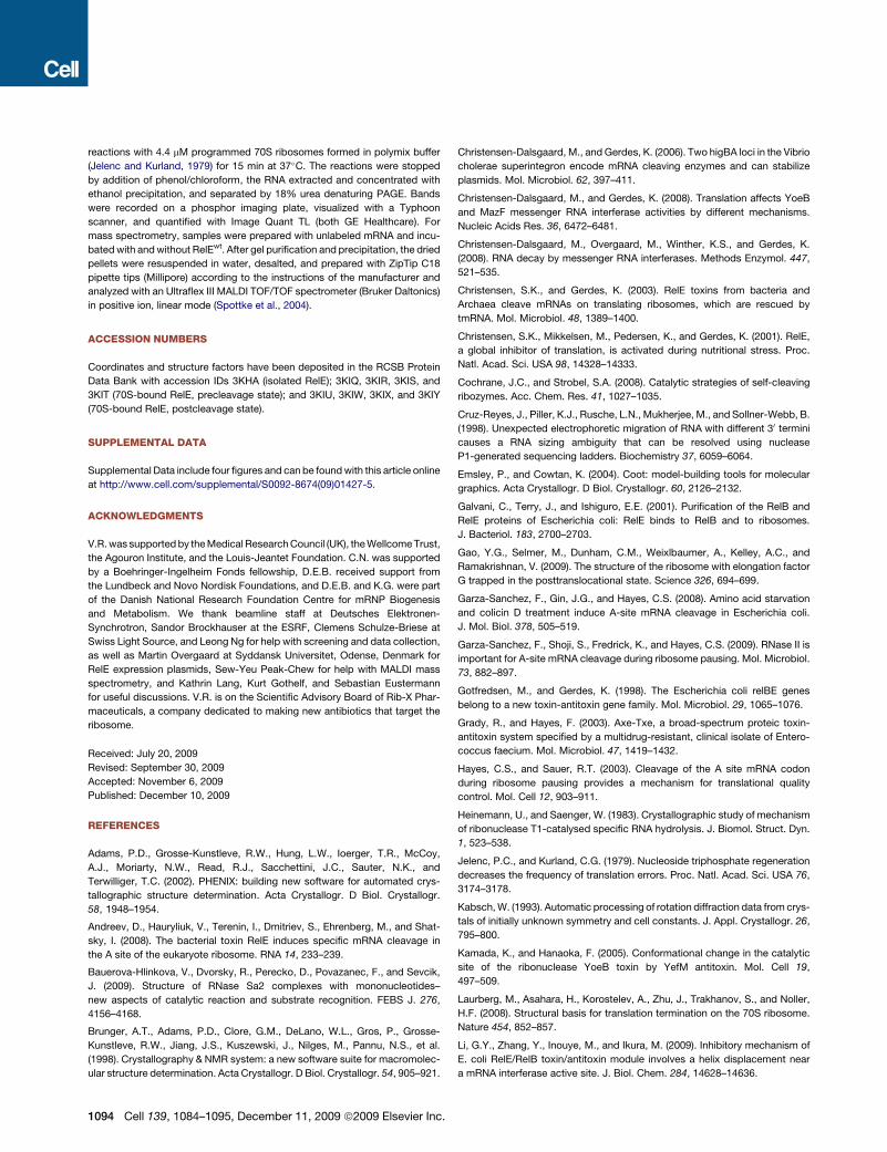
RelE Cleaves the mRNA after the Second Positionof the A Site CodonNext, we decided to confirm that cleavage occurs at theobserved position under the conditions used for crystallizationby analyzing RelE-mediated cleavage of the mRNA on the ribo-some in vitro. E. coli or T. thermophilus 70S ribosomes wereassembled in the same way as for structure determination,but with 50-32P-labeled mRNAs. After incubation in the presenceof RelE, resulting RNA fragments were isolated by gel purifica-tion and analyzed by matrix-assisted laser desorption ionization(MALDI) mass spectrometry. Spectra collected from thesamples incubated in the absence of RelE showed of a peakwith mass 8203 Da corresponding to the full-length mRNA
Cell 139, 1084–1095, December 11, 2009 ª2009 Elsevier Inc. 1087

C
K43
L44 O2’
A1493
A1492
D
K54
U19
A20
G21R61
(R81)K80
R56
K52
R83 1st
2nd
3rd
A B
G530R56
K54
K29K28
α2β2
β3
R93
R10K13
K17
α3
α1
helix 18(530-loop)
helix 31
U956U957
C518
U961
C519
E F
P siteA site
RelE RelE
P-site tRNA P-site tRNA
Y87
(R81)K54
R56
Y87
R81K54
R56
precleavage postcleavage
A site
C1054
K52
R61 K52
R61
C1054 P site
A20
G21
A20
U19U19
R45(R45)
α1
α2
α3
β2
β3
β4
G517
Figure 2. Interactions with rRNA and mRNA(A) Interactions between RelE (blue) and the head region of the 30S (helix 31, wheat). (A)–(E) are based on the precleavage structure. All rRNA references corre-
spond to the E. coli sequences.
(B) Interactions between RelE and the body region of the 30S (helix 18, wheat).
(C) Interactions between RelE (blue, semitransparent surface with interacting residues shown as sticks) and the decoding center bases A1492/A1493 (green
sticks) of 16S rRNA helix 44 (wheat). The unbound conformation of the decoding site is shown with white sticks.
(D) Overview of the contacts between RelE and mRNA. RelE is shown as a white cartoon with basic residues near the mRNA as blue sticks. Residues known to
affect the activity of P. horikoshii RelE are shown in green. The normal mRNA path is shown with white sticks and the RelE-bound mRNA path with purple sticks.
The position of the R81 side chain is inferred from the Cb position.
(E and F) Details of the interactions withmRNA in the precleavage (E) or post-cleavage (F) state showing RelE (blue) with relevant side chains as sticks, P site tRNA
(green cartoon), the 16S conserved rRNA helix 34 bulge (wheat), and the mRNA (purple sticks) with the A and P site codons labeled. C1054 is shown in red. The
positions of the side chains of R45 and R81 are inferred from Cb positions and are therefore shown in gray.
See also Figure S2.
1088 Cell 139, 1084–1095, December 11, 2009 ª2009 Elsevier Inc.
(Figure 3C, blue), while a mass of 6602 Da was observed forsamples containing RelE, confirming that cleavage occursfollowing the second position of the A site codon (Figures 3Band 3C, red).
We also analyzed the resultingmRNA fragments by denaturinggel electrophoresis by comparison to a reference RNA com-pletely synthesized without A site codon (‘‘Trunc Asite mRNA’’)(Figure 3D). Incubation of programmed E. coli or T. thermophilus
K
5’-GGC AAG GAG GUA AAA AUG UAG AAA A-3’
mRNA (25 nt)
A siteP siteSD
Trunc Asite mRNA (18 nt)
C82036602
R-UAGAAAA-3’OHR-U-3’OHR-U-3’PR-U-2’,3’-cPR-UA-3’OHR-UA-3’PR-UA-2’,3’-cP
8203 Da6212 Da6292 Da6274 Da6541 Da6621 Da6603 Da
9000800070006000
B
without RelEwith RelE
25 nt 3’-OH
18 nt 3’-OH20 nt 2’,3’-P
D
m/z
β1 α1 α2
M A Y F L D F D E R A L K E W R K L G S T V R E Q L K K K L V E L E SP R I E A N
P D C K I K L R S S G Y R L V Y Q V I D E K V V V F V I S V R E S E S A V K R I L
0
50 60 70 80 90V Y
U956 U957 U961 G517 C518 A1493
U1052 U1196
β2 β3 β4 α3
C519U531
mRNAmRNA mRNA
R
mRNA
T.th. 70S E.co. 70S T.th. 70S T.th. 70S o/n
TruncAsite MAP MAO MAO2 MAO3 MAD MAO3 MAO3
A
wtRelEwtRelE wtRelE dmRelE
1 2 3 4 5 6 7 8 9 10 11 12 13 14 15 16 17 18mRNA
Lane
wtRelE
20 30 40101G ML
KGY
V
R E
Figure 3. In Vitro mRNA Cleavage Assay on the 70S Ribosome(A) Sequence of E. coli RelE with the conservation among homologs indicated as increasing strength of red color and the conserved tyrosine at the C terminus in
light blue. The secondary structure is shown above the sequence and the interactions to rRNA and mRNA below (all numbers correspond to the E. coli 16S
sequence). Residues in the P. horikoshii RelE homolog that affect the activity are indicated with black boxes (Takagi et al., 2005).
(B) Overview of the mRNAs used for the in vitro cleavage assays. The 25 nt mRNA consists of a Shine-Dalgarno element (SD) followed by a spacer and the P site
(AUG) and A site (UAG) codons. ‘‘Trunc Asite’’ ends after the P site codon with a 30-OH. The table shows predictedmasses of full length mRNA and fragments that
would result from cleavage after position 1 or 2 of the A site codon leaving either a 30-OH, 30-phosphate (30-P), or 20-30 cyclic phosphate (20,30-cP).
(C) MALDI mass spectrometry spectra and masses of RNA fragments isolated from complexes in the absence (blue) or presence (red) of RelE.
(D) In vitro cleavage assay using 50 32P-labeled mRNA substrates. # is the 25 nt unmodified mRNA; MAP has phosphorothioate linkages after A site codon
positions 1 and 2;MAO,MAO2, andMAO3 are 20-O-methylated at position 1, positions 1 + 2, or all three positions, respectively; andMAD contains a deoxyribose
at position 1. The mRNAs were incubated with either T. thermophilus (T.th.) or E. coli (E.co.) 70S ribosomes, tRNAfMet, and either RelEwt or RelER81A/R45A (RelEdm)
as indicated for either 1 hr (lanes 1–16) or overnight (lanes 17 and 18). The size markers indicate the positions of the full-length (25 nt 30-OH) and Trunc Asite (18 nt
30-OH) RNAs and the 20 nt 20-30 cyclic phosphate cleavage product, which runs approximately 1 nt faster than the corresponding 30-OH species.
See also Figure S3.
Cell 139, 1084–1095, December 11, 2009 ª2009 Elsevier Inc. 1089

ribosomes with RelEwt consistently resulted in RNA fragmentsappearing 1 nucleotide longer than the referenceRNA (Figure 3D,lanes 3, 4, and 7), while incubation of the mRNA alone or in thepresence of either programmed 70S ribosomes or RelEwt, butnot both, did not (Figure 3D, lanes 1, 2, 6, and 15). Althoughthis could be interpreted as cleavage after position 1 of the Asite codon, it must be borne in mind that RNAs arising fromhydrolysis by internal attack of the 20-OH on the phosphatecontain an additional 30 phosphate group compared to synthe-sized RNAs and typically run between 1⁄2 and 2 nucleotides fasterthan their 30-OH counterparts (Cruz-Reyes et al., 1998). Thecombined results are therefore consistent with a model wherebycleavage occurs at position 2 of the A site codon by amechanisminvolving the 20-OH as an internal nucleophile.
mRNA Cleavage by RelE Follows a Mechanism Involving20-OH-Induced HydrolysisTo precisely define the involvement of the ribose 20-OH of mRNAin the cleavage reaction, we repeated the experiments usingRNAs modified by 20-O-methylation at A site codon position 1(MAO), positions 1 and 2 (MAO2), or all three positions (MAO3),containing a 20-deoxy ribose at position 1 (MAD), or phosphoro-thioate linkages after positions 1 and 2 (MAP) (Figure 3D). RelEcleaved both the phosphorothioate-modified RNA (lane 5) aswell as mRNAs containing a 20-O-methyl group (MAO, lane 11)or a 20-deoxyribose at position 1 of the A site codon (MAD,lane 14) to the same extent as unmodified RNA, showing thatthe 20-OH of the first nucleotide of the codon is not directlyinvolved in the reaction. In contrast, 20-O-methylation of bothpositions 1 and 2 (MAO2, lane 12) or all three codon positions(MAO3, lane 13) shifted the cleavage position downstream tothe first or second unmodified nucleotide, indicating that RelEonly cleaves at positions where the 20-OH is available. Surpris-ingly, the mutant RelER45A/R81A did cleave mRNA to someextent (data not shown), but not the mRNAs modified by 20-O-methylation at all three positions even after overnight incubation(lane 18), suggesting that the same conserved basic residues areinvolved in all the cleavage events observed. This stronglysuggests that there is only a single active site in RelE and, conse-quently, that the mRNA most likely shifts inside the ribosome toallow cleavage in other places than between A site position 2 and3. Ultimately, it is possible that even cleavage of mRNA in theribosomal E site is a result of such mRNA frame-shifting (Peder-sen et al., 2002). On the other hand, our results also demonstratethat R81, which is one of themost conserved amino acids in RelEand is known to be required for efficient inhibition of translationin vivo (Pedersen et al., 2002), is not absolutely essential forcatalysis, but greatly enhances the reaction rate.
The involvement of the 20-OH in the reaction can be confirmedfurther by close inspection of the mass spectrometry spectra(Figure 3C). After cleavage, we observe an RNA fragment witha mass of 6602 Da, more closely matching a situation wherecleavage occurs after the second position of the A site codonwith a 30-phosphate (6621 Da) than a 30-OH (6541 Da, Figure 3B).But most precisely, the measured mass matches that of a cyclic20-30 phosphate (predicted to 6603 Da), which lacks an oxygenatom compared to a 30-phosphate monoester and can only begenerated by internal attack of the 20-OH. The combined results
therefore strongly support a model in which RelE-mediatedcleavage of mRNA occurs after position 2 of the A site codonby a 20-OH-induced hydrolytic mechanism, leading to a 20-30
cyclic phosphate at the newly formed 30 end and a 50-OH. Inaddition, and unlike other structurally related nucleases suchas RNase T1, RelE does not appear to catalyze the subsequenthydrolysis of the cyclic phosphate to 30-phosphate (Heinemannand Saenger, 1983).
mRNA Cleavage Requires Both RelE and the RibosomeRelE is structurally related to a group of sequence-specific,extracellular microbial endoribonucleases that cleave polyribo-nucleotides independently of the ribosome specifically afterguanosine residues, but it lacks their conserved active site resi-dues (Heinemann and Saenger, 1983; Li et al., 2009; Takagiet al., 2005). Structural alignment of RNase T1 bound to twoguanosine molecules representing a postcleavage state (Zegerset al., 1994) with RelE bound tomRNA, however, reveals a similarposition and to some extent orientation of nucleotides 2 and 3 ofthe A site codon in the precleavage structure to the guanosineresidues in RNase T1 (Figure S4A). This observation suggeststhat RelE nevertheless shares the location of its active site withthe microbial RNases and, consequently, that it might recognizeRNA by a similar mechanism.The reaction mechanism proposed for RNase T1 involves E58
acting as a general base to activate the 20-OH of guanosine for anSN2-type nucleophilic attack on its 30 phosphate group, H92(general acid) stabilizing the leaving 50-OH, and formation ofa 20-30 cyclic phosphodiester intermediate product (Figure 4A)(Heinemann and Saenger, 1983). In RNase T1, correct orienta-tion of the substrate for inline attack is achieved by ‘‘sandwich-ing’’ of the guanine base between Y42 and Y45, similar to thestacking of Y87 of RelE with the second base of the A site codon(Figure 4). It can be noted that Y87 is shifted about 14 A awayin the RelE-RelB complex, thus effectively destroying the activesite when the antitoxin is bound. In addition, Y87 is the closestfunctional group to the 20-OH of the second A site nucleotidein the ribosome-bound structure (3.8 A), suggesting that it couldbe important for the catalytic function of RelE. In RNase T1, thedownstream nucleotide is held tightly in place by an aromaticstack formed by H92 and F100, but such aromatic residuesare absent in RelE and the nucleotide is instead stabilized bystacking with C1054 of 16S rRNA. The structurally relatedYoeB, which has retained both the active site histidine (H83)and glutamate (E46) and therefore most probably employsthe classical cleavage mechanism, has been shown to be ableto cleave RNA independently of the ribosome in vitro (Kamadaand Hanaoka, 2005). However, recent evidence suggests thatit only acts on the ribosome in vivo (Christensen-Dalsgaardand Gerdes, 2008), and comparison with RelE reveals thatYeoB also lacks the hydrophobic stack required for orientationof the nucleotide downstream of the cleavage point. It may there-fore be that it is the combination of a noncanonical active site andlack of the hydrophobic stack that together leads to the strictrequirement for the ribosome in the case of RelE. In summary,structural comparison suggests that RelE cleaves mRNAwith an active site related to that used by extracellular bacterialendonucleases like RNase T1 and, consequently, that the
1090 Cell 139, 1084–1095, December 11, 2009 ª2009 Elsevier Inc.

ribosome is not directly involved in catalysis. However, theinvolvement of 16S rRNA in orienting the mRNA correctly forthe cleavage reaction rationalizes why the ribosome is criticalfor RelE activity.
A Mechanism for the Ribosome-Dependent mRNACleavage by RelERelE contains no functional equivalents of H92 and E58 that arerequired for catalysis in RNase T1, but instead depends on a setof conserved basic residues (e.g., K52, R61, andR81) for activity,as shown for the archaeal RelE (Figure S3) (Takagi et al., 2005). Inribosome-bound RelE, most of these residues (Figure 4B,marked with *) cluster around the scissile phosphate esterbond (Figure 4B, arrow) and the stack between Y87 and thesecond base of the A site codon, but their involvement in catal-ysis has so far been unclear. However, cleavage of RNA byattack of the 20-OH is a chemically well understood mechanismemployed bymany ribozymes, and a set of general requirementscan be defined (Cochrane and Strobel, 2008). First, the 20-OHmust be activated by abstraction of the proton by a generalbase, allowing it to act as a nucleophile and the RNA to orientitself for an inline attack at the phosphate; second, the negativelycharged, trigonal bipyramidal transition state must be stabilized;and third, the leaving 50-OH group should be stabilized throughdonation of a proton by a general acid. Superposition of RelEwith RNase Sa2, another related bacterial endonuclease, revealsthat the general base in RNase Sa2, E56 (E58 in RNase T1),aligns with K52 in RelE, while the general acid, H86 (H92 inRNase T1), coincides with R81 (Bauerova-Hlinkova et al., 2009)(Figure S4B). In addition, R61 and K54 in RelE align well withtwo arginine residues critical for stabilizing the transition statein RNase Sa2, R71 and R67, and are both conserved in manyRelE homologs, suggesting that these residuesmayplay a similarrole here.To clarify the involvement of the individual residues in catal-
ysis, we constructed K52A, K54A, R61A, R81A, Y87A, andY87F mutants of RelE and measured their cleavage efficiencieson T. thermophilus 70S ribosomes in vitro (Figure 5A). Based
on our structures, we additionally designed a R81A/Y87F doublemutant that we expected to have the strongest effect. Neither thelysine (K52A, K54A) nor the Y87F mutants showed a significantdecrease in activity. However, mutation of either of the arginines(R61A, R81A) or the tyrosine (Y87A) significantly lowered thecleavage efficiency. Most remarkably, the R81A/Y87F doublemutant almost completely abolished RelE activity. Together,these data indicate that both the arginines and the stacking ofY87 with the mRNA are important for the cleavage reactionand that the loss of the Y87 h-OH group can be compensatedonly when R81 is retained.Both the structural and functional data thus indicate that Y87
and the arginines R61 and R81 are critically involved in catalysis.The lysines, K52 and K54, also have the potential to function ingeneral acid/base catalysis, but our cleavage assay suggeststhat they are less important. However, they might contributetoward stabilization of the negatively charged transition stateby analogy with RNase Sa2. The combined data thereforesuggest a model for catalysis in which the mRNA is drawn intothe active site by electrostatic forces from several basic sidechains, while Y87 and 16S rRNA C1054 orient the substratecorrectly for a nucleophilic attack of the 20-OH of the secondnucleotide in the A site on its own 30-phosphate (Figure 5B).We propose that the high local concentration of positive chargesin the active site lowers the pKa of Y87 by stabilizing the nega-tively charged, deprotonated form. Y87 is then able to activatethe 20-OH by acting as a general base, possibly stabilized byhydrogen bonding to a water molecule observed at this locationin the precleavage structure (Figures 4B, 6, and S1C). The reac-tion then proceeds toward inversion of the phosphate geometryvia a negatively charged, trigonal bipyramidal transition state,most likely stabilized by R61 and possibly K54. A central rolefor R61 in transition state stabilization is consistent with thestrong influence on RelE activity and the function of the corre-sponding R71 in RNase Sa2 (Bauerova-Hlinkova et al., 2009).Finally, R81 is in position to stabilize the leaving 50-OH groupby donating a proton during the final stage of the cleavage reac-tion and hence acting as a general acid (Figure 5B). One question
A
K52
K54
Y87
R61
(R81)
F100
H92
Y42E58
R77
R45
3’P-G105
G106
RelERNase T1
mRNA A20
mRNA G21
Y45L44
*
*
*
H+
H+
B
*
H2O
R83
Figure 4. Comparison of the Active Sites ofRelE and RNase T1(A) TheactivesiteofRNaseT1 (orangecartoon)with
critical aminoacidsshownas labeledsticks. The30-
phosphateguanosine representing the30 endof the
cleavedRNAand the guanosine representing the 50
end are shown as purple sticks. Movements of
protons during the reaction are shownwith straight
arrows (marked H+), and the nucleophilic attack of
the 20 oxyanion with a curved arrow.
(B) The corresponding region in RelE (blue
cartoon) with residues implied in catalysis as
blue sticks, and other nearby basic residues as
yellow sticks. Residues critical for the activity of
P. horikoshii RelE are marked with a * (Takagi
et al., 2005). The nucleotides at positions 2 and 3
of the A site codon are shown as purple sticks
and the scissile bond is marked with an arrow.
The water molecule near the 20-OH of position
2 (A20) is shown as a red sphere.
See also Figure S4.
Cell 139, 1084–1095, December 11, 2009 ª2009 Elsevier Inc. 1091

that this mechanism raises is why the effect of the single muta-tions Y87F and R81A is so relatively weak when the combinedeffect of their mutation effectively abolishes the activity of theenzyme. It is possible that even in the absence of the Y87 OHgroup, the high concentration of arginine and lysine residues inthe active site lowers the pKa of the 20-OH enough to alleviatethe requirement for a specific general base. Furthermore, the
side chain of Y87 is held in check by hydrogen binding to R81,so it is possible that lack of both the OH group and the R81side chain leave the tyrosine in a conformation in which it is ener-getically unfavorable to stack with the critical second base of thecodon. In summary, the structural and functional data presentedhere have allowed us to propose a detailed mechanism for RelE-mediated mRNA cleavage on the ribosome; however, a fullunderstanding of all aspects of the reaction mechanism willprobably require additional work by complementary techniques.
The Sequence Specificity of the ReactionRelE cleaves different codons with varying efficiencies, andalthough there appears to be no consensus sequence, most effi-ciently cleaved codons seem to follow the pattern Py-Pu-G,where Py is a pyrimidine and Pu a purine (Pedersen et al.,2003). In the precleavage structure, the first base of the A sitecodon is rotated back and contacts the P site tRNA (Figure 2E).This conformation would be difficult to achieve with a largepurine base and could contribute to the subtle preference forpyrimidines at the first position. Likewise, the stacking interac-tions seen at positions 2 and 3 might be more stable in the pres-ence of the larger purine bases. In the third position, the pre-cleavage structure is consistent with an interaction betweenthe guanine base and the region in RelE around E82, which couldconfer a more direct specificity toward G. However, none ofthese interactions seem essential for RelE binding, explainingwhy most codons are be cleaved with some efficiency, butthey may explain some of the observed variations in activity.
mRNA
K54OHOH
Y87
R45
R56
tRNAP site
A siteRelE
A UG
P
C1054
16S rRNA
R61R81
P
P
2
31
K52
P
P
H2O
Figure 6. Overview of the RelE Cleavage MechanismRelE (blue) occupies the A site where it pulls the mRNA (purple) into its active
site (arrows). Here, the second nucleotide (‘‘2’’) stacks with Y87 (blue), while
the third nucleotide (‘‘3’’) stacks with C1054 (orange). Relevant basic side
chains and the water molecule in the active site are shown. The red arrow indi-
cates the nucleophilic attack of the 20-O!.
A B
0%
25%
50%
75%
100%
wt K52A K54A R61A R81A Y87A Y87F R81AY87F
uncleaved
cleaved
O
O
OO
O
P-O
O
OOH
2
O
N
N
NNH
N
N
N
O
NH
NHN
Y87
NHNH
NH
R61
O
OHU19
(pos. 1)
ONH
O
N
O
OO- OP
H
N
N
NH2
OC1054
2’
3’
2
2
A20
(pos. 2)
G21
(pos. 3)
NH3+ K52
R81
NHNH
NH3
+
K54
+
2
2
NHH+
-:
:
Figure 5. A Mechanism for Ribosome-Dependent mRNA Cleavage by RelE(A) Relative inhibition of cleavage in RelEmutants shown as%uncleavedmRNA after 15min incubation at 37"C,mean value ±SEM. Below, a denaturing RNA gel
showing substrate and products for each mutant.
(B) Proposed reaction mechanism for the cleavage of mRNA by RelE with residues from RelE shown in blue, 16S C1054 in green, and the mRNA in purple. Stack-
ing of the second A site basewith Y87 and the third basewith 16S rRNA nucleotide C1054 (double arrows) first orients the RNA correctly for an inline attack. A high
local concentration of positive charge shifts the pKa of Y87 to allow it to act as a general base and abstract a proton from the 20-OH promoting its attack on the
phosphate between A site positions 2 and 3. The negatively charged bipyramidal transition state is stabilized by R61, while R81 acts as a general acid to protonate
the 50 OH leaving group, generating a 20-30 cyclic phosphate at the new 30 end.
1092 Cell 139, 1084–1095, December 11, 2009 ª2009 Elsevier Inc.

ConclusionIn this paper, we present crystal structures of the isolatedribosome-dependent endonuclease, RelE, and its complexwith the ribosome in both pre- and postcleavage states. Wepresent evidence that cleavage of mRNA on the ribosome iscarried out by RelE and not by the ribosome itself, and we areable to propose a detailed reaction mechanism for RNA cleav-age. These results have important implications for our under-standing of the roles of RelE and other ribosome-dependentendonucleases in translational regulation.The classical description of RelE as a toxin implies that it is
a nonspecific inhibitor of translation, most compatible witha role in cell growth stasis or even cell death. However, the effectof RelE induction is more likely a reduction of the global rate oftranslation during amino acid starvation, leading to increasedlevels of charged tRNA and increased fidelity of translation(Christensen et al., 2001; Pedersen et al., 2002; Sorensen,2001). This is supported by the observation that deletion of relBEand four other toxin-antitoxin loci encoding mRNA cleavingenzymes in E. coli significantly increases the global translationalerror rate, consistent with a role of RelE in quality control (K.G.,unpublished data). The enzymes have more likely evolved toallow fast adaptation of bacterial cells to changing environmentsby modulation of the global rate of translation, and the term‘‘toxin’’ is therefore misleading.Ribosome-dependent endonucleases are widespread in free-
living bacteria, and many are probably yet to be discovered. Butsequence similarity can be very low between members of thefamily, so many are often only discovered after structural studies.One such example is RegB from bacteriophage T4, which wasrecently shown to be a member of the RelE superfamily by struc-tural homology (Odaert et al., 2007). RegB participates in the lifecycle of the phage by favoring the degradation of early mRNAwith specific sequences inside the ribosome. It is therefore quitepossible that the overwhelming number of RelE-like nucleasesin bacteria act as ‘‘adaptation enzymes,’’ each appropriate for aspecific environmental situation. Sequence-specific degradationofmRNAby ribosome-associatednucleasesmight therefore allowthe control of translation as response to a range of conditions,including stress response, viral infections, and ribosome stalling.
EXPERIMENTAL PROCEDURES
Preparation of RelEA bicistronic construct encoding His-tagged RelB and untagged RelE-R81A
mutant was synthesized (GenScript) and inserted into plasmid pMG25 (Chris-
tensen-Dalsgaard et al., 2008). The RelBE complex was expressed in E. coli
BL21 DE3 (Novagen) and purified on Ni-NTA agarose (QIAGEN) equilibrated
in 50 mM Tris (pH 8), 0.3 M NaCl, 5 mM MgCl2, and 5 mM 2-mercaptoethanol
at 20"C. After wash with lysis buffer plus 15 mM imidazole, on-column dena-
turation in 50 mM Tris (pH 8), 0.3 M NaCl, 9 M urea, and 5 mM 2-mercaptoe-
thanol allowed elution of free RelE-R81A. After shock refolding by 10-fold dilu-
tion into 50 mM Tris (pH 8), 10% glycerol, and 5 mM 2-mercaptoethanol, the
protein was purified on a MonoS column (GE Healthcare) with a 0.05–1 M
NaCl gradient followed by Superdex 75/300 GL gel filtration (GE Healthcare)
in 15 mM Tris (pH 8), 0.1 M NaCl, and 5 mM BME, and concentration to
4–6 mg/ml on a spin filter (Milipore). The mutants used were generated by
site-directed mutagenesis and purified similarly. For the ribosome complexes,
the plasmid pSC2524HE was used for coexpression of His-tagged RelE and
RelB (Christensen-Dalsgaard et al., 2008). The complex was denatured in
50 mM Tris (pH 8), 0.3 M NaCl, and 6 M guanidinium chloride while bound to
Ni-NTA beads to remove RelB and renatured on column in 50 mM Tris
(pH 8) plus 0.3 M NaCl. Subsequent purification steps were similar to the
above. The RelER45A/R81A double mutant was constructed by site-directed
mutagenesis.
Structure Determination of Isolated RelESingle crystals of untagged RelE grew up to 200 mm size by sitting drop vapor
diffusion in 0.1 M Mes (pH 6.5), 0.2M (NH4)2SO4, and 30% w/v PEG
5000 monomethylether at 4"C. X-ray data to 2.5 A were collected at the
DESY X12 beamline (EMBL, Hamburg) from a single crystal flash-frozen in
the mother liquor, and the data were processed with XDS (Kabsch, 1993).
The structure was determined by molecular replacement in Phaser/PHENIX
with a poly-alanine searchmodel based on PDB 2KC9 (Li et al., 2009). Amodel
covering residues 2–95 for three monomers was built in Coot (Emsley and
Cowtan, 2004) and refined by iterative model building in PHENIX to a final
R/Rfree of 22.6% / 26.8%.(Adams et al., 2002).
Preparation of 70S Ribosomes, tRNA, and mRNAsT. thermophilus ribosomes and E. coli tRNAfMet were purified as described
previously (Selmer et al., 2006). mRNAs with the sequence 50-GGCAAGGAG
GUAAAAAUGUAGAAAA-30 were synthesized with appropriate modifications,
phosphorothioate linkages after nt 19 and 20 (MAP), 20-O-methylation at nt 19
(MAO1), 19 + 20 (MAO2), or 19 + 20 + 21 (MAO3), or a 20 deoxy ribose at nt 19
(MAD),while ‘‘TruncAsite’’ had thesequence50-GGCAAGGAGGUAAAAAUG-30.
Structure Determination of Ribosome-RelE ComplexesComplexes of RelEwt or RelER45A/R81A with the 70S ribosome, tRNAfMet, and
unmodified or MAO3 mRNA were crystallized as described previously (Selmer
et al., 2006). All complexes were formed in 5 mM HEPES (pH 7.5), 10 mM
Mg(CH3COO)2, 50 mM KCl, 10 mM NH4Cl, and 6 mM 2-mercaptoethanol. In
brief, 70S ribosomes at a final concentration of 4.4 mM were incubated with
a 2-fold excess of mRNA and a 2.5-fold excess of tRNAfMet at 55"C for
30 min. RelE was added in 5-fold excess and incubated for 10 min at 37"C
and 30 min at 20"C. Crystals were grown by sitting-drop vapor diffusion as
2.4 ml complex including 2.8 mMDeoxy Big Chap (Calbiochem) plus 2 ml reser-
voir solution containing 0.1 M Tris-HAc (pH 7.2), 0.2 M KSCN, 3%–4.5% w/v
PEG 20K, and 3%–4.5%w/v PEG 550monomethylether and left to equilibrate
at 20"C. The crystals grew within 2 weeks to a size of up to 803 1003 600 mm
and were gradually transferred into cryoprotecting solution (0.1 M Tris-HAc
[pH 7.2], 0.2 M KSCN, 10 mM NH4Cl, 10 mM MgAc, 5% PEG 20K, and 25%
PEG550MME), and frozen in liquid nitrogen for data collection at 100 K.
Data Collection, Refinement, and Model BuildingCrystals were screened at beamlines ID14-4 at the European Synchrotron
Radiation Facility (ESRF) in Grenoble, France, and X06SA PXI at the Swiss
Light Source (SLS), and data were collected from suitable crystals at six posi-
tions on four crystals for the precatalytic state and at seven positions of two
crystals for the postcleavage state at PXI (SLS) and processed with XDS
(Kabsch, 1993). An empty 70Smodel was used for initial refinement and phase
calculation (Voorhees et al., 2009) using both CNS (Brunger et al., 1998) and
Phenix (Adams et al., 2002) as described previously (Selmer et al., 2006;Weixl-
baumer et al., 2008). 3mFo-2DFc and mFo-DFc difference maps were used for
model building of initially P site tRNAfMet, noncognate E site tRNAfMet, and the
mRNA except the A site codon. The ribosomemodel wasmodified by compar-
ison with a recent, more complete model (Gao et al., 2009). Changes in the 16S
rRNA at the decoding center and near C1054 were manually fitted, and, finally,
the models of mRNA and RelE were built starting with the isolated RelE crystal
structure. The final refinement in CNS had R/Rfree of 21.9% / 24.7% and
21.5% / 24.8% for the precleavage and postcleavage structures, respectively.
Analysis of Cleaved mRNAsAll mRNAs were 50 end labeled with T4 polynucleotide kinase and ATP-g32P
and complexed formed essentially as described above but only with 3.5 mM
mRNA. Reactions contained a 5-fold excess of RelE and were incubated for
10 min at 37"C followed by 30 min at 20"C. For the mutagenesis screen,
2.2 mM of each RelE mutant (0.5-fold) was incubated in triplicate independent
Cell 139, 1084–1095, December 11, 2009 ª2009 Elsevier Inc. 1093
reactions with 4.4 mM programmed 70S ribosomes formed in polymix buffer
(Jelenc and Kurland, 1979) for 15 min at 37"C. The reactions were stopped
by addition of phenol/chloroform, the RNA extracted and concentrated with
ethanol precipitation, and separated by 18% urea denaturing PAGE. Bands
were recorded on a phosphor imaging plate, visualized with a Typhoon
scanner, and quantified with Image Quant TL (both GE Healthcare). For
mass spectrometry, samples were prepared with unlabeled mRNA and incu-
batedwith andwithout RelEwt. After gel purification and precipitation, the dried
pellets were resuspended in water, desalted, and prepared with ZipTip C18
pipette tips (Millipore) according to the instructions of the manufacturer and
analyzed with an Ultraflex III MALDI TOF/TOF spectrometer (Bruker Daltonics)
in positive ion, linear mode (Spottke et al., 2004).
ACCESSION NUMBERS
Coordinates and structure factors have been deposited in the RCSB Protein
Data Bank with accession IDs 3KHA (isolated RelE); 3KIQ, 3KIR, 3KIS, and
3KIT (70S-bound RelE, precleavage state); and 3KIU, 3KIW, 3KIX, and 3KIY
(70S-bound RelE, postcleavage state).
SUPPLEMENTAL DATA
Supplemental Data include four figures and can be foundwith this article online
at http://www.cell.com/supplemental/S0092-8674(09)01427-5.
ACKNOWLEDGMENTS
V.R.was supportedby theMedical ResearchCouncil (UK), theWellcomeTrust,
the Agouron Institute, and the Louis-Jeantet Foundation. C.N. was supported
by a Boehringer-Ingelheim Fonds fellowship, D.E.B. received support from
the Lundbeck and Novo Nordisk Foundations, and D.E.B. and K.G. were part
of the Danish National Research Foundation Centre for mRNP Biogenesis
and Metabolism. We thank beamline staff at Deutsches Elektronen-
Synchrotron, Sandor Brockhauser at the ESRF, Clemens Schulze-Briese at
Swiss Light Source, and Leong Ng for help with screening and data collection,
as well as Martin Overgaard at Syddansk Universitet, Odense, Denmark for
RelE expression plasmids, Sew-Yeu Peak-Chew for help with MALDI mass
spectrometry, and Kathrin Lang, Kurt Gothelf, and Sebastian Eustermann
for useful discussions. V.R. is on the Scientific Advisory Board of Rib-X Phar-
maceuticals, a company dedicated to making new antibiotics that target the
ribosome.
Received: July 20, 2009
Revised: September 30, 2009
Accepted: November 6, 2009
Published: December 10, 2009
REFERENCES
Adams, P.D., Grosse-Kunstleve, R.W., Hung, L.W., Ioerger, T.R., McCoy,
A.J., Moriarty, N.W., Read, R.J., Sacchettini, J.C., Sauter, N.K., and
Terwilliger, T.C. (2002). PHENIX: building new software for automated crys-
tallographic structure determination. Acta Crystallogr. D Biol. Crystallogr.
58, 1948–1954.
Andreev, D., Hauryliuk, V., Terenin, I., Dmitriev, S., Ehrenberg, M., and Shat-
sky, I. (2008). The bacterial toxin RelE induces specific mRNA cleavage in
the A site of the eukaryote ribosome. RNA 14, 233–239.
Bauerova-Hlinkova, V., Dvorsky, R., Perecko, D., Povazanec, F., and Sevcik,
J. (2009). Structure of RNase Sa2 complexes with mononucleotides–
new aspects of catalytic reaction and substrate recognition. FEBS J. 276,
4156–4168.
Brunger, A.T., Adams, P.D., Clore, G.M., DeLano, W.L., Gros, P., Grosse-
Kunstleve, R.W., Jiang, J.S., Kuszewski, J., Nilges, M., Pannu, N.S., et al.
(1998). Crystallography & NMR system: a new software suite for macromolec-
ular structure determination. Acta Crystallogr. D Biol. Crystallogr. 54, 905–921.
Christensen-Dalsgaard, M., and Gerdes, K. (2006). Two higBA loci in the Vibrio
cholerae superintegron encode mRNA cleaving enzymes and can stabilize
plasmids. Mol. Microbiol. 62, 397–411.
Christensen-Dalsgaard, M., and Gerdes, K. (2008). Translation affects YoeB
and MazF messenger RNA interferase activities by different mechanisms.
Nucleic Acids Res. 36, 6472–6481.
Christensen-Dalsgaard, M., Overgaard, M., Winther, K.S., and Gerdes, K.
(2008). RNA decay by messenger RNA interferases. Methods Enzymol. 447,
521–535.
Christensen, S.K., and Gerdes, K. (2003). RelE toxins from bacteria and
Archaea cleave mRNAs on translating ribosomes, which are rescued by
tmRNA. Mol. Microbiol. 48, 1389–1400.
Christensen, S.K., Mikkelsen, M., Pedersen, K., and Gerdes, K. (2001). RelE,
a global inhibitor of translation, is activated during nutritional stress. Proc.
Natl. Acad. Sci. USA 98, 14328–14333.
Cochrane, J.C., and Strobel, S.A. (2008). Catalytic strategies of self-cleaving
ribozymes. Acc. Chem. Res. 41, 1027–1035.
Cruz-Reyes, J., Piller, K.J., Rusche, L.N., Mukherjee, M., and Sollner-Webb, B.
(1998). Unexpected electrophoretic migration of RNA with different 30 termini
causes a RNA sizing ambiguity that can be resolved using nuclease
P1-generated sequencing ladders. Biochemistry 37, 6059–6064.
Emsley, P., and Cowtan, K. (2004). Coot: model-building tools for molecular
graphics. Acta Crystallogr. D Biol. Crystallogr. 60, 2126–2132.
Galvani, C., Terry, J., and Ishiguro, E.E. (2001). Purification of the RelB and
RelE proteins of Escherichia coli: RelE binds to RelB and to ribosomes.
J. Bacteriol. 183, 2700–2703.
Gao, Y.G., Selmer, M., Dunham, C.M., Weixlbaumer, A., Kelley, A.C., and
Ramakrishnan, V. (2009). The structure of the ribosome with elongation factor
G trapped in the posttranslocational state. Science 326, 694–699.
Garza-Sanchez, F., Gin, J.G., and Hayes, C.S. (2008). Amino acid starvation
and colicin D treatment induce A-site mRNA cleavage in Escherichia coli.
J. Mol. Biol. 378, 505–519.
Garza-Sanchez, F., Shoji, S., Fredrick, K., and Hayes, C.S. (2009). RNase II is
important for A-site mRNA cleavage during ribosome pausing. Mol. Microbiol.
73, 882–897.
Gotfredsen, M., and Gerdes, K. (1998). The Escherichia coli relBE genes
belong to a new toxin-antitoxin gene family. Mol. Microbiol. 29, 1065–1076.
Grady, R., and Hayes, F. (2003). Axe-Txe, a broad-spectrum proteic toxin-
antitoxin system specified by a multidrug-resistant, clinical isolate of Entero-
coccus faecium. Mol. Microbiol. 47, 1419–1432.
Hayes, C.S., and Sauer, R.T. (2003). Cleavage of the A site mRNA codon
during ribosome pausing provides a mechanism for translational quality
control. Mol. Cell 12, 903–911.
Heinemann, U., and Saenger, W. (1983). Crystallographic study of mechanism
of ribonuclease T1-catalysed specific RNA hydrolysis. J. Biomol. Struct. Dyn.
1, 523–538.
Jelenc, P.C., and Kurland, C.G. (1979). Nucleoside triphosphate regeneration
decreases the frequency of translation errors. Proc. Natl. Acad. Sci. USA 76,
3174–3178.
Kabsch,W. (1993). Automatic processing of rotation diffraction data from crys-
tals of initially unknown symmetry and cell constants. J. Appl. Crystallogr. 26,
795–800.
Kamada, K., and Hanaoka, F. (2005). Conformational change in the catalytic
site of the ribonuclease YoeB toxin by YefM antitoxin. Mol. Cell 19,
497–509.
Laurberg, M., Asahara, H., Korostelev, A., Zhu, J., Trakhanov, S., and Noller,
H.F. (2008). Structural basis for translation termination on the 70S ribosome.
Nature 454, 852–857.
Li, G.Y., Zhang, Y., Inouye, M., and Ikura, M. (2009). Inhibitory mechanism of
E. coli RelE/RelB toxin/antitoxin module involves a helix displacement near
a mRNA interferase active site. J. Biol. Chem. 284, 14628–14636.
1094 Cell 139, 1084–1095, December 11, 2009 ª2009 Elsevier Inc.

Murgola, E.J., Hijazi, K.A., Goringer, H.U., and Dahlberg, A.E. (1988). Mutant
16S ribosomal RNA: a codon-specific translational suppressor. Proc. Natl.
Acad. Sci. USA 85, 4162–4165.
Odaert, B., Saida, F., Aliprandi, P., Durand, S., Crechet, J.B., Guerois, R.,
Laalami, S., Uzan, M., and Bontems, F. (2007). Structural and functional
studies of RegB, a new member of a family of sequence-specific ribonucle-
ases involved in mRNA inactivation on the ribosome. J. Biol. Chem. 282,
2019–2028.
Ogle, J.M., Brodersen, D.E., Clemons, W.M., Jr., Tarry, M.J., Carter, A.P., and
Ramakrishnan, V. (2001). Recognition of cognate transfer RNA by the 30S
ribosomal subunit. Science 292, 897–902.
Overgaard, M., Borch, J., Jorgensen, M.G., and Gerdes, K. (2008). Messenger
RNA interferase RelE controls relBE transcription by conditional cooperativity.
Mol. Microbiol. 69, 841–857.
Pedersen, K., Christensen, S.K., and Gerdes, K. (2002). Rapid induction and
reversal of a bacteriostatic condition by controlled expression of toxins and
antitoxins. Mol. Microbiol. 45, 501–510.
Pedersen,K., Zavialov,A.V., Pavlov,M.Y., Elf, J.,Gerdes,K., andEhrenberg,M.
(2003). The bacterial toxin RelE displays codon-specific cleavage of mRNAs in
the ribosomal A site. Cell 112, 131–140.
Potrykus, K., andCashel, M. (2008). (p)ppGpp: still magical? Annu. Rev. Micro-
biol. 62, 35–51.
Prescott, C., Krabben, L., and Nierhaus, K. (1991). Ribosomes containing the
C1054-deletion mutation in E. coli 16S rRNA act as suppressors at all three
nonsense codons. Nucleic Acids Res. 19, 5281–5283.
Prysak, M.H., Mozdzierz, C.J., Cook, A.M., Zhu, L., Zhang, Y., Inouye, M., and
Woychik, N.A. (2009). Bacterial toxin YafQ is an endoribonuclease that asso-
ciates with the ribosome and blocks translation elongation through
sequence-specific and frame-dependent mRNA cleavage. Mol. Microbiol.
71, 1071–1087.
Schmidt, O., Schuenemann, V.J., Hand, N.J., Silhavy, T.J., Martin, J., Lupas,
A.N., and Djuranovic, S. (2007). prlF and yhaV encode a new toxin-antitoxin
system in Escherichia coli. J. Mol. Biol. 372, 894–905.
Selmer, M., Dunham, C.M., Murphy, F.V., 4th, Weixlbaumer, A., Petry, S.,
Kelley, A.C., Weir, J.R., and Ramakrishnan, V. (2006). Structure of the 70S
ribosome complexed with mRNA and tRNA. Science 313, 1935–1942.
Sorensen, M.A. (2001). Charging levels of four tRNA species in Escherichia coli
Rel(+) and Rel(-) strains during amino acid starvation: a simple model for the
effect of ppGpp on translational accuracy. J. Mol. Biol. 307, 785–798.
Spottke, B., Gross, J., Galla, H.J., and Hillenkamp, F. (2004). Reverse Sanger
sequencing of RNA by MALDI-TOF mass spectrometry after solid phase
purification. Nucleic Acids Res. 32, e97.
Sunohara, T., Jojima, K., Tagami, H., Inada, T., and Aiba, H. (2004). Ribosome
stalling during translation elongation induces cleavage of mRNA being trans-
lated in Escherichia coli. J. Biol. Chem. 279, 15368–15375.
Takagi, H., Kakuta, Y., Okada, T., Yao, M., Tanaka, I., and Kimura, M. (2005).
Crystal structure of archaeal toxin-antitoxin RelE-RelB complex with implica-
tions for toxin activity and antitoxin effects. Nat. Struct. Mol. Biol. 12,
327–331.
Voorhees, R.M.,Weixlbaumer, A., Loakes, D., Kelley, A.C., and Ramakrishnan,
V. (2009). Insights into substrate stabilization from snapshots of the peptidyl
transferase center of the intact 70S ribosome. Nat. Struct. Mol. Biol. 16,
528–533.
Weixlbaumer, A., Jin, H., Neubauer, C., Voorhees, R.M., Petry, S., Kelley, A.C.,
and Ramakrishnan, V. (2008). Insights into translational termination from the
structure of RF2 bound to the ribosome. Science 322, 953–956.
Wimberly, B.T., Brodersen, D.E., Clemons, W.M., Jr., Morgan-Warren, R.J.,
Carter, A.P., Vonrhein, C., Hartsch, T., and Ramakrishnan, V. (2000). Structure
of the 30S ribosomal subunit. Nature 407, 327–339.
Yusupova, G., Jenner, L., Rees, B., Moras, D., and Yusupov, M. (2006).
Structural basis for messenger RNA movement on the ribosome. Nature
444, 391–394.
Zegers, I., Haikal, A.F., Palmer, R., and Wyns, L. (1994). Crystal structure
of RNase T1 with 30-guanylic acid and guanosine. J. Biol. Chem. 269,
127–133.
Cell 139, 1084–1095, December 11, 2009 ª2009 Elsevier Inc. 1095


![Antisense Oligodeoxynucleotide Inhibition as an ...tary to the mRNA of a target gene would cause RNase H cleavage, inhibiting target gene mRNA transcription [7] or forming a complex](https://img.dokumen.tips/doc/110x75/5ed1e93df7ad4a0e2b5015e2/antisense-oligodeoxynucleotide-inhibition-as-an-tary-to-the-mrna-of-a-target.jpg)

![Journal of Falkenhagen et al, J Antivir Antiretrovir 213 ... · CCR5 gene via Zinc finger nucleases [4], cleavage of CCR5 mRNA by multimeric ribozymes [5], inhibition of CCR5 mRNA](https://img.dokumen.tips/doc/110x75/5fd3f8f670db7b30b42beea9/journal-of-falkenhagen-et-al-j-antivir-antiretrovir-213-ccr5-gene-via-zinc.jpg)