Embed Size (px)
Citation preview
The structural basis for infection of root hairs of Trifolium repens by Rhizobium
DALE A. CALLAHAM AND JOHN G. TORREY' Department of Botany, University of Massachusetts, Amherst, MA, U . S . A . 01003
Received November 12, 1980
CALLAHAM, D.A., and J. G. TORREY. 1981. The structural basis for infection of root hairs of Trifolium repens by Rhizobium. Can. J. Bot. 59: 1647-1664.
Root hair infection of Trifoliurn repens L. by Rhizobium trifolii was investigated with regard to the structural basis of infection thread origin. Most infected root hairs were shown to have in common an enclosed region at- the site of thread origin formed by specialized root hair growth or contacts. Electron micrographs of diverse infection sites showed in every case a degradation of the root hair wall at the site of thread origin within the enclosure. The thread wall is a new layer formed by apposition of material by the host cytoplasm near the penetrated wall and surrounding the break as encapsulation of the invading rhizobia. It is suggested that rhizobial enzymes provide for degradative penetration of the root hair cell wall and that localized concentrated activity of hydrolytic enzymes as well as protection from cell lysis is favored by physical constraints provided by the deformed root hair enclosures.
Introduction tion of the site of active root hair tip growth by bacterial ~h~ initiation of nodules on the roots of plants of the action resulted in an invagination of the hair wall back
family Leguminosae by soil bacteria of the genus into the hair, forming the tubular wall structure of the Rhizobium involves a complex sequence of events infection thread. which despite extensive study is only poorly under- Subsequently Fahraeus and Ljunggren (1959) report- stood. early as 1887 ward observed in roots of vicia ed that inoculation with infective bacteria resulted in an faba that nodulation by Rhizobium commenced with the increase of constitutively produced polygalacturonase infection of root hairs through the formation of a by the root itself. These authors interpreted their results bacterial thread which grew into the root cortex, thus as compatible with the invagination mechanism pro- initiating the nodule. In a careful study blccoy (1932) posed by Phtman (1956). Ljunggren (1969) attempted demonstrated that shortly after inoculation of seedling to~onfronttheobvious problem presented by the lack of
roots with nodulating bacteria, the root hairs showed ability of a loosely attached rhizobial colony to generate strong deformation, typically a curling or branching an inward force greater than the pressure potential of the response, and it was among these root hairs that root hair cell which would be required to initiate infections were found, M ~ c ~ ~ observed that the bacte- invagination at a locally softened region of cell wall. ria were found after infection in a thread of tubular form Ljunggren expressed the belief that rhizobia enclosed by which was continuous with the root hair wall. ~h~ thread the induced deformation derived an advantage from the
originated with no apparent structural disruption of the enclosed "pocket." The combination of rhizobial multi- hair wall and appeared to have the same chemical plication, bacterial slime secretion, and wall softening composition as the hair cell wall. The rhizobia within the induced within the pocket allowed expansion of the infection thread wall were surrounded by a "thread colon^, ensheathed by the extending wall, with forma- matrixv which is probably a rhizobial exopo~ysacc~a- tion of an infection thread. Further thread growth was ride. The rhizobia within the infection thread are extra- thought to involve the redirection of active root hair "tip cellular. In a search for hydrolytic enzyme activity growtK' to the tip Of the thread. McCoy (1932) could obtain no evidence for growth of efforts to repeat the pectinase assay of Fahraeus Rhizobium on pectin or cellulosic polysaccharides in and LJunggren (1959) using whole seedlings have failed culture. (cf. reviews by Dart 1974, 1975, 1977). Using more
The lack of evidence of a chemical or physical sensitive techniques to detect polysaccharide degrada- penetration of the hair cell wall led Nutman (1956) to tion activity, Hubbell et a l . (1978) reported ~ e c t o l ~ t i c propose that the bacteria penetrated by a process similar activity by Rhizobium colonies in culture. Subsequent to intussusception, intercalating themselves into the root (Martinez-Mo1ina et a l . 1979) showed hair cell wall polymers and initiating the thread as they degrading enzymes can also be detected from ~ h i z o b i u m contacted the plasmalemma. He proposed that a redirec- cultures, thus reestablishing the possibility of poll'-
saccharide-hydrolyzing enzymes of rhizobial rather than host origin.
on ail in^ address: Cabot Foundation, Harvard University, Direct observations of the events of infection have Petersham, MA, U.S.A. 01366. been sparse. Ultrastructural observations are limited to
0008-402618 1/09 1647- 18$0 1 .OO/O 01981 National Research Council of CanadaIConseil national de recherches du Canada
Can
. J. B
ot. D
ownl
oade
d fr
om w
ww
.nrc
rese
arch
pres
s.co
m b
y H
AR
VA
RD
UN
IVE
RSI
TY
HE
RB
AR
IA o
n 08
/30/
11Fo
r pe
rson
al u
se o
nly.
1648 CAN. J. BOT. VOL. 59. 1981
three studies, all interpreting the infection process in support of the hypothesis of invagination. Studies by Sahlman and Fahraeus (1963) and Higashi (1966) preceded the availability of techniques suitable for adequate fixation and ultrastructural examination. In their electron microscopic study Napoli and Hubbell (1975) properly interpreted the overall structure of sections of curled root hairs. Their conclusions in favor of the invagination mechanism were based on illustra- tions which showed that the root hair cell wall was continuous into the infection thread wall and that n o discontinuities were found in the hair wall. Some of their illustrations, however, suggested a degradation of the original hair cell wall at the point of transition and showed an inner layer of the hair cell wall which continued into the thread wall. The occurrence of such a cell wall change would be in conflict with their interpre- tation of the lack of a physical penetration of the hair cell wall and would considerably alter the currently accepted view of the invagination mechanism of root hair infection.
The questions raised regarding the feasibility of the invagination mechanism, the uncertain interpretation of the limited structural evidence, and recent new evidence of polysaccharide-hydrolyzing activity of Rhizobium taken together provided the impetus for the reexamina- tion of the structural basis of root hair infection. The results of these studies which provide an alternative mechanism to the invagination hypothesis are presented here.
Materials and methods Slide cultures
Seeds of Trifolium repens L. were surface sterilized for 20 min in full-strength commercial bleach (5.25% sodium
hypochlorite), rinsed three times in sterile distilled water, and left to soak in a small volume of water overnight. Following a brief water rinse in the morning, seeds were planted on water-agar plates, inverted, and incubated at 20°C with a 16-h photoperiod allowing the roots to germinate into the moist atmosphere. Slide cultures were observed at 1, 5, 12, 24, 48, and 72 h after inoculation. Stages of Rhizobiutn-clover root hair association, curling, and infection were photographed with a Reichert Zetopan microscope using X25 and X40 phase contrast objectives. At 48 h after sterilization the seedlings had roots 5-10 mm in length and were transferred to modified Fahraeus slide cultures (Fahraeus 1957) in which the macro- nutrient salts of the Fahraeus slide culture medium were supplemented with microelements (in milligrams per litre) H3BO3, 0.015; ZnS04.7H20, 0.015; MnS04.H20, 0.045; Na2Mo04.2H10, 0.0025; CuS04.5Hz0, 0.0004) and M Fe(II1) NaEDTA. Several loopfuls of a 4-day subculture of Rhizobium trifolii strain 162 E7 (courtesy of Dr. J . C. Burton, Nitragin Co.) were mixed with 50mL of the agar medium cooled to 30°C or in liquid medium, and 5 drops of medium, three seedlings, and a cover slip were then placed on a slide. These inoculated assemblies were incubated in 150 mL Fleak- ers containing 40 mL of liquid Fahraeus slide medium and covered by a 6-cm Petri dish cover (Fig. 1).
Electron microscopy At 48 h after inoculation, specimens for electron micro-
scopic observation were fixed 2-4 h in 2.5% glutaraldehyde in 0.025 M sodium phosphate buffer at pH 6.8, postfixed in 1% buffered Os04 for 2 h, and dehydrated through 50, 75, and 100% 2-methoxyethanol, 5050 2-methoxyethanol-ethanol, 100% ethanol, 5050 ethanol-propylene oxide, and three changes of 100% propylene oxide, all at 30-min intervals. Specimens were infiltrated in a very gradually increasing concentration of Epon-Araldite (Mollenhauer 1964) in pro- pylene oxide and transferred to fresh resin for 16 h vacuum infiltration before embedding between release-coated glass slides (Hepler 1976).
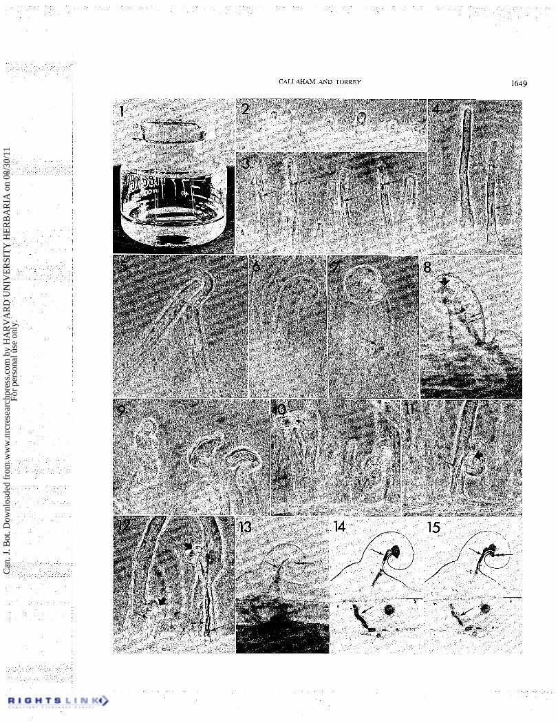
FIGS. 1-15. Root hair development in seedling roots of T. repens. Fig. 1. Fahraeus slide culture apparatus utilizing 150-mL Fleaker, 40-mL Fahraeus slide culture medium, and slide with three seedlings. x$. Fig. 2. Early stage of root hair development. A thick cap of cytoplasm is present at the hair tip and the nucleus is within the hair base. Phase contrast. x450. Fig. 3. Intermedi- ate stage of root hair elongation. A thickcytoplasmic cap is present at the hair tip and the nucleus (arrows) lies proximal to the tip. Phase contrast. X 330. Fig. 4. Full elongate root hairs a short time after inoculation. Rhizobia attached to the root hair surface are seen as small dark dots. The nucleus (arrow) is present in the basal one-third of the hair at this stage. Seedling grown in liquid slide culture. Phase contrast,. X350. Fig. 5. Deformation growth in uninoculated root hairs due to tactile stimuli. Phase contrast. X500. Fig. 6. Early stage of root hair tip curling response 12 h after inoculation in agar slide culture. No rhizobia attached to the root hair surface are visible. x700. Fig. 7. Late stage in the tip curling process. A region of wall has been enclosed within the curl. The nucleus is present proximal to the curled tip (arrow). No infection thread is present. Phase contrast. ~ 7 0 0 . Fig. 8. Tip curled root hair which has been infected from within the tip curl (large arrow). Two infection threads arise from the infection site (small arrows). Phase contrast. X750. Fig. 9. Branching, swelling, and twisting of root hairs induced by inoculation with rhizobia. Phase contrast. X500. Fig. 10. Various growth deformities induced by inoculation include root hair enlargement and initiation of multiple branches. Phase contrast. X500. Fig. 11. Root hair infection arising at the enclosed side of a root hair basal branch (large arrow) from which one infection thread (small arrow) penetrates to the hair base. Phase contrast. X500. Fig. 12. Root hair infections arising from deformed lateral branches of root hairs (large arrows). One infection thread issues from the infection site of the root hair on the left (small arrow) while two infection threads developed from the infection site of the root hair on the right (small arrows). Phase contrast. x 500. Figs. 13- 15. Micrographs of an infected tip-curled root hair from embedded slide culture (Fig. 13) and thickepoxy sections (Figs. 14, 15). Figure 13 illustrates the phase contrast appearance of the tip-curled root hairs with an infection thread (single arrow) originating from the inside of the curl (double-headed arrow). Phase contrast. x750. Figures 14 and 15 illustrate the appearance of an infected tip-curled root hair in section. Infection threads (arrows) arise at the infection site (double-headed arrow) and penetrate into the outer cortex. Bright field. X750.
Can
. J. B
ot. D
ownl
oade
d fr
om w
ww
.nrc
rese
arch
pres
s.co
m b
y H
AR
VA
RD
UN
IVE
RSI
TY
HE
RB
AR
IA o
n 08
/30/
11Fo
r pe
rson
al u
se o
nly.
CALLAHAM AND TORREY 1649
Can
. J. B
ot. D
ownl
oade
d fr
om w
ww
.nrc
rese
arch
pres
s.co
m b
y H
AR
VA
RD
UN
IVE
RSI
TY
HE
RB
AR
IA o
n 08
/30/
11Fo
r pe
rson
al u
se o
nly.
1650 CAN. J. BOT. VOL. 59, 1981
Polymerized specimens from which one slide had been removed were studied under a x250 phase contrast objective and root hairs in different stages of infection were photo- graphed. The distance from the resin surface to the root hair was measured and recorded from the calibrated fine focus of the Reichert Zetopan microscope. The specimens were cut out and glued to Beemcapsule blanks and sectioned to the measured depth of the infected root hair on a Reichert OMU2 ultrami- crotome. Ribbons of sections were cut with a diamond knife on the Reichert OMU2 ultramicrotome and mounted onto lightly carbon-coated Formvar films on 1 X 2 mm oval-hole copper grids. Sections were stained lOmin in 2% aqueous uranyl acetate and 5 min in lead citrate (Venable and Coggeshall 1965). Sections were observed and photographed on a Zeiss EM9A electron microscope.
Observations The initiation of root hair infections: light microscopic
observations Root hairs are initiated immediately behind the root
apex and in the early stages of elongation possess dense cytoplasm at the tip of the hair (Fig. 2). The root hair cell nucleus is located at the base of the cell at this stage. Intermediate stages in the elongation of rapidly develop- ing root hairs also show accumulation of cytoplasm at the root hair tip with the nucleus located in the distal third of the hair (Fig. 3). When elongation of the hair has ceased, dense cytoplasm is no longer evident at the tip of the hair and the nucleus lies in thin, peripheral cyto- plasm in the basal third of the root hair (Fig. 4). Uninoculated root hairs typically show straight growth but some contact-induced deformations occur where root hairs touch the cover slip, slide, or each other (Fig. 5).
The infection process is initiated shortly after the application of inoculum of Rhizobium to the clover seedlings growing in sterile conditions. Interaction between rhizobia and the seedling root is influenced markedly by the cultural conditions. Many more root hair infections were formed in seedlings grown in liquid medium than in agar medium. In agar cultures, the
motile bacteria were observed to swim freely within the agar but were not observed to accumulate at the root hair surfaces. Seedlings inoculated in liquid cultures pro- duced a more uniform response to the inoculum, showing attachment of bacteria to root hairs, sloughed root cap cells, and debris. Even so, sometimes only one seedling of three in a slide assembly would show extensive root hair deformation with the other roots showing only localized response or no response at all. The causes of this variability were not known.
Beginning about 5 h after inoculation of the liquid cultures, the root hairs begin to show tip curling (Figs. 6, 7 ,8 ,9 , 13) or branching (Figs. 10, 11, 12). The root hair curling arises from the continual displacement of the growth center of the root hair tip to the outside of the curl until curvatures of up to 360' or more may occur. Root hairs in active stages of elongation at inoculation generally develop tip deformities, either curling or enlargement followed by new sites of tip growth (Fig. 10).
Branches may develop at any point along the root but their form bears a distinct relationship to the growth phase of the root hair. Older root hairs which have ceased to elongate most commonly show basal branching of the hair. The formation of basal branches usually occurs near where the nucleus is found at that stage (Figs. 11, 12) and involves quite a different process from deforma- tion of tip growth. Basal branching necessitates the removal of existing root hair cell wall comprising aligned cellulosic microfibrillar structure before the development of a new site for tip growth can occur (Callaham 1979). Thus, by tip curling and branching, root hair deformation leading to rhizobial infections can be found at many locations on the seedling root surface simultaneously.
By 16-24 h the early stages of root hair deformation have developed in white clover, forming fully curled or branched root hairs, but no infection threads are yet evident. Active cytoplasmic streaming is present in all root hairs and dense accumulations of cytoplasm usually
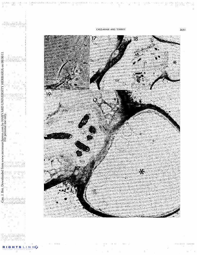
FIGS. 16- 19. Micrographs of an early root hair infection. Fig. 16. Phase contrast micrograph of a root hair infected from the axil of a lateral branch. The lateral branch (asterisk) arises on the back edge of the root hair and grows in a direction coming out of the plane of the photograph and enclosing a region between the branch and original hair axis. An internal proliferation of rhizobia (rc) is evident just adjacent to the branch and two infection threads (it, arrows) penetrate to the root hair base. Phase contrast. x750. Fig. 17. Electron micrograph of early section of series through the branch axil infection site seen in Fig. 16. The lateral branch (br) is seen as completely separate from the root hair main body in this plane. Space between the branch and hair main axis shows the outermost extension of the rhizobial capsule (c). Alteration of the staining characteristics of the major axis wall is evident adjacent and to the left of the rhizobial capsule. X6300. Fig. 18. Electron micrograph of section from late in the series shows the lateral branch (*) connection to the main root hair axis in a plane cut near the back of the root hair seen in Fig. 16. The disorganized rhizobial colony is encapsulated by irregular deposits of wall material. x3000. Fig. 19. Electron micrograph from the same series cut at the level showing the lateral branch junction with the root hair main axis within the enclosure. The plant cell wall appears degraded along the junction in contact with the enclosed rhizobia and at the lower edge (dhw) where the thread matrix (tm) is evident external to the plant cell walls within the enclosed space. Fibrillar thread encapsulation (tw) encloses the rhizobia (rh). Lateral branch is marked by an asterisk. x 13 500.
Can
. J. B
ot. D
ownl
oade
d fr
om w
ww
.nrc
rese
arch
pres
s.co
m b
y H
AR
VA
RD
UN
IVE
RSI
TY
HE
RB
AR
IA o
n 08
/30/
11Fo
r pe
rson
al u
se o
nly.
C A L L A H A M A N D TORREY 1651
Can
. J. B
ot. D
ownl
oade
d fr
om w
ww
.nrc
rese
arch
pres
s.co
m b
y H
AR
VA
RD
UN
IVE
RSI
TY
HE
RB
AR
IA o
n 08
/30/
11Fo
r pe
rson
al u
se o
nly.
1652 CAN. I. BOT. VOL. 59. 1981
surrounding the nucleus are seen in the curled tip or branch axil where infections will later be found. No large accumulations of attached rhizobia are observed on these root hairs at this time.
By 32-40 h after inoculation infection threads are observed in some of the root hairs and the number of observable infection threads increases with time. Few of the numerous sites of tip curling or branching finally give rise to infection threads. These sites of incipient infection could not be distinguished by phase contrast microscopy during the 18-h interval between deforma- tion and evident thread formation. Only when the thread tip and its cytoplasmic sheath within the hair cell have advanced from the initial site is a strongly refractile zone evident (Figs. 11, 12) due to the long optical path through the double wall and the accumulated cytoplasm. Dart ( 1974) and others before him described the refrac- tile spot as the first sign of the occurrence of infection. Specimens which have been embedded in epoxy resin for electron microscopy appear dark in this region (Figs. 8, 13, 16, 32) due to closer matching of the indices of refraction of the wall and the embedding medium. Under our experimental conditions, both tip-curled and branched root hairs gave rise to infection threads in
u
approximately equal proportions. Infection threads in tip-curled root hairs originated from the area enclosed by the folded or curled root hair tip (Figs. 8, 13). In branched root hairs infection threads developed from the axil of the branch (Figs. 1 1 , 12, 16).
By 40-48 h after inoculation new threads are still being initiated and the earlier formed infection threads have progressed to the base of the root hairs. The advancing tips of the infection threads are indistinct within a mass of root hair cell cytoplasm which is actively streaming. It is not uncommon to find several infection threads arising within the infection site from the proliferated refractile material (Figs. 8, 12) and these threads may run separately or anastamose on their path to the root hair base.
Ultrastructural observations The examination of diversely infected root hairs
reveals that, except for the very rare instance of infection in a slightly curved root hair (Fig. 32), all infections originate from sites in root hairs which have in common an enfolding by appression of portions of the root hair cell wall upon itself. Three major variations of this process were observed (i) in the axils of branched root hairs, (ii) in root hairs simply folded upon themselves, and (iii) in root hairs with marked curling. Each of these cases together with a case of infection at a slight tip curvature is described below. Representative specimens were selected and studied by serial thin sections in the electron microscope. Roots were chosen .for analysis which showed curvature parallel to the plane of section-
ing and which presented the initiation region of the thread in an orientation which could be unambiguously interpreted. Light micrographs of the embedded root hairs were made prior to sectioning, to aid in the interpretation of the electron micrographs.
Infection in the axil of a root hair branch The infection from the axil of a lateral branch
photographed in the living condition is shown by the phase contrast micrograph of Fig. 16. The branch tip was growing toward the observer. The plane of subsequent sectioning was approximately the same as the plane of focus of the root hair of Fig. 16. In this specimen, two infection threads (arrows) can be seen which originated from the proliferation of rhizobia and wall material just adjacent to the lateral branch. The infection occurred from within the fold enclosure of this specimen by penetration of the root hair wall at the side of the original axis (Figs. 19, 20). The penetration involved degradation of root hair wall from a region near the back of the fold (Fig. 19) and extending toward the opening of the fold to a site where the lateral branch is seen in section as separate from the main hair axis (Figs. 17, 20). A portion of the rhizobial capsular material from the enclosed cells extends to the opening of the branch axil (Fig. 17).
The penetrated region of the hair wall is characterized by a loose, fibrillar appearance of the edges of the hair wall (Figs. 19, 20) which protrudes into thread matrix. This wall layer is more densely stained than the new inner wall layer and thread wall. The facing wall of the lateral branch also appears more fibrillar and lightly stained only within the enclosed area (Figs. 19, 20), although these walls retain their overall structural integrity.
A large amount of material is deposited presumably by the host in response to the rhizobial attack on the hair wall and this material is evident as a more lightly stained layer to the inside of the original hair wall (Figs. 19,20). The thread wall, which is the intracellular encapsulation of the penetrating rhizobia, is the continuation of this new wall layer beyond the terminus of the original hair wall as a defined layer of fibrillar appearance approxi- mately 0.5 p m thick which encloses the rhizobia and their capsule, the thread matrix.
Internally, near the site of infection there is a proliferation of bacteria, thread matrix, and the thread wall before the more nearly tubular infection threads are developed (Figs. 18, 19). This proliferation appears to be evidence of an initial imbalance in the symbiotic growth processes. Also present in this site is an irregular deposition of large blobs of material of slightly lighter staining reaction than the thread wall itself and often overlaying the more defined thread wall (Figs. 18, 19, 20).
Can
. J. B
ot. D
ownl
oade
d fr
om w
ww
.nrc
rese
arch
pres
s.co
m b
y H
AR
VA
RD
UN
IVE
RSI
TY
HE
RB
AR
IA o
n 08
/30/
11Fo
r pe
rson
al u
se o
nly.
CALLAHAM AND TORREY
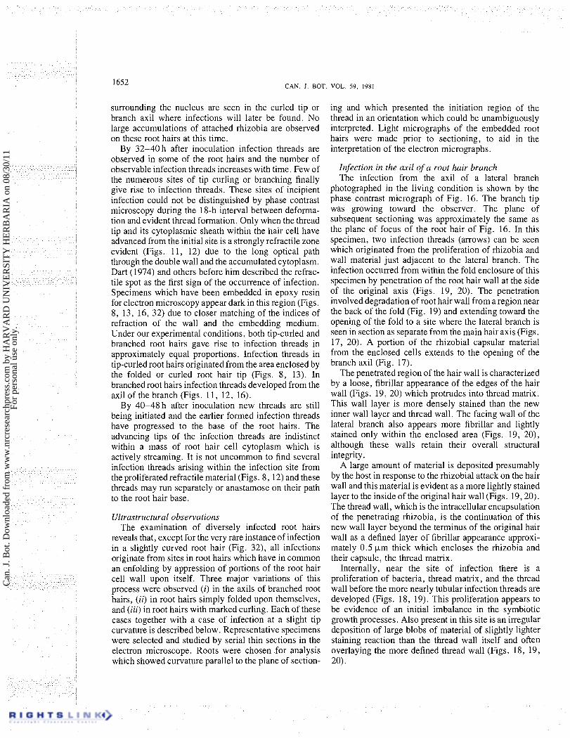
FIG. 20. Electron micrograph from the series shown in Figs. 16- 19, showing section median to the root hair wall penetration. Lateral branch at bottom is separated from the main hair axis in this plane and rhizobia (rh) and thread matrix (tm) are evident inside the root hair and protruding into enclosed space. Protruding edge of the penetrated root hair wall is marked by small arrows and the inner layer of wall material which forms the thread wall (tw) is marked in that region by double-headed arrows. x 13 500.
Infection of a folded root hair tip Figures 21-26 illustrate the infection of a root hair
that has occurred from within a root hair tip folded 180" back upon itself. A section of this root hair above the plane of thread origin shows the appearance of the wall layers which are pressed together within the fold (Fig. 21). The line of wall contact seen here has been interpreted as the "invagination" (Sahlman and Fahraeus 1963; Higashi 1966), although a three-dimensional reconstruction shows this to be a continuous surface of the enclosed root hair wall, not a tubular structure. In the series of sections, the line of wall contact in each section inscribes a plane of wall area enclosed by the tip deformation. The infection thread in this root hair originates from this folded region.
Further sections of this series show the proximal edge of an enclosed space within the innermost region of the fold (Fig. 23) as the plane of sectioning begins to graze
the infection thread. This enclosed space enlarges in further sections until the region is seen to be occupied by a large globular proliferation of rhizobia and thread matrix (Fig. 24). Figure 24 is nearly median to the infection site and shows the two-layered folded wall at the upper left opening into the enclosed colony. The innermost part of the folded wall appears more lightly stained and loosely fibrillar suggesting that degradation had occurred at the tip of the fold. In Figs. 23-26 the original hair wall has been largely or entirely degraded in contact with the thread matrix. Patches with similar staining characteristics appear at sites external to the thread wall on the circumference of the rhizobial colony (Fig. 24). These wall fragments were displaced as degraded remnants of the original root by expansion of the rhizobial colony.
As illustrated in the preceding branched root hair infection site, a new layer of the wall is present to the
Can
. J. B
ot. D
ownl
oade
d fr
om w
ww
.nrc
rese
arch
pres
s.co
m b
y H
AR
VA
RD
UN
IVE
RSI
TY
HE
RB
AR
IA o
n 08
/30/
11Fo
r pe
rson
al u
se o
nly.
1654 CAN. J. BOT. VOL. 59. 1981
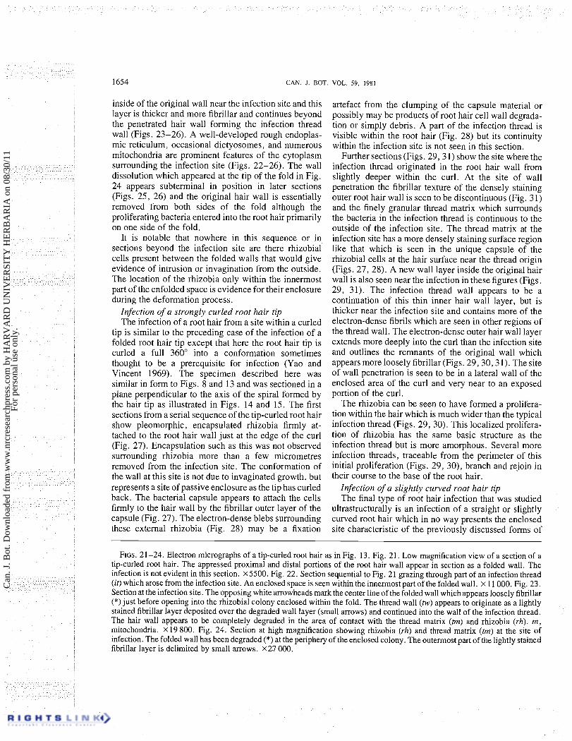
inside of the original wall near the infection site and this layer is thicker and more fibrillar and continues beyond the penetrated hair wall forming the infection thread wall (Figs. 23-26). A well-developed rough endoplas- mic reticulum, occasional dictyosomes, and numerous mitochondria are prominent features of the cytoplasm surrounding the infection site (Figs. 22-26). The wall dissolution which appeared at the tip of the fold in Fig. 24 appears subterminal in position in later sections (Figs. 25, 26) and the original hair wall is essentially removed from both sides of the fold although the proliferating bacteria entered into the root hair primarily on one side of the fold.
It is notable that nowhere in this sequence or in sections beyond the infection site are there rhizobial cells present between the folded walls that would give evidence of intrusion or invagination from the outside. The location of the rhizobia only within the innermost part of the enfolded space is evidence for their enclosure during the deformation process.
Infection of a strongly curled root hair tip The infection of a root hair from a site within a curled
tip is similar to the preceding case of the infection of a folded root hair tip except that here the root hair tip is curled a full 360" into a conformation sometimes thought to be a prerequisite for infection (Yao and Vincent 1969). The specimen described here was similar in form to Figs. 8 and 13 and was sectioned in a plane perpendicular to the axis of the spiral formed by the hair tip as illustrated in Figs. 14 and 15. The first sections from a serial sequence of the tip-curled root hair show pleomorphic, encapsulated rhizobia firmly at- tached to the root hair wall just at the edge of the curl (Fig. 27). Encapsulation such as this was not observed surrounding rhizobia more than a few micrometres removed from the infection site. The conformation of the wall at this site is not due to invaginated growth, but represents a site of passive enclosure as the tip has curled back. The bacterial capsule appears to attach the cells firmly to the hair wall by the fibrillar outer layer of the capsule (Fig. 27). The electron-dense blebs surrounding these external rhizobia (Fig. 28) may be a fixation
artefact from the clumping of the capsule material or possibly may be products of root hair cell wall degrada- tion or simply debris. A part of the infection thread is visible within the root hair (Fig. 28) but its continuity within the infection site is not seen in this section.
Further sections (Figs. 29 ,3 1) show the site where the infection thread originated in the root hair wall from slightly deeper within the curl. At the site of wall penetration the fibrillar texture of the densely staining outer root hair wall is seen to be discontinuous (Fig. 31) and the finely granular thread matrix which surrounds the bacteria in the infection thread is continuous to the outside of the infection site. The thread matrix at the infection site has a more densely staining surface region like that which is seen in the unique capsule of the rhizobial cells at the hair surface near the thread origin (Figs. 27, 28). A new wall layer inside the original hair wall is also seen near the infection in these figures (Figs. 29, 31). The infection thread wall appears to be a continuation of this thin inner hair wall layer, but is thicker near the infection site and contains more of the electron-dense fibrils which are seen in other regions of the thread wall. The electron-dense outer hair wall layer extends more deeply into the curl than the infection site and outlines the remnants of the original wall which appears more loosely fibrillar (Figs. 29 ,30 ,3 1). The site of wall penetration is seen to be in a lateral wall of the enclosed area of the curl and very near to an exposed portion of the curl.
The rhizobia can be seen to have formed a prolifera- tion within the hair which is much wider than the typical infection thread (Figs. 29, 30). This localized prolifera- tion of rhizobia has the same basic structure as the infection thread but is more amorphous. Several more infection threads, traceable from the perimeter of this initial proliferation (Figs. 29, 30), branch and rejoin in their course to the base of the root hair.
Infection of a slightly curved root hair tip The final type of root hair infection that was studied
ultrastructurally is an infection of a straight or slightly curved root hair which in no way presents the enclosed site characteristic of the previously discussed forms of
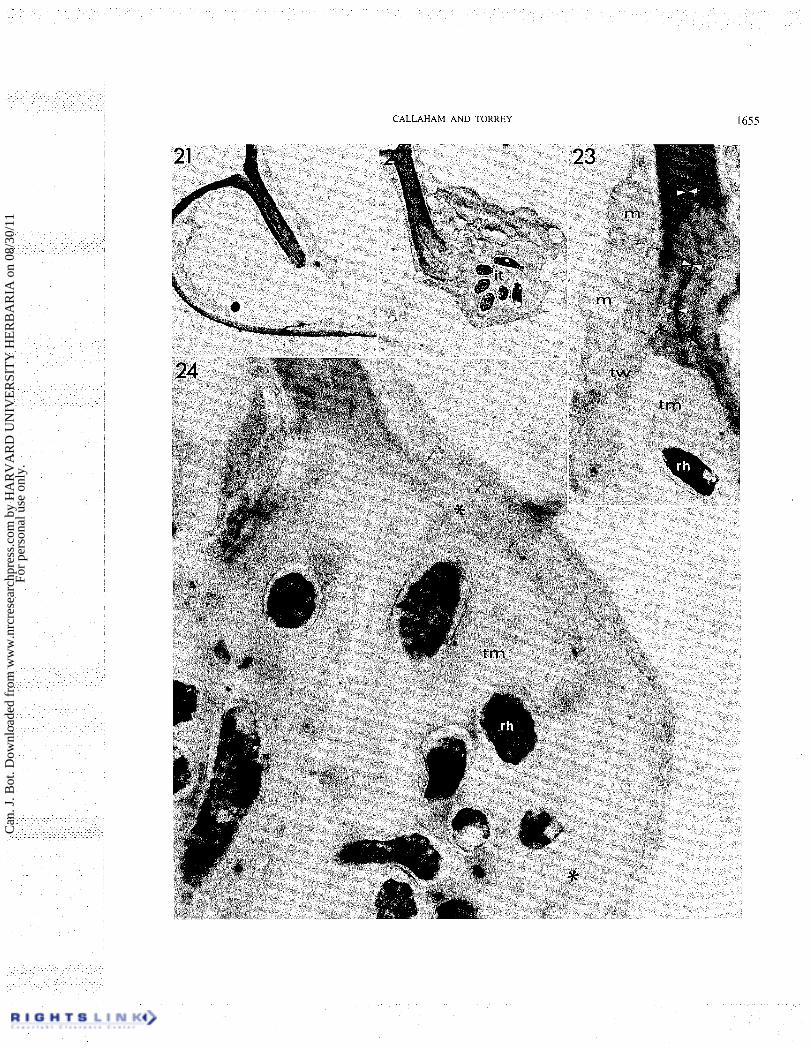
FIGS. 2 1-24. Electron micrographs of a tip-curled root hair as in Fig. 13. Fig. 2 1. Low magnification view of a section of a tip-curled root hair. The appressed proximal and distal portions of the root hair wall appear in section as a folded wall. The infection is not evident in this section. X5500. Fig. 22. Section sequential to Fig. 21 grazing through part of an infection thread (it) which arose from the infection site. An enclosed space is seen within the innermost part of the folded wall. x 11 000. Fig. 23. Section at the infection site. The opposing white arrowheads mark the center line of the folded wall which appears loosely fibrillar (*)just before opening into the rhizobial colony enclosed within the fold. The thread wall (tw) appears to originate as a lightly stained fibrillar layer deposited over the degraded wall layer (small arrows) and continued into the wall of the infection thread. The hair wall appears to be completely degraded in the area of contact with the thread matrix (tm) and rhizobia (rh). m, mitochondria. X 19 800. Fig. 24. Section at high magnification showing rhizobia (rh) and thread matrix (tm) at the site of infection. The folded wall has been degraded (*) at the periphery of the enclosed colony. 'The outermost part of the lightly stained fibrillar layer is delimited by small arrows. X U 000.
Can
. J. B
ot. D
ownl
oade
d fr
om w
ww
.nrc
rese
arch
pres
s.co
m b
y H
AR
VA
RD
UN
IVE
RSI
TY
HE
RB
AR
IA o
n 08
/30/
11Fo
r pe
rson
al u
se o
nly.
CALLAHAM AND TORREY 1655
Can
. J. B
ot. D
ownl
oade
d fr
om w
ww
.nrc
rese
arch
pres
s.co
m b
y H
AR
VA
RD
UN
IVE
RSI
TY
HE
RB
AR
IA o
n 08
/30/
11Fo
r pe
rson
al u
se o
nly.
CAN J. BOT. VOL. 59, 1981
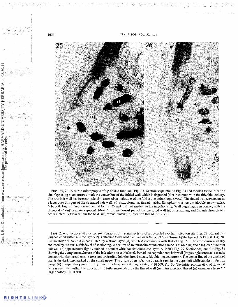
FIGS. 25, 26. Electron micrographs of tip-folded root hair. Fig. 25. Section sequential to Fig. 24 and median to the infection site. Opposing black arrows mark the center line of the folded wall which is degraded (dw) in contact with the rhizobial colony. The root hair wall has been completely removed on both sides of the fold at one point (large arrow). The thread wall (tw) occurs as a layer over this part of the degraded hair wall. rh, rhizobium; tm, thread matrix. ~ n d o ~ l a s m i c reticulum (double arrowheads). x 16 000. Fig. 26. Section sequential to Fig. 25 and just past median to the infection site. Wall degradation in contact with the rhizobial colony is again apparent. More of the innermost part of the enclosed wall (ft) is remaining and the infection clearly occurs laterally from within the fold. tm, thread matrix; it, infection thread. x 12 300.
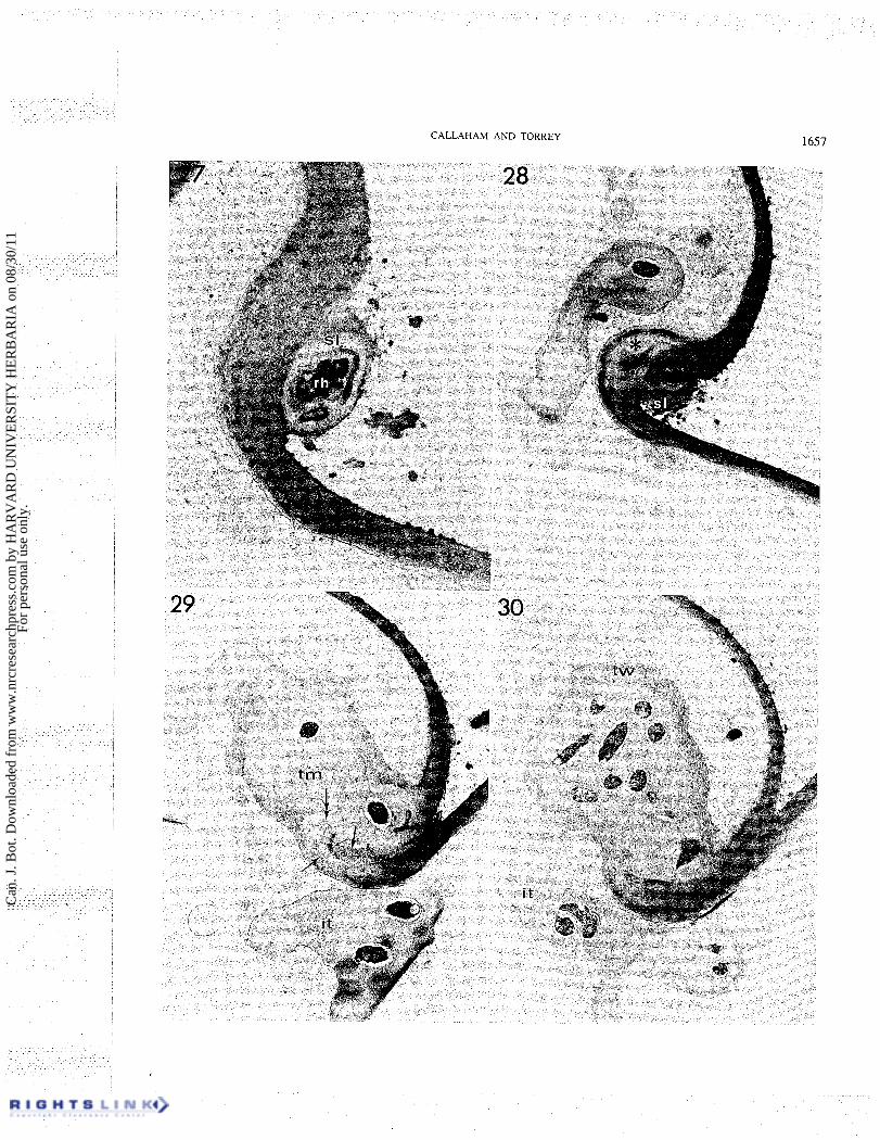
FIGS. 27-30. Sequential electron micrographs from serial sections of a tip-curled root hair infection site. Fig. 27. Rhizobium (rh) enclosed within a slime layer (sl) is attached to the root hair wall near the point of enclosure by the tip curl. x 17 000. Fig. 28. Extracellular rhizobium encapsulated by a slime layer (sf) which is continuous with that of Fig. 27. The rhizobium is nearly enclosed by the curl at this level of sectioning. A section of an intracellular infection thread is visible (it) and a region of the root hair wall (*) appears more lightly stained in contact with the rhizobial slime layer. X 10 500. Fig. 29. Section sequential to Fig. 3 1 showing the complete enclosure of the infection site at this level. Part of the degraded root hair wall (large single arrows) is seen in contact with the thread matrix (tm) and protruding into the thread matrix (double-headed arrow). The center line of the enclosed wall is the dark line marked by the small arrow. The origin of an infection thread is seen in the upper left while another infection thread (it) of separate origin from the infectionsite appears at lower center. X 10 500. Fig. 30. Theinitial proliferationof rhizobial cells is seen just within the infection site fully surrounded by the thread wall (tw). An infection thread (it) originates from the larger colony. X 10 500.
Can
. J. B
ot. D
ownl
oade
d fr
om w
ww
.nrc
rese
arch
pres
s.co
m b
y H
AR
VA
RD
UN
IVE
RSI
TY
HE
RB
AR
IA o
n 08
/30/
11Fo
r pe
rson
al u
se o
nly.
CALLAHAM AND TORREY 1657
Can
. J. B
ot. D
ownl
oade
d fr
om w
ww
.nrc
rese
arch
pres
s.co
m b
y H
AR
VA
RD
UN
IVE
RSI
TY
HE
RB
AR
IA o
n 08
/30/
11Fo
r pe
rson
al u
se o
nly.
1658 CAN. J. BOT. VOL. 59, 1981
infected root hairs. This type of root hair infection is only rarely observed or reported (Napoli and Hubbell 1975) and the specimen illustrated (Figs. 32-37) was the only root hair infection of this type encountered during the course of this work.
The phase contrast image of this root hair after embedment (Fig. 32) shows the origin of the infection thread from the inside of a very slight curve of the root hair at a site where the wall appears protruding. A microcolony of rhizobial cells is present externally surrounding this infection site with the densest portion of the colony spatially separated from the protruding wall. A single infection thread has traversed the length of the root hair from the point of infection to the root hair base. Electron micrographs of this cell are shown in Figs. 33-37.
Figure 33 is a low magnification electron micrograph of the root hair in cross section, approximately median to the site of infection, illustrating the general structure of this region. The rhizobial colony surrounds the protruding wall region from which the infection thread originates. The rhizobial cells are generally not asso- ciated with the hair wall since most of the colony is at least several micrometres removed (Figs. 33, 35). A loose, globular layer of densely stained material is present a t the wall surface in those sections.
The structure of the wall at the infection site presents evidence of a direct penetration of the hair wall. At the proximal edge of the infection site the original densely staining hair wall can be seen to terminate abruptly in contact with the thread matrix (Figs. 34, 35). The distal edge of the root hair wall is seen in Fig. 35 as distinctly protruding beyond the line of the thread matrix. Figures 34 and 35 show the thread matrix extending slightly beyond the line of the hair wall on the proximal edge.
The thread wall is here again seen to arise from an inner layer of the hair wall progressively thickened toward the site of penetration and giving rise to the
thread encapsulation where it extends beyond the break. 'The infection site in this specimen shows no evidence of the initial disorganization of wall deposition and rhizo- bial proliferation which was evident in the previously described specimens.
The lack of rhizobia closely associated with the infection site is very noticeable. One encapsulated rhizobial cell was observed attached to the surface in the series of sections (Fig. 34), although several conspicu- ous capsules without rhizobia were evident in later sections (Figs. 36, 37). As in Figs. 27 and 28 of the tip-curled hair, this particularly evident capsule has only been observed surrounding rhizobia attached to the hair wall within a few micrometres of the infection site. The capsule appears firmly associated with the hair wall (Figs. 34, 35, 36, 37) which appears degraded at this area of contact (Figs. 34, 35). The rhizobial cell and capsule remnants show a lateral attachment to the root hair wall.
This exposed infection site lacks the physical con- straint provided by a curled tip or branch and there is a correlated structural deformation of this region where the hair wall has been attacked. The greatest part of the protrusion is due to the hair wall on the distal edge of the infection site being forced outward (Figs. 33, 34, 35). Rhizobia are attached to the region of the wall which also appears degraded, thinner, and structurally weak- ened. The hair wall on the proximal edge of the infection site is not degraded, thinner, or forced outward. The distal section of hair wall which is forced outward would almost close the gap if it were moved back into alignment with the proximal edge but a space, about the size of one rhizobial cell, would remain. It appears then that the wall may have been degraded completely by a rhizobial cell attached across the region by its capsule and this covering may have prevented cell lysis when the wall was breached and the hair cell pressure potential forced the distal edge of the infection site outward.
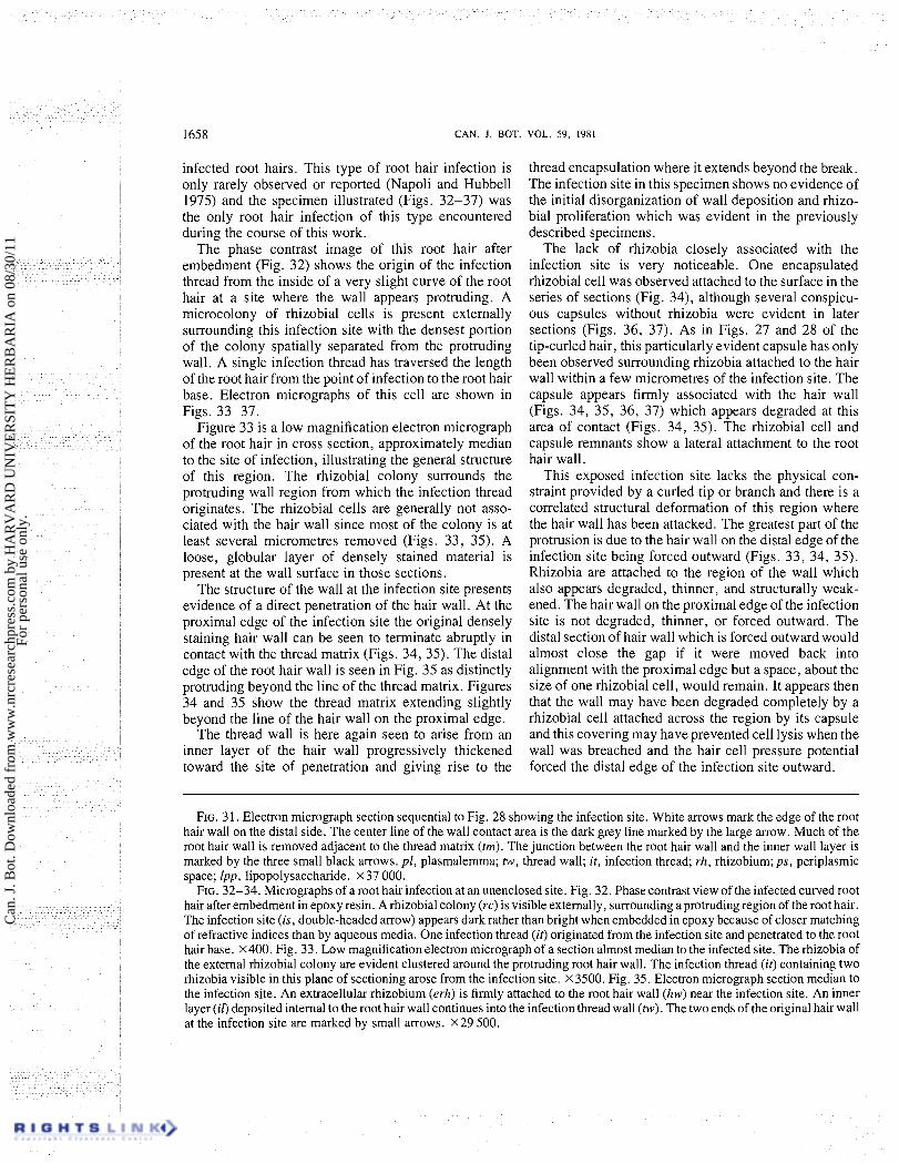
FIG. 31. Electron micrograph section sequential to Fig. 28 showing the infection site. White arrows mark the edge of the root hair wall on the distal side. The center line of the wall contact area is the dark grey line marked by the large arrow. Much of the root hair wall is removed adjacent to the thread matrix ( tm) . The junction between the root hair wall and the inner wall layer is marked by the three small black arrows. pl, plasmalemma; tw, thread wall; it, infection thread; rh, rhizobium; ps, periplasmic space; lpp, lipopolysaccharide. X 37 000.
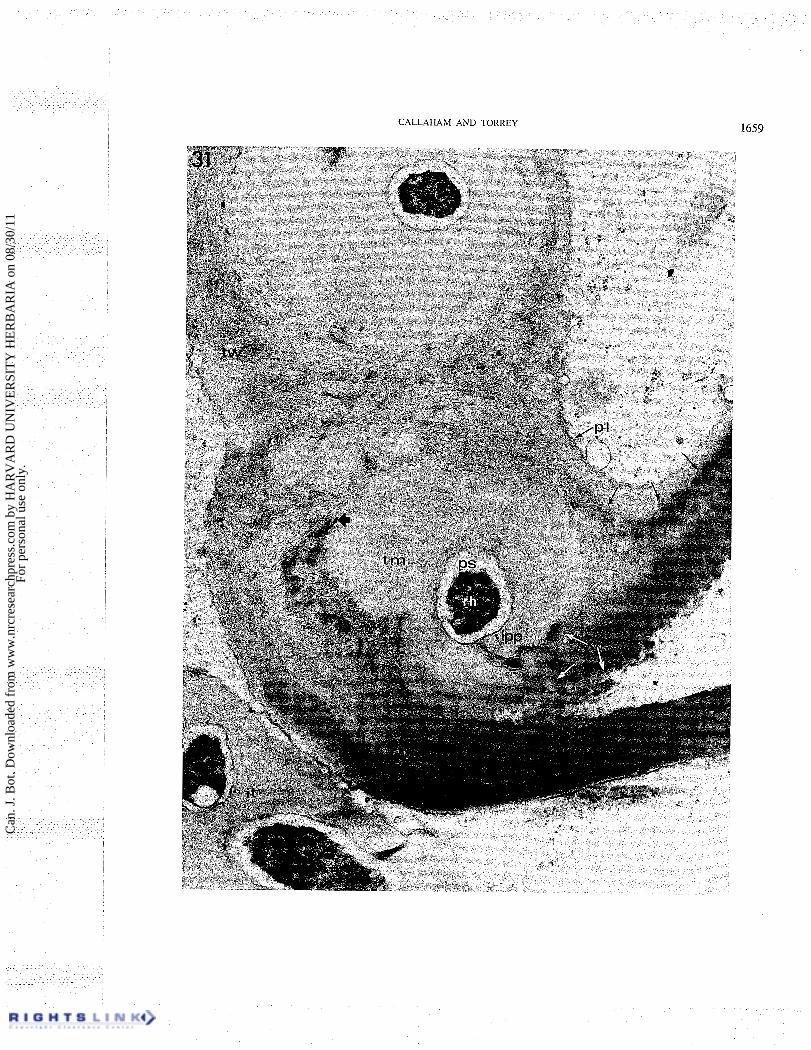
FIG. 32-34. Micrographs of a root hair infection at an unenclosed site. Fig. 32. Phase contrast view of the infected curved root hair after embedment in epoxy resin. A rhizobial colony (rc) is visible externally, surrounding a protruding region of the root hair. The infection site ( i s , double-headed arrow) appears dark rather than bright when embedded in epoxy because of closer matching of refractive indices than by aqueous media. One infection thread (i t) originated from the infection site and penetrated to the root hair base. x400. Fig. 33. Low magnification electron micrograph of a section almost median to the infected site. The rhizobia of the external rhizobial colony are evident clustered around the protruding root hair wall. The infection thread (i t) containing two rhizobia visible in this plane of sectioning arose from the infection site. X 3500. Fig. 35. Electron micrograph section median to the infection site. An extracellular rhizobium (erh) is firmly attached to the root hair wall (hw) near the infection site. An inner layer (i l) deposited internal to the root hair wall continues into the infection thread wall ( tw) . The two ends of the original hair wall at the infection site are marked by small arrows. X29 500.
Can
. J. B
ot. D
ownl
oade
d fr
om w
ww
.nrc
rese
arch
pres
s.co
m b
y H
AR
VA
RD
UN
IVE
RSI
TY
HE
RB
AR
IA o
n 08
/30/
11Fo
r pe
rson
al u
se o
nly.
CALLAHAM AND TORREY 1659
Can
. J. B
ot. D
ownl
oade
d fr
om w
ww
.nrc
rese
arch
pres
s.co
m b
y H
AR
VA
RD
UN
IVE
RSI
TY
HE
RB
AR
IA o
n 08
/30/
11Fo
r pe
rson
al u
se o
nly.
CAN. J. BOT. VOL. 59. 1981
Can
. J. B
ot. D
ownl
oade
d fr
om w
ww
.nrc
rese
arch
pres
s.co
m b
y H
AR
VA
RD
UN
IVE
RSI
TY
HE
RB
AR
IA o
n 08
/30/
11Fo
r pe
rson
al u
se o
nly.
CALLAHAM AND TORREY 1661
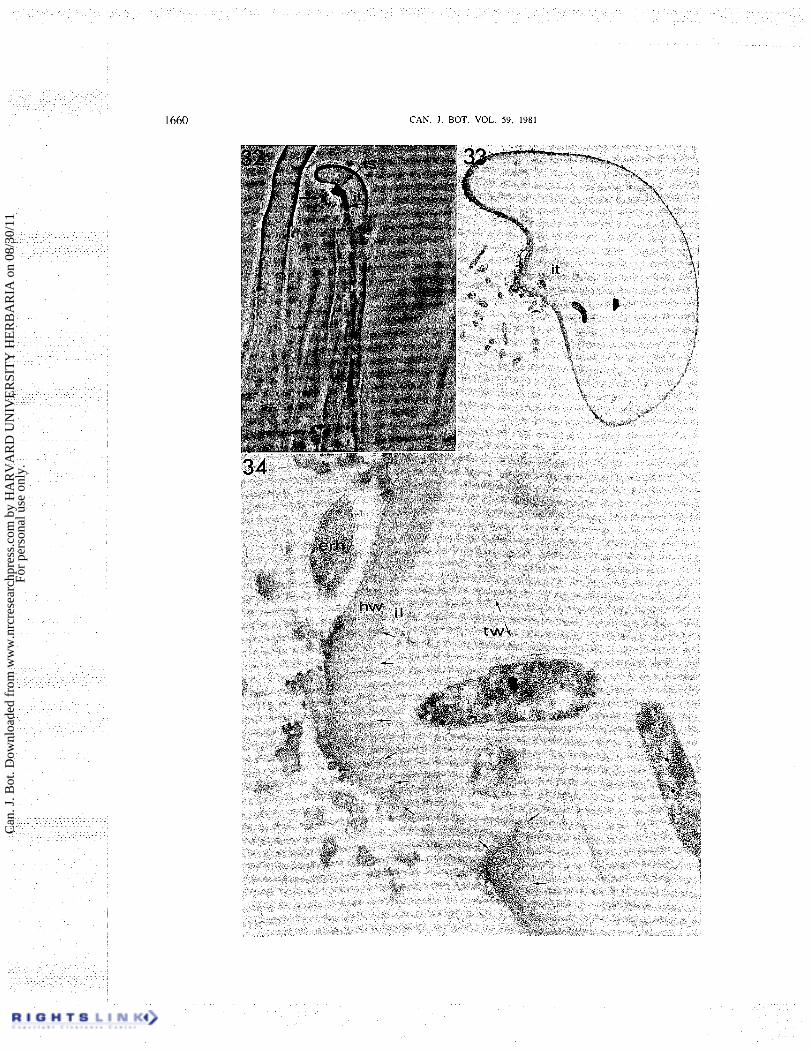
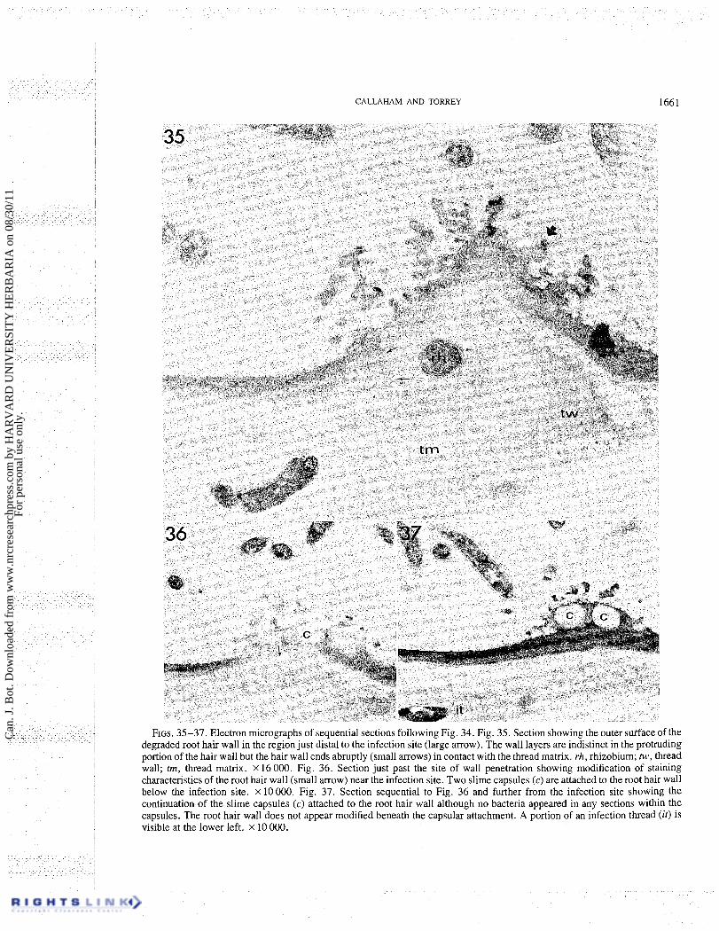
FIGS. 35-37. Electron micrographs of sequential sections following Fig. 34. Fig. 35. Section showing the outer sutface of the degraded root hair wall in the region just distal to the infection site (large arrow). The wall layers are indistinct in the protruding portion of the hair wall but the hair wall ends abruptly (small arrows) in contact with the thread matrix. rh, rhizobium; tw, thread wall; trn, thread matrix. x 16 000. Fig. 36. Section just past the site of wall penetration showing modification of staining characteristics of the root hair wall (small arrow) near the infection site. Two slime capsules (c) are attached to the root hair wall below the infection site. X 10 000. Fig. 37. Section sequential to Fig. 36 and further from the infection site showing the continuation of the slime capsules (c) attached to the root hair wall although no bacteria appeared in any sections within the capsules. The root hair wall does not appear modified beneath the capsular attachment. A portion of an infection thread (it) is visible at the lower left. X 10 000.
Can
. J. B
ot. D
ownl
oade
d fr
om w
ww
.nrc
rese
arch
pres
s.co
m b
y H
AR
VA
RD
UN
IVE
RSI
TY
HE
RB
AR
IA o
n 08
/30/
11Fo
r pe
rson
al u
se o
nly.
1662 CAN. J. BOT. VOL. 59 . 1981
Discussion The results of this study indicate that rhizobia directly
penetrate the existing root hair wall by a process involving the alteration and degradation of cell wall polysaccharides at a very localized site. The attack on the hair wall is probably enzymatic, emanating from the attached or enclosed bacterial cells. Thus the hair wall is completely degraded at the site of contact with the enclosed colony while adjacent wall areas appear modified but structurally intact.
The apparent continuity of the root hair wall with the infection thread wall has been previously interpreted as evidence of the operation of an invagination mechanism (Nutman 1956; Sahlman and Fahraeus 1963; Higashi 1966; Napoli and Hubbell 1975), but this apparent continuity of the wall is due to a new wall layer, presumably deposited by the host cell internal to the existing wall and continuing beyond the penetration point to form the intracellular encapsulation of the invading rhizobia or the thread wall. The thread wall is a new layer which is distinctly internal to and therefore ontogenetically more recent than the original hair wall. Both the evidence that the wall is actively degraded and the evidence that such threads are established at sites which are not active in elongation distinguish these observations from the invagination process proposed by Nutman (1956). In Figs. 24-31 for example, penetra- tion has occurred into the side of the enclosed region bounded by the original fully mature hair wall and not the thin wall of the induced branch.
Kumarasinghe and Nutman (1977) reported the pres- ence of callose at the site of infection thread initiation based on aniline blue induced fluorescence at the site. The materials deposited at the infection site and in conjunction with the formation of infection threads appear fibrillar by our electron microscopic observations and this is unlike the textureless electron-transparent ap- pearance of callose prepared by similar techniques (Echlin and Godwin 1968). Smith and McCully (1978) have found complications with the positive identifica- tion of callose using aniline blue due to autofluorescence and nonspecific staining of cellulose. Certain controls (Smith and McCully 1978) which effectively block autofluorescence and ensure specific staining were not used in the study of Kumarasinghe and Nutman (1977). In other studies using aniline blue staining of whole mounts of infected root hairs and thin sections in which the epoxy resin was removed and stained (Callaham 1979) it has been shown that the material deposited at the infection was not callose.
The existence of a mechanism of direct penetration of the root hair wall does not conflict with evidence that only one rhizobial strain is usually present within a nodule (Jones and Russell 1971; Hughes and Vincent 1942; MacGregor and Alexander 1972). Although a
hole is formed in the wall, entry into the root hair is restricted by both the small size of the hole and access to the hole, the branch or curled tip serving the latter function. The occurrence of multiple infections within a close region of the root (Dart 1974) could place different rhizobial strains at the inner cortex where both could contribute to the nodule bacterial population accounting for the occasional finding of two rhizobial types within one nodule (Linderman et a1. 1974).
The role of the curling and branching responses which precede the infection is now confirmed as a facilitation of the rhizobial wall penetration and not an absolute requirement. The occasional infection of straight root hairs (Napoli and Hubbell 1975; these observations) and at sites of contact between straight root hairs (Ljunggren 1969; Callaham 1979) illustrates less common alterna- tives. The high coincidence of infections at these strongly curled or branched sites has been previously noted and a function of the enclosure in restricting osmotic forces and biochemical interactions has been postulated (Ljunggren 1969; Napoli and Hubbell 1975). Although branch deformations and particularly induced basal branching are different in nature from tip curl deformations (Callaham 1979), one still is justified to consider, contrary to the view of Yao and Vincent (1969), that branching may be a sign of events leading to infection. In natural conditions root hair contact with soil particles may also play a role in facilitation of infection.
It seems appropriate now to reconsider the concept of thread origin. Evidence presented in this work suggests that the existing root hair wall is removed in contact with enclosed rhizobia; under conditions of enclosure or restriction, as observed, this results in the formation of the infection thread. A wall layer is observed on the inner side of the original hair wall which continues into the infection thread wall at this site. The origin and dynamics of thread wall formation are keys to a further understanding of the infection process. If the host cell is stimulated to form this layer in advance of the complete penetration of the original hair wall, then the infection thread is at all times sheathed or enclosed by this cell wall layer which must be continually degraded or stretched to allow the elongation of the thread. If thread wall deposition does not commence until the original wall is fully penetrated, the deposition of wall material could follow the stretching of plasmalemma at the point of contact with the expanding rhizobial colony and the dynamics of rhizobial proliferation and wall deposition could allow for the growth of an infection which is "open" at the end, not being covered by wall polysaccharides. The enclosed thread growth mecha- nism, as proposed above, is very much like the "in- vagination" mechanism proposed by Ljunggren (1969) and Napoli and Hubbell (1975) except that the initial
Can
. J. B
ot. D
ownl
oade
d fr
om w
ww
.nrc
rese
arch
pres
s.co
m b
y H
AR
VA
RD
UN
IVE
RSI
TY
HE
RB
AR
IA o
n 08
/30/
11Fo
r pe
rson
al u
se o
nly.
CALLAHAM AND TORREY 1663
phase of root hair wall penetration and each subsequent cell wall crossing would involve the observed degrada- tion prior to reestablishment of the thread.
The reality of the open-tip thread growth mechanism is not well established but is equally likely o n the basis of our present knowledge. This type of thread growth would have the property of a more intimate cell surface contact between the legume plasmalemma and the rhizobial cells a t the thread tip. Even so, the distinction between these possible types of infection threads is subtle and is more a matter of degree than kind. LTltrastructural observations of actively growing tips of infection threads should help to clarify this process.
Understanding the infection process involves a de- tailed knowledge of all the steps, biochemical and structural, whereby rhizobial cells in the soil approach the root hair surface, establish contact, enter the root hair, stimulate the formation of the infection thread, proliferate as the thread develops along the root hair cell, and then invade root cortical cells which respond by nuclear DNA replication, cell multiplication, and the rest of the events leading to nodule formation. A s has been pointed out by Martinez-Molina et al. (1979), the specificity of host cell - Rhizobium interaction may be controlled not only by the cell recognition events, involving surface cell recognition factors, but also perhaps by the specificity of the bacterial enzymes involved in cell wall degradation and the chemical nature of the wall polysaccharides which must be hydrolyzed for infection to occur. Some of the implica- tions of these considerations have been reviewed re- cently by Dazzo and Hubbell (1981). Clear evidence that cell wall penetration is an integral part of the pro- cess of root hair infection by Rhizobium should help t o interpret the other events in the process more accurately.
Acknowledgments This research was supported in part by Research grant
NIH/RO/GM 25 120-04 to Peter Hepler and by the Maria Moors Cabot Foundation for Botanical Research, Har- vard University. Part of this work was submitted by the senior author in partial fulfillment of the requirements for the M.Sc. degree of the Department of Botany, University of Massachusetts, Amherst.
CALLAHAM, D. 1979. A structural basis for infection of root hairs of Trifolium repens by Rhizobium trifolii. M.Sc. thesis, University of Massachusetts, Amherst.
DART, P. J. 1974. The infection process. In The biology of nitrogen fixation. Edited by A. Quispel. North Holland Publishing Co., Amsterdam. pp. 38 1-429.
1975. Legume root nodule initiation and development. In The development and function of roots. Edited by J. G. Torrey and D. T. Clarkson. Academic Press, London. pp. 467-506.
1977. Infection and development of leguminous nodules. In A treatise of dinitrogen fixation. Section 111, Biology. Edited by R. W. F. Hardy and W. S. Silver. J. Wiley & Sons, London. pp. 367-472.
D ~ z z o , F. B., and D. H. HUBBELL. 1981. Control of root hair infection. In Ecology of nitrogen fixation. Vol. 2. Rhizo- bium. Edited by W. J. Broughton. Oxford University Press, Oxford, England. In press.
ECHLIN, P., and H. GODWIN. 1968. The ultrastructure and ontogeny of pollen in Helleborus foetidus L. 11. Pollen grain development through the callose special wall stage. J. Cell Sci. 3: 175-186.
FAHRAEUS, G. 1957. The infection of clover root hairs by nodule bacteria studied by a simple glass slide technique. J. Gen. Microbiol. 14: 374-381.
FAHRAEUS, G., and H. LJUNGGREN. 1959. The possible significance of pectic enzymes in root hair infection by nodule bacteria. Physiol. Plant. 12: 145-154.
HEPLER, P. K. 1976. The blepharoplast of Marsilea: its de novo formation and spindle association. J. Cell Sci. 21: 361-390.
HIGASHI, S. 1966. Electron microscopic studies on the infection thread developing in the root hair of Trifolium repens L. infected with Rhizobium trifolii. J. Gen. Appl. Microbiol. 12: 147- 156.
HUBBELL, D. H., V. M. MORALES, and M. UMALI-GARCIA. 1978. Pectolytic enzymes in Rhizobium. Appl. Environ. Microbiol. 35: 2 10-2 13.
HUGHES, D. Q., and J. M. VINCENT. 1942. Serological studies of the root nodule bacteria. 111. Test of neighboring strains of the same species. Proc. Linn. Soc. N.S.W. 67: 142-152.
JONES, D. G., and P. E. RUSSELL. 1971. The application of immunofluorescence techniques to host plant/nodule bac- teria selectively using Trifolium repens. Soil Biol. Bio- chem. 4: 277-282.
KUMARASINGHE, R. M. K., and P. S . NUTMAN. 1977. Rhizobium stimulated callose formation in clover root hairs and its relation to infection. J. Exp. Bot. 28: 961-976.
LINDERMAN, W. C., E. L. SCHMIDT, and G. E. HAM. 1974. Evidence for double infection within soybean nodules. Soil Sci. 118: 274-279.
LJUNGGREN, H. 1969. Mechanism and pattern of Rhizobium invasion into leguminous root hairs. Physiol. Plant. Suppl. v .
MACGREGOR, A. N., and M. ALEXANDER. 1972. Comparison of nodulating and non-nodulating strains of Rhizobium trifolii. Plant Soil, 46: 129-139.
MARTINEZ-MOLINA, E., V. M. MORALES, and D. H. HUB- BELL. 1979. Hydrolytic enzyme production of Rhizobium. Appl. Environ. Microbiol. 38: 1186-1 188.
McCoy, E. F. 1932. Infection by Bacterium radicicola in relation to the microchemistry of the host cell walls. Proc. R. Soc. London, Ser. B, 110: 514-533.
MOLLENHAUER, H. H. 1964. Plastic embedding mixtures for use in electron microscopy. Stain Technol. 39: 11 1-1 14.
NAPOLI, C. A., and D. H. HUBBELL. 1975. Ultrastructure of Rhizobium-induced infection threads in clover root hairs. Appl. Microbiol. 30: 1003- 1009.
NUTMAN, P. S. 1956. The influence of the legume in root
Can
. J. B
ot. D
ownl
oade
d fr
om w
ww
.nrc
rese
arch
pres
s.co
m b
y H
AR
VA
RD
UN
IVE
RSI
TY
HE
RB
AR
IA o
n 08
/30/
11Fo
r pe
rson
al u
se o
nly.
CAN. 1. BOT. VOL. 59, 1981
nodule symbiosis: a comparative study of host determinants and functions. Biol. Rev. Cambridge Philos. Soc. 31: 109-151.
SAHLMAN, K. , and G. FAHRAEUS. 1963. An electron micro- scope study of root hair infection by Rhizobium. J . Gen. Microbiol. 33: 425-427.
SMITH, M. M., and M. E. MCCULLY. 1978. A critical evaluation of the specificity of aniline blue induced fluores- cence. Protoplasma, 95: 229-254.
VENABLE, J . H., and R. COGGESHALL. 1965. A simplified lead citrate stain for use in electron microscopy. J . Cell Biol. 25: 407-408.
WARD, H. M. 1887. On the tubercular swellings on the roots of Vicia faba. Philos. Trans. R. Soc. London, Ser. B, 178: 539-562.
YAO, P. Y., and J . M. VINCENT. 1969. Host specificity in the root hair "curling factor" of Rhizobium spp. Aust. J . Biol. Sci. 22: 413-423.
Can
. J. B
ot. D
ownl
oade
d fr
om w
ww
.nrc
rese
arch
pres
s.co
m b
y H
AR
VA
RD
UN
IVE
RSI
TY
HE
RB
AR
IA o
n 08
/30/
11Fo
r pe
rson
al u
se o
nly.