Embed Size (px)
Citation preview

Journal of Insect Physiology 56 (2010) 253–259
The strepsipteran endoparasite Xenos vesparum alters the immunocompetenceof its host, the paper wasp Polistes dominulus
Fabio Manfredini a,*, Daniela Benati a, Laura Beani b
a Department of Evolutionary Biology, University of Siena, via Aldo Moro 2, 53100 Siena, Italyb Department of Evolutionary Biology, University of Florence, via Romana 17, 50125 Florence, Italy
A R T I C L E I N F O
Article history:
Received 22 May 2009
Received in revised form 19 October 2009
Accepted 21 October 2009
Keywords:
Polistes dominulus
Endoparasite
Hemocyte counts
Phagocytosis
Antibacterial response
A B S T R A C T
It is unexplained how strepsipteran insects manipulate the physiology of their hosts in order to undergo
endoparasitic development without being entrapped by the innate immune defences of the host. Here
we present pioneering work that aimed to explore for the first time several components of the cellular
and humoral immune response among immature stages of the paper wasp Polistes dominulus, in both
unparasitized insects and after infection by the strepsipteran endoparasite Xenos vesparum. We carried
out hemocyte counts, phagocytosis assays in vitro and antibacterial response in vivo. On the whole,
hemocyte load does not seem to be drastically affected by parasitization: a non-significant increase in
hemocyte numbers was observed in parasitized wasps as respect to control, while the two dominant
hemocyte types were present with similar proportions in both groups. On the other hand, phagocytosis
was significantly reduced in hemocytes from parasitized wasps while the antibacterial response seemed
to be less effective in control. These somewhat unexpected results are discussed, along with the
implications of a multiple approach in immune response studies.
� 2009 Elsevier Ltd. All rights reserved.
Contents lists available at ScienceDirect
Journal of Insect Physiology
journa l homepage: www.e lsev ier .com/ locate / j insphys
1. Introduction
The paper wasp Polistes dominulus Christ is the primary host ofthe strepsipteran endoparasite Xenos vesparum Rossi (Hugheset al., 2004a). Coexistence between the two organisms may occurfrom the first larval instars of the wasp, the target of infection(Hughes et al., 2003), till the emergence of the parasite, if male, orfor months, if female; i.e. the sex of the parasite affects hostlifespan (Beani, 2006). The Xenos neotenic female, living perma-nently in the wasp abdomen, is the vehicle of infective 1st instarlarvae to the next colony cycle (Hughes et al., 2004b). Due to thisprolonged association, it is appropriate to include X. vesparum
within the specific category of ‘‘koinobiont endoparasitoids’’(Brodeur and Boivin, 2004). These organisms penetrate into sessilehost stages and their development is finely tuned to thedevelopment of the holometabolous host, which continues togrow and metamorphoses. Thus, the damage is reduced incomparison to other parasitoids (Pennacchio and Strand, 2006),but the host is unable to reproduce because it is rendered sterile. Inour model system, the wasp is both ‘‘softly invaded’’ by the parasite(Manfredini et al., 2007a) and not heavily depleted during itsdevelopment (Hughes and Kathirithamby, 2005).
* Corresponding author. Tel.: +39 0577 234494; fax: +39 0577 234476.
E-mail address: [email protected] (F. Manfredini).
0022-1910/$ – see front matter � 2009 Elsevier Ltd. All rights reserved.
doi:10.1016/j.jinsphys.2009.10.009
Host–parasitoid associations have been described sometimes asa ‘‘physiological race’’: the outcome depends on a balance betweenhost’s potential to mount an immune response and parasitoidpotential to disrupt this response (Prevost et al., 2005). Susceptiblehosts provide all the requirements for successful parasitoiddevelopment, while resistant hosts are capable of avoiding oreliminating undesired intruders through both cellular andhumoral factors (for example, encapsulation and melanization)as well as by means of behavioural, chemical and genetic defences(Cremer et al., 2007; Stow and Beattie, 2008). On the other hand,parasitoids are able to escape host immune responses throughpassive evasion and/or active suppression (Strand and Pech, 1995),i.e. they avoid triggering host defences without compromisingdirectly the components of its immune system or by temporarilyinhibiting/destroying it. Passive evasion strategies have alsoalready been described in strepsipteran insects; for example, theydevelop within the host inside a ‘‘bag’’ derived from the hostepidermis (Kathirithamby et al., 2003), and the profile of theircuticular hydrocarbons is similar to that of their hosts (Beani et al.,2005). On the other hand, parasitoids may also employ activeimmunosuppressive factors, primarily affecting hemocytesthrough inhibition of spreading (reorganization of the cytoskele-ton), changes in their number, alterations of putative hemato-poietic tissues and induced apoptosis: this occurs in Drosophila
melanogaster and Spodoptera littoralis parasitized by Leptopilina
boulardi and Chelonus inanitus, respectively (Labrosse et al., 2005;Stettler et al., 1998). A second target may be the humoral

F. Manfredini et al. / Journal of Insect Physiology 56 (2010) 253–259254
components of host immunity: a reduction in hemolymphviscosity or in phenoloxidase activity occurs in Heliothis virescens
after parasitization by Toxoneuron nigriceps (Consoli et al., 2005).Finally, parasitoids may also cause depression of host phagocytosisof bacteria and encapsulation, as has been recently observed in thesystem Cotesia plutellae/Plutella xylostella (Ibrahim and Kim, 2006).
This study is focused on cellular and humoral changes in theinnate immunity of P. dominulus immatures induced by X. vesparum.Similarly to hymenopteran endoparasitoids (Schmidt et al., 2001),Xenos must overcome host defences to achieve successful develop-ment but also to preserve the host from premature death, thuscontrolling its own virulence. After the invasion of X. vesparum
(Manfredini et al., 2007b), as a first point the encapsulation process isnot suppressed but delayed: hemocytes start aggregating only 48 hafter infection and complete capsules are visible within thefollowing week. Second, the capsule surrounds a pseudo-target,which is the empty exuvium of the 1st instar-infective stage, whilethe real target – the living parasite from 2nd larval instar till maturestages – is not trapped inside. Third, no melanization reaction isvisible anywhere, either within the formed capsule or in theproximity of the wound site. Thus X. vesparum appears to adopt acomplex strategy, involving both active suppression and passiveevasion of host defence reaction, but it is not clear yet whichmolecular processes underlie these mechanisms. Here, by investi-gating both the cellular and humoral components of the immunity ofwasp larval stages, we obtained the first data about hemocytecounts, in vitro phagocytosis and in vivo antibacterial response afterparasitization by strepsipterans.
2. Materials and methods
2.1. Animal rearing, hemolymph collection and laboratory infections
with the parasite
Hibernating clusters of P. dominulus were collected in SanGimignanello (Siena, Italy) at the end of the winter. They were placedin groups of three individuals inside 20 cm� 20 cm � 20 cmPlexiglas cages with sugar, water and Sarcophaga sp. larvae ad
libitum, under 15L/9D and 28 � 2 8C, to allow colony foundation(24 large nests after 4–6 weeks). No more than 2–3 larvae of the samestage were selected from the same colony, in order to reduce the riskof pseudo-replications due to relatedness. Our sources of X. vesparum
1st instar larvae, the infective stages of the parasite (otherwise calledtriungulins), were 12 overwintered wasps coming from the samehibernating clusters as above and parasitized by a single (rarely 2)Xenos female. These females, extruding their cephalothorax throughwasp abdomen, released batches of triungulins after 4 weeks at 15L/9Dand 28 8C, i.e. when wasp larvae began to develop inside nests.
Hemolymph was collected from immatures of P. dominulus bymaking a small incision in the cuticle of chilled larvae andcollecting the oozing hemolymph drop with a precision microliterpipette; samples were kept in ice to prevent hemolymphcoagulation and cell impairment (Manfredini et al., 2008). Thisprocedure was followed for 3rd (‘‘small’’), 4th and 5th (‘‘large’’)instars larvae, both naıve (‘‘control’’) and parasitized by X.
vesparum.To obtain parasitized larvae in the lab, we performed artificial
(i.e. laboratory) infections from mid-June to mid-July; thus wepresumably infected early and late workers. The procedure was thesame as described in a previous work (Manfredini et al., 2007a):after the temporary removal of adult wasps from the colony, thenest was placed under a stereomicroscope. Using a needle, smallgroups of triungulins (around five) were transferred from theabdomen of parasitized adult wasps to different larval stages,without removing them from the nest. We used a pool oftriungulins from several parasitized wasps for each test, to
simulate a natural infection (Beani and Massolo, 2007). After10 min the nest was then tied in its cage, so the adult wasps wouldnot abandon the colony. The cells containing larvae of the samestage, parasitized and not (control) were colour-marked. Afterhemolymph collection, we verified for the successful entry of atleast one parasite, i.e. for the presence of triungulins’ exuvia and/or2nd instar parasites.
2.2. Hemocyte counts, types and functional tests
For hemocyte counts we selected a pool of 9 nests. From eachof a total of 30 insects (15 small and 15 large larvae, 16 controland 14 parasitized, 48 h post-infection) 5 ml hemolymph wassampled and diluted 4� in a mixture of Grace’s Insect Medium(Sigma–Aldrich) and Mead’s Anticoagulant Buffer (NaOH98 mM, NaCl 145 mM, EDTA 17 mM, citric acid 41 mM; pH4.5), added in equal proportions. Thereafter, 16 ml of themixture were transferred in a haemocytometer (Burker), loading8 ml of solution per spot; after 10 min at room temperature (RT)in the dark, samples were observed under a Leica DMRB lightmicroscope in phase contrast and for each specimen the number(THC = total hemocyte counts) and type (DHC = differentialhemocyte counts) of observed hemocytes were registered.Based on previous work (Manfredini et al., 2008), we couldeasily distinguish between ‘‘granulocytes’’ (small, very refractivein phase contrast, spherical) and ‘‘plasmatocytes’’ (larger, lessrefractive and elongated). Prohemocytes, the third hemocytetype, were not considered in this count.
Additional aliquots of hemolymph were used for functionaltests, after dilution in the same Grace–Mead solution as above. Toevaluate mortality, samples were treated with Trypan Blue(Sigma–Aldrich) at the final concentration of 0.04 mg/ml andplaced on a glass slide for 5 min in a wet chamber; for spreadingactivity (i.e. adhesion modality on glass), diluted hemolymph wasdirectly settled on the glass slide for 30 min. Thereafter the slidewas observed with the phase contrast microscope. In the formertest (7 control and 13 parasitized larvae), we counted how manycells out of 100 randomly observed in one field were dead(intensively blue-coloured after Trypan Blue pre-treatment).Spreading activity (6 control and 13 parasitized) was evaluatedby means of changes in cell conformation, from a roundish-traditional shape (no spreading performed) to an elongated-activeform, typical of plasmatocytes and clearly distinguishable due tobidirectional fibroblastic extensions (Manfredini et al., 2008).
2.3. Phagocytosis assay
To carry out phagocytosis assays, we used 10 P. dominulus nests.Total hemolymph from pools of five immatures of 3rd, 4th and 5thinstar clumped together (n = 75 at all, 40 control and 35 parasitizedlarvae, i.e. 8 and 7 pools, respectively) was collected 72 h post-infection and diluted in 200 ml of the Grace’s/Mead’s (1:1) mixture.We spotted 100 ml of the final solution onto Teflon1-printedmicrowell glass slides (VWR International); 1 ml of a solution offluorescent beads (total 5 � 105 beads, Sigma–Aldrich) in phos-phate buffered saline (PBS) 1� was added to each sample. Slideswere kept for 1 h at 30 8C in wet and dark conditions, and thensamples were fixed for 10 min in paraformaldehyde 4% in PBS 1�at 4 8C. Thereafter, samples were resuspended in Trypan Blue, toquench signal from beads not internalized, and analyzed by flowcytometry using a FACScan flow cytometer (Becton Dickinson, SanJose, CA). Data were acquired using CellQuest software: the outputwas the percentage of phagocytosis, i.e. the increase of fluores-cence in the population of hemocytes due to the presence offluorescent beads within the cytoplasm. Cell populations rangingfrom 2000 to 10,000 hemocytes were included in this analysis.

F. Manfredini et al. / Journal of Insect Physiology 56 (2010) 253–259 255
Two additional pools of P. dominulus immatures (10 at all, 5control and 5 parasitized) were used for observation at thefluorescence microscope: these samples underwent a prolongedfixation (20 min at RT) in the same conditions as above. As ablocking solution (to reduce background staining) we used fetalbovine serum (FBS) 1% in PBS 1� for 20 min at RT in the dark; toenhance the process slides were covered with a parafilm layer.We performed permeabilization with FBS 1% + Triton X-100 0.1%in PBS 1� for 3 h; during this step primary anti-actin antibody(1:50) (Sigma–Aldrich) was added. At this point samples wereincubated with secondary antibody for 1 h; 10 min before thisstep was concluded, we added 1 mg/ml of the DNA-specific dyeHOECHST 33258 (Sigma–Aldrich) for nuclei staining. Finally,samples were mounted on glycerol for observation under aLeica DMRB light microscope equipped with UV light source,fluorescein and UV filters.
2.4. Antibacterial response
Antibacterial response was measured by challenging P.
dominulus larvae from 5 nests with both Gram positive andGram negative bacteria, which are normally associated with thedigestive tract and absent from the hemolymph. For thispurpose, we chose two bacteria which have been commonlyused for experimental challenge of insect immune systems, i.e.Staphylococcus aureus, strain ATCC 25923 and Escherichia coli,strain ATCC 23739. They were grown overnight at 37 8C in Luria-Bertani Broth (LB) and used for immune challenge at theconcentration of 1.6 � 106 (E. coli) and 2.6 � 106 (S. aureus)colony forming units (CFU) ml�1. Both bacterial solutions wereput together and then reduced to a thick pellet by centrifuga-tion. At this point, a thin needle was sterilized in 70% ethanoland repeatedly dipped into the bacterial pellet; this needle wasgently sunk into wasp larvae, without removing them from theirnest cells. Totally, 19 non-parasitized 3rd to 4th instar larvae(control) and 23 larvae 24 h after artificial infection werepricked with the needle bearing bacteria. Then, wasp nests wereleft in standard conditions for 24 h.
The next day, 5 ml hemolymph was collected from each larvaafter surface sterilization with 70% ethanol; next, aliquots werediluted in 200 ml PBS 1� and then plated on LB agar (50 ml of theoriginal solution and the same amount of the 1000� dilution).Plates were maintained for 1 day at 30 8C, thereafter CFU wereoptically recorded. The morphology of the two bacterial strainsafter plating was different enough to easily distinguish them. Thesame procedure was followed for hemolymph samples collectedfrom 6 non-parasitized non-challenged larvae (‘‘negative con-trols’’). As a culture medium we decided to use LB agar, which isusually indicated for Enterobacteriacea for two reasons: first, it is asuitable medium for the growth of both E. coli and S. aureus
colonies second, in order to prevent the growth of residentconstitutive bacteria, i.e. the microbial flora potentially associatedto the hemolymph of the wasp.
2.5. Statistical analysis
Statistical analysis was carried on by means of SPSS statisticalpackage for Windows. Normality was tested using the Kolmo-gorov–Smirnov 1-sample test (Siegel and Castellan, 1988), whilehomogeneity of variances was tested by Levene test of Equalityof Error Variances for the 2-way ANOVA. The combined effects ofparasite and larval size on the hemocyte parameters (THC, DHC,mortality and spreading) have been compared between treat-ment groups using a 2-way ANOVA (Sokal and Rohlf, 1995); onecase has been excluded from analyses due to anomaloushemocyte count (outlier).
3. Results
3.1. Hemocyte counts, types and functional tests
Due to the high intra-group variability of both control andparasitized wasps, specimens analyzed by hemocytometer weresplit for their size into two subgroups, i.e. small larvae (10 controland 5 parasitized) and large larvae (6 and 9, respectively). In fact,preliminary observations in our model (pers. obs.) as well as inother insects (Silveira et al., 2003) suggested that the totalhemocyte count (THC) could depend on the animal size/instar. THC(Fig. 1(A)) was not significantly different for any variablesconsidered, i.e. size (2-way ANOVA, F = 1.969, df = 1; P = 0.173),treatment (F = 0.882, P = 0.357) and interaction (F = 0.007,P = 0.934). Concerning the functional tests (Fig. 1(B)), on averagethe percentage of dead cells did not significantly differ amonghemocyte populations of control and parasitized wasps (F = 1.794,P = 0.199), neither for their size (F = 3.852, P = 0.067) nor interac-tion (F = 1.221, P = 0.286). As previously observed (Manfrediniet al., 2008), the most common spreading type was theplasmatocyte, both in control and parasitized wasps (61% and64%, respectively, out of the total number of spreading cells).Spreading activity (see Fig. 3(B), (E) and (F)) did not significantlydiffer for any variables (treatment F = 0.387, P = 0.543; sizeF = 0.325, P = 0.577; interaction F = 0.449, P = 0.513).
The relative abundance of two principal cell types (DHC) wasmonitored in 28 specimens at phase contrast, as describedelsewhere (Manfredini et al., 2008). Granulocytes were predomi-nant, on average, in both parasitized and control wasps (Fig. 1(C)),in line with data formerly obtained in other insect models (Ibrahimand Kim, 2006). The hemolymph composition (frequency of thetwo cell types) was not significantly different for all the variableshere considered (treatment F = 3.486, P = 0.074; size F = 1.814,P = 0.191; interaction F = 0.132, P = 0.720).
3.2. Phagocytosis assay
FACS analysis revealed the level of phagocytosis to besignificantly lower in parasitized wasps of different larval stages(Fig. 2), where it was observed on average in 9% of cell populationsvs 14.9% of control (Student T: t = 2.86, df = 13, P = 0.013). Samplesanalyzed by fluorescence microscopy (Fig. 3) confirmed what hadbeen observed using FACS. We counted the same numbers ofhemocytes both in parasitized wasps (n = 262) and control(n = 261) and calculated the percentage of phagocytizing cells;the value was 9.5% for parasitized wasps and 13.8% for control.Due to similar numbers of fluorescent beads phagocytized by asingle cell (1.72 in parasitized specimens and 1.86 in control, onaverage), the higher value recorded in control wasps is evidentlythe result of higher numbers of control hemocytes undertakingphagocytosis rather than the presence of larger numbers of beadswithin each cell.
3.3. Antibacterial response
In all six negative controls, hemolymph contained hardly anybacteria able to form colonies when plated out (on average,0.44 CFU ml�1), but viable bacteria were recovered from bothcontrol, i.e. non-parasitized wasps challenged with bacteria, andchallenged parasitized wasps (Fig. 4(A)). On average, in the rangeof 101 CFU ml�1 were recovered from parasitized wasps, whilearound 102 CFU ml�1 were recovered from unparasitized controls.The difference was just significant (Student T: t = 2.5, df = 28,P = 0.018), i.e. injected bacteria were less well able to survive inparasitized wasps, or these wasps were more able to restrictbacterial growth. In general, colonies of E. coli were less abundant
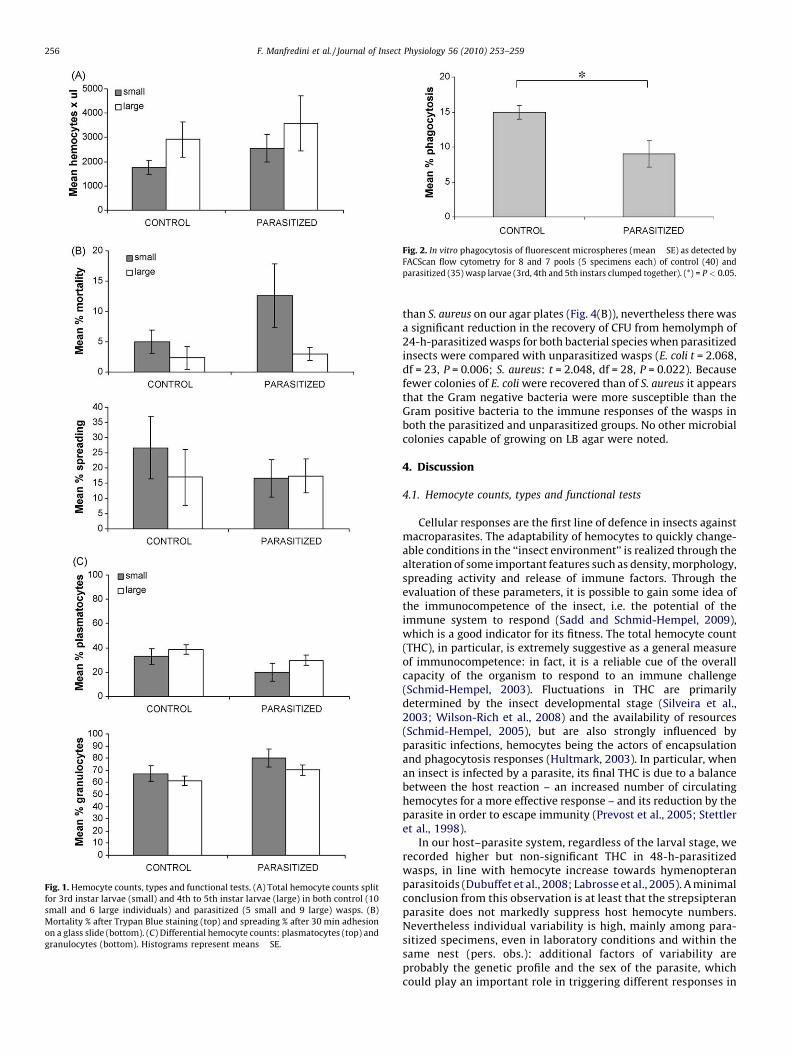
Fig. 1. Hemocyte counts, types and functional tests. (A) Total hemocyte counts split
for 3rd instar larvae (small) and 4th to 5th instar larvae (large) in both control (10
small and 6 large individuals) and parasitized (5 small and 9 large) wasps. (B)
Mortality % after Trypan Blue staining (top) and spreading % after 30 min adhesion
on a glass slide (bottom). (C) Differential hemocyte counts: plasmatocytes (top) and
granulocytes (bottom). Histograms represent means � SE.
Fig. 2. In vitro phagocytosis of fluorescent microspheres (mean � SE) as detected by
FACScan flow cytometry for 8 and 7 pools (5 specimens each) of control (40) and
parasitized (35) wasp larvae (3rd, 4th and 5th instars clumped together). (*) = P < 0.05.
F. Manfredini et al. / Journal of Insect Physiology 56 (2010) 253–259256
than S. aureus on our agar plates (Fig. 4(B)), nevertheless there wasa significant reduction in the recovery of CFU from hemolymph of24-h-parasitized wasps for both bacterial species when parasitizedinsects were compared with unparasitized wasps (E. coli t = 2.068,df = 23, P = 0.006; S. aureus: t = 2.048, df = 28, P = 0.022). Becausefewer colonies of E. coli were recovered than of S. aureus it appearsthat the Gram negative bacteria were more susceptible than theGram positive bacteria to the immune responses of the wasps inboth the parasitized and unparasitized groups. No other microbialcolonies capable of growing on LB agar were noted.
4. Discussion
4.1. Hemocyte counts, types and functional tests
Cellular responses are the first line of defence in insects againstmacroparasites. The adaptability of hemocytes to quickly change-able conditions in the ‘‘insect environment’’ is realized through thealteration of some important features such as density, morphology,spreading activity and release of immune factors. Through theevaluation of these parameters, it is possible to gain some idea ofthe immunocompetence of the insect, i.e. the potential of theimmune system to respond (Sadd and Schmid-Hempel, 2009),which is a good indicator for its fitness. The total hemocyte count(THC), in particular, is extremely suggestive as a general measureof immunocompetence: in fact, it is a reliable cue of the overallcapacity of the organism to respond to an immune challenge(Schmid-Hempel, 2003). Fluctuations in THC are primarilydetermined by the insect developmental stage (Silveira et al.,2003; Wilson-Rich et al., 2008) and the availability of resources(Schmid-Hempel, 2005), but are also strongly influenced byparasitic infections, hemocytes being the actors of encapsulationand phagocytosis responses (Hultmark, 2003). In particular, whenan insect is infected by a parasite, its final THC is due to a balancebetween the host reaction – an increased number of circulatinghemocytes for a more effective response – and its reduction by theparasite in order to escape immunity (Prevost et al., 2005; Stettleret al., 1998).
In our host–parasite system, regardless of the larval stage, werecorded higher but non-significant THC in 48-h-parasitizedwasps, in line with hemocyte increase towards hymenopteranparasitoids (Dubuffet et al., 2008; Labrosse et al., 2005). A minimalconclusion from this observation is at least that the strepsipteranparasite does not markedly suppress host hemocyte numbers.Nevertheless individual variability is high, mainly among para-sitized specimens, even in laboratory conditions and within thesame nest (pers. obs.): additional factors of variability areprobably the genetic profile and the sex of the parasite, whichcould play an important role in triggering different responses in

Fig. 3. Phagocytosis assay as observed by fluorescence microscopy. (A–C) Phagocytosis in hemolymph samples extracted from control P. dominulus larvae and (D–F) from
parasitized wasps at 72 h post-infection. Higher fluorescence is noticeable in A–C than in D–F. Spreading activity of hemocytes is evident in B, E and F.
F. Manfredini et al. / Journal of Insect Physiology 56 (2010) 253–259 257
the host. Moreover, different generations of Polistes larvae (earlyand late workers) could be differently equipped in their immuneresponses. In the future we will investigate individual variability inrelation to host castes (although Polistes are primitively eusocialwasps and castes are very flexible), host sex and parasite load.
A further approach in our future analysis will be to monitor thetemporal dynamics of the cellular response during the initial phaseof the infection process, which is ‘‘a particularly relevant but oftenneglected aspect’’ (Korner and Schmid-Hempel, 2004). We selected48 h post-infection for THC and DHC because at this time the 2ndinstar of the parasite abandons its exuvia to successfully establishinside the host (Manfredini et al., 2007a). In Galleria mellonella
infected with Steinerma nematodes, hemocyte numbers decreasein the first 4 h, but then increase gradually over the next 12 h, andfinally decline to values lower than control: here time spans arebrief (within first 24 h) and the final decline in hemocyte numbersis associated with approaching host death (Bergin et al., 2003).
In our Xenos–Polistes system, the general hemocyte profile of thehost is rather stable: spreading activity, mortality and relativeabundances of two main morpho-types are not dramaticallyaffected by the presence of the parasite, as well as THC, and thisis in line with the requirement for X. vesparum to maintain the hostalive and healthy (i.e. capable of fighting other possible invadingpathogens), in the perspective of a long and sustainable coexistence(Hughes and Kathirithamby, 2005). This fine-tuning of host immune
responses suggests that instead of decreasing the host’s capacity tomount immune responses by destroying or otherwise interferingwith the production of immune cells, the parasite concentrates onavoiding, whether by evasion or suppression, the responses that thehemocytes would normally mount.
4.2. Phagocytosis assay
Though parasitoids are too large to be phagocytized, phago-cytosis analysis by means of synthetic microspheres is a goodparameter for evaluating several mechanisms of the innateimmunity (Williams et al., 2006; Wood and Jacinto, 2007). Infact, phagocytosis is an actin-dependent mechanism, requiring aproper cytoskeleton to internalize the foreign body. When thisprocess is compromised by the action of a pathogen, further actin-dependent mechanisms are affected, in particular encapsulationand wound healing, since they rely on the ability of hemocytes toperform migration and conformational changes. Moreover, theability to undertake phagocytosis may be considered symptomaticfor the general health condition of the insect, being involved inrelated signalling pathways of non-cellular immune compart-ments, for example antimicrobial peptides production (Hoffmann,2003; Hultmark, 2003).
After parasitization by X. vesparum, phagocytosis in vitro,relatively low in our insect model, is inhibited by about one-third,

Fig. 4. Antibacterial response. (A) Total CFU (log10 transformed) per ml of
hemolymph sampled from 19 non-parasitized larvae challenged with bacteria
(control) and 23 larvae challenged with bacteria at 24 h post-infection with Xenos
(parasitized). (B) Separate analysis for the two bacterial components:
Staphylococcus aureus (S. aureus) and Escherichia coli (E. coli). Means � SE.
(*) = P < 0.05; (**) = P < 0.01.
F. Manfredini et al. / Journal of Insect Physiology 56 (2010) 253–259258
the number of engulfed particles being reduced from 14% incontrol to 9% in parasitized larvae. The inhibition does not appearas extensive as has been noted to occur in other host–parasitoidsystems. For example, the parasitic wasp Eulophus pennicornis
triggers a 87% reduction of phagocytosis in the lepidopteran hostLacanobia oleracea (Richards and Edwards, 2002). Again, whatobserved in our model is in line with the strategy of the parasite,which might be disadvantaged by drastically compromising theimmunity of its host. Thus, phagocytosis is not totally suppressed,since this mechanism is important for the clearance of potentiallypathogenic bacteria which could jeopardize host survival. More-over, the similar number of fluorescent beads internalized per cellwithin the two treatments suggests that X. vesparum reducesphagocytosis by reducing the number of host hemocytes under-taking phagocytosis. How this accomplished is unclear; the data donot suggest that the parasite acts directly on any specificphagocytic cell type, as indicated by DHC and spreading activity.
There are many possible routes whereby X. vesparum – similarlyto parasitoid insects – might interfere with P. dominulus
phagocytosis and the literature of host–parasite systems is fullof interesting examples. A peculiar family of proteins could be ofgreat interest for our future analyses, since they are regulators ofthe master signalling Rho GTPases (Rho, Rac and Cdc42), highlyconserved across both invertebrate and vertebrate animals, keycellular factors in cytoskeleton regulation and vesicle trafficking(Qualmann and Mellor, 2003; Takai et al., 2001). This family ofproteins, belonging to the Rho-GAPs (Ras homologous GTPaseActivating Protein) domain, may be exploited by different parasitesto disrupt host cellular response (Labrosse et al., 2005). In thisperspective, encapsulation and phagocytosis appear strictlyconnected. In Drosophila mutants defective for the protein Rac2,for example, hemocytes recognize and attach to the eggs of theparasitoid wasp L. boulardi but fail to spread around them andcapsules fail to melanize (Williams, 2007). The similarity betweenwhat happens in Drosophila mutants and the peculiar immune
response of P. dominulus towards X. vesparum infective larvae(delayed encapsulation and absence of melanization) is striking.
4.3. Antibacterial response
Among the immature stages of social insects, two effectivebarriers towards bacteria are represented by social prophylaxis, i.e.the cleaning behaviour by nursing adults and the ‘‘nest environ-ment’’ itself (Cremer et al., 2007; Stow and Beattie, 2008). Ourexperimental challenge with bacteria not already present in thehemolymph of larval wasps has shown a significant but notstriking decrease in bacterial survival in 24-h-parasitized wasps vs
control. Unfortunately, the ‘‘pricking system’’ does not allow tomonitor the precise number of bacterial colonies introduced ineach specimen, thus we do not know the total power of P.
dominulus immatures to respond to a bacterial challenge. Thereproducibility of the procedure among our experimental groups,however, is sufficient to support a reliable comparison.
The more effective antibacterial response after bacterialchallenge in parasitized vs unparasitized wasps is apparentlyinconsistent with the decreased phagocytosis capability shown bythe former group. We are not currently able to account for this. Itshould be noted however that we observed phagocytosis dynamicsin vitro while the antibacterial response was measured in vivo,where additional variables may be involved (for example, thepricking itself). Secondly, check points were fixed at different timesof the infection process and immune dynamics rapidly evolveduring parasitism: phagocytosis was measured at 72 h post-infection, while bacterial challenge was performed at 24 h post-infection and measured 24 h later (i.e. 48 h post-infection). Third,phagocytosis is not the sole mechanism adopted by insects to clearbacteria from the hemolymph: among other routes, the productionof antimicrobial peptides (AMPs) and reactive oxygen species(ROS), both contributing to bacterial killing and cooperating withthe cellular response (Elrod-Erickson et al., 2000). It is possible thatin parasitized wasps these pathways are already activated by thepresence of X. vesparum larvae, thus they are prompter inresponding against the bacterial challenge. An additional possi-bility is that when the injected bacteria are less efficiently clearedby phagocytosis, then they may be more effective in eliciting thesynthesis of AMPs.
Finally, we should not forget that it is possible that the X.
vesparum parasites may also fight, together with their host, againstinoculated microbes. We have already hypothesized (Manfrediniet al., 2007a) that X. vesparum 1st instar larvae, similar to other insectparasitoids, release some secretions into the host to allow entranceand settlement into the hemocoel. In several cases the antibioticpower of such secretions has been demonstrated (Doury et al., 1995;Richards and Edwards, 2002). In our system, they could either killbacteria directly or create an unsuitable environment for theirgrowth. From this perspective, the action of X. vesparum parasiteswould be extremely intriguing: they ‘‘softly’’ inactivate waspimmune response either to undergo their own first developmentalsteps within the host, or to preserve the defence reactions of hostlarvae against other pathogens. Moreover, X. vesparum evidently atleast cooperates with its host in eliminating invading bacteria,which could be doubly dangerous, being a possible source ofinfection for both the parasite and the host. It will be our plan in thefuture to identify which component(s) of the wasp immune systemis (are) mainly responsible for the differential antibacterial activity(whether cellular or humoral) and to elucidate the mechanismswhereby the parasite is actually operating within the hemolymph ofthe host. To understand this long-term, complex parasite–hostassociation requires assessment of more than one index of immunestatus, strictly connected to each other: hemocyte counts, phago-cytosis and antibacterial response.
F. Manfredini et al. / Journal of Insect Physiology 56 (2010) 253–259 259
Acknowledgments
The authors are grateful to Romano Dallai and Enzo Ottavianifor fruitful discussions on the experimental design and for theiruseful advices during laboratory activity, Cosima Tatiana Baldarifor her precious technical support and for revising the manuscriptand Alessandro Massolo for his essential help in the statisticalanalysis. The authors would also like to thank the three anonymousreferees and Stuart E. Reynolds for their contributions in improvingthe manuscript with good advice and helpful comments.
References
Beani, L., 2006. Crazy wasps: when parasites manipulate the Polistes phenotype.Annales Zoologici Fennici 43, 564–574.
Beani, L., Massolo, A., 2007. Polistes dominulus wasps (Hymenoptera Vespidae), ifparasitized by Xenos vesparum (Strepsiptera Stylopidae), wander among nestsduring the pre-emerging phase. Redia XC 161–164.
Beani, L., Theodora, P., Dallai, R., Turillazzi, S., 2005. Epicuticular hydrocarbons ofXenos vesparum (Strepsiptera Stylopidae), parasite of Polistes dominulus (Hyme-noptera Vespidae): preliminary data. Redia 87, 167–169.
Bergin, D., Brennan, M., Kavanagh, K., 2003. Fluctuations in haemocyte density andmicrobial load may be used as indicators of fungal pathogenicity in larvae ofGalleria mellonella. Microbes and Infection 5, 1389–1395.
Brodeur, J., Boivin, G., 2004. Functional ecology of immature parasitoids. AnnualReview of Entomology 49, 27–49.
Consoli, F.L., Brandt, S.L., Coudron, T.A., Vinson, S.B., 2005. Host regulation andrelease of parasitism-specific proteins in the system Toxoneuron nigriceps–Heliothis virescens. Comparative Biochemistry and Physiology B-Biochemistry,Molecular Biology 142, 181–191.
Cremer, S., Armitage, S.A.O., Schmid-Hempel, P., 2007. Social immunity. CurrentBiology 17, R693–R702.
Doury, G., Rojasrousse, D., Periquet, G., 1995. Ability of Eupelmus orientalis ectopar-asitoid larvae to develop on an unparalysed host in the absence of femalestinging behavior. Journal of Insect Physiology 41, 287–296.
Dubuffet, A., Doury, G., Labrousse, C., Drezen, J.M., Carton, Y., Poirie, M., 2008.Variation of success of Leptopilina boulardi in Drosophila yakuba: the mechan-isms explored. Developmental and Comparative Immunology 32, 597–602.
Elrod-Erickson, M., Mishra, S., Schneider, D., 2000. Interactions between the cellularand humoral immune responses in Drosophila. Current Biology 10, 781–784.
Hoffmann, J.A., 2003. The immune response of Drosophila. Nature 426, 33–38.Hughes, D.P., Beani, L., Turillazzi, S., Kathirithamby, J., 2003. Prevalence of the
parasite Strepsiptera in Polistes as detected by dissection of immatures. InsectesSociaux 50, 62–68.
Hughes, D.P., Kathirithamby, J., 2005. Cost of strepsipteran macroparasitism forimmature wasps: does sociality modulate virulence? Oikos 110, 428–434.
Hughes, D.P., Kathirithamby, J., Beani, L., 2004a. Prevalence of the parasite Strep-siptera in adult Polistes wasps: field collections and literature overview. Ethol-ogy Ecology, Evolution 16, 363–375.
Hughes, D.P., Kathirithamby, J., Turillazzi, S., Beani, L., 2004b. Social wasps desertthe colony and aggregate outside if parasitized: parasite manipulation? Beha-vioral Ecology 15, 1037–1043.
Hultmark, D., 2003. Drosophila immunity: paths and patterns. Current Opinion inImmunology 15, 12–19.
Ibrahim, A.M.A., Kim, Y., 2006. Parasitism by Cotesia plutellae alters the hemocytepopulation and immunological function of the diamondback moth, Plutellaxylostella. Journal of Insect Physiology 52, 943–950.
Kathirithamby, J., Ross, L.D., Johnston, J.S., 2003. Masquerading as self? Endopar-asitic strepsiptera (Insecta) enclose themselves in host-derived epidermal bag.Proceedings of the National Academy of Sciences of the United States of America100, 7655–7659.
Korner, P., Schmid-Hempel, P., 2004. In vivo dynamics of an immune responsein the bumble bee Bombus terrestris. Journal of Invertebrate Pathology 87,59–66.
Labrosse, C., Eslin, P., Doury, G., Drezen, J.M., Poirie, M., 2005. Haemocyte changes inD. melanogaster in response to long gland components of the parasitoid waspLeptopilina boulardi: a Rho-GAP protein as an important factor. Journal of InsectPhysiology 51, 161–170.
Manfredini, F., Dallai, R., Ottaviani, E., 2008. Circulating hemocytes from larvae ofthe paper wasp Polistes dominulus (Hymenoptera, Vespidae). Tissue, Cell 40,103–112.
Manfredini, F., Giusti, F., Beani, L., Dallai, R., 2007a. Developmental strategy of theendoparasite Xenos vesparum (Strepsiptera, Insecta): host invasion and elusionof its defence reactions. Journal of Morphology 268, 588–601.
Manfredini, F., Giusti, F., Beani, L., Dallai, R., 2007b. Preliminary data on the cellularresponse of Polistes immatures (Hymenoptera Vespidae), hosts of the endopar-asite Xenos vesparum (Strepsiptera Stylopidae). Redia XC 155–159.
Pennacchio, F., Strand, M.R., 2006. Evolution of developmental strategies in parasitichymenoptera. Annual Review of Entomology 51, 233–258.
Prevost, G., Eslin, P., Doury, G., Moreau, S.J.M., Guillot, S., 2005. Asobara, braconidparasitoids of Drosophila larvae: unusual strategies to avoid encapsulationwithout VLPs. Journal of Insect Physiology 51, 171–179.
Qualmann, B, Mellor, H., 2003. Regulation of endocytic traffic by Rho GTPases.Biochemistry Journal 15, 233–241.
Richards, E.H., Edwards, J.P., 2002. Larvae of the ectoparasitic wasp, Eulophuspennicornis, release factors which adversely affect haemocytes of their host,Lacanobia oleracea. Journal of Insect Physiology 48, 845–855.
Sadd, B.M., Schmid-Hempel, P., 2009. Principles of ecological immunology. Evolu-tionary Applications 2, 113–121.
Schmid-Hempel, P., 2003. Variation in immune defence as a question of evolu-tionary ecology. Proceedings of the Royal Society of London Series B-BiologicalSciences 270, 357–366.
Schmid-Hempel, P., 2005. Evolutionary ecology of insect immune defenses. AnnualReview of Entomology 50, 529–551.
Schmidt, O., Theopold, U., Strand, M., 2001. Innate immunity and its evasion andsuppression by hymenopteran endoparasitoids. Bioessays 23, 344–351.
Siegel, S., Castellan Jr., N.J., 1988. Nonparametric Statistics for the BehavioralSciences, 2nd ed. McGraw-Hill—A Division of the McGraw-Hill Companies,London.
Silveira, E.B., Ribeiro, B.M., Bao, S.N., 2003. Characterization of larval haemocytesfrom the velvetbean caterpillar Anticarsia gemmatalis (Hubner) (Lepidop-tera: Noctuidae). Journal of Submicroscopic Cytology and Pathology 35,129–139.
Sokal, R.R., Rohlf, F.J., 1995. Biometry: The Principles and Practice of Statistics inBiological Research, 3rd ed. W.H. Freeman and Co, New York, 887 pp., ISBN: 0-7167-2411-1.
Stettler, P., Trenczek, T., Wyler, T., Pfister-Wilhelm, R., Lanzrein, B., 1998. Overviewof parasitism associated effects on host haemocytes in larval parasitoids andcomparison with effects of the egg-larval parasitoid Chelonus inanitus on its hostSpodoptera littoralis. Journal of Insect Physiology 44, 817–831.
Stow, A., Beattie, A., 2008. Chemical and genetic defenses against disease in insectsocieties. Brain Behavior and Immunity 22, 1009–1013.
Strand, M.R., Pech, L.L., 1995. Immunological basis for compatibility in parasitoidhost relationships. Annual Review of Entomology 40, 31–56.
Takai, Y., Sasaki, T., Matozaki, T., 2001. Small GTP-binding proteins. PhysiologicalReviews 81, 153–208.
Williams, M.J., 2007. Drosophila hemopoiesis and cellular immunity. Journal ofImmunology 178, 4711–4716.
Williams, M.J., Wiklund, M.L., Wikman, S., Hultmark, D., 2006. Rac1 signalling inthe Drosophila larval cellular immune response. Journal of Cell Science 119,2015–2024.
Wilson-Rich, N., Dres, S.T., Starks, P.T., 2008. The ontogeny of immunity: develop-ment of innate immune strength in the honey bee (Apis mellifera). Journal ofInsect Physiology 54, 1392–1399.
Wood, W., Jacinto, A., 2007. Drosophila melanogaster embryonic haemocytes: mas-ters of multitasking. Nature Reviews Molecular Cell Biology 8, 542–551.