Embed Size (px)
Citation preview
Journal of Nematology 42(2):139–150. 2010.� The Society of Nematologists 2010.
Vittatidera zeaphila (Nematoda: Heteroderidae), a new genus and speciesof cyst nematode parasitic on corn (Zea mays)
ERNEST C. BERNARD,1 ZAFAR A. HANDOO,2 THOMAS O. POWERS,3 PATRICIA A. DONALD,4 ROBERT D. HEINZ5
Abstract: A new genus and species of cyst nematode, Vittatidera zeaphila, is described from Tennessee. The new genus is superficiallysimilar to Cactodera but is distinguished from other cyst-forming taxa in having a persistent lateral field in females and cysts, persistentvulval lips covering a circumfenestrate vulva, and subventral gland nuclei of the female contained in a separate small lobe. Infectivejuveniles (J2) are distinguished from all previously described Cactodera spp. by the short stylet in the second-stage juvenile (14-17 mm);J2 of Cactodera spp. have stylets at least 18 mm long. The new species also is unusual in that the females produce large egg masses.Known hosts are corn and goosegrass. DNA analysis suggests that Vittatidera forms a separate group apart from other cyst-forminggenera within Heteroderinae.
Key words: cyst nematode, Eleusine indica, goosegrass, maize, molecular analysis, new genus, taxonomy, Vittatidera zeaphila, Zea mays.
Cyst nematodes are widespread but with the excep-tions of Heterodera avenae Wollenweber and H. zeae Koshi,Swarup & Sethi (Baldwin & Mundo-Ocampo 1991) arenot significant parasites of Poaceae. In the late 1970s thefirst author collected specimens of a cyst nematode fromgoosegrass (Eleusine indica (L.) Gaertn.) growing in a to-mato field in west Tennessee. These specimens were sentto the late A. Morgan Golden, who was of the opinion (inlitt.) that they represented an undescribed species. Thespecies was not described at that time, but in 2006 thefourth author collected a similar nematode from corn(Zea mays) in the same general region of the state. Thesecond author compared specimens of the two collec-tions and confirmed that they were the same species.Further examination revealed that this taxon has fea-tures unlike any other genus of cyst-forming nematodes.
The objective of this paper is the description of a newgenus and species of Heteroderidae, Vittatidera zeaphila,which parasitizes corn and goosegrass. A companionpaper (Donald et al., in prep.) provides information onhost range studies and environmental requirements.
MATERIALS AND METHODS
Specimens used to prepare the description wereobtained from greenhouse corn cultures derived fromthe collection of an isolate from Obion County, Tennessee,in 2006, and from specimens collected in the late 1970sfrom Lauderdale County, Tennessee. Specimens werefixed in either warm (408C) or hot (808C) 4% formalin,
processed to glycerin with a rapid method (Seinhorst1959), and mounted in anhydrous glycerin on micro-scope slides.
DNA analyses Juvenile nematodes of Vittatidera zeaphilawere obtained from culture. Because of an initial concernthat V. zeaphila might be conspecific with H. zeae, frozenindividuals of H. zeae were sequenced for each of thegenetic markers and included in sequence comparisons.
Specimens of V. zeaphila were individually selected andmanually disrupted to provide template for DNA ampli-fications (Powers & Harris 1993). Small (18S) and large(28S) subunit rDNA and the internal transcribed spacer1 (ITS1) regions were amplified in 50-ml reactions, eachcontaining: 31.5 ml distilled water, 5 ml 10x PCR buffer(100 mM Tris-HCl (pH 8.3), 500 mM KCl, 15 mM MgCl2,0.01% gelatin), 1 ml dNTP mixture (2.5 mM each ofdATP, dCTP, dGTP, and dTTP), 3.0 ml 25 mM MgCl2, 1.0ml of each primer (20 mM), 2.5 ml of JumpStart REDTaqpolymerase (Sigma, St. Louis, MO; 1.0 u/ml), and 5 ml ofDNA template. All PCR reactions were performed ona DNA Engine PTC-200 Peltier thermal cycler (MJ Re-search, Watertown, MA) with the following run parame-ters: one initial denaturation cycle at 95oC for 3 min,followed by 50 cycles at 95oC for 15 sec, 55 or 50oC for 15sec, ramped increase at 0.5oC per sec to 72oC for 1 min. Afinal elongation step was run at 72oC for 5 min. Negativecontrols were included in each amplification series. Thefollowing primer sets were used in this study.
18S ribosomal DNA: Near full-length 18S sequence of1,565 bases was obtained in three separate amplifications.The primer sets were:
59-first primer pair:G18S4 (59 to 39) –> GCTTGTCTCAAAGATTAAGCC18s721R (59 to 39) –> AGCACTCTAATTTTTTCAAAGMiddle third primer pair:18s550a(59 to 39) –> AGCCGCGGTAATTCCAG18s977R (59 to 39) –> TTTACGGTTAGAACTAGGGCGG39-third primer pair:18s1.2a(59 to 39) –> CGATCAGATACCGCCCTAG18sr2b (59 to 39) –> TACAAAGGGCAGGGACGTAAT
Primer 18Sr2b (positions 1567 to 1547) is the reversecomplement of primer rDNA2 from Vrain et al. (1992).
Received for publication May 7, 2010.1Entomology & Plant Pathology, The University of Tennessee, 2431 Joe
Johnson Drive, 205 Plant Sciences, Knoxville, TN 37996-4560, USA, [email protected].
2Nematology Laboratory, USDA ARS, Beltsville, MD 20705, USA, [email protected].
3Department of Plant Pathology, University of Nebraska, 406 Plant ScienceHall, Lincoln, NE 68583-0722, USA, [email protected].
4USDA ARS, West Tennessee Research and Education Center, 605 AirwaysBlvd., Jackson, TN 38301, USA, [email protected].
5Division of Plant Sciences and Bond Life Sciences Center, University ofMissouri-Columbia, Columbia, MO 65211, USA, [email protected].
Mention of trade names or commercial products in this publication is solelyfor the purpose of providing specific information and does not imply recom-mendations or endorsement by the United States Department of Agriculture.
E-mail: [email protected] paper was edited by Daniel Bumbarger.
139

28S -D2/3 primer set: This primer set produces aproduct of 741 bases in V. zeaphila.
D2A (59 to 39) –> ACAAGTACCGTGAGGGAAAGTTGD3B (59 to 39) –> TCGGAAGGAACCAGCTACTAITS1 primer set:rDNA2: 59-TTGATTACGTCCCTGCCCTTT-39
rDNA1.58Sa: 59-ACGAGCCGAGTGATCCACC-39
Primer rDNA2 is a modified version of the reversecomplement of 18Sr2b (above) and is paired with primerrDNA1.58Sa, which is located in the 5’ region of the 5.8SrDNA gene. This primer set produces a fragment of 582nucleotides in V. zeaphila, excluding primers, of which176 nucleotides are the 3’ end of 18S rDNA. Sequencecomparisons of the ITS1 region were confined to thespacer region only. Annealing temperature for this ITS1primer set was 558C.
PCR products were purified and concentrated withMicrocon-100 centrifugal filter units (Millipore Inc., Bed-ford, Massachusetts) and sent to the DNA Sequencing Lab(University of Arkansas for Medical Sciences) for directsequencing in both directions. Amplification primers wereused as sequencing primers. DNA sequences were editedand assembled using CodonCode Aligner (CodonCodeCorp, Dedham, Massachusetts). DNA alignment was byMUSCLE 3.7 (Edgar, 2004). Maximum likelihood anal-ysis (100 bootstrap replicates for estimation of branchsupport) was carried out with PHYML 3.0 (http://www.phylogeny.fr) using the HKY85 substitution model, withgamma parameters and proportion of invariant sites es-timated, and a transition/tranversion ratio of 4.
DESCRIPTION
Vittatidera new genusCysts orange-brown to brown, lemon-shaped, necks
short; secretions around neck persistent. Cyst surfacewith weak longitudinal ridges between neck and coneand with underlying transverse rows of punctations; zig-zag pattern absent. Lateral field in both females andcysts arched or sinuous, represented by short, transverselines between neck and cone area. Cuticle in neck regiongranulated. Vulval cone slightly protuberant, membra-nous vulval lips persistent; vulval aperture circular torhomboid, circumfenestrate, with irregular denticle-likeprotuberances around the periphery of orifice. Bullae,vulval bridge, vulval underbridge, and internal denticlesabsent. Cone tip encircled with short, wavy ridgesextending to vicinity of anus. Cone region with numerousminute, irregularly distributed duct-like tubes extendingto pores on cuticle surface. Cuticle around anus thin-ner than rest of cyst but not developed as a fenestra. Anussubterminal. Subventral gland nuclei of female containedin a discrete lobe extending from the large dorsal gland.Phasmid apertures present on white females, approxi-mately at level of anus. Males of variable length; styletknobs rounded. Second-stage juveniles heteroderiform,
tail conoid with narrowly rounded tip, phasmid aperturespore-like. Egg shell smooth.
Type species: Vittatidera zeaphila n. sp.Etymology: Combined Latin vittatus (striped) and Greek
dera (skin or hide), referring to the presence of a lateralfield in the females and cysts.
Differential diagnosis Superficially Vittatidera n. g. re-sembles Cactodera Krall & Krall in having a circum-fenestrate vulva in the cyst stage. It differs from all cyst-forming genera in possessing a distinct lateral field inthe adult female and cyst stages. The new genus also hasunusually persistent vulva lips that remain intact wellafter the cyst has formed. Unlike Cactodera, females ofthe new genus produce large egg masses with numerouseggs, a character shared with a few Heterodera spp. Theunique characters of this new genus require modificationof the diagnosis of Heteroderinae Filipjev & SchuurmansStekhoven, the subfamily of Heteroderidae that containsthe cyst-forming taxa.
Emended diagnosis of Heteroderinae (After Siddiqi, 2000):Heteroderidae. Mature females spherical, oval, pear orlemon-shaped with a short neck, turning into a tough,hard-walled, yellowish, light to dark brown, or blackishcyst containing eggs and juveniles, eggs sometimes laidin a large gelatinous matrix (egg mass). Cuticle surfacewith zigzag or lace-like pattern, or with elongated, fusi-form ridges, overlaying fine pattern of annulations, orannulations absent in mature females and cysts. Lateralfield on females and cysts rarely present; if present (Vit-tatidera), lateral field arching or sinuate, extending fromneck to cone base. Vulva and anus close together, almostterminal, on raised vulval cone or in flat to concave vulvalbasin. Clear hyaline single (circumfenestrate) or doubled(ambifenestrate, bifenestrate) vulval fenestrae present;anal fenestra present in Punctodera, vulva lips rarely per-sistent (Vittatidera). Esophageal gland nuclei containedin single lobe or subventral gland nuclei of the femalecontained in separate small lobe (Vittatidera) Male de-veloping through metamorphosis with labial region an-nulated, four incisures in lateral field, tail short, hemi-spherical, without bursa. Second-stage juvenile stylet over14 um long; with three to four incisures in lateral field.
Type genus: Heterodera Schmidt, 1871Other genera (after Wouts & Baldwin 1998, Sturhan
2002, Sturhan et al. 2007, Mundo-Ocampo et al. 2008):
Betulodera Sturhan, 2002Cactodera Krall & Krall, 1978Dolichodera Mulvey & Ebsary, 1980Globodera Skarbilovich, 1959Paradolichodera Sturhan, Wouts & Subbotin, 2007Punctodera Mulvey & Stone, 1976Vittatidera n. gen.
Vittatidera zeaphila n. sp.(Figs. 1-7)
Females Femalemeasurements: length389-534mm(mean455 ± 17.3 SE, CV = 11.4, n = 20), neck length 71-117 mm
140 Journal of Nematology, Volume 42, No. 2, June 2010
(92.7 ± 5.1, CV = 16.5), anterior end to metacorpal valve 50.0-88.6mm(66.0±5.3,CV=8.1),a ratio 1.5-2.1 (1.8 ± 0.07, CV =11.4). Holotype female: length 434 mm, neck length 100 mm,anterior end to metacorpal valve 61.2 mm, a ratio 1.6.
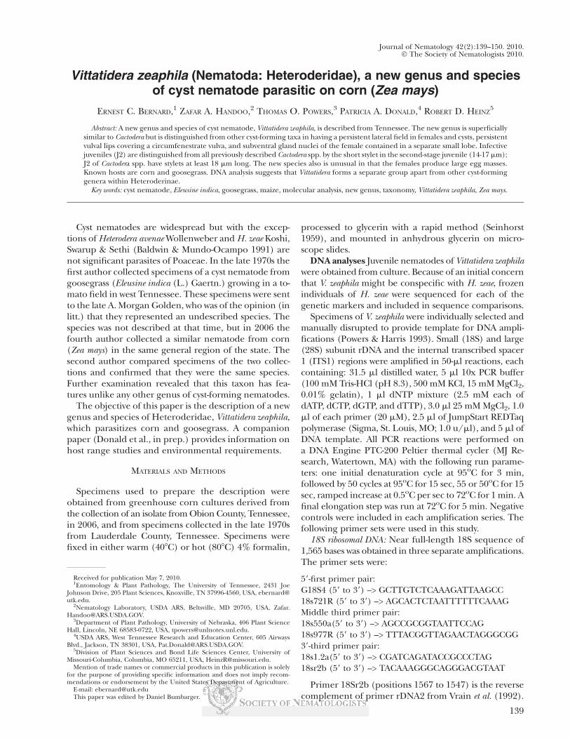
Female oval (Figs. 1A,C, 2A), white in reflected light,often with large, attached egg mass (Figs. 1A,B). Cuticleof neck region with annulations consisting of fine rowsof granules (Fig. 1D). Cuticle of swollen part of body
FIG. 1. Light micrographs of Vittatidera zeaphila n. sp. A. White females, one with attached egg mass; B. Cysts with attached egg masses;C. Female lateral view (a: anus); D. Cuticular granulation in neck region (arrow points anteriorly); E. Longitudinal striations and lateral field (lf)on white female (large arrow points toward neck); F. Longitudinal striations on senescent female (arrow points toward neck).
Vittatidera zeaphila n. g. n. sp.: Bernard et al. 141

anterior to vulval cone with elongated, fusiform ridgesoverlaying fine pattern of annulations (Figs. 1E,F, 2D).Lateral field arching or sinuate, extending from neck tocone base (Figs. 2D,E), indicated by short, fine trans-
verse striae (Figs. 1E, 2D,E). Phasmid apertures minute,pore-like, level with anus (Fig. 2F). Cone area delimitedby shallow, transverse, wavy striae; vulval lip region smooth(Figs. 2D,F; 3A). Anterior end with one prominent head
FIG. 2. Females of Vittatidera zeaphila n. sp. A. Outlines of females and cysts; B. Anterior end; C. Esophageal variation; D. Cuticular sculp-turing and arched lateral field, some sculpturing omitted; E. Sinuous lateral field; F. Perineal region, arrows indicate phasmids.
142 Journal of Nematology, Volume 42, No. 2, June 2010

annule (Fig. 2B); anterior structure usually obscured byhardened mucilage-like secretion. Stylet slender, withrounded or slightly posteriorly inclined knobs. Dorsal
gland orifice 3-4 mm from stylet base. Subventral glandnuclei in small lobe extending ventro-posteriorly fromdorsal gland lobe. Excretory pore usually level with
FIG. 3. Cyst features of Vittatidera zeaphila n. sp. A. Vulval cone of old female; B. Vulval cone of young cyst; C. Posterior end of mature cyst(a: anus); D. Vulval opening of mature cyst; E. Lateral field, cuticular striations, and annular punctations.
Vittatidera zeaphila n. g. n. sp.: Bernard et al. 143

midregion of esophageal glands, occasionally posteriorto glands (Figs. 2B,C).
Cysts Cyst measurements: length 385-608 mm (mean470 ± 14.6 SE, CV = 13.1, n = 18), width 179-391 mm,(287 ± 11.9, CV = 17.6), neck length 60-119 mm (79.5 ±4.4, CV = 21.6), a ratio 1.4-2.2 (1.67 ± 0.05, CV = 12.9);vulva width 28-48 mm (36); vulva-anus distance 52-74mm (63).
Cysts lemon-shaped, vulval cone not protuberant,large egg masses persistent (Fig. 1B). Vulval aperture indeveloping cysts fusiform, lips smooth, surroundingstriae long and sinuous, prominent only near cone apex(Fig. 3B). In mature cysts vulval aperture circumfenestrate,circular to rhomboidal, membranous lips persistent onmature cysts (Figs. 3C, 4A-C); underbridge, bullae, andother internal thickenings absent. Old cysts with irregulardenticles around the inner edge of aperture (Figs. 3D,4C). Cuticle around anus thinned but not fenestrate (Figs.3C, 4A-C). Cuticular sculpturing on mature cyst similar butless pronounced than that of female, with faint longitu-dinal fusiform figures, underlying transverse annulations,and ladder-like lateral field (Fig. 3E); cuticle of neckregion granulated (Fig. 4D).
Males Male length highly variable (Table 1, Fig. 6E).Lip region rounded, slightly set off, with three annules(Figs. 5D, 6F). Lateral field with four incisures, outer bandswith scattered incomplete areolation. Tail tip rounded orbluntly conoid (Figs. 6G-I). Stylet knobs rounded, dorsalgland orifice near base of stylet. Esophageal glands insingle lobe, subventral nuclei posterior to dorsal nucleus.Excretory pore just posterior to hemizonid, at anterior halfof glands (Fig. 6F). Male gonad about 40-60% of bodylength. Spicules slightly curved, tips bifid (Fig. 6H). Inventral view, gubernaculum star-shaped, with two smallanterior processes, two broad, lateral wings, and poste-rior process (Fig. 6I)
Juveniles Second-stage juveniles heteroderiform (Fig.6A); stylet length less than 18 mm (Table 2). Lip regionrounded, slightly offset, with three annules (Figs. 5A,B,6B). Stylet knobs rounded (Fig. 5B). Lateral field withfour incisures, outer incisures weakly crenulated. Phasmidapertures minute, in middle of lateral field midway be-tween anus and tail tip. Anus without cuticular flap.Esophageal glands in single long lobe, subventral glandnuclei posterior to dorsal nucleus (Fig. 6B). Tail elongate-conoid, tip narrowly rounded (Figs, 5C, 6C,D), regularly
FIG. 4A-C. Vulval cones and surface sculpture of Vittatidera zeaphila n. sp. (arrows point to anus); D. Surface granulation in neck region(arrow points toward anterior end)
144 Journal of Nematology, Volume 42, No. 2, June 2010

annulated in anterior two-thirds, posterior third with ir-regular or incomplete annulation; proximal margin ofhyaline region irregular, usually with two invasive lobes ofthe body lumen (Figs. 6C,D).
Etymology The specific name zeaphila means ‘‘corn-loving,’’ a reference to its economically important host.
Type locality and host USA, Tennessee, Obion County,Troy, 36821.35’N, 89811.1’W, maize (corn) field, 6 Oc-tober 2006, Patricia Donald, collector.
Type designation and deposition Type specimens de-posited in USDA Nematode Collection, Beltsville, MD:Holotype female (T641t) selected from greenhouseculture originating from type locality collection. Para-types on slides, same data: T-5929p to T-5942p, cysts(anterior and posterior halves on alternating slides); T-5943p, white female; T-5944p to T-5947p, second-stagejuveniles. Paratypes in vials, same data: T-553p, mixedstages. T-554p: juveniles. Paratypes on slides, culturedfrom same field collection by R. D. Heinz and harvested
December 2006: T-5479p, males; T-5480p to T-5482p,second-stage juveniles; T-5815p to T-5817p: cyst parts.Paratypes in vials, same origin: T-502p, cysts and juve-niles; T-526p, eggs. Other paratype females, males,juveniles, and cysts maintained in the University ofTennessee Nematode Collection, Knoxville, TN.
Other specimens examined: USA, Tennessee, Lau-derdale County, 35845’N, 89835’W, on roots of goosegrass,August 1978, E. C. Bernard, coll.: on slides, T-5908p toT-5916p, vulval cones; T-5917p to T-5919p, anteriorhalves of cysts; T-5920p, cyst wall; T-5921p to T-5927p,one cyst (ant. + post.) on each slide; T-5928p, second-stage juveniles. In vials: T-551p, second-stage juveniles;T-552p, females.
DISCUSSION
The placement of this taxon in the classification ofHeteroderidae is problematical. Wouts (1985) proposed
FIG. 5A-C. Second-stage juveniles of Vittatidera zeaphila n. sp. A. Anterior end; B. Head end; C. Tail; D. Male lip region.
Vittatidera zeaphila n. g. n. sp.: Bernard et al. 145

six subfamilies within Heteroderidae. Baldwin andSchouest (1990) rearranged the family, reducing it tosubfamily Heteroderinae and proposing six monophyletic
tribes within Heteroderinae, but Siddiqi (2000) elevatedHeteroderinae back to family rank, a proposal acceptedby Subbotin et al. (2006). Of the six taxa proposed by
FIG. 6A-D. Second-stage juveniles of Vittatidera zeaphila n. sp. A. Outlines; B. Anterior end; C. Tails in lateral view; D. Tail in ventral view. E-I.Males. E. Outlines; F. Anterior end; G. Posterior region, subventral view; H. Posterior region, lateral view; I. Tail region, ventral view showingshape of gubernaculum.
146 Journal of Nematology, Volume 42, No. 2, June 2010

Baldwin and Schouest (1990), only Heteroderini (nowHeteroderinae) possesses the cyst-forming character.Therefore, V. zeaphila should be placed in Hetero-derinae. However, V. zeaphila possesses some uniquefeatures that do not appear to fit the confines of thisgroup. The basic cuticular pattern consists of longitu-dinally oriented and elongated fusiform ridges, similar
to the cuticle of Ekphymatodera thomasoni, a non-cystformer (Baldwin et al. 1989). Unlike all other describedheteroderids, V. zeaphila has a distinct lateral field con-sisting of a dense ladder-like row of short, transverse linesrunning from the neck base to the cone base. Finally,females appear to have phasmid apertures, a featurenot before reported in heteroderid females. Previous
FIG. 7A-C. Molecular relationships of Vittatidera zeaphila to other Heteroderidae. A. 18S; B. D2D3; C. ITS. Within each tree the terminal nodewith V. zeaphila is surrounded by a bold box. Bootstrap values are presented for all internal nodes.
Vittatidera zeaphila n. g. n. sp.: Bernard et al. 147

examination of female cyst nematode cuticle generallyhas been focused on the anterior and posterior regions(Othman et al., 1988; Wouts & Baldwin, 1998).
The presence of a separate small esophageal lobecontaining the subventral gland nuclei is a feature that V.zeaphila n. sp. has in common with many non-cyst-formingHeteroderinae, such as Atalodera festucae, A. trilineata,Ekphymatodera thomasoni, and Verutus californicus (Baldwinet al. 1989). It is tempting to hypothesize that V. zeaphilais a cyst-forming member of one of the non-cyst-formingheteroderid tribes on the basis of this apparently un-usual gland nuclei arrangement. However, beside the
incontrovertible fact of a cyst stage, the J2 has pore-likephasmids, which are typical of the Heteroderini. Second-stage juveniles of most of the non-cyst-forming specieshave large phasmidial ampullae and the phasmids ap-pear scutelliform. Finally, the sequestration of the sub-ventral gland nuclei may not be unusual and could bethe norm in Heteroderidae. We were unable to finddetailed illustrations of the esophagus of any Heteroderaor Globodera spp. comparable to those in Baldwin et al.(1989). A similar lobe was illustrated for H. orientalis(Kazachenko) (Mundo-Ocampo et al. 2008) but thesubventral gland nuclei were reported to be in the ante-rior third of the esophageal lobe. Heterodera pakistanensiswas described as having esophageal glands in a single lobe(Maqbool & Shahina 1986), and Stone & Rowe (1976)stated that the esophageal gland lobe of H. cruciferae waslarge and not differentiated into dorsal and subventralsections. Whether this character is of supraspecific valuecannot be determined until more typical cyst nematodeshave been carefully examined.
Molecular comparison of V. zeaphila with availableheteroderid sequences in GenBank suggests this spe-cies is basal to the other cyst-forming Heteroderidae,and is concordant with the unusual morphology ofthis nematode. Phylogenetic analysis firmly establishedthat Vittatidera zeaphila is genetically distant from theother cyst nematode on corn, Heterodera zeae. Phyloge-netic trees constructed from the three genetic markers
FIG. 7A-C. Continued.
TABLE 1. Morphometrics of males of Vittatidera zeaphila n. sp.(n = 10).
Mean 6 SE Range CVa
Measurements (mm)Length 691 6 50.3 478-912 21.8Stylet length 18.9 6 0.36 17.3-21.0 5.8Stylet knobs to dorsal gland
orifice3.3 6 0.31 2.2-4.8 26.6
Head end to excretory pore 94 6 6.4 68-115 19.2Spicule length 24.0 6 0.59 22.1-26.5 6.9
Ratiosa 35.7 6 1.45 29.2-42.7 12.2S (stylet length/body
width at knobs)1.5 6 0.06 1.3-1.8 10.3
a CV: coefficient of variation.
148 Journal of Nematology, Volume 42, No. 2, June 2010

consistently grouped H. zeae within a large Heteroderaclade that does not include V. zeaphila (Fig. 7). The 18Stree (Fig. 7A) places V. zeaphila outside a Heterodera/Globodera clade that is relatively well-supported (0.8bootstrap value) in the data set. The limited number offull-length 18S sequences in GenBank of cyst speciesother than Heterodera and Globodera precludes a moredefinitive statement of the position of Vittatidera withinHeteroderidae based on 18S. The D2/D3 sequenceincluded a wider set of available heteroderid genera forcomparison, including Dolichodera, Cactodera, Rhizo-nema, Atalodera, Cryphodera and Meloidodera (Fig 7B).Vittatidera was weakly supported (0.68 bootstrap value)as a member of a clade that includes the genera Hetero-dera, Cactodera, Punctodera, Globodera and Dolichodera, ex-cluding Atalodera and Rhizonema. Vittatidera may occupya sister taxon relationship with the aforementioned in-group genera; however, additional DNA sequence isnecessary to confirm that relationship. A recent phylo-genetic tree based on D2/D3 sequence indicates thatVittatidera may group with Betulodera apart from the othercyst-forming nematodes in Heteroderinae (S. Subbotinpers. comm.). ITS1 sequence could provide insight intothe systematic position of Vittatidera, but comparativesequences of cyst genera other than Heterodera are lack-ing (Fig. 7C).
It should be kept in mind that the molecular portionof this study was an attempt to determine where V. zeaphilafits among the Heteroderidae based on available se-quences, but was not intended to be a phylogeneticstudy of Heteroderidae. The DNA analyses, as well asthe morphology of this nematode, support a positionoutside of the Heterodera/Globodera clade. Available se-
quences for the minor cyst and non-cyst-forming spe-cies are limited, and phylogenetic refinement of theplacement of V. zeaphila in Heteroderidae awaits fur-ther study.
The first and second authors measured separategroups of juveniles and obtained similar measurementsexcept for stylet length. Specimens fixed in hot for-malin had stylets averaging about 1.5 mm longer thanspecimens fixed in warm formalin (Table 2). The firstauthor confirmed these figures by measuring both groupsof juveniles with the same equipment and methods.Separate measurements of the conus and shaft indicatedthat both the conus and shaft were slightly shorter inwarm-fixed specimens than in hot-fixed specimens. Otherfactors in processing, such as the temperature of the al-cohol-evaporation oven, could have contributed to thisdifference, which could be either shrinkage or swelling.Regardless of the processing steps that may have led tothis discrepancy, this finding points out the variability thatcan occur in specimen preparation.
LITERATURE CITED
Baldwin, J. G., Bernard, E. C., and Mundo-Ocampo, M. 1989. Fournew species of Heteroderidae including Ekphymatodera n. gen. fromCalifornia. Journal of Nematology 21:48–68.
Baldwin, J. G., and Mundo-Ocampo, M. 1991. Heteroderinae, cyst-and non-cyst-forming nematodes. Pp. 273–362 in W. R. Nickle, ed.Manual of agricultural nematology. Marcel Dekker, New York.
Baldwin, J. G., and Schouest, L. P., Jr. 1990. Comparative detailedmorphology of the Heteroderinae Filip’ev & Schuurmans Stekhoven,1941, sensu Luc et al. (1988): Phylogenetic systematics and revisedclassification. Systematic Parasitology 15:81–106.
Edgar, R. C. 2007. MUSCLE protein multiple sequence alignmentsoftware, version 3.7. http://www.drive5.com/muscle/download3.6.html.Accessed 22 April 2010.
Maqbool, M. A., and Shahina, F. 1986. New species of cyst nematodeHeterodera pakistanensis (Nematoda: Heteroderidae) attacking wheatin Pakistan. Journal of Nematology 18:541–548.
Mundo-Ocampo, M., Troccoli, A., Subbotin, S. A., Del Cid, J.,Baldwin, J. G., and Inserra, R. N. 2008. Synonymy of Afenestratawith Heterodera supported by phylogenetics with molecular andmorphological characterization of H. koreana comb. n. and H. ori-entalis comb. n. (Tylenchida: Heteroderidae). Nematology 10:611–632.
Othman, A. B., Baldwin, J. G., and Mundo-Ocampo, M. 1988.Comparative morphology of Globodera, Cactodera, and Punctodera spp.(Heteroderidae) with scanning electron microscopy. Revue de Nem-atologie 11:53–63.
Powers, T. O., and Harris, T. S. 1993. A polymerase chain methodfor identification of five major Meloidogyne species. Journal of Nema-tology 25:1–6.
Seinhorst, J. W. 1959. A rapid method for the transfer of nematodesfrom fixative to anhydrous glycerin. Nematologica 4:67–69.
Siddiqi, M. R. 2000. Tylenchida: Parasites of plants and insects. 2ndedition. CABI Publishing, Wallingford, UK.
Stone, A. R., and Rowe, J. A. 1976. Heterodera cruciferae. C.I.H. De-scriptions of Plant-parasitic Nematodes, Set 6, No. 90. CommonweathInstitute of Helminthology, St. Albans, UK. 4 pp.
Sturhan, D. 2002. Notes on the genus Cactodera Krall & Krall, 1978and proposal of Betulodera betulae gen. nov., comb. nov. (Nematoda:Heteroderidae). Nematology 4:875–882.
TABLE 2. Morphometrics of second-stage juveniles of Vittatiderazeaphila n. sp. (n = 20).
Mean 6 SE Range CVa
Measurements (mm)Length 365 6 5.20 346-400 4.7Stylet lengthb 16.7 6 0.20 15.7-17.5 3.9Stylet lengthc 15.1 6 0.09 14.4-16.0 3.8Stylet knobs to dorsal gland
orifice2.9 6 0.04 2.2-3.2 3.3
Head end to excretory pore 148 6 1.8 142-161 4.1Metacorpal valve to excretory
pore26.1 6 0.62 23.2-30.0 7.9
Tail length 41.5 6 1.30 33.0-48.0 10.4Hyaline terminus length 15.3 6 0.57 12.4-17.8 12.3
Ratiosa 24.0 6 0.44 22.3-26.9 5.9b 2.5 6 0.10 2.4-2.7 4.2c 8.9 6 0.23 7.8-10.5 8.6c’ 4.2 6 0.07 3.9-4.6 5.3S (stylet length/body width
at knobs)1.5 6 0.04 1.3-1.8 10.0
H (%) (hyaline terminuslength/tail length 3 100)
37.0 6 1.14 30.7-43.7 10.2
a CV: coefficient of variation.b Live juveniles fixed with hot (808C) 4% formalin (E. C. Bernard).c Live juveniles fixed with warm (408C) 3% formalin (Z. A. Handoo).
Vittatidera zeaphila n. g. n. sp.: Bernard et al. 149

Sturhan, D., Wouts, W. M., and Subbotin, S. A. 2007. An unusualcyst nematode from New Zealand, Paradolichodera tenuissima gen. n.,sp. n. (Tylenchida: Heteroderidae) Nematology 9:561–571.
Subbotin, S. A., Sturhan, D., Chizhov, V. N., Vovlas, N., andBaldwin, J. G. 2006. Phylogenetic analysis of Tylenchida Thorne, 1949as inferred from D2 and D3 expansion fragments of the 28S rRNAgene sequences. Nematology 8:455–474.
Vrain, T. C., Wakarchuk, D. A., Levesque, A. C., and Hamilton, R. I.1992. Intraspecific rDNA restriction-fragment-length-polymorphism
in the Xiphinema-americanum group. Fundamental and Applied Nema-tology 15:563–573.
Wouts, W. M. 1985. Phylogenetic classification of the family Het-eroderidae (Nematoda: Tylenchida). Systematic Parasitology 7:295–328.
Wouts, W. M., and Baldwin, J. G. 1998. Taxonomy and iden-tification. In: Sharma, S. B. (Ed.). The cyst nematodes. KluwerAcadamic Publishers, Dordrecht, The Netherlands, pp. 83–122.
150 Journal of Nematology, Volume 42, No. 2, June 2010


![MS 212 Society of Nematologists Records, 1907-[ongoing]findingaids.lib.iastate.edu/spcl/manuscripts/MS212.pdf · 2014. 4. 3. · accomplishment of these educational and scientific](https://img.dokumen.tips/doc/110x75/5ffa23b93f9ce9499e591b6d/ms-212-society-of-nematologists-records-1907-ongoing-2014-4-3-accomplishment.jpg)