Embed Size (px)
DESCRIPTION
sistema esqueletico. Osteologia
Citation preview
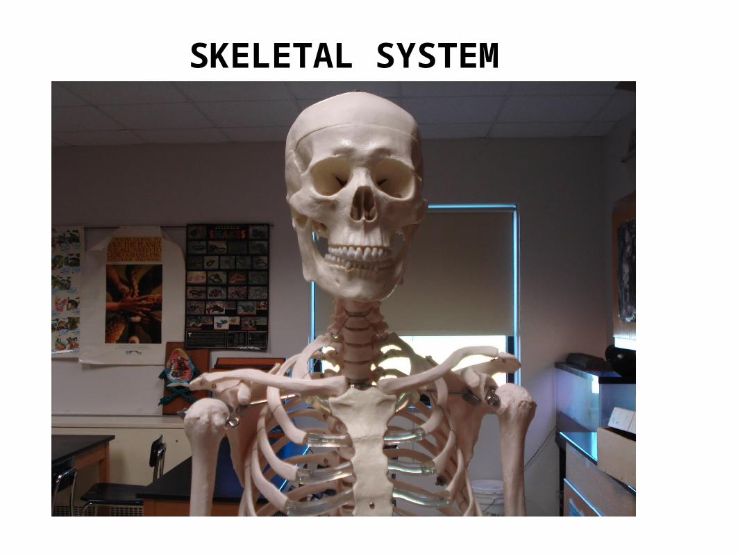
SKELETAL SYSTEM
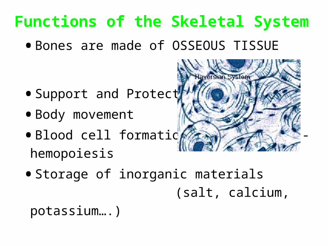
Functions of the Skeletal System
•Bones are made of OSSEOUS TISSUE
•Support and Protection
•Body movement
•Blood cell formation (bone marrow) - hemopoiesis
•Storage of inorganic materials
(salt, calcium, potassium….)
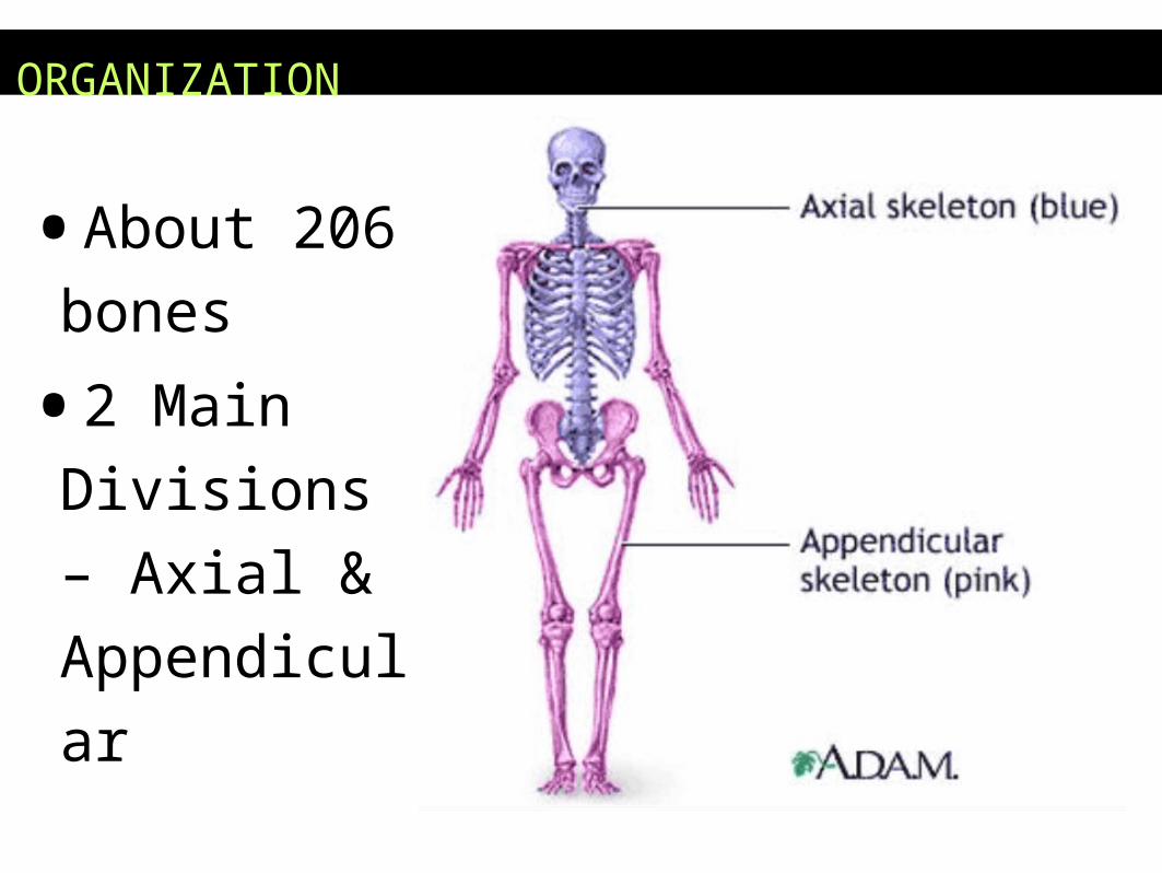
ORGANIZATION
•About 206
bones
•2 Main
Divisions –
Axial &
Appendicular
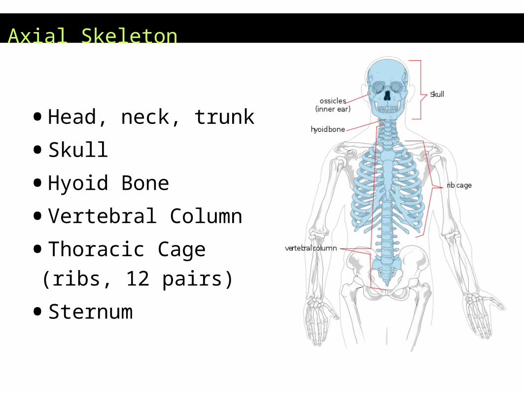
Axial Skeleton
•Head, neck, trunk
•Skull
•Hyoid Bone
•Vertebral Column
•Thoracic Cage (ribs, 12 pairs)
•Sternum

Hyoid Bone
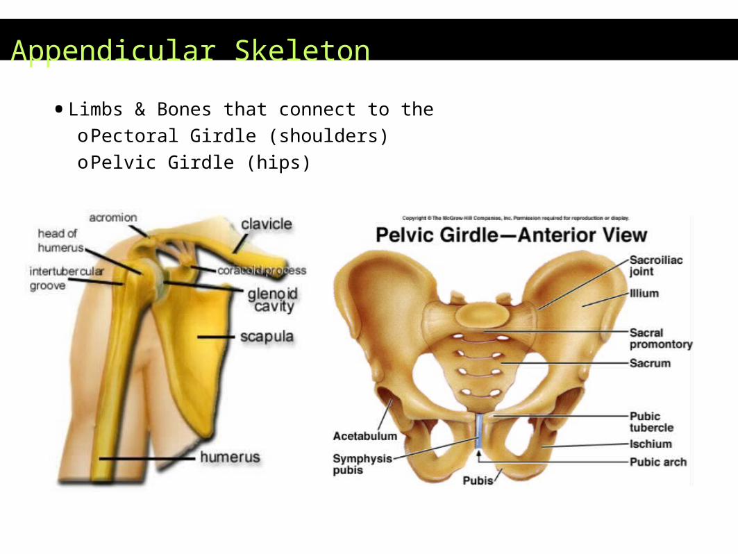
Appendicular Skeleton
•Limbs & Bones that connect to theoPectoral Girdle (shoulders)oPelvic Girdle (hips)
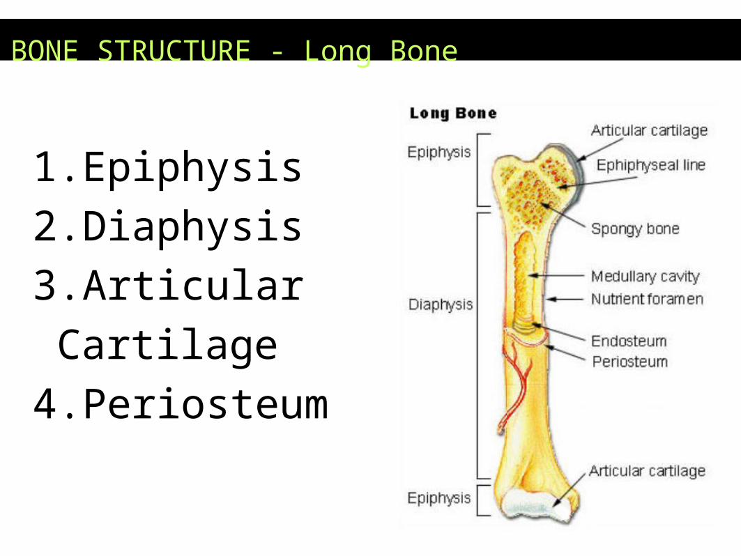
BONE STRUCTURE - Long Bone
1.Epiphysis
2.Diaphysis
3.Articular Cartilage
4.Periosteum
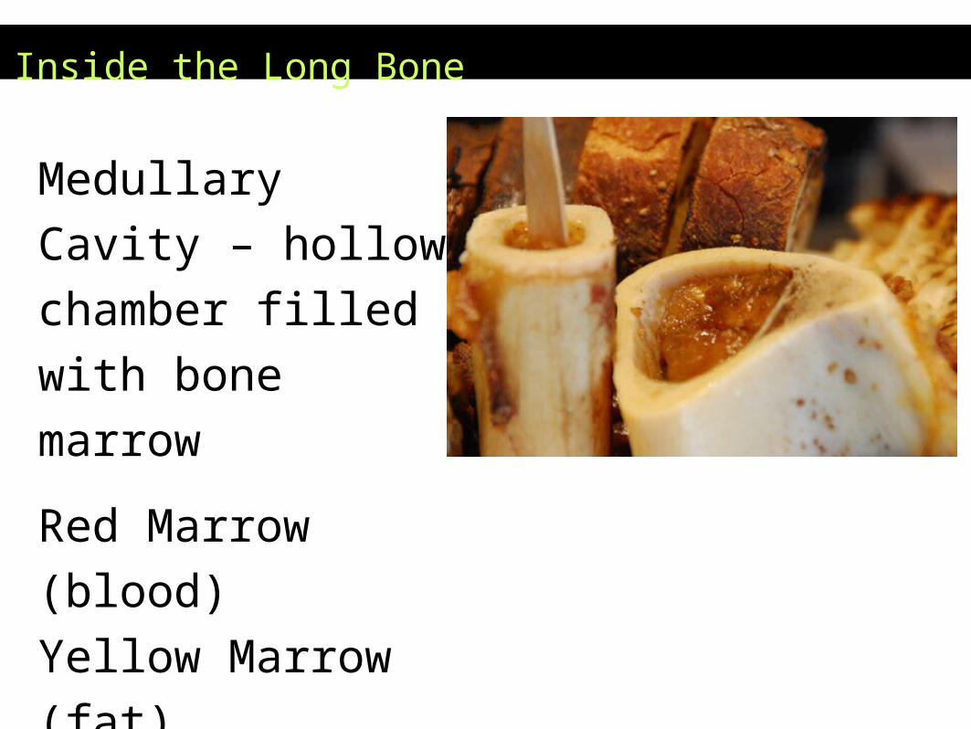
Inside the Long Bone
Medullary Cavity –
hollow chamber filled
with bone marrow
Red Marrow (blood)
Yellow Marrow (fat)
Endosteum
– lining of the
medullary
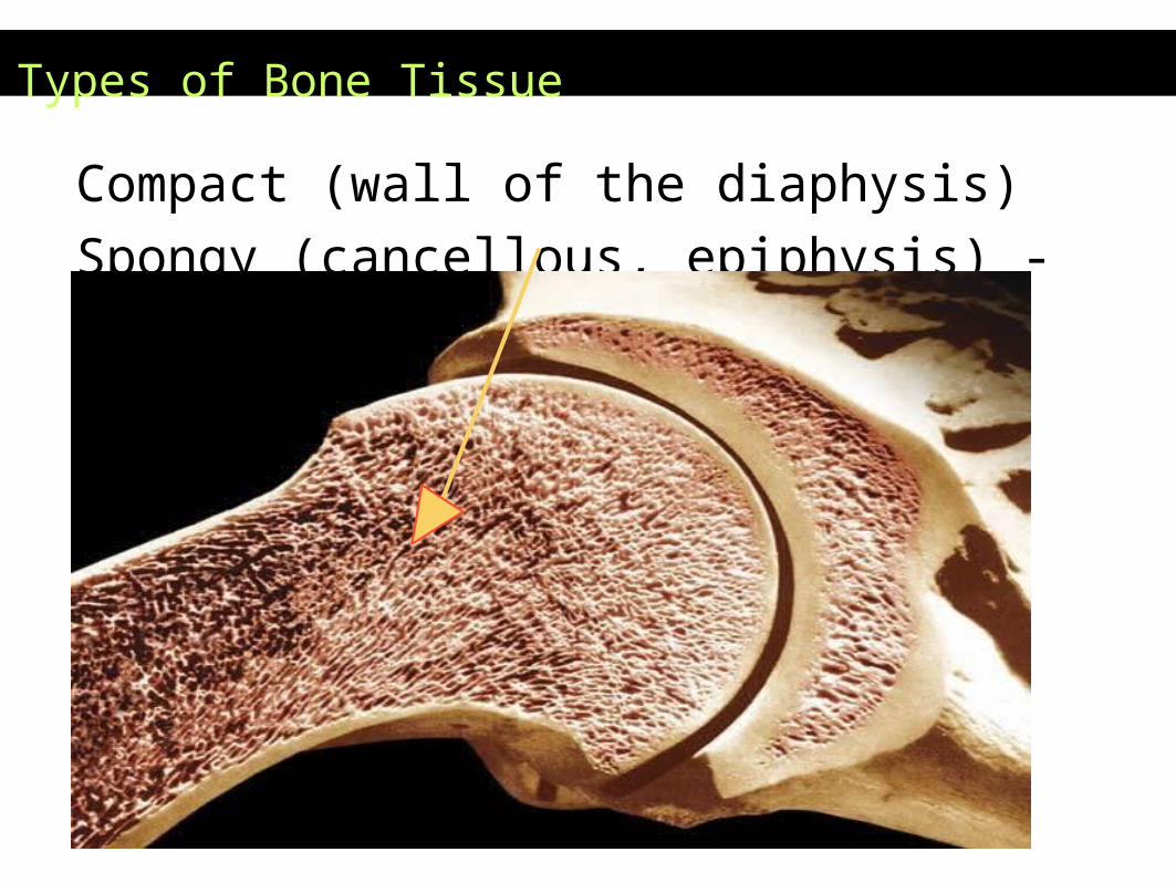
Types of Bone Tissue
Compact (wall of the diaphysis)
Spongy (cancellous, epiphysis) - red marrow
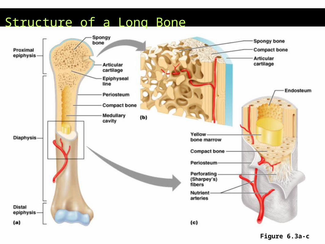
Structure of a Long Bone
Figure 6.3a-c

* Assignment
– Coloring of a Long
Bone
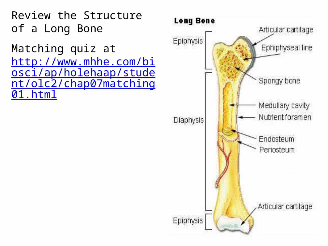
Review the Structure of a Long Bone
Matching quiz at http://www.mhhe.com/biosci/ap/holehaap/student/olc2/chap07matching01.html
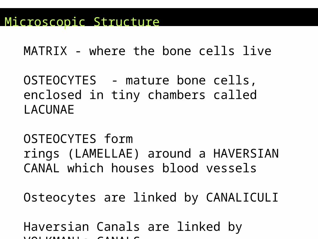
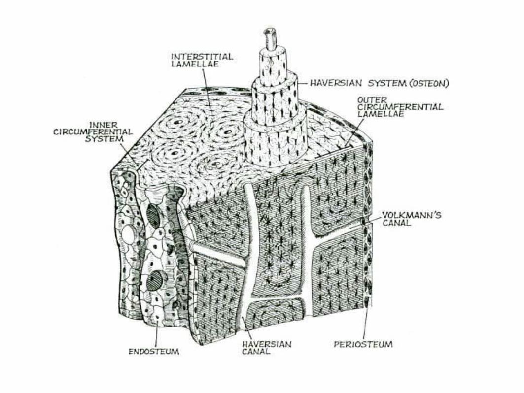
Microscopic Structure
MATRIX - where the bone cells live OSTEOCYTES - mature bone cells, enclosed in tiny chambers called LACUNAE OSTEOCYTES form rings (LAMELLAE) around a HAVERSIAN CANAL which houses blood vessels Osteocytes are linked by CANALICULI Haversian Canals are linked by VOLKMAN's CANALS
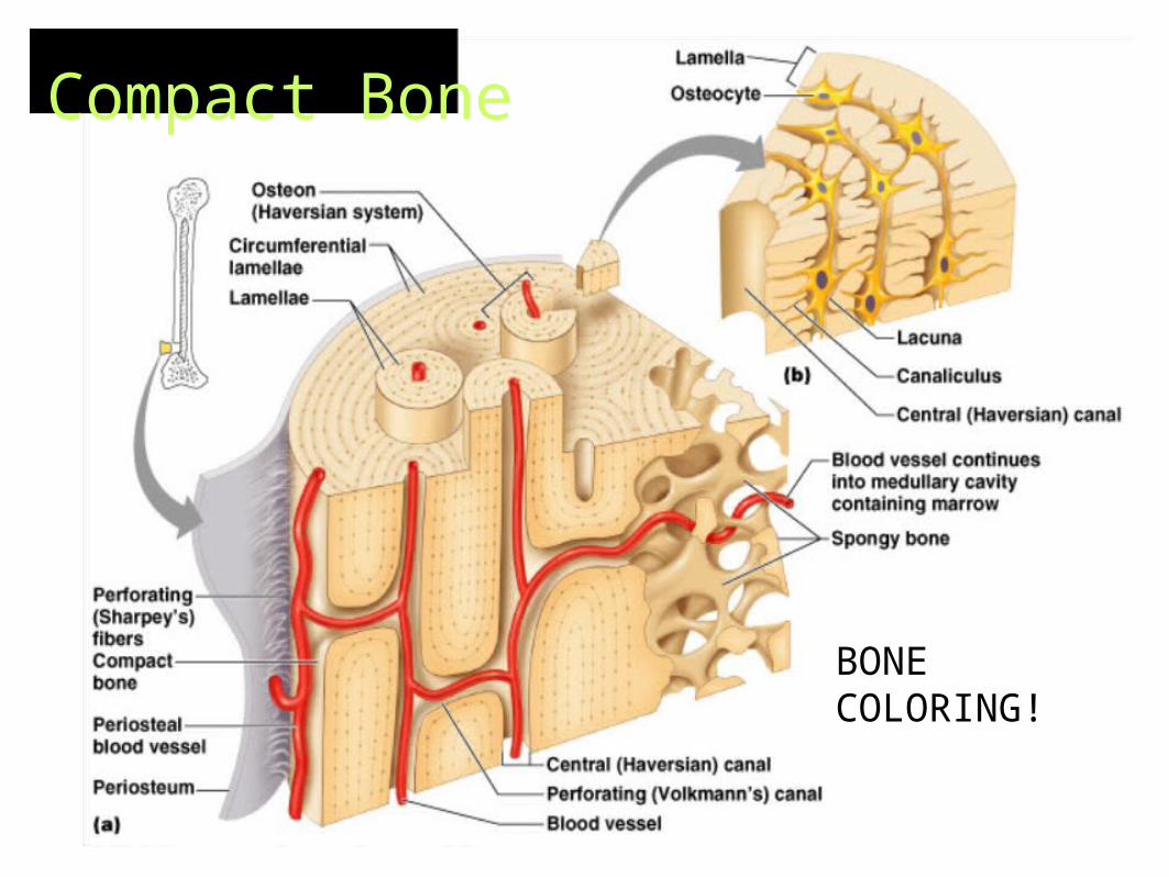
Compact Bone
BONE COLORING!

Test Yourself
Find the...
Haversian CanalVolkman's Canal
Lamellae
Spongy BoneCompact Bone
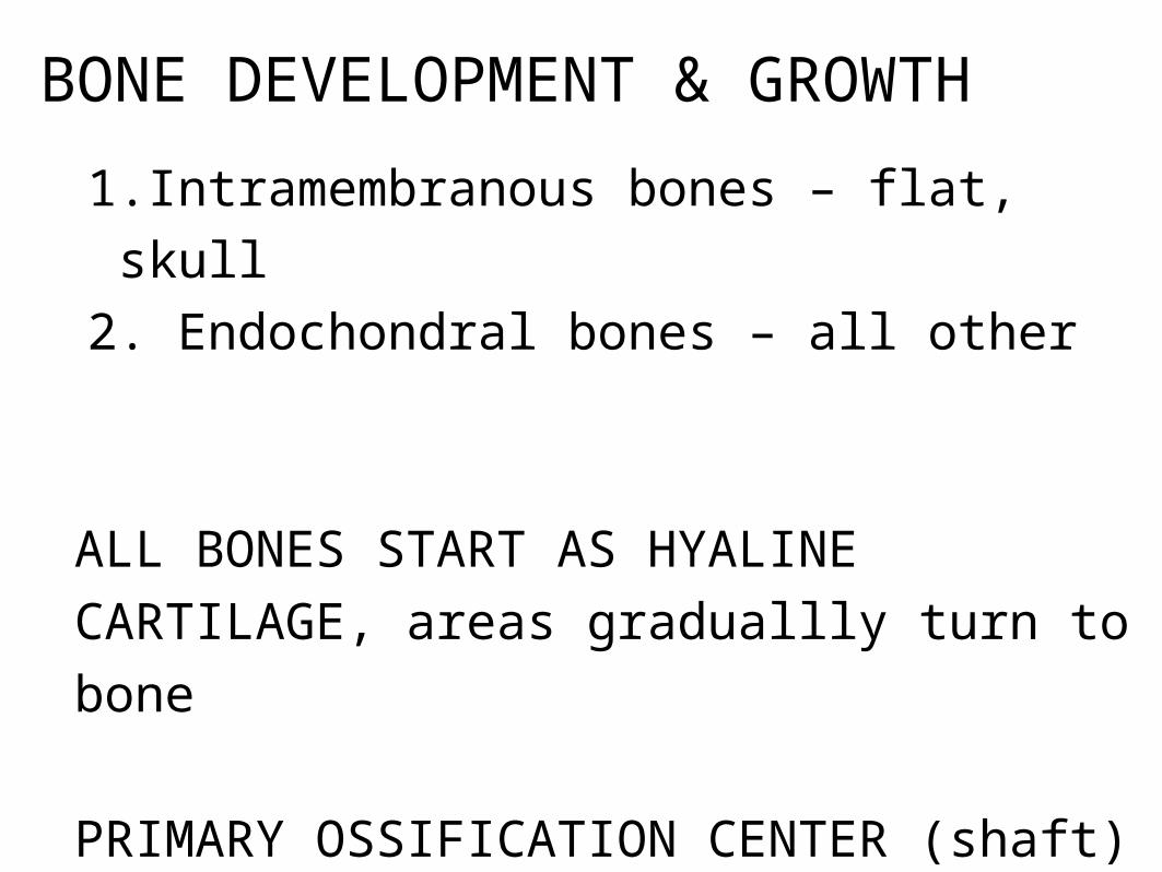
BONE DEVELOPMENT & GROWTH
1.Intramembranous bones – flat, skull
2. Endochondral bones – all other
ALL BONES START AS HYALINE
CARTILAGE, areas graduallly turn to bone
PRIMARY OSSIFICATION CENTER (shaft)
SECONDARY OSSIFICATION CENTER (ends)
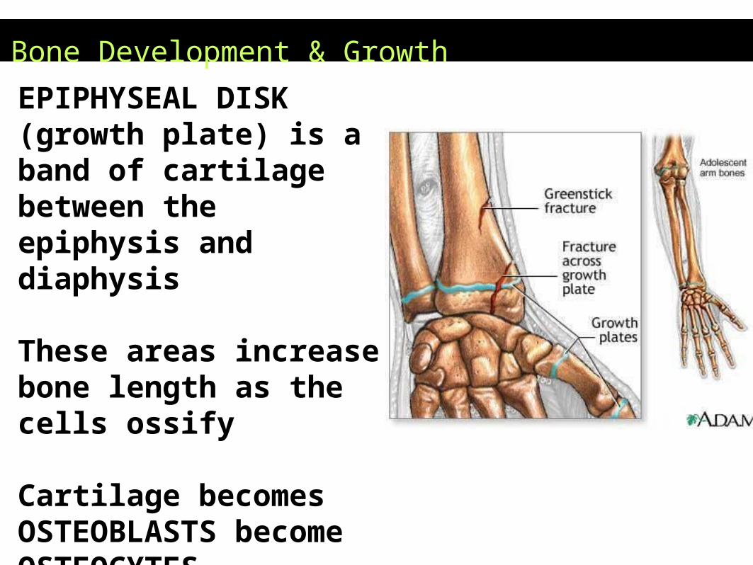
Bone Development & Growth
EPIPHYSEAL DISK (growth plate) is a band of cartilage between the epiphysis and diaphysis These areas increase bone length as the cells ossify Cartilage becomes OSTEOBLASTS become OSTEOCYTES
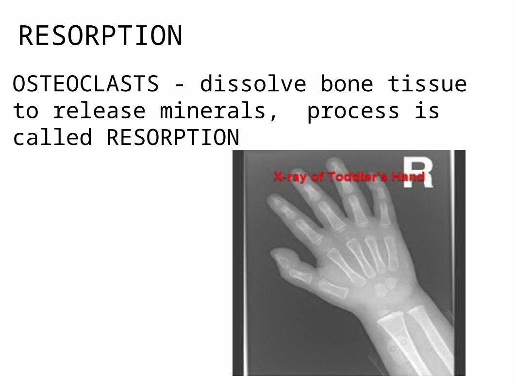
RESORPTION
OSTEOCLASTS - dissolve bone tissue to release minerals, process is called RESORPTION
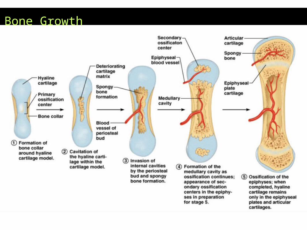
Bone Growth
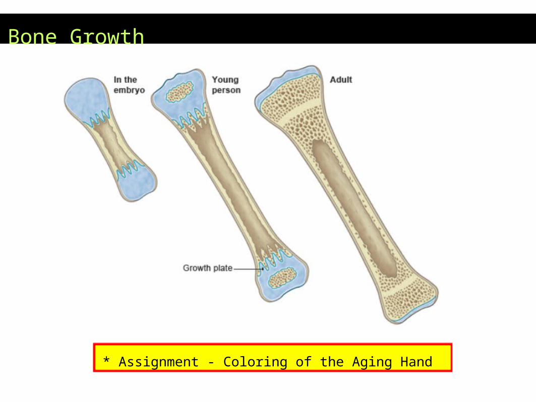
Bone Growth
* Assignment - Coloring of the Aging Hand
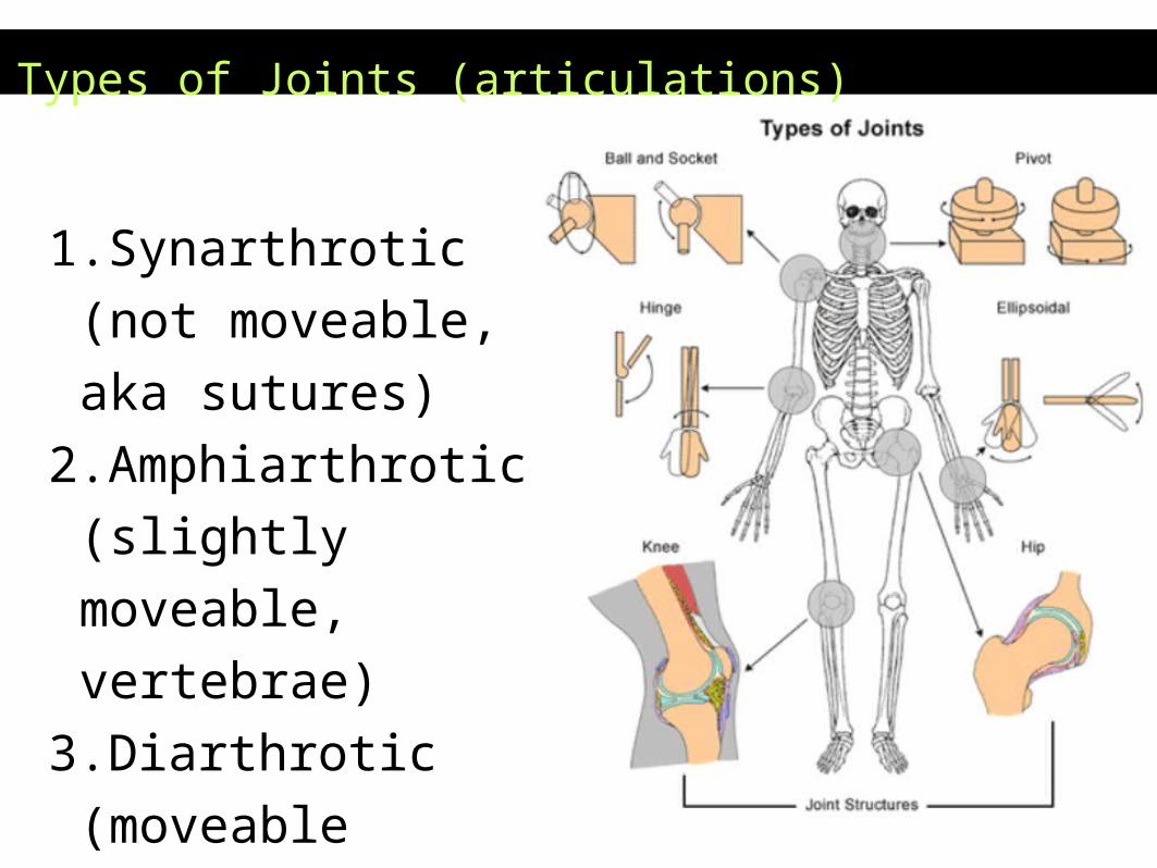
Types of Joints (articulations)
1.Synarthrotic (not
moveable, aka sutures)
2.Amphiarthrotic
(slightly moveable,
vertebrae)
3.Diarthrotic (moveable
joint, aka synovial
joints)
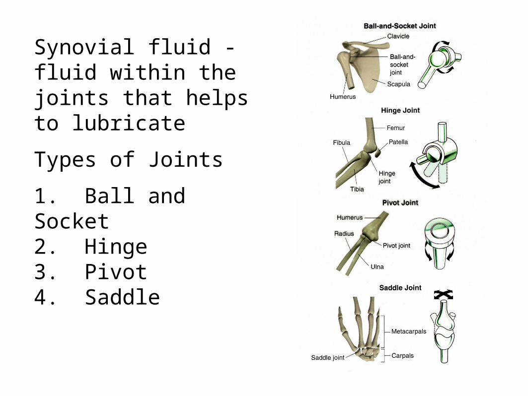
Synovial fluid - fluid within the joints that helps to lubricate
Types of Joints
1. Ball and Socket2. Hinge3. Pivot4. Saddle
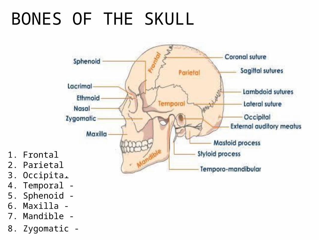
BONES OF THE SKULL
1. Frontal -2. Parietal - 3. Occipital -4. Temporal - 5. Sphenoid - 6. Maxilla - 7. Mandible -
8. Zygomatic -

TOPOGRAPHY OF THE SKULL
Foramen - refers to any tiny opening, nerves and blood vessels leave this opening to supply the face
Mental Foramen
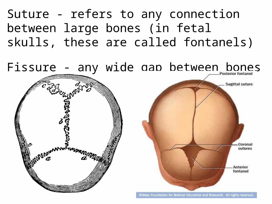
Suture - refers to any connection between large bones (in fetal skulls, these are called fontanels)
Fissure - any wide gap between bones
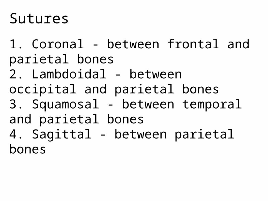
Sutures
1. Coronal - between frontal and parietal bones2. Lambdoidal - between occipital and parietal bones3. Squamosal - between temporal and parietal bones4. Sagittal - between parietal bones

Bones of the Skull & Sutures
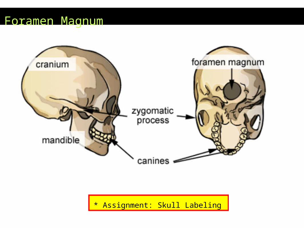
Foramen Magnum
* Assignment: Skull Labeling
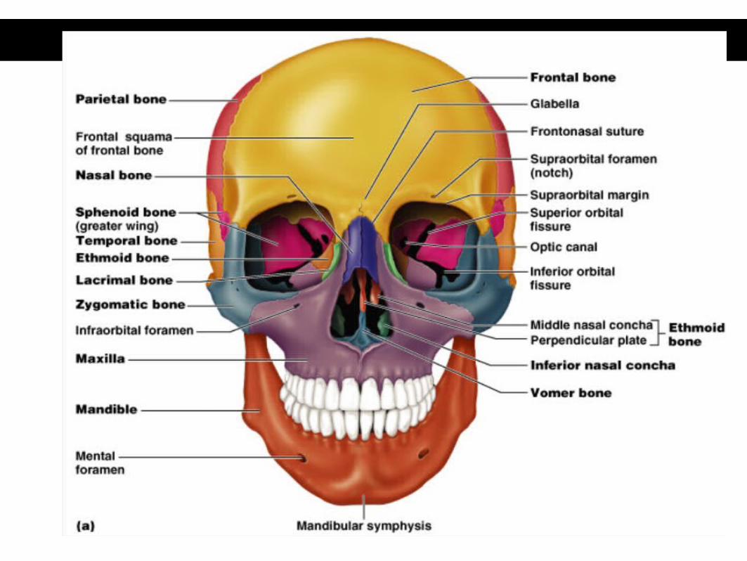
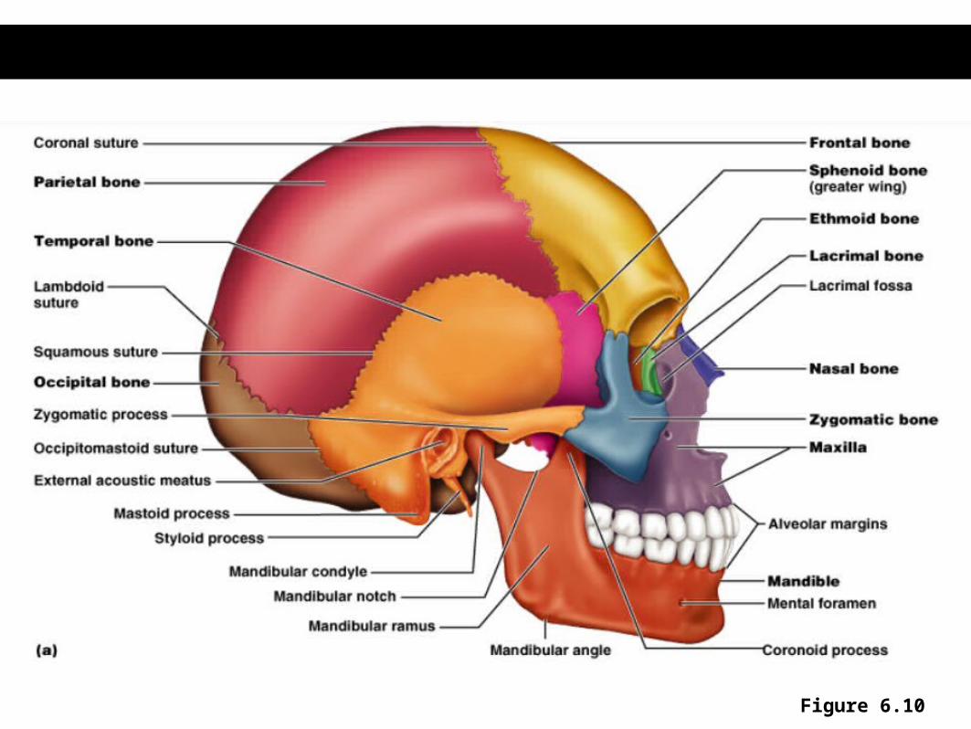
Figure 6.10
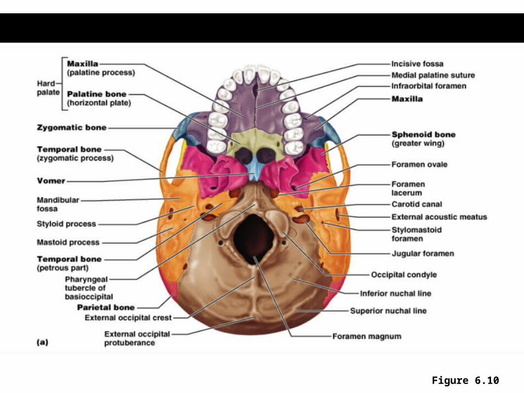
Figure 6.10

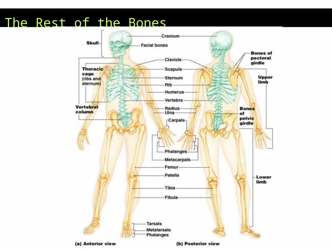
The Rest of the Bones

Vertebrae
Neck = cervical
Middle Back = thoracic
Lower Back = lumbar
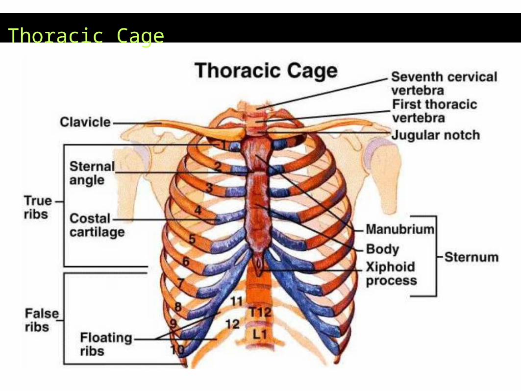
Thoracic Cage
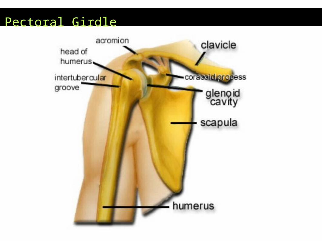
Pectoral Girdle

Bones of the Arm
Ulna goes to pinky (P-U)
Radius goes to thumb
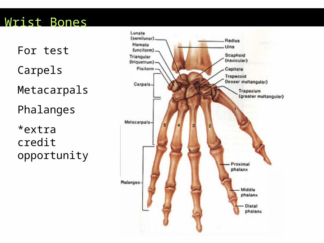
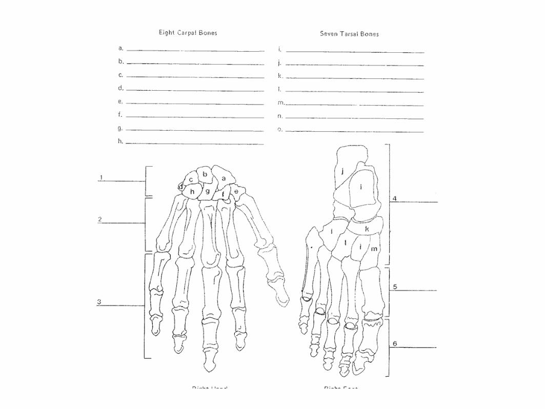
Wrist Bones
For test
Carpels
Metacarpals
Phalanges
*extra credit opportunity
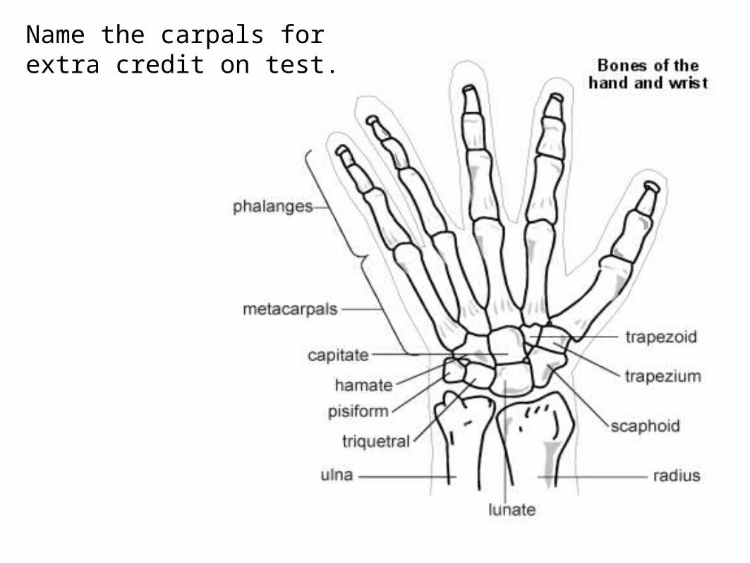
Name the carpals for extra credit on test.

Pelvic Girdle
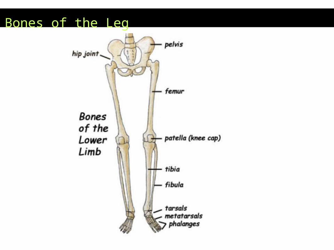
Bones of the Leg

Bones of the Ankle
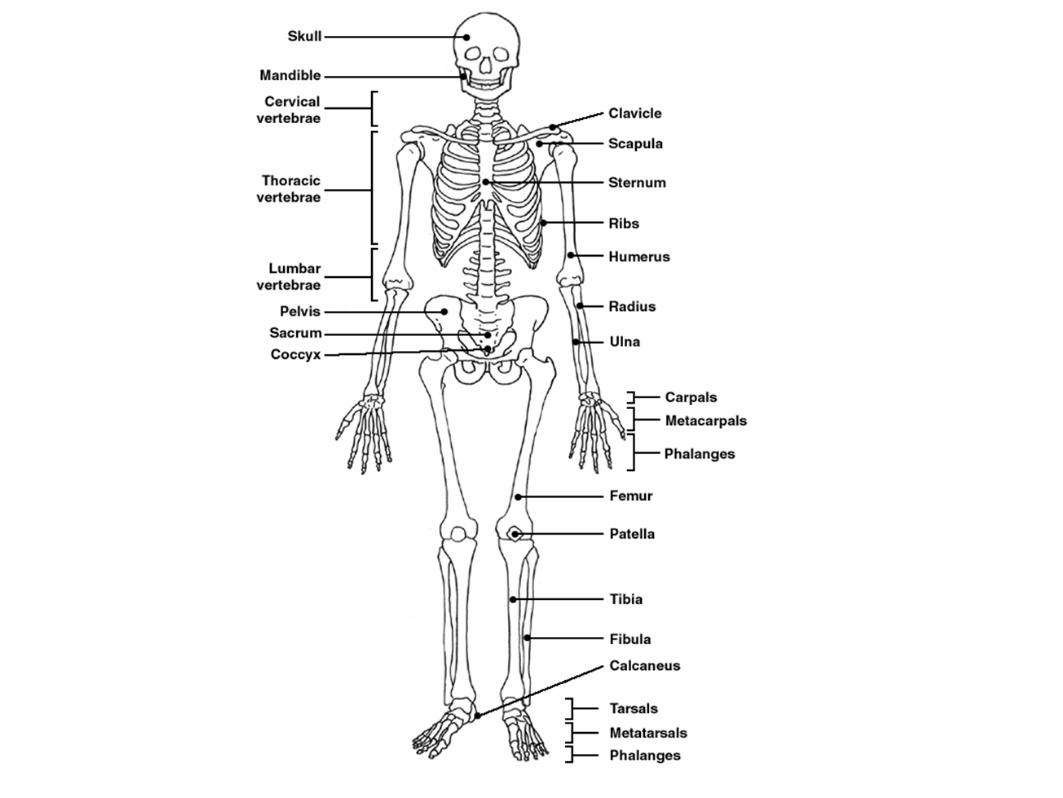
Assignment –
Skeleton Labeling
For Test
Calcaneous
Tarsals
Metatarsals
Phalanges
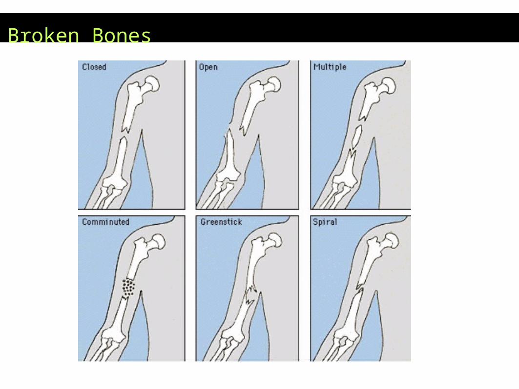
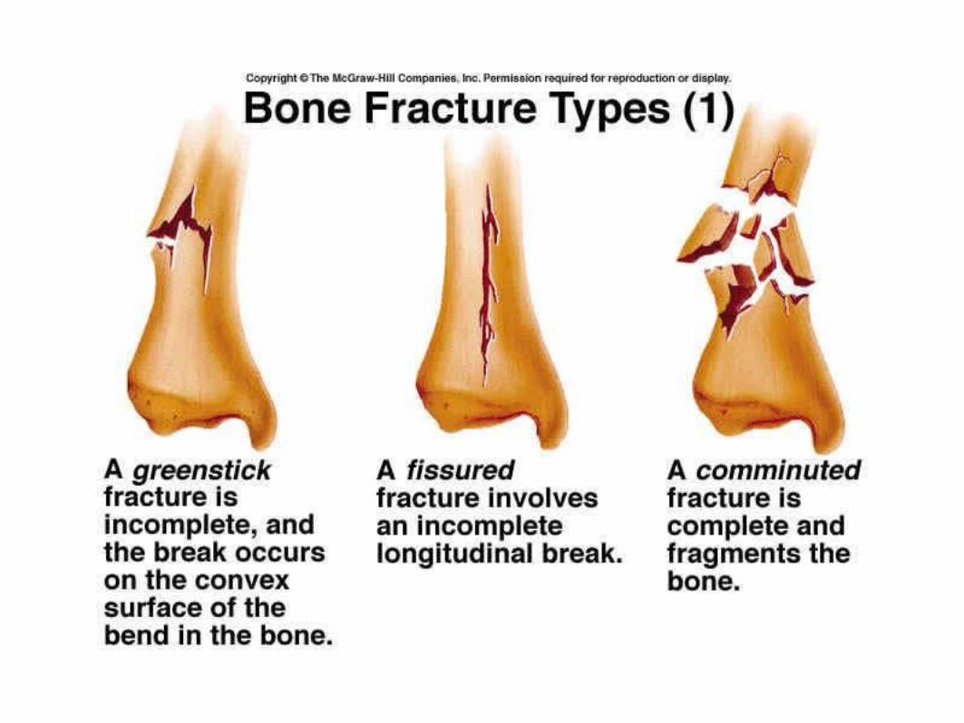
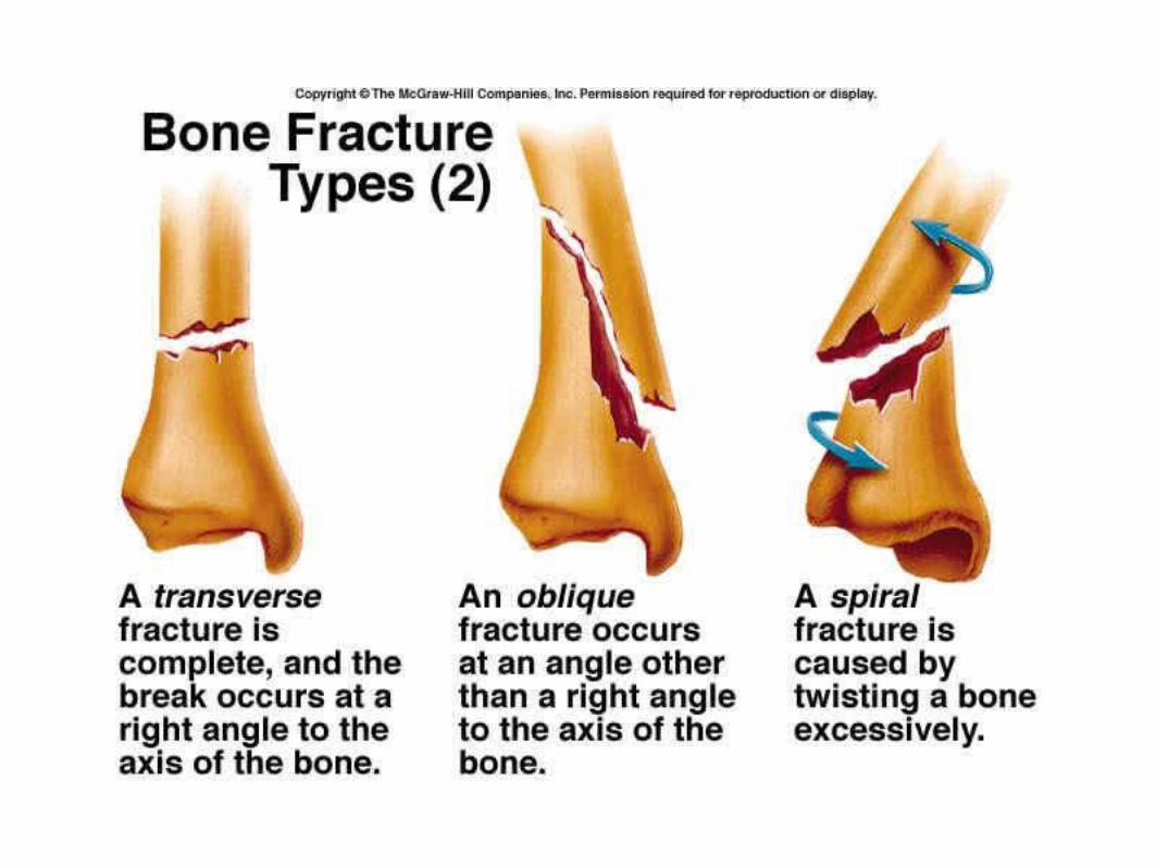
Broken Bones
Upcoming Assignments
•The Skeleton Mystery - read about a “crime
scene” and reconstruct skeletons to identify
the remains
•Watch a Bones Episode
•Identify Bones on a real skeleton • Lab
Practical Test
•Medical Imaging – learn how doctors view
bones and diagnose problems
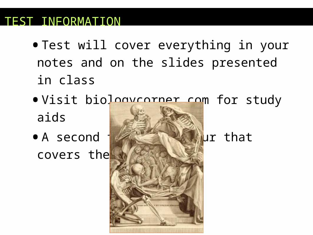
TEST INFORMATION
•Test will cover everything in your notes and on the
slides presented in class
•Visit biologycorner.com for study aids
•A second test will occur that covers the lab portion
Abnormal Bone Conditions
•BONE SPURS: abnormal growth. Can occur on any bone (e.g. heel).
•OSTEOPOROSIS: Increased activity of osteoclasts cause a break down bone, and the subsequent fewer minerals in the extracellular matrix make it fragile. The spongy bone especially becomes more porous.
•Men get it as well as women. What’s the best way to prevent osteoporosis? Exercise! What does exercise do? Makes bones bigger.
•The most common bone used for a bone graft is the iliac bone of the hip.

Osteoporosis
Figure 6.15
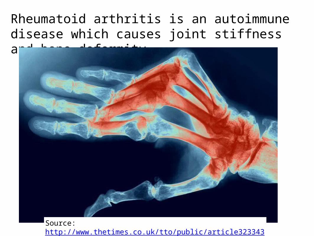
Rheumatoid arthritis is an autoimmune disease which causes joint stiffness and bone deformity
Source: http://www.thetimes.co.uk/tto/public/article3233439.ece
ABNORMALITIES OF THE SPINE
ABNORMALITIES OF THE SPINE
•SCOLIOSIS is a lateral curve in the spine
•KYPHOSIS is a hunchback curve
•LORDOSIS is a swayback in the lower region.
•ANKYLOSIS is severe arthritis in the spine and
the vertebrae fuse.
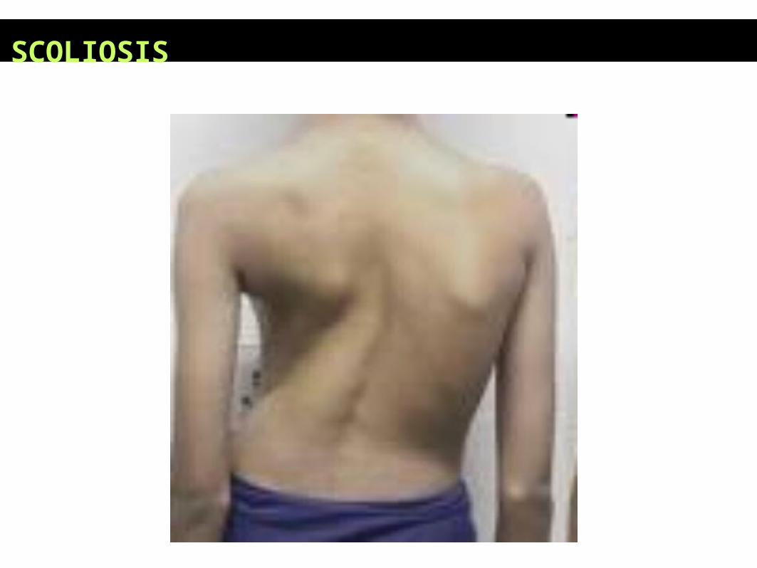
SCOLIOSIS
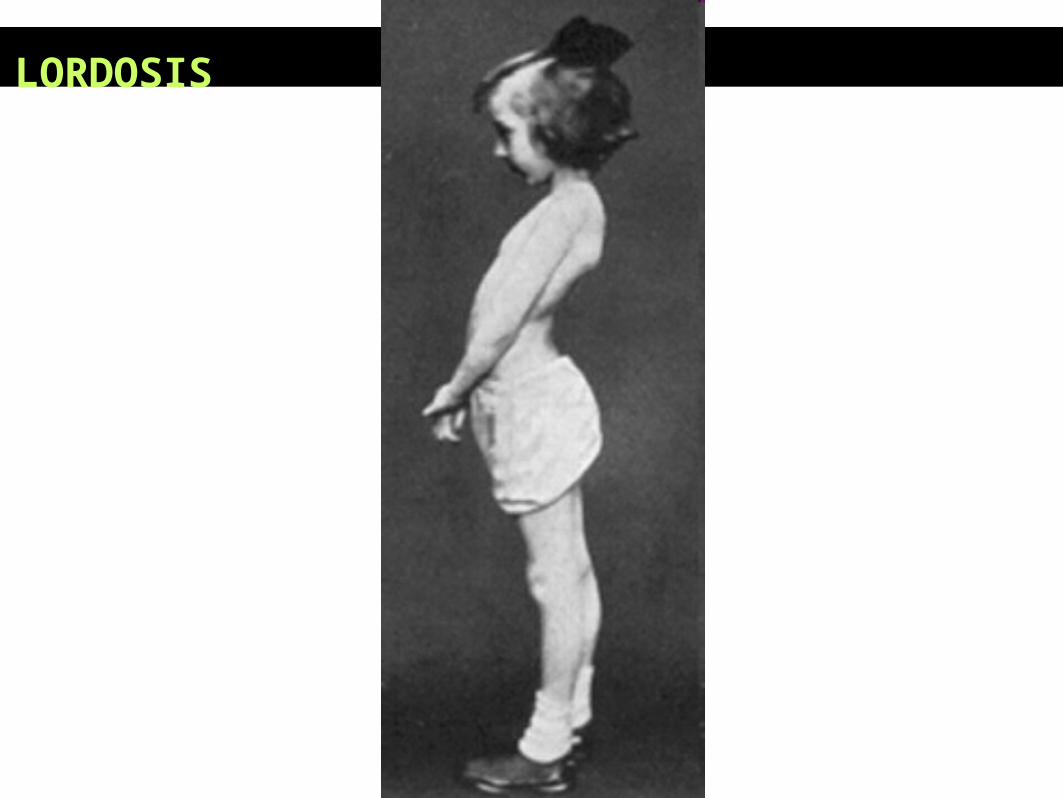
LORDOSIS

ANKYLOSIS
FUN FACTS ABOUT BONESBone is made of the same type of minerals as limestone.
•Babies are born with 300 bones, but by adulthood we have only 206 in our bodies.
•The giraffe has the same number of bones in its neck as a human: seven in total.
•The long horned ram can take a head butt at 25 mph. The human skull will fracture at 5mph.