Embed Size (px)
Citation preview

Charles F. Lanzieri1 Paul M. Duchesneau
Scott A. Rosenbloom Alison S. Smith
ArthurE. Rosenbaum
Received October 5, 1987; accepted after revision April 13, 1988.
Presented at the annual meeting of the American Society of Neuroradiology, Chicago, May 1988.
, All authors: Section of Neuroradiology, Division of Radiology, The Cleveland Clinic Foundation, 9500 Euclid Ave. , Cleveland, OH 44106. Address reprint requests to C. F. Lanzieri.
AJNR 9:1201-1204, November/December 1988 0195-6108/88/0906-1201 © American Society of Neuroradiology
1201
The Significance of Asymmetry of the Foramen of Vesalius
The foramen of Vesalius is a small, variable but consistently symmetrical structure located anteromedial to the foramen ovale and lateral to the foramen rotundum and vidian canal. It transmits an emissary vein through which the cavernous sinus and pterygoid plexus communicate. Fifty high-resolution CT scans of the skull base and two three-dimensional (Cemax) reconstructions were reviewed to determine criteria for defining the normal appearance of the foramen of Vesalius. Three normal types were classified: (1) a well-formed foramen, 1-2 mm in size (n = 32); (2) lack of visualization of the foramen (n = 11); and (3) partial assimilation of the foramen with the foramen ovale (n = 7). The foramen was remarkably symmetric in a large number of cases (n = 48).
Asymmetry signified abnormality in four of the six cases. Abnormal causes of asymmetry included invasion by nasopharyngeal melanoma, angiofibroma, carotid cavernous fistula with drainage through the emissary vein, and neurofibromatosis. Thus, for these usually symmetriC foramina of Vesalius, asymmetry is more likely the result of a pathologic process than a normal variant.
The importance of evaluating the skull base foramina has been well demonstrated in the literature [1, 2] . A variety of neoplastic and nonneoplastic processes has been documented to involve the foramen ovale and foramen rotundum as well as the pterygoid and transphenoidal canals [1, 3, 4].
A detailed examination of the skull base with all its foramina is indicated when searching for the cause of trigeminal neuralgia or neoplastic invasion. And an understanding of the normal morphology of the skull base is essential for correct interpretation of high-resolution CT scans. Tumors of nasopharyngeal origin are the most likely infracranial tumors to invade the middle portion of the skull base. The foramen of Vesalius is one of the foramina that may be affected .
The purpose of the present study is to establish criteria for defining the normal appearance of the foramen of Vesalius and to illustrate the significance of this structure in patients with potential skull-base abnormalities.
Materials and Methods
We reviewed the high-resolution CT scans of 50 patients who had been studied for suspected temporal bone abnormalities . All studies were done on a Synerview 1200 SX scanner (R)* with overlapping 2-mm or 3-mm-thick slices. Multiplanar reconstructions were used in selected cases. The foramen of Vesalius was identified and evaluated for position , size, shape, and symmetry.
During a 12-month period we also encountered four patients with pathologic changes at the skull base that involved the foramen of Vesalius .
Results
The foramen of Vesalius was best visualized with multiplanar reconstructions in 34 of 54 patients. Three patterns were identified in normal patients. Most commonly
• Picker International , Cleveland , OH .

1202 LANZIERI ET AL. AJNR:9 , November/December 1988
(n = 32), well-formed foramina measuring less than 2 mm were identified immediately anteromedial to the foramen ovale (Fig . 1). The exocranial opening occurs along the posterolateral surface of the lateral pterygoid plate, lateral to the insertion of the pharyngobasilar fascia, the spine of Civinini, and the pterygospinous ligament [5] . In most instances cuts were not obtained low enough to identify these small structures. The exocranial opening was seen in eight of 11 cases when cuts sufficiently inferior were available. The foramen of Vesalius was not demonstrable in 11 patients (Fig. 2), perhaps owing to its small size in some patients in whom a condensation of cortical bone could be seen even when a true foramen was not apparent (three patients). In seven patients
A B
o E
A B Fig. 2.-Normal variations.
there was partial assimilation of the foramen of Vesalius within the anterior angle of the foramen ovale.
The foramina of Vesalius were judged symmetrical in 48 patients. In addition to the two normal patients with incidental asymmetry, four cases were observed in which asymmetry was believed to indicate pathologic change. In two of these cases, asymmetry may have been due to invasion of the emissary vein and foramen of Vesalius by tumors; specifically, nasopharyngeal melanoma and juvenile angiofibroma (Fig. 3). Asymmetry of the foramen of Vesalius was also seen in association with a carotid-cavernous fistula in which one of the pathways of drainage from the cavernous sinus was through the emissary vein into the pterygoid plexus (Fig . 4).
c
c
Fig. 1.-Normal anatomy. A, Axial high-resolution CT scan through skull
base shows endocranial opening of foramen of Vesalius (larger black arrow) just anteromedial to foramen ova Ie (smaller black arrows) and lateral to pterygoid canal (curved white arrow).
B, Exocranial opening (curved arrow) can be seen just lateral to spine of Civinini (larger straight arrow) on medial surface of lateral pterygoid plate (smaller straight arrows).
C, Direct coronal image occasionally shows foramen (larger straight arrow) between foramen lacerum (curved arrow) and foramen ova Ie (smaller straight arrows).
D and E, Off-axis coronal reconstructions usually show foramen well in its entire length (larger arrow). Smaller arrows point to foramen ova Ie.
A and B, In 14 patients, either the foramen of Vesalius was not visible on CT scans (A) or bone condensation was visible without a demonstrable foramen (arrows in B).
C, In seven patients, there was partial assimilation with the foramen ova Ie (arrows), which was symmetrical in all cases.
AJNR:9 , November/December 1988
Fig, 3,-lnvasion by tumors. A-C, High-resolution CT scans with off-axis
coronal reconstruction (A and B) and coronal reconstruction (C) were obtained in this 14-yearold boy with recurrent juvenile angiofibroma invading the skull base. The foramen of Vesalius is enlarged and irregular (curved arrows) in both views. Straight arrows point to foramen ova Ie.
D and E, Axial (D) and off-axis coronal reconstruction (E) CT scans of 63-year-old woman with nasopharyngeal melanoma invading the skull base show enlarged, irregular foramen of Vesalius (wide arrows), foramen ovale (straight arrows), and foramen spinosum (curved arrow in E).
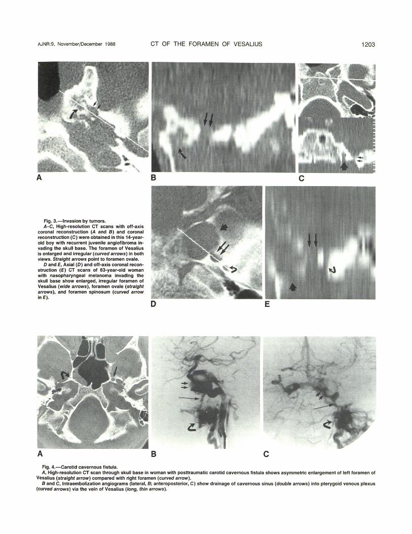
Fig. 4.-Carotid cavernous fistula.
CT OF THE FORAMEN OF VESALIUS 1203
B c A, High-resolution CT scan through skull base in woman with posttraumatic carotid cavernous fistula shows asymmetric enlargement of left foramen of
Vesalius (straight arrow) compared with right foramen (curved arrow). Band C, Intra embolization angiograms (lateral, B; anteroposterior, C) show drainage of cavernous sinus (double arrows) into pterygoid venous plexus
(curved arrows) via the vein of Vesalius (long, thin arrows).

1204 LANZIERI ET AL. AJNR:9, November/December 1988
One patient with neurofibromatosis exhibited asymmetric foramina.
Discussion
The foramen of Vesalius is a small , inconstant but symmetrical opening in the medial aspect of the greater wing of the sphenoid bone anteromedial to the foramen ovale [5]. Through it an emissary vein courses, which affords communication between the cavernous sinus in the middle cranial fossa and the pterygoid plexus of veins within the parapharyngeal space [6]. The exocranial opening is most often found along the medial surface of the lateral pterygoid plate and spine of Civinini . Another useful landmark is the pterygospinous ligament (or bar, when ossified, as in 11 % of patients), which is immediately medial to the foramen of Vesalius [5]. The exocranial opening enters the scaphoid fossa, where the tensor veli palatini muscle originates, and is therefore peripheral to the pharyngobasilar fascia. Anatomic studies have found that this structure sometimes ends within the diploic space and rarely exceeds 2 mm in size [5]. When the foramen is absent the respective emissary vein leaves the skull through the foramen ovale [6]; therefore, nonvisualization is a known anatomic variation.
The foramen of Vesalius was first recognized on plain film by Schuller, and may be seen in only 15% of 50 axial skull radiographs [5]. It is distinctive to humans and has not been found in other primates [7] .
The results of the present CT study suggest that the foramen of Vesalius may be a much more constant and symmetrical structure than previously thought. In the two asymptomatic patients there was an approximately 2-mm difference in the size of the foramina. An intact cortical rim was present, and it is tempting to postulate that these are incidental findings. A larger series of studies with long-term follow-up may be needed for confirmation. Enlargement and especially asymmetry of this structure may be abnormal in the majority of cases. Invasion of the skull base due to perineural extension is well known [7]. Similarly, the mechanism for tumorous invasion of the foramen of Vesalius from the nasopharynx may be along the course of the tensor veli palatini muscle and eustachian tube via a small gap between
the skull base and the pharyngobasilar fascia called the foramen of Morgagni [8, 9] . The scaphoid foramen at the skull base is common to both the origin of the tensor muscle and the exocranial opening of the foramen of Vesalius. Thus, a neoplasm may follow the tensor muscle into the scaphoid foramen and subsequently into the foramen of Vesalius and cavernous sinus.
To our knowledge, ipsilateral enlargement of the foramen of Vesalius has not been previously described in carotidcavernous fistulas [10]. In the single case reported here, enlargement was caused by participation of the emissary vein of Vesalius in the drainage of the cavernous sinus.
Finally, in the single case of asymmetry associated with neurofibromatosis, it is tempting to postulate that this may be another result of mesodermal dysplasia.
In summary, evaluation of this consistently symmetrical structure is of some importance when studying the skull base, since asymmetry is more likely the result of a pathologic process than a normal variant.
REFERENCES
1. Pandolfa I, Gaeta M, Blandino A, Longo M. The radiology of the pterygoid canal: normal and pathologic findings. AJNR 1987;8:479-483
2. Curtin AD, Williams R, Johnson J. CT of perineural tumor extension: pterygopalatine fossa. AJNR 1984;5:731-737
3. Currarino G, Maravilla KR, Salyer KE. Transsphenoidal canal and its pathologic implications. AJNR 1985;6:39-43
4. Curtin HD, Williams R. Computed tomographic anatomy of the pterygopalatine fossa. Radiographies 1985;5:429-440
5. Sondheimer FK. Basal foramina and canals. In: Newton TH , Potts DG, eds. Radiology of the skull and brain. St. Louis: Mosby, 1971 :305-321
6. Henderson WR. A note on the relationship of the maxillary nerve to the cavernous sinus and to an emissary sinus passing through the foramen ovale. J Anat 1966;100 :905-908
7. Wood-Jones F. The non-metrical morphological characters of the skull as criteria for racial diagnosis. Part I. General discussion of the morphological characters employed in racial diagnosis. J Anat 1931 ;65: 179-195
8. Mancuso AA, Hanafee WN. Nasopharynx and parapharyngeal space in computed tomography and magnetic resonance imaging of the head and neck, 2nd ed. Baltimore: Williams & Wilkins, 1985
9. Silver AJ, Mawad ME, Hilal SK, Sane P, Ganti SR. Computed tomography of the nasopharynx and related spaces. part I: anatomy. Radiology 1983;147:725-731
10. Ahmadi J, Teal JS, Segall HD, Zee CS, Han JS, Becker TS. Computed tomography of carotid-cavernous fistula. AJNR 1983;4 :131-136