Embed Size (px)
Citation preview

Available online at www.sciencedirect.com
www.elsevier.com/locate/yebeh
Epilepsy & Behavior 12 (2008) 445–455
The selective amobarbital test in the anterior choroidal artery:Perfusion pattern assessed by intraarterial SPECT and prediction
of postoperative verbal memory
S. Vulliemoz a,*, A.J. Pegna a, J.-M. Annoni a, H. Yilmaz b, J.P. Willi c,L. Spinelli a, T. Landis a, M. Seeck a
a Department of Neurology, University Hospital of Geneva, Geneva, Switzerlandb Neuro-interventional Section, Department of Clinical Neurosciences, University Hospital of Geneva, Geneva, Switzerland
c Department of Nuclear Medicine, University Hospital of Geneva and Faculty of Medicine, University of Geneva, Switzerland
Received 30 September 2007; revised 22 November 2007; accepted 26 November 2007Available online 8 January 2008
Abstract
To screen for patients at risk for memory decline after temporal lobe epilepsy (TLE) surgery, selective amobarbital procedures, suchas injection into the anterior choroidal artery (ACA-IAT), are sometimes used. We investigated the extent of the territory affected duringACA-IAT and its predictive value with respect to postoperative memory. Seventeen patients with TLE underwent ACA-IAT. In 9 of 17patients, intraarterial SPECT co-registrated to MRI allowed delineation of amobarbital-perfused structures. Another subgroup of 9 of17 patients underwent anterior temporal lobectomy. Verbal memory was tested pre- and postoperatively and during ACA-IAT. Majorvariations in the ACA-IAT perfusion pattern occurred and were not correlated with the verbal memory scores during ACA-IAT. Post-operatively, no patient experienced a severe verbal memory decline, but individual postoperative performance was not correlated withresults during ACA-IAT. Our study suggests that ACA-IAT can be used to screen for severe postoperative amnesia in inconclusive cases,but cannot predict individual outcome, even when the perfusion pattern is taken into account.� 2007 Elsevier Inc. All rights reserved.
Keywords: Temporal lobe epilepsy; Amobarbital; Anterior choroidal artery; Single-photon-emission computed tomography; Memory
1. Introduction
Temporal lobe epilepsy (TLE) is the most frequent causeof pharmacoresistant focal epilepsy in adults and adoles-cents. Resection of anterior and mesial temporal lobe struc-tures can lead to complete seizure control in well-definedcases and has become an established treatment [1]. Postop-erative impairment of memory, particularly verbal mem-ory, remains a feared complication, and several methodshave been developed to determine the risk of such an amne-sic syndrome [2,3]. The most recognized risk factors for sig-
1525-5050/$ - see front matter � 2007 Elsevier Inc. All rights reserved.
doi:10.1016/j.yebeh.2007.11.015
* Corresponding author. Address: Presurgical Evaluation Unit, Depart-ment of Neurology, University Hospital of Geneva, Micheli-du-Crest 24,1211 Geneva 14, Switzerland. Fax: +41 22 372 84 75.
E-mail address: [email protected] (S. Vulliemoz).
nificant postoperative verbal memory decline includedominant hemisphere surgery, intact preoperative interictalmemory, and absence of hippocampal sclerosis on MRI [4–6]. Older age [4,7], male gender [8,9], extent of surgicalresection [10,11], low preoperative verbal IQ, as well asthe presence of cortical dysgenesis [12], are other risk fac-tors and should all be taken into account before surgery.
The intraarterial amobarbital test (IAT), initially devel-oped to identify the language-dominant hemisphere [13],consists of injection of a short-acting barbiturate into a cere-bral artery and produces transient deactivation of the corre-sponding vascularized structure. Behavioral testing of thepatient during IAT reveals the functioning of the unanesthe-tized structures, that is, the integrity of the structures thatwill remain after the intervention. Intraarterial injection ofamobarbital into the carotid artery is a validated procedure

446 S. Vulliemoz et al. / Epilepsy & Behavior 12 (2008) 445–455
widely employed to screen for the risk of postoperativeamnesia, to predict verbal memory decline after temporallobe epilepsy (TLE) surgery, and to lateralize the seizurefocus [14,15]. It compares the cognitive performance afterinjection into the hemisphere ipsilateral and contralateralto the epileptogenic focus by using indices of hemisphericasymmetry. However, its role in memory evaluation maybe hampered by the aphasia induced after injection intothe dominant hemisphere or by confusion after injection intothe nondominant hemisphere. Furthermore, although amo-barbital injected into the carotid artery is known to producehypoperfusion in the mesial temporal structures, as deter-mined by intravenous SPECT [16–18], other reports usingintraarterial injection of 99mTc-hexamethylpropyleneamineoxime (99mTc-HMPAO) during IAT have reported that theanesthetic agent did not perfuse the mesial temporal struc-tures in up to 75% of the patients [16,19,20]. Repetition ofthe IAT in the same patients produces less reliable resultsfor memory than for language testing [21].
These observations, combined with the development ofmore selective epilepsy surgery, led to the design of selec-tive amobarbital tests targeting the mesial temporal struc-tures. The mesial temporal lobe and, more specifically,the amygdaloid body and hippocampal formation are vas-cularized by the anterior choroidal artery (ACA) and theposterior choroidal artery (a branch of the posterior cere-bral artery), with anastomoses between both vessels[22,23]. IAT of the anterior choroidal artery (ACA-IAT)[24] and the posterior cerebral artery (PCA-IAT) [24–27]have been found useful in this respect.
In the present case series, we were interested in evaluat-ing the usefulness of ACA-IAT by determining exactlywhich brain regions are perfused by the test, how thisaffects memory performance (verbal and visual recogni-tion), and, ultimately, whether the procedure results inaccurate prediction of postoperative verbal memorydecline.
2. Methods
2.1. Patients
Between 1995 and 2006, 17 patients aged 15–56 years (8 women, 9men, mean age = 34.3 years) with intractable temporal epilepsy who wereconsidered candidates for resective surgery were evaluated with ACA-IAT. All except two (patients 9 and 17) were right-handed. On MRI, 14had hippocampal sclerosis and 3 had normal hippocampi (patients 7, 8and 11). Four patients had evidence of bilateral temporal dysfunctionon MRI or intracranial EEG (patients 6, 7, 8, and 11). This study wasapproved by the local ethics committee.
The indication for ACA-IAT was related to (1) the presence of domi-nant-hemisphere epilepsy or (2) a focus in the nondominant hemisphere,but significantly impaired interictal verbal memory scores. In our group,9 of 17 patients (4 women, 5 men) underwent additional assessment ofthe perfusion pattern with intraarterial SPECT. In all patients, the proce-dure was carried out ipsilaterally to the potential surgical resection, as ini-tially described by Wieser et al. [24].
Nine patients (3 patients with SPECT, 6 patients without) underwent tem-poral lobe surgery consisting of amygdalohippocampectomy and resection ofthe anterior third of the temporal lobe (anterior temporal lobectomy).
Table 1 summarizes the clinical features and the results of the patientworkups.
2.2. Selective amobarbital test in the anterior choroidal artery
Percutaneous transfemoral catheterization under local anesthesia wasperformed, and selective digital subtraction angiograms (DSAs) of theinternal carotid artery were obtained with biplanar angiography equip-ment (Philips Integris BV/BN 3000, Best, The Netherlands). The proce-dure was performed under intravenous heparin coverage (3000 IU), anda 5-French Envoy guiding catheter (Cordis Endovascular, Miami Lakes,FL, USA) was used. Depending on the size of the ACA and on the anat-omy of the carotid siphon, a previously steam-shaped microcatheter wasused (Vasco 10 microcatheter, Balt, Montmorency, France, or Fastracker18 microcatheter, Boston Scientific, Natick, MA, USA). A microguidewire (Transend Ex 14, Boston Scientific) was used to navigate throughthe carotid siphon. The previously steam-shaped microcatheter enableddirect catheterization of the ACA without the use of the microguide wire,thus avoiding the risk of arterial damage or spasm during the procedure.The microcatheter position was checked by angiographic series in theanteroposterior and lateral positions. Amobarbital (75 mg) was thenslowly and manually injected into the ACA over about 20 seconds to avoidreflux into the middle cerebral artery. Angiograms were performed afteramobarbital and 99mTc-ethyl cysteinate diethylester (99mTc-ECD) injec-tions to verify that the microcatheter remained at the same location.
2.3. Combined intraarterial and intravenous 99mTc-ECD cerebral
SPECT
Immediately after the amobarbital injection, a maximum of 0.5 mL of99mTc-ECD, corresponding to a dose of 26.4 ± 6.3 MBq, was slowlyinjected manually into the ACA. Following intraarterial injection, a doseof 460.8 ± 163.6 MBq of 99mTc-ECD was injected intravenously to bettervisualize the brain parenchyma in six of eight patients. The first twopatients (patients 1 and 2) in the series received only intraarterial injec-tions. SPECT imaging was performed 30 to 60 minutes after completionof the angiography on a three-head Toshiba GCA-9300 A/HR camerawith fan beam collimators. Data were acquired and reconstructed in a128 · 128 matrix. A Shepp and Logan filter was used. The scatter correc-tion was applied using the FBP/Sorenson method. Whole-brain volumewas covered.
2.4. Co-registration of intraarterial and intravenous SPECT to
MRI
Nuclear medicine images were transferred on the dedicated image pro-cessing workstation in Analyze format. The images were then resampled inan isotropic set and co-registered to the patient’s own millimetric three-dimensional MRI scan using a six-parameter rigid-body algorithm. Forthis purpose, we used AIR5.2 software, a voxel-based intensity algorithmavoiding the use of external markers.
In one of the two patients (patients 1 and 7) who received only anintraarterial injection, co-registration was possible (patient 7) becausehemispheric contamination provided anatomical landmarks. In the other(patient 1), co-registration was not possible and perfused structures wereidentified from the native SPECT images alone.
2.5. Neuropsychological protocol
ACA-IAT was performed according to the following protocol, asdescribed elsewhere [24]. Sixty cards, each containing a verbal item anda visual item to memorize (dual encoding run), were presented in centralvision to the patients. Verbal and visual stimuli were positioned one underthe other on the card; on half of the cards, the visual stimulus was on top,and on the other half, on the bottom. The cards were presented in pseudo-random order. Subjects were instructed to press a button (thumb of the

Table 1Clinical data
Gender/age/handedness
ACA-IAT side
Age at onset,frequency
Clinical aspects ATCD/etiology MRI EEG I Interictal PEThypometabolism
Operation
Patient 1 F/44y/R
Ra 4 years 0–10/month
LOC, automatismsPostictal: complete amnesia
— R HS TRIctal 6/6b
Interictal 85%c
R anteriortemporal
No
Patient 2 F/16/R
L 4 years 8/month
Anxiety fi LOC, automatismsPostictal aphasia
Febrile seizures L HS TLIctal and interictal
L temporal Yes
Patient 3 M/56/R
R 5 years 1/month
Epigastric aura fi LOCPostictal: prolonged confusionOccasional GTCS
Probableneonatalhypoxia
R HS TLIctal: 8/8 withcontralateralpropagationInterictal TR>>TL
R temporal Yes
Patient 4 F/32/R
L Childhood 1/week
Nausea fi LOC, automatismsPostictal: amnesia and aphasia
Febrile seizures L HS TLIctal and interictal
L anteriortemporal
Yes
Patient 5 M/15/R
L 11 years 2–8/year
Blurred vision, nausea fi LOC,automatisms, aphasia fi occasionalGTCS
— L HS TLIctal and interictal
L anteriortemporal
Yes
Patient 6 M/41/R
R 16 yearsCPS 1/monthSPS 1/week
‘‘Deja vu,’’ olfactogustatoryhallucination fi epigastric aura fi LOC,automatisms, occasional GTCS
— R>L HS TR>TLIctal 4/6Interictal 80%
R anteriortemporal
No
Patient 7 F/31/R
L 25 years withSE 3–6/month
Thoracic aura fi LOC, automatismsPostictal: aphasia, disorientation
— Normal ScalpInterictalbitemporalIctal: TLSEEG TL>TRIctal 5/7Interictal L>R
L temporal No
Patient 8 M/30/R
L 14 years 2/month
LOC, automatismsPostictal: amnesia
— Normal ScalpInterictal TLIctal late TLSEEG TL>TRIctal 11/15Interictal TL>TR
L temporal No
Patient 9 M/35/L
L 4 years 1/month
Epigastric aura fi LOC,automatisms fi GTCS
Early ischemiclesion
L HSL hemispheric atrophy
Interictal: LhemisphericmultifocalIctal: TL
L hemisphere No
Patient 10M/25/R
R 5 yearsSPS: 1–4/monthCPS: 2–8/month
Anxiety fi LOC, automatisms,urine loss fi GTCS
L hemiparesis andhemiatrophy
R HSR hemispheric atrophyR temporal schizencephaly
TRIctal and interictal
R temporal Yes
Patient 11M/51/R
L 16 months 4–7/week
Thoracic sensation, strange feeling fiLOC, automatismsPostictal: aphasia
Postvaccinalencephalitis(smallpox) at age 7
L temporal atrophy Scalp: TL>TRIctal late TLInterictal TL 90%SEEG: TL>TR
R temporal Yes
(continued on next page)
S.
Vu
lliemo
zet
al.
/E
pilep
sy&
Beh
avio
r1
2(
20
08
)4
45
–4
55
447

Table 1 (continued)
Gender/age/handedness
ACA-IAT side
Age at onset,frequency
Clinical aspects ATCD/etiology MRI EEG I Interictal PEThypometabolism
Operation
Patient 12 F/31/R
R 5 years 1–7/week
Epigastric aura, palpitations, olfactoryhallucination fi LOC, cloni of L arm fiGTCS
Fetal distressFebrile seizures
R HSProbable L HS
TRIctal interictal
R>L temporal Yes
Patient 13 F/40/R
L 3 years 4–24/year
Thoracic aura fi LOC or motor aphasiaand R arm weaknessPostictal: aphasia
— L HS TLIctal interictal
L temporal Yes
Patient 14M/34/R
R 17 yearsSPS:15/monthCPS: 5/month
Epigastric aura fi LOC, automatisms,hypersalivation fi rare GTCSPostictal amnesia
— R HS statuspostresection of Rtemporo-parietalangioma
TR a R parietalIctal interictal
R temporal No
Patient 15M/26/R
R 13 yearsSPS:10–0/monthCPS: 5/month
Thoracic aura fi LOC, dystonia of bothhands, wandering fi rare GTCS
— R HS TRInteri l and ictalPropa tion to TL
R temporal Yes
Patient 16 F/32/R
R 3 years 10/month
Epigastric aura fi LOC, automatismsPostictal: confusion
— R HS TRIctal: 24/26Interi l: 66%
R temporal No
Patient 17 F/37/L
L 24 years 2–4/month
Nausea fi LOC, grasping Febrile seizures L HS TLInteri l TLslowiIctal: 0% late TL
L anteriortemporal
No
a R/L, right/left; TR/TL, right/left temporal lobe; LOC, loss of contact; SPS/CPS, simple/complex partial seizures; GTCS, generalized tonic–cl c seizures; HS, hippocampal sclerosis; BG, basalganglia; SE, status epilepticus.
b Ictal: numbers indicate proportion of seizures starting from the main focus.c Interictal: percentages indicate proportion of interictal spikes located at the main focus.
448S
.V
ulliem
oz
eta
l./
Ep
ilepsy
&B
eha
vior
12
(2
00
8)
44
5–
45
5
and
and
ndand
ctaga
TLcta
ctang10
oni
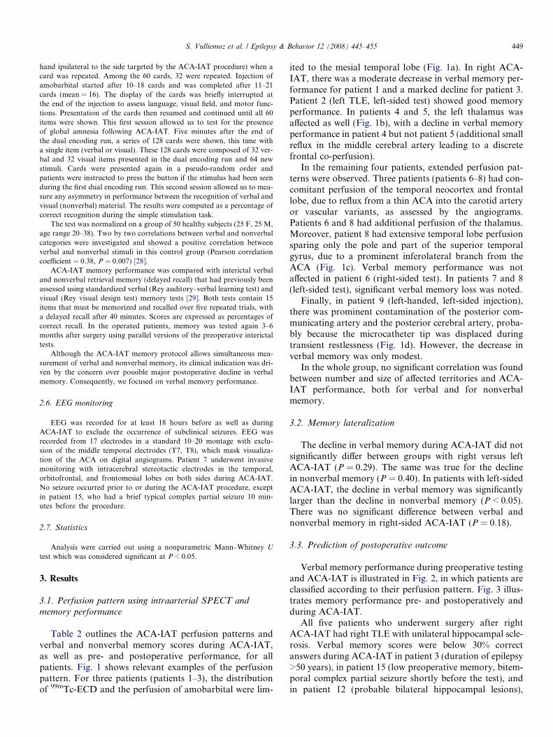
S. Vulliemoz et al. / Epilepsy & Behavior 12 (2008) 445–455 449
hand ipsilateral to the side targeted by the ACA-IAT procedure) when acard was repeated. Among the 60 cards, 32 were repeated. Injection ofamobarbital started after 10–18 cards and was completed after 11–21cards (mean = 16). The display of the cards was briefly interrupted atthe end of the injection to assess language, visual field, and motor func-tions. Presentation of the cards then resumed and continued until all 60items were shown. This first session allowed us to test for the presenceof global amnesia following ACA-IAT. Five minutes after the end ofthe dual encoding run, a series of 128 cards were shown, this time witha single item (verbal or visual). These 128 cards were composed of 32 ver-bal and 32 visual items presented in the dual encoding run and 64 newstimuli. Cards were presented again in a pseudo-random order andpatients were instructed to press the button if the stimulus had been seenduring the first dual encoding run. This second session allowed us to mea-sure any asymmetry in performance between the recognition of verbal andvisual (nonverbal) material. The results were computed as a percentage ofcorrect recognition during the simple stimulation task.
The test was normalized on a group of 50 healthy subjects (25 F, 25 M,age range 20–38). Two by two correlations between verbal and nonverbalcategories were investigated and showed a positive correlation betweenverbal and nonverbal stimuli in this control group (Pearson correlationcoefficient = 0.38, P = 0.007) [28].
ACA-IAT memory performance was compared with interictal verbaland nonverbal retrieval memory (delayed recall) that had previously beenassessed using standardized verbal (Rey auditory–verbal learning test) andvisual (Rey visual design test) memory tests [29]. Both tests contain 15items that must be memorized and recalled over five repeated trials, witha delayed recall after 40 minutes. Scores are expressed as percentages ofcorrect recall. In the operated patients, memory was tested again 3–6months after surgery using parallel versions of the preoperative interictaltests.
Although the ACA-IAT memory protocol allows simultaneous mea-surement of verbal and nonverbal memory, its clinical indication was dri-ven by the concern over possible major postoperative decline in verbalmemory. Consequently, we focused on verbal memory performance.
2.6. EEG monitoring
EEG was recorded for at least 18 hours before as well as duringACA-IAT to exclude the occurrence of subclinical seizures. EEG wasrecorded from 17 electrodes in a standard 10–20 montage with exclu-sion of the middle temporal electrodes (T7, T8), which mask visualiza-tion of the ACA on digital angiograms. Patient 7 underwent invasivemonitoring with intracerebral stereotactic electrodes in the temporal,orbitofrontal, and frontomesial lobes on both sides during ACA-IAT.No seizure occurred prior to or during the ACA-IAT procedure, exceptin patient 15, who had a brief typical complex partial seizure 10 min-utes before the procedure.
2.7. Statistics
Analysis were carried out using a nonparametric Mann–Whitney U
test which was considered significant at P < 0.05.
3. Results
3.1. Perfusion pattern using intraarterial SPECT and
memory performance
Table 2 outlines the ACA-IAT perfusion patterns andverbal and nonverbal memory scores during ACA-IAT,as well as pre- and postoperative performance, for allpatients. Fig. 1 shows relevant examples of the perfusionpattern. For three patients (patients 1–3), the distributionof 99mTc-ECD and the perfusion of amobarbital were lim-
ited to the mesial temporal lobe (Fig. 1a). In right ACA-IAT, there was a moderate decrease in verbal memory per-formance for patient 1 and a marked decline for patient 3.Patient 2 (left TLE, left-sided test) showed good memoryperformance. In patients 4 and 5, the left thalamus wasaffected as well (Fig. 1b), with a decline in verbal memoryperformance in patient 4 but not patient 5 (additional smallreflux in the middle cerebral artery leading to a discretefrontal co-perfusion).
In the remaining four patients, extended perfusion pat-terns were observed. Three patients (patients 6–8) had con-comitant perfusion of the temporal neocortex and frontallobe, due to reflux from a thin ACA into the carotid arteryor vascular variants, as assessed by the angiograms.Patients 6 and 8 had additional perfusion of the thalamus.Moreover, patient 8 had extensive temporal lobe perfusionsparing only the pole and part of the superior temporalgyrus, due to a prominent inferolateral branch from theACA (Fig. 1c). Verbal memory performance was notaffected in patient 6 (right-sided test). In patients 7 and 8(left-sided test), significant verbal memory loss was noted.
Finally, in patient 9 (left-handed, left-sided injection),there was prominent contamination of the posterior com-municating artery and the posterior cerebral artery, proba-bly because the microcatheter tip was displaced duringtransient restlessness (Fig. 1d). However, the decrease inverbal memory was only modest.
In the whole group, no significant correlation was foundbetween number and size of affected territories and ACA-IAT performance, both for verbal and for nonverbalmemory.
3.2. Memory lateralization
The decline in verbal memory during ACA-IAT did notsignificantly differ between groups with right versus leftACA-IAT (P = 0.29). The same was true for the declinein nonverbal memory (P = 0.40). In patients with left-sidedACA-IAT, the decline in verbal memory was significantlylarger than the decline in nonverbal memory (P < 0.05).There was no significant difference between verbal andnonverbal memory in right-sided ACA-IAT (P = 0.18).
3.3. Prediction of postoperative outcome
Verbal memory performance during preoperative testingand ACA-IAT is illustrated in Fig. 2, in which patients areclassified according to their perfusion pattern. Fig. 3 illus-trates memory performance pre- and postoperatively andduring ACA-IAT.
All five patients who underwent surgery after rightACA-IAT had right TLE with unilateral hippocampal scle-rosis. Verbal memory scores were below 30% correctanswers during ACA-IAT in patient 3 (duration of epilepsy>50 years), in patient 15 (low preoperative memory, bitem-poral complex partial seizure shortly before the test), andin patient 12 (probable bilateral hippocampal lesions),

Table 2Clinical, angiographic, and memory data obtained with the ACA-IATa
Gender/age/handedness
Clinical change during test Angio-graphy Intraarterial SPECT PreopV%
PreopNV%
ACA-IAT V%
ACA-IATNV%
PostopV%
PostopNV%
Patient 1 F/44/R
Emotional reaction before injection R MTb 47 93 34 47
Patient 2 F/16/R
None L MT 80 67 69 59 40 60
Patient 3 M/56/R
None R MT 73 97 28 47 87 60
Patient 4 F/32/R
None L MT + thalamus 73 87 19 53 47 NA
Patient 5 M/15/R
None L MT + lateral thalamus + frontal 27 73 27 43
Patient 6 M/41/R
None R MT + thalamus fronto-orbital + lateral temporal
53 57 53 44
Patient 7 F/31/R
Faciobrachial weakness and hypesthesia, dysarthria L MT + temporal lateral + frontalb 73 70 6 69
Patient 8 M/30/R
None L MT + inferior temporal + thalamus+ orbitofrontal
87 100 22 50
Patient 9 M/35/L
R hemianopsia, ‘‘heavy R arm’’ Disinhibition L MT + thalamus + inferior posterior temporal+ occipital + orbitofrontal
47 37 34 59
Patient 10 M/25/R
Discrete disinhibition and psychomotor slowing R NA 87 83 75 75 93 100
Patient 11 M/51/R
None L NA 47 87 16 53 53 NA
Patient 12 F/31/R
Hypophony without dysarthria or aphasia R NA 80 40 25 28 60 37
Patient 13 F/40/R
None L NA 87 80 56 88 60 100
Patient 14 M/34/R
Moderate brachial weakness R NA 67 60 41 56 87 60
Patient 15 M/26/R
Habitual CPS 10 min before the procedureModerate brachial weakness and dystonia,hypophony
R NA 27 57 25 38 40 77
Patient 16 F/32/R
Long-lasting angiography, emotional reactionduring the test
R NA 60 70 13 50
Patient 17 F/37/L
Dysarthria, motor aphasia L NA 67 73 22 84
a Baseline memory performance is expressed as a percentage of delayed recall (15 words/figures). Preop/Postop, before/after surgery; R/L, right/left; MT, mesiotemporal; V, verbal memory; NV,nonverbal memory; CPS, complex partial seizure; NA, not available.
b No concomitant intravenous radiotracer, hence lower spatial definition.
450S
.V
ulliem
oz
eta
l./
Ep
ilepsy
&B
eha
vior
12
(2
00
8)
44
5–
45
5

Fig. 1. Perfusion pattern of ACA-IAT assessed by SPECT during ACA-IAT, co-registered to MRI (left: axial view, right: coronal view). (a) Patient 2:selective perfusion of mesial temporal lobe. (b) Patient 4: additional perfusion of the thalamus. (c) Patient 8: additional extensive perfusion of the inferiorand lateral temporal lobe due to anatomical variation of ACA as well as frontal perfusion due to reflux into the internal carotid artery (arrow). (d) Patient9: additional temporo-occipital perfusion due to reflux into the posterior communicating artery.
S. Vulliemoz et al. / Epilepsy & Behavior 12 (2008) 445–455 451

Fig. 2. Verbal memory performance during the ACA-IAT (gray) forindividual patients compared with preoperative scores (white). Scores aregiven as percentages of correct answers. The vertical bar separates patientsinto two groups: selective perfusion (patients 1–3) and extended perfusion(patients 4–9). In parentheses is the side of ACA-IAT.
Fig. 3. Preoperative (white), ACA-IAT (gray), and postoperative (striped)verbal memory outcome for each patient who underwent surgery. Fromleft to right are preoperative, ACA-IAT, and postoperative. The verticalbar separates patients with a right-sided procedure (first five patients) frompatients with a left-sided procedure. In parentheses is the side of ACA-IAT.
452 S. Vulliemoz et al. / Epilepsy & Behavior 12 (2008) 445–455
who was the only one to show a postoperative decline.Both patients who scored higher than 30% during rightACA-IAT showed postoperative improvement of verbalmemory.
Three of the four patients who underwent surgery afterleft-sided ACA-IAT had left TLE with unilateral hippo-campal sclerosis; the fourth, patient 11, had nonlesionalbilateral TLE. Two patients (4 and 11) had verbal memoryscores below 30% correct answers during the procedure,but the scores were not predictive of postoperative decline.Postoperatively, memory scores declined for all exceptpatient 11, who had low preoperative verbal memory.Moreover, patient 2 showed a strong decline in verbal
memory that was not predicted by the ACA-IAT score.Fortunately, she could still retain 40% of items; that is,she had no postoperative amnesia.
There was no significant correlation between verbalmemory performance during ACA-IAT and pre-/postoper-ative decline whether considering the whole group or sub-groups of patients with right and left ACA-IAT. Therewas no significant correlation between preoperative andpostoperative verbal memory performance.
4. Discussion
In our institution, ACA-IAT is carried out when there isa concern about any potentially significant postoperativeverbal memory decline. Thus, only a small group ofpatients with intractable TLE undergo this procedure.Given its invasive nature, thorough evaluation of its useful-ness in terms of estimation of the postoperative memorydeficit is mandatory. Despite the modest size of the studygroup, we feel that important conclusions can be drawnregarding decisions on operability, which are always madeon an individual basis. Our study based on this series ofpatients with different types of TLE shows that:
1. Performed by experienced interventional neuroradiol-ogists, the procedure did not lead to complications.
2. The perfusion pattern of amobarbital varies greatlybetween patients.
3. No case of severe postoperative amnesia was observedin the patients screened preoperatively with ACA-IAT.Performance during the test was lower than postopera-tive performance in all except one patient; that is, ACA-IAT provides a pessimistic estimate of the postopera-tive situation in the vast majority.
4. The ACA-IAT memory test gives an insufficient esti-mate of the individual decline in postoperative verbalmemory. Underestimation and overestimation ofpostoperative decline were observed.
4.1. Perfusion pattern
The perfusion pattern during ACA-IAT has beenreported for only two patients so far, but details of theirclinical history were not given [24]. In our study, selectiveamobarbital injection into the anterior choroidal arteryled to a very variable perfusion pattern due to a combina-tion of anatomical variations and technical aspects (reflux,catheter displacement). The pattern varied from selectiveperfusion of the anterior mesial temporal structures tar-geted to widespread involvement of frontal, temporal,and parieto-occipital lobes, as well as subcortical struc-tures. This might explain, at least partially, the lack of pre-dictive value of ACA-IAT.
Amobarbital affected more than the mesial temporallobe in six of nine patients, including co-perfusion of thethalamus in five cases. In four of six patients with extended

S. Vulliemoz et al. / Epilepsy & Behavior 12 (2008) 445–455 453
perfusion, the decrease in verbal memory was significant.Interpreting significant memory decline in patients withan extensive pattern of perfusion is particularly challeng-ing: thalamic, frontal, as well lateral temporal structuresare known to be involved in memory function and theirinactivation would thus worsen memory scores [30]. Inthese cases, the memory test does not mimic selective surgi-cal resection. Consequently, knowledge of the perfusionpattern of amobarbital could be relevant in decidingwhether surgery is contraindicated in patients with a severedecrease in verbal memory during ACA-IAT.
A study of PCA-IAT found significant variability inneurological/neuropsychological deficits in patients withco-perfusion of the thalamus (11/14 patients) [31,32]. Vari-ations in the perfusion pattern are also found in intracaro-tid IAT: the mesial temporal region is perfused only inabout one-third of cases [16,18,20,33]. More data are there-fore needed to determine the influence of co-perfusion ofthe thalamus or other structures on memory performance.Other factors, such as side of TLE, presence of hippocam-pal sclerosis, and age at onset of the disease are known tobe relevant to postoperative verbal memory outcome andshould also be taken into account. Unfortunately, ourgroup was too small for multivariate analysis.
4.2. Memory lateralization
In our series of patients, nonverbal memory was lessaffected by the ACA-IAT procedure than verbal memory,consistent with evidence that nonverbal memory is less welllateralized and, thus, less severely affected after surgery[34,35]. This asymmetrical decline was largest in the groupwith left-sided ACA-IAT, confirming the importance of theleft-sided mesial temporal structures in our group of right-handed patients. Postoperative tests on our patients withright-sided ACA-IAT (all right-handed) confirmed thatverbal memory function did not strongly rely on the righthippocampus, despite doubts cast by the preoperative non-invasive workup.
4.3. Prediction of postoperative verbal memory outcome
The predictive value of amobarbital distribution in ver-bal memory decline during the test appears to be modest.Its main usefulness thus appears to be as an additionalscreening tool for patients at risk of global postoperativeamnesia, rather than as a predictor of the extent of thepostoperative deficit. In their study, Wieser et al. foundno statistically significant difference between ACA-IATverbal memory and postoperative verbal memory, but thecorrelation between the two is not given [24]. In our study,the extent of decline in verbal memory was not correlatedwith individual ACA-IAT performance. In patients withgood preoperative verbal memory, ACA-IAT scores below30% were associated with a postoperative decline in patient12 (right TLE) and patient 4 (left TLE), However, inpatient 3 (right TLE), ACA-IAT scores overestimated the
postoperative decline. In another patient (patient 2, lefthippocampal sclerosis), postoperative neuropsychologicaltesting revealed a major verbal memory decline (assessedpsychometrically but not as severely noted by the patient),which was not suspected by ACA-IAT results (underesti-mation). The two patients with low preoperative verbalmemory (patient 11 with left TLE and patient 15 with rightTLE) had low ACA-IAT scores but no postoperativedecline.
Knowledge of the whole clinical picture and of the per-fusion pattern could help in understanding the discrepantcases. Patients 7 and 8 had bilateral TLE with left-sidedpredominance and no hippocampal sclerosis. They hadgood preoperative verbal memory but very low verbalmemory during the test, in the context of an extended pat-tern of amobarbital perfusion. These results, in addition tothe known elevated risk of verbal memory decline in theabsence of hippocampal sclerosis [5], were interpreted asa contraindication to epilepsy surgery. As no operationwas performed, we do not know if the extended patternof amobarbital perfusion turned these patients into false-positive cases. Patient 9 had early ischemic lesions as wellas early epileptogenic activity, reportedly associated withatypical localization of language and memory functions[36,37]. This might explain the discrepancy between theextended territories revealed by SPECT and the moderateneuropsychological deficits, so that the clinical pictureappears more relevant than the perfusion pattern revealedby SPECT.
4.4. Clinical implications
Established clinicoradiological predictive factors ofpoor verbal memory outcome are surgery on the dominanthemisphere, intact preoperative memory, absence of ipsi-lateral hippocampal sclerosis [5], older age [4,7], male gen-der [8,9], extent of surgical resection [10,11], lowpreoperative verbal IQ, as well as the presence of corticaldysgenesis [12]. These should all be taken into accountbefore surgery.
Although less validated and technically more difficultthan the carotid IAT, ACA-IAT is sometimes envisagedwhen there is a risk of severe postoperative verbal memorydecline or when global amnesia is feared. Although it maybe valid for these purposes, the test seems to be insufficientin providing a good estimate of the degree of postoperativeverbal memory decline. The results should always be inter-preted with all relevant clinical data, to prevent incorrectjudgments concerning the operability of an individualpatient.
With respect to carotid IAT, two recent studies foundthat noninvasive tests (neuropsychology, volumetricMRI) were as reliable as carotid IAT in predicting postop-erative decline in verbal memory [38,39]. These observa-tions, as well as the ongoing development of functionalMRI using memory tasks [40,41], will certainly reduce

454 S. Vulliemoz et al. / Epilepsy & Behavior 12 (2008) 445–455
the importance of amobarbital procedures for TLE surgeryin the future.
Acknowledgments
The authors thank Nicolas Ruffieux and Boris Pimonowfor their help in gathering the neuropsychological data.
This study was supported by SNF Grants IB74B0-111086, 3200B0-104146, 3200-068105, and 3200-113766.Serge Vulliemoz is supported by the Fonds de Perfectionn-ement of the University Hospital of Geneva.
References
[1] Wiebe S, Blume WT, Girvin JP, Eliasziw M. A randomized,controlled trial of surgery for temporal-lobe epilepsy. N Engl JMed 2001;345:311–8.
[2] Kapur N, Prevett M. Unexpected amnesia: are there lessons to belearned from cases of amnesia following unilateral temporal lobesurgery? Brain 2003;126:2573–85.
[3] Akanuma N, Alarcon G, Lum F, et al. Lateralising value ofneuropsychological protocols for presurgical assessment of temporallobe epilepsy. Epilepsia 2003;44:408–18.
[4] Alpherts WC, Vermeulen J, van Rijen PC, da Silva FH, van VeelenCW. Verbal memory decline after temporal epilepsy surgery? A 6-year multiple assessments follow-up study. Neurology2006;67:626–31.
[5] Stroup E, Langfitt J, Berg M, McDermott M, Pilcher W, Como P.Predicting verbal memory decline following anterior temporal lobec-tomy (ATL). Neurology 2003;60:1266–73.
[6] Chelune GJ, Naugle RI, Luders H, Awad IA. Prediction of cognitivechange as a function of preoperative ability status among temporallobectomy patients seen at 6-month follow-up. Neurology1991;41:399–404.
[7] Gleissner U, Sassen R, Schramm J, Elger CE, Helmstaedter C.Greater functional recovery after temporal lobe epilepsy surgery inchildren. Brain 2005;128(Pt. 12):2822–9.
[8] Bjornaes H, Stabell KE, Roste GK, Bakke SJ. Changes in verbal andnonverbal memory following anterior temporal lobe surgery forrefractory seizures: effects of sex and laterality. Epilepsy Behav2005;6:71–84.
[9] Trenerry MR, Jack Jr CR, Cascino GD, Sharbrough FW, Ivnik RJ.Gender differences in post-temporal lobectomy verbal memory andrelationships between MRI hippocampal volumes and preoperativeverbal memory. Epilepsy Res 1995;20:69–76.
[10] Jooma R, Yeh HS, Privitera MD, Gartner M. Lesionectomy versuselectrophysiologically guided resection for temporal lobe tumorsmanifesting with complex partial seizures. J Neurosurg1995;83:231–6.
[11] Katz A, Awad IA, Kong AK, et al. Extent of resection in temporallobectomy for epilepsy: II. Memory changes and neurologic compli-cations. Epilepsia 1989;30:763–71.
[12] Baxendale S, Thompson PJ, Harkness W, Duncan JS. Predictingmemory decline following epilepsy surgery: a multivariate approach.Epilepsia 2006;47:1887–94.
[13] Wada J, Rasmussen T. Intracarotid injection of sodum amytal for thelateralization of cerbral speech dominance: experimental and clinicalobservations. J Neurosurg 1960:266–82.
[14] Acharya JN, Dinner DS. Use of the intracarotid amobarbitalprocedure in the evaluation of memory. J Clin Neurophysiol1997;14:311–25.
[15] Bell BD, Davies KG, Haltiner AM, Walters GL. Intracarotidamobarbital procedure and prediction of postoperative memory inpatients with left temporal lobe epilepsy and hippocampal sclerosis.Epilepsia 2000;41:992–7.
[16] Kim BG, Lee SK, Nam HW, Song HC, Lee DS. Evaluation offunctional changes in the medial temporal region during intracarotidamobarbital procedure by use of SPECT. Epilepsia 1999;40:424–9.
[17] McMackin D, Dubeau F, Jones-Gotman M, Gotman J. Medialtemporal lobe structures have reduced blood flow immediately afterthe intracarotid injection of amobarbital. Epilepsia 1999;40:1673–4.
[18] Setoain X, Arroyo S, Lomena F, et al. Can the Wada test evaluatemesial temporal function? A SPECT study. Neurology2004;62:2241–6.
[19] de Silva R, Duncan R, Patterson J, Gillham R, Hadley D. Regionalcerebral perfusion and amytal distribution during the Wada test. JNucl Med 1999;40:747–52.
[20] Hart Jr J, Lewis PJ, Lesser RP, et al. Anatomic correlates of memoryfrom intracarotid amobarbital injections with technetium Tc 99mhexamethylpropyleneamine oxime SPECT. Arch Neurol1993;50:745–50.
[21] Loddenkemper T, Morris HH, Lineweaver TT, Kellinghaus C.Repeated intracarotid amobarbital tests. Epilepsia 2007;48:553–8.
[22] Brassel F, Weissenborn K, Ruckert N, Hussein S, Becker H.Superselective intra-arterial amytal (Wada test) in temporal lobeepilepsy: basics for neuroradiological investigations. Neuroradiology1996;38:417–21.
[23] Huther G, Dorfl J, Van der Loos H, Jeanmonod D. Microanatomicand vascular aspects of the temporomesial region. Neurosurgery1998;43:1118–36.
[24] Wieser HG, Muller S, Schiess R, et al. The anterior and posteriorselective temporal lobe amobarbital tests: angiographic, clinical,electroencephalographic, PET, SPECT findings, and memory perfor-mance. Brain Cogn 1997;33:71–97.
[25] Weissenborn K, Ruckert N, Brassel F, Becker H, Dietz H. Aproposed modification of the Wada test for presurgical assessment intemporal lobe epilepsy. Neuroradiology 1996;38:422–9.
[26] Stabell KE, Bakke SJ, Andresen S, et al. Selective posterior cerebralartery amobarbital test: its role in presurgical memory assessment intemporal lobe epilepsy. Epilepsia 2004;45:817–25.
[27] Jack Jr CR, Nichols DA, Sharbrough FW, Marsh WR, Petersen RC.Selective posterior cerebral artery amytal test for evaluating memoryfunction before surgery for temporal lobe seizure. Radiology1988;168:787–93.
[28] Schiess R. Visuelle Erinnerungsfahigkheit unter besonder Ber-ucksichtigung des limbischen Systems. University of Zurich; 1992.
[29] Rey A. L’examen clinique en psychologie. Paris: Presses Universi-taires de France; 1964.
[30] Kopelman MD. Disorders of memory. Brain 2002;125(Pt. 10):2152–90.
[31] Urbach H, Kurthen M, Klemm E, et al. Amobarbital effects on theposterior hippocampus during the intracarotid amobarbital test.Neurology 1999;52:1596–602.
[32] Von Oertzen J, Klemm E, Urbach H, et al. SATSCOM—Selectiveamobarbital test intraarterial SPECT coregistered to MRI: descrip-tion of a method assessing selective perfusion. NeuroImage2000;12:617–22.
[33] Jeffery PJ, Monsein LH, Szabo Z, et al. Mapping the distribution ofamobarbital sodium in the intracarotid Wada test by use of Tc-99mHMPAO with SPECT. Radiology 1991;178:847–50.
[34] Barr WB, Chelune GJ, Hermann BP, et al. The use of figuralreproduction tests as measures of nonverbal memory in epilepsysurgery candidates. J Int Neuropsychol Soc 1997;3:435–43.
[35] Hermann BP, Seidenberg M, Schoenfeld J, Davies K. Neuropsycho-logical characteristics of the syndrome of mesial temporal lobeepilepsy. Arch Neurol 1997;54:369–76.
[36] Janszky J, Jokeit H, Heinemann D, Schulz R, Woermann FG, EbnerA. Epileptic activity influences the speech organization in medialtemporal lobe epilepsy. Brain 2003;126(Pt. 9):2043–51.
[37] Vikingstad EM, Cao Y, Thomas AJ, Johnson AF, Malik GM, WelchKM. Language hemispheric dominance in patients with congenitallesions of eloquent brain. Neurosurgery 2000;47:562–70.

S. Vulliemoz et al. / Epilepsy & Behavior 12 (2008) 445–455 455
[38] Baxendale S, Thompson P, Harkness W, Duncan J. The role of theintracarotid amobarbital procedure in predicting verbal memorydecline after temporal lobe resection. Epilepsia 2007;48:546–52.
[39] Lineweaver TT, Morris HH, Naugle RI, Najm IM, Diehl B,Bingaman W. Evaluating the contributions of state-of-the-artassessment techniques to predicting memory outcome after unilateralanterior temporal lobectomy. Epilepsia 2006;47:1895–903.
[40] Janszky J, Jokeit H, Kontopoulou K, et al. Functional MRI predictsmemory performance after right mesiotemporal epilepsy surgery.Epilepsia 2005;46:244–50.
[41] Richardson MP, Strange BA, Duncan JS, Dolan RJ. MemoryfMRI in left hippocampal sclerosis: optimizing the approachto predicting postsurgical memory. Neurology 2006;66:699–705.

![Unilateral Choroidal Osteoma with Choroidal Neovascularization...Surgical evacuation of the choroidal neovascular membrane has been reported [12] but the visual outcome was not favorable](https://img.dokumen.tips/doc/110x75/6053732923e31173be575e28/unilateral-choroidal-osteoma-with-choroidal-neovascularization-surgical-evacuation.jpg)