Embed Size (px)
Citation preview
TELEFLEX MEDICAL PAEDIATRIC PRODUCTSOnly the best is good enough for children
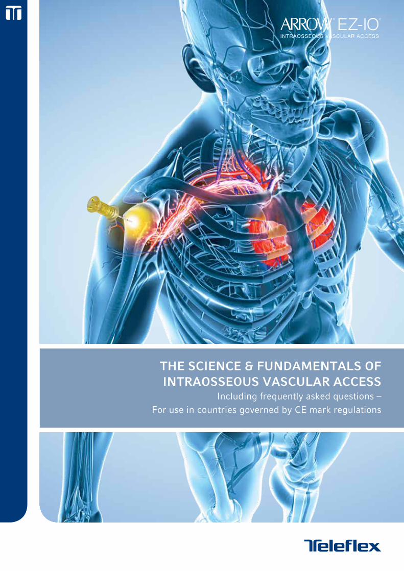
THE SCIENCE & FUNDAMENTALS OF INTRAOSSEOUS VASCULAR ACCESS
Including frequently asked questions – For use in countries governed by CE mark regulations
2 content
2014 SECOND EDITION
INTRODUCTION ............................................................................................................................... 4
INDICATIONS/CONTRAINDICATIONS AND GENERAL IO USE ................................................. 5
ANATOMY AND PHYSIOLOGY ...................................................................................................... 6
ANATOMY AND PHYSIOLOGY OF THE IO SPACE ....................................................................... 6
TECHNIQUE/TRAINING .................................................................................................................. 8
FLOW RATES AND INFUSION UNDER PRESSURE ................................................................... 11
SELECTION OF APPROPRIATE INSERTION SITE AND NEEDLE SET ..................................... 12
PROXIMAL HUMERUS .................................................................................................................. 13
EZ-IO® PROXIMAL HUMERUS IDENTIFICATION AND INSERTION TECHNIQUE ................. 14
PROXIMAL TIBIA ........................................................................................................................... 17
DISTAL TIBIA ................................................................................................................................. 18
DISTAL FEMUR .............................................................................................................................. 19
CARE AND MAINTENANCE OF THE EZ-IO® .............................................................................. 20
EZ-IO G3 POWER DRIVER AND TRAINING DRIVER ................................................................. 21
EZ-CONNECT® PROVIDED BY TELEFLEX .................................................................................. 22
COMPLICATIONS ........................................................................................................................... 22
INTRAOSSEOUS COMPLICATIONS ............................................................................................. 23
COMPARTMENT SYNDROME ...................................................................................................... 24
EFFECTS OF IO ACCESS ON GROWTH PLATES AND BONE REPAIR ..................................... 25
EMBOLISM ..................................................................................................................................... 25
OSTEOMYELITIS ............................................................................................................................ 27
MEDICATIONS/FLUIDS ................................................................................................................. 27
PAIN MANAGEMENT FOR IO INFUSION .................................................................................... 28
INTRAOSSEOUS ADMINISTRATION OF PRESERVATIVE-FREE LIDOCAINE ........................ 30
PAEDIATRICS: NEWBORNS, INFANTS, CHILDREN AND ADOLESCENTS ............................ 31
LABORATORY ANALYSIS/BLOOD SAMPLING .......................................................................... 35
LABORATORY ANALYSIS/BLOOD SAMPLING FROM IO ACCESS .......................................... 35
INDUCED HYPOTHERMIA AND THE EZ-IO® .............................................................................. 38
SPECIAL CONSIDERATIONS ........................................................................................................ 38
RESEARCH ......................................................................................................................................40
MYTHS ABOUT INTRAOSSEOUS VASCULAR ACCESS ............................................................ 41
REFERENCES ................................................................................................................................. 42
content 3
4 introduction
INTRODUCTION
Teleflex is pleased to provide you with an updated version of "The Science & Fundamentals of Intra osseous Vascular Access". This document represents the body of knowledge garnered from years of research (clinical and preclinical), laboratory experimentation, clinical experience (end-users and researchers), and expert opinion (intraosseous experts and key opinion leaders).
The document is divided by topics into several sections. The first part of each section contains a list of short, concise responses to the most commonly asked questions on intra osseous (IO) vascular access. Where more detailed information and research is available, the reader is directed to the appropriate section. For clarity, most cita-tions and references are confined to the expand-ed, detailed sections.
Citations which are followed by the superscript VS indicate studies that have been sponsored in part or conducted by Teleflex.
A superscript EO after a statement or title signi-fies infor mation based on Expert Opinion and will occur after relevant statements (in lieu of a cited reference). Unless otherwise noted, expert opinion is provided by Dr. Larry J. Miller, former Chief Medical Officer of Vidacare Corporation, and the Vidacare research team. Dr. Miller’s extensive experience with intraosseous access over the past two decades has proven to be a vital resource for guiding current practice and continues to provide significant contributions to IO research.
The authors of this document have diligently examined the cited sources and made every effort to assure the information provided is reliable, com-plete, and in accord with the standards of current practice at the time of publication. However, this document does not constitute any official recom-mendations by Teleflex for patient care. Use of IO devices is the responsibility of the treating clinician, medical director or qualified prescriber.
We hope these answers will assist our team mem-bers and clinicians in taking full advantage of the benefits – and minimising the risks – of IO vascu-lar access and the ARROW® EZ-IO®.
This document is disseminated for medical and scientific/educational purposes only, and some cited studies may contain references to indications for IO access or insertion sites that are not indi-cated in the CE marking relating to the ARROW® EZ-IO product, which is manufactured/marketed/ distributed by Teleflex. This information should not be construed to suggest that any Vidacare product may or should be used in any manner that differs from its CE marking Indications / Directions for use.
Your Teleflex team
EO = Expert opinion

indications/contraindications and General io use 5
INDICATIONS/CONTRAINDICATIONS AND GENERAL IO USE
WHEN CAN THE EZ-IO BE USED?
Indications for the ARROW® EZ-IO Intraosseous Infusion System:• The EZ-IO can be used for adult and paediatric patients,
and is indicated any time vascular access is difficult to obtain in emergent, urgent, or medically necessary (non-emergent) cases. Intraosseous (IO) sites for the EZ-IO include the proximal humerus, proximal tibia and distal tibia in adults and paediatrics, and the distal femur in paediatrics only. EZ-IO may remain in place for up to 72 hours.
Contraindications for the ARROW® EZ-IO Intraosseous Infusion System:• fracture in targeted bone• excessive tissue or absence of adequate anatomical
landmarks• infection at area of insertion site• previous, significant orthopedic procedure at site
(e.g. prosthetic limb/joint)• IO access in targeted bone within past 48 hours
IN WHAT TYPE OF CLINICAL SCENARIOS IS IO USED?
Emergent/urgent conditions in which IO vascular access may be beneficial:
• Sepsis• Therapeutic hypothermia• Altered level of
consciousness• Respiratory compromise/
arrest• Cardiac compromise/arrest• Seizures/status epilepticus• End stage renal disease• Diabetes• Hemodynamic instability• Shock• Cardiac arrest
• Respiratory arrest• Major trauma• Hypovolemia• Sickle cell crisis• Morbid obesity• Rapid sequence induction• Bridge to central line• Stroke• Drug overdose• Burns• Dehydration• Anaphylaxis• Cardiac arrhythmias
Non-urgent conditions in which IO vascular access may be beneficial:
• difficult vascular access• antibiotic therapy• sedation for procedures• analgesia for pain• chest pain • laboratory analysis*
• general anesthesia• metabolic disorders• rehydration• induction of labor• surgical procedures
*refer to section on Laboratory Analysis/Blood Sampling
CAN THE EZ-IO BE USED IN THE STERNUM?
The EZ-IO Sternal Intraosseous System and the Tactically Advanced Lifesaving Intraosseous Needle (T.A.L.O.N.™) are for use by the military and tactical medical teams only, and are not intended for use in the civilian sector. Only the EZ-IO Sternal Intraosseous System and the Tactically Advanced Lifesaving Intraosseous Needle (T.A.L.O.N.) as directed in the Instructions for Use specific to the sternum may be used safely in the sternum. Neither the EZ-IO needle sets nor the Power Driver should ever be used for sternal insertion.
WHAT IS OFF-LABEL USE OF THE EZ-IO?
Off-label use is defined as use of a medical device for an indication not specifically identified in the CE marking. Physicians (or qualified prescribers) may prescribe, order or use drugs and devices for indications not identified in the CE marking according to their best medical judgment; however, the manufacturing company is prohibited from any promotion of the off-label use. Therefore, Vidacare cannot recommend, promote or endorse off-label use of the EZ-IO product.1
CAN NURSES AND MEDICS PERFORM IO INSERTIONS?
RN: In some countries a licensed, qualified and trained regis-tered nurse is permitted to place and manage IO devices, if it is determined by regulation, position statement or decision-making model to be within that professional’s scope of practice. The appropriate organizational officials, chief nursing officer/ nurse supervisor, hospital or country regulatory official should be consulted to determine whether placement and use of IO devices is currently within an individual’s scope of practice.
6 anatomy and PhysioloGy
EMT-P, EMT-I, EMT-B: Most countries permit a licensed, trained and qualified EMS professional to place and use IO devices upon the order of a medical director. The appropriate regulatory agency, medical director and system protocols should be consulted to determine if placement and manage-ment of IO devices is currently within an individual’s scope of practice.
• Each country has laws and regulations that govern the medical procedures licensed personnel may perform within their respective scope of licensure. These laws, regulations and directives are occasionally modified as new medical technology becomes standard and accepted within the healthcare industry. The appropriate regulatory agency, medical director, chief nursing officer and/or healthcare system protocols should be consulted prior to implementing an intraosseous device protocol.
• A variety of professional organizations have developed position statements supporting IO use for their respective specialties.
IS SPECIAL TRAINING OR CERTIFICATION REQUIRED PRIOR TO USING THE EZ-IO?
There is no official “certification” process unless mandated by an agency/organization, medical director or hospital. The EZ-IO is similar to an IV catheter in that specific training must occur in order to use the device safely and correctly. Vidacare offers a comprehensive training program for the EZ-IO and recommends completion prior to using the device. Online training information is available at www.arrowezio.com; or contact your Vidacare representative to set up a training session.
ANATOMY AND PHYSIOLOGY
HOW DOES THE INTRAOSSEOUS (IO) VASCULAR ROUTE WORK?
IO catheters are usually placed in the proximal and distal ends (epiphyses) of long bones due to the thinner compact bone and abundance of cancellous (spongy) bone at these sites. Within the epiphysis of the medullary space lies a vast system of blood vessels. When accessed with an IO needle, blood and fluid pass from the medullary space through the vascular system into the central circulation. [See Anatomy and Physiology of the IO Space, page 7]
WHICH INSERTION SITE WORKS BEST?
IO site selection depends on patient age, size, anatomy, pre-senting condition, ability to locate anatomical landmarks, and clinical judgment and experience. Studies and articles suggest the humerus may be a superior site for flow rates, drug delivery, and management of infusion pain. [See Selec-tion of Appropriate Insertion Site and Needle Set, page 12]
ANATOMY AND PHYSIOLOGY OF THE IO SPACE
ANATOMY
Within the epiphysis (proximal and distal end) of the medul-lary space of the bone lies a vast system of blood vessels running both vertically (Haversian canals) and horizontally (Volksmann’s canals). Due to this large network, blood and fluid travel quickly through this component of the vascular system to reach the central circulation. See Figures 1 and 2.

anatomy and PhysioloGy of the io sPace 7
Intraosseous (IO) needles/catheters are placed in the epiphyses of long bones such as the tibia and humerus where compact bone is relatively thin and there is an abundance of cancellous (spongy) bone. This area of the bone allows for easier entry through the cortex of the bone, with rapid access into the IO vasculature.
PHYSIOLOGY
A 25-patient clinical study published in 2008 compared the pharmacokinetics of IO access using an implantable IO device
vs. IV administration of morphine sulfate in adults. The investigators reported no differences between IO and IV administration of morphine for several pharmacokinetic parameters, including maximum plasma concentration, time to maximum plasma concentration, and area under plasma concentration-time curve.2
A preclinical study measured peak serum concentrations of epinephrine after IO infusion.3 The authors concluded that peak serum concentrations of epinephrine were equivalent between humeral IO and central line administration.
1
2
FIGURE 1.
FIGURE 2.

8 techniQue/traininG
In a healthy adult volunteer study conducted in May 2013, contrast media was injected through the proximal humerus site and captured under fluoroscopy as it entered the heart. The mean time it took from injection at the insertion site to visualise contrast entry into the superior vena cava was 2.3 seconds.4
INTRAMEDULLARY PRESSURE
A preclinical study demonstrated pressure in the IO medullary cavity measures approximately 25 % of arterial pressure without significant difference between IO sites tested.5
DURING CPR
In a series of preclinical studies, Hoskins et al evaluated effi-cacy of the IO route compared to other IV access routes. In one study, researchers demonstrated that fluid infused into the IO space gains access to the central circulation within several seconds to less than 2 minutes, even during CPR.6 The authors suggested that results demonstrated IO equiva-lence to IV access during CPR. A follow up study comparing sternal and humerus IO routes concluded the proximal humerus and sternal IO routes were comparable to central venous drug delivery during CPR.7
TECHNIQUE/TRAINING
NOTEFor comprehensive training information, please refer to www.arrowezio.com or contact Customer Service.
HOW SHOULD THE SKIN BE PREPARED FOR IO INSERTION?
Similar to a peripheral IV site: prior to insertion, the site should be thoroughly cleaned with chlorhexidine (e.g. ChloraPrep®) or the cleansing agent required by specific protocol.
IS A LOCAL ANESTHETIC NECESSARY FOR EZ-IO INSERTION IN AN ALERT PATIENT?
EZ-IO insertion does not generally require local anesthesia, though discomfort resulting from the insertion is variable; and local infiltration of an anesthetic prior to insertion may be done. However, infusion of fluids is often painful for patients responsive to pain; therefore following the IO needle insertion, IO anesthetic (1 % or 2 % preservative-free lido-caine without epinephrine) may be considered for use under institutional protocols or policies.
HOW IS APPROPRIATE NEEDLE SET LENGTH DETERMINED? CAN THE “PAEDIATRIC” NEEDLE SETS BE USED IN ADULTS, OR “ADULT” NEEDLE SETS IN PAEDIATRIC PATIENTS?
The EZ-IO needle sets have guidelines based on approximate weight and age ranges. The needle sets are 15 gauge and available in 3 different lengths including 15 mm (for 3–39 kg), 25 mm (40 kg or over), and 45 mm (40 kg or over, for excessive tissue depth). Clinical judgment should be used to determine appropriate needle set selection based on patient anatomy, weight and tissue depth. For example, a small geriatric female may require a shorter length catheter whereas an obese child may require a longer catheter. The longer 45 mm needle set should be used when there is excessive tissue overlying the insertion site, and for the proximal humerus site in adults.
The EZ-IO catheter is marked with a black line 5 mm from the hub. If the EZ-IO needle set is inserted through the soft tissue and does not reach the bone or the 5 mm needle-mark from the hub is not visible above the skin, a longer needle set or alternate site should be chosen prior to penetration of the cortex. [See Selection of Appropriate Insertion Site and Needle Set, page 12]
techniQue/traininG 9
HOW CAN THE HUMERUS SITE BE USED IN THE PERIOPERATIVE SETTING?
In the perioperative setting, the EZ-IO should be inserted into the proximal humerus as described in the instructions for use. After placement, the patient’s arm can be repositioned as needed but extreme care should be taken if the arm is abducted more than 45 degrees from the side of the body. [See Pain Management for IO Infusion, page 28]
HOW DEEP SHOULD THE NEEDLE SET BE INSERTED WHEN POWERING THE EZ-IO INTO THE BONE?
Push needle set tip through the skin until tip rests against the bone. The 5 mm mark from the hub must be visible above the skin for confirmation of adequate needle set length. Squeeze driver trigger and apply moderate, steady pressure.
Paediatrics: Immediately release the trigger when you feel the “pop” or “give” as the needle set enters the medullary space.
Adults: Advance needle set approximately 1–2 cm after feel-ing a change in resistance that indicates entry into the medullary space or until the needle set hub is close to the skin. In the humerus, for most adults a 45 mm needle set should be advanced until catheter hub is flush with the skin. Clinicians are strongly encouraged to study the training materials and obtain hands-on experience with a Vidacare representative or clinical trainer to become competent in safe and proper use of the EZ-IO. [See Selection of Appropriate Insertion Site and Needle Set, page 12] [See Paediatrics: Newborns, Infants, Children & Adoles-cents, page 31]
WHAT IF THE DRIVER SEEMS TO BE LOSING POWER AND SLOWS DOWN?
The most common cause of the driver slowing or stalling is improper use; specifically, applying too much downward pressure during insertion. The needle set should always be inserted with moderate pressure, allowing the driver to do the work. If the driver fails in a clinical emergency, the EZ-IO needle set may be inserted manually without the driver. If the condition persists despite proper use, please contact Customer Service. See page 42 for Customer Service contact information. [See EZ-IO G3 Power Driver and Training Driver, page 21]
HOW SHOULD THE EZ-IO BE STABILIZED?
After insertion of the EZ-IO, use the EZ-Stabilizer™ to secure the needle and prevent accidental dislodgement. Remove the stylet, place EZ-Stabilizer over the hub, then attach the primed EZ-Connect® extension set to the hub; firmly secure by twisting clockwise. If an EZ-Stabilizer is unavailable, other methods should be used to secure the device. [Refer to EZ-Stabilizer Directions for Use]
SHOULD THE EZ-CONNECT BE PRIMED WITH FLUID PRIOR TO USE?
Yes. Always prime the EZ-Connect with fluid before attaching to the EZ-IO hub. (Note: If the patient is responsive to pain, consider priming the EZ-Connect with 1 % or 2 % preser-vative-free lidocaine without epinephrine). [See EZ-Connect by Vidacare, page 22]
DOES THE EZ-CONNECT MEET HOSPITAL INFECTION CONTROL STANDARDS?
Yes. The EZ-Connect uses the Robertsite® needleless connector by Halkey-Roberts. The connector valve satisfies all requirements of the United States Center for Disease Control for needleless intravascular systems. [See EZ-Connect by Teleflex, page 22]
WHAT IS THE EZ-IO NEEDLE SET MADE OF?
The catheter and stylet are 304 stainless steel. The plastic hub is medical grade polycarbonate.
IS A SYRINGE FLUSH NECESSARY AFTER IO INSERTION?
Yes. It is essential to inject a syringe flush into the IO space before attempting to infuse fluids through the IO catheter. A syringe flush helps clear the marrow and fibrin from the medullary space, allowing for effective infusion rates. No Flush = No Flow.
It is important not to use extreme pressure for the flush, as it may increase the risk of extravasation. [See Flow Rates and Infusion Under Pressure, page 11]
10 techniQue/traininG
IS IT NECESSARY TO FLUSH THE IO LINE WITH SALINE AFTER INFUSING MEDICATIONS VIA THE IO ROUTE?
Yes. As with an IV infusion, an IO line should be flushed before and after infusion to ensure all prescribed medication has entered the vascular space in the proper amount and concentration. The volume of the EZ-Connect is approx-imately 1.0 ml.
WHAT FLOW RATES CAN BE ACHIEVED WITH THE IO? HOW CAN FLOW RATES BE OPTIMISED?
In published literature, IO flow rates (delivered under pres-sure) range from 200 ml/hour to 9,900 ml/hour.8,9,10 A more likely estimate for flow rates in adults (based on a human volunteer study) might be 5 liters per hour through the hu-merus and one liter per hour through the tibia; both with 300 mmHg of pressure.11 As with other vascular access lines, IO flow rates will vary among patients and anatomical sites. Generally, adequate flow rates are dependent on performing a syringe flush prior to IO infusion and infusing fluids and medications under pressure (e.g. infusion pressure pump or pressure bag). Gravity alone will rarely generate adequate flow rates; the higher the pressure, the faster the flow. Gen-erally, the proximal humerus allows higher flow rates than tibial sites. [See Flow Rates and Infusion Under Pressure, page 11]
DOES THE SYRINGE FLUSH HAVE TO BE REPEATED WITH PROLONGED USE? WILL THE IO CATHETER CLOT OFF IF UNUSED FOR A FEW HOURS?
Possibly. IO access may be compromised if the line is not used for prolonged periods. Often, IO lines can be opened by an additional syringe flush.
CAN A HEPARIN LOCK/SALINE LOCK BE USED TO MAINTAIN PATENCY OF AN IO LINE? WHAT SHOULD BE DONE IF THE LINE CLOTS?EO
Confirm IO catheter placement in the medullary cavity. Attempt to administer a syringe flush to open the line. Theoretically, as with intravenous lines, a small amount of standard heparin or saline lock solution may allow the IO site to stay open and prevent clotting for a longer period. (To minimise the amount of heparin patients receive, aspi-rate and discard any heparin lock solution in the catheter and EZ-Connect prior to re-establishment of flush or fluid administration).EO Depending on frequency of IO site access,
a repeat syringe flush may be necessary to re-open the line. Organizational policies and procedures should dictate whether instillation of medications should be used to open an obstructed IO catheter.
IS THERE ADDITIONAL GUIDANCE ON CARE AND MAINTENANCE OF THE EZ-IO OVER A 72-HOUR PERIOD (E.G. LIDOCAINE RE-DOSING FOR PAIN CONTROL, MONITORING SITE)?
Yes. [See Care and Maintenance of the EZ-IO, page 20]
CAN ANOTHER IO CATHETER BE PLACED IN THE SAME BONE FOLLOWING A FAILED INSERTION OR INFUSION?
No. After a failed insertion (or once an IO catheter is removed), another IO catheter placement cannot be attempted in the same bone for 48 hours. If multiple attempts are made in the same bone, repeated penetration of the cortex will likely result in extravasation, which may lead to more serious com-plications (e.g. compartment syndrome). An alternate site must be chosen. [See Effects of IO Access on Growth Plates and Bone Repair, page 25]
HOW IS THE EZ-IO REMOVED?
To withdraw the catheter, remove the EZ-Connect and EZ-Stabilizer. Stabilize catheter hub and attach a Luer-lock syringe to the hub. Maintaining axial alignment, twist the syringe and catheter clockwise, while pulling straight out. Do not rock or bend the catheter during removal. Dispose of all sharps in a proper sharps container. Apply pressure as needed, dress the site.
WHAT CAN BE DONE IF THE IO CATHETER BREAKS OFF THE HUB OR IS IMPOSSIBLE TO REMOVE BY THE REC-OMMENDED METHOD?
If the plastic hub breaks loose from the catheter, grasp the catheter with a haemostat or mechanical tool and rotate the catheter while firmly pulling outward. If the catheter cannot be visualised, a surgeon or emergency physician should be consulted. Refer to company training materials for proper removal technique.
EO = Expert opinion
flow rates and infusion under Pressure 11
DOES THE INSERTION SITE LEAK AFTER EZ-IO REMOVAL?
A small amount of bleeding may occur after EZ-IO removal. Apply direct pressure to the site for 1–2 minutes to control bleeding. More time may be required for anticoagulated patients.
DOES THE INSERTION SITE REQUIRE SPECIAL DRESS-ING OR CARE AFTER DEVICE REMOVAL?
No special dressing is required after removal of the EZ-IO. A Band-Aid® or any clean dressing is appropriate.
CAN A PATIENT AMBULATE WITH A TIBIAL IO CATHETER IN PLACE?
Yes, but ambulation should be discouraged until the tibial IO catheter is removed.
ARE THERE ANY EXERCISE RESTRICTIONS AFTER EZ-IO REMOVAL?
No.
FLOW RATES AND INFUSION UNDER PRESSURE
FLUSHING AND FLOW RATES
A preclinical study indicates that the proximal humerus allows higher fluid flow rates than tibial sites.12 Described in published literature, intraosseous (IO) flow rates (deliver- ed under pressure) range from 200 ml/hour to 9,900 ml /hr.13,14,15,16 During a randomized, controlled human volunteer study, mean flow rates were 5 liters per hour through the humerus and one liter per hour through the tibia; both with 300 mmHg of pressure.13 As with other vascular access lines, IO flow rates will vary among patients and anatomical sites. Flow rates are dependent on performing a syringe flush prior to IO infusion and infusing fluids and medications un-der pressure. Failure to perform a syringe flush is a common reason for lack of flow and/or inadequate flow rates. Other factors affecting IO flow rates include bone structure, cathe-ter position within bone, types of fluids being infused and specific patient characteristics.
IO INFUSION DURING CPR
Hoskins et al. conducted a series of preclinical studies to re-search the efficacy of the IO route during CPR. In one study, the researchers demonstrated that fluid instilled into the intraosseous space gains access to the central circulation effectively during CPR.17 A follow up study comparing ster-nal and humerus IO routes concluded the proximal humerus and sternal IO routes were comparable to central venous drug delivery during CPR.18 Another study measured peak serum concentrations of epinephrine during CPR and
concluded IO humeral delivery of epinephrine during cardi-ac arrest is as effective as central intravenous infusion.19
In a 1996 article, physicians from Japan described successful experiences using the IO route for resuscitation medications during CPR.20 A 1992 Statement for the Advanced Life Support Working Party of the European Resuscitation Council describes the IO route as a rapid route to central circulation, and recom-mends it as a viable route for drug administration during CPR.21
INFUSING UNDER PRESSURE
An IV pressure bag capable of generating 300 mmHg pressure or a standard IV infusion pump is usually required. The small efferent vessels within the medullary space act as a filter and further restrict flow rate.EO Sufficient pressure usually cannot be attained by manually squeezing the IV bag.
PRESSURE PUMPS
Pressure is the rate-limiting factor in achieving adequate flow rates in IO infusions; the higher the pressure, the greater the flow rate. Many electronic IV pumps (including rapid in-fusers such as the Level 1®) are designed to administer large volumes of fluid rapidly, but often work by measuring volume rather than infusion pressure. Most infusion pumps, including rapid infusers, automatically shut off when pressure exceeds 300 mmHg. Therefore, these pumps may limit the ability to de-liver desired IO flow rates due to limits on infusion pressures.
EO = Expert opinion

12 selection of aPProPriate insertion site and needle set
If adequate IO flow rates cannot be achieved with an infusion pump, a simple pressure bag may be used.
MAXIMUM PRESSURES FOR INFUSION
In clinical practice, a maximum of 300 mmHg is generally used. Therefore there is no known “maximum” infusion pressure. There are several considerations:
1. Equipment limits. Most pressure bag infusion systems will not exceed 300 mmHg.
2. Flow rates. Pressure and flow rates are directly correlated: greater applied pressure will generally result in higher flow rates.
3. Pain management. Pressure and infusion pain are directly correlated; therefore, higher pressures will generally re-sult in higher pain in the conscious patient, and a greater need for pain management.
4. Potential for bone damage. It is unknown whether higher infusion pressures have potential for medullary damage. A preclinical study by Lairet resulted in two instances of a distal extra-osseal leak of unknown cause (thought to be clinically insignificant) with the use of the higher pres-sures (approximately 600 mmHg) of the power injector. However, subsequent histological evaluations showed no damage in the limbs that received power-infused contrast media (under high pressure).22
HIGH PRESSURE INFUSIONS/POWER INJECTION
The EZ-IO catheter has been shown to withstand up to 325 psi (approximately 16,800 mmHg) without leakage or rupture in an engineering study.23 However, in another study the EZ-Connect (extension set) did not withstand this level of pressure and should not be used for high pressure infusion /power injection.24
SELECTION OF APPROPRIATE INSERTION SITE AND NEEDLE SET
SITE SELECTION
Intraosseous (IO) site selection depends on patient age, size, anatomy, presenting condition, ability to locate anatomical landmarks, and clinical judgment and experience. Site selec-tion is also dependent on the absence of contraindications, accessibility of the site and the ability to monitor and secure the site. Comparative studies in the literature may also help guide decision-making. Articles suggest the humerus may be a superior site for flow rates, drug delivery, and management of infusion pain.25,26,27 Regardless of IO site selected, clinician experience and comfort level must be taken into consideration.
NEEDLE SET SELECTION
Clinical judgment should always be used to determine appro-priate needle set selection based on patient anatomy, weight and tissue depth. The EZ-IO is 15 gauge and comes in three different needle set lengths: 15 mm (pink hub: 3–39 kg weight range), 25 mm (blue hub: 40 kg or over), and 45 mm (yellow hub: 40 kg or over and excessive soft tissue). Tissue depth over the insertion site should always be assessed when determining
the most appropriate needle set length and prior to insertion of an IO needle set.
The EZ-IO catheter is marked with a black line 5 mm from the hub. If the EZ-IO needle set is inserted through the soft tissue and does not reach the bone or the 5 mm needle-mark from the hub is not visible above the skin, a longer needle set or alternate site should be chosen prior to penetration of the cortex. Clinical experience with the device will ultimately present a more rapid approach to needle set selection, but the 5 mm mark from the hub will safely establish which needle set length is appropriate for the patient.
Adults – Generally the 25 mm EZ-IO needle set is used for tibial access. The 45 mm needle set should be considered for the proximal humerus site in most adults, and any time excessive tissue overlies the insertion site.
Infants and small children – With any insertion site or needle set length, as the needle set is powered into the bone, a “pop” or “give” of the needle set indicates entry into the medullary space. Once the pop or decrease in resistance has been felt, the operator should immediately release the trigger to stop the
Proximal humerus 13
driver. Do not jerk back on the driver (recoil) when releasing the trigger. A bench study, with models designed to mimic bone density, has demonstrated the ease of identifying this
“stopping point” for IO insertion through tactile feedback.28 If resistance is felt again with further penetration, the needle may be penetrating the distal cortex.
PROXIMAL HUMERUS
For adult patients, the proximal humerus is recommended as a superior site for IO vascular access. Studies support the proximal humerus as a successful or even superior IO route for flow rates, drug delivery, and management of infusion pain.29,30,31,32,33 One of the first descriptions of humeral IO ac-cess use was published in Journal of Trauma. The study compared various vascular access methods in-cluding proximal humerus IO, peripheral venous access and central venous access routes. The study concluded that access into the proximal humerus was significantly faster than the other two routes.34
DRUG DELIVERY
A preclinical study in 2006 using epinephrine demonstrated that humeral IO access generated higher mean arterial pres-sures than central venous access. The authors concluded IO humeral head delivery of epinephrine during cardiac arrest is as effective as intravenous infusion.35 A 2007 study in swine compared the proximal humerus and sternal routes during CPR, and concluded the humerus route is an effective alternative to IO sternal delivery during CPR.36
FLOW RATES
Pre-clinical, volunteer, and clinical studies have evaluated flow rates using various sites and pressure methods. With the exception of one study by Ong et al., evidence to date supports the proximal humerus as the site of choice when maximum flow rates are desired.32,33,37,38,39
PAIN MANAGEMENT
A series of studies in healthy volunteers demonstrated reasonable relief of IO infusion pain with initial lidocaine dosages of 40 mg, and a subsequent 20 mg dose after flush-ing.33 For IO infusions in the proximal humerus, pain relief
was sustained for 90 minutes without re-dosing. The proximal humerus may be the preferred site for conscious patients due to less infusion pain and the ability to better manage pain. Lidocaine and appropriate dosages must be prescribed by a qualified prescriber.
ABDOMINAL / LOWER EXTREMITY TRAUMA
The preferred intraosseous (IO) site for fluid and drug admin-istration in patients with lower extremity or pelvic injuries is the proximal humerus. Fluids given through the proximal humerus reach the central circulation via the superior vena cava, thereby bypassing pelvic and abdominal vasculature. In cases of major trauma to a lower extremity with suspected vascular injury, IO access should not be attempted in that extremity.EO
Despite evidence demonstrating a high success rate in access-ing the humeral IO site, two pre-hospital studies by Reades et al compared proximal humerus and tibia IO routes, and reported lower success for humerus site insertions.40,41 Training issues, needle selection and lack of stabilization may have contributed to the lower success rates.EO In a prehospital study by Wampler et al high first attempt success rates for proximal humerus placements were reported. A standardized training program guiding proper needle selection and catheter securing was given to the EMS providers prior to the start of the study.42
The proximal humerus requires specific training for insertion site identification. (Note: Vidacare has additional information on humeral IO insertion, including literature with step-by-step instructions, anatomic visuals, written training materials, video demonstrations, and on-site personal clinical instruc-tion). When accessing the humerus site, the following should be considered:
EO = Expert opinion

14 eZ-io Proximal humerus identification and insertion techniQue
1. Needle Set Selection. The proximal humerus is covered by layers of muscle. Therefore, the longer EZ-IO 45 mm needle set (yellow hub) is recommended for this site in adult patients. Clinical judgment should be used for needle set selection in pediatric patients, considering the overlay-ing tissue depth.
2. Site Identification. Identification of the correct insertion site is a critical aspect of accessing the proximal humerus. The surgical neck and the greater tubercle of the proximal humerus are key landmarks.
EZ-IO PROXIMAL HUMERUS IDENTIFICATION AND INSERTION TECHNIQUE
IDENTIFY THE PROXIMAL HUMERUS:
Place the patient’s hand over the abdomen (elbow adducted and humerus in ternally rotated.)
Place the ulnar aspect of one hand vertically over the axilla.
Place your palm on the patient’s shoulder an-teriorly.
• The area that feels like a “ball” under your palm is the general target area.
• You should be able to feel this ball, even on obese patients, by pushing deeply.
Place the ulnar aspect of the opposite hand alon g the midline of the upper arm laterally.
3
5
4
6
FIGURE 3.
FIGURE 5.
FIGURE 4.
FIGURE 6.

eZ-io Proximal humerus identification and insertion techniQue 15
Insertion:
• Prepare the site by using antiseptic so-lution of your choice (e.g. Chlorhexidine).
• Remove the needle cap.
• Aim the needle tip downward at a 45-degree angle to the horizontal plane. See Figure 10. The correct angle will result in the needle hub lying perpendicular to the skin.
• Push the needle tip through the skin until the tip rests against the bone.
• The 5 mm mark from the hub must be visible above the skin for confirmation of adequate needle length.
• Gently drill into the humerus 2 cm or until the hub reaches the skin in an adult. Stop when you feel the “pop” or “give” in infants and children. Avoid recoil by actively releasing the trigger when you feel the needle set enter the medullary space – do NOT pull back on the driver when releasing the trigger.
Place your thumbs together over the arm.
• This identifies the ver-tical line of insertion on the proxi-mal humerus.
• Hold the hub in place and pull the driver straight off. See Fig-ure 11.
If necessary, for further confirmation, locate the inter-tubercular groove:
• With your finger on the insertion site, keeping the arm ad-ducted, externally ro-tate the humerus 90- degrees. You may be able to feel the inter-tubercular groove.
• Rotate the arm back to the original position for insertion. The in-sertion site is 1-2 cm lateral to the inter-tubercular groove.
Palpate deeply as you climb up the humerus to the surgical neck.
• It will feel like a golf ball on a tee – the spot where the “ball” meets the “tee” is the surgical neck.
• The insertion site is on the most prominent aspect of the greater tubercle, 1 to 2 cm above the surgical neck.
7
9
11
8
10
FIGURE 7.
FIGURE 9.
FIGURE 8.
FIGURE 10.
FIGURE 11.
16 eZ-io Proximal humerus identification and insertion techniQue
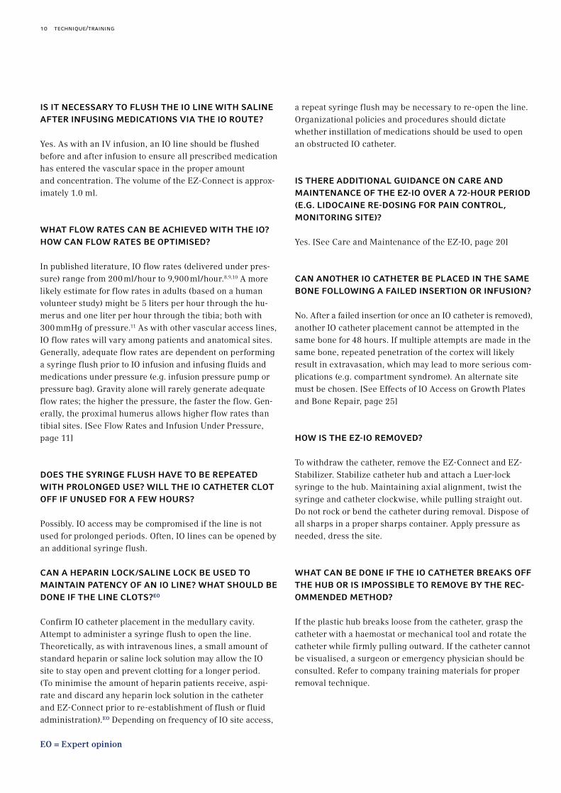
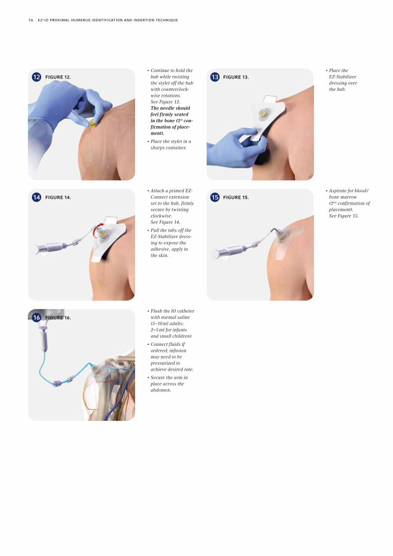
• Continue to hold the hub while twisting the stylet off the hub with counterclock-wise rotations. See Figure 12. The needle should feel firmly seated in the bone (1st con-firmation of place-ment).
• Place the stylet in a sharps container.
• Attach a primed EZ-Connect extension set to the hub, firmly secure by twisting clockwise. See Figure 14.
• Pull the tabs off the EZ-Stabilizer dress-ing to expose the adhesive, apply to the skin.
• Place the EZ-Stabilizer dressing over the hub.
• Flush the IO catheter with normal saline (5–10 ml adults; 2–5 ml for infants and small children).
• Connect fluids if ordered; infusion may need to be pressurized to achieve desired rate.
• Secure the arm in place across the abdomen.
• Aspirate for blood /bone marrow (2nd confirmation of placement). See Figure 15.
12
14
16
13
15
FIGURE 12.
FIGURE 14.
FIGURE 16.
FIGURE 13.
FIGURE 15.
Proximal tibia 17
PROXIMAL TIBIA
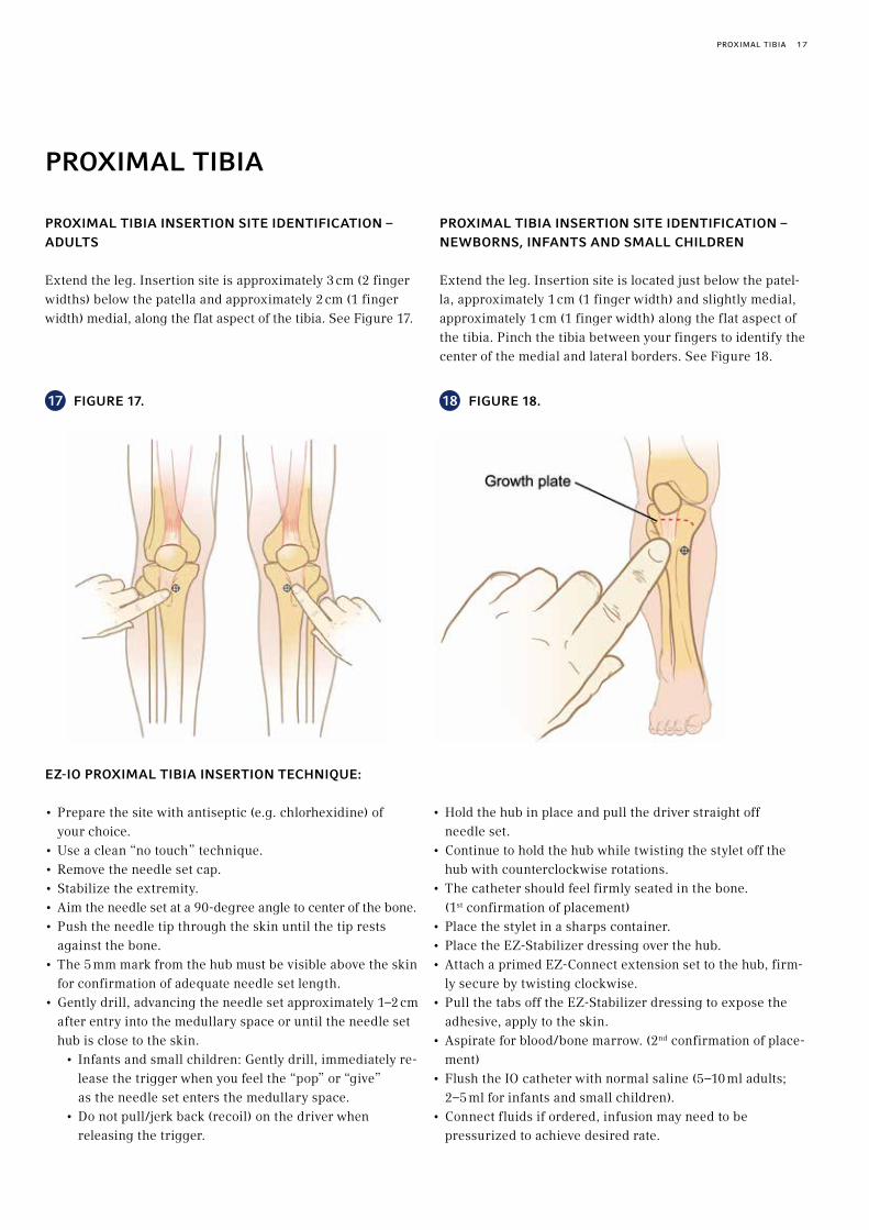
PROXIMAL TIBIA INSERTION SITE IDENTIFICATION – ADULTS
Extend the leg. Insertion site is approximately 3 cm (2 finger widths) below the patella and approximately 2 cm (1 finger width) medial, along the flat aspect of the tibia. See Figure 17.
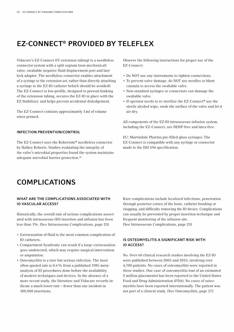
PROXIMAL TIBIA INSERTION SITE IDENTIFICATION – NEWBORNS, INFANTS AND SMALL CHILDREN
Extend the leg. Insertion site is located just below the patel-la, approximately 1 cm (1 finger width) and slightly medial, approximately 1 cm (1 finger width) along the flat aspect of the tibia. Pinch the tibia between your fingers to identify the center of the medial and lateral borders. See Figure 18.
EZ-IO PROXIMAL TIBIA INSERTION TECHNIQUE:
• Prepare the site with antiseptic (e.g. chlorhexidine) of your choice.
• Use a clean “no touch” technique.• Remove the needle set cap.• Stabilize the extremity.• Aim the needle set at a 90-degree angle to center of the bone.• Push the needle tip through the skin until the tip rests
against the bone.• The 5 mm mark from the hub must be visible above the skin
for confirmation of adequate needle set length. • Gently drill, advancing the needle set approximately 1–2 cm
after entry into the medullary space or until the needle set hub is close to the skin.
• Infants and small children: Gently drill, immediately re-lease the trigger when you feel the “pop” or “give” as the needle set enters the medullary space.
• Do not pull/jerk back (recoil) on the driver when releasing the trigger.
• Hold the hub in place and pull the driver straight off needle set.
• Continue to hold the hub while twisting the stylet off the hub with counterclockwise rotations.
• The catheter should feel firmly seated in the bone. (1st confirmation of placement)
• Place the stylet in a sharps container.• Place the EZ-Stabilizer dressing over the hub. • Attach a primed EZ-Connect extension set to the hub, firm-
ly secure by twisting clockwise.• Pull the tabs off the EZ-Stabilizer dressing to expose the
adhesive, apply to the skin.• Aspirate for blood/bone marrow. (2nd confirmation of place-
ment)• Flush the IO catheter with normal saline (5–10 ml adults;
2–5 ml for infants and small children).• Connect fluids if ordered, infusion may need to be
pressurized to achieve desired rate.
17 18FIGURE 17. FIGURE 18.

18 distal tibia
EZ-IO DISTAL TIBIA INSERTION TECHNIQUE:
• Prepare the site with antiseptic (e.g. chlorhexidine) of your choice.
• Use a clean “no touch” technique.• Remove the needle set cap.• Stabilize the extremity. • Aim the needle set at a 90-degree angle to center of the bone.• Push the needle tip through the skin until the tip rests
against the bone.• The 5 mm mark from the hub must be visible above the skin
for confirmation of adequate needle set length. • Gently drill, advancing the needle set approximately 1–2 cm
after entry into the medullary space or until the needle set hub is close to the skin.
• Infants and small children: Gently drill, immediately re-lease the trigger when you feel the “pop” or “give” as the needle set enters the medullary space.
• Do not pull/jerk back (recoil) on the driver when releasing the trigger.
• Hold the hub in place and pull the driver straight off needle set.
• Continue to hold the hub while twisting the stylet off the hub with counterclockwise rotations.
• The catheter should feel firmly seated in the bone. (1st confirmation of placement)
• Place the stylet in a sharps container.• Place the EZ-Stabilizer dressing over the hub. • Attach a primed EZ-Connect extension set to the hub,
firmly secure by twisting clockwise. • Pull the tabs off the EZ-Stabilizer dressing to expose the
adhesive, apply to the skin.• Aspirate for blood/bone marrow (2nd confirmation of
placement)• Flush the IO catheter with normal saline (5–10 ml adults;
2–5 ml for infants and small children).• Connect fluids if ordered, infusion may need to be
pressurized to achieve desired rate.
DISTAL TIBIA
DISTAL TIBIA INSERTION SITE IDENTIFICATION – ADULTS
Insertion site is located approximately 3 cm (2 finger widths) proximal to the most prominent aspect of the medial malleo-lus. Palpate the anterior and posterior borders of the tibia to assure that your insertion site is on the flat center aspect of the bone. See Figure 19.
DISTAL TIBIA INSERTION SITE IDENTIFICATION – NEWBORNS, INFANTS AND SMALL CHILDREN
Insertion site is located approximately 1–2 cm (1 finger width) proximal to the most prominent aspect of the medial malleolus. Palpate the anterior and posterior borders of the tibia to assure that your insertion site is on the flat center aspect of the bone. See Figure 20.
19 20FIGURE 19. FIGURE 20.

distal femur 19
Growth plate
DISTAL FEMUR
DISTAL FEMUR SITE IDENTIFICATION – NEWBORNS, INFANTS AND SMALL CHILDREN ONLY
Secure the leg out-stretched to ensure the knee does not bend. The insertion site is just proximal to the patella (maximum 1 cm) and approximately 1 cm medial to the mid-line. See Figure 21.EZ-IO distal femur insertion technique – newborns, infants and small children only:
EZ-IO DISTAL FEMUR INSERTION TECHNIQUE – NEWBORNS, INFANTS AND SMALL CHILDREN ONLY:
• Prepare the site by using antiseptic (e.g. chlorhexidine) of your choice.
• Use a clean, “no touch” technique.• Remove the needle set cap. • Aim the needle set at a 90-degree angle to center of the bone. • Push the needle tip through the skin until the tip rests
against the bone.• The 5 mm mark from the hub must be visible above the skin
for confirmation of adequate needle set length. • Gently drill, immediately release the trigger when you feel
the “pop” or “give” as the needle set enters the medullary space.
• Do not pull/jerk back (recoil) on the driver when releasing the trigger.
• Hold the hub in place and pull the driver straight off needle set.
• Continue to hold the hub while twisting the stylet off the hub with counterclockwise rotations,
• The catheter should feel firmly seated in the bone (1st confirmation of placement),
• Place the stylet in a sharps container,• Place the EZ-Stabilizer dressing over the hub, • Attach a primed EZ-Connect extension set to the hub,
firmly secure by twisting clockwise.• Pull the tabs off the EZ-Stabilizer dressing to expose the
adhesive, apply to the skin.• Aspirate for blood/bone marrow (2nd confirmation of
placement)• Flush the IO catheter with normal saline (2–5 ml for
infants and small children).• Connect fluids if ordered, infusion may need to be
pressurized to achieve desired rate.
21 FIGURE 21.

20 care and maintenance of the eZ-io
CARE AND MAINTENANCE OF THE EZ-IO
Care and maintenance of the EZ-IO is similar to other venous access routes: confirming placement prior to medication administration, maintaining catheter patency, monitoring the insertion site for signs of extravasation, and appropriate device removal. The EZ-IO may remain in place up to 72 hours.
CONFIRMING EZ-IO PLACEMENT
Prior to administration of medications or fluids, confirm EZ-IO placement with the following methods:
• ability to aspirate blood• stability of catheter • adequate flow rate
EZ-IO PATENCYEO
Theoretically, as with intravenous lines, a small amount of standard heparin or saline lock solution may allow the IO site to remain patent.EO (To minimise the amount of heparin patients receive, aspirate and discard any heparin lock solution in the catheter and EZ-Connect prior to re-establishment of flush or fluid administration). Depending on the duration of IO site access, a repeat syringe flush may be necessary to re-open the line. Refer to organizational policies and proce-dures to determine whether instillation of medications should be used to open an obstructed IO catheter.
PAIN MANAGEMENT IN CONSCIOUS PATIENTS (RESPONSIVE TO PAIN)
The pain associated with IO insertion is variable whereas pain associated with IO infusion under pressure is often severe.43 One percent (1 %) and 2 % intravenous preservative-free lidocaine without epinephrine has been shown to be effective in limiting or alleviating IO infusion pain. Duration of the anesthetic effect will vary among patients. Repeat doses of lidocaine may be necessary to maintain anesthetic effect.
One series of studies in healthy volunteers measured the duration of pain relief during IO infusion in the proximal humerus and proximal tibia. During the 90-minute obser-vation period in the tibial study, 8 of 10 volunteers who had previously received 100 mg lidocaine required an additional
20 mg lidocaine to keep the IO infusion pain level below 5 (on a scale of 0–10). No volunteers in the humeral study – who had previously received 60 mg lidocaine – required additional lidocaine dosing to keep pain levels below 5.44
Lidocaine and appropriate dosages must be prescribed by a physician or qualified prescriber. The Intraosseous Lidocaine Guideline – developed by Dr Richard Hixson, a recognized anesthesiologist who has published drug dosages, provides a concise guide to administration and dosing of lidocaine via the IO route for all age and weight ranges.45 The guide may be accessed online at Hixson R. Intraosseous administration of preservative-free lidocaine. [See Pain Management for IO Infusion, page 28]
SITE MAINTENANCE/MONITORING
Extravasation is the most common complication associated with IO insertion, and can lead to serious complications such as compartment syndrome and necrosis. The IO insertion site should be monitored frequently for any signs of extrava-sation, localised inflammation, or dislodgement, particularly in the first half hour after insertion and anytime the IO catheter is manipulated. After an initial observation period, the site should be monitored at least hourly. Organizational policy should dictate care of the insertion site.
PATIENT ACTIVITY
Ambulation should be discouraged with a tibial EZ-IO cathe-ter in place. With distal femur IO access the knee should not be flexed and ambulation should be prohibited. There are no activity restrictions after EZ-IO removal.
REMOVAL
To withdraw the catheter, remove the EZ-Connect and EZ-Stabilizer. Stabilize catheter hub and attach a Luer-lock syringe to the hub. While maintaining axial alignment, twist the syringe and catheter clockwise while pulling straight out. Do not rock or bend the catheter during removal. Dispose of all sharps in a proper sharps container. Apply pressure over the site as needed. Dress the site.
EO = Expert opinion

eZ-io G3 Power driver and traininG driver 21
EZ-IO G3 POWER DRIVER AND TRAINING DRIVER
LIFE OF DRIVER
The EZ-IO G3 Power Driver can achieve several hundred EZ-IO needle set insertions under ideal conditions. However, storage and use conditions may result in substantially fewer insertions. The drivers contain non-rechargeable manganese dioxide lithium batteries, which have significantly better per-formance and storage characteristics than previous batteries.
All batteries deteriorate somewhat over time. If the driver is being regularly transported on an EMS truck in extreme or constantly changing climate conditions, or if the driver is used frequently in extreme conditions, driver life may be significantly less.
Other factors can also reduce the longevity of the driver. Daily equipment checks (pulling the trigger to activate the driver) is one of the most significant contributors to the need for driver replacement. The presence of a green light on the driver handle indicates adequate battery life. The light will turn red when approximately 10 % of battery life remains, providing notification of the need for driver replacement.
DRIVER CLEANING
Specific information and direction on driver cleaning is outlined below. This information can also be found in the driver’s Instructions for Use. The instructions can also be found on Vidacare’s web site at www.arrowezio.com.
CLEANING AND DISINFECTION OF THE EZ-IO G3 POW-ER DRIVER
1. Maintain BSI or PPE precautions.2. Wipe entire exterior surface of G3 Power Driver with soft,
clean moistened cloth. (If supplied, detach, clean and soak lanyard and trigger guard). Use soft, bristled brush to re-move any visible soil or debris, paying particular attention to crevices and seam.
3. Spray exterior surface of G3 Power Driver with the antimi-crobial commonly used by your institution, making sure to follow the antimicrobial manufacturer’s recommendations.
4. Gently wipe exterior surfaces with gauze pads until visible debris is removed.
5. Clean and manipulate trigger using cloth moistened with selected antimicrobial.
6. Using sterile swabs, moisten with selected antimicrobial solution, gently clean inside opening around metal drive shaft.
7. After cleaning, inspect to ensure no visible debris re-mains, and no damage has occurred to the driver.
8. Dry driver with soft, clean cloth (re-attach lanyard and trigger guard), and return to appropriate location.
Do not immerse or use excessive amount of liquid when performing cleaning and disinfecting. In the unlikely event of driver failure, remove the G3 Power Driver, grasp the needle set by hand and advance the needle set into the medullary space while twisting the needle set.
DRIVER STERILISATION
If the clinical environment requires driver sterilisation, the G3 Power Driver can be sterilised using the STERRAD® 100S, NX Standard cycle, and 100NX Standard cycle. STERRAD® is a product of Advanced Sterilization Products.
TRAINING DRIVER
The G3 Training Driver is intended for training and demonstration only. Training drivers typically incur heavier usage, more frequent transport, and frequent handling by inexperienced users. Regulatory, quality, and cleaning differences preclude the training driver from being used for patient care.
TRIGGER GUARD/VASCULAR ACCESS PACK (VAP)
The EZ-IO vascular access pack includes a built-in cradle for the driver. Storing the driver in the cradle with the trigger guard in place may cause inadvertent activation of the driver, resulting in depletion of the batteries. To prevent this situa-tion, the trigger guard should be completely removed when storing the driver in the cradle.
22 eZ-connect by vidacare / comPlications
EZ-CONNECT® PROVIDED BY TELEFLEX
Vidacare’s EZ-Connect (IV extension tubing) is a needleless connector system with a split septum (non-mechanical) valve, swabable negative fluid displacement port and luer lock adapter. The needleless connector enables attachment of a syringe to the extension set, rather than directly attaching a syringe to the EZ-IO catheter (which should be avoided). The EZ-Connect is low-profile; designed to prevent kinking of the extension tubing, secures the EZ-IO in place with the EZ-Stabilizer, and helps prevent accidental dislodgement.
The EZ-Connect contains approximately 1 ml of volume when primed.
INFECTION PREVENTION/CONTROL
The EZ-Connect uses the Robertsite® needleless connector by Halkey Roberts. Studies evaluating the integrity of the valve’s microbial properties found the system maintains adequate microbial barrier protection.46
Observe the following instructions for proper use of the EZ-Connect:
• Do NOT use any instruments to tighten connections.• To prevent valve damage, do NOT use needles or blunt
cannula to access the swabable valve. • Non-standard syringes or connectors can damage the
swabable valve.• If operator needs to re-sterilize the EZ-Connect® use the
sterile alcohol wipe, swab the surface of the valve and let it air dry.
All components of the EZ-IO intraosseous infusion system, including the EZ-Connect, are DEHP-free and latex-free.
EU: Martindale Pharma pre-filled glass syringes: The EZ-Connect is compatible with any syringe or connector made to the ISO 594 specification.
COMPLICATIONS
WHAT ARE THE COMPLICATIONS ASSOCIATED WITH IO VASCULAR ACCESS?
Historically, the overall rate of serious complications associ-ated with intraosseous (IO) insertion and infusion has been less than 1%. [See Intraosseous Complications, page 23]
• Extravasation of fluid is the most common complication of IO catheters.
• Compartment Syndrome can result if a large extravasation goes undetected, which may require surgical intervention or amputation.
• Osteomyelitis is a rare but serious infection. The most often-quoted rate is 0.6 % from a published 1985 meta-analysis of IO procedures done before the availability of modern techniques and devices. In the absence of a more recent study, the literature and Vidacare records in-dicate a much lower rate – fewer than one incident in 100,000 insertions.
Rare complications include localized infections, penetration through posterior cortex of the bone, catheter bending or clogging, and difficulty removing the IO device. Complications can usually be prevented by proper insertion technique and frequent monitoring of the infusion site. [See Intraosseous Complications, page 23]
IS OSTEOMYELITIS A SIGNIFICANT RISK WITH IO ACCESS?
No. Over 60 clinical research studies involving the EZ-IO were published between 2005 and 2013, involving over 4,700 patients. No cases of osteomyelitis were reported in these studies. One case of osteomyelitis (out of an estimated 2 million placements) has been reported to the United States Food and Drug Administration (FDA). No cases of osteo-myelitis have been reported internationally. The patient was not part of a clinical study. [See Osteomyelitis, page 27]
intraosseous comPlications 23
WILL INFUSING DRUGS THROUGH THE IO SPACE CAUSE LONG-TERM DAMAGE TO THE BONE MARROW?
No long-term damage to human bone has been documented in known medical literature. One preclinical study in swine demonstrated marrow damage after receiving multiple infu-sions of Adriamycin® via the IO route. Another preclinical study in swine reported marrow damage after multiple in-fusions of hypertonic saline. Any drug with the potential to cause sclerosis or damage to veins has the potential to damage intraosseous vessels. As such, the risk vs. benefit of administering these drugs via the IO route should be carefully evaluated prior to use. [See Effects of IO Access on Growth Plates and Bone Repair, page 25]
DOES IO INSERTION OR INFUSION AFFECT THE GROWTH PLATE IN PEDIATRIC PATIENTS?
Though often listed as a theoretical complication of IO access, no growth plate damage in pediatric patients has been docu-mented in known medical literature. [See Effects of IO Access on Growth Plates and Bone Repair, page 25]
IS FAT EMBOLISM OR THROMBOEMBOLISM AN ISSUE WITH IO INFUSION?
Clinically significant fat embolism from IO administration has not been reported in known medical literature. [See Embolism, page 25]
IS AIR EMBOLISM A POSSIBILITY THROUGH AN IO CATHETER?
Air embolism can be introduced into the circulatory system by any vascular route including peripheral venous access, central venous access, arterial access, or intraosseous access. A primed syringe, extension set or infusion tubing should always be placed on the IO catheter hub immediately after insertion. The inherent intraosseous pressure precludes spontaneous air embolism that is more likely to occur with central venous catheters. [See Embolism, page 25]
DOES VIDACARE TRACK COMPLICATIONS ASSOCIATED WITH THE EZ-IO?
Vidacare tracks any reported problems or complications associated with the EZ-IO in accordance with applicable regulatory agency requirements.
INTRAOSSEOUS COMPLICATIONS
Historically, the documented overall complication rate asso-ciated with intraosseous (IO) insertion and infusion is less than 1%. In a 1985 meta-analysis of over 4,200 patients, the most common IO complication was infection, including osteomyelitis (0.6%), and was attributed to IO placement in bacteremic patients or prolonged infusions.47
With modern technological devices and procedures, extra-vasation is the more prevalent complication reported.48,49 While simple extravasation itself may be unremarkable, com-partment syndrome may occur if extravasation continues undetected. Therefore, careful monitoring of the insertion site is strongly recommended. [See Compartment Syndrome, page 24]
Although uncommon, other reported complications have included fracture, and failure to infuse due to catheter bend-ing or clogging.50,51,52,53,54
IO COMPLICATIONS
As of November 2013, over 60 clinical trials or case studies involving the EZ-IO have been reported in the clinical litera-ture, involving over 4,700 patients.55 The rate of EZ-IO serious complications reported in literature is <0.001 % (less than one per 100,000 IO placements). In aggregate, these reports described 6 serious complications. Four cases of compart-ment syndrome have been reported with two resulting in
24 comPartment syndrome
amputations.56,57,58 The fifth and sixth cases involved extensive tissue necrosis, one case requiring operative intervention subsequent to administration of fibrinolytics via the EZ-IO.59,60 No cases of osteomyelitis were reported, a commonly cited concern for IO infusions. Minor complications included extra-vasation, infiltration, slow flow rate, dislodgement, inability to flush, leakage, problems with device, difficulty with re-moval of device, and local inflammation.
In a 2005 prospective study of the EZ-IO in 250 adult patients, Davidoff et al. reported an overall complication rate of 3 %, with failure to deliver medications the most predominant.61 These complications were generally associated with failure to syringe-flush the catheter following insertion – a critical step for IO infusions. There were no cases of osteomyelitis, embolism, fracture, infection, extravasation, or compart-ment syndrome.
COMPARTMENT SYNDROME
COMPARTMENT SYNDROME “101”
“Compartments” are composed of muscle tissue, nerves and blood vessels separated and surrounded by thick layers of non-expandable tissue (fascia). Compartment syndrome occurs when swelling within that confined space causes the com-pression of those nerves, blood vessels, and muscle due to the lack of ability to expand outward. This may be caused by instillation of fluid into the soft tissue outside the vascular space. The most common site for compartment syndrome is the lower leg. The swelling within the compartment can progress to compression of blood vessels within the compart-ment causing a lack of oxygenation and eventually, necrotic tissue. When the condition is attributed to IO access, com-partment syndrome is usually secondary to extravasation – the number one complication of IO vascular access, including the EZ-IO. Compartment syndrome usually occurs when clinicians do not recognize early signs of extravasation. In extreme cases, amputation of a limb is necessary if the compartment syndrome is not recognized soon enough or is not adequately treated. Once recognized, treatment of compartment syndrome consists primarily of removing the increased pressure source and careful monitoring, assuming circulation has not been compromised. In more severe cases, fasciotomy (surgically opening the fascia surrounding the compartment to release the pressure) may be required to re-store circulation. Necrosis of tissue resulting in amputation can occur if circulation is not restored within about 4 hours.
IO ACCESS-ASSOCIATED COMPARTMENT SYNDROME IN THE LITERATURE
Five paediatric cases of compartment syndrome were report-ed in the medical literature between 2011 and 2013, and 2 in 2008.62,63,64,65,66,67 Contributing factors included improper technique, catheter dislodgement, and prolonged infusion with caustic agents. In one case, further investigation (for United States FDA reporting) suggested multiple IO attempts in the same bone, causing the underlying extravasation. One case of adult compartment syndrome related to IO infusion was reported in 2013 in which IO access was established in the fractured tibia of an adult multi-trauma patient for fluid infusion.68 IO catheter placement is contraindicated for frac-tured bones. Other cases of compartment syndrome have occurred using older IO devices; two resulting in limb ampu-tations.69,70,71,72,73,74,75,76,77
These cases underscore the importance of adequate training, appropriate selection of IO needle set and insertion site, prop-er technique, confirming proper placement of the IO catheter within the medullary space, and stabilisation of the IO device. Early detection of extravasation and prevention of compart-ment syndrome can be accomplished through frequent monitoring of the insertion site and the involved extremity, particularly during prolonged infusion, prolonged transport times and administration of large fluid volumes.

effects of io access on Growth Plates and bone rePair / embolism 25
EFFECTS OF IO ACCESS ON GROWTH PLATES AND BONE REPAIR
EFFECT ON EPIPHYSEAL (GROWTH) PLATES
A 1990 review article published in the New England Journal of Medicine stressed the relative safety of IO and reported earlier findings of no lasting negative effects of IO infusion on the bone, growth plates and marrow elements.78 Lack of negative effect on the epiphyseal plate subsequent to intra-osseous (IO) infusion has been demonstrated in several radio-graphic studies in the paediatric population. Preclinical studies in swine have supported similar conclusions. A clini-cal report of 72 patients who received D50W injections disclosed no disturbance of the growth plate over a three year observation period.79 A 2003 overview article is also supportive of these findings.80
CLINICAL RESEARCH
A 1946 clinical study examined long-term bone abnormalities during radiographic follow-up in 36 paediatric patients who received IO insertion. No patient exhibited radiographic bone abnormality and bone growth was normal for all patients in the study.78 In a 1986 study, investigators found no bone defects or distortions at 6 and 12 weeks post IO insertion in ten paediatric patients.81 A 1997 study performed radio-graphic measurements of the tibias in paediatric patients 12 months after IO infusion. Results demonstrated no signif-icant difference in tibial lengths.82 In 2003, a clinical study of paediatric patients receiving IO infusions revealed no radiographic differences in tibia width or length. The follow-up radiographs were performed on average 29 months after infusion.83
PRECLINICAL RESEARCH
One preclinical study of IO infusion in young swine found no growth disturbances or growth plate abnormalities after 2 and 6 months.84 Another preclinical study found IO infusion of saline and bicarbonate did not damage growth plates. The researchers observed loss of bone trabeculae supporting the growth plate, but the loss was rapidly repaired.85 A 1993 preclinical study found no changes in bone growth of epiphy-seal injury related to IO infusion.86
BONE REPAIR AFTER IO INFUSION
The Rosetti meta-analysis (1985) reported multiple follow-up studies on marrow and bone from 24-hours post infusion through 22 months.87 Periostitis at the injection site cleared within 2–3 weeks; marrow cellularity after isotonic infusion was slightly less or normal; no long-term bone changes were noted post isotonic infusion.
Based on a 2010 preclinical study, sealing of the bone (to the point at which IO placement and infusion can be accomplished in the same bone) takes approximately 48 hours post IO removal.
By that time, fibrin formation and clotting are sufficient to prevent extravasation through the previous IO hole. Com-plete healing, to the point where x-ray can no longer detect the hole, varies from days to weeks.88
EMBOLISM
THROMBOEMBOLISM
Thromboembolism is not typically a complication associated with intraosseous (IO) infusions due to anatomy and physiol-ogy of circulation within the medullary cavity. Only one known case reported arterial thrombosis in a patient receiv-ing an IO infusion. The authors were not certain of the cause of the thrombosis and exact mechanisms of the disease pro-cess were unclear.89
AIR EMBOLISM
As with any vascular access route, an air embolism can be introduced into the circulatory system by IO access. The determining factors favoring air embolism are relative pressure gradients between the vascular access site and atmospheric pressure, and the size of the catheter.
26 embolism
There are two known reports in published literature of air embolism in patients with an IO infusion. In the first case, a cerebral arterial air embolism was noted on autopsy in a 7-month-old child.90
While the cause of death remained undetermined, it was noted that the patient had no other vascular access other than the IO route. However, the authors noted that air could have been introduced during any of the attempts at central venous or arterial access, as well as by the IO route.
In the second case, multiple gas emboli were discovered in a post-mortem computed tomography (CT) scan in a 4-month-old child who died of sudden infant death syndrome.91 A bone marrow aspiration needle had been used to provide IO vas-cular access. No alternative attempts at vascular access were noted. The author concluded that gas could have been intro-duced subsequent to the method of infusing IO medications, and concluded that resuscitation with an inserted, disconnect-ed intraosseous needle/catheter should be avoided.
Since pressure in the IO space is higher than atmospheric pressure, air embolism via the IO route is less likely than with other vascular access route. A primed syringe, extension set or infusion tubing should always be placed on the IO catheter hub immediately after insertion, and should remain in place until catheter removal.
FAT EMBOLISM
No known case of clinically significant fat embolism resulting from IO administration has been reported in the medical literature or in actual practice, although preclinical trials have shown microscopic fat emboli in the lungs after high pressure IO infusion.92,93
The risk of fat embolism associated with IO infusion has been studied in preclinical trials and reported in the clinical literature for two decades. In a canine study, Orlowski et al. examined the prevalence of fat and bone marrow emboli in the lung following IO infusion of hypertonic and emergency drugs.94 Researchers found no difference in the mean num-ber of fat and bone marrow emboli per square millimeter of lung tissue compared to the control group, who received normal saline. In 1995, Plewa reported a preclinical study examining hematologic parameters with IO and IV autologous blood transfusions.95 The authors found all haematologic parameters remained within normal limits in both IO and IV groups, and concluded that IO blood transfusions were hematologically safe, without risk of appreciable hemolysis, disseminated intravascular coagulation, or fat embolism syndrome.
A 1997 swine study examined IO infusion during CPR and found no increase in fat embolism in the IO group compared to control groups, which received no IO infusions.93 Another swine study found low levels of fat embolism (one to three emboli per high powered field in 30 % of the specimens). The researchers concluded that the risk of fat emboli exists with IO, but its clinical relevance is unclear.96 In 2012, Lairet et al. reported finding fat emboli in the lungs of 32 of 39 swine that had received blood transfusions using high pres-sure averaging 604 mmHg.97 The infusion pressures were double the maximum pressure typically used for IO infusion and, in contrast to Orlowski’s earlier work, Lairet’s study did not include control animals in which there was no IO infu-sion or infusion with saline, or infusion with typical pres-sure (300 mmHg).
In a case series of 18 paediatric patients receiving IO infu-sion during resuscitation, one complication of minor fat aembolism was reported but had no clinical significance.98
osteomyelitis / medications/fluids 27
OSTEOMYELITIS
Numerous research studies and reports in clinical literature have addressed the low incidence of osteomyelitis risk in the intraosseous (IO) space.
In a 1985 meta-analysis of IO complications in over 4,200 patients, the most common IO complication was osteomyelitis at 0.6 % and was attributed to IO access placement in bac-teremic patients or prolonged infusions.99 Six osteomyelitis cases have been reported in known literature since the 1985 meta-analysis by Rosetti et al.100,101,102,103,104,105
As of November 2013, over 60 clinical trials or case studies in-volving the EZ-IO have been reported in the clinical literature, involving over 4,700 patients. In aggregate, study results
have reported no cases of osteomyelitis. While not part of a published study, one case of osteomyelitis subsequent to EZ-IO use has been reported to Vidacare. The pediatric patient had multiple co-morbidities, including sepsis. Upon initial follow up the patient was steadily improving, but com-plete follow up data was unavailable.106
As of November 2013, the EZ-IO had been used in an estimated 2,000,000 insertions since being introduced to the market in 2004, with only one case of osteomyelitis being recorded. Considering the total number of uses, the risk of osteomyelitis remains low.EO
MEDICATIONS/FLUIDS
WHAT FLUIDS AND MEDICATIONS CAN BE INFUSED VIA THE IO ROUTE?
Virtually any fluid or medication that can be safely infused via peripheral IV route may be safely infused through the in-traosseous (IO) route. Incompatible drugs and fluids should be infused sequentially in a manner consistent with standard IV infusion practice. Caution should be exercised with repeat doses of hypertonic fluids. We do not recommend infusing chemotherapy agents.
WHAT DOSAGES ARE REQUIRED FOR IO INFUSION COMPARED WITH IV DOSAGES?
Intraosseous dosages are typically identical to IV dosages. Drugs and fluids reach the central circulation at essentially the same concentrations through the IO route and the IV route.110,111,112,113
WHAT MEDICATIONS HAVE BEEN ADMINISTERED SUCCESSFULLY TO DATE (VIA THE IO ROUTE)?
See the following list of medications that have been adminis-tered effectively via the IO route without reported adverse events. [See Reference List, page 42]

28 Pain manaGement for io infusion
PAIN MANAGEMENT FOR IO INFUSION
While the discomfort associated with intraosseous (IO) insertion is variable, pain associated with IO infusion under pressure is often severe.111 One percent (1 %) and 2 % pre-servative-free intravenous lidocaine without epinephrine (i.e. cardiac lidocaine) has been shown to be effective in lim-iting or alleviating IO infusion pain. Lidocaine administered via the IO route for anesthetic effect should be delivered slowly into the IO space prior to administering the saline flush.
CONSCIOUS PATIENTS/LIDOCAINE DOSING
Vidacare does not manufacture lidocaine, and therefore cannot make specific dosage recommendations (follow man-ufacturer recommendations). A number of articles in the literature describe clinical experience with lidocaine admin-istration for IO infusion in patients responsive to pain.112–121 These cited sources document initial lidocaine doses ranging from 20–80 mg, with varying doses for maintenance. The Intraosseous Lidocaine Guideline – developed by Dr Richard
• Adenosine (e.g. Adenocard)• Albumin• Alfentanil (e.g. Alfenta)• Aminophylline• Amiodarone (e.g. Cordarone)• Ampicillin • Anascorp (scorpion antivenin)• Anesthetic agents• Antibiotics (multiple)• Antitoxins (various)• Atracurium besylate (e.g. Tracrium)• Atropine• Azactam (e.g. Aztreonam)• Blood and blood products• Calcium chloride• Calcium gluconate• Cefepime hydrochloride
(e.g. Maxipime)• Ceftriaxone (e.g. Rocephin)• Contrast media (e.g Omnipaque)• Dexamethasone • (e.g. Decadron)• Dextran• D5 ½NS• Dextrose 10 %• Dextrose 25 %• Dextrose 50 %• Diazepam (e.g. Valium)• Diazoxide (e.g. Hyperstat)• Digoxin (e.g. Lanoxin)• Diltiazem (e.g. Cardizem)• Diphenhydramine (e.g. Benadryl)• Dobutamine hydrochloride
(e.g. Dobutrex)
• Dopamine• Ephedrine • Epinephrine• Esmolol (e.g. Brevibloc)• Etomidate• Fentanyl• Fluconazole (e.g. Diflucan)• Flumazenil (e.g. Romazicon)• Fosphenytoin
(e.g. Cerebyx, Prodilantin)• Furosemide (e.g. Lasix)• Gentamycin • Haloperidol (e.g. Haldol)• Heparin• Hydroxo-cobalamin (B12)• Hydropmorphone (e.g. Dilaudid)• Insulin• Isoprenaline
(e.g. isoproterenol, Isuprel)• Ketamine• Labetalol (e.g. Normodyne)• Levetiracetam (e.g. Keppra)• Lidocaine (e.g. Xylocaine)• Linezolid (e.g. Zyvox)• Lorazepam (e.g. Ativan)• Magnesium sulfate• Mannitol• Methyl-prednisolone
(e.g. Solu-Medrol) • Metoprolol (e.g. Lopressor)• Midazolam (e.g. Versed)• Mivacurium (e.g. Mivacron)• Morphine sulfate• Nalbuphine (e.g. Nubain)
• Naloxone (e.g. Narcan)• Neostigmine (e.g. Prostigmin)• Nitroglycerin• Nitroprusside (e.g. Nipride) • Norcuron• Norepinephrine
(Levarterenol, Levophed)• Normal saline• Odansetron (e.g. Zofran)• Pancuronium (e.g. Pavulon)• Paracetomol (i.e. acetominophen)• Phenobarbital • Phenylephrine (e.g. Neo-Synephrine)• Phenytoin (e.g. Dilantin)• Piperacillin (e.g. Zosyn)• Plasmanate• Potassium chloride • Promethazine (e.g. Phenergan)• Propofol (e.g. Diprivan)• Propranolol (e.g. Inderal)• Remifentanil (e.g. Ultiva)• Ringer’s lactate• Rocuronium (e.g. Zemuron)• Sodium bicarbonate• Standard IV solutions• Succinylcholine (e.g. Anectine)• Tenectaplase (e.g. TNKase)• Thiamine• Thiopental (e.g. Pentothal)• Tobramycin sulfate• Vancomycin • Vasopressin
(e.g. Pitressin, Argipressin)• Vecuronium

Pain manaGement for io infusion 29
Hixson, a recognized anaesthesiologist who has published drug dosages – provides a concise guide to administration and dosing of lidocaine via the IO route for all age and weight ranges.122 See Figure 22.
A 2010 article reported the combined results of two studies examining pain management with IO vascular access.111 These volunteer studies using the humerus and tibia demon-strated that less pressure was required to infuse through the humerus than the tibia route, and demonstrated a direct correlation between infusion pressure and pain level (i.e. in-creased pain with greater infusion pressures).
During the 90-minute observation period in the tibial study, 8 of 10 volunteers who had previously received 100 mg lido-caine required an additional 20 mg lidocaine to keep the IO infusion pain level below 5 (on a scale of 0–10). No volun-teers in the humeral study – who had previously received 60 mg lidocaine – required additional lidocaine dosing to keep pain levels below 5. This article demonstrates the prox-imal humerus may be a preferred IO site for conscious pa-tients. Lidocaine and appropriate dosages must be pre-scribed by a qualified prescriber.
TECHNIQUE
Recommended anesthetic for adult patients responsive to pain: • Observe recommended cautions/contraindications to using
1 % and 2 % preservative-free and epinephrine free lido-caine (intravenous lidocaine).
• Confirm lidocaine dose with your medical director, treating physician, or institutional protocol.
• Prime EZ-Connect extension set with lidocaine. • Note that the priming volume of the EZ-Connect is
approximately 1.0 ml.• If primed with 1 % preservative-free lidocaine,
this will be approximately 10 mg.• If primed with 2 % preservative-free lidocaine,
this will be approximately 20 mg.• Slowly infuse 40 mg of lidocaine IO over 120 seconds
(2 minutes). • Allow lidocaine to dwell in IO space 60 seconds (1 minute). • Flush the IO catheter with 5 to 10 ml of normal saline. • Slowly administer an additional 20 mg of lidocaine IO over
60 seconds (1 minute). • Repeat PRN for pain. • Consider systemic pain control for patients not responding
to IO lidocaine.
Recommended anesthetic for infants and children (and those weighing less than 80 kg) responsive to pain: • Observe recommended cautions/contraindications to using
1 % and 2 % preservative-free and epinephrine free lido-caine (intravenous lidocaine).
• Confirm lidocaine dose with your medical director, treating physician, or institutional protocol.
• Usual initial dose is 0.5 mg/kg, not to exceed 40 mg.• Prime EZ-Connect extension set with lidocaine.
• Note that the priming volume of the EZ-Connect is approximately 1.0 ml.
• If primed with 1 % preservative-free lidocaine, this will be approximately 10 mg.
• If primed with 2 % preservative-free lidocaine, this will be approximately 20 mg.
• For small doses of lidocaine, consider administering by carefully attaching syringe directly to needle hub (prime EZ-Connect with normal saline).
• Slowly infuse lidocaine IO over 120 seconds (2 minutes). • Allow lidocaine to dwell in IO space 60 seconds (1 minute). • Flush the IO catheter with 2 to 5 ml of normal saline. • Slowly administer subsequent lidocaine (half the initial
dose – .25 mg/kg) IO over 60 seconds (1 minute). • Repeat PRN for pain. • Consider systemic pain control for patients not responding
to IO lidocaine.
Refer to Dr. Hixson’s chart, Intraosseous administration of preservative-free lidocaine, for lidocaine dosages related to patients weighing less than 80 kg. See Figure 21.
DisclaimerThe use of any medication, including lidocaine, given IV or IO is the responsibility of the treating physician, medical direc-tor or qualified prescriber and not an official recommendation of Vidacare. Vidacare is not the manufacturer of lidocaine, and the user should be familiar with the manufacturer’s instruc-tions or directions for use for all indications, side-effects, contraindications, precautions and warnings of lidocaine. Vidacare disclaims all liability for the use, application or interpretation of the use of this information in the medical treatment of any patient.
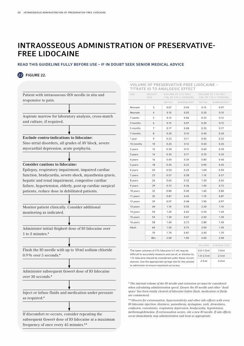
30 intraosseous administration of Preservative-free lidocaine
* The internal volume of the IO needle and extension set must be considered when calculating administration speed. Ensure the IO needle and other ‘dead space’ has been totally cleared of lidocaine before flush, medication or fluids are commenced.
** Observe for extravasation, hypersensitivity and other side-effects with every IO lidocaine injection: dizziness, parasthesia, nystagmus, rash, drowsiness, confusion, convulsions, respiratory depression, bradycardia, hypotension, methemoglobinemia. If extravasation occurs, site a new IO needle. If side effects occur immediately stop administration and treat as appropriate.
VOLUME OF PRESERVATIVE-FREE LIDOCAINE – TITRATE IO TO ANALGESIC EFFECTaGe weiGht
(kG)volume of 2 % (ml) 1 ml of 2 % = 20 mG/ml
volume of 1 % (ml) 1 ml of 1 % = 10 mG/ml
initial subseQuent initial subseQuent
Neonate 3 0.07 0.03 0.15 0.07
Neonate 4 0.10 0.05 0.20 0.10
7 weeks 5 0.12 0.06 0.25 0.12
3 months 6 0.15 0.07 0.30 0.15
5 months 7 0.17 0.08 0.35 0.17
7 months 8 0.20 0.10 0.40 0.20
1 year 9 0.22 0.11 0.45 0.22
15 months 10 0.25 0.12 0.50 0.25
2 years 12 0.30 0.15 0.60 0.30
3 years 14 0.35 0.17 0.70 0.35
4 years 16 0.40 0.20 0.80 0.40
5 years 18 0.45 0.22 0.90 0.45
6 years 20 0.50 0.25 1.00 0.50
7 years 23 0.57 0.28 1.10 0.57
8 years 26 0.65 0.32 1.30 0.65
9 years 29 0.72 0.36 1.40 0.72
10 years 32 0.80 0.40 1.60 0.80
11 years 35 0.87 0.43 1.70 0.87
12 years 39 0.97 0.48 1.90 0.97
13 years 44 1.10 0.55 2.20 1.10
14 years 50 1.20 0.62 2.50 1.20
15 years 54 1.30 0.67 2.60 1.30
16 years 58 1.40 0.72 2.80 1.40
Adult 60 1.50 0.75 3.00 1.50
70 1.70 0.87 3.40 1.70
80+ 2.00 1.00 4.00 2.00
volume syrinGe siZe
The lower volumes of 2 % lidocaine (<1 ml) may be
difficult to accurately measure and use of, or dilution to,
1 % lidocaine should be considered under these circum-
stances. Use the appropriate syringe site for the volume
to administer to ensure maximum accuracy:
0.0–1.0 ml 1.0 ml
1.0–2.5 ml 2.5 ml
2.5 ml 5.0 ml
INTRAOSSEOUS ADMINISTRATION OF PRESERVATIVE-FREE LIDOCAINE READ THIS GUIDELINE FULLY BEFORE USE – IF IN DOUBT SEEK SENIOR MEDICAL ADVICE
Patient with intraosseous (IO) needle in situ and responsive to pain.
Aspirate marrow for laboratory analysis, cross-match and culture, if required.
Exclude contra-indications to lidocaine: Sino-atrial disorders, all grades of AV block, severe myocardial depression, acute porphyria.
Consider cautions to lidocaine: Epilepsy, respiratory impairment, impaired cardiac function, bradycardia, severe shock, myasthenia gravis, hepatic and renal impairment, congestive cardiac failure, hypertension, elderly, post-op cardiac surgical patients, reduce dose in debilitated patients.
Monitor patient clinically. Consider additional monitoring as indicated.
Administer initial (higher) dose of IO lidocaine over 1 to 2 minutes.*
Flush the IO needle with up to 10 ml sodium chloride 0.9 % over 5 seconds.*
Administer subsequent (lower) dose of IO lidocaine over 30 seconds.*
Inject or infuse fluids and medication under pressure as required.*
If discomfort re-occurs, consider repeating the subsequent (lower) dose of IO lidocaine at a maximum frequency of once every 45 minutes.**
22 FIGURE 22.
Paediatrics: newborns, infants, children and adolescents 31
PAEDIATRICS: NEWBORNS, INFANTS, CHILDREN AND ADOLESCENTS
The US Food and Drug Administration (FDA) defines the paediatric population from newborn to 21 years of age using four paediatric subgroups. The FDA recognizes that the descriptions are somewhat arbitrary and that weight, body size, physiological development, neurological development, and neuromuscular coordination may often be more appro-priate indicators than chronological age.
• Birth to 1 month of age . . . . . . . newborn• > 1 month to 2 years of age . . . . infant• > 2 to 12 years of age . . . . . . . . . child• > 12 to 21 years of age . . . . . . . . adolescent
For the purpose of describing paediatric patients in relation-ship to intraosseous (IO) vascular access, the above sub-groups will be used. For IO devices, adolescents are usually considered adults.
CAN A 25 MM OR 45 MM NEEDLE SET BE USED IN A PAEDIATRIC PATIENT?
The EZ-IO needle sets do not have “adult” or “paediatric” designations. Each needle set has guideline weight ranges.* The 25 mm needle set is cleared for 40 kg or over. The 45 mm needle set should be used when the patient’s weight is 40 kg or over or there is excessive tissue overlying the insertion site and for adult humeral insertions. Clinical judgment should be used to determine appropriate needle set selection based on patient anatomy, weight and tissue depth. Just as a small geriatric female may require a shorter length catheter, an obese child may require a longer catheter.
The EZ-IO catheter is marked with a black line 5 mm from the hub. If the EZ-IO needle set is inserted through the soft tissue and does not reach the bone or the 5 mm mark from the hub is not visible above the skin, a longer needle set or alternate site should be chosen prior to penetration of the cortex. [See Selection of Appropriate Insertion Site and Needle Set, page 12]
* 15 mm EZ-IO needle set (3–39 kg) * 25 mm EZ-IO needle set (40 kg and over) * 45 mm EZ-IO needle set (40 kg and over)
IN NEWBORNS, INFANTS, AND CHILDREN, HOW DEEP SHOULD THE IO NEEDLE BE INSERTED?
Squeeze driver trigger and apply moderate, steady pressure. Release trigger when sudden “give” or “pop” is felt, indicating entry into medullary space. A 2010 study with a power-driven intraosseous (IO) needle confirmed a high reliability in discerning accurate needle set placement by tactile feedback (in models designed to mimic bone density differences).123
IS THERE A RISK OF OVER-PENETRATION WITH EZ-IO?
Penetration of the IO needle set through the posterior cortex of the bone is a possible complication, but avoidable with se-lection of appropriate needle set length and proper insertion technique.
IS COMPARTMENT SYNDROME OF CONCERN IN PAEDIATRIC PATIENTS?
Yes. Compartment syndrome is a serious complication that can result if a large extravasation goes undetected. The IO insertion site should be monitored frequently for any signs of extravasation.
WHAT IS THE RISK OF INJURY TO THE EPIPHYSEAL (GROWTH) PLATE IN PEDIATRIC PATIENTS?
Several clinical and preclinical studies have reported no cases of impaired growth or bone abnormalities as a result of IO insertion through the epiphyseal plate. [See Effects of IO Access on Growth Plates and Bone Repair, page 25]
WHAT CAN BE DONE TO MANAGE IO INFUSION PAIN IN PAEDIATRICS?
One percent (1 %) and 2 % intravenous preservative-free lidocaine without epinephrine (i.e. cardiac lidocaine) has been shown to be effective in limiting or alleviating IO infusion pain. Lidocaine administered via the IO route for infusion pain should be administered very slowly (over 60–120 seconds) so as to remain in the IO space for local anesthetic effect, rather than immediately entering vascular circulation.
32 Paediatrics: newborns, infants, children & adolescents
The Intraosseous Lidocaine Guideline – developed by Dr Richard Hixson, a recognized anaesthesiologist who has published drug dosages – provides a concise guide to ad-ministration and dosing of lidocaine via the IO route for all age and weight ranges. [See Pain Management for IO Infusion, page 28]
HOW MUCH “DEAD SPACE” IS IN THE EZ-CONNECT?
The approximate volume of the EZ-Connect is 1.0 ml. The 15 mm EZ-IO catheter itself contains 0.03 ml; a 25 mm cathe-ter contains 0.045 ml; a 45 mm catheter contains 0.07 ml. These volumes must be factored into dosing requirements for paediatric patients. The EZ-Connect must always be primed prior to attaching it to the EZ-IO hub.
FOR INFANTS AND SMALL CHILDREN, SHOULD THE EZ-IO BE SECURED IN ANY SPECIAL MANNER?
Yes. The EZ-IO Stabilizer is strongly recommended for all patients, but particularly for infants, due to softer, thinner bones in this population. For further protection, consider stabilizing the leg with an arm board or splint.
GENERAL DISCUSSION OF INTRAOSSEOUS ACCESS IN PAEDIATRICS INCLUDING LITERATURE AND TECHNIQUE
The American Heart Association (AHA) and the European Resuscitation Council (ERC) recognizes intraosseous (IO) access as a viable vascular access route for the paediatric population. The 2010 AHA guidelines for Pediatric Advanced Life Support states: “IO access is a rapid, safe, effective and acceptable route for vascular access in children. All intra-venous medications can be administered intraosseously, in-cluding epinephrine, adenosine, fluids, blood products and catecholamines.”124 The ERC recommends initial securement of vascular access by IO if attempts at peripheral access are unsuccessful after one minute.125 AHA guidelines also em-phasize that providers should limit the time spent attempting to establish peripheral venous access in a critically ill or injured child. AHA supports IO or IV vascular access as the preferred route for drug delivery during CPR, and recom-mends against central venous access as the initial route of vascular access during emergencies.
Medical literature reflects several decades of IO use in paediatric patients. A majority of these references cite the safety and efficacy of IO vascular access in paediatrics. A complete list of known sources on paediatric IO use can be found at www.arrowezio.com.
BRIEF LITERATURE REVIEW
An early (1947) study of 495 paediatric patients undergoing IO procedures emphasized its “great advantage in paediat-rics.”126 A 1986 article by Iserson et al discussed successful utilization of IO access in ten paediatric patients, and de-scribed IO as a safe, rapid method to gain access to venous circulation.127
A 1990 review article published in the New England Journal of Medicine stressed the relative safety of IO and reported earlier findings of no lasting negative effects of IO infusion on the bone, growth plates and marrow elements.128
A 2005 retrospective study demonstrated the safety and efficacy of IO needle/catheter placement during paediatric critical care transport. Investigators identified 47 patients requiring 58 IO placements; first attempt success rate was 78%.129 Complications were noted in 12% of patients, all limited to local edema or infiltration.
A second 2005 retrospective study examined data from 129 paediatric major trauma patients who received IO vascular access. The authors noted that the relatively high mortality rate (64 %) was likely due to severity of injuries and difficulty obtaining venous access. Investigators concluded IO use to be safe, simple, and effective, and suggested IO training for personnel involved with paediatric trauma resuscitation.130
A 2008 retrospective study of 95 paediatric patients evaluat-ed safety and effectiveness of the EZ-IO intraosseous access device in this population.131,132 Successful insertion and infu-sion was achieved in 94 % of the patients; 77 % achieved in one attempt. The authors reported 4 minor complications (4.2 %), but none significant. The study conclusions supported use of the EZ-IO for children in emergency situations.
Paediatrics: newborns, infants, children & adolescents 33
A prospective study by Frascone examined prehospital use of the EZ-IO in paediatric patients. Successful insertion was achie-ved in 95 % of patients.133 A majority of providers (paramed-ics, nurses) reported feeling “comfortable” or “very comfort-able” with the device, and recommended its use over a man-ual IO needle. Reported complications included infiltration (2), slow flow rate (2), and needle dislodgement (1) during transport.
A 2010 article reviewed IO vascular access in the paediatric population, for use by anaesthesiologists.134 Three articles in 2011 and 2012, including one in The New England Journal of Medicine provided overviews of IO use in paediatric patients.135,136,137 A joint policy statement endorsed by multiple professional societies provided guidelines for care of children in the Emergency Department, included a recommendation for IO equipment in adult and paediatric sizes.138
Recently, several case studies have been cited in the literature. A 2012 article describes a case study of a 5-month-old infant that suffered a head injury resulting in a large epidural hematoma with subsequent severe hemorrhagic shock. She received 100 ml of red blood cells (RBC) over 10 minutes via a 15 gauge IO needle in the proximal tibia.139
A 2013 paper describes a case study of a 31-month-old child that suffered hypovolemic shock due to severe epistaxis. The child received 300 ml of Ringers Lactate in one hour then 200 ml of blood and Cloxacillin via IO access.140
EFFECT ON EPIPHYSEAL (GROWTH) PLATES
A 1990 review article published in the New England Journal of Medicine stressed the relative safety of IO and reported earlier findings of no lasting negative effects of IO infusion on the bone, growth plates and marrow elements.127 Lack of negative effect on the epiphyseal plate subsequent to IO infusion has been demonstrated in several radiographic studies in the paediatric population. Preclinical studies in swine have supported similar conclusions. A clinical report of 72 patients who received D50W injections disclosed no disturbance of the growth plate over a three year observation period.125 A 2003 overview article is also supportive of these findings.141 In 2004, Baren’s summary discussed a study of 23 children that received IO tibial infusions and concluded that long-term growth abnormalities are unlikely in children after IO infusions.131
SUMMARY TECHNIQUE FOR THE EZ-IO IN PAEDIATRICS
The EZ-IO is indicated in the CE marking for all patients weighing greater than 3 kg.
SITE SELECTIONIdentifying and locating appropriate landmarks is essential to successful IO insertion. For step by step insertion tech-nique please see section titled “Technique and Training” specific to each insertion site.
PROXIMAL TIBIAProximal Tibia Insertion Site Identification – Newborns, infants and small children: Extend the leg. Insertion site is located just below the patella, approximately 1 cm (1 finger width) and slightly medial, approximately 1 cm (1 finger width) along the flat aspect of the tibia. Pinch the tibia between your fingers to identify the center of the medial and lateral borders. See Figure 18.
Proximal Tibia Insertion Site Identification – Larger children and adolescents: Extend the leg. Insertion site is approximately 3 cm (2 finger widths) below the patella and approximately 2 cm (1 finger width) medial, along the flat aspect of the tibia. See Figure 17.
DISTAL TIBIADistal Tibia Insertion Site Identification – Newborns, infants and small children: Insertion site is located approxi-mately 1–2 cm (1 finger width) proximal to the most prominent aspect of the medial malleolus. Palpate the anterior and posterior borders of the tibia to assure that your insertion site is on the flat center aspect of the bone. See Figure 20.
Distal Tibia Insertion Site Identification – Larger children and adolescents: Insertion site is located approximately 3 cm (2 finger widths) proximal to the most prominent aspect of the medial malleolus. Palpate the anterior and posterior borders of the tibia to assure that your insertion site is on the flat center aspect of the bone. See Figure 19.
DISTAL FEMURDistal Femur Insertion Site Identification – Newborns, infants and small children only: Secure the leg out-stretched to ensure the knee does not bend. Insertion site is just proxi-mal to the patella (maximum 1 cm) and approximately 1 cm medial to the midline. See Figure 21.

34 Paediatrics: newborns, infants, children & adolescents
PROXIMAL HUMERUSProximal Humerus EZ-IO Insertion Site Identification and Technique – Infants and small children: For optimal in-sertion, rotate the arm inward and place patient’s hand on abdomen (over the umbilicus). Palpate the greater tubercle of the proximal humerus, just above the surgical neck. The greater tubercle is the insertion site. Insert needle set into greater tubercle, using a slight downward angle. The proxi-mal humerus should only be used in pediatric patients when landmarks can be clearly identified. See Figures 3–14 for step by step directions.
Proximal Humerus EZ-IO Insertion Site Identification and Technique- Larger children and adolescents: For optimal insertion, rotate the arm inward and place patient’s hand on abdomen (over the umbilicus). Palpate the greater tubercle of the proximal humerus, just above the surgical neck. The most prominent aspect of the greater tubercle is the insertion site, 1 to 2 cm above the surgical neck. Insert needle set into greater tubercle, aiming the needle tip downward at 45- degree angle to the horizontal plane. See Figures 3–14 for step by step directions.
INSERTIONEnsure at least 5 mm of the catheter is visible using the depth indicator line (black line closest to the hub on EZ-IO catheter). Gentle, steady pressure is required for insertion. Due to softer, smaller paediatric bones, special care must be taken during insertion to avoid both excessive pressure and recoil. Recoil occurs when the clinician feels the lack of resistance upon entry into the medullary space and inadvertently jerks back on the driver. Recoil may displace the needle set from the medullary space and prevent additional attempts for IO access at that site. Release trigger when sudden “give” or “pop” is felt, indicating entry into medullary space. A 2010 study with a power-driven IO needle confirmed a high reliability in discerning accurate needle set placement by tactile feedback (in models designed to mimic bone density differences).142
Stabilisation: After insertion of the EZ-IO, use the EZ-Stabi-lizer to secure the device and prevent accidental dislodgement. The EZ-Stabilizer can be cut to size, and telescopes to accom-modate varying insertion depths. Generally the EZ-Stabilizer should not be placed circumferentially around an extremity. If an EZ-Stabilizer is unavailable, other methods should be used to secure the device. For further protection, consider securing the leg with an arm board or splint.
Syringe flush: Infants and small children will not be able to accommodate the same syringe flush volume as recommended for adults. Clinical judgment should be used to determine an appropriate volume for the syringe flush in paediatric pa-tients, and will be dependent on the child’s weight and size. Infants and small children may require less force (than an adult) for the syringe flush and infusion of fluids.
Conscious patients (or patients responsive to pain): Consider the use of 1 % or 2 % intravenous preservative-free lidocaine without epinephrine (i.e. cardiac lidocaine) for anaesthesia prior to the initial saline flush; prime the EZ-Connect with the appropriate amount of lidocaine. Weight-based resuscitation guides may be used to determine dosing. The Intraosseous Lidocaine Guideline – developed by Dr. Richard Hixson, a recognized anaesthesiologist who has published drug dosages – provides a concise guide to administration and dosing of lidocaine via the IO route for all age and weight ranges.143 [See Pain Management for IO Infusion, page 28]
(Note: While these named sources may provide guidance for administration of lidocaine via the IO route, Vidacare does not manufacture lidocaine, and therefore cannot make spe-cific dosage recommendations).
Confirm placement: Correct IO placement should be con-firmed with the following methods:
• stability of catheter • ability to aspirate blood or marrow• adequate flow rate
Infusion: Infants and children may require less infusion pressure (than adults) for adequate flow rates.
Monitor site: The IO insertion site must be monitored frequently for extravasation.
For additional guidance on IO use in infants and children, please contact Customer Service. See page 42.

laboratory analysis/blood samPlinG / laboratory analysis/blood samPlinG from io access 35
LABORATORY ANALYSIS/BLOOD SAMPLING
ARE BLOOD SPECIMENS DRAWN VIA THE INTRA-OSSEOUS (IO) ROUTE ADEQUATE FOR LABORATORY ANALYSIS?
The most recent clinical studies in healthy volunteers exam-ining IO vs. IV laboratory values demonstrated statistically significant correlation for many commonly ordered lab stud-ies, with some exceptions noted. In these studies, IO blood proved to be reliable for:
• red blood cell count • creatinine• hemoglobin and hematocrit • chloride• glucose • total protein• blood urea nitrogen • lactate• albumin
Statistically significant correlation was not achieved for sodium, potassium, CO2, calcium, platelets or white blood cell count. However, sodium and potassium values were clinically similar. [See Laboratory Analysis/Blood Sampling from IO Access for research detail]
From a review of all known published literature examining IO vs. IV laboratory values, the list below summarizes aggregate results of IO correlation with IV blood values.
The following laboratory values have produced statistically significant correlation between IO and IV values in human studies:
• glucose • total protein• hemoglobin • red blood cell count• hematocrit • albumin• BUN • chloride• creatinine
The following laboratory values have shown mixed results in producing statistically significant correlation between IO and IV values (some values were clinically similar, but not statistically correlated):
• CO2 • SGOT• potassium • LDH• sodium • alkaline phosphatase• calcium • bicarbonate• platelet count • pH• phosphorus • pO2 (venous values)• uric acid/area • pCO2 (venous values)• total bilirubin • base excess
IO and IV values for white blood count do not correlate in any known study.
LABORATORY ANALYSIS/BLOOD SAMPLING FROM IO ACCESS
Several preclinical and human studies have compared intra-osseous (IO) blood samples to traditionally obtained venous and arterial values. A number of common laboratory values correlated well; other values showed clinical similarity but with-out statistically significant correlation. In the latter group, IO laboratory values may prove useful as an alternative to IV values, but caution should be exercised with their interpretation.
SUMMARY AND RECOMMENDATIONS
Overall review of the clinical evidence suggests that early in the resuscitation process, blood gas values derived from IO
blood may be used to assess central venous acid-base status, and that a number of blood count and chemistry values will equal venous samples. Other values will approximate venous values; few will not correlate. IO samples should be used with caution after resuscitation efforts beyond the immediate phase. The work of Brickman et al. provides evidence that blood typing and screening can be done accurately and reliably using IO blood. For a tabular summary of laboratory values that have produced statistically significant correlation between IO and IV in human studies, refer to previous section.
36 laboratory analysis/blood samPlinG from io access
CLINICAL STUDIES
From a series of healthy volunteer studies conducted in 2012 and 2013, Montez et al. compared IO and venous blood to determine if there is a clinical similarity and/or correlation between samples from the two sources for serum lactate level. From each arm of 15 study subjects, peripheral venous specimens were collected, followed by a proximal humerus IO blood sampling. Each IO and venous sample was analysed for lactate levels, using the I-Stat point-of-care analyser. Means and Pearson’s correlation were computed to assess the relationship between IO blood lactate levels and venous blood lactate levels. Of 30 specimens from each source, 23 matched pairs of samples were obtained. The mean IO blood lactate level was 1.00 ± 0.54 mmol/l; and the mean venous blood lactate level was 1.08 ± 0.50 mmol/l. There was a posi-tive correlation between IO blood lactate and venous blood lactate (R2 = 0.623, n = 23, p < 0.001). Investigators concluded that lactate levels obtained from IO blood appear comparable to lactate levels from venous blood, and those values are re-flected in positive correlation. While results are promising, the subjects in this study were healthy and results may not accurately reflect the results that may be seen in patients who are septic or have other illnesses and injuries. Further investigation is needed in patients to determine if the relation-ship between IO and IV values continues to exist in non-healthy patients.144
A 2009 study (unpublished) in healthy volunteers examined the reliability of IO cardiac enzyme and blood gas values.145 The study compared venous and IO samples of two common cardiac enzymes (Troponin-I and creatinine phosphokinase), and also compared venous, arterial and IO samples for blood gas analyses. Values for IO blood gases fell between arterial and venous blood sample values. Results demonstrated a significant correlation between venous and IO blood for creatinine phosphokinase, and for pH and base excess. Arterial and IO blood correlated well for pCO2. Correlation analysis was not possible for Troponin-I. However, results were identical or clinically similar for 7 of the 10 samples.
A study using adult volunteers conducted in 2009 examined the relationship between IO and venous blood samples for complete blood count and chemistry profile.146 Researchers concluded that IO and IV laboratory values had statistically significant correlation for many commonly ordered lab studies, with some exceptions noted. The IO space proved to be a reliable source for red blood cell count, hemoglobin and hematocrit, glucose, blood urea nitrogen, creatinine, chloride, total protein and albumin. No statistically significant correlation was achieved for sodium, potassium, CO2, calcium,
platelets or white blood cell count. However, sodium and po-tassium values were clinically similar.
A 2000 study by Hurren examined IO blood samples for routine blood analysis in pediatric patients.147 The laboratory values for hemoglobin, hematocrit, sodium, urea, creatinine, calcium were considered to be clinically similar. Potassium levels were elevated in most samples, and study authors recommend-ed “great caution should be exercised in their interpretation.” Authors also recommended that blood samples obtained intraosseously may give a useful guide to peripheral blood levels of some hematological and biochemical values, but cautioned the values should be “interpreted with care.”
In 1994, Ummenhofer and associates found bone marrow and venous blood samples to be similar in regard to hemo-globin, sodium, chloride, glucose, bilirubin, BUN, creatinine, pH and bicarbonate in 30 children with blood disorders.148 IO blood was also moderately accurate for hematocrit, potas-sium, and total protein, but not for alkaline phosphatase, aspirate aminotransferase, alanine aminotransferase, thrombocytes, pCO2, pO2, and leukocytes.
In a 1991 15-patient study, Grisham and Hastings reported that bone marrow aspirate from the iliac crest was a reliable source for blood gas and serum chemistries.149 In a 28-patient clinical trial the following year, Brickman et al. compared IO aspirates against standard peripheral IV blood with regard to ABO and Rh typing.150 Researchers concluded that IO blood can be used for accurate and reliable typing and screening of blood. Their study did not address whether IO blood can be used for cross-matching.
PRECLINICAL STUDIES
In 1986, Unger and associates reported that electrolytes, calcium, glucose, BUN and creatinine were not different in bone marrow and venous blood in swine.151 In a 1989 canine study, Orlowski et al. compared blood laboratory values for IO, arterial and venous samples. No significant differences were found among the 3 source sites for most blood electro-lytes, chemistry values and hemoglobin. Results for liver enzymes (lactate dehydrogenase, alkaline phosphatase) varied among the 3 sites. While blood gases were signifi-cantly different among all sites, pH, pO2, pCO2, HCO3 and SpO2 were consistently intermediate between arterial and venous samples, suggesting a possible correlation with arte-rialized capillary blood gases.152
laboratory analysis/blood samPlinG from io access 37
In the 1990s, Kissoon and associates conducted a series of swine studies to determine the relationship between IO and venous blood for determining acid-base values. In a 1993 study the authors reported that acid-base status of IO blood is similar to status of central venous blood, and may be an acceptable alternative to central venous blood gas values in determining central acid-base status during CPR.153 In a 1994 study comparing pH and pCO2 values of samples simultane-ously obtained from central venous and IO lines, researchers concluded that pH and pCO2 values were similar.154 In a 1997 study, Kissoon’s group compared the acid-base values of blood obtained through the IO route and mixed venous blood. The authors concluded that IO blood may reflect local acidosis, yield lower pCO2 and higher pH values than central venous blood as CPR progresses.155
A 1999 study by Abdelmoneim et al. examined the acid-base status of blood samples from IO and mixed venous sites during prolonged CPR and drug infusions in swine.156 The investi-gators found no difference in pH and pCO2 levels during the first 15 minutes of CPR. However, this correlation did not continue during resuscitations of longer duration or after bicarbonate infusion. Large volumes of saline infusion and the use of epinephrine did not affect the association in re-suscitation times under 15 minutes.
In another 1999 swine study, Johnson et al. found no differ-ences in sodium, potassium, magnesium, lactate, and calcium levels in IO aspirates versus central venous blood samples during the first five minutes of CPR.157 After 30 minutes, differences were noted in magnesium and potassium values, but the investigators observed no differences in biochemical (i.e. chemistry values) and hemoglobin values if no drugs were given though the intraosseous site.
In a 2012 preclinical study, anaesthetised pigs were instru-mented with bilateral tibial intraosseous cannulae and an arterial catheter. Samples were collected hourly for six hours and analysed for blood gases, acid base status, haemoglobin and electrolytes using an I-Stat® point of care analyser. For most variables, there seemed to be some degree of systematic difference between intraosseous and arterial results; and the direction of the difference seemed to be predictable. Investi-gators concluded that cartridge based point of care instruments appear suitable for the analysis of intraosseous samples. The agreement between intraosseous and arterial analysis seemed to be clinically useful.158
TECHNICAL CONSIDERATIONS
Blood samples for laboratory analysis can be drawn from the EZ-IO by connecting a syringe directly to the EZ-IO hub. (Note: the only times a syringe should be connected directly to an EZ-IO catheter hub is for drawing laboratory samples, for administration of medications that require very small fluid volumes for precise doses to infants and small children, or for catheter removal). For most laboratory studies, the first 2 ml should be aspirated and discarded prior to withdrawal of laboratory samples. If necessary (e.g. paediatrics), the first 2 ml may be saved for certain tests, such as cultures or blood typing. Aspiration of adequate volumes for laboratory samples may vary greatly between patients; therefore, sam-ples should be prioritised in order of importance.
Consider drawing initial blood samples into smaller volume syringes, and placing them immediately into sample tubes. In one (unpublished) preclinical study by Vidacare, heparin-ization of the IO catheter (using a small amount of heparin) prior to withdrawal of the sample was found effective to pre-vent clotting. If this technique is used, sample tubes should be gently rotated and mixed immediately after sample transfer for adequate admixture. Mark the samples as heparinised for laboratory analysis. Note that heparinisation will alter some lab results, such as coagulation studies. Samples must be identified as IO blood so laboratory personnel can accurate-ly interpret results based on the possible presence of stem cells.

38 induced hyPothermia and the eZ-io / sPecial considerations
INDUCED HYPOTHERMIA AND THE EZ-IO
The 2010 Guidelines from the American Heart Association and the 2010 Guidelines from the European Resuscitation Council include recommendations for therapeutic hypo-thermia.159,166
A 2007 article described successful use of the EZ-IO for induction of therapeutic hypothermia at an urban EMS service.160 For the observed period, paramedics adminis-tered chilled saline for post-resuscitation induction of hypo-thermia in 68 patients. The IO route was used alone or with a peripheral IV line in 74 % of the TH cases.
Truhlar et al. described use of the EZ-IO to induce therapeu-tic hypothermia (post-resuscitation) in a 2-year-old paediat-ric patient.161 The patient survived and was ultimately dis-charged home without neurological consequences.
Though there have been case reports on therapeutic hypo-thermia induction via the IO route, there have been no clinical studies evaluating the effects of chilled fluids on the bone marrow.162
PRECLINICAL STUDIES
In a 2007 published study in swine, investigators concluded that mild therapeutic hypothermia can be effectively induced after successful resuscitation of prolonged ventricular fibril-lation through infusion of chilled saline via the IO catheter.163
A 2011 swine study compared the efficacy of chilled saline administration between the IO and IV routes.164 Endpoints were brain, esophageal and rectal temperatures. The results suggested no clinical or statistical difference between IV and IO routes for infusion of chilled saline for therapeutic hypothermia.
One 2011 swine study concluded the peripheral IV route was superior to IO for induction of therapeutic hypothermia.165 However, total infusion volume of chilled saline in the IV group was approximately 2.5 times that of the IO route. Additionally, there was a significantly slower infusion rate in the IO route.
SPECIAL CONSIDERATIONS
CAN THE EZ-IO REMAIN IN PLACE DURING A CT SCAN?
There are no known reports of problems associated with inserted IO catheters during computed tomography (CT). If an IO catheter is placed in the proximal humerus, the affected arm should be secured in the adducted position. An IO catheter may cause slight scatter effect on the image.
CAN THE EZ-IO REMAIN IN PLACE DURING AN MRI SCAN?
No. The EZ-IO is made of 304 stainless steel, and should not be present during MRI procedures. The metal in the EZ-IO needle could cause problems if subjected to high magnetic forces.
CAN THE EZ-IO BE USED IN PATIENTS WITH OSTEOPOROSIS?
Yes. In fact, the EZ-IO is ideal for insertion into osteoporotic bones. EZ-IO intraosseous access involves a relatively mod-erate procedure that does not disrupt bone architecture during the insertion process. The EZ-IO device cuts a precise hole in the bone, requires minimal force for insertion, and provides greater control than a needle set manually rotated into bone. Note: Use of the EZ-Stabilizer is strongly recommended for additional stabilisation. Osteoporosis is a contraindication for the sternal site with the T.A.L.O.N. device available only to the military and tactical medical community.
sPecial considerations 39
EO = Expert opinion
SHOULD AN IO DEVICE BE USED IN PATIENTS WITH MAJOR TRAUMA TO THE ABDOMEN, PELVIS OR LOWER EXTREMITIES?
The preferred intraosseous (IO) site for fluid and drug adminis-tration in patients with lower extremity or pelvic injuries is the proximal humerus. Fluids given through the proximal humerus reach the central circulation via the superior vena cava, thereby bypassing pelvic and abdominal vasculature. In cases of major trauma to a lower extremity with suspected vascular injury, IO access should not be attempted in that ex-tremity.EO
SHOULD AN IO DEVICE BE INSERTED IN THE HUMERUS ON THE SAME SIDE AS A MASTECTOMY?
It is recommended that proximal humerus IO insertion be avoided on the affected (post-mastectomy) side.EO
IS IO ACCESS CONTRAINDICATED IN A PATIENT WITH AVASCULAR NECROSIS?
By definition, a patient with avascular necrosis lacks adequate vasculature at the affected site. IO access should be avoided at the avascular site, and an alternative insertion site should be selected.EO
IS IO ACCESS CONTRAINDICATED IN OSTEOGENESIS IMPERFECTA?
While osteogenesis imperfecta is not an absolute contra-indication for IO use, the degree of osteogenesis imperfecta may present challenges to IO vascular access. The soft bones of an osteogenesis imperfecta patient may prevent the IO catheter from maintaining an adequate seal for infusion. It may also be difficult or impossible to adequately stabilise an IO catheter in a patient with osteogenesis imperfecta.
There is one known case regarding IO vascular access in an osteogenesis imperfecta patient. A 2009 article describes the case of an adult male in cardiac arrest requiring emer-gency vascular access. Several IO attempts resulted in the IO catheters immediately becoming loose, and the inability to secure or flush the IO devices. It was later determined that the patient suffered from Type III osteogenesis imperfecta, a more severe form of the disease.167
CAN AN IO DEVICE BE USED IN BURN PATIENTS?
Yes. An IO device can be inserted through burned skin as long as the underlying bone has not been compromised.168
CAN THE EZ-IO BE USED IN HYPERBARIC MEDICINE?
While there have been anecdotal reports of successful EZ-IO use in hyperbaric chambers, there are no published articles. Under normal circumstances, vascular access is obtained prior to entry into the hyperbaric chamber. There is no contra-indication to IO infusion in the hyperbaric chamber.EO
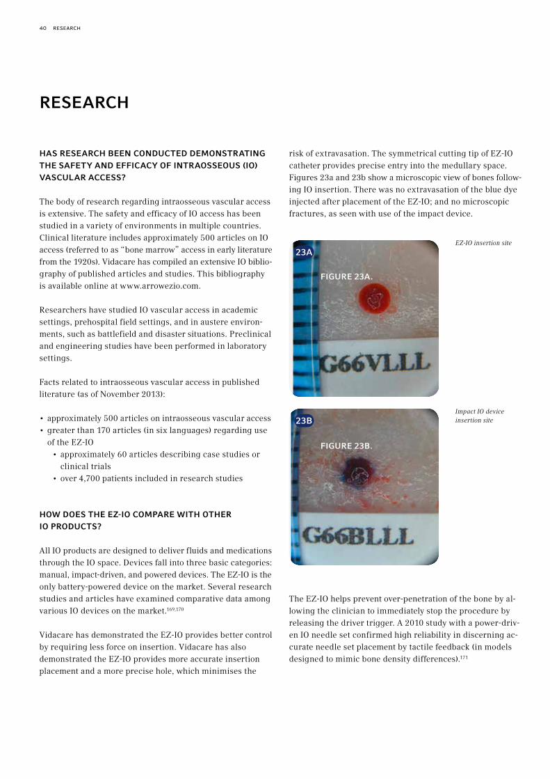
40 research
RESEARCH
HAS RESEARCH BEEN CONDUCTED DEMONSTRATING THE SAFETY AND EFFICACY OF INTRAOSSEOUS (IO) VASCULAR ACCESS?
The body of research regarding intraosseous vascular access is extensive. The safety and efficacy of IO access has been studied in a variety of environments in multiple countries. Clinical literature includes approximately 500 articles on IO access (referred to as “bone marrow” access in early literature from the 1920s). Vidacare has compiled an extensive IO biblio-graphy of published articles and studies. This bibliography is available online at www.arrowezio.com.
Researchers have studied IO vascular access in academic settings, prehospital field settings, and in austere environ-ments, such as battlefield and disaster situations. Preclinical and engineering studies have been performed in laboratory settings.
Facts related to intraosseous vascular access in published literature (as of November 2013):
• approximately 500 articles on intraosseous vascular access• greater than 170 articles (in six languages) regarding use
of the EZ-IO• approximately 60 articles describing case studies or
clinical trials• over 4,700 patients included in research studies
HOW DOES THE EZ-IO COMPARE WITH OTHER IO PRODUCTS?
All IO products are designed to deliver fluids and medications through the IO space. Devices fall into three basic categories: manual, impact-driven, and powered devices. The EZ-IO is the only battery-powered device on the market. Several research studies and articles have examined comparative data among various IO devices on the market.169,170
Vidacare has demonstrated the EZ-IO provides better control by requiring less force on insertion. Vidacare has also demonstrated the EZ-IO provides more accurate insertion placement and a more precise hole, which minimises the
risk of extravasation. The symmetrical cutting tip of EZ-IO catheter provides precise entry into the medullary space. Figures 23a and 23b show a microscopic view of bones follow-ing IO insertion. There was no extravasation of the blue dye injected after placement of the EZ-IO; and no microscopic fractures, as seen with use of the impact device.
The EZ-IO helps prevent over-penetration of the bone by al-lowing the clinician to immediately stop the procedure by releasing the driver trigger. A 2010 study with a power-driv-en IO needle set confirmed high reliability in discerning ac-curate needle set placement by tactile feedback (in models designed to mimic bone density differences).171
EZ-IO insertion site
Impact IO device insertion site
23A
23B
FIGURE 23A.
FIGURE 23B.
myths about intraosseous vascular access 41
MYTHS ABOUT INTRAOSSEOUS VASCULAR ACCESS
MYTH 1: THERE IS LITTLE EVIDENCE-BASED RESEARCH TO SUPPORT USE OF INTRAOSSEOUS (IO) VASCULAR ACCESS AS A VIABLE VASCULAR ACCESS ROUTE.
The body of research regarding intraosseous vascular access is extensive. The safety and efficacy of IO access has been studied in a variety of environments in multiple countries. Vidacare has compiled an extensive IO bibliography of published articles and studies. This bibliography is available online at www.arrowezio.com.
Researchers have studied IO in academic settings, prehospital field settings, and in austere environments, such as battlefield and disaster situations. Preclinical and engineering studies have been performed in laboratory settings. Facts related to intraosseous vascular access in published literature (as of November 2013):
• approximately 500 articles on intraosseous vascular access• more than 170 articles (in six languages) regarding use of
the EZ-IO• approximately 60 articles describing case studies or
clinical trials• over 4,700 patients studied
Multiple professional organizations recommend and/or support use of IO vascular access as an important vascular access route. Organizations include:
• Air & Surface Transport Nurses Association• American Association of Critical-Care Nurses• American College of Emergency Physicians• American Heart Association• Consortium on Intraosseous Vascular Access in
Healthcare Practice• Emergency Nurses Association• European Resuscitation Council• Infusion Nurses Society• International Liaison Committee on Resuscitation• National Association of EMS Physicians• Society of Pediatric Nurses
MYTH 2: EPIPHYSEAL PLATE INJURY IS A SIGNIFICANT RISK FOR IO CATHETER INSERTION.
Several research studies have been conducted to determine the effect of IO access on epiphyseal (growth) plates. Aggre-gate results have concluded that IO infusion does not result in any long-term effect on the epiphyseal plate. For research detail, refer to the section on Effects of IO Access on Growth Plates and Bone Repair.
MYTH 3: FAT EMBOLISM IS A SERIOUS RISK FOR IO INFUSION.
The risk of fat embolism associated with IO infusion has been studied in preclinical trials and reported in the clinical litera-ture for two decades. No known cases of clinically significant fat embolism resulting from IO administration have been re-ported in the medical literature or in actual practice. Preclinical trials have shown microscopic fat emboli in the lungs after high pressure IO infusion; none were considered clinically sig-nificant. For research detail, refer to the section on Embolism.
MYTH 4: OSTEOMYELITIS IS A SIGNIFICANT RISK FOR IO INFUSION.
Numerous research studies and reports in clinical literature have addressed the low incidence of osteomyelitis risk in IO vascular access. As of November 2013, over 60 clinical trials or case studies involving the EZ-IO have been reported in the clinical literature, involving over 4,700 patients; there were no cases of osteomyelitis. In the largest known meta-analysis (1985) of more than 4,200 patients, the rate of osteo-myelitis was reported to be 0.6 %. For research detail, refer to the section on Osteomyelitis.

42 references
REFERENCES
1 Food and Drug Administration. “Off-Label” and Investigational Use Of Marketed Drugs, Biologics, and Medical Devices – Information Sheet. Internet: Accessed: 01/23/12. Available at http://www.fda.gov/RegulatoryInformation/Guidances/ucm126486.htm.
2 Von Hoff DD, Kuhn JG, Burris HA, Miller LJ. Does intraosseous equal intravenous? A pharmacokinetic study. Amer J Emerg Med 2008;26:31-8.
3 Hoskins SL, Kramer GC, Stephens CT, Zachariah BS. Efficacy of epinephrine delivery via the intraosseous humeral head route during CPR. Circulation 2006;114:II_1204.VS
4 Internal study report. Protocol 2013-06: Clinical Studies to Determine the Optimal Technique to Identify the Proximal Humerus Intraosseous Vascular Access Insertion Site. Vidacare Corporation, May 2013.
5 Frascone RJ, Salzman JG, Bliss P, Adams A, Wewerka SS, Dries DJ. Intraosseous pressure tracings mimic arterial pressure tracings in timing and contour. Annals of Emergency Medicine 2013;62(4s):s13.
6 Hoskins S, Nascimento P, Espana J, Kramer G. Pharmacokinetics of intraosseous drug delivery during CPR. Shock 2005;23:35.VS
7 Hoskins SL, Nascimento P Jr., Lima RM, Espana-Tenorio, JM, Kramer GC. Pharmacokinetics of intraosseous and central venous drug delivery during cardiopulmonary resuscitation. Resuscitation 2012;83(1):107-12.VS
8 Dubick MA, Holcomb JB. A review of intraosseous vascular access: current status and military application. Mil Med 2000;165:552-9.
9 Tan BKK, Chong S, Koh ZX, et al. EZ-IO in the ED: an observational, prospective study comparing flow rates with proximal and distal tibia intraosseous access in adults. Amer J Emerg Med 2012;Early Online:1-4.VS
10 Ong MEH, Chan YH, Oh JJ, Ngo AS-Y. An observational, prospective study comparing tibial and humeral intraosseous access using the EZ-IO. Amer J Emerg Med 2009;27:8-15.VS
11 Philbeck TE, Miller LJ, Montez D, Puga T. Hurts so good; easing IO pain and pressure. JEMS 2010;35(9):58-69.VS
12 Lairet J, Bebarta V, Lairet K, Kacprowicz R, Lawler C, Pitotti R, Bush A, King J. A comparison of proximal tibia, distal femur, and proximal humerus infusion rates using the EZ-IO intraosseous device on the adult swine (sus scrofa) model. Prehospital Emergency Care 2013;17:280-4.
13 Dubick MA, Holcomb JB. A review of intraosseous vascular access: current status and military application. Mil Med 2000;165:552-9.
14 Philbeck TE, Miller LJ, Montez D, Puga T. Hurts so good; easing IO pain and pressure. JEMS 2010;35(9):58-69.VS
15 Tan BKK, Chong S, Koh ZX, et al. EZ-IO in the ED: an observational, prospective study comparing flow rates with proximal and distal tibia intraosseous access in adults. Amer J Emerg Med 2012;Early Online:1-4.VS
16 Ong MEH, Chan YH, Oh JJ, Ngo AS-Y. An observational, prospective study comparing tibial and humeral intraosseous access using the EZ-IO. Amer J Emerg Med 2009;27:8-15.VS
17 Hoskins S, Nascimento P, Espana J, Kramer G. Pharmacokinetics of intraosseous drug delivery during CPR. Shock 2005;23:35.VS
18 Hoskins SL, Zachariah BS, Copper N, Kramer GC. Comparison of intraosseous proximal humerus and sternal routes for drug delivery during CPR. Circulation 2007;116:II_933.VS
19 Hoskins SL, Kramer GC, Stephens CT, Zachariah BS. Efficacy of epinephrine delivery via the intraosseous humeral head route during CPR. Circulation 2006;114:II_1204.VS
20 Iwama H, Katsumi A. Emergency fields, obtaining intravascular access for cardiopulmonary arrest patients is occasionally difficult and time-consuming. J Trauma 1996; 41: 931-2.
21 Hapnes SA, Robertson C. CPR--drug delivery routes and systems. A statement for the Advanced Life Support Working Party of the European Resuscitation Council. Resuscitation 1992; 24:137-42.
22 Lairet JR, Bebarta V, Lairet K et al. A comparison of proximal tibia, proximal humerus, and distal femur infusion rates under high pressure (>300 mmHg) using the EZ-IO intraosseous device on an adult swine (sus scrofa) model. Prehospital Emergency Care 2011;15(1):117.
23 Internal Report. ISO 594-2 and high pressure liquid leakage testing for the Vidacare Corporation EZ-IO Needles. Report number 1105192. DDL. June 15, 2011.
24 Vidacare Internal Report. Pre-clinical study and bench testing to determine the utility of the EZ-IO Vascular Access System for delivering contrast dye using a power injector for computerized tomography studies. June 2010.
25 Philbeck TE, Miller LJ, Montez D, Puga T. Hurts so good; easing IO pain and pressure. JEMS 2010;35(9):58-69.VS
26 Hoskins SL, Zachariah BS, Copper N, Kramer GC. Comparison of intraosseous proximal humerus and sternal routes for drug delivery during CPR. Circulation 2007;116:II_933.VS
27 Miller L, Philbeck T, Montez D, Puga T. A two-phase study of fluid administration measurement during intraosseous infusion. Ann Emerg Med 2010;56(3):S151.VS
28 Miller L, Philbeck T, Bolleter S, Garcia G. Tactile feedback comparison of three types of intraosseous access devices for needle insertion accuracy. Ann Emerg Med 2010;56(3):S133.VS
29 Kovar J, Gillum L. Alternate route: the humerus bone—a viable option for IO access. JEMS 2010;35(8):52-9.
30 Wampler D, Schwartz D, Shumaker J, et al. Paramedics successfully perform humeral EZ-IO intraosseous access in adult out-of-hospital cardiac arrest patients. Am J Emerg Med 2010;56(3):S88. VS doi.org/10.1016/ j.ajem.2011.07.010.
31 Miller L, Philbeck T, Montez D, Puga T. A two-phase study of fluid administration measurement during intraosseous infusion. Ann Emerg Med 2010;56(3):S151.VS
32 Ong MEH, Chan YH, Oh JJ, Ngo AS-Y. An observational, prospective study comparing tibial and humeral intraosseous access using the EZ-IO. Amer J Emerg Med 2009;27:8-15.VS
33 Philbeck TE, Miller LJ, Montez D, Puga T. Hurts so good; easing IO pain and pressure. JEMS 2010;35(9):58-69.VS
34 Paxton JH, Knuth TE, Klausner HA. Proximal humerus intraosseous infusion: a preferred emergency venous access. The J Trauma 2009;67(3):606-11.VS

references 43
35 Hoskins SL, Kramer GC, Stephens CT, Zachariah BS. Efficacy of epinephrine delivery via the intraosseous humeral head route during CPR. Circulation 2006;114:II_1204.VS
36 Hoskins SL, Zachariah BS, Copper N, Kramer GC. Comparison of intraosseous proximal humerus and sternal routes for drug delivery during CPR. Circulation 2007;116:II_933.VS
37 Warren DW, Kissoon N, Sommerauer JF, Rieder MJ. Comparison of fluid infusion rates among peripheral intravenous and humerus, femur, malleolus, and tibial intraosseous sites in normovolemic and hypovolemic piglets. Ann Emerg Med 1993;22(2):183-6.
38 Lairet J, Bebarta V, Lairet K et al. A comparison of proximal tibia, proximal humerus, and distal femur infusion rates under high pressure (>300 mmHg) using the EZ-IO intraosseous device on an adult swine (sus scrofa) model. Prehosp Emerg Care 2011;15(1):117.
39 Lairet JR, Bebarta V, Lairet K et al. Intraosseous pressure infusion comparison using a rapid infusion device and a pressure bag in a swine model. Ann Emerg Med 2010;56(3):S26.
40 Reades R, Studneck J, Garrett J, Vandeventer S, Blackwell T. Comparison of first-attempt success between tibial and humeral intraosseous insertions during out-of-hospital cardiac arrest. Prehosp Emerg Care 2011;15(2):278-81.doi:10.3109/10903127.2010.545479.
41 Reades R, Studneck J, Vandeventer S, Garrett J. Intraosseous versus intravenous vascular access during out-of-hospital cardiac arrest: a randomized controlled trial. Ann Emerg Med 2011;58(6):509-16.
42 Wampler D, Schwartz D, Shumaker J, Bolleter S, Beckett R, Manifold C. Paramedics successfully perform humeral EZ-IO intraosseous access in adult out-of-hospital cardiac arrest patients. Am J of Emerg Med 2012;30:1095-9.doi:10.1016/j.ajem.2011.07.010.
43 Fowler RL, Pierce A, Nazeer S et al. 1,199 case series: Powered intraosseous insertion provides safe and effective vascular access for emergency patients. Ann Emerg Med 2008;52(4):S152.VS
44 Philbeck TE, Miller LJ, Montez D, Puga T. Hurts so good; easing IO pain and pressure. JEMS 2010;35(9):58-69.VS
45 Hixson R. Intraosseous administration of preservative-free lidocaine. http://www.arrowezio.com.
46 Halkey-Roberts Corp. http://www.halkeyroberts.com/products/medical/rnv-marketing-booklet.aspx. Accessed February 14, 2012.
47 Rosetti VA, Thompson BM, Miller J, et al. Intraosseous infusion: an alternative route of pediatric intravascular access. Ann Emerg Med 1985;14(9):885-8.
48 Gluckman W. Intraosseous cannulation. Emedicine June 2012. Available at: http://emedicine.medscape.com/article/908610-overview#aw2aab6b4. Accessed October 21, 2013.
49 Fowler RL, Pierce A, Nazeer S et al. 1,199 case series: Powered intraosseous insertion provides safe and effective vascular access for emergency patients. Ann Emerg Med 2008;52(4):S152.VS
50 Vidal R, Kissoon N, Gayle M. Compartment syndrome following intraosseous infusion. Pediatrics 1993;91(6):1201-2.
51 Smith R, Davis N, Bouamra O, Lecky F. The utilisation of intraosseous infusion in the resuscitation of paediatric major trauma patients. Injury 2005;36(9):1034-8.
52 Katz DS, Wojtowycz AR. Tibial fracture: a complication of intraosseous infusion. Am J Emerg Med 1994;12(2):258-9.
53 Bowley DM, Loveland J, Pitcher GJ. Tibial fracture as a complication of intraosseous infusion during pediatric resuscitation. J Trauma 2003; 55(4):786-7.
54 La Fleche FR, Slepin MJ, Vargas J, Milzman DP. latrogenic bilateral tibial fractures after intraosseous infusions attempts in a 3-month-old infant. Ann Emerg Med 1989;18(10):1099-1101.
55 www. arrowezio.com.
56 Dolister M, Miller S, Borron S, et al. Intraosseous vascular access is safe, effective and costs less than central venous catheters for patients in the hospital setting. J Vasc Access 2012; doi: 10.5301/jva.5000130.
57 Khan LAK, Anakwe RE, Murray A, Godwin Y. A severe complication following intraosseous infusion used during resuscitation of a child. Inj Extra 2011;doi:10.1016/j.injury.2011.05.015.
58 Taylor CC, Clarke NMP. Amputation and intraosseous access in infants. BMJ 2011;342:d2778 doic: 10.1136/bmj.d2778.
59 Fetissof H, Nadaud J, Landy C, Millot I, Paris R, Plancade D. Amines on intraosseous vascular access: A case of skin necrosis. Ann Fr Anesth Reanim. 2013;32(5):e89-90. http://dx.doi.org/10.1016/j.annfar.2013.02.022.
60 Landy C, Plancade D, Gagnon N, Schaeffer E, Nadaud J, Favier JC. Complication of intraosseous administration of systemic fibrinolysis for a massive pulmonary embolism with cardiac arrest. Resuscitation 2012;83:e149-e150. doi: 10.1016/j.resuscitation.2012.01.044.
61 Davidoff J, Fowler R, Gordon D, Klein G et al. Clinical evaluation of a novel intraosseous device for adults. J Emerg Med 2005;30(10 Suppl):S20-3.VS
62 Dolister M, Miller S, Borron S, et al. Intraosseous vascular access is safe, effective and costs less than central venous catheters for patients in the hospital setting. J Vasc Access 2012; doi:10.5301/jva.5000130.
63 Khan LAK, Anakwe RE, Murray A, Godwin Y. A severe complication following intraosseous infusion used during resuscitation of a child. Inj Extra 2011;doi:10.1016/j.injury.2011.05.015.
64 Carreras-Gonzáles E, Brio-Sanagustin S, Guimera I, Crespo C. [Spanish]. Complication of the intraosseous route in a newborn infant. [Complicación de la via intraósea en un neonate]. Med Intensiva 2011;doi:10.1016/j.medin.2011.05.004.
65 Taylor CC, Clarke NMP. Amputation and intraosseous access in infants. BMJ 2011;342:d2778 doic: 10.1136/bmj.d2778.
66 Atanda A Jr, Statter MB. Compartment syndrome of the leg after intraosseous infusion: guidelines for prevention, early detection, and treatment. Am J Orthop 2008; 37:E198-200.
67 Moen TC, Sarwark JF. Compartment syndrome following intraosseous infusion. Orthopedics 2008;37(12):E198-200.
68 d’Heurle A, Archdeacon MT. Compartment syndrome after intraosseous infusion associated with a fracture of the tibia. JBJS Case Connect. 2013:3:e20:1-3.
69 Wright R, Reynolds SI, Nachtsheim B. Compartment syndrome secondary to prolonged intraosseous infusion. Pediatr Emerg Care 1994;10(3):157-9.
70 Moscati R, Moore GP. Compartment syndrome with resultant amputation following intraosseous infusion. Am J Emerg Med 1990; 8(5):470-1.
71 Galpin RD, Kronick JB, Willis RB, Frewen TC. Bilateral lower extremity compartment syndromes secondary to intraosseous fluid resuscitation. J Pediatr Orthop 1991; 11(6):773-6.
44 references
72 Gayle M, Kissoon N. A case of compartment syndrome following intraosseous infusions. Pediatr Emerg Care 1994; 10(6):378.
73 Launay F, Paut O, Katchburian M, Bourelle S, Jouve JL, Bollini G. Leg amputation after intraosseous infusion in a 7-month-old infant: a case report. J Trauma 2003; 55(4):788-90.
74 Vidal R, Kissoon N, Gayle M. Compartment syndrome following intraosseous infusion. Pediatrics 1993; 91(6):1201-2.
75 Burke T, Kehl DK. Intraosseous infusion in infants. Case report of a complication. J Bone Joint Surg Am 1993; 75(3):428-9.
76 Ribeiro JA, Price CT, Knapp DR Jr. Compartment syndrome of the lower extremity after intraosseous infusion of fluid. A report of two cases. J Bone Joint Surg Am 1993; 75(3):430-3.
77 Simmons CM, Johnson NE, Perkin RM, van Stralen D. Intraosseous extravasation complication reports. Ann Emerg Med 1994; 23(2):363-6.
78 Fiser DH. Intraosseous infusion. N Engl J Med 1990;322(22):1579-81.
79 Heinild S, Sondergaard T, Tuvad F. Bone marrow infusion in childhood. J Pediatr 1947;30(6):400-2.
80 LaRocco BG, Wang HE. Intraosseous infusion. Prehosp Emerg Care 2003;7:280-5.
81 Iserson KV, Criss E. Intraosseous infusions: a usable technique. Am J Emerg Med 1986;4(6):540-2.
82 Fiser RT, Walker WM, Seibert JJ, et al. Tibial length following intraosseous infusion: a prospective, radiographic analysis. Pediatr Emerg Care 1997;13(3):186-8.
83 Claudet I, Baunin C, Laporte-Turpin E. Long-term effects on tibial growth after intraosseous infusion: a prospective, -radiographic analysis. Pediatr Emerg Care 2003;19(6):397-401.
84 Brickman KR, Rega P, Koltz M, Guinness M. Analysis of growth plate abnormalities following intraosseous infusion through the proximal tibia epiphysis in pigs. Ann Emerg Med 1988;17(2):121-3.
85 Dedrick DK, Mase C, Ranger W, Burney RE, The effects of intraosseous infusion on the growth plate in a nestling rabbit model. Ann Emerg Med 1992;21(5):494-7.
86 Bielski RJ, Bassett GS, Fideler B, Tolo VT. Intraosseous infusions: effects on the immature physis – an experimental model in rabbits. J Pediatr Orthop 1993; 13: 511-5.
87 Rosetti VA, Thompson BM, Miller J, et al. Intraosseous infusion: an alternative route of pediatric intravascular access. Ann Emerg Med 1985;14(9):885-8.
88 Miller LJ, Philbeck TE, Puga TA, Montez DF, Escobar GP. A pre-clinical study to determine the time to bone sealing and healing following intraosseous vascular access. Ann Emerg Med 2011;58(4S):S240.VS
89 Launay F, Paut O, Katchburian M, et al. Leg amputation after intraosseous infusion in a 7-month-old infant : a case report. J of Trauma 2003;55(4):788-90.
90 van Rijn RR, Knoester H, Maes A, van der Wal AC, Kubat B. Cerebral arterial air embolism in a child after intraosseous infusion. Emerg Radiol 2008; 15(4): 259-62.
91 Hillewig E, Aghayev E, Jackowski C, et al. Gas embolism following intraosseous medication application proven by post-mortem multislice computed tomography and autopsy. Resuscitation 2007:72:149-53.
92 Byrick R. Pulmonary fat embolism and intraosseous infusion. Pediatr Crit Care Med 2001;2(2):184-5.
93 Fiallos M, Kissoon N, Abdelmodium T, et al. Fat embolism with the use of intraosseous infusion during cardiopulmonary resuscitation. Am J Med Sci 1997;314(2):73-9.
94 Orlowski JP, Julius CJ, Petras RE, Porembka DT, Gallagher JM. The safety of intraosseous infusions: risks of fat and bone marrow emboli to the lungs. Ann Emerg Med 1989;18(10):1062-7. 95 Plewa MC, King RW, Fenn-Buderer N, Gretzinger K, Renuart D, Cruz R. Hematologic safety of intraosseous blood trans-fusion in a swine model of pediatric hemorrhagic hypovolemia. Acad Emerg Med 1995;2(9):799-809.
96 Hasan MY, Kissoon N, Khan TM, Saldajeno V, Goldstein J, Murphy SP. Intraosseous infusion and pulmonary fat embolism. Pediatr Crit Care Med 2001;2(2):133-8.
97 Lairet JR, Berbata V, Lariet K, et al. A comparison of proximal tibia, distal femur, and proximal humerus infusion rates using the EZ-IO intraosseous device on the adult swine (Sus scrofa) model. Prehospital Emergency Care 2013;17:280-84. doi:10.3109/10903127.2012.755582.
98 Moller JC, Tegtmeyer FK, Schaible TF, Sussmane JB. Intraosseous puncture as vascular access in pediatric emergency and intensive care medicine. Anaesthesiol Reanim 1996;21(4):103-7.
99 Rosetti VA, Thompson BM, Miller J, et al. Intraosseous infusion: an alternative route of pediatric intravascular access. Ann Emerg Med 1985;14(9):885-8.
100 Platt SL, Notterman DA, Winchester P. Fungal osteomyelitis and sepsis from intraosseous infusion. Pediatr Emerg Care 1993; 9: 149-50.
101 Rosovsky M, FitzPatrick M, Goldfarb CR, Finestone H.Bilateral osteomyelitis due to intraosseous infusion: case report and review of the English-language literature. Pediatr Radiol 1994; 24: 72-3.
102 Barron BJ, Tran HD, Lamki LM. Scintigraphic findings of osteomyelitis after intraosseous infusion in a child. Clin Nucl Med 1994; 19: 307-8.
103 Stoll E, Golej J, Burda G, Hermon M, Boigner H, Trittenwein G. Osteomyelitis at the injection site of adrenalin through an intraosseous needle in a 3-month-old infant. Resuscitation 2002; 53: 315-8.
104 Dogan A, Irmak H, Harman M, Ceylan A, Akpinar F, Tosun N. Tibial osteomyelitis following intraosseous infusion: a case report. Acta Orthop Traumatol Turc 2004; 38:357-60.
105 Henson NL, Payan JM, Terk MR. Tibial subacute osteomyelitis with intraosseous abscess: an unusual complication of intraosseous infusion. Skeletal Radiol 2010;doi:10.1007/s00256-010-1027-1029.
106 Vidacare Internal Database (as of November 2013).
107 Von Hoff DD, Kuhn JG, Burris HA, Miller LJ. Does intraosseous equal intravenous? A pharmacokinetic study. Amer J Emerg Med 2008;26:31-8.
108 Hoskins SL, Kramer GC, Stephens CT, Zachariah BS. Efficacy of epinephrine delivery via the intraosseous humeral head route during CPR. Circulation 2006;114:II_1204.VS
109 Hoskins S, Nascimento P, Espana J, Kramer G. Pharmacokinetics of intraosseous drug delivery during CPR. Shock 2005;23:35.VS
110 Hoskins SL, Nascimento P Jr., Lima RM, Espana-Tenorio, JM, Kramer GC. Pharmacokinetics of intraosseous and central venous drug delivery during cardio-pulmonary resuscitation. Resuscitation 2012;83(1):107-12.VS
111 Philbeck TE, Miller LJ, Montez D, Puga T. Hurts so good; easing IO pain and pressure. JEMS 2010;35(9):58-69.VS

references 45
112 Ong MEH, Chan YH, Oh JJ, Ngo AS-Y. An observational, prospective study comparing tibial and humeral intraosseous access using the EZ-IO. Am J Emerg Med 2009;27:8-15.VS
113 Fowler RL, Pierce A, Nazeer S et al. 1,199 case series: Powered intraosseous insertion provides safe and effective vascular access for emergency patients. Ann Emerg Med 2008;52:S152.VS
114 Paxton JH, Knuth TE, Klausner HA. Proximal humerus intraosseous infusion: a preferred emergency venous access. The Journal of Trauma 2009;67(3):606-11.
115 Wayne MA. Intraosseous vascular access: devices, sites and rationale for IO use. JEMS 2007;32:S23-5.
116 Frascone RJ, Jensen JP, Kaye K, Salzman JG. Consecutive field trials using two different intraosseous devices. Prehosp Emerg Care 2007;11:164-71.VS
117 Fowler R, Gallagher JV, Isaacs SM, et al. The role of intraosseous vascular access in the out-of-hospital environment (resource document to NAEMSP position statement). Prehosp Emerg Care 2007;11:63-6.VS
118 Miller L, Kramer GC, Bolleter S. Rescue access made easy. JEMS 2005;30:S8-18.VS
119 Davidoff J, Fowler R, Gordon D, et al. Clinical evaluation of a novel intraosseous device for adults: prospective, 250-patient, multi-center trial. JEMS 2005;30:S20-3.VS
120 Gillum L, Kovar J. Powered intraosseous access in the prehospital setting: MCHD EMS puts the EZ-IO to the test. JEMS 2005;30:S24-6.
121 Cooper BR, Mahoney PF, Hodgetts TJ, Mellor A. Intra-osseous access (EZIO®) for resuscitation: UK military combat experience. JR Army Med Corps 2008;153(4):314-6.
122 Hixson R. Intraosseous administration of preservative-free lidocaine.
123 Miller L, Philbeck T, Bolleter S, Garcia G. Tactile feedback comparison of three types of intraosseous access devices for needle insertion accuracy. Ann Emerg Med 2010;56(3):S133.VS
124 American Heart Association 2010 American Heart Association guidelines for CPR and ECC. Part 14-Pediatric Advanced Life Support. Circulation 2010;122(3 suppl.):S881-5.
125 Nolan J.P., Soar J, Zideman D, et al. European Resuscitation Council Guidelines for Resuscitation 2010. Resuscitation 2010. 81:1219-1276. doi:10.1016/j.resuscitation.2010.08.021. Accessed 11/25/2013.
126 Heinild S, Sondergaard T, Tuvad F. Bone marrow infusion in childhood. J Pediatr 1947;30:400-12.
127 Iserson KV, Criss E. Intraosseous infusions: a usable technique. Am J Emerg Med 1986;4(6):540-2.
128 Fiser DH. Intraosseous infusion. N Eng J Med 1990;322(22):1579-81.
129 Fiorito BA, Mirza F, Doran TM, Oberle AN, et al. Intraosseous access in the setting of pediatric critical care. Pediatr Crit Care 2005;6(1):50-3.
130 Smith R, Davis N, Bouamra O, Lecky F. The utilisation of intraosseous infusion in the resuscitation of paediatric major trauma patients. Injury 2005;36(9):1034-8.
131 Horton MA, Beamer C. Powered intraosseous insertion provides safe and effective vascular access for pediatric emergency patients. Pediatr Emerg Care 2008;24(6):347-50.
132 Baren J. Are Powered Intraosseous Insertion Devices Safe and Effective in Children? Journal Watch Emergency Medicine. Aug 2008.
133 Frascone RJ, Jensen J, Wewerka SS, Salzman JG.Use of the pediatric EZ-IO needle by emergency medical services providers. Pediatr Emerg Care 2009;25: 329-32.VS
134 Tobias JD, Ross AK. Intraosseous infusions: A review for the anesthesiologist with a focus on pediatric use. Anesth Analg 2010;110(2):391-401.
135 Nagler J, Krauss B. Intraosseous catheter placement in children. NEJM 2011; 364(8):e14-8.
136 Cullen PM. Intraosseous cannulation in children. Paediatric Critical Care 2011;13(1): 28-30.
137 Cleugh FM, Maconochie I. Management of the multiply injured child. Paediatrics and Child Health 2012. 23:5. 194-199.
138 Joint Policy Statement. Guidelines for care of children in the emergency department: American Academy of Pediatrics. JEN 2013;39(2):116-27.
139 Weiser G, Poppa E, Katz Y, Bahouth H, Shavit I. Intraosseous blood transfusion in infants with traumatic hemorrhagic shock- a case report and review of the literature. Am J Emerg Med 2012;http>//dx.doi.org/10.1016/j.ajem.2012.10.036.
140 Chansa E, et al. An intraosseous blood transfusion in a critically ill child. Afr J emerg Med 2013;http://dx.doi.org/10.1016/j.afjem.2013.05.003.
141 LaRocco BG, Wang HE. Intraosseous infusion. Prehosp Emerg Care 2003;7:280-5.
142 Miller L, Philbeck T, Bolleter S, Garcia G. Tactile feedback comparison of three types of intraosseous access devices for needle insertion accuracy. Ann Emerg Med 2010;56(3):S133.VS
143 Hixson R. Intraosseous administration of preservative-free lidocaine.
144 Montez DF, Puga TA, Garcia MR et al. Intraosseous blood correlates with venous blood in healthy subjects using point-of-care analyzers. Annals of Emergency Medicine 2013;62(4s):S40.
145 Philbeck T, Miller L, Montez, Powell T. IO blood for enzymes and blood gas analysis. Unpublished manuscript. 2010.VS
146 Miller LJ, Philbeck TE, Montez DF, Spadaccini CJ A new study of intraosseous blood for laboratory analysis. Arch Pathol Lab Med 2010;134(9):1253-60.VS
147 Hurren JS. Can blood taken from intraosseous cannulations be used for blood analysis? Burns 2000;26(8):727-30.
148 Ummenhofer W, Frei FJ, Urwyler A, et al. Are laboratory values in bone marrow predictable for venous blood in paediatric patients? Resuscitation 1994;27(2):123-8.
149 Grisham J, Hastings C. Bone marrow aspirates an accessible and reliable source for critical laboratory studies. Ann Emerg Med 1991;20(10):1121-24.
150 Brickman KR, Krupp K, Rega P, Alexander J, Guinness M. Typing and screening of blood from intraosseous access. Ann Emerg Med 1992;21(4):414-7.
151 Unger H, Spivey WH, McNamara RM, et al. Comparison of intraosseous and intravenous CBC and Astra 8 in swine. Ann Emerg Med 1986;15:647.
152 Orlowski JP, Proemka DT, Gallagher JM et al. The bone marrow as a source for laboratory studies. Ann Emerg Med 1989;18(12):1348-51.
153 Kissoon N, Rosenberg H, Gloor J et al. Comparison of the acid-base status of blood obtained from intraosseous and central venous sites during steady- and low-flow states. Crit Care Med 1993;21(11):1765-9.

46 references
154 Kissoon N, Peterson R, Murphy S, Gayle M, Ceithaml E, Harwood-Nuss A. Comparison of pH and carbon dioxide tension values of central venous and intraosseous blood during changes in cardiac output. Crit Care Med 1994 June;22(6):1010-5.
155 Kissoon N, Idris A, WenzelV, Murphy S, Rush W. Intraosseous and central venous blood acid-base relationship during cardiopulmonary resuscitation. Pediatr Emerg Care 1997;13(4):250-3.
156 Abdelmoneim T, Kissoon N, Johnson L, Fiallos M, Murphy S. Acidbase status of blood from intraosseous and mixed venous sites during prolonged cardiopulmonary resuscitation and drug infusion. Crit Care Med 1999;27 (9):1923-8.
157 Johnson L, Kissoon N, Fiallos M, Abdelmoneim T, Murphy S. Use of intraosseous blood to assess blood chemistries and hemoglobin during cardiopulmonary resuscitation with drug infusions. Crit Care Med 1999 June;27(6):1147-52.
158 Strandberg G, Eriksson M, Gustafsson MG et al. Analysis of intraosseous samples using point of care technology – an experimental study in the anaesthetized pig. Resuscitation (2012), doi:10.1016/j.resuscitation.2012.04.007.
159 American Heart Association 2010 American Heart Association guidelines for CPR and ECC. Part 9-Post-Cardiac Arrest Care. Circulation 2010;122(3 suppl.):S769-78.
160 Myers BJ, Lewis R. Induced cooling by EMS (ICE): year one in Raleigh/Wake County. JEMS 2007;32:S13-5.
161 Truhlar A, Skulec R, Rozsival P, Cerny V. Efficient prehospital induction of therapeutic hypothermia via intraosseous infusion. Resuscitation 2010;81(2):262-3.
162 Ornato JP, Peberdy MA, Kurz MC. Abstract P134: A building block strategy for optimizing outcomes from out of hospital cardiac arrest. Circulation 2009;120:S1470-a.
163 Mader TJ, Walterscheid JK, Kellog AR, Lodding CC. The feasibility of inducing mild therapeutic hypothermia after cardiac resuscitation using iced saline infusion via an intraosseous needle. Resuscitation 2010;81:82-6.VS
164 Miller L, Montez DF, Philbeck TE, Puga TA, Morgan J. Infusing chilled saline via the Intraosseous route is equivalent to infusion via the intravenous route in reducing the core temperature in swine. Prehosp Emerg Care 2012;16(1):152. doi:10.3109/10903127.2011.624676.VS
165 Larabee TM, Campbell JA, Severyn FA, Little CM. Intraosseous infusion of ice cold saline is less efficacious than intravenous infusion for induction of mild therapeutic hypothermia in a swine model of cardiac arrest. Resuscitation 2011;82:603-6.doi:10.1016/j.resuscitation.2011.01.007.
166 Nolan JP, Soar J, Zideman D, et al. European Resuscitation Council guidelines for resuscitation 2010. Resuscitation 2010;81:1219-76. doi:10.1016/j.resuscitation.2010.08.021. Accessed 11/25/2013.
167 Nutbeam T, Fergusson A. Intraosseous access in osteogenesis imperfecta (IO in OI). Resuscitation. 2009,80(12);1442-1443. doi:10.1016/j.resuscitation.2009.08.016.
168 Frascone R, Kaye K, Dries D, Solem L. Successful placement of an adult sternal intraosseous line through burned skin. J Burn Care Rehabil 2003; 24: 306-8.
169 Weiser G, Hoffmann Y, Galbraith R, Shavit I. Current advances in intraosseous infusion – a systematic review. Resuscitation. 2012;83(1):20-6. doi:10.1016/j.resuscitation.2011.07.020.
170 Paxton JH. Intraosseous vascular access: A review. Trauma 2012;14(3):195-232. doi:10.1177/1460408611430175.
171 Miller L, Philbeck T, Bolleter S, Garcia G. Tactile feedback comparison of three types of intraosseous access devices for needle insertion accuracy. Ann Emerg Med 2010;56(3):S133.VS
Vidacare disclaims all liability for the use, application or interpretation of this information in the medical treatment of any patient.

47

Telefl ex is a leading global provider of specialty medical devices used for diagnostic and therapeutic procedures in critical care, urology and surgery. Our mission is to provide solutions that enable healthcare providers to improve outcomes and enhance patient and provider safety.We specialise in devices for general and regional anaesthesia, cardiac care, respiratory care, urology, vascular access and surgery and we serve healthcare providers in more than 150 countries. Telefl ex also provides specialty products for medical device manufacturers.
Our well known brands include arrow®, deknatel®, Gibeck®, hudson rci®, kmedic®, lma™, PillinG®, Pleur-evac®, rÜsch®, sheridan®, taut®, tfx oem®, vasonova®, vidacare® and weck®, all of which are trademarks or registered trademarks (in the U.S. and/or other countries) of Telefl ex Incorporated.
Telefl ex global operations: Australia, Austria, Belgium, Canada, China, Czech Republic, France, Germany, Greece, India, Ireland, Italy, Japan, Korea, Malaysia, Mexico, Netherlands, New Zealand, Portugal, Singapore, Slovak Republic, South Africa, Spain, Switzerland, United Kingdom, Uruguay and USA.
YOUR INTERNATIONAL CONTACTS:
TELEFLEX HEADQUARTERS INTERNATIONAL, IRELAND Telefl ex Medical Europe Ltd., IDA Business and Technology Park, Dublin Road, Athlone, Co WestmeathPhone +353 (0)9 06 46 08 00 · Fax +353 (0)14 37 07 73orders.intl@telefl ex.com AUSTRALIA/NEW ZEALAND 1300 360 226AUSTRIA +43 (0)1 402 47 72BELGIUM +32 (0)2 333 24 60CHINA (SHANGHAI) +86 (0)21 6163 0965CHINA (BEIJING) +86 (0)10 6418 5699CZECH REPUBLIC +420 (0)495 759 111FRANCE +33 (0)5 62 18 79 40GERMANY +49 (0)7151 406 0GREECE +30 210 67 77 717INDIA +91 (0)44-2836 5040ITALY +39 0362 58 911JAPAN +81 (0)3 3379 1511KOREA +82 2 536 7550NETHERLANDS +31 (0)88 00 215 00PORTUGAL +351 22 541 90 85SINGAPORE +65 6439 3000SLOVAK REPUBLIC +421 (0)3377 254 28 SOUTH AFRICA +27 (0)11 807 4887 SPAIN +34 918 300 451SWITZERLAND +41 (0)31 818 40 90UNITED KINGDOM +44 (0)1494 53 27 61
For detailed information see www.telefl ex.com
Telefl ex is a registered trademark of Telefl ex Incorporated or its affi liates.The products in this catalogue may not be available in all countries. Please contact your local representative. All data current at time of printing (09/2016). Subject to technical changes without further notice. © 2016 Telefl ex Incorporated. All rights reserved.
94 07 79 - 00 00 01 · REV B · MC / SF · 09 16 0.05