Embed Size (px)
Citation preview

1
The SaeRS two-component system is a direct and dominant transcriptional 1
activator of toxic shock syndrome toxin-1 in Staphylococcus aureus 2
3
Miren L. Baroja1,3, Christine A. Herfst1,3, Katherine J. Kasper1, Stacey X. Xu1*, Daniel A. 4
Gillett1, Jingru Li4*, Gregor Reid1,2,3 and John K. McCormick1,3 5
6 1Department of Microbiology and Immunology, and 2Surgery, Schulich School of Medicine and 7
Dentistry, Western University, London, ON, Canada. 8 3Lawson Health Research Institute, London, Ontario, Canada. 9 4Corporate Research & Engineering, Kimberly-Clark Corporation, 1400 Holcomb Bridge Road, 10
Roswell, Georgia, USA 11
12
Address correspondence to John K. McCormick, [email protected] 13
Tel. (+1) 519-661-3309 14
Fax (+1) 519-661-3499 15
16
* Present address: Stacey X. Xu, Department of Molecular Genetics, University of Toronto, 17
Toronto, Ontario, Canada; Jingru Li, Philips Oral Healthcare, Bothell, WA, USA 18
19
Short title: SaeRS regulation of TSST-1 20
21
JB Accepted Manuscript Posted Online 25 July 2016J. Bacteriol. doi:10.1128/JB.00425-16Copyright © 2016, American Society for Microbiology. All Rights Reserved.
on April 10, 2018 by guest
http://jb.asm.org/
Dow
nloaded from

2
ABSTRACT 22
Toxic shock syndrome toxin-1 (TSST-1) is a Staphylococcus aureus superantigen that has been 23
implicated in both menstrual-associated and non-menstrual-associated toxic shock syndrome 24
(TSS). Despite the important role of TSST-1 in severe human disease, a comprehensive 25
understanding of staphylococcal regulatory factors that control TSST-1 expression remains 26
incomplete. The S. aureus exotoxin expression (Sae) operon contains a well-characterized two-27
component system that regulates a number of important exotoxins in S. aureus, although 28
regulation of TSST-1 by the Sae system has not been investigated. We generated a defined 29
deletion of the Sae histidine kinase sensor (saeS) in the prototypic menstrual-associated TSS 30
strain S. aureus MN8. Mutation of saeS resulted in a complete loss of TSST-1 expression. Using 31
both luciferase reporter experiments and quantitative real-time PCR we demonstrate that the Sae 32
system is an important transcriptional activator of TSST-1 expression. Recombinant SaeR was 33
able to bind directly to the tst promoter to a region containing two SaeR consensus binding sites. 34
Although the stand-alone SarA transcriptional regulator has been shown to be both a positive and 35
negative regulator of TSST-1, deletion of sarA in S. aureus MN8 resulted in a dramatic 36
overexpression of TSST-1. As expected, mutation of agr also reduced TSST-1 expression, but 37
this phenotype appeared to be independent of Sae. A double mutant of saeS and sarA resulted in 38
the loss of TSST-1 expression. This work indicates that the Sae system is a dominant and direct 39
transcriptional activator that is required for expression of TSST-1. 40
on April 10, 2018 by guest
http://jb.asm.org/
Dow
nloaded from

3
IMPORTANCE 41
The TSST-1 superantigen is an exotoxin, produced by some strains of S. aureus, that has a clear 42
role in both menstrual-associated and non-menstrual-associated TSS. Although the well-43
characterized agr quorum sensing system is a known positive regulator of TSST-1, the molecular 44
mechanisms that directly control TSST-1 expression are only partially understood. Our studies 45
demonstrate that the Sae two-component regulatory system is a positive transcriptional regulator 46
that binds directly to the TSST-1 promoter, and furthermore, our data suggest that Sae is required 47
for expression of TSST-1. This work highlights how major regulatory circuits can converge to 48
fine tune exotoxin expression, and suggests that the Sae regulatory system may be an important 49
target for anti-virulence strategies. 50
on April 10, 2018 by guest
http://jb.asm.org/
Dow
nloaded from

4
INTRODUCTION 51
Staphylococcus aureus is both a common commensal of humans and a prominent bacterial 52
pathogen that is responsible for an assortment of self-limiting superficial infections to life-53
threatening invasive diseases (1). While most infections caused by S. aureus involve the 54
coordinated expression of numerous virulence factors, a few select diseases, including 55
staphylococcal scalded skin syndrome, food poisoning, and toxic shock syndrome (TSS), require 56
the expression of specific staphylococcal exotoxins (2). 57
Staphylococcal TSS is a rare but devastating disease that is caused by S. aureus strains 58
which secrete high levels of superantigens. Superantigen exotoxins function by binding directly 59
to lateral surfaces of both T cell receptor β-chains and MHC class II molecules, which forces the 60
massive activation of numerous T cells and can induce a T cell-dependent cytokine storm (3). 61
TSS symptoms are characterized by the acute onset of fever, rash formation, hypotension, 62
multisystem involvement, and desquamation in patients that recover (4), although strict use of 63
this definition likely underestimates the actual number of TSS cases (5). There are two major 64
forms of TSS including menstrual-associated TSS (mTSS) and non-menstrual-associated TSS (3). 65
Although the latter can occur from virtually any S. aureus infection, mTSS typically occurs 66
within ~2 days prior or ~2 days following menstruation, and is often associated with the use of 67
tampons (6, 7). The staphylococcal superantigen toxic shock syndrome toxin-1 (TSST-1) is 68
believed to be responsible for nearly all cases of menstrual-associated TSS (3, 8), as well as 69
many cases of non-menstrual TSS (5, 9). 70
Despite the well-recognized role of TSST-1 in both forms of TSS, a comprehensive 71
understanding of the genetic elements required for the regulation and expression of TSST-1 72
remains incomplete. Although the prototypical positive regulator of TSST-1 is the well-73
characterized accessory gene regulator (agr) quorum sensing system (10), a number of additional 74
regulatory factors also impact on TSST-1 expression. For example, mutation of the 75
staphylococcal respiratory response (srrAB) two-component system resulted in a decrease in 76
TSST-1 production, particularly under conditions of low oxygen (11). However, overexpression 77
of srrAB in trans massively upregulated RNAIII, the major effector molecule of the agr system, 78
with a near complete repression of TSST-1, demonstrating that regulation of TSST-1 by agr can 79
be uncoupled (11). This is also consistent with a study that could not correlate agr 80
polymorphisms, including agrC-inactivating mutations, with varied production of TSST-1 (12). 81
on April 10, 2018 by guest
http://jb.asm.org/
Dow
nloaded from

5
Also, the staphylococcal accessory regulator (SarA) has previously been demonstrated to bind 82
directly to the tst promoter region (13), and depending upon the genetic background, has been 83
reported to function as either a positive or negative regulator of TSST-1 expression (13–15). 84
Furthermore, carbon catabolite protein A (CcpA) acts as a repressor of TSST-1 (16), which 85
explains the initial observation that TSST-1 expression is repressed by glucose (17). Although 86
mutation of the repressor of toxins (rot) transcriptional regulator was reported to have little affect 87
on TSST-1 expression, overexpression of Rot in trans strongly repressed TSST-1 (15). Further to 88
these experiments, prior work from our laboratory has demonstrated that interspecies 89
communication between Lactobacillus reuteri and S. aureus can result in a dramatic repression 90
of TSST-1 expression (18). Although this phenotype correlated with repression of agr, and 91
deletion of agr in S. aureus MN8 reduced TSST-1 expression, TSST-1 could be further repressed 92
in the agr-null background by the L. reuteri signaling molecules. This additional repression of 93
TSST-1 correlated with decreased transcription of the S. aureus exotoxin expression (Sae) 94
system, and increased transcription of stand-alone transcriptional regulator SarA (18). The aim of 95
the present study was to investigate the role of both Sae and SarA on production of TSST-1 in S. 96
aureus MN8, a prototypic menstrual TSS S. aureus strain. 97
98
MATERIALS AND METHODS 99
Bacterial strains and growth conditions. Bacterial strains used in this study are listed in Table 100
1. All cloning was carried out utilizing E. coli XL1-Blue (Stratagene) grown on Luria-Bertani 101
(LB) Broth (Difco) or Brain Heart Infusion (BHI) (Becton Dickinson) plates supplemented with 102
1.5% agar. For E. coli, antibiotics (Sigma-Aldrich) were added to growth media at the following 103
concentrations: 200 µg/mL ampicillin (Amp), 150 µg/mL erythromycin (Erm), and 10 µg/ml 104
chloramphenicol (Cm). S. aureus MN8 was isolated prior to 1980 and is a prototypic mTSS 105
strain (17). S. aureus strains were routinely grown in Tryptic Soy Broth (TSB) (Difco) or BHI 106
broth or on solid agar with antibiotics as appropriate. For S. aureus, antibiotics were added to 107
growth media at the following concentrations: 5 µg/mL Erm, and 10 µg/ml Cm. 108
109
Molecular cloning. Plasmids and primers used in this work are listed in Tables 2 and 3, 110
respectively. Plasmid DNA was isolated from E. coli using the Qiagen Miniprep Kit. Routine 111
PCR amplifications were carried out using an MJ Research PTC-200 instrument with primers 112
on April 10, 2018 by guest
http://jb.asm.org/
Dow
nloaded from
6
obtained from Sigma. Digestions were carried out utilizing restriction enzymes provided by New 113
England Biolabs or Roche. Ligations were performed utilizing T4 DNA Ligase (New England 114
Biolabs). E. coli cells were made competent using the RbCl2 method (3). Transformation of 115
plasmids into S. aureus was carried out as described (18) and S. aureus RN4220 (19) or E. coli 116
SA30B (20) were used as cloning intermediates to methylate DNA prior to electroporation in S. 117
aureus MN8. 118
To generate a markerless deletion in the saeS gene, 610-bp of DNA (including the first 2 119
codons of saeS) was PCR amplified and cloned into the BamHI and SalI sites of the temperature 120
sensitive integration plasmid pMAD (21). Next, 616-bp of DNA downstream and within saeS 121
was PCR amplified and cloned into the EcoRI and BglII sites of this plasmid to create 122
pMAD::ΔsaeS (Table 2). The saeS deletion was constructed in the S. aureus MN8 chromosome 123
as described (21) to create a 381-bp deletion within the 5’ region of the saeS gene, which was 124
replaced by a 31-bp ‘scar’ (Figure 1A and 1B). The correct deletion was confirmed by PCR and 125
DNA sequencing analysis. 126
To generate a markerless deletion in the sarA gene, 1122-bp of DNA (including the first 127
3 codons of sarA) and 1063-bp of DNA downstream of sarA (including the last 3 codons) was 128
PCR amplified from the chromosome of S. aureus MN8. These products were ligated together 129
and cloned into pKOR1 using the Gateway BP Clonase II system (Life Technologies). The 130
sarA deletion was constructed in the S. aureus MN8 chromosome as described (22) to create a 131
358-bp, in-frame deletion within the sarA coding region (Figure 1A and 1B). The correct 132
deletion was confirmed by PCR and DNA sequencing analysis. 133
To generate a complementation plasmid for the saeS mutation, a 2755-bp fragment 134
containing full length saeQ, saeR and saeS genes was PCR amplified and directionally cloned 135
using KpnI and EcoRI into pALC2073. To generate a complementation plasmid for the sarA 136
mutation, a 1321-bp fragment contain full length sarA and upstream promoter region was PCR 137
amplified and directionally cloned using KpnI and SacI into pALC2073. As pALC2073 contains 138
a tetracycline inducible promoter, the complementation genes were over-expressed in some 139
experiments by the addition of increasing concentrations of anhydrotetracycline (Sigma-Aldrich), 140
as previously described (23). 141
142
Analysis of exoprotein profiles and TSST-1 expression. S. aureus MN8 and the various 143
on April 10, 2018 by guest
http://jb.asm.org/
Dow
nloaded from

7
mutants were subcultured to a starting OD600 of 0.05 in BHI medium and grown for indicated 144
time points at 37°C in a shaking incubator. Cells were centrifuged and cell-free supernatants 145
equivalent to 42 OD600 units (~41010 CFUs) of cultures were collected. Proteins were 146
precipitated 30 min on ice using trichloroacetic acid (TCA, Fisher Scientific) at a final 147
concentration of 6%, washed with ice-cold acetone and resuspended in 250 μl 8M Urea. Proteins 148
were analyzed by 12% SDS-PAGE and specific proteins of interest were ‘picked’ from 149
Coomassie Brilliant Blue R-250 (Bio-Rad) stained gels using an Ettan Spot Picker (GE 150
Healthcare), in-gel digested using a Waters MASSPrep Automated Digestor (PerkinElmer), and 151
identified by MALDI-TOF mass spectrometry at the University of Western Ontario MALDI 152
Mass Spectrometry Facility. 153
For Western blot analysis of TSST-1 expression, protein samples were transferred to 154
polyvinylidene difluoride (PVDF) membranes (Millipore) at 100V for 1 hour. The membrane 155
was blocked at room temperature for 2 hours with PBS containing 5% skimmed milk, 10% 156
normal horse serum (NHS) (Life Technologies) and 10% fetal calf serum (FCS) (Wisent Inc.). 157
After removal of the blocking solution, the membrane was incubated at 4°C overnight with 158
rabbit polyclonal anti-TSST-1 antisera (kindly provided by Dr. Patrick Schlievert, University of 159
Iowa, IA, USA) diluted 1:500 in PBS containing 2.5% skimmed milk, 5% NHS and 5% FCS. 160
Membranes were washed 3 times with PBS containing 0.1% Tween-20 (PBST) (Fisher 161
Scientific) followed by incubation with IRDye800-conjugated donkey anti-rabbit IgG antibody 162
(Rockland) diluted 1:10000 in PBS containing 2.5% skimmed milk, 5% NHS and 5% FCS at 163
room temperature for 1 hour in the dark. After washing the membrane 3 times with PBST, the 164
membrane was imaged using an Odyssey imager (LI-COR Biosciences, Lincoln, NB, USA). 165
166
TSST-1 Enzyme Linked Immunosorbent Assay. A sandwich ELISA procedure was used for 167
TSST-1 quantification in S. aureus culture supernatants. Affinity purified rabbit IgG anti-TSST-168
1 antibody (Abcam) diluted to 10μg/ml in 0.01M carbonate buffer pH 9.6 (Coating buffer) was 169
adsorbed to high binding polystyrene 96 well plates (Corning) at 37°C for 18 hours. Unbound 170
anti-TSST-1 was removed by washing 4 times with PBST. A 1% bovine serum albumin (Sigma) 171
blocking solution prepared in coating buffer was added to the wells at 37°C for 1 hour and wells 172
were washed with PBST. Filtered supernatant samples were diluted in 1% normal rabbit serum 173
(Thermo Fisher) prepared in PBST. Each diluted supernatant was incubated at room temperature 174
on April 10, 2018 by guest
http://jb.asm.org/
Dow
nloaded from

8
for 30 min to eliminate possible interference of protein A. Recombinant TSST-1 reference 175
standards (Serially diluted in PBST from 10 to 0.039 ng/ml) and supernatants were added to the 176
wells and incubated at 37°C for 2 hours. The plate wells were washed, and incubated with 177
horseradish peroxidase-conjugated affinity purified rabbit IgG anti-TSST-1 antibody (Abcam) 178
diluted 1:300 in PBS at room temperature for 1 hour. After washing the wells, TMB substrate 179
solution (BD) was added and plates were incubated at 37°C for 20 minutes. A 2N H2SO4 solution 180
was added to stop the enzymatic reaction. Optical density was read at 450nm in a microplate 181
reader (Biotek Synergy H4). A standard curve was generated using the data of the reference 182
standards. The model R2 of linear regression analysis was ≥98%. Total amounts of TSST-1 are 183
reported as ng/ml. 184
185
Luciferase reporter assays. The tst gene promoter (Ptst) was previously cloned into the 186
luciferase reporter plasmid pAmilux (18, 24). The pAmilux::Ptst plasmid was transformed into 187
the different MN8 clones. Cells were grown in BHI medium and subcultured to a starting OD600 188
of 0.01 (200μl/well) in half-BHI medium (1:2 dilution of BHI medium in Milli-Q water). Each 189
strain was tested for expression of luminescence over time by growth in a Biotek Synergy H4 190
multimode plate reader at 37°C with shaking in flat, clear bottom 96-well Microfluor 2 White 191
plate (Thermo) sealed using Acetate Plate Sealers (Thermo). OD600 and luminescence readings 192
were taken every hour with individual clones grown separately in quadruplicate. 193
194
Quantitative real-time polymerase chain reaction (qRT-PCR). S. aureus strains were grown 195
in 5ml of TSB medium for 18 hours. Cells were then subcultured at OD600 0.05 in 3 ml of media 196
and grown for 4 and 8 hours. RNA extraction was carried out using the RNeasy Mini Kit 197
(Qiagen) with the addition of 250 μg/ml lysostaphin to the lysis solution. First-strand cDNA was 198
synthesized using Super-Script II reverse transcriptase and random primers (Invitrogen) from 199
500 ng of total RNA. PCR reactions with primers listed in Table 3 were prepared using iQ 200
SYBR Green Supermix (Bio-Rad), and performed with the Rotor-Gene Real-Time Analyzer 201
(Corbett Life Science). All RNA samples were prepared using four biological replicates, and 202
analyzed against the expression of the housekeeping rpoB gene (18). 203
204
Recombinant protein expression and purification. The protein expression plasmids for full 205
on April 10, 2018 by guest
http://jb.asm.org/
Dow
nloaded from

9
length SaeR and the C-terminal effector domain of SaeSc (25) were kindly provided by Dr. 206
Taeok Bae (Indiana University School of Medicine). Protein purification was carried out as 207
previously described, with some modifications (25). Briefly, cells were grown at 37°C in LB 208
media to OD600 ~ 0.6 and 1mM isopropyl-β-D-thiogalactopyranoside (IPTG) was added. The 209
culture for SaeR expression was further incubated at room temperature overnight while the 210
culture for SaeSC expression was induced at 16°C with shaking. Cells were resuspended in 211
10mM Tris-HCl, 500mM NaCl, pH 7.5 and lysed at 25,000 psi using a cell disruptor (Constants 212
Systems Ltd.). The His6-tagged proteins were purified using nickel column chromatography 213
using a previously described protocol (26). The purified SaeR and SaeSc were dialyzed in 10mM 214
Tris-HCl, 138mM NaCl, 2.7mM KCl, pH 7.5 and 50mM Tris-HCl, 50mM KCl, 1mM MgCl2, 215
pH 8.0 buffers, respectively (27), and concentrated up to 10-fold using Amicon Centrifugal Filter 216
devices (Millipore). Protein concentration was determined by the Bicinchoninic acid assay 217
(Thermo Scientific), and purified proteins were stored in buffers containing 10% glycerol at -218
80°C until used. 219
220
Electrophoretic mobility shift assays (EMSAs). EMSA assays were performed using double 221
stranded fluorescently labeled DNA probes. The DNA probes for the PsaeP1 and Ptst promoter 222
sequences were PCR amplified with 5’ biotin end-labeled primers (Sigma-Aldrich) and 223
IRDye800-conjugated streptavidin (Rockland) was used (1:2500 dilution) to detect DNA 224
binding. Custom 5’ IRDye800 end-labeled oligonucleotides (for Probes 1, 2 and 3) were 225
purchase from Integrated DNA technologies (IDT) (Table 3). Equal amounts of forward and 226
reverse DNA oligonucleotides (20 μM) were heated to 100°C for 3 minutes and allowed to 227
anneal. Phosphorylation of SaeR and the EMSA assay were done as previously described (25, 228
28), with some modifications. Proteins and DNA probes were incubated at room temperature in a 229
25 µl volume containing phosphorylation buffer (10mM Tris-HCl, pH 7.4, 50mM KCl, 5mM 230
MgCl2, 10% glycerol), 240 μg/ml of bovine serum albumin and 12 μg/ml poly [d(I-C)]. First, 231
SaeSC was preincubated with 1.6mM ATP (Sigma-Aldrich) for 10 min. Increasing amounts of 232
SaeR were added to the mixture and further incubated for 10 min. DNA probes (40nM) were 233
mixed with various amounts of test proteins and incubated for 20 min. Samples were separated in 234
8% non-denaturing polyacrylamide gels in TBE buffer (10mM Tris, 89mM Tris Borate, 2mM 235
EDTA, pH8.3) at 85V for 85 min. Gels were visualized using an Odyssey imager (LI-COR 236
on April 10, 2018 by guest
http://jb.asm.org/
Dow
nloaded from

10
Biosciences). 237
238
RESULTS 239
The Sae two-component system is a positive regulator of TSST-1 expression. To investigate 240
a role for the Sae regulatory system on TSST-1 expression, we generated a markerless deletion 241
within the histidine kinase encoding saeS gene in the prototypical menstrual TSS strain S. aureus 242
MN8 (Figs. 1A and 1B). Analysis of the exoprotein profile of S. aureus MN8 ΔsaeS resulted in 243
a dramatic reduction of multiple proteins, including the effectual elimination of TSST-1, as 244
determined by mass spectrometry and Western blot analysis (Fig. 2A). Also as identified by 245
mass spectrometry, we saw a large reduction in both the pro- and mature forms of the glycerol 246
ester hydrolase (Geh), and a minor reduction in components of the gamma hemolysin (Hlg) (Fig. 247
2A). Using the inducible complementation plasmid pALC2073, in trans complementation of the 248
sae operon (including saeQRS) restored production of TSST-1, although induction with 500 249
ng/ml anhydrotetracycline was necessary to fully restore pro-Geh expression. These experiments 250
indicate that the Sae two-component system is a positive regulator of TSST-1 expression in S. 251
aureus MN8. 252
253
The SarA stand-alone transcription regulator is a repressor of TSST-1 expression in S. 254
aureus MN8. Previous work has shown that depending upon the genetic background, SarA may 255
function as either a positive (13), or negative (15), regulator of TSST-1 expression. To 256
investigate a role for TSST-1 regulation by SarA in S. aureus MN8, we generated a markerless 257
deletion within the sarA gene (Fig. 1A and 1B). Analysis of the exoprotein profile of S. aureus 258
MN8 ΔsarA resulted in a dramatic increase in TSST-1, the aureolysin protease (Aur), and the 259
staphopain A protease (ScpA), a minor increase in nuclease (Nuc), and a notable reduction of 260
ProGeh (Fig. 2B). Using the inducible complementation plasmid pALC2073, in trans 261
complementation of sarA restored production of TSST-1, although similar to the saeQRS 262
complementation plasmid, induction with 500 ng/ml anhydrotetracycline was necessary to fully 263
restore pro-Geh to wild-type levels. These experiments indicate that SarA is an important 264
negative regulator of TSST-1 expression in S. aureus MN8. 265
266
Transcriptional control of TSST-1 by Sae, SarA and Agr. Given the striking, but qualitative, 267
on April 10, 2018 by guest
http://jb.asm.org/
Dow
nloaded from

11
data in Figure 2, we quantified TSST-1 production by ELISA after 18h of growth (Fig. 3A). 268
Compared with wild-type S. aureus MN8, TSST-1 production was reduced to barely detectable 269
levels in the MN8 ΔsaeS mutant. In S. aureus MN8 Δagr, TSST-1 protein was significantly 270
reduced, but not abolished, whereas MN8 ΔsarA produced ~2.5 the amount of TSST-1 relative 271
to wild-type MN8. 272
To evaluate if the changes in TSST-1 expression were mediated at the transcriptional 273
level, we transformed the luciferase reporter plasmid pAmilux (24) containing the tst promoter 274
(Ptst) (18) into each of the S. aureus strains. Although each strain containing pAmilux::Ptst grew 275
equivalently (Fig. 3B), we noted significant differences in luciferase activity for each of the three 276
regulatory mutants (Fig. 3C). Transcription of tst in wild-type MN8 peaked at late exponential 277
phase (~7h), and activity declined during stationary phase. Deletion of the agr locus resulted in a 278
shift to earlier expression, peaking by 4h during, corresponding to early exponential phase, and 279
was subsequently reduced to background levels when cells entered stationary phase. Consistent 280
with the lack of TSST-1 protein expression (Figs. 2A & 3A), deletion of saeS resulted in a 281
complete reduction of tst expression over the entire growth curve. However, and also consistent 282
with enhanced TSST-1 protein expression (Figs. 2B & 3A), the sarA mutant demonstrated a 283
temporally similar curve to wild-type MN8, although with a marked increase in luminescent 284
activity from mid- to late-exponential phase. 285
In order to confirm our findings with the luciferase reporter system, we conducted qRT-286
PCR experiments at both 4 and 8h, representing early and late exponential phase of growth, 287
respectively. In this analysis, we included the tst gene, as well as saeR, sarA, agrA and the 288
RNAIII transcripts. The qRT-PCR experiments of tst largely confirmed the luciferase assays 289
(Fig. 4A), although qRT-PCR could detect transcripts prior to the detection of luminescence by 290
~2h, and tst transcripts were thus very low at the 8h time point in wild-type MN8. Both the agr 291
and saeS mutants demonstrated clear reduction in tst transcript levels for both time points, while 292
the sarA mutant demonstrated a clear increase in tst transcript levels. Importantly, for each of the 293
regulatory mutants, transcription of the corresponding mutant gene was not detectable above 294
background; however, none of the regulatory mutants had a significant impact on transcription of 295
the other regulatory systems at either time point. Secretion of TSST-1 from the different 296
regulatory mutants at 4 and 8h (Fig. 4B), was consistent with the transcriptional analysis at these 297
time points. These collective experiments demonstrate that the Sae system is a positive 298
on April 10, 2018 by guest
http://jb.asm.org/
Dow
nloaded from

12
transcriptional regulator, while SarA is a negative transcriptional regulator, of tst expression in S. 299
aureus MN8. These experiments also further confirm that the Agr system is a positive regulator 300
of tst (10, 18). 301
302
SaeR binds to the TSST-1 promoter. Although SarA is known to bind to Ptst (13), given the 303
complete repression of TSST-1 in the saeS deletion background, we hypothesized that SaeR, the 304
response regulatory of the Sae system, would directly regulate TSST-1 transcription. We first 305
assessed the Ptst promoter for a potential SaeR binding motif (25, 29). We located two sequences 306
that could theoretically represent SaeR binding motifs, highlighted by the TTAA(N7)TTAA 307
motif (Fig. 5A). One of the potential SaeR motifs was located immediately upstream of the -35 308
site (13), which is a common feature of genes directly regulated by SaeR (25, 29). To test 309
binding of SaeR to the Ptst promoter, we produced recombinant SaeR, as well as the cytoplasmic 310
C-terminal domain of the Sae histidine kinase SaeSc (25). SaeSc was used to phosphorylate SaeR 311
in vitro, and we first assessed the ability of phosphorylated SaeR to bind to a known target 312
promoter, namely the P1 promoter of the Sae operon (PsaeP1) (25). Phosphorylated SaeR 313
(SaeR~P) bound to PsaeP1 in a concentration dependent manner (Fig. 5B). Removal of ATP from 314
the in vitro phosphorylation reaction to preclude phosphorylation of SaeR prevented binding to 315
PsaeP1 (Fig. 5B, lane 6). Next, we tested binding of SaeR~P to an intergenic 271-bp fragment 316
located immediately upstream of tst (Fig. 5C). This experiment demonstrated binding of SaeR~P 317
to the Ptst promoter region. Again, no binding was detected when ATP was omitted from the 318
phosphorylation reaction. Competition experiments using excess, unlabeled Ptst, inhibited 319
binding to labelled Ptst in a concentration dependent manner (Fig. 5C, right panel). Next, we 320
generated three sets of smaller probes to contain each potential SaeR binding motif (Probes 1 and 321
3), or one probe to contain both potential SaeR binding motifs (Probe 2) (Fig. 5A). A very weak 322
shift was detected with Probe 1 (Fig. 5D), Probe 2 demonstrated two apparent shifts (Fig. 5E), 323
while Probe 3 demonstrated a single, weak shift similar to Probe 1 (Fig. 5F). In each case, 324
binding was dependent upon the phosphorylation status of SaeR. These collective experiments 325
demonstrate that SaeR~P binds directly to Ptst, and that tandem sequences consistent with the 326
SaeR consensus binding site appear to be important for optimum binding. 327
328
Sae is a dominant regulatory system that is essential for TSST-1 expression. Given the data 329
on April 10, 2018 by guest
http://jb.asm.org/
Dow
nloaded from

13
demonstrating that mutation of sarA dramatically increased TSST-1 expression (Figs. 2A, 3A, 330
3C and 4), while mutation of saeS dramatically reduced TSST-1 expression (Figs. 2B, 3A, 3C 331
and 4), to investigate which regulatory system played a more dominant role, we generated a 332
double mutation of both regulatory systems in S. aureus MN8 (Fig. 1). Despite the lack of the 333
SarA repressor, S. aureus ΔsaeS ΔsarA demonstrated a dramatic reduction in TSST-1 expression, 334
that essentially phenocopied the single saeS mutation (Fig. 6), while Aur and ScpA expression 335
appeared to phenocopy the sarA mutation. These findings indicate that although both SarA and 336
Sae are key and opposing regulators of TSST-1, expression of TSST-1 requires functional Sae 337
even in the absence of repression by SarA. 338
339
DISCUSSION 340
Prior work from our laboratory discovered that particular strains of lactobacilli are able to 341
produce small signaling cyclic dipeptide molecules that repressed the expression of the TSST- 342
superantigen in S. aureus MN8 (18). In that work, repression of TSST-1 was clearly correlated to 343
repression of agr, a well-established positive regulator of TSST-1 expression (10), although the 344
precise details as to how TSST-1 is regulated by agr are not known. Nevertheless, mutation of 345
agr does not completely abolish TSST-1 expression (10, 13, 15, 18), and quorum sensing 346
inhibitors based on the agr auto-inducing peptide structure also do not eliminate TSST-1 347
expression (30). Furthermore, in the agr mutant background, TSST-1 could be further repressed 348
by the lactobacilli signals (18), indicating that repression of TSST-1 was not entirely dependent 349
upon agr. Herein, this work confirms that although agr functions as an important positive 350
regulator of TSST-1, the sae regulatory system is the principal regulator that directly controls 351
TSST-1 production and is essential for expression of this important exotoxin. 352
During the kinetic analysis experiments, transcription of tst in wild-type MN8 was 353
predictably initiated at late exponential phase (Fig. 3) (10). In MN8 ΔsarA, tst transcription 354
occurred at coinciding time points relative to wild-type MN8, but at much higher levels, which 355
was entirely consistent with the protein expression levels (Fig. 2B, 3A and 4B) and qRT-PCR 356
analysis (Fig. 4A). Since SarA binds directly to the tst promoter (13), this implies that in the 357
context of TSST-1 regulation in S. aureus MN8, the role of SarA is to compete with SaeR-358
mediated activation of tst transcription. This is also consistent with the overlapping binding sites 359
of SarA (13), and the SaeR binding motifs localized by the EMSA assays (Fig. 5). SarA has been 360
on April 10, 2018 by guest
http://jb.asm.org/
Dow
nloaded from

14
reported to upregulate RNAIII in S. aureus, through enhancing transcription of the agr P2 361
promoter (31). With S. aureus MN8 however, there was no apparent cross-talk between agr, sae 362
and sarA during early (4h) and late (8h) exponential phase (Fig. 4A), consistent with other S. 363
aureus strains (32). Thus, the reduced levels of TSST-1 in MN8 Δagr were likely, for the most 364
part, independent of sae or sarA. 365
Earlier studies using a Ptst::lux reporter system integrated into the lipase gene of S. aureus 366
8325-4 (14) provided evidence that SarA may function as a transcriptional activator of tst 367
expression (13). However, 8325-4 does not encode tst, but as noted (15), is known to harbor a 368
mutation in rsbU resulting in a Sigma B defect (33). Deletion of the alternative sigma factor sigB 369
gene in strain RN4282, which does encode tst, resulted in increased expression of tst, which also 370
resulted in decreased levels of sarA transcript, and increased levels of RNAIII (15). In S. aureus 371
RN4282, SarA functioned to repress TSST-1 production (15). As the effects of sigB disruption 372
on sarA have been previously noted (34), SigB likely acts indirectly, through the derepression of 373
tst, due to decreased sarA. Combined with our work in S. aureus MN8, which is a prototypical 374
menstrual TSS isolate (17), SarA is a TSST repressor in tst encoding S. aureus strains. 375
S. aureus MN8, similar to other contemporary clonal complex 30 isolates, contains a 376
G55R mutation within AgrC that is known to result in decreased levels of RNAIII and reduced 377
virulence in a mouse model of bacteremia (35). Consistent with this mutation, levels of RNAIII 378
transcripts are reduced in S. aureus MN8 compared with other S. aureus agr groups (18). Yet, S. 379
aureus MN8 Δagr consistently produced reduced, but detectable, levels of TSST-1. The shift to 380
the early expression profile in MN8 Δagr is entirely consist with the protein analysis where 381
expression was not abolished, but peaked at a relatively low cell density accounting for the low 382
quantities of TSST-1 produced. Evidence exists that agr can antagonize Sae (36), and thus in the 383
absence of agr, the sae system may potentially function prior to 4h, which could account for the 384
temporal shift in transcription. In addition, activation of agr may function to relieve repression of 385
TSST-1 by the repressor of toxins (Rot), which would no longer be repressed by RNAIII (37, 38). 386
There are two proposed DNA binding motifs for SaeR (25, 29). Both motifs indicate a 387
highly conserved ‘primary’ TTAAN7TTAA sequence that overlaps with the 5’ end of the -35 388
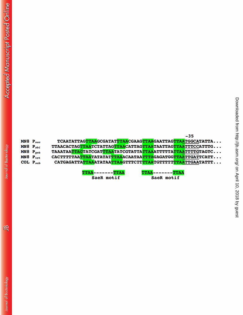
promoter region. As noted previously, many Sae regulated genes contain an additional, but less 389
conserved ‘secondary’ TTAA repeat motif (25). Figure 7 shows an alignment of the SaeP1 390
promoter region, and the Sbi promoter region, both of which are highly regulated by the Sae 391
on April 10, 2018 by guest
http://jb.asm.org/
Dow
nloaded from

15
system in S. aureus USA300 (29). In addition, the Geh promoter appears to be Sae regulated in S. 392
aureus MN8, as well as the TSST-1 promoter. Mutation analysis of the SaeP1 promoter 393
demonstrated that the presence of a G residue upstream of either primary TTAA sequence 394
facilitates direct binding by SaeR (25). Of note, the Ptst ‘primary’ motif sequence does not appear 395
to be optimal, yet tst transcript is completely repressed in the absence of a functional Sae system. 396
The evident weak binding of SaeR to Ptst may reflect, in part, the in vitro nature of the system 397
which lacks RNA polymerase known to enhance SaeR binding to PsaeP1 (39). However, based on 398
these findings we predict that an additional element may function to enhance SaeR binding to Ptst. 399
Although Rot is generally considered an exotoxin repressor, Rot and SaeR both cooperate to 400
enhance promoter activity for the staphylococcal superantigen-like genes (40). Since Rot is 401
likely a repressor of tst transcription (15), we do not expect Rot promotes SaeR binding to Ptst. 402
The staphylococcal respiratory response (srr) two-component system has been shown to 403
influence TSST-1 expression, primarily under microaerophilic conditions. SrrA has also been 404
demonstrated to weakly bind Ptst, and thus it is possible that Sae cooperates with other systems, 405
such as Srr, in the regulation of TSST-1. Nevertheless, the apparent weak binding of SaeR to Ptst 406
in vitro is consistent with other studies where Sae is a key regulator (e.g. nuc and ssl promoters) 407
(40, 41). 408
Apart from regulation of TSST-1 in S. aureus MN8, the reduction of pro-Geh expression 409
in MN8 ΔsaeS (Fig. 2) is consistent with Sae functioning as a positive transcriptional regulator 410
of geh in USA300 (29), as well as a potential SaeR binding site (25, 29) located upstream of geh 411
(Fig. 7). In MN8 ΔsarA, as expected (42, 43), we noted a prominent increase in expression of the 412
aureolysin (Aur) metalloprotease and staphopain A (ScpA) cysteine protease. Aur is responsible 413
for maturation of Pro-Geh to mature Geh (44), which explains the near complete lack of Pro-Geh 414
in the sarA mutant. Thus, although the regulatory role of SarA and Sae for these other important 415
exoproteins was largely predicted, a dominant role for regulation by Sae is selective for 416
particular exotoxins. 417
Consistent with a prior model as to how phosphorylated SaeR recruits RNA polymerase 418
to Sae-regulated target promoters (25), we herein show that the Sae system is required for 419
expression of TSST-1 and thus functions as a dominant transcriptional activator of TSST-1 420
expression. Although the regulatory queues that control Sae activation are complex, the specific 421
signals that govern Sae control of TSST-1 expression during the context of menstrual TSS 422
on April 10, 2018 by guest
http://jb.asm.org/
Dow
nloaded from

16
remain uncharacterized. Given that increased expression of saeRS from the inducible 423
complementation system did not enhance TSST-1 expression (Fig. 2A), we would predict that 424
host factors that alter SaeR phosphorylation, rather than autoinduction of the sae system, would 425
play a more important role for enhanced TSST-1 expression. Apart from the role of TSST-1 in 426
both menstrual and non-menstrual forms of TSS, another major superantigen with a clear role in 427
non-menstrual TSS is staphylococcal enterotoxin B (SEB) (9). SEB expression is known to be 428
controlled by Sae (32, 45), while this two-component regulator had no effect on transcription of 429
sea or sel-k (45). Immediately upstream of the -35 promoter region of seb (46) is a potential 430
SaeR consensus binding sequence (Fig. 7). In an in vivo rabbit model of non-menstrual TSS, agr 431
activation was not necessary for the expression of SEB and the development of TSS (47). Thus 432
potentially, Sae may also be the dominant regulator of SEB expression in S. aureus. A picture 433
has therefore emerged whereby the Sae system seems to have evolved to be a critical regulator of 434
S. aureus “trademark” superantigen exotoxins involved in TSS, and thus Sae may represent an 435
important target for the rational design of targeted therapeutics with a focus to disarm the 436
exotoxin arsenal of S. aureus. 437
438
ACKNOWLEDGEMENTS 439
We thank Dr. Taeok Bae (Indiana University) for the kind gift of the SaeR and SaeSc expression 440
plasmids. Dr. Patrick Schlivert (University of Iowa) for the kind gift of anti-TSST-1 antisera, and 441
Dr. Timothy Foster (Trinity College Dublin) for the kind gift of E. coli SA30B. This work was 442
supported by the Kimberly-Clark Corporation. 443
on April 10, 2018 by guest
http://jb.asm.org/
Dow
nloaded from

17
REFERENCES 444
1. Lowy FD. 1998. Staphylococcus aureus infections. N Engl J Med 339:520–32. 445
2. Dinges MM, Orwin PM, Schlievert PM. 2000. Exotoxins of Staphylococcus aureus. 446
Clin Microbiol Rev 13:16–34. 447
3. McCormick JK, Yarwood JM, Schlievert PM. 2001. Toxic shock syndrome and 448
bacterial superantigens: an update. Annu Rev Microbiol 55:77–104. 449
4. Centers for Disease Control and Prevention. 2011. Toxic shock syndrome; 2011 Case 450
Definition. 451
5. DeVries AS, Lesher L, Schlievert PM, Rogers T, Villaume LG, Danila R, Lynfield R. 452
2011. Staphylococcal toxic shock syndrome 2000-2006: epidemiology, clinical features, 453
and molecular characteristics. PLoS One 6:e22997. 454
6. Davis JP, Chesney PJ, Wand PJ, LaVenture M. 1980. Toxic-shock syndrome: 455
epidemiologic features, recurrence, risk factors, and prevention. N Engl J Med 303:1429–456
35. 457
7. Shands KN, Schmid GP, Dan BB, Blum D, Guidotti RJ, Hargrett NT, Anderson RL, 458
Hill DL, Broome C V, Band JD, Fraser DW. 1980. Toxic-shock syndrome in 459
menstruating women: association with tampon use and Staphylococcus aureus and clinical 460
features in 52 cases. N Engl J Med 303:1436–42. 461
8. Spaulding AR, Salgado-Pabón W, Kohler PL, Horswill AR, Leung DYM, Schlievert 462
PM. 2013. Staphylococcal and streptococcal superantigen exotoxins. Clin Microbiol Rev 463
26:422–47. 464
9. Schlievert PM. 1986. Staphylococcal enterotoxin B and toxic-shock syndrome toxin-1 are 465
significantly associated with non-menstrual TSS [letter]. Lancet 1:1149–1150. 466
10. Recsei P, Kreiswirth B, O’Reilly M, Schlievert P, Gruss A, Novick RP. 1986. 467
Regulation of exoprotein gene expression in Staphylococcus aureus by agr. Mol Gen 468
Genet 202:58–61. 469
11. Yarwood JM, McCormick JK, Schlievert PM. 2001. Identification of a novel two-470
component regulatory system that acts in global regulation of virulence factors of 471
Staphylococcus aureus. J Bacteriol 183:1113–1123. 472
12. Nagao M, Okamoto A, Yamada K, Hasegawa T, Hasegawa Y, Ohta M. 2009. 473
Variations in amount of TSST-1 produced by clinical methicillin resistant Staphylococcus 474
on April 10, 2018 by guest
http://jb.asm.org/
Dow
nloaded from

18
aureus (MRSA) isolates and allelic variation in accessory gene regulator (agr) locus. 475
BMC Microbiol 9:52. 476
13. Andrey DO, Renzoni A, Monod A, Lew DP, Cheung AL, Kelley WL. 2010. Control of 477
the Staphylococcus aureus toxic shock tst promoter by the global regulator SarA. J 478
Bacteriol 192:6077–6085. 479
14. Chan PF, Foster SJ. 1998. Role of SarA in virulence determinant production and 480
environmental signal transduction in Staphylococcus aureus. J Bacteriol 180:6232–6241. 481
15. Andrey DO, Jousselin A, Villanueva M, Renzoni A, Monod A, Barras C, Rodriguez 482
N, Kelley WL. 2015. Impact of the regulators SigB, Rot, SarA and SarS on the toxic 483
shock tst promoter and TSST-1 expression in Staphylococcus aureus. PLoS One 484
10:e0135579. 485
16. Seidl K, Bischoff M, Berger-Bächi B. 2008. CcpA mediates the catabolite repression of 486
tst in Staphylococcus aureus. Infect Immun 76:5093–9. 487
17. Schlievert PM, Blomster DA. 1983. Production of staphylococcal pyrogenic exotoxin 488
type C: influence of physical and chemical factors. J Infect Dis 147:236–242. 489
18. Li J, Wang W, Xu SX, Magarvey NA, McCormick JK. 2011. Lactobacillus reuteri-490
produced cyclic dipeptides quench agr-mediated expression of toxic shock syndrome 491
toxin-1 in staphylococci. Proc Natl Acad Sci U S A 108:3360–3365. 492
19. Novick R. 1967. Properties of a cryptic high-frequency transducing phage in 493
Staphylococcus aureus. Virology, 1967/09/01 ed. 33:155–166. 494
20. Monk IR, Tree JJ, Howden BP, Stinear TP, Foster TJ. 2015. Complete bypass of 495
restriction systems for major Staphylococcus aureus lineages. MBio 6:e00308–15. 496
21. Arnaud M, Chastanet A, Debarbouille M. 2004. New vector for efficient allelic 497
replacement in naturally nontransformable, low-GC-content, gram-positive bacteria. Appl 498
Env Microbiol 70:6887–6891. 499
22. Bae T, Schneewind O. 2006. Allelic replacement in Staphylococcus aureus with 500
inducible counter-selection. Plasmid 55:58–63. 501
23. Bateman BT, Donegan NP, Jarry TM, Palma M, Cheung AL. 2001. Evaluation of a 502
tetracycline-inducible promoter in Staphylococcus aureus in vitro and in vivo and its 503
application in demonstrating the role of sigB in microcolony formation. Infect Immun 504
69:7851–7857. 505
on April 10, 2018 by guest
http://jb.asm.org/
Dow
nloaded from

19
24. Mesak LR, Yim G, Davies J. 2009. Improved lux reporters for use in Staphylococcus 506
aureus. Plasmid 61:182–187. 507
25. Sun F, Li C, Jeong D, Sohn C, He C, Bae T. 2010. In the Staphylococcus aureus two-508
component system sae, the response regulator SaeR binds to a direct repeat sequence and 509
DNA binding requires phosphorylation by the sensor kinase SaeS. J Bacteriol 192:2111–510
2127. 511
26. Rahman AK, Bonsor DA, Herfst CA, Pollard F, Peirce M, Wyatt AW, Kasper KJ, 512
Madrenas J, Sundberg EJ, McCormick JK. 2011. The T cell receptor beta-chain 513
second complementarity determining region loop (CDR2beta) governs T cell activation 514
and Vbeta specificity by bacterial superantigens. J Biol Chem 286:4871–4881. 515
27. Liu Q, Cho H, Yeo W-S, Bae T. 2015. The extracytoplasmic linker peptide of the sensor 516
protein SaeS tunes the kinase activity required for staphylococcal virulence in response to 517
host signals. PLoS Pathog 11:e1004799. 518
28. Laakso HA, Marolda CL, Pinter TB, Stillman MJ, Heinrichs DE. 2016. A heme-519
responsive regulator controls synthesis of staphyloferrin B in Staphylococcus aureus. J 520
Biol Chem 291:29–40. 521
29. Nygaard TK, Pallister KB, Ruzevich P, Griffith S, Vuong C, Voyich JM. 2010. SaeR 522
binds a consensus sequence within virulence gene promoters to advance USA300 523
pathogenesis. J Infect Dis 201:241–254. 524
30. Tal-Gan Y, Stacy DM, Foegen MK, Koenig DW, Blackwell HE. 2013. Highly potent 525
inhibitors of quorum sensing in Staphylococcus aureus revealed through a systematic 526
synthetic study of the group-III autoinducing peptide. J Am Chem Soc 135:7869–82. 527
31. Reyes D, Andrey DO, Monod A, Kelley WL, Zhang G, Cheung AL. 2011. 528
Coordinated regulation by AgrA, SarA, and SarR to control agr expression in 529
Staphylococcus aureus. J Bacteriol 193:6020–6031. 530
32. Rogasch K, Rühmling V, Pané-Farré J, Höper D, Weinberg C, Fuchs S, Schmudde 531
M, Bröker BM, Wolz C, Hecker M, Engelmann S. 2006. Influence of the two-532
component system SaeRS on global gene expression in two different Staphylococcus 533
aureus strains. J Bacteriol 188:7742–58. 534
33. Giachino P, Engelmann S, Bischoff M. 2001. Sigma B activity depends on RsbU in 535
Staphylococcus aureus. J Bacteriol 183:1843–1852. 536
on April 10, 2018 by guest
http://jb.asm.org/
Dow
nloaded from

20
34. Bischoff M, Dunman P, Kormanec J, Macapagal D, Murphy E, Mounts W, Berger-537
Bächi B, Projan S. 2004. Microarray-based analysis of the Staphylococcus aureus 538
sigmaB regulon. J Bacteriol 186:4085–99. 539
35. DeLeo FR, Kennedy AD, Chen L, Bubeck Wardenburg J, Kobayashi SD, Mathema 540
B, Braughton KR, Whitney AR, Villaruz AE, Martens CA, Porcella SF, McGavin 541
MJ, Otto M, Musser JM, Kreiswirth BN. 2011. Molecular differentiation of historic 542
phage-type 80/81 and contemporary epidemic Staphylococcus aureus. Proc Natl Acad Sci 543
U S A 108:18091–6. 544
36. Giraudo AT, Cheung AL, Nagel R. 1997. The sae locus of Staphylococcus aureus 545
controls exoprotein synthesis at the transcriptional level. Arch Microbiol 168:53–8. 546
37. Geisinger E, Adhikari RP, Jin R, Ross HF, Novick RP. 2006. Inhibition of rot 547
translation by RNAIII, a key feature of agr function. Mol Microbiol 61:1038–1048. 548
38. Boisset S, Geissmann T, Huntzinger E, Fechter P, Bendridi N, Possedko M, 549
Chevalier C, Helfer AC, Benito Y, Jacquier A, Gaspin C, Vandenesch F, Romby P. 550
2007. Staphylococcus aureus RNAIII coordinately represses the synthesis of virulence 551
factors and the transcription regulator Rot by an antisense mechanism. Genes Dev 552
21:1353–66. 553
39. Cho H, Jeong D-W, Li C, Bae T. 2012. Organizational requirements of the SaeR binding 554
sites for a functional P1 promoter of the sae operon in Staphylococcus aureus. J Bacteriol 555
194:2865–76. 556
40. Benson MA, Lilo S, Nygaard T, Voyich JM, Torres VJ. 2012. Rot and SaeRS 557
cooperate to activate expression of the staphylococcal superantigen-like exoproteins. J 558
Bacteriol 194:4355–65. 559
41. Olson ME, Nygaard TK, Ackermann L, Watkins RL, Zurek OW, Pallister KB, 560
Griffith S, Kiedrowski MR, Flack CE, Kavanaugh JS, Kreiswirth BN, Horswill AR, 561
Voyich JM. 2013. Staphylococcus aureus nuclease is an SaeRS-dependent virulence 562
factor. Infect Immun 81:1316–24. 563
42. Ziebandt AK, Weber H, Rudolph J, Schmid R, Höper D, Engelmann S, Hecker M. 564
2001. Extracellular proteins of Staphylococcus aureus and the role of SarA and sigma B. 565
Proteomics 1:480–93. 566
43. Jones RC, Deck J, Edmondson RD, Hart ME. 2008. Relative quantitative comparisons 567
on April 10, 2018 by guest
http://jb.asm.org/
Dow
nloaded from

21
of the extracellular protein profiles of Staphylococcus aureus UAMS-1 and its sarA, agr, 568
and sarA agr regulatory mutants using one-dimensional polyacrylamide gel 569
electrophoresis and nanocapilla. J Bacteriol 190:5265–78. 570
44. Cadieux B, Vijayakumaran V, Bernards MA, McGavin MJ, Heinrichs DE. 2014. 571
Role of lipase from community-associated methicillin-resistant Staphylococcus aureus 572
strain USA300 in hydrolyzing triglycerides into growth-inhibitory free fatty acids. J 573
Bacteriol 196:4044–56. 574
45. Kusch K, Hanke K, Holtfreter S, Schmudde M, Kohler C, Erck C, Wehland J, 575
Hecker M, Ohlsen K, Bröker B, Engelmann S. 2011. The influence of SaeRS and σ(B) 576
on the expression of superantigens in different Staphylococcus aureus isolates. Int J Med 577
Microbiol 301:488–99. 578
46. Mahmood R, Khan SA. 1990. Role of upstream sequences in the expression of the 579
staphylococcal enterotoxin B gene. J Biol Chem 265:4652–6. 580
47. Yarwood JM, McCormick JK, Paustian ML, Kapur V, Schlievert PM. 2002. 581
Repression of the Staphylococcus aureus accessory gene regulator in serum and in vivo. J 582
Bacteriol 184:1095–1101. 583
584 on April 10, 2018 by guest
http://jb.asm.org/
Dow
nloaded from

22
Figure Legends 585
586
Figure. 1. Schematic and PCR analysis of the saeS and sarA in-frame deletions in S. aureus 587
MN8. A) Scale schematic of the 381-bp deletion in saeS (top) and the 358-bp deletion in sarA 588
(bottom) and location of the PCR products used for analysis and sequencing of the corresponding 589
deletions. The black bar indicates a 500-bp size fragment. B) DNA agarose gel analysis using 590
PCR products indicated in Panel A for the individual and double MN8 sarA and saeS deletions 591
as indicated. 592
593
Figure 2. Sae is a positive regulator, and SarA is a negative regulator, of TSST-1 1 594
expression in S. aureus MN8. Exoprotein profiles (top panels) and Western blot (bottom 595
panels) analysis of TSST-1 for wild-type S. aureus MN8 and the corresponding (A) saeS and (B) 596
sarA deletion and complemented strains. Concentrated supernatants from the indicated strains 597
grown in BHI medium for 18h were loaded onto 12% SDS-PAGE gels. Increasing 598
concentrations of anhydrotetracyclin (0, 5, 50 and 500 ng/ml) was used to induce the promoter in 599
the complemented strains. Molecular mass markers were loaded on the left and labeled in kDa, 600
and purified recombinant TSST-1 was loaded on the right and is indicated with the black 601
arrowhead. 602
603
Figure 3. TSST-1 quantitation, growth curve analysis, and TSST-1 promoter activity of 604
wild-type S. aureus MN8 and the corresponding saeS, sarA and agr mutants. (A) S. aureus 605
MN8 strains were grown for 18h in BHI medium and TSST-1 secretion was quantified by 606
ELISA. Data represented the means (± SEM) of three biological replicates (**, p < 0.05 by one-607
way ANOVA with Tukey's Multiple Comparison Test). (B and C) The indicated S. aureus 608
strains were grown at 37°C in a Biotek Synergy H4 multimode plate reader and OD600 and 609
luminescence readings were taken every hour for 15h. Results were expressed (B) OD600 units 610
and (C) relative light units (defined as counts min−1). Data represent the means of three 611
independent experiments each done with quadruplicate technical replicates. 612
613
Figure 4. Quantitative real-time PCR (qRT-PCR) analysis of tst transcripts and relevant 614
regulators in wild-type S. aureus MN8 and the saeS, sarA and agr mutants. (A) cDNA was 615
on April 10, 2018 by guest
http://jb.asm.org/
Dow
nloaded from

23
prepared at the indicated time points and copies were normalized to the housekeeping rpoB gene. 616
Data represent the means (± SEM) of four biological replicates (*, p < 0.05; **, p < 0.01; ***, p 617
< 0.001 by one-way ANOVA with Tukey's Multiple Comparison Test). (B) Exoprotein profiles 618
(top panel) and anti-TSST-1 Western blot (bottom panel) analysis of the indicated S. aureus 619
strains at 4 and 8h time points. Molecular mass markers were loaded on the left and labeled in 620
kDa, and purified recombinant TSST-1 was loaded on the right and indicated with the black 621
arrowhead. 622
623
Figure 5. DNA binding analysis of phosphorylated SaeR (P~SaeR) to the PsaeP1 and Ptst 624
promoters. (A) Schematic map and nucleotide sequence of Ptst in S. aureus MN8 (Locus tag 625
HMPREF0769). The characterized transcriptional start site and -10 and -35 regions are labelled 626
with boxes, and the characterized SarA binding sites is highlighted with blue (13). The proposed 627
SaeR binding sequences are highlighted with green. Probes 1, 2 and 3 correspond to the EMSAs 628
shown in Panels D, E, and F, respectively. DNA binding analysis by P~SaeR to PsaeP1 (B), Ptst 629
(C), Probe 1 (D), Probe 2 (E) and Probe 3 (F). Panel C (right panel) also shows a competitive 630
binding analysis using 0, 5, 10 and 50-fold excess unlabeled Ptst (gradient denoted by the 631
triangle). Unbound DNA probes are labelled with open arrow heads, and DNA shifts are labelled 632
with solid arrowheads. Protein concentrations, and the presence of ATP in the reaction buffer, 633
are indicated for each lane. 634
635
Figure 6. The Sae two-component regulator is necessary for TSST-1 expression in the 636
absence of repression by SarA. Exoprotein profiles (top panel) and Western blot (bottom panel) 637
analysis of TSST-1 for wild-type S. aureus MN8 and the corresponding mutants. Concentrated 638
supernatants from the indicated strains grown in BHI medium were loaded onto 12% SDS-639
PAGE gels. Molecular mass markers in kDa are shown on the left, followed by purified 640
recombinant TSST-1 that is indicated with the black arrowhead. The locations of pro-Geh, 641
mature Geh, Aur, ScpA, and Nuc are labelled with open arrowheads. 642
643
Figure 7. Alignment of promoter element nucleotide sequences of SaeR binding motifs 644
located upstream of PsaeP1, Psbi, Pgeh, Ptst, and Pseb. The -35 promoter binding regions are 645
underlined, and green highlights outline the primary and secondary SaeR binding motifs. 646
on April 10, 2018 by guest
http://jb.asm.org/
Dow
nloaded from
24
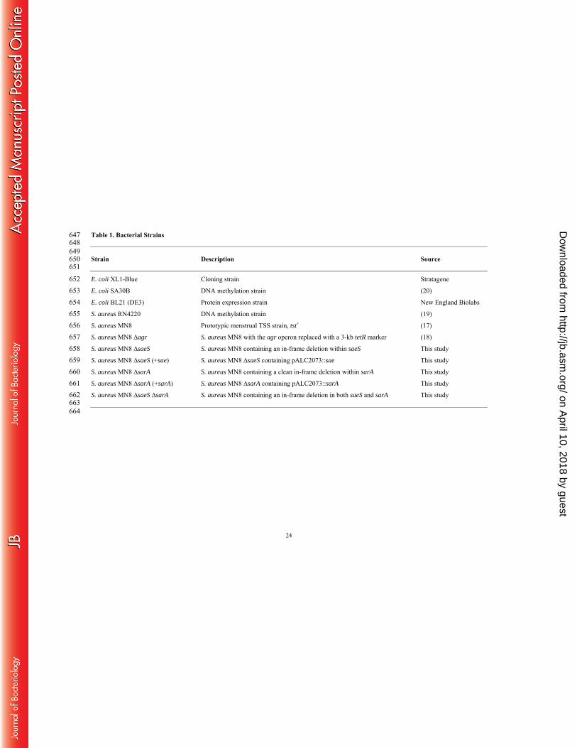
Table 1. Bacterial Strains 647 648 649 Strain Description Source 650 651 E. coli XL1-Blue Cloning strain Stratagene 652 E. coli SA30B DNA methylation strain (20) 653 E. coli BL21 (DE3) Protein expression strain New England Biolabs 654 S. aureus RN4220 DNA methylation strain (19) 655 S. aureus MN8 Prototypic menstrual TSS strain, tst+ (17) 656 S. aureus MN8 ∆agr S. aureus MN8 with the agr operon replaced with a 3-kb tetR marker (18) 657 S. aureus MN8 ∆saeS S. aureus MN8 containing an in-frame deletion within saeS This study 658 S. aureus MN8 ∆saeS (+sae) S. aureus MN8 ∆saeS containing pALC2073::sae This study 659 S. aureus MN8 ∆sarA S. aureus MN8 containing a clean in-frame deletion within sarA This study 660 S. aureus MN8 ∆sarA (+sarA) S. aureus MN8 ∆sarA containing pALC2073::sarA This study 661 S. aureus MN8 ∆saeS ∆sarA S. aureus MN8 containing an in-frame deletion in both saeS and sarA This study 662 663 664
on April 10, 2018 by guest
http://jb.asm.org/
Dow
nloaded from

25
Table 2. Plasmids 665 666 667 Plasmid Descriptiona Source 668 669 pMAD Temperature sensitive integration vector with blue-white selection, EmR (21) 670 pMAD:: ΔsaeS saeS deletion plasmid, EmR This study 671 pKOR1 Temperature sensitive integration vector with inducible counter selection, EmR (22) 672 pKOR1::ΔsarA sarA deletion plasmid, EmR This study 673 pALC2073 S. aureus complementation plasmid, CmR (23) 674 pALC2073::sae Complementation plasmid containing the saeQRS operon, CmR This study 675 pALC2073::sarA Complementation plasmid containing sarA, CmR This study 676 pAmilux Luminescence reporter plasmid, CmR (24) 677 pAmilux::Ptst TSST-1 promoter reporter plasmid, CmR (18) 678 pET28::saeR Protein expression clone containing full length SaeR, KanR (25) 679 pMCSG19::saeSC Protein expression clone encoding residues 93 to 351 of SaeS, AmpR (25) 680 681 a EmR, erythromycin resistant; CmR, chloramphenicol resistant; KanR, kanamycin resistant. 682 683
on April 10, 2018 by guest
http://jb.asm.org/
Dow
nloaded from

26
Table 3. Primers 684 685 686 Primer Sequence (5’ - 3’) a, b 687 688 689 Primers for saeS deletion and complementation plasmid construction 690 SaeS up BamHI (forward) GGGGGATCCAACAACGACAACTAGCGGTAAAGA 691 SaeS up Sall (reverse) AGTGTCGACACACCATTATCGGCTCCTTTCA 692 SaeS down EcoRI (forward) CCCGAATTCTAGCCCATGATTTAAAAACACCTT 693 SaeS down Bglll (reverse) CCCAGATCTTTCCTACATCTATATCACTGCTTACACTG 694 SaeRS Comp II KpnI For TTTTTGGTACCGGTAAATTAATGCTTACTAACTACAA 695 SaeRS Comp II EcoRI Rev CCCCCGAATTCTTATGTCGTAATGTCTAATTTGTG 696 697 Primers for sarA deletion and complementation plasmid construction 698 SarA upstream For attB1 GGGGACAAGTTTGTACAAAAAAGCAGGCTAGGTGCAGCATTAACAACACT 699 SarA upstream Rev* AATTGCCATGGTTAAAACCTC 700 SarA downstream For* GAACTATAATTTTGTTTAGCG 701 SarA downstream rev primer attB2 GGGGACCACTTTGTACAAGAAAGCTGGGTTGAGGGAGGTGTCACAATGA 702 SarA complement KpnI For TTTTTGGTACCTTTAACATTTAGCTTATCATTTTAA 703 SarA complement SacI Rev CCCCCGAGCTCTTATAGTTCAATTTCGTTGTTTGCT 704
705 Sequencing Primers 706 SaeRS screen For CTGGGGGATATGTTTTACC 707 SaeRS screen Rev GTCCCTATGCGTATTAAGGA 708 pMAD seq FP GGGGAAGGCCATCCAGCCTCGCGTC 709 pMAD seq RP AATCTAGCTAATGTTACGTTACACA 710 SarA flank for TCTTATCATTAAAACTGCACTGGGA 711 SarA flank rev GCGGTGGCAATTCGTTCATT 712 pKOR1For CAGATCCATATCCTTCTTTTCTGA 713 pKOR1 Rev GTGTGGAATTGTGAGCGGATA 714 pALC2073 Seq For GGTTGCATGCCTGCAGGTCGACGG 715 pALC2073 Seq Rev CAGTCACGACGTTGTAAAACG 716 717 Primers for protein expression 718 SarA-Forward-NcoI CCCCCATGGCAATTACAAAAATCAAT 719
on April 10, 2018 by guest
http://jb.asm.org/
Dow
nloaded from

27
SarA-Reverse-BamHI CCCGGATCCTTATAGTTCAATTTCGTTGTTTGC 720 721
Primers for qRT-PCR analysis 722 tst-For CTGATGCTGCCATCTGTGTT 723 tst-Rev GTAAGCCCTTTGTTGCTTGC 724 agrA-For (P2) GTGAAATTCGTAAGCATGACCCAGTTG 725 agrA-Rev (P2) TGTAAGCGTGTATGTGCAGTTTCTAAAC 726 rnaIII-For (P3) TAGATCACAGAGATGTGA 727 rnaIII-Rev (P3) CTGAGTCCAAGGAAACTAACTC 728 saeR-For CCAAGGGAACTCGTTTTACG 729 saeR-Rev ACGCATAGGGACTTCATGAC 730 sarA-For TGTTTGCTTCAGTGATTCGTTT 731 sarA-rev CATCAGCGAAAACAAAGAGAAA 732 rpoB-For TCCTGTTGAACGCGCATGTAA 733 rpoB-Rev GCTGGTATGGCTCGTGATGGTA 734 735 Primers for EMSA experiments 736 SaeP1-For-biotin ATTAGTTAAGCGATATTTAAACGAAGTTAAGAATTAGTTAATGGCA 737 SaeP1-Rev TGCCATTAACTAATTCTTAACTTCGTTTAAATATCGCTTAACTAAT 738 ptst–For-biotin AAAGTGACTTTAAAGAATATAACTA 739 ptst-Rev TTTTAATTCTCCTTCATTCAAATGT 740 Probe1-For-IRDye800 GTAACAAACACTTTTTAATTAATATATATTTAAACAATAATTTAGA 741 Probe1-Rev TCTAAATTATTGTTTAAATATATATTAATTAAAAAGTGTTTGTTAC 742 Probe 2-For-IRDye800 TTTTTAATTAATATATATTTAAACAATAATTTAGAGATGGTTAATTGATT 743 Probe2-Rev AATCAATTAACCATCTCTAAATTATTGTTTAAATATATATTAATTAAAAA 744 Probe3-For-IRDye800 AATTTAGAGATGGTTAATTGATTCATTTAAATAATATTTATACATTCT 745 Probe3-Rev AGAATGTATAAATATTATTTAAATGAATCAATTAACCATCTCTAAATT 746 747 * primer contained a phosphate group for blunt end ligation 748 a restriction sites are indicated in the primer name and underlined in the primer sequence 749 b attB sites are shown in bold 750
on April 10, 2018 by guest
http://jb.asm.org/
Dow
nloaded from