Embed Size (px)
Citation preview

Supervisor: Fredrik Palm, PhD Laboratory supervisor: Malou Friederich Persson, PhD
The role of uncoupling protein-2 in
regulating mitochondrial oxygen
consumption in the diabetic kidney.
A regulatory mechanism by glutamine.
Jonas Gustafsson
Bachelor thesis in Biomedicine, 15 HP
Department of Medical cell biology, Division of Integrative Physiology

Abstract
Diabetes is one of our times most common diseases, affecting millions every year and it is said to
stand for almost 50% of inpatient care in the United States. Diabetes has two general forms, type I,
insulin dependent diabetes and type II, noninsulin dependent diabetes. Diabetes is associated with
increased production of reactive oxygen species (ROS) which cause a dysfunction of the cells, a
phenomenon known as oxidative stress. In the kidney increased production of ROS can activate the
mitochondrial protein uncoupling protein-2 (UCP-2), which can shuffle protons over the
mitochondrial membrane independent of ATP-production. This decreases the membrane potential and
therefore reduces production of ROS. However, an important side effect is increased oxygen
consumption (QO2) which may result in hypoxia and nephropathy.
The aim of the present study was to investigate the regulatory effects of glutamine on the UCP-2
and thus the QO2 in kidney mitochondria isolated from wild type and UCP-2 knockout mice. Isolated
kidney cortex mitochondria were analyzed in terms of QO2 with an Oxygraph-2k.
An increase in QO2 in the groups treated with glutamine and was evident and there was also an
increase inhibition of UCP-2 by GDP in the same groups. The combined results indicated that
glutamine had a regulatory effect on mitochondrial uncoupling, possibly by UCP-2 or other isoforms.

Sammanfattning
Diabetes är en av vår tids mest utbredda sjukdomar, den påverkar miljoner av folks normala liv och
sägs stå för ungefär 50 % av kostnaderna för patientvård i USA. Det finns två typer av diabetes, typ I,
insulin beroende diabetes och typ II, icke-insulin beroende diabetes. Diabetes ökar produktionen av
reaktiva syre former (ROS) vilket kan skada många av kroppens celler, detta kallas ofta för oxidativ
stress. I njuren resulterar den ökade produktionen av ROS en aktivering av mitokondrieproteinet UCP-
2 som transporterar protoner över mitokondriemembranet och sänker membranpotentialen vilket
minskar risken för ROS. Denna transport har en viktig biverkning, ökad syrekonsumtion (QO2) i
njuren, vilket kan resultera i hypoxi och orsaka nefropati.
Målet med denna studie var att undersöka den regulatoriska effekten av glutamin på UCP-2 och
därigenom QO2 i isolerade mitokondrier från vildtyp och UCP-2 knockout möss. Hos de isolerade
mitokondrier analyserades QO2 med en Oxygraph-2k.
Resultaten visade en ökning i QO2 hos grupperna som var behandlade med glutamin, inhiberingen
av UCP-2 med GDP var också ökad i dessa grupper. De sammanlagda resultaten indikerade en
regulatorisk effekt av glutamin på mitokondriell frikoppling, troligen via UCP-2 eller andra isoformer.

Introduction
The kidney and oxygen consumption
The kidney is responsible for removing waste material through the production of urine and to control
the volume and composition of our body fluids. The kidneys produce 180 liters of primary urine per
day and 99% is reabsorbed in terms of electrolytes, osmotic particles and water to maintain
homeostasis. 65% of sodium and water are reabsorbed in the proximal tubule in the kidney cortex via
active transportation Because of high requirement of adenosine triphosphate (ATP) to support the
active transport of electrolytes, proximal tubular epithelial cells have a high concentration of
mitochondria, producing ATP though the electron transport chain (ETC) and ATP-synthase1. The
renal blood flow consists of 25% of cardiac output and the kidney extract 10-15% of oxygen from the
blood. This is in contrast to other organs with up to 45% of oxygen extraction, such as the heart2,3
.
Despite a high perfusion, kidney cortex oxygen tension is low4. This can be explained by the structural
design of the renal vasculature where arteries and veins run in parallel thus allowing oxygen to diffuse
from the arteries to the venous system5. Therefore cortical tissues are at risk of developing hypoxia if
the oxygen consumption (QO2) were to increase5,6
.
Electron transport chain
The production of ATP occurs in the inner membrane of the mitochondria using the ETC2,5,6
. The ETC
consists of four complexes (Fig. 1), NADH deliver electrons (e-) to complex I and FADH2 delivers
electrons to complex II7. The electrons are then transported within the membrane using carrier
molecules to complex III and then transferred from complex III to complex IV using cytochrome C via
the intermembrane space6,7
. In complex IV the electrons reacts with oxygen (O2) and protons (H+) to
create water6,7
.
O2 +4H++4e
- 2 H2O
Each time two electrons are transferred trough complex I, III and IV protons are transported from
the mitochondrial matrix into the intermembrane space, altogether ten protons are transported for
every two electrons passing through the ETC6,7
. This transport of protons creates a H+
gradient that
constitutes the mitochondrial membrane potential driving the ATP-synthase6. For ATP production the
ATP-synthase require adenosine diphosphate (ADP), which is transported over the membrane using
adenosine nucleotide transporter (ANT), translocation ADP and ATP into the intermembrane space
and subsequently, the cystole.

Figure 1. The electron transport chain in the kidney with inhibitors and the mechanism for uncoupling via adenosine nucleotide
transporter (ANT) and uncoupling protein-2 (UCP-2). FA – Fatty acids; GDP – Guanidine diphosphate; CAT – Carboxyatractyloside;
ADP – Adenosine diphosphate; ATP – Adenosine triphosphate. Modified from17.
Reactive oxygen species and oxidative stress
Reactive oxygen species (ROS) is a general term for free radicals of oxygen that include pure oxygen
radicals, such as superoxide (O2.-) and hydroxyl (OH
.), and non-radical oxidizing agents, such as
hydrogen peroxide (H2O2). The most common radical is O2.-8
. ROS can cause cellular damage by
modifying protein, DNA and fatty acyl groups in the cellular membrane, thus interfering with normal
cell functions. Damage by ROS is known as oxidative stress9. The body’s immune system,
macrophages and neutrophils, purposely generated ROS to kill pathogens and ROS play a major part
in many cellular signaling pathways9–11
. ROS is produced in many metabolic pathways but the major
source of ROS is the ETC9, complex I and III, which is increasedby a hyperpolarized mitochondrial
membrane12
.
The increased delivery of electron donating molecules such as NADH and FADH2 causes increased
transport of protons over the membrane to the point where inhibition of electron transport occurs12
.
Excess electrons will be trapped in the ETC and may slip and react directly with O2 creating O2.- 13
.
Protection from oxidative stress is provided by, among other enzymes, superoxide dismutase (SOD)14
.
SOD catalyzes the reaction:
2 O2.-
+ 2H+
H2O2 + O2
Mitochondrial matrix
Intermembrane space

The reaction creates H2O2 and O2. H2O2 is significantly more stable and can easily be dismuted into
water and O2 by the enzyme catalase14
. Mitochondrial production of O2.- can also be prevented by
decreasing the mitochondrial membrane potential, thus preventing hyperpolarization and inhibition of
complexes I and III. This can be achieved by mitochondrial uncoupling usually performed by
uncoupling proteins (UCP’s), these include UCP-1 through UCP-515
.
Uncoupling Protein-2
UCP-2 is the only isoform of UCP’s expressed in the kidney16
. Mitochondrial uncoupling denotes
shunting of protons over the inner membrane independently of ATP-production. This lowers the
membrane potential and the formation of ROS is therefore decreased6,16–19
. UCP-2 does not by itself
transport protons over the membrane, a charged fatty acid anion (FA) binds to the proton and can then
diffuse across the membrane where it becomes deprotonated. UCP-2 works as a transportation channel
for the FA back to the intermembrane space, this is called fatty acid cycling (Fig. 1)20
. The amino acid
glutamine has been shown to regulate UCP-2 expression by increasing translation of UCP-2 mRNA
and is for now the only amino acid known to have an regulatory effect on UCP-2 expression21
.
Increased mitochondrial uncoupling using UCP-2 come at the cost of increased oxygen consumption,
this side effect may be harmful to the kidney as it may cause kidney hypoxia5,6
.
Diabetes Mellitus and its consequences
There are two kinds of diabetes, Type I, insulin dependent diabetes and type II, non-insulin dependent
diabetes. Type I diabetes debuts at around 14-15 years of age and is caused by destruction of the
insulin producing β-cells in the pancreas by an autoimmune disorder, resulting in an insufficient
production of insulin. In turn, insufficient production of insulin results in decreased uptake of glucose
and increased blood glucose. Excess glucose is lost in the urine and by being an osmotic particle,
glucoseuria results in osmotic diuresis that may result in dehydration. Type I diabetes can also cause
neural, cardiac and vascular disorders, such as stroke, heart attack and ischemia in the limbs1.
Furthermore, diabetes is known to increased QO2, resulting decreased oxygen tension in the kidney
that may ultimately result in hypoxia and nephropathy22–25
. Importantly, diabetes increases UCP-2
expression which might account for increased QO2, resulting in hypoxia in the diabetic kidney18,26
.
Type II diabetes, also called insulin resistance diabetes, is caused by an increased resistance to the
effects of insulin1,27
. Compared to type I diabetes the type II is more common, more than 90% of all
the diabetes cases in the world is type II and it is most common between ages of 50 and 60. This type
of diabetes does not cause the all the same complications as type I, it will however cause a number of
different cardiovascular diseases such as, arteriosclerosis, hypertension and organ damage due to high
blood glucose levels1. Almost 80% of type II diabetes is related to overweight and high body mass
index (BMI) and it stands for almost 50% of the total health care inpatient costs in the United States28
.

Hypothesis and aim
The diabetic kidney has increased mitochondrial membrane potential and increased production of
ROS18
. Glutamine can also increase the expression of UCP-2 by increasing translation of UCP-2
mRNA21
. Both glutamine and diabetes ma cause increased expression of UCP-2 and increased
uncoupling 18
. This can result in increased QO2, decreased oxygen tension and may result in hypoxia
and renal damage in the kidney.
The hypothesis in the present study is that production and activation of UCP-2 in the diabetic
kidney, and therefore QO2, can be regulated by glutamine. The aim was therefore to investigate this by
using kidney cortex mitochondria from mice with induced diabetes and increase intake of glutamine to
detect alterations in QO2. The specific role of UPC-2 was investigated using UCP-2 knockout mice.

Material and method
All chemicals were from Sigma Aldrich CO. LLC. St. Louis, USA, if nothing else is mentioned.
All solutions and isolated mitochondria were kept on ice or in 4 ˚C during the entire experiment.
UCP-2 knockout mice and corresponding wild-type littermates were either untreated, treated with
glutamine (Dipetiven, 1mg/kg body weight, Fresenius Kabi AB, Uppsala, Sweden, 2 weeks), alloxan
to induce diabetes (75 mg/kg body weight dissolved in 0.2 ml physiological saline, 2 weeks) or
glutamine and alloxan in combination.
Isolation of kidney cortex mitochondria
After cervical dislocation the kidneys were removed and placed in mitochondrial buffer A, containing
(in mmol/l): 250 sucrose, 10 4-(2-Hydroxyethyl)piperazine-1-ethanesulfonic acid (HEPES), 1
Ethylene glycol-bis(2-aminoethylether)-N,N,N′,N′-tetraacetic acid (EGTA), 1 mg/ml Bovine Serum
Albumine (BSA, Fraction V), pH 7.4 (controlled with KOH), 300 mOsm/kg H20. The kidney cortex
was isolated on ice and the isolated cortex pieces were then transferred to 5 ml of new mitochondrial
buffer A. The isolated cortex was then homogenized in a Potter-Elvehjem homogenizer at 600-800
rpm (4-6 strokes in total) until no visible pieces were observed. The homogenizer was kept in ice
mixed with water to ensure a low temperature throughout the homogenization process. The
homogenizesed cortex were transferred to eppendorf tubes and centrifuged, 800*g, for 10 minutes at 4
˚C and the supernatant transferred to new tubes, carefully not including the buffy coat. The supernatant
was then centrifuged at 8000*g for 10 minutes at 4 ˚C the pellets rinsed with mitochondrial buffer A
and stored on ice.
Wild-type – Control UCP-2 Knockout – Control
Wild-type – Glutamine UCP-2 Knockout – Glutamine
Wild-type – Diabetes UCP-2 Knockout – Diabetes
Wild-type – Glutamine + Diabetes UCP-2 Knockout – Glutamine + Diabetes
Table 1. Groups used in the present study.

Mitochondrial oxygen consumption
Mitochondrial oxygen consumption was measured in an Oxygraph-2k (O2k) (Oroboros Instruments
Corp, Innsbruck, Austria) after calibration by air saturation.
The chamber of the O2k was filled with 2 ml of mitochondrial buffer B containing (in mmol/l): 68
sucrose, 198 mannitol, 2 EGTA, 5 MgCl2, 5 KPO4- (from a 1M mix of K2HPO4
- and KH2PO4
-), 10
HEPES, 3 mg/ml BSA, pH 7.1, 330 mOSM/kg H2O and 20 µl of glutamate (1 M) was added to both
chamber A (control chamber) and chamber B (inhibitor chamber). The saturated fatty acid palmitate
48µmol/l (2.3µg/ml) and the ATP-synthase inhibitor oligomycin were also added to chamber B before
the addition of mitochondria. The mitochondria pellet was dissolved in 200 µl of mitochondrial buffer
A and 100 µl of suspended mitochondria was added to each chamber. After stable QO2 was achieved
20 µl ADP (30 mM in 10 mM HEPES, 0.6mol MgCl2/mol ADP) was added to chamber A, to achieve
maximal respiration and calculate respiratory control ratio (RCR). In chamber B 20 µl Guanosine 5′-
diphosphate (GDP, 4.4 mg Guanosine 5′-diphosphate sodium salt dissolved in 200 ml mitochondrial
buffer B) was added and thereafter 2 µl Carboxyatractyloside (CAT, 2 mg Carboxyatractyloside
potassium salt dissolved in 444 µl water) was inserted. Analysis of QO2 was made using DATALAB 4
software (Oroboros instruments Corp, Innsbruck, Austria).
Respiratory control ratio (RCR) was calculated by dividing the QO2 after the addition of ADP with
the QO2 before ADP. RCR is indicative of mitochondrial health and a RCR ≥ 4 is required before
performing additional experiments. A sample was taken from each chamber to correct QO2 for protein
content using DC Protein Assay (Bio-Rad Laboratories, CA, USA).

0,00 1,00 2,00 3,00 4,00 5,00 6,00 7,00 8,00 9,00
10,00
WT - Untreated
(n=5)
WT - Glutamine
(n=5)
WT - Diabetes
(n=5)
WT - Diabetes + Glutamine
(n=6)
UCP-2 Knockout - Untreated
(n=6)
UCP-2 Knockout - Glutamine
(n=6)
UCP-2 Knockout - Diabetes
(n=5)
UCP-2 Knockout - Diabetes + Glutamine
(n=3)
Oxy
gen
co
nsu
mp
tio
n
(pm
ol/
(s*m
l)/m
g p
rote
in)
Mitochondria Oxygen consumption
0,00 0,50 1,00 1,50 2,00 2,50 3,00 3,50 4,00 4,50
WT - Untreated
(n=5)
WT - Glutamine
(n=5)
WT - Diabetes
(n=5)
WT - Diabetes + Glutamine
(n=6)
UCP-2 Knockout - Untreated
(n=6)
UCP-2 Knockout - Glutamine
(n=6)
UCP-2 Knockout - Diabetes
(n=5)
UCP-2 Knockout - Diabetes + Glutamine
(n=3)
Oxy
gen
co
nsu
mp
tio
n d
ecr
eas
e
(pm
ol/
(s*m
l)/m
g p
rote
in)
Oxygen consumption decrease after GDP
Results
There was a tendency to increased mitochondrial QO2 in wild-type groups after glutamine treatment
but the UCP-2 knockout groups did show the same tendency (Fig. 2.). There was a tendencey of
increased inhibition by GDP, thus decreasing QO2, in the wild-type groups that were treated with
glutamin. The UCP-2 kcockout groups did also show this tendency, however not as strong (Fig. 3.)
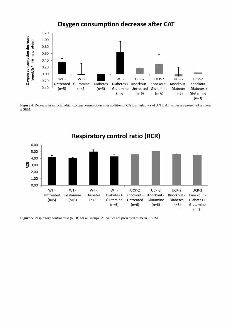
There was a decrease in QO2 in the wild-type group that had both alloxan and glutamine treatment
when CAT was added (Fig. 4.). All the groups had a RCR above 4 (Fig. 5.).
Figure 2. Baseline mitochondrial oxygen consumption for all the groups. All groups have ATP-synthase inhibited by oligomycin. All
values are presented as mean ± SEM.
Figure 3. Decrease in mitochondrial oxygen consumption after addition of GDP, an inhibitor of UCP-2. All values are presented as
mean ± SEM.
-0,40
-0,20
0,00
0,20
0,40
0,60
0,80
1,00
1,20
WT - Untreated
(n=5)
WT - Glutamine
(n=5)
WT - Diabetes
(n=5)
WT - Diabetes + Glutamine
(n=6)
UCP-2 Knockout - Untreated
(n=6)
UCP-2 Knockout - Glutamine
(n=6)
UCP-2 Knockout - Diabetes
(n=5)
UCP-2 Knockout
- Diabetes + Glutamine
(n=3)
Oxy
gen
co
nsu
mp
tio
n d
ecr
eas
e
(pm
ol/
(s*m
l)/m
g p
rote
in)
Oxygen consumption decrease after CAT
0,00
1,00
2,00
3,00
4,00
5,00
6,00
WT - Untreated
(n=5)
WT - Glutamine
(n=5)
WT - Diabetes
(n=5)
WT - Diabetes + Glutamine
(n=6)
UCP-2 Knockout - Untreated
(n=6)
UCP-2 Knockout - Glutamine
(n=6)
UCP-2 Knockout - Diabetes
(n=5)
UCP-2 Knockout - Diabetes + Glutamine
(n=3)
RC
R
Respiratory control ratio (RCR)
Figure 4. Decrease in mitochondrial oxygen consumption after addition of CAT, an inhibitor of ANT. All values are presented as mean
± SEM.
Figure 5. Respiratory control ratio (RCR) for all groups. All values are presented as mean ± SEM.

Discussion
The major finding of the present study is that mitochondrial QO2 can be affected with glutamine in
wild type mice. There was also a small increase in QO2 in the UCP-2 knockout groups after glutamine
treatment. It’s possible that other UCP-isoforms, UCP-4 and UCP-5 which is also expressed in mouse
kidney29
, may be involved in regulation mitochondrial QO2. It is known that diabetes can increase
expression of UCP-2 in the kidney26
and increasing amounts of NADH and FADH2 sequentially
increase the O2 required for electron transport6. This increasing QO2 may result in hypoxia
5,6. The
results demonstrated increased QO2 in the animals treated with glutamine. However, diabetes alone did
not affect QO2 in wild-type, which was unexpected since diabetes increases QO2 in rats26
. It may be
postulated that UCP-2 may not be activated in mouse kidneys. Further measurements of UCP-2
protein excretion will clarify this issue. Since wild type groups treated with glutamine displayed a
greater increase in QO2, it may be postulated that the increased QO2 is caused by increased uncoupling
by UCP-2 and that is activated and regulated by glutamine. This is supported by increased inhibition
by GDP, a known inhibitor of UCP’s30
, in the same groups. However, these effects were also evident
in UCP-2 knockout mice, indicating that mitochondrial QO2 is directly regulated by glutamine. It
cannot be excluded that glutamine may regulate other UCP-isoforms that is present in mouse kidney.
Furthermore, an increased ANT uncoupling was evident in diabetic wild-type treated with
glutamine, indicating that glutamine might have regulatory effects on ANT uncoupling during
increased stress caused by diabetes and increased glutamine intake. ANT is known for its ability to
replace UCP-2’s functions if UCP-2 function is severely reduced18
. However, interestingly, similar
effects by CAT were not evident in UCP-2 knockout mice. Additional studies are warranted due to
interesting tendencies before making further conclusions.
Conclusions
The present study concludes that glutamine can activate and regulate uncoupling through UCP-2,
resulting in increased QO2. This finding is supported by an increased inhibition by GDP.
Future aspects
The present study revealed some interesting tendencies that need to be thoroughly investigated. The
study only featured 5-6 mice in each group and thus, needs to be increased with added experiments.
Future studies should also investigate the effect of glutamine on ANT and it’s uncoupling in
mitochondria. Furthermore the possible role of other UCP-isoforms in mediating observed effects
needs to be investigated.

References
1. Hall, J. E. & Guyton, A. C. in 307–377, 972–977 (Saunders/Elsevier, 2006).
2. Hansell, P., Welch, W. J., Blantz, R. C. & Palm, F. Determinants of kidney oxygen consumption
and their relationship to tissue oxygen tension in diabetes and hypertension. Clin. Exp.
Pharmacol. Physiol. 40, 123–37 (2013).
3. Levy, M. N. Effect of variations of blood flow on renal oxygen extraction. Am. J. Physiol. -- Leg.
Content 199, 13–18 (1960).
4. Aukland, K. & Krog, J. Renal Oxygen Tension. Nature 188, 671–671 (1960).
5. Eckardt, K.-U. et al. Role of hypoxia in the pathogenesis of renal disease. Kidney Int. 68, S46–
S51 (2005).
6. Friederich, M., Hansell, P. & Palm, F. Diabetes, oxidative stress, nitric oxide and mitochondria
function. Curr. Diabetes Rev. 5, 120–144 (2009).
7. Harvey, R. A., Ferrier, D. R. & Champe, P. C. in 73–77 (Wolters Kluwer Health, 2011).
8. Halliwell, B. in Free Radicals Biol. Med. (Oxford University Press, 2007).
9. Lodish, H. F. in 502–503 (W.H. Freeman ; Palgrave [distributor], 2013).
10. Bayr, H. Reactive oxygen species. Crit. Care Med. 33, S498–S501 (2005).
11. Kowaltowski, A. J., de Souza-Pinto, N. C., Castilho, R. F. & Vercesi, A. E. Mitochondria and
reactive oxygen species. Free Radic. Biol. Med. 47, 333–343 (2009).
12. Korshunov, S. S., Skulachev, V. P. & Starkov, A. A. High protonic potential actuates a
mechanism of production of reactive oxygen species in mitochondria. Febs Lett. 416, 15–18
(1997).
13. Brownlee, M. Biochemistry and molecular cell biology of diabetic complications. Nature 414,
813–820 (2001).
14. Fridovich, I. & Freeman, B. Antioxidant Defenses in the Lung. Annu. Rev. Physiol. 48, 693–702
(1986).
15. Miwa, S. & Brand, M. D. Mitochondrial matrix reactive oxygen species production is very
sensitive to mild uncoupling. Biochem. Soc. Trans. 31, 1300–1301 (2003).
16. Jezek, P. et al. Existence of uncoupling protein-2 antigen in isolated mitochondria from various
tissues. Febs Lett. 455, 79–82 (1999).
17. Duval, C. et al. Increased reactive oxygen species production with antisense oligonucleotides
directed against uncoupling protein 2 in murine endothelial cells. Biochem. Cell Biol. Biochim.
Biol. Cell. 80, 757–764 (2002).
18. Friederich-Persson, M. et al. Acute knockdown of uncoupling protein-2 increases uncoupling via
the adenine nucleotide transporter and decreases oxidative stress in diabetic kidneys. Plos One 7,
e39635 (2012).
19. Skulachev, V. P. Uncoupling: new approaches to an old problem of bioenergetics. Biochim.
Biophys. Acta 1363, 100–124 (1998).
20. Garlid, K. D., Jabůrek, M., Jezek, P. & Varecha, M. How do uncoupling proteins uncouple?
Biochim. Biophys. Acta 1459, 383–389 (2000).
21. Hurtaud, C., Gelly, C., Chen, Z., Lévi Meyrueis, C. & Bouillaud, F. Glutamine stimulates
translation of uncoupling protein 2mRNA. Cell. Mol. Life Sci. 64, 1853–60 (2007).
22. Cameron, N. E. & Cotter, M. A. Metabolic and vascular factors in the pathogenesis of diabetic
neuropathy. DIABETES 46 Suppl 2, S31–7 (1997).
23. Fine, L. G. & Norman, J. T. Chronic hypoxia as a mechanism of progression of chronic kidney
diseases: from hypothesis to novel therapeutics. Kidney Int. 74, 867–872 (2008).
24. Fine, L. G., Orphanides, C. & Norman, J. T. Progressive renal disease: the chronic hypoxia
hypothesis. Kidney Int. Suppl. 65, S74–78 (1998).
25. Palm, F., Cederberg, J., Hansell, P. & Liss, P. Reactive oxygen species cause diabetes-induced
decrease in renal oxygen tension. DIABETOLOGIA CLINICAL AND EXPERIMENTAL
DIABETES AND METABOLISM (2003).
26. Friederich, M., Fasching, A., Hansell, P., Nordquist, L. & Palm, F. Diabetes-induced up-
regulation of uncoupling protein-2 results in increased mitochondrial uncoupling in kidney
proximal tubular cells. Biochim. Biophys. Acta Bba - Bioenerg. 1777, 935–940 (2008).
27. Gadsby, R. Epidemiology of diabetes. Adv. Drug Deliv. Rev. 54, 1165–1172 (2002).

28. Bruno, G. & Landi, A. Epidemiology and Costs of Diabetes. Transplant. Proc. 43, 327–329
(2011).
29. Alán, L., Smolková, K., Kronusová, E. & Santorová, J. Absolute levels of transcripts for
mitochondrial uncoupling proteins UCP2, UCP3, UCP4, and UCP5 show different patterns in rat
and mice tissues. J. Bioenerg. Biomembr. 41, 71 (2009).
30. Nègre Salvayre, A. et al. A role for uncoupling protein-2 as a regulator of mitochondrial hydrogen
peroxide generation. Faseb J. 11, 809–15 (1997).